thepainful heel - ard.bmj.com · the calcaneus). talalgia wasconsidered to besevere...
TRANSCRIPT
Annals of the Rheumatic Diseases, 1977, 36, 343-348
The painful heelComparative study in rheumatoid arthritis, ankylosing spondylitis, Reiter'ssyndrome, and generalized osteoarthrosis
J. C. GERSTER, T. L. VISCHER, A. BENNANI, AND G. H. FALLET
From the Department of Medicine, Division of Rheumatology, University Hospital, Geneva, Switzerland
SUMMARY This study presents the frequency of severe and mild talalgias in unselected, consecutivepatients with rheumatoid arthritis, ankylosing spondylitis, Reiter's syndrome, and generalizedosteoarthosis. Achilles tendinitis and plantar fasciitis caused a severe talalgia and they were observedmainly in males with Reiter's syndrome or ankylosing spondylitis. On the other hand, sub-Achillesbursitis more frequently affected women with rheumatoid arthritis and rarely gave rise to severetalalgias. The simple calcaneal spur was associated with generalized osteoarthrosis and its frequencyincreased with age. This condition was not related to talalgias. Finally, clinical and radiologicalinvolvement of the subtalar and midtarsal joints were observed mainly in rheumatoid arthritis andoccasionally caused apes valgoplanus.
A 'painful heel' syndrome occurs at times in patientswith inflammatory rheumatic disease or osteo-arthrosis, causing significant clinical problems. Veryfew studies have investigated the frequency andcharacteristics of this syndrome. Therefore we havestudied unselected groups of patients with rheuma-toid arthritis (RA), ankylosing spondylitis (AS),Reiter's syndrome (RS), and generalized osteo-arthrosis (OA) in order to characterize the heeltenderness (talalgia) occurring in each group and todetermine its frequency. The results indicate thateach disease has its particular expression in theheel of the foot.
Patients and methods
RA 100 PATIENTSAll patients had classical or definite RA accordingto the criteria of the American RheumatismAssociation (1959). The disease had been present formore than 6 months in all of our patients and 87 hadpositive rheumatoid factor tests (87%).
AS 35 PATIENTSAll patients were typical and fulfilled the criteria asdefined by Ogryzlo (1974). None suffered from
Accepted for publication November 9, 1976Correspondence to Prof. G. H. Fallet, H6pital cantonal,Avenue Beau-S6jour, 1211, Geneve 4, Switzerland
psoriasis, urethritis, conjunctivitis, or enterocolitis.The antigen HLA B27 was present in 29 patients(80%O).RS 16 PATIENTSAll of our patients had the complete triad (non-gonococcal urethritis, arthritis, and conjunctivitis).The antigen HLA B27 was present in 12 patients(74%/O) and sacroiliitis in 5 (31 %.).OA 70 PATIENTSAll of the patients in this group conformed to thecriteria of Kellgren et al. (1963).
Table 1 gives the essential characteristics of these4 groups.
Talalgia was defined as spontaneous or elicitedpain either at the posterior part of the heel (along
Table 1 PatientsNo. of Malesl Mean age Mean durationpatients females (range) ofdisease (years)
(range)
RA 100 31/69 59.5 9(23-84) (0-5-40)
AS 35 32/3 43-5 11-4(19-75) (1-29)
RS 16 16/0 34.8 5.6(22-56) (1-16)
OA 70 16/54 73.4 9.7(56-98) (2-40)
343
on 13 July 2019 by guest. Protected by copyright.
http://ard.bmj.com
/A
nn Rheum
Dis: first published as 10.1136/ard.36.4.343 on 1 A
ugust 1977. Dow
nloaded from

344 Gerster, Vischer, Bennani, Fallet
the Achilles tendon and at its insertion, or in thearea of the sub-Achilles bursa) or at the plantarsurface (at the insertion of plantar aponeurosis onthe calcaneus). Talalgia was considered to be severewhen the pain occurred on weight bearing, decreasedvery slowly after prolonged rest, and was elicitedby slight local pressure. Talalgia was considered tobe mild when pain was inconsistent on weightbearing, decreased immediately with rest, and couldonly be provoked by moderate to marked localpressure. In each patient the presence of valgus orvarus deformities of the hindfoot and of soft tissueswelling was recorded together with the articularmobility of the various joints of the foot.A standard lateral radiograph was taken each
time. When there was a suspicion of soft tissueswelling (Achilles tendon, sub-Achilles bursa, or theplantar fascia), a lateral xeroradiograph of theposterior foot was made following the techniquedescribed previously (Gerster et al., 1975).
Results
The frequency of talalglas in the four groups ofpatients is given in Table 2. Severe talalgia was mostfrequent in RS, followed by AS. Mild talalgiaoccurred most frequently in RA. In Table 3 theclinical features of the heel are given. Achillestendinitis was present in 8 male patients: 5 with RS(31% of the group) and 3 with AS (8 6% of thegroup). The 5 patients with diffuse thickening of thetendon (Fig. I) had severe talaglia, whereas the 3patients with nodular tendinitis (Fig. 2) had only
Table 2 Incidence of talalgia
Severe talalgia Mild talalgia(0/) ( /0)
RA (100 patients) 2 27AS (35 patients) 8-6 17.1RS (16 patients) 31 19OA (70 patients) 1-4 15-7
Table 3 Incidence and distribution ofobjective softtissue changes
Achillestendinitis(/)
Plantarfasciitis(/)
Achillobursitis(/)
RA (100 patients) 0 1 4AS (35 patients) 8*6 8-6 2-8RS (16 patients) 31 18.7 0OA (74 patients) 0 0 0
Fig. 1 Reiter's syndrome in a 45-year-old male.Xeroradiography of the right foot, showing diffuseswelling of the Achilles tendon (inferior diameter 80 mm,normal 5. 1+1 *0; diameter ofmiddle part 8 - 0 mm,normal 6* 2±1 6). Note a simple, regular plantar spur.
Fig. 2 Reiter's syndrome in a 37-year-old male.Xeroradiography of the right foot, showing nodularthickening (arrow) at middle part of the Achilles tendon.
on 13 July 2019 by guest. Protected by copyright.
http://ard.bmj.com
/A
nn Rheum
Dis: first published as 10.1136/ard.36.4.343 on 1 A
ugust 1977. Dow
nloaded from
The painful heel. Comparative study 345
A~~~~~~~~~~'W
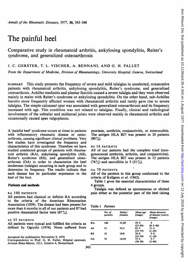
Fig. 3 Rheumatoid arthritis in a 42-year-old female. Xeroradiography of(A) left foot, (B) right foot, (C) detailofB. Note plantar fasciitis at right with thickened heel pad (arrow); the overgrowth measures 24 mm on the originalxerography (normal left heel is only 19 mm). An irregular erosion is present on the plantar aspect ofright calcaneus(C) and demineralization of its posterior part is visible (B)
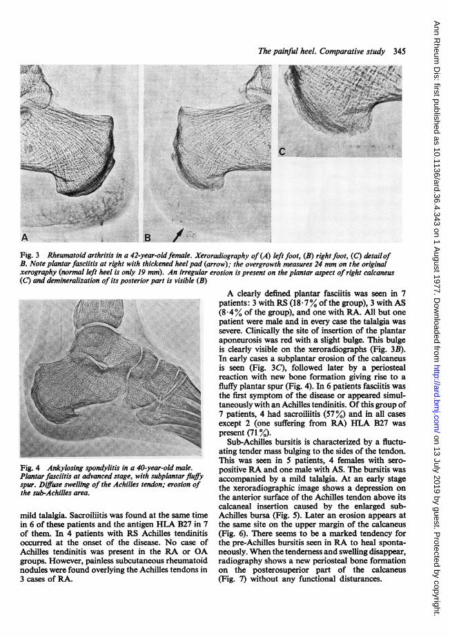
Fig. 4 Ankylosing spondylitis in a 40-year-old male.Plantar fasciitis at advanced stage, with subplantar fluffyspur. Diffuse swelling of the Achilles tendon; erosion ofthe sub-Achilles area.
mild talalgia. Sacroiliitis was found at the same timein 6 of these patients and the antigen HLA B27 in 7of them. In 4 patients with RS Achilles tendinitisoccurred at the onset of the disease. No case ofAchilles tendinitis was present in the RA or OAgroups. However, painless subcutaneous rheumatoidnodules were found overlying the Achilles tendons in3 cases of RA.
A clearly defined plantar fasciitis was seen in 7patients: 3 with RS (18X7% of the group), 3 with AS(8 4% of the group), and one with RA. All but onepatient were male and in every case the talalgia wassevere. Clinically the site of insertion of the plantaraponeurosis was red with a slight bulge. This bulgeis clearly visible on the xeroradiographs (Fig. 3B).In early cases a subplantar erosion of the calcaneusis seen (Fig. 3C), followed later by a periostealreaction with new bone formation giving rise to afluffy plantar spur (Fig. 4). In 6 patients fasciitis wasthe first symptom of the disease or appeared simul-taneously with an Achilles tendinitis. Of this group of7 patients, 4 had sacroiliitis (57%Y) and in all casesexcept 2 (one suffering from RA) HLA B27 waspresent (71 %O).
Sub-Achilles bursitis is characterized by a fluctu-ating tender mass bulging to the sides of the tendon.This was seen in 5 patients, 4 females with sero-positive RA and one male with AS. The bursitis wasaccompanied by a mild talalgia. At an early stagethe xeroradiographic image shows a depression onthe anterior surface of the Achilles tendon above itscalcaneal insertion caused by the enlarged sub-Achilles bursa (Fig. 5). Later an erosion appears atthe same site on the upper margin of the calcaneus(Fig. 6). There seems to be a marked tendency forthe pre-Achilles bursitis seen in RA to heal sponta-neously. When the tenderness and swelling disappear,radiography shows a new periosteal bone formationon the posterosuperior part of the calcaneus(Fig. 7) without any functional disturances.
--N.1
,*to
on 13 July 2019 by guest. Protected by copyright.
http://ard.bmj.com
/A
nn Rheum
Dis: first published as 10.1136/ard.36.4.343 on 1 A
ugust 1977. Dow
nloaded from

346 Gerster, Vischer, Bennani, Fallet
Ir-U1 A -4 ;#xXtn D
Fig. 5 Xeroradtography of the left foot of a SS-year-oldfemtale wtth rheumatoid arthritis. Active sub-Achillesbursittts wtth an impression on the anterior part of theAchilles tendon (arrow).
The incidence and distribution of the osseouschanges on x-rays are presented in Table 4. A simplecalcaneal spur with well-defined borders wasfrequently seen in the group with QA. Frequentlythe spurs are bilateral and occur more often on theplantar surface than on the posterior surface of thecalcaneus. Except in RS, these simple spurs aregenerally present in patients over 60 years of age.Irregular calcaneal spurs are most frequently seenon the plantar surface and represent the naturalevolution of a fasciitis. Occasionally they are foundon the posterior pole of the calcaneus and are theresult of an Achilles tendinitis. Thus, the incidenceof such irregular spurs is greatest in adults withRS or AS.As expected, the radiological changes of the
subtalar articulations, frequently associated withchanges in the midtarsal joint, were most often seenin RA. In 38% of the patients in this group the tarsal
1 auie 4t nciaenCe unu UaLsriDuion (JJ oJsJsouradiological changes
RA (100 AS (35 RS (16 OA (70patients) patients) patients) patients)(%) (%) (/) (%)
CalcaneusSimple spur 20 17 18.7 56Irregular spur
Posterior 2 2-8 0 0Plantar 2 11.4 18*7 0
ErosionPosterior 6 5*7 6-2 0Plantar 1 2-8 6-2 0
Involvement ofsubtalarjoint 27 2-8 0 17.1
Involvement ofmidtarsal joint 23 2-8 6.2 15.7
joints were affected and in all but 4 cases the diseasehad lasted for more than 4 years. In 5 of thesepatients subtalar involvement was so severe that itcaused a painful valgoplanus deformity of the foot.This condition was found only once in the othergroups (a patient with OA). We did not observe adistinct clinical correlation between tarsal involve-ment and the presence of talalgia in any of thegroups.
Fig. 6 Rheumatoid arthritis in a 57-year-old female.Erosion of the upper margin of calcaneus from activesub-Achilles bursitis-negative xeroradiographic picture.
on 13 July 2019 by guest. Protected by copyright.
http://ard.bmj.com
/A
nn Rheum
Dis: first published as 10.1136/ard.36.4.343 on 1 A
ugust 1977. Dow
nloaded from
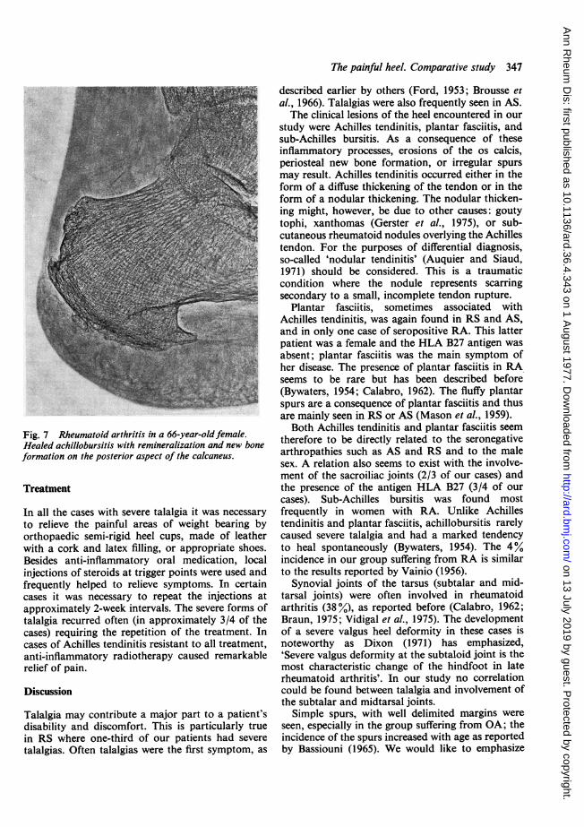
Fig. 7 Rheumatoid arthritis in a 66-year-old female.Healed achillobursitis with remineralization and new boneformation on the posterior aspect of the calcaneus.
Treatment
In all the cases with severe talalgia it was necessary
to relieve the painful areas of weight bearing byorthopaedic semi-rigid heel cups, made of leatherwith a cork and latex filling, or appropriate shoes.Besides anti-inflammatory oral medication, localinjections of steroids at trigger points were used andfrequently helped to relieve symptoms. In certaincases it was necessary to repeat the injections atapproximately 2-week intervals. The severe forms oftalalgia recurred often (in approximately 3/4 of thecases) requiring the repetition of the treatment. Incases of Achilles tendinitis resistant to all treatment,anti-inflammatory radiotherapy caused remarkablerelief of pain.
Discussion
Talalgia may contribute a major part to a patient'sdisability and discomfort. This is particularly truein RS where one-third of our patients had severe
talalgias. Often talalgias were the first symptom, as
The painful heel. Comparative study 347
described earlier by others (Ford, 1953; Brousse etal., 1966). Talalgias were also frequently seen in AS.The clinical lesions of the heel encountered in our
study were Achilles tendinitis, plantar fasciitis, andsub-Achilles bursitis. As a consequence of theseinflammatory processes, erosions of the os calcis,periosteal new bone formation, or irregular spursmay result. Achilles tendinitis occurred either in theform of a diffuse thickening of the tendon or in theform of a nodular thickening. The nodular thicken-ing might, however, be due to other causes: goutytophi, xanthomas (Gerster et al., 1975), or sub-cutaneous rheumatoid nodules overlying the Achillestendon. For the purposes of differential diagnosis,so-called 'nodular tendinitis' (Auquier and Siaud,1971) should be considered. This is a traumaticcondition where the nodule represents scarringsecondary to a small, incomplete tendon rupture.
Plantar fasciitis, sometimes associated withAchilles tendinitis, was again found in RS and AS,and in only one case of seropositive RA. This latterpatient was a female and the HLA B27 antigen wasabsent; plantar fasciitis was the main symptom ofher disease. The presence of plantar fasciitis in RAseems to be rare but has been described before(Bywaters, 1954; Calabro, 1962). The fluffy plantarspurs are a consequence of plantar fasciitis and thusare mainly seen in RS or AS (Mason et al., 1959).Both Achilles tendinitis and plantar fasciitis seem
therefore to be directly related to the seronegativearthropathies such as AS and RS and to the malesex. A relation also seems to exist with the involve-ment of the sacroiliac joints (2/3 of our cases) andthe presence of the antigen HLA B27 (3/4 of ourcases). Sub-Achilles bursitis was found mostfrequently in women with RA. Unlike Achillestendinitis and plantar fasciitis, achillobursitis rarelycaused severe talalgia and had a marked tendencyto heal spontaneously (Bywaters, 1954). The 4%incidence in our group suffering from RA is similarto the results reported by Vainio (1956).
Synovial joints of the tarsus (subtalar and mid-tarsal joints) were often involved in rheumatoidarthritis (38 %), as reported before (Calabro, 1962;Braun, 1975; Vidigal et al., 1975). The developmentof a severe valgus heel deformity in these cases isnoteworthy as Dixon (1971) has emphasized,'Severe valgus deformity at the subtaloid joint is themost characteristic change of the hindfoot in laterheumatoid arthritis'. In our study no correlationcould be found between talalgia and involvement ofthe subtalar and midtarsal joints.
Simple spurs, with well delimited margins wereseen, especially in the group suffering from OA; theincidence of the spurs increased with age as reportedby Bassiouni (1965). We would like to emphasize
on 13 July 2019 by guest. Protected by copyright.
http://ard.bmj.com
/A
nn Rheum
Dis: first published as 10.1136/ard.36.4.343 on 1 A
ugust 1977. Dow
nloaded from
348 Gerster, Vischer, Bennani, Fallet
the absence of a clinical correlation between thesimple spur and talalgias in our groups; this is inagreement with observations made by Serre et al.(1968).In many patients with inflammatory or degenera-
tive joint diseases suffering from mild talalgias aspecial cause could not be determined and xero-radiography failed to show any soft tissue changesin this study. It seems that minor inflammatoryprocesses exist at the calcaneal insertion of theplantar fascia or Achilles tendon which cannot beshown. These processes could be assimilated intothe larger group of ligamentous attachment anoma-lies called 'enthesopathy' (Niepel et al., 1966).
References
American Rheumatism Association (1959). Diagnosticcriteria for rheumatoid arthritis. 1958 revision. Annals oftheRheumatic Diseases, 18, 49-53.
Auquier, L., and Siaud, J. R. (1971). Tendinites nodulairesdu tendon d'Achille. Revue du Rhumatisme, 38, 373-381.
Bassiouni, M. (1965). Incidence of calcaneal spurs in osteo-arthrosis and rheumatoid arthritis, and in control patients.Annals of the Rheumatic Diseases, 24, 490-493.
Braun, S. (1975). Le pied dans les grands rhumatismesinflaminatoires. Rhumatologie, 27, 47-56.
Brousse, J. P., Braun, S., Amor, B., and Coste, F. (1966).Etude comparative de quelques types de talalgie et decalcan6ite. Semaine des Hdpitaux de Paris, 13, 795-802.
Bywaters, E. G. L. (1954). Heel lesions of rheumatoidarthritis. Annals of the Rheumatic Diseases, 13, 42-51.
Calabro, J. J. (1962). A critical evaluation of the diagnosticfeatures of the feet in rheumatoid arthritis. Arthritis andRheumatism, 5, 19-29.
Dixon, A. St. J. (1971). The rheumatoid foot. Modern Trendsin Rheumatology, 2, p. 158. Ed. by A. G. S. Hill. Butter-worths, London.
Ford, D. K. (1953). Natural history of arthritis followingvenereal urethritis. Annals of the Rheumatic Diseases, 12,177-194.
Gerster, J. C., Hauser, H., and Fallet, G. H. (1975). Xero-radiographic techniques applied to assessment of Achillestendon in inflammatory or metabolic diseases. Annals ofthe Rheumatic Diseases, 34, 479-488.
Kellgren, J. H., Lawrence, J. S., and Bier, F. (1963). Geneticfactors in generalized osteo-arthrosis. Annals of theRheumatic Diseases, 22, 237-255.
Mason, R. M., Murray, R. S., Oates, J. K., and Young, A. C.(1959). A comparative radiological study of Reiter'sdisease, rheumatoid arthritis and ankylosing spondylitis.Journal ofBone andJoint Surgery, 41B, 137-148.
Niepel, G. A., Kostka, D., Kopecky, S., and Manca, S.(1966). Enthesopathy. Acta Rheumatologica et Balneo-logica, Pistiniana, No. 1. Czechoslovak State Spa Piestany,Publ. Piestany.
Ogryzlo, M. A. (1974). Ankylosing spondylitis. Arthritis andAllied Conditions, 4th ed., p. 699. Ed. by J. L. Hollanderand D. L. McCarty. Lea and Febiger, Philadelphia.
Serre, H., Simon, L., and Claustre, J. (1968). Les algiestalonni6res communes. Gazette des HOpitaux Civils etMilitaires, Paris, 140, 641-652.
Vainio, S. (1956). The rheumatoid foot; a clinical study withpathological and roentgenological comments. AnnalesChirurgiae et Gynaecologiae Fenniae, 45, Suppl. 1.
Vidigal, E., Jacoby, R. K., Dixon, A. St. J., Ratliff, A. H.,and Kirkup, J. (1975). The foot in chronic rheumatoidarthritis. Annals of the Rheumatic Diseases, 34, 292-297.
on 13 July 2019 by guest. Protected by copyright.
http://ard.bmj.com
/A
nn Rheum
Dis: first published as 10.1136/ard.36.4.343 on 1 A
ugust 1977. Dow
nloaded from