thesis for the degree master of pharmacy investigation and - munin
TRANSCRIPT

THESIS FOR THE DEGREE MASTER OF PHARMACY
INVESTIGATION AND OPTIMIZATION OF LIPOSOME FORMULATION FOR USE AS DRUG CARRIER FOR THE
ANTICANCER AGENT CAMPTOTHECIN
BY
ELENAZ NADERKHANI
2011
Supervisors
Assoc. Professor Gøril Eide Flaten
& Post-doc Ragnhild Dragøy Whitaker
Drug Transport and Delivery Research Group Department of Pharmacy
Faculty of Health Sciences University of Tromsø

ii

iii
TABLE OF CONTENTS
LIST OF FIGURES .............................................................................................vi
LIST OF TABLES..............................................................................................vii
ACKNOWLEDGMENTS ....................................................................................ix
ABSTRACT .......................................................................................................x
ABBREVIATIONS............................................................................................xii
1. INTRODUCTION.......................................................................................... 1
1.1 Camptothecin ................................................................................................................................ 1
1.2 Liposomes...................................................................................................................................... 4
1.2.1 Definition and background..................................................................................................... 4
1.2.2 Lipids in liposome products.................................................................................................... 5
1.2.2.1 Phospholipid (PL)............................................................................................................. 5
1.2.2.2 Cholesterol (Chol) and other employed lipids................................................................. 7
1.2.3 Characterization of liposomes.................................................................................................... 9
1.2.3.1 The role of liposome size..................................................................................................... 9
1.2.3.2 The role of the surface charge and membrane characteristics ........................................ 10
1.3 Long circulating liposomes .......................................................................................................... 11
1.3.1 Liposomes in cancer therapy................................................................................................ 12
2 MATERIALS & METHODS ............................................................................14
2.1 Materials...................................................................................................................................... 14
2.2 Methods ...................................................................................................................................... 18
2.3 Liposome preparation ................................................................................................................. 20
2.3.1 Lipid‐CPT film preparation.................................................................................................... 20
2.3.2 Lipid‐CPT freeze‐drying preparation .................................................................................... 21
2.3.3 Rehydration .......................................................................................................................... 21
2.4 Size reduction of liposomes ........................................................................................................ 21
iv
2.4.2 Direct probe sonication ........................................................................................................ 22
2.4.3 Size determination by photon correlation spectroscopy (PCS) ........................................... 23
2.5 Ultracentrifugation...................................................................................................................... 24
2.5.1 Fluorescence microscopy ..................................................................................................... 24
2.6 CPT liposomes retention ability................................................................................................... 25
2.6.1 Spin column method ............................................................................................................ 25
2.7 Quantification of Camptothecin.................................................................................................. 26
2.8 Quantification of phosphatidylcholine........................................................................................ 27
2.9 Determination of zeta Potential of liposomes ............................................................................ 28
2.10 Fluorescence depolarization anisotropy ................................................................................... 29
3. RESULTS AND DISCUSSIONS ......................................................................30
3.1 Method development ................................................................................................................. 30
3.1.1 Sonication procedure ........................................................................................................... 30
3.1.1.1 Probe sonication............................................................................................................ 31
3.1.2 Optimization of ultracentrifugation for separation of free CPT from liposomes................. 32
3.1.3 Comparison of film and freeze‐drying method.................................................................... 35
3.2 Characterization of how lipid composition influences CPT incorporation and retention........... 37
3.2.1 CPT liposome incorporation study ....................................................................................... 37
3.2.2 CPT liposomes retention ability............................................................................................ 39
3.2.3 CPT liposomes incorporation ability and the influence of the zeta potential...................... 41
3.3 Preliminary studies on PEGylated liposomes .............................................................................. 43
3.3.1 PEGylated CPT liposomes incorporation study .................................................................... 43
3.3.2 PEGylated CPT liposomes retention ability .......................................................................... 45
3.3.3 PEGylated liposomes CPT incorporation and the influence of zeta potential ..................... 46
3.4 Fluorescence anisotropy............................................................................................................. 47
3.5 Fusing of liposomes and size measurements using PCS.............................................................. 48
4. Conclusions ...............................................................................................51

v
5. Future Perspectives ...................................................................................52
6. REFERENCES ..............................................................................................54
vi
LIST OF FIGURES
Figure 1.1: Schematic illustration of CPT binding to Topo-I and arrest the replication fork
Figure 1.2: Equilibrium between the carboxylate and lactone form of CPT
Figure 1.3: Schematic description of lactone and carboxylate equilibrium in both liposome
membrane and in blood circulation
Figure 1.4: Liposomes with lipophilic drugs incorporated in the phospholipid bilayer
Figure 1.5: Structural formula of glycerophospholipid
Figure 1.6: General structure of cholesterol (Chol)
Figure 1.7: General structure of DOTAP
Figure 1.8: Modified illustration of sterically stabilized liposome surrounded with PEG
Figure 1.9: Accumulation of liposomes in tumour tissues due to EPR effect
Figure 2.1: Enzymatic quantification of phosphatidylcholine (PC)
Figure 3.1: Fluorescence pictures of supernatant after ultracentrifugation of 20 minutes
Figure 3.2: Fluorescence pictures of supernatant after ultracentrifugation of 25 minutes
Figure 3.3: Camptothecin-incorporation capacity of EPC by film and freeze-drying method
Figure 3.4: Camptothecin-incorporation capacity of the different liposome formulations
Figure 3.5: The different liposome formulations ability to retain CPT associated with the
liposomes over time in buffer at room temperature
Figure 3.6: The different liposome formulations ability to retain CPT associated with the
liposomes over time in serum
Figure 3.7: Zeta potential (mV) of the different liposome formulations
Figure 3.8: Correlation between liposomal Zeta potential (mV) and incorporation efficacy.
The zeta potential is plotted against absolute value of the zeta potential
Figure 3.9: Comparison of Camptothecin-incorporation between the PEGylated and non-
PEGylated liposomes
Figure 3.10: The different PEGylated liposomal formulations ability to retain CPT associated
with the liposomes over time in buffer and serum
Figure 3.11: Zeta potential of PEGylated liposomal formulations
Figure 3.12: Anisotropy in different liposome formulations

vii
LIST OF TABLES
Table 1.1: The most common glycerophospholipids
Table 2.1: Chemicals
Table 2.2: Lipids
Table 2.3: Equipment
Table 2.4: The lipid compositions (mol %) of the different formulations
Table 3.1: Bath sonicated EPC liposomes
Table 3.2: Probe sonicated liposomes
Table 3.3: PCS results of some liposome formulations after sonication

viii

ix
ACKNOWLEDGMENTS This study was conducted at the Drug Transport and Delivery Research Group, Institute of
pharmacy, University of Tromsø.
First I would like to express my deep gratitude to my supervisors, Assoc. Professor Gøril Eide
Flaten & Post-doc Ragnhild Dragøy Whitaker for their outstanding contribution, valuable
guidance and support during this thesis. Gøril: thank you for the guidance through the
different stages of this project, you always took time to help me during the writing process,
something I always will remember. Ragnhild: thank you for all the time you spent with me in
the laboratory and for always supporting me whatever issues were arising. Your support
cannot be appreciated highly enough.
I am grateful to Merete Skar for all the time she spent in the laboratory helping me with the
technical problems, especially with the HPLC.
My final thanks goes to my family, for always supporting me during these years.
Elenaz Naderkhani
May, 2011

x
ABSTRACT In this thesis, the method development and investigation of different liposomal formulations
to incorporate and retain Camptothecin (CPT) is described. CPT is a potent anticancer drug
that has shown to be active against a broad spectrum of cancers. However, due to its
challenging physicochemical properties, like poor water solubility, severe toxic effects to
normal tissues and instability, its clinical development has been limited for nearly 40 years. A
strategy to overcome CPT’s challenging properties is to use liposome-based carrier system.
By taking advantage of this carrier system, we may solubilise CPT in the phospholipid bilayer
of liposomes, protect it from blood proteins and achieve a selective drug accumulation in
tumor tissues or tumor-associated cells by enhanced permeability and retention effect (EPR).
A good liposome formulation of clinical utility must fulfil two important criteria. The
liposomal drug carrier must incorporate CPT in the liposomal bilayer in a relevant therapeutic
concentration and be able to retain the drug within the liposome to make it bioavailable at the
target site after i.v. administration. The focus of this thesis was to study different liposomal
formulations and their ability to incorporate and retain CPT. Screening of eight different
liposome formulations with respect to association with CPT was performed. The 1,2-di-oleyl-
3 trimethyl-ammonium-propane (DOTAP) containing formulations showed superior
incorporation capacity, giving an CPT incorporation of 250 µg/130 µmoles lipid. The
DOTAP containing formulations exhibited as well a trend toward higher retention ability in
serum compared to the other formulations. Although they showed better retention ability, only
25 % of the drug was associated with the liposomes, which is far from being optimal. One of
the important criteria mentioned above for liposomes as drug delivery systems is their ability
to remain stable in blood circulation for prolonged time in order to reach the specific target
and to avoid rapid clearance by RES after i.v. injection. To achieve this, PEG decoration on
the liposome surface can be employed. We chose to PEGylate DOTAP formulations in order
to get a better understanding of this system. PEGylation lead, as expected, to increased
stability of the liposomes, however a reduced incorporation capacity was observed. The
presence of 1 % and 10 % PEG gave better retention and slower leakage from the liposomes.
We conclude that DOTAP inclusion in our liposomes increased the incorporation of CPT into
the lipid bilayer, that liposomal retention in our current formulations must be improved, and
while PEGylation is necessary in order to prevent rapid in vivo clearance, the inclusion of
PEG reduces incorporation, and therefore further studies are needed in order to improve
incorporation of CPT in PEGylated liposomes.

xi

xii
ABBREVIATIONS AcCN Acetonitrile
Chol Cholesterol
CPT Camptothecin
CPT-11 Irinotecan
DMSO Dimethyl sulphoxide
DOPG Dioleoylphosphatidylglycerol
DOTAP 1,2-di-oleyl-3 trimethyl-ammonium-propane
DPH 1,6-Diphenyl-1,3,5-hexatriene
EPC Egg phosphatidylcholine
EPR Enhanced permeability and retention
HPLC High performance liquid chromatography
HSA Human serum albumin
i.v. Intra venous
LC Liquid chromatography
LUV Large unilamellar vesicle
MLV Multilamellar vesicle
PA Phosphatidic acid
PB Phosphate buffer
PBS Phosphate buffered saline
PC Phosphatidylcholine
PCS Photon correlation spectroscopy
PE Phosphatidylethanolamine
PEG Poly(ethylene glycol)
PG Phosphatidylglycerol
PI Phosphatidylinositol
PL Phospholipid
PS Phosphatidylserine
RES Reticuloendothelial system
SD Standard deviation
SUV Small unilamellar vesicle
Topo-I Topoisomerase I
UV Ultraviolet
v/v Volume ratio

1
1. INTRODUCTION
Over the past thirty years, liposomes are becoming important as a delivery system for
therapeutic agents, chemotherapeutics, antigens, immunomodulators, imaging and genetic
materials. A large number of liposome based drugs are in preclinical and clinical research.
Cosmetic industry has also shown great interest in liposomes, and today there are many
liposome-based cosmetic formulations e.g. skin-care products available on the market
(Garidel et al., 2000).
1.1 Camptothecin
Camptothecin (CPT) is a potent anticancer agent that has shown significant cytotoxic activity
(Watanabe et al., 2008). CPT is an alkaloid derived from the bark of the Chinese tree
Camptotheca acuminate, and was isolated already in 1966 by the group of Wall and Wani
(Wall et al., 1966). But due to its limitations, like poor water solubility and the undesired
physical and chemical properties described below, their pharmaceutical development and
clinical implementation has been impaired and research on other chemotherapeutic agents
prioritized. In the 1980s the molecular target for CPT was however identified, again raising
researchers’ interest in and attention to CPT as an anticancer agent (Hsiang and Liu, 1988)
(Mattern et al., 1987).
CPT’s anticancer effect lays in its ability to bind to DNA and inhibit Topoisomerase I (Topo-
I) at physiological pH as shown in Figure 1.1. Topo-I is a central enzyme in the process of
DNA replication. It is responsible for winding and unwinding of the supercoiled DNA
composing the chromosomes. Transcription of DNA cannot occur, if the chromosomes are
not unwound. Further protein synthesis is inhibited, inducing apoptosis (Venditto and
Simanek, 2010). The primary mechanism of CPT is S-phase specific, which means that it is
very toxic to cells undergoing DNA synthesis (Burke and Bom, 2000).

2
Figure 1.1: CPT is binding to Topo-I and arrests the replication fork leading to cell death (reprinted with permission from Dr. Holsæter (Saetern, 2004))
It is known that CPT exists in a pH dependent equilibrium between a lactone form and a
carboxylate form (Figure 1.2). CPT is present in its active lipophilic lactone form at pH
below 6. At physiological pH, CPT is largely present as its significantly less biologically
active and more water soluble carboxylate form. Under physiological conditions and in
human plasma CPT-lactone is rapidly hydrolysed to the open carboxylate form. The
carboxylate form of CPT binds with a 100-150 fold higher affinity to human serum albumin
(HSA) compared to the lactone form, which drives the equilibrium toward the open-ring
carboxylate form (Figure 1.3). HSA-CPT binding makes the inactive carboxylate form
inaccessible for cellular uptake. Moreover, the inactive carboxylate form is excreted by the
kidneys, and causes several toxicity problems like haemorrhagic cystitis and myelotoxicity.
Red blood cells/cell membranes on the other hand stabilize the lactone form, also shown in
Figure 1.3 (Mi and Burke, 1994) (Saetern et al., 2004a). The biologically active form of CPT
has a very short half-life (approximately 12 min), and in presence of human plasma 99% of
the drug is converted to its less active and potentially toxic carboxylate form (Burke, 1996).

3
Figure 1.2: Equilibrium between the carboxylate and lactone form of CPT (reprinted with permission from Dr. Holsæter (Saetern, 2004)).
Currently, there are only two CPT-analogues that have passed the clinical trials. Irinotecan
(Campto®) and topotecan (Hycamptin®) are the two derivatives and have been approved for
treatment of the ovarian carcinoma, small-cell lung cancer and colorectal cancers. These two
CPT derivatives are both water soluble, due to molecular modifications, and also exhibit a
reduced binding affinity for HSA (Li et al., 2006). However, there are still problems with
these derivatives’ unstable E-ring, which is converted to the carboxylate form at physiological
pH (Emerson, 2000). Researchers are still interested in finding new and better ways to solve
CPTs’ stability problems, and this may be achieved by incorporating this drug in liposomal
drug carrier, thereby overcome some of CPT’s challenges and keeping it in its active lactone
form (Watanabe et al., 2008). This can be achieved by the fact that lipids can dissolve the
lipophilic drug and the pH can be controlled inside the liposome in such a way that the
equilibrium is forced toward the active lactone form (see Figure 1.3). The lactone form has
also shown to be stable when harboured in the liposome bilayer (Burke et al., 1992). The drug
is in addition protected from HSA and the complexing with the carboxylate form is avoided
(Emerson, 2000).
( ac ti v e) ( i n a c ti v e)
O H ‐
H+
C am p to th e c i n ‐ L ac to n e C am p to th e c i n ‐ C a rb o x y l a te
N N
O
O
O O H H 3 C
N
N O
O H H 3 C O
O H
O -

4
Liposome pH 6.0 Liposome membrane Blood pH 7.4
Carboxylate Lactone Lactone Lactone Carboxylate
Lipid compartments HSA stabilisation
Figure 1.3: Schematic description of lactone and carboxylate equilibrium in both liposome membrane and in blood circulation
1.2 Liposomes
1.2.1 Definition and background
Liposomes are self-assembling spherical vesicles with a size ranging from 20 nm to 10 µm.
Liposomes may exist as uni- and multilamellar vesicles. Unilamellar vesicles consist of a lipid
bilayer separating the aqueous core from an outer aqueous environment, while multilamellar
vesicles have multiple lipid bilayers separating the different aqueous environments (Brandl,
2001). Liposomes normally consist of different types of naturally occurring phospholipids,
but other lipids such as cholesterol can be included to tune the liposome properties. In order to
obtain liposomes of mixed composition, the lipids are dissolved in an organic solvent, which
is subsequently evaporated from the lipid mixture and a dried lipid film is obtained.
Liposomes are formed spontaneously when dry lipids is dispersed in an aqueous media. They
have the ability to function as drug carriers for both hydrophilic, lipophilic and amphiphilic
drugs. Hydrophilic drugs can be encapsulated in to the aqueous core, while lipophilic and
amphiphilic drugs can be incorporated within the lipophilic bilayer (see Figure 1.4) (Chrai et
al., 2002) (Brandl, 2001).

5
Figure 1.4: Liposomes with lipophilic drugs incorporated in the phospholipid bilayer (reprinted with permission from Dr. Holsæter (Saetern, 2004))
By using liposomes as drug-delivery carriers, we may keep CPT in its active lactone form by
protecting CPT from degradation and HSA binding in the blood stream (Emerson, 2000). And
because liposomes themselves are formed from naturally occurring lipids of low intrinsic
toxicity, they are non-immunogenic and biodegradable in the body (Chrai et al., 2002).
1.2.2 Lipids in liposome products
1.2.2.1 Phospholipid (PL)
Phospholipids (PLs) typically found in high proportions in cell membranes of living matter,
are an important component in liposome formulations. PL consists of two fatty acids linked to
a polar head group, and they have either glycerol (Figure 1.5) or sphingomyeline as the back
bone. PLs are amphipathic molecules, and have both hydrophobic and hydrophilic groups.
The two hydrocarbon chains constitute the hydrophobic tails, while the phosphate group and
its polar attachment constitute the hydrophilic group (Cooper and Hausman, 2009).
PLs can consist of different head and tail groups that affect the surface charge and bilayer
permeability of the liposomes (Perrie and Rades, 2010).
6
CH
CH2
CH2 O
O
C
O P
O
O
O
-O
R3
satured fatty acid, e.g. myristic acid
C
O
unsatured fatty acid, e.g. oleic acid
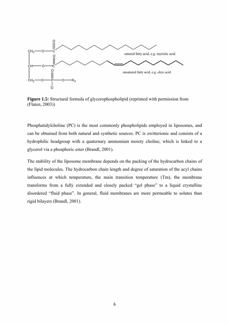
Figure 1.5: Structural formula of glycerophospholipid (reprinted with permission from (Flaten, 2003))
Phosphatidylcholine (PC) is the most commonly phospholipids employed in liposomes, and
can be obtained from both natural and synthetic sources. PC is zwitterionic and consists of a
hydrophilic headgroup with a quaternary ammonium moiety choline, which is linked to a
glycerol via a phosphoric ester (Brandl, 2001).
The stability of the liposome membrane depends on the packing of the hydrocarbon chains of
the lipid molecules. The hydrocarbon chain length and degree of saturation of the acyl chains
influences at which temperature, the main transition temperature (Tm), the membrane
transforms from a fully extended and closely packed “gel phase” to a liquid crystalline
disordered “fluid phase”. In general, fluid membranes are more permeable to solutes than
rigid bilayers (Brandl, 2001).
7
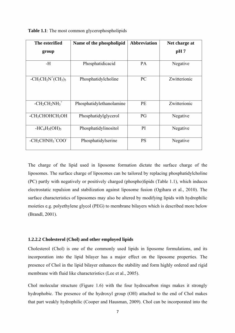
Table 1.1: The most common glycerophospholipids
The esterified
group
Name of the phospholipid Abbreviation Net charge at
pH 7
-H Phosphatidicacid PA Negative
-CH2CH2N+(CH3)3
Phosphatidylcholine PC Zwitterionic
-CH2CH2NH3+ Phosphatidylethanolamine PE Zwitterionic
-CH2CHOHCH2OH Phosphatidylglycerol PG Negative
-HC6H5(OH)5 Phosphatidylinositol PI Negative
-CH2CHNH3+COO- Phosphatidylserine PS Negative
The charge of the lipid used in liposome formation dictate the surface charge of the
liposomes. The surface charge of liposomes can be tailored by replacing phosphatidylcholine
(PC) partly with negatively or positively charged (phospho)lipids (Table 1.1), which induces
electrostatic repulsion and stabilization against liposome fusion (Ogihara et al., 2010). The
surface characteristics of liposomes may also be altered by modifying lipids with hydrophilic
moieties e.g. polyethylene glycol (PEG) to membrane bilayers which is described more below
(Brandl, 2001).
1.2.2.2 Cholesterol (Chol) and other employed lipids
Cholesterol (Chol) is one of the commonly used lipids in liposome formulations, and its
incorporation into the lipid bilayer has a major effect on the liposome properties. The
presence of Chol in the lipid bilayer enhances the stability and form highly ordered and rigid
membrane with fluid like characteristics (Lee et al., 2005).
Chol molecular structure (Figure 1.6) with the four hydrocarbon rings makes it strongly
hydrophobic. The presence of the hydroxyl group (OH) attached to the end of Chol makes
that part weakly hydrophilic (Cooper and Hausman, 2009). Chol can be incorporated into the

8
lipid bilayers at concentrations up to 1:1 molar ratio, and does not form a bilayer on its own.
Therefore other phospholipids are needed to form a bilayer. Due to its amphiphatic properties,
Chol inserts itself in the bilayer with its OH-group oriented towards the aqueous core, and the
rigid hydrophobic tail toward the phospholipid bilayers (Perrie and Rades, 2010).
Figure 1.6: General structure of cholesterol (Chol) (www.avantilipids.com)
1,2-di-oleyl-3-trimethyl-ammonium-propane (DOTAP) is another example of lipids used in
liposome formation. DOTAP is a cationic lipid with two unsaturated fatty acids. It consists of
propane as backbone and trimethylammonium as the hydrophilic head group as shown in
(Figure 1.7).
Figure 1.7: General structure of DOTAP (www.avantilipids.com)
9
1.2.3 Characterization of liposomes
Classifications of liposomes are based on their size and lamellarity. Different size and
lamillarity depends on their composition and their method of preparation.
Liposomes are usually categorized in to three main types, based on the size and lamellarity, as
follows.
• Multilamellar vesicles (MLVs) is one of the three categorizes. These are vesicles with
a size ranging from 100 nm to several micrometers, depending on the method of
preparation. They consist of a large number concentric lamellar, and due to their large
lamellarity they are more suited to incorporation of lipophilic molecules compared to
hydrophilic substances
• Small unilamellar vesicles (SUVs) are vesicles consisting of single bilayer and can
theoretically be as small as about 20 nm. They are more suitable for parenteral
administration than MLVs, because of their homogeneity in size. Their small size
results in lower amount of encapsulation of hydrophilic drugs.
• Large unilamellar vesicles (LUVs) are vesicles generally with size in the order of
100 nm, consisting of one single lamellar. They can entrap a higher amount of
hydrophilic drugs due to their larger aqueous core compared with SUVs (Perrie and
Rades, 2010).
1.2.3.1 The role of liposome size
The rate of the opsonisation and clearance by the reticuloendothelial system (RES) of the
injected liposomes from the blood circulation is dependent on the composition and size (Liu
et al., 1995). RES is part of the immune system and their main function is to eliminate foreign
materials from the body (Harashima et al., 1994) (Perrie and Rades, 2010). RES consists of
cells such as blood monocytes and macrophages found mainly in the Kupffer cells in liver, the
lung and the spleen. Shortly after i.v injection, the liposomes become coated by serum
proteins called opsonins. Once they are opsonised, they will rapidly be phagocyted by the
RES cells, and the major part of the injected liposomes will be accumulated in the liver and
spleen (Maurer et al., 2001).

10
Large liposomes (>200 nm in diameter) are rapidly opsonised and taken up by the (RES)
disappear from the blood circulation within short time and primarily end up in the spleen.
Opsonisation decreases with a decreasing in liposome size. Small liposomes have a relatively
larger surface area, and will have a lower density of opsonins on the membrane surface which
results in lower uptake by the macrophages (Liu et al., 1995). Liposomes with a size of 70 to
200 nm will have a greater chance to escape from RES and remain in the circulation longer
and then reach the target. Due to extravasations through the fenestrated capillary walls in the
liver, the small liposomes (< 70 nm in diameter) show shorter circulation time. The structure
and architecture of the blood capillary walls varies in different organs and tissues. There are
structure differences between healthy and tumour capillaries and blood supply to the organs
and tissues is somewhat different (Brandl, 2001).
1.2.3.2 The role of the surface charge and membrane characteristics
Lipid organization in the liposome membranes has a major role on the physical membrane
properties such as permeability, membrane elasticity, surface charge and binding properties of
proteins, and is of equal importance for clearance as compared to liposome size (Garidel et
al., 2000).
Neutral-charged liposomes with tightly packed membranes tend to remain longer in the
circulation and exhibit increased drug retention, compared to charged systems. Protein
opsonisations onto the liposome surface are reduced due to the tightly packed and rigid
membrane. The presence of Chol in liposome formulations may change the packing of the
phospholipids to a more ordered and rigid membrane and may stabilize to avoid drug leakage.
Moreover, this may reduce binding of opsonins on the liposomes and may improve stability
and retention of liposomes in vivo (Maurer et al., 2001).
Certain plasma proteins have an affinity for liposomes, and the affinity is enhanced if the
liposome is charged. In particular cationic systems are expected quickly interaction with
various components in systemic circulation and thus having shorter half life in vivo (Maeda,
2001). It is also known that anionic liposomes containing negatively charged lipids such as
phospatidylserine (PS), phosphatidicacid (PA) and phosphatidylglycerol (PG) are quickly
taken up by macrophages and thus disappear from the circulation in short time (Liu et al.,
1995) (Massing and Fuxius, 2000).

11
1.3 Long circulating liposomes
Liposomes for use as drug delivery systems must be stable in the blood circulation for
prolonged time to reach the specific target other than RES. In order to avoid rapid clearance
by the RES after i.v. injection and thus allowing them to remain in the circulation for
prolonged periods, PEG attachment on the liposome surface can be used. PEG is a
hydrophilic polymer with varying in molecular weight due to the number on monomer repeat
units. The polymer acts as a steric barrier with the flexible chains forming “brushes” which
extending out from the surface (se Figure 1.8), thereby preventing interaction of opsonins and
uptake by phagocytic cells. These liposomes are known as “stealth liposomes”, and have good
solubility properties in aqueous media (Torchilin and Papisov, 1994) (Allen, 1994). Although
PEG is non-biodegradable, it does not form any metabolites, has a very low toxicity profile
and does not accumulate in the RES (Perrie and Rades, 2010).
Figure 1.8: Modified illustration of sterically stabilized liposome surrounded with PEG (www.uni-magdeburg.de)

12
1.3.1 Liposomes in cancer therapy
Liposomes are used for drug delivery in cancer therapy due to their unique properties. They
have the distinct advantages of being non-toxic and degradable in the body because of their
naturally occurring lipids as main content. Liposomes have also a unique ability to entrap both
hydrophilic and lipophilic drugs to its compartment and lead to a controlled release effect
(Massing and Fuxius, 2000). Drug entrapment in the liposomes has also shown reduced drug
toxicity due to minimized uptake in other tissues such as heart, kidneys and gut. Beside their
ability to protect the entrapped drugs from degradation in the blood stream, their most
important properties is the ability to accumulate in the tumors by passive targeting due to the
enhanced permeability and retention effect (EPR) (Figure 1.9). The EPR effect is due to the
differences between the vasculature in tumors and healthy tissues. Because of the
angiogenesis, the blood vessels in tumor are more leaky and have less perfect cellular packing
leading to bigger gaps between the cells. Furthermore, the lymphatic system which is
responsible for removing substances such as liposomes or other nanoparticles from the tissues
is marginally expressed compared to normal tissue (Jain, 1987). By utilizing the EPR effect,
small liposomes (< 70 nm) are able to escape vasculature within tumors and accumulate there
via passive targeting effect (Brandl, 2001).
Figure 1.9: Accumulation of liposomes in tumour tissues due to EPR effect (reprinted with permission from Dr. Holsæter (Saetern, 2004)).

13
A range of water soluble, low-molecular weight anticancer drug compounds such as e.g.
doxorubicin, have as said above been demonstrated to show significantly enhanced
accumulation within solid tumors upon entrapment in liposomes when administered i.v. due
to the EPR effect. Unfortunately such tumor-targeting by liposomal carriers so far could not
be achieved to the same extent for other cytostatics, especially for the class of poorly water
soluble compounds. We hypothesize that a premature loss of the anticancer compound from
the liposome carrier is the reason for this (Fahr et al., 2006). A central prerequisite for
successful delivery of the anticancer drug, namely that the drug remains associated with the
liposome carrier during transit in the blood stream and is only released upon arrival at the
target site, may not have been sufficiently fulfilled with the so far investigated liposome
formulations of such drugs.
The first and the most important aim of this thesis was to investigate CPT incorporation as
well as the retention ability of different liposome formulations in order to identify which
factors are crucial for obtaining the optimal liposome formulation for in vivo CPT delivery.
The second aim was to come up with a formulation exhibiting surface characteristics that
makes the liposomes likely to circulate over longer time periods in the blood. In order to
perform these studies, appropriate methods were needed, thus a third aim was to establish
suitable protocols for our purposes.

14
2 MATERIALS & METHODS
2.1 Materials Table 2.1: Chemicals
Chemicals Purity Quality Producent
Acetic acid 100 % Glacial, p.a.* Merck, Germany
Acetone Min. 99.5 % p.a.* Merck, Germany
Acetonitrile ≥ 99.5 % Gradient grade
for LC
Sigma-Aldrich chemie,
GmbH, Germany
(S)-(+)-Camptothecin 96.1 % For laboratory
use only
Sigma-Aldrich chemie
GmbH, Germany
Chloroform 99.0 % For analysis Merck, Germany
Chromatography Agarose
Beads
GFU-04-500
4 % ACL Agarose
Beads
Sooner Scientific.Inc.,
USA
di-
Sodiumhydrogenphosphate-
dihydrate
Min. 99.0 % Extra pure Merck, Germany
DMSO 99.5 % GC Sigma-Aldrich chemie
GmbH,Germany
Ethanol 96 % For analysis Sigma-Aldrich chemie,
GmbH, Germany
Methanol Min. 99.9 % For high
performance
liquid
chromatography
Sigma-Aldrich chemie,
GmbH,Germany
Phospholipids B test kit Wako Chemicals, USA
15
Potassium
dihydrogenphosphate
Min. 99.5 % p.a. Merck, Germany
Sodium Chloride Min. 99.8 % p.a. Sigma-Aldrich chemie
GmbH, Germany
Triton X-100 97 % Sigma-Aldrich Chemie
GmbH, Germany
Triethylamin Min. 99 % For synthesis Merck, Germany
p.a.= pro analysis
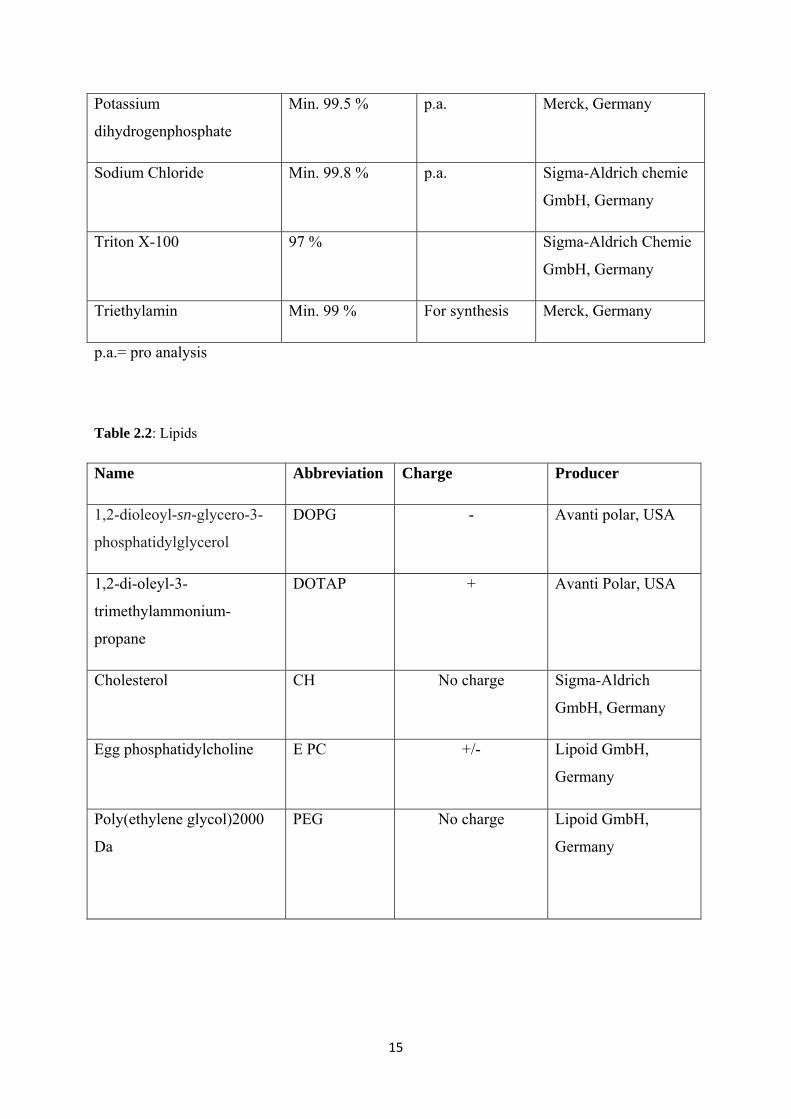
Table 2.2: Lipids
Name Abbreviation Charge Producer
1,2-dioleoyl-sn-glycero-3-
phosphatidylglycerol
DOPG - Avanti polar, USA
1,2-di-oleyl-3-
trimethylammonium-
propane
DOTAP + Avanti Polar, USA
Cholesterol CH No charge Sigma-Aldrich
GmbH, Germany
Egg phosphatidylcholine E PC +/- Lipoid GmbH,
Germany
Poly(ethylene glycol)2000
Da
PEG No charge Lipoid GmbH,
Germany

16
Table 2.3: Equipment
Equipment Type Producer
Bath sonicator Branson®1510 Branson ultrasonics, USA
Bath sonicator Model G112SPIT Laboratory supplies Co.,
Inc., USA
Centrifuge tubes Polycarbonat tubes, thick-wall Beckman Instrument, USA
Centrifuge Biofuge Startos Heraeus Instruments, UK
Chromatography
columns
Bio-Spin®Disposable
chromatography columns
Bio-Rad Laboratories, USA
Dialyse cassette Slide_A-Lyzer Dialysis cassette
10.000 MWCO
0.5-3.0 ml capacity
Thermo scientific, USA
Eppendorf-tube Safe-Lock tubes 2.0 ml Eppendorf AG, GmbH,
Germany
Rotary evaporator Büshi R-124 rotary evaporator with
vacuum pump v-500-system
Büshi, Switzerland
HPLC Waters 2690 Separation module
Waters 474 Scanning Fluorescence
detector
Waters 2487 Dual λ Absorbance
detector
Symmetry C18-columm (3,9x150
mm)
Waters,USA
Filter 0.22 µm non-sterile syringe filters Pall Life Sciences, USA

17
Filter 0.22 µm cellulose acetate filter Sartorius AG, GmbH,
Germany
Fluorescence
microscope
Leica CTR 6000 microsystem Leica, Germany, GmbH
Fluorescence
spectrophotometer
Perklin Elmer LS 55 Fluorescence
spectrometer
Perklin Elmer, UK
Microtitre plates Costar® UV 96-well plate with UV
transparent flat bottom, Acrylic
Costar®, USA
Microtitre plate reader Spectra Max 190 Microplate
Spectrophotometer
Molecular devices, USA
PCS Submicron Particle Sizer, model
370
Nicomp,USA
pH meter 744 pH meter Metrohm Metrohm Ltd, Switzerland
Probe-Sonicator Ultrasonics Vibra Cell VC 754
750 Watt ultrasonic processor
CVR 234 converter
Needle probe 19 mm
Sonics and Materials, USA
Probe-Sonikator Sonics high intensity ultrasonic
processor 500 Watt model
Needle probe 13 mm
Sonics and Materials, USA
Ultracentrifuge Optima LE-80 Beckman,USA
Vortex MS2 Minishaker IKA Chiron AS
Zetasizer Nano series Malvern instruments, UK
Zetasizer capillary cells Folded capillary cells Malvern instruments, UK

18
2.2 Methods Buffer solutions
Isotonic phosphate buffered salines (PBS) 0.025 M
1. 0.025 M di-sodiumhydrogenphasphate-dihydrat
I. di-sodiumhydrogenphosphate-dihydrat 8.90 g
II. Sodium chloride 15.16 g
III. Distilled water ad 2000.0 ml
I and II are dissolved in III
2. 0.025 M Potassium dihydrogen phosphate
I. Potassium dihydrogen phosphate 6.804 g
II. Sodium chloride 15.0 g
III. Distilled water ad 2000.0 ml
I and II are dissolved in III
Solution 1 and 2 are filtered through a 0.22 µm filter (cellulose acetate filters, Sartorius AG,
Germany) and mixed to achieve the desired pH which is pH 6.0.

19
Phosphate buffer (PB) 0.025 M pH 3.0
I. Ortho-Phosphoric acid 85 % 1.038 g
II. Sodium hydroxide ad pH 3.0
III. Distilled water ad 1000.0 ml
I is dissolved in 250 ml III, II are added to the solution. Rest of III is added up to 1000.0 ml.
The solution is filtered through a 0.22 µm filter (cellulose acetate filter, Sartorius AG,
Germany).
Triton- solution 10 % (w/w)
I. Triton X-100 10.0 g
II. PB 0.025 M pH 3.0 90.0 g (ad 100 g)
I is dissolved in II.
Mobile phase for HPLC analysis
TEAA buffer 1 v/v %
I. Triethylamine 20 ml
II. Distilled water ad 2000.0 ml
III. Acetic acid ad pH 5.5
I and 1500 ml II are mixed by a magnetic stirrer. III is added to obtain pH 5.5. Then the
volumetric bottle is filled to 2000.0 ml with II. The pH value is controlled to be 5.5 using a
pH-meter. The solution is filtered through a 0.22 µm filter (cellulose acetate filter, Sartorius
AG, Germany)

20
2.3 Liposome preparation
Liposomes can be prepared by several techniques (Torchilin and Weissig, 2003). We
employed both film hydration and freeze drying method, in order to determine if there are any
differences in the incorporation of CPT in liposomes. The most appropriate method was then
used further.
2.3.1 Lipid-CPT film preparation
Lipid-CPT films were prepared by mixing the lipid solutions in the desired composition with
solution of the active drug CPT in a round bottom flask. Stock solutions of 100 mg/ml were
made with different lipids in chloroform or a chloroform:methanol mixture. Stock solutions of
CPT in a mixture of chloroform:methanol (4:1 volume ratio) with a concentration of 2 mg/ml
were also prepared. After mixing the desired components, containing totally 1 mg CPT/130
µmoles lipid, the solvents were removed on a Büshi R-124 rotary evaporator with vacuum
pump 500-system (Büshi, Switzerland) for 45 minutes at 200 mPa on a water bath at 45 ˚C.
After 45 minutes, the round bottom flask was removed from the water bath and the pressure
was adjusted to 50 mPa for about 3 hours to remove traces of solvent and obtain a dry film.
The lipid compositions of the different formulations which were prepared are given in Table
2.4.
Table 2.4: The lipid compositions (mol %) of the different formulations
Lipids Formulations
1 2 3 4 5 6 7 8
Chol 10
E PC 100 90 90 85 80 85 85 85
DOPG 10
DOTAP 15 20 15 15 15
PEG 1 5 10

21
2.3.2 Lipid-CPT freeze-drying preparation
The freeze-dried samples were prepared by mixing solutions of the desired lipid EPC and the
active ingredient CPT in injection vials. 1 ml of lipid-CPT solution containing 100 mg lipid
and 1 mg CPT stock solution was used for freeze-drying. The vials with the mixture were
shock-frozen in liquid nitrogen for 1 minute and placed in a freeze dryer (beta 2-16 equipped
with an LMC-2 controller, Martin Christ Gefriertrocknungsanlagen GmbH, Osterode am
Herz, Germany) with opened stoppers. The freeze-drying method are described earlier
(Saetern et al., 2004b) and was performed for 65 hours at temperatures from - 40 °C to 45 °C
with pressure declining from 800 mbar to 0.008 mbar. After freeze-drying the PL-CPT vials
were sealed with aluminium closures and stored at – 80 °C.
2.3.3 Rehydration
The lipid-CPT samples from freeze-drying or film preparation were brought to room
temperature for about 15 minutes before rehydration. Subsequently 3 ml 0.025 M PBS with
pH 6.0 was added to the lipid film and 1ml to the freeze-dried cake for hydration. The lipid-
CPT mixture was vortexed and shaken to ensure that all the lipids were dispersed in the
buffer. The dispersion was then ready for further size reduction.
2.4 Size reduction of liposomes
After rehydration with buffer, it is assumed that the liposomes are present in multilamellar
vesicles (MLVs). To reduce the size and lamellarity of liposomes high energy must be
delivered to the liposome dispersion. There are several methods to reduce MLVs to SUVs,
and some of the most frequently used techniques are bath and probe-sonication (Brandl,
2001).

22
2.4.1 Bath sonication
The hand shaken MLVs film were sonicated in 3 ml portions in 15 minutes intervals using a
bath sonicator G112SPIT Special Ultrasonic Cleaner (Laboratory supplies Co.,Inc.,USA). The
sonicator and the samples were allowed to cool for ten minutes between each interval.
2.4.2 Direct probe sonication
Sonication of liposomes made by the film hydration method
The hand shaken MLVs film were placed in an ice bath and sonicated in 3 ml quantity with an
ultrasonic vibra cell (Sonics and Materials, USA) using a 19 mm needle probe tip and an
output of 40 % max. The different formulations were sonicated in intervals of two minutes
until the desired size was reached. The probe and the dispersion were allowed to cool for ten
minutes between each interval. Upon sonication, the sample were placed in the fridge at 4 °C
for equilibration overnight before further experiments were performed.
Sonication of liposomes from the freeze-drying hydration methods
The freeze dried samples were sonicated in a similar procedure as the hydrated liposome
films. The sonication intervals were the same, but due to the smaller 1 ml quantity, the
samples were sonicated using a Sonics high intensity ultrasonic processor (Sonics and
Materials, USA) with a 13 mm needle tip that can accommodate a volume of 1 ml. Upon
sonication, the sample were placed in the fridge at 4 °C for equilibration overnight before
further experiments were performed.

23
2.4.3 Size determination by photon correlation spectroscopy (PCS)
The particle size and distribution of the sonicated liposomes can be measured by photon
correlation spectroscopy (PCS), which is based on dynamic light scattering. The principle is
based upon Brownian motion of particles in the solution. Small particles diffuse much faster
than large particles, affecting the rate of fluctuation of scattered light intensity. The PCS
instrument focuses laser light to the sample, and registers any movement from particles in
solution (Torchilin and Weissig, 2003) (User manual, Nicomp Model 380,1997).
PCS measurements of particle size and distribution were performed on Nicomp TM model
380 particle sizing system (USA). In order to avoid impurities in the sample, sample
preparation was measured in clean environment using particle free equipments. The cuvettes
(borosilicate glass) were bath sonicated (Branson® 1510) for 10 minutes in freshly filtered
PBS pH 6.0. Then all the samples and equipment needed were carried out in a laminar air-
flow LAF bench prior to use. The test tubes were rinsed with PBS pH 6.0 filtered through a
0.22 µm pore size syringe filter. For measurement, the sample was diluted in filtered PBS pH
6.0 until a stable intensity of approximately 250-350 kHz was achieved (User manual,
Nicomp Model 380, 1997).
The following instrument parameters described below were used in accordance with
(Ingebrigtsen, 2001) with some exceptions. The buffer used was 0.025 M PBS pH 6.0.
• Nicomp distribution
• Automatic choice of channel width
• Number weighting
• Temperature 23 ˚C
• Liquid index of refraction: 1.333
• Laser wavelength : 632.8 nm (Helium-Neon)
• Liquid viscosity: 0.933 CP
• Scattering angle: 90˚ (Fixed angle)

24
• Number of cycles: 3
• Run time: 15 minutes
2.5 Ultracentrifugation
To separate excess CPT, crystals, titanium particles from the sonication probe and lipid
aggregates from the liposomes, an ultracentrifuge was employed. CPT crystals and lipid
aggregates have a higher density than the small probe sonication liposomes, and will settle in
the pellet upon ultracentrifugation. The SUVs with associated CPT will be present in the
supernatant. 500 µl PBS pH 6.0 was added to 2 ml of the liposomal dispersion in a 3-ml thick
wall polycarbonate centrifuge tube to raise the volume enough to fill the centrifuge tube. The
samples were vortexed for 30 seconds prior to centrifugation, and were then centrifuged using
Beckman Optima L8-M centrifugation with SW60Ti rotor (Beckman Inc.,USA). The
centrifugation speed was 100 000 g, the temperature 10 ˚C and duration was optimized to
separate free CPT crystals and lipid aggregates from the liposomes (Saetern et al., 2004b).
Totally 900 µl of the supernatant was then carefully withdrawn for further determination of
amount of CPT and PC as well as further studies on retention ability.
2.5.1 Fluorescence microscopy
In order to ensure that the ultracentrifugation method was optimized so that there were no
CPT crystals in the supernatant, the supernatant was examined using a fluorescence
microscopy Leica CTR 6000 microsystem (Germany). CPT is itself fluorescent so no external
labelling of the CPT crystals was needed. The supernatant was examined by applying a drop
on a glass clean slide and put a cover glass on top. It was important to avoid air bobbles
between the glass slide and the cover glass. The preparation was examined under the
microscope using a 20 x objective and filter set A, yielding an excitation wavelength of 360
nm with a bandwidth of 40nm and recording fluorescence at 470 nm (bandwidth 40 nm) with
a dichromatic mirror at 400 nm. Images were recorded using Leica Application Suite version
2.5.0 R1 (Germany).

25
2.6 CPT liposomes retention ability
The different formulations ability to retain CPT after incorporation was investigated using the
spin column method. The principle behind the column filtration is that liposomes do not
penetrate into the pores of the beads packed in the column, but instead percolates through the
interbead spaces. Proteins as well as free drug are smaller in size and will be retarded in the
pores of the bead pack. It is therefore assumed that CPT associated liposomes will be
separated from the serum proteins as well as free CPT and will be collected in the early
fractions. The formulations’ retention ability both in buffer and serum could be obtained using
this method.
2.6.1 Spin column method
The different formulations’ ability to retain the drug in buffer and serum was investigated by
using the spin column method (Torchilin and Weissig, 2003). 2.5 ml gel SeparatorGel
Agarose Beads ACL 4% (Sooner Scientific) was packed in spin columns (Bio-Rad) using
centrifuge Biofuge Startos (Heraeus Instruments, UK). The centrifuge was stopped manually
when speed of 400 rpm was reached.
300 µl of supernatant was diluted 1:2 v/v with PBS pH 6.0 and incubated at room
temperature. Another 300 µl of supernatant was diluted 1:2 v/v with serum and incubated at
37 ˚C. Separations of the incubated samples were measured at 0 hours, 5 hours and 24 hours
and the samples were separated into 4 different fractions. Separations were done by adding
100 µl of CPT-liposome dilution to a column. First fraction was collected in an eppendorf-
tube by centrifugation of the column until a speed of 400 rpm was reached. Further, the
second fraction was collected in a new eppendorf-tube by adding 100 µl PBS buffer pH 6.0 to
the column and performing the same centrifuge procedure as describe above. The same
process was repeated twice more using 100 µl PBS buffer pH 6.0 and the fractions collected
in eppendorf- tubes. Above, every fraction was diluted 1:2 v/v with Triton 10 % for further
PC and CPT determination.

26
2.7 Quantification of Camptothecin High performance liquid chromatography (HPLC) was used to quantify Camptothecin (CPT)
in the liposomes. The method described by Warner and Burke (Warner and Burke, 1997) was
used with some modifications. Samples were diluted 1:2 with 10 % triton solution to dissolve
the liposomes and release the incorporated CPT from the liposomes prior the analysis. Each
sample was analyzed in triplicates.
Quantification was achieved using CPT standards both in lactone and carboxylate form within
a concentration range from 0.5 µM to 5.0 µM. The standard curve exhibited good linearity
with a correlation coefficient of 0.997 ± 0.001 (n = 3).
Following HPLC-method was used:
• Mobile phase:
A: 25% Acetonitrile in 1% (v/v) triethylamine acetate buffer pH 5.5 and adjusted to
pH 5.99
B: 35% Acetonitrile in 1% (v/v) triethylamine acetate buffer pH 5.5 and adjusted
pH 6.32
C: 95% Acetonitrile in 5% distilled water (v/v)
• 474 scanning fluorescence detector
• Detection Wavelengths: Excitation λ=360 nm, Emission λ=440
• Column: Waters Symmetri C18-column (3.9x150 mm)
• Injection volume: 10 µl
• Flow rate: 1.0 ml/min
• Run time: 15 min
• Sample temperature: 25˚C
• Column temperature: 30˚C

27
2.8 Quantification of phosphatidylcholine Quantification of amount of phosphatidylcholines in liposomes was performed by using an
enzyme assay, Wako LabAssay Phospholipid B test kit (USA). The assays are based on
phospholipids (lecithin, sphingomyelin, lysolecithin) being hydrolyzed by phospholipase D
enzyme yielding choline as the product. Choline is further oxidized by choline oxidase in a
reaction which forms hydrogen peroxide. The latter takes part in a peroxidase-catalyzed
coupling which produces a blue pigment. The amount of phospholipids in the sample can be
determined by measuring the absorbance of the blue colour (Grohganz et al., 2003) (User
manual, Wako Chemicals).
Figure 2.1: Enzymatic quantification of phosphatidylcholine (PC) (User manual,WAKO LabAssay Phospholipid, Wako Chemicals)

28
The assay was performed using a microtiter plate. Each microtiterplate was filled with 25 µl
of the sample and 275 µl of colouring reagent. The plate was subsequently incubated at 37 ˚C
for 15 minutes prior the absorbance measurements at 600 nm performed with a microtiterplate
reader. All samples and standards were prepared and measured in triplicate (Grohganz et al.,
2003).
This method was used after ultracentrifugation of liposomes to quantify phosphatidylcholine
(PC) in the supernatant and pellet as well as to quantify the amount of PC in the fractions
from the retention study.
PC recovery in the supernatant after ultracentrifugation was calculated as follows:
• PC recovery = (Mean amount of PC in supernatant / mean amount of PC in total
dispersion)
The results from the incorporation study were adjusted based on the recovery to make the
comparison easier.
PC content in fractions collected from the columns were used to determine the percentage of
liposomes contained in each fraction compared to the original sample (either diluted in buffer
or serum) used for the retention analysis. For each sample, the fraction with the highest PC
content was used for CPT determination. If the PC content in this fraction was 40% of the
original sample, it was assumed that 40% of the liposomes were eluted in this fraction. The
results from the retention study were adjusted according to the percentage of liposomes in the
fraction to relate to the amount of lipids in the original sample.
2.9 Determination of zeta Potential of liposomes
Surface properties of liposome formulations can vary depending on the composition of the
lipid. There are cationic, anionic and neutral lipids, which can be used for preparation of
liposomes. Zeta potential can be used to identify any correlation between the liposome
incorporation and retention of CPT and surface charge properties of the liposomes (Brgles et
al., 2008). The zeta potential is charge at the slip plane of the particle surface, and although it
is not a direct measurement of the surface charge, it is a good estimation. Zeta potential is one
of the important factors affecting liposomes stability, incorporation efficiency and interactions

29
with biological system in vivo(Gjelstrup Kristensen, 2000) (Labhasetwar et al., 1994). The
zeta potential of liposomes was measured using a zetasizer Nano ZS (Malvern, UK).
The samples were thus analyzed using the zetasizer. Prior to analysis the samples were diluted
1 in 10 with PBS pH 6.0. The zetasizer capillary cell was also rinsed with 96 % ethanol and
distilled water using a 1-ml syringe prior to analyses as recommended by the manufacturer.
The diluted samples were then analyzed for ten cycles with a voltage of 4 mV.
2.10 Fluorescence anisotropy
To evaluate distribution of CPT in the liposomes, we used intrinsic fluorescence of CPT by
fluorescence polarization measurements to examine mobility of CPT in the liposomal bilayer.
Polarization measurements were performed on a fluorescence spectrophotometer (Perklin
Elmer, UK) and a number of liposomal compositions were investigated. The samples were
analyzed in a rectangular quartz fluorometer cell, and the excitation and emission wavelengths
were set at 360 and 440 nm, respectively.

30
3. RESULTS AND DISCUSSIONS
3.1 Method development In this study the method of incorporation capacity screening presented by Saeterns group
(Saetern et al., 2004b) was used. However, due to different type of equipment and a desire to
improve the feasibility of the method some changes were done as described below.
3.1.1 Sonication procedure It is well known that the sonication process may influence the size and size distribution of
liposomes (Woodbury et al., 2006). The goal was to obtain a sufficient size reduction and
monodispersed liposomal size. Since small liposomes is preferred for i.v. application, the
liposomal size was set to be <200 nm in diameter (Saetern, 2004). In the study by Saeterns
group (Saetern et al., 2004b) they used probe sonication with a (Labsonic U,B.Braun Biotech
International, Leverkusen, Germany) at 50 W, but this equipment was not available for us so
we had to find another way to prepare SUVs. In order to determine the optimal sonication
process for our purpose, size determination by PCS was performed upon sonication. Due to
the toxicity of CPT, we wanted to keep the CPT containing liposomes in closed containers for
not to expose the environment. We therefore chose to employ the bath sonicator to obtain
SUVs. The result of bath sonicated liposomes is displayed in Table 3.1.
Table 3.1: Bath sonicated EPC liposomes with duration of 5 x 15 minutes
Liposomal composition Mean particle size
(nm ± SD)
Polydispersity
Index (P.I.)*
EPC 704.3 nm ± 41.5 0.755
*P.I. represents the polydispersity index used as indication of size distribution of vesicles.
Lower values of P.I. indicate more homogeneous liposomal sample.
As described in Table 3.1 the measured size of the liposomes was found to be very large even
after sonication for five intervals of fifteen minutes. The P.I is as well quit high indicating that
the efficiency of size reduction is low and the samples are containing highly polydispersed
population of liposome.

31
The liposome size, standard deviation of the mean particle size and the large P.I suggests that
bath sonicator was not appropriate for size reduction of our liposomes, in addition the process
was quit time consuming. It was therefore decided to employ a more powerful sonicator to
reduce the size in a more rapid and reproducible manner.
3.1.1.1 Probe sonication Since bath sonication in a closed container showed not to be appropriate we had to choose
probe sonication in an open container and place the sonicator in a closed box in an appropriate
room (cytostatic laboratory) instead. The size reduction of the liposomes was performed using
an Ultrasonics Vibra Cell (USA) with a needle probe of 19 mm diameter. In order to
determine an optimal sonication process for size reduction of the liposomes, it was necessary
to present number of trials and evaluate the impact of number of intervals and time duration
on the liposomes.
Due to the high temperature during sonication, there is a risk of lipid degradation. It is
therefore necessary to keep the sonication time short, keep the system cooled and include a
break between the sonication cycles. Optimal sonication conditions were evaluated in regard
to vesicle size and size distribution by using EPC and EPC/Chol (90%:10%). The reason why
we chose a formulation with cholesterol in addition to pure EPC was that it is expected that
the inclusion of Chol in the liposomal bilayer makes it more rigid and more resistance to size
reduction (New, 1990).
The sonicated lipsomes was tested by PCS upon sonication and results are shown in Table
3.2. The liposomes obtained after 2x2min of sonication showed a satisfied size for the EPC
formulation. In the case of EPC/Chol formulation the results show larger vesicle size in
comparison with the EPC formulation. The sonication time was therefore increased for this
formulation, and the results show an obvious size reduction for our purpose after three
intervals of two minutes.

32
Table 3.2: Probe sonicated liposomes
Duration Liposomal composition Mean particle size± SD
Polydispersity Index (P.I.)
2 x 2 min EPC 42.9 nm ± 2 0.328
2 x 2 min EPC-Chol (90%/10%) 228.9 nm ± 5 0.712
3 x 2 min EPC-Chol (90%/10%) 32.5 nm ± 2 0.392
3.1.2 Optimization of ultracentrifugation for separation of free CPT from liposomes
Separation of CPT crystals from liposomes by ultracentrifugation was used to be able to see
how much drug is associated with the liposomes. CPT crystals and lipid aggregates have
higher density than SUV liposomes, and will settle in the pellet upon ultracentrifugation. In
order to ensure that the ultracentrifugation earlier described (Saetern et al., 2004b) was
optimized for our liposomes, and that there were no CPT crystals in the supernatant, the
supernatant obtained after 20 min of centrifugation at 100 000 g was examined using a
fluorescence microscopy Leica CTR 6000 microsystem, as described in 2.7. Averages of 10
to 20 pictures were taken of each sample and several different formulations were examined.
Figures 3.1 and 3.2 are representative examples.
Crystals of CPT could be visually distinguee from dissolved CPT because they appear as an
intense spot while CPT in liposomes results in a less intense fluorescing background. The
microscopy pictures (Figure 3.1) clearly show CPT-crystals in the supernatant upon
ultracentrifugation of 100 000 g for 20 minutes.

33
Refocusing of the lens to see the liquid-air interface in the glass slide
Figure 3.1: Fluorescence pictures from the examination under fluorescence microscope, Leica CTR 6000 microsystem, of the supernatant from the formulation number 1 after ultracentrifugation of 20 minutes. Observations were conducted using a 20 X objective.
34
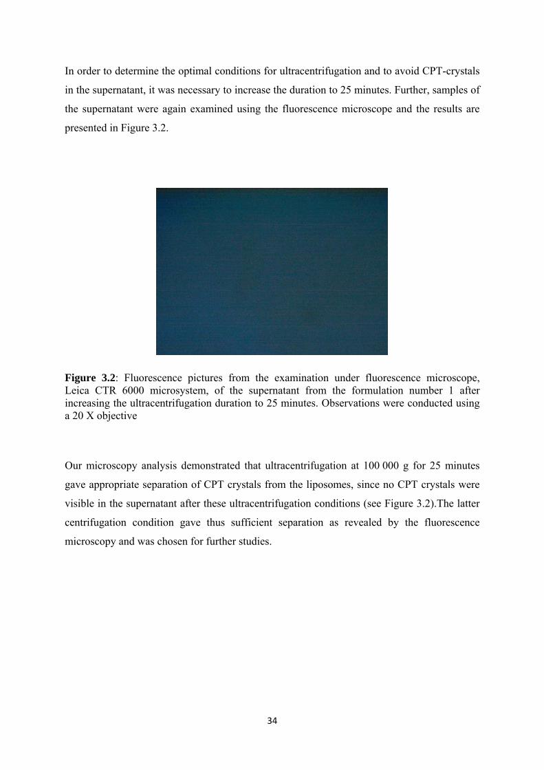
In order to determine the optimal conditions for ultracentrifugation and to avoid CPT-crystals
in the supernatant, it was necessary to increase the duration to 25 minutes. Further, samples of
the supernatant were again examined using the fluorescence microscope and the results are
presented in Figure 3.2.
Figure 3.2: Fluorescence pictures from the examination under fluorescence microscope, Leica CTR 6000 microsystem, of the supernatant from the formulation number 1 after increasing the ultracentrifugation duration to 25 minutes. Observations were conducted using a 20 X objective
Our microscopy analysis demonstrated that ultracentrifugation at 100 000 g for 25 minutes
gave appropriate separation of CPT crystals from the liposomes, since no CPT crystals were
visible in the supernatant after these ultracentrifugation conditions (see Figure 3.2).The latter
centrifugation condition gave thus sufficient separation as revealed by the fluorescence
microscopy and was chosen for further studies.

35
3.1.3 Comparison of film and freeze-drying method As part of the development of a method to detect incorporation and retention of CPT in
different liposome formulations, we compared CPT incorporation using two different methods
for removing organic solvents from the lipid:CPT mix. We wanted to employ the film
methods for our research, while the freeze-drying method had previously been employed for
similar research in this lab. The purpose of the comparison of film and freeze-drying method
was to determine if there are any significant differences in the incorporation capacity of CPT
in the liposomes when using these two methods. The reason for this was that the freeze-drying
method caused a lot of problems (M. Skar, personal communication) and we wanted to do it
in a more appropriate way. The freeze drying method reported by Saetern and co-workers
(Saetern et al., 2004b) is anyway a more suitable method when DMSO is used as the solvent
for CPT. The method removes organic solvent by sublimation, and in order for this procedure
to work properly the lipid mixture needs to be in the solid state at – 40 ˚C . We employed
organic solvents Chloroform:Methanol (4:1 volume ratio) in the CPT stock solution as
recommended by the manufacturer. DMSO was not used as we expected better mixing of the
lipids and the CPT in organic phase before drying, using the Chloroform:Methanol mixture. It
is more difficult to remove chloroform to obtain a dry cake using freeze-drying. For this
reason it was decided in advance to use the film method if there were no major differences
between the incorporation capacities of CPT between these two methods.
However, due to drying problems with the Freeze dryer (beta 2-16 equipped with an LMC-2
controller, Martin Christ Gefriertrocknungsanlagen GmbH, Osterode am Herz, Germany) we
only achieved one parallel of formulation number 1 using this method. The result of the
comparison is displayed in Figure 3.3. It appears that there are no significant differences in
CPT-incorporation between these two methods. The variations are within the standard
deviation that we expect from incorporation studies using lipids.

36
Figure 3.3: Camptothecin-incorporation capacity of EPC (formulation 1) by film (n=3) and freeze-drying method (n=1).
The freeze-drying problems is mainly related to our choice of solvents for preparation of CPT
stock solution which was Chloroform:Methanol (4:1 volume ratio). Chloroform has a melting
point of - 63 ˚C and by using the procedure as described in 2.3.2 it appeared some difficulties
to sublimate the solvent to vapour. The melting point of chloroform might indicate that the
solvent did not pass through the solid state and become sublimated, but rather evaporated
when raising the temperature from – 40 ˚C to 45 ˚C and lowering the pressure from
800 to 0.08 mAtm within 65 hours. Moreover, CPT-incorporated liposomes approach
performed earlier by Saetern and co-workers (Saetern et al., 2004b) gave reproducible data in
their studies, probably due to their choice of solvent. Saetern et al (Saetern et al., 2004b)
employed DMSO for preparation of CPT stock solution, and gained dry lipid cakes. DMSO
has a melting point of 18.4 ˚C, and will be in the solid state and further become sublimated
when raising the temperature from – 45 ˚C to 45 ˚C, while reducing the pressure.
However, it looks like these two methods is comparable as was expected and the film method
was chosen as the method for further liposome preparation due to less troublesome
preparations.
CPT incorporation with two different methods
0
20
40
60
80
100
120
140
160
180
Film method Freeze drying method
µg C
PT/1
30 µ
mol
es li
pid

37
3.2 Characterization of how lipid composition influences CPT incorporation and retention
Earlier studies in our research group has revealed that the lipid composition of the liposomes
have an effect on the CPT incorporation capacity (Saetern et al., 2004b) (unpublished results).
In this study we wanted to investigate these effects in more detail by changing the lipid
composition based on the previously studies. We also wanted to look at the different
formulations retention ability since it is not enough that the drug is incorporated it also needs
to stay with the liposomes in circulation.
3.2.1 CPT liposome incorporation study
Five liposome formulations with lipids of varying carbon chain length, saturation and charge
as shown in Table 2.4 were prepared with a total concentration of 1 mg CPT/130 µmoles
lipid, and the content of CPT in the liposomes was quantified after ultracentrifugation. As the
molecular weight of the lipids varied significantly, especially later when PEG lipids were
used, we based the formulations on 1 mg CPT/130 µmol lipid which is equivalent to 1 mg
CPT/100 mg of EPC. Results from incorporation in the tested formulations are presented in
Figure 3.4.
Figure 3.4: Camptothecin-incorporation capacity of the different liposome formulations (n = 3)
CPT Incorporation in liposome formulations
0
50
100
150
200
250
300
350
EPC EPC/DOPG (90%:10%) EPC/Chol (90%:10%) EPC/DOTAP(85%:15%)
EPC/DOTAP(80%:20%)
CPT
µg
/ 130
µm
oles
lipi
d

38
The most promising formulations in this study were formulation 4 and formulation 5, which
exhibited a significantly higher CPT-incorporation capacity compared to the other liposome
formulations displayed in Figure 3.4. Both these formulations contain DOTAP and this is
likely the main reason for the higher incorporation capacity. As we can see in Figure 3.4 the
incorporation efficacy increases with a raising DOTAP content from 15 mol % to 20 mol %,
although the two are not significantly different. Increased incorporation capacity observed in
our results, corresponds with earlier studies and observations (Saetern et al., 2004b).
Correlation between presence of DOTAP in liposomes and increased incorporation capacity
previously studied by Saetern and co-workers (Saetern et al., 2004b), indicates that CPT have
a higher affinity for cationic lipids such as DOTAP as compared with other non-charged
lipids. Since CPT exists in a pH dependent equilibrium between its lactone isomer and
negatively charged carboxylate isomer, the latter may bind to the cationic liposomes through
electrostatic interactions and lead to increased incorporation of CPT within the cationic
liposomes.
The non-charged formulations, 1 and 3 do show lower incorporation capacities compared to
the negative charged formulation. In comparison with literature, Cortesi and co-workers
(Cortesi et al., 1997) reported that addition of Chol seems to raise the incorporation from 50
% to 57 %. In the contrary, other studies done by Saetern and co-workers (Saetern et al.,
2004b) and Daouds group (Daoud et al., 1995) observed that addition of Chol presented in the
membrane reduced CPT-incorporation capacity. However, a direct comparison of these
results can not be made due to differences in experimental approach, as well as the higher
amount of Chol in the liposomes from Saeterns group compared to those in our research.
A slight tendency toward increased incorporation was observed in formulation 2, when the
negative charged lipid DOPG was presented in the PC membrane. This formulation contained
10 % DOPG and 90 % EPC, and as we can see in Figure 3.4, addition of DOPG raised the
incorporation capacity as compared with formulation 1 containing 100 % EPC. This result are
corresponding with the study done by Sugarmans group (Sugarman et al., 1996), which
suggested that use of DOPG solubilised the drug-lipid particles to a higher degree. Also
Saeterns group reported increased incorporation capacity when negative charged DPPG was
present in the liposome formulation (Saetern et al., 2004b). Any effect by using the longer and
more unsaturated oleyl fatty acid chain instead of palmitoyl was however not observed
compared to what has been reported earlier (Saetern et al., 2004b).

39
3.2.2 CPT liposomes retention ability
A liposome formulations’ ability to retain the incorporated drug is a prerequisite for
successful delivery of drug to the target. The retention abilities for the different formulations
were therefore investigated.
The retention ability of CPT of the five formulations shown in Table 2.4 was investigated
over time, with withdrawal of samples after 0, 5, and 24 hours of incubation both in buffer
and serum as described in section 2.6.1.
Figure 3.5: The different liposome formulations ability to retain CPT, associated with the liposomes over time in buffer at room temperature (n = 3)
The results of retention incubated in buffer at room temperature are displayed in Figure 3.5,
and show different retention ability depending on the liposome formulations. There are no
significant differences between the formulation containing EPC (formulation 1, Table 2.4)
and EPC-DOPG (formulation 2, Table 2.4), which exhibit a slightly lower and decreasing
retention ability during the incubation at room temperature. However, the most prominent
results observed was the formulations containing EPC-Chol (formulation 3, Table 2.4) and
0 %
10 %
20 %
30 %
40 %
50 %
60 %
70 %
80 %
EPC, buffer EPC-DOPG, buffer EPC-Chol, buffer EPC-DOTAP 15%,buffer
EPC-DOTAP 20%,buffer
Perc
ent C
PT a
ssoc
iate
d w
ith th
e lip
osom
es
Hr 0Hr 5Hr 24
40
EPC-DOTAP 15% (formulation 4, Table 2.4), which leads to less drug leakage from the
liposomes as compared to the other formulations. In general, none of the formulations (Figure
3.5) appears to retain CPT within the liposomes to high degree. However, the results exhibit
high standard deviations. We assume that the high standard deviations are due to the packing
of the gel (SeparatorGel Agarose Beads ACL 4%) in the columns. The columns were packed
using a centrifuge without set time function, and had to be stopped manually when it reached
the desired rpm. We assume that this resulted in different packing of the gel material in the
columns, which may have led to incomplete or varying elution of the liposomes of the
columns.
Liposomes in serum are most likely disposed to interact with blood components. One effect of
this is drug loss to aggregates such as lipoproteins (Silvander et al., 1998).
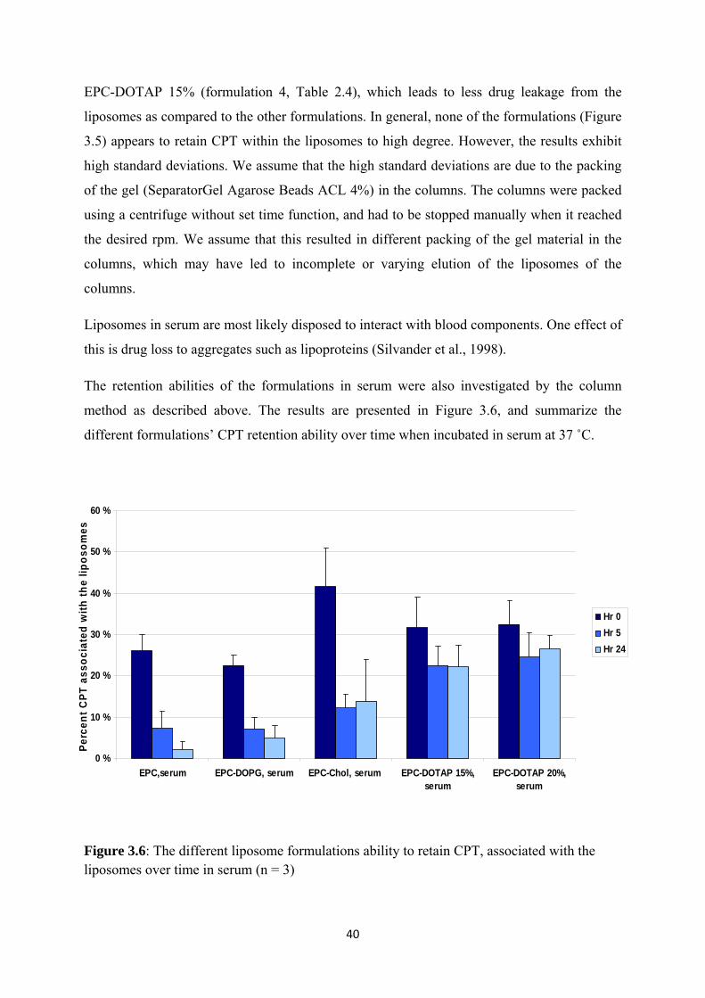
The retention abilities of the formulations in serum were also investigated by the column
method as described above. The results are presented in Figure 3.6, and summarize the
different formulations’ CPT retention ability over time when incubated in serum at 37 ˚C.
Figure 3.6: The different liposome formulations ability to retain CPT, associated with the liposomes over time in serum (n = 3)
0 %
10 %
20 %
30 %
40 %
50 %
60 %
EPC,serum EPC-DOPG, serum EPC-Chol, serum EPC-DOTAP 15%,serum
EPC-DOTAP 20%,serum
Perc
ent C
PT a
ssoc
iate
d w
ith th
e lip
osom
es
Hr 0Hr 5Hr 24

41
As we can see in Figure 3.5 and 3.6 there are no significant differences between retention
ability in buffer and serum for the three first formulations right after the dilution i.e. t = 0.
Moreover, the liposomes retention ability seems to decrease when these three formulations
were incubated in serum compared to in buffer. The most promising results are the DOTAP
containing formulations incubated in serum, which exhibit a trend toward higher liposomal
retention compared to the other liposome formulations. Although formulation 4 and 5 shows
better retention ability, only 25 % of the drug is associated with the liposomes, which is far
from being optimal.
In the case of serum containing samples, variations in the column production may have led to
incomplete separation of liposomes and serum proteins. This might be the major reason of the
high standard deviations. Due to the high standard deviation as described, more studies have
to be done before any conclusion can be made. Another separate method to analyse retention,
based on dialysis, is being developed so that the above results can be confirmed.
3.2.3 CPT liposomes incorporation ability and the influence of the zeta potential
Zeta potential of the different liposome formulations were measured to identify if there are
any correlations between incorporation and retention ability and surface charge properties of
the liposomes. The result is given in Figure 3.7 and shows a plot of measured zeta potential of
the different liposomes.
Figure 3.7: Zeta potential (mV) of the different liposome formulations (n = 3)
-15
-10
-5
0
5
10
15
20
25
30
EPC EPC/DOPG(90%:10%)
EPC/Chol(90%:10%)
EPC/DOTAP(85%:15%)
EPC/DOTAP(80%:20%)
Zeta
pot
entia
l (m
V)

42
Liposomes which consist of EPC and EPC-Chol show a zeta potential of around 0 mV. A
decrease of zeta potential is seen for the anionic lipid DOPG included in the liposome
formulation 2. Further, the zeta potential starts to increase when the cationic DOTAP is
employed in the liposomes, which is all according to what was expected.
A correlation between absolute zeta potential and incorporation efficacy, are presented in
Figure 3.8 and a trend toward increased CPT retention ability is seen when the absolute
surface charge of the liposomes becomes higher. However it is hard to draw any clear
conclusions since increasing concentration of DOTAP also has shown increased CPT
incorporation.
Figure 3.8: Correlation between liposomal Zeta potential (mV) and incorporation efficacy. The incorporation efficacy is plotted against absolute value of the zeta potential
EPC-DOPG
EPC-DOTAP 15%
EPC-DOTAP 20%
EPC
EPC-Chol
75
95
115
135
155
175
195
215
235
255
275
0 5 10 15 20 25Zeta potential
µg C
PT/
130
µmol
es li
pid

43
3.3 Preliminary studies on PEGylated liposomes
As mentioned earlier, PEGylation of parenteraly administered liposomes are important to
avoid opsonisation and rapid clearance from the blood circulation in vivo. The aim of the PEG
inclusion to these liposomes was therefore to investigate whether the PEG had an effect on the
CPT-incorporation capacity as well as retention ability, and to avoid rapid in vivo clearance in
future studies.
3.3.1 PEGylated CPT liposomes incorporation study
As the EPC-DOTAP formulations showed increased CPT- incorporation in comparison to the
other formulations displayed in Figure 3.4, these formulations were chosen for further PEG
studies. Moreover, since the incorporation capacity of formulation 4 and 5 was quite similar,
formulation 4 containing EPC-DOTAP 15 % was chosen for further PEG studies.
Furthermore, DOTAP is more expensive than the other naturally occurring lipids and has a
higher toxicity in vivo. The option was therefore to start with the lowest DOTAP content,
which was 15 %, and see if same incorporation results could be obtained with inclusion of
different ratios of PEG.
Incorporation capacity of PEGylated liposomes (formulation 6-8 in Table 2.4) were prepared
with a total concentration of 1 mg CPT/130 µmoles lipid, and the content of CPT in the
liposomes was quantified after ultracentrifugation as described in section 2.5. We utilized
DSPE-PEG lipids where the PEG had a molecular weight of 2000 Da. The results of the
incorporations are displayed in Figure 3.9.

44
Figure 3.9: Comparison of Camptothecin-incorporation between the PEGylated and non-PEGylated liposomes. Formulation 5 (n = 3), formulation 6 (n = 4), formulation 7 (n = 1), formulation 8 (n = 2)
The use of PEG resulted in an important observation. Reduced CPT incorporation capacity
was observed when different ratios of PEG were included in the DOTAP/EPC formulation
compared to the EPC/DOTAP formulation whiteout PEG (see Figure 3.9). As we observed,
there are no significant differences between formulation 6 which contains 1 % PEG and
formulation 8 containing 10 % PEG. Formulation 7 shows a relatively higher incorporation
capacity as compared with formulation 6 and 8. Due to technical problems that appeared
during the preparation, unfortunately we obtained only one parallel of formulation 7. This
makes the comparison with the other formulations more difficult. Overall, PEG inclusion into
the cationic liposomes reduced the incorporation capacity. The reason for the reduced
incorporation when PEG is included is not completely understood, but we hypothesize that
the DSPE-PEG chains might affect the fluidity of the liposomes, decreasing the incorporation
of the drug into the membrane. PEG has a neutralization effect on the liposomes, and as seen
in Figure 3.11 PEG also reduces the zeta potential as expected, thereby possible diminish the
favourable effect of the cationic lipids. The neutralization effect of PEG was linearly
proportional to the molar inclusion of the PEG lipids.
0
50
100
150
200
250
300
EPC/DOTAP(85%:15%)
EPC/DOTAP/PEG(85%:15%:1%)
EPC/DOTAP/PEG(85%:15%:5%)
EPC/DOTAP/PEG(85%:15%:10%)
µg C
PT/1
30 µ
mol
es li
pid

45
3.3.2 PEGylated CPT liposomes retention ability
The CPT retention ability of the PEGylated formulations shown in Table 2.4 was investigated
over time, with withdrawal of samples after 0, 5, and 24 hours of incubation both in buffer
and serum as described in section 2.8 and results are presented in Figure 3.10.
Figure 3.10: The different PEGylated liposomal formulations ability to retain CPT, associated with the liposomes over time in buffer and serum (n = 1 for PEG 1 % and 5 %, and n = 2 for PEG 10 %)
Depending on PEG ratios employed in the liposomes, different leakage profiles were
observed. The results show that inclusion of different ratios of PEG to the liposomes
influences the formulations’ ability to retain the drug. The presence of 1 % and 10 % PEG
show better retention and slower drug leakage from the liposomes compared to 5 % PEG
formulation as displayed in Figure 3.10. Although we do not have results from several
parallels of 1 % and 5 % PEG formulations, the results indicate that inclusion of PEG
influence the formulations’ ability to retain the drug and a tendency toward improved
retention ability is observed especially when 1 % PEG is included. Leakage of drugs from the
bilayer is thought to be related to fluidity and packing order of the membrane. It seems like
inclusion of PEG gives higher degree of retention which is good. PEG inclusion in liposomes
0 %
10 %
20 %
30 %
40 %
50 %
60 %
EPC-DOTAP 15%-PEG 1%, serum
EPC-DOTAP 15%-PEG 5%, buffer
EPC-DOTAP 15%-PEG 5%, serum
EPC-DOTAP 15%-PEG 10%, buffer
EPC-DOTAP 15%-PEG 10% serum
Pre
cent
CPT
ass
ocia
ted
with
the
lipos
omes
Hr 0Hr 5Hr 24

46
is assumed to alter the surface characteristics and reduce the leakage from the liposomes
(Brandl, 2001)
3.3.3 PEGylated liposomes CPT incorporation and the influence of zeta potential
Zeta potential of the different PEGylated formulation was measured to see how PEG is
influencing the zeta potential as well as how much one have to include to get a zeta potential
around zero. The results are displayed in Figure 3.11.
Figure 3.11: Zeta potential of PEGylated liposomal formulations (n = 3)
As expected, inclusion of neutral PEG into EPC/DOTAP liposomes resulted in decreasing
zeta potential shifting towards zero. The zeta potential decreased with an increasing PEG
inclusion leading to a neutralization of the liposomal surface charge.
The decreased zeta potential resulted in decreased CPT incorporation within the liposomes,
thereby removing the favourable effect of the cationic lipids. Although the zeta potential
decreases with an increasing PEG ratio, this seems not to decrease the liposomes retention
-4
-2
0
2
4
6
8
10
12
14
16
18
EPC/DOTAP(85%:15%)
EPC/DOTAP/PEG(85%:15%:1%)
EPC/DOTAP//PEG(85%:15%:5%)
EPC/DOTAP//PEG(85%:15%:10%)
Zeta
pot
entia
l (m
V)

47
ability further. The research on PEGylated liposomes so far indicates that the PEG inclusion
could have a favourable effect on CPT retention.
3.4 Fluorescence anisotropy
The fluidity of the liposomal bilayer membrane was investigated by fluorescence
depolarization anisotropy in an attempt to find a relation between drug incorporation and
retention ability with the membrane fluidity. The incorporated CPT was employed as a
fluorescent probe to investigate changes in mobility and packing of the hydrocarbon acyl
chains in the bilayer. CPT’s rotational freedom in the bilayer dictates the degree of
polarization. An increased anisotropy value indicates less rotational freedom for the drug
trapped in the liposome, which conserves the polarization of the incoming light. We used the
intrinsic fluorescence of CPT to measure anisotropy, so that a lower anisotropy value would
indicate that the CPT molecules had a more rotational freedom in the membrane, and were
therefore more loosely trapped. A number of liposomal compositions were investigated, and
Figure 3.12 summarizes the findings.
Figure 3.12: Anisotropy in different liposome formulations
0,12
0,14
0,16
0,18
0,2
0,22
0,24
0,26
0 10 20 30 40 50 60 70 80 90 100
Temperature (Celcius)
Ani
sotro
py
EPC
DOPG
Cholesterol
Dotap 15%
Dotap 20%
Dotap 15% PEG1%
Dotap 15% PEG5%
48
As shown in Figure 3.12 we saw little differences between the non-PEGylated formulations,
most likely due to that these measurements are not specific enough toward retention to expose
the small differences we discovered in the column experiments. We expected to see increased
anisotropy with increased retention and decreased incorporation in liposomal formulations,
hypothesizing that these drugs were more tightly trapped in the liposomal membrane.
Contrary to what we expected, the PEG containing formulations as a group exhibited lower
anisotropy while demonstrating an increased retention of the drug on the column experiments.
In light of these results we conclude that anisotropy alone is an incomplete measure of the
liposomes’ ability to retain CPT over time. The reason for this can be manifold; I) The
temperature dependent anisotropy measurements are done over a short time period just after
ultracentrifugation, thereby not exposing the differences between the formulations after
extended incubation with buffer. II) Leakage of CPT during the temperature dependent
experiment will result in some background scattering of free CPT in solution, and it is unclear
if this background scatter is linearly proportional to a reduction in anisotropy value. III)
Different levels of CPT incorporation into the liposomes can result in different anisotropy
values. It can be more optimal to measure anisotropy at different time point in the incubation
period similar to the retention studies. The anisotropy measurements must be further
optimized with another marker to yield meaningful data (see also section 4: Future
perspectives)
3.5 Fusing of liposomes and size measurements using PCS
Charged liposomes are shown to be unstable in the presence of serum components, as
indicated by rapid fusion, and leading to rapid clearance from the blood circulation (Mori et
al., 1998). During the retention studies, we noticed that the sample solutions containing serum
changed colour during the 24 hours of incubation to become whiter, more milk like. We
hypothesize that fusion of liposomes is the reason of this. In order to check whether the
hypothesis is correct or not, we analyzed some of the formulations on PCS. The results from
the PCS analysis are displayed in Table 3.2.

49
Table 3.3: PCS results of some liposome formulations right after sonication (0 Hr) and after 24 Hr incubation in buffer and serum
0 Hr 24 Hr buffer 24 Hr serum
Duration Liposomal composition Mean particle size
(nm ± SD)
2 x 2 min EPC 42.9 nm ± 2 40 nm ± 2 259.6 nm ± 11
2 x 2 min EPC-DOPG (90%:10%) 37.3 nm ± 1 50.2 ± 0.1 105.2 nm ± 2
3 x 2 min EPC-Chol (90%/10%) 32.5 nm ± 2 36.7 nm ± 2 280.2 nm ± 23
2 x 2 min EPC-DOTAP (80%: 20%) 30 nm ± 3 70 nm ± 2 120 nm ± 5
2 x 2 min EPC-DOTAP (85%:15%) 28.9 nm ± 2 80.8 nm ±2 107.8 nm ± 3
2 x 2 min EPC-DOTAP-PEG (85%:15%:1%)
75.1 nm ± 4 78.6 nm ± 4 77.3 nm ± 5
2 x 2 min EPC-DOTAP-PEG (85%:15%:5%)
68.9 nm ± 1 71.3 nm ± 3 72.6 nm ± 4
2 x 2 min EPC-DOTAP-PEG (85%:15%:10%)
48.5 nm ± 0.5 49.8 nm ± 1 49 nm ± 1
The results represent the mean of tree cycles (3 x 15 minutes)
*Hr = hours
Results displayed in Table 3.2 indicate that the size of liposome formulations without PEG
attachment increases in presence of serum during the incubation time. This indicates that the
milk like appearance is related to the liposome fusion which represents an irreversible
process. As described above, it is known that charged liposomes exhibit instability and
interacts with different serum components. This might be one of the reasons of the charged
liposomes rapid fusion. It is also known that temperature conditions may influence liposome
fusion, and since the serum containing samples were incubated respectively at 37 ˚C, this may
have influenced the fusion rate of the neutral liposomes.
In order to use fusogenic liposomes in drug delivery systems in vivo, they have to be designed
in such way to become stable in the circulation for an extend period of time, and to avoid
opsonisation before they reach the desired target site.

50
The PCS results of PEG containing liposomes do not show any size alteration during the
incubation period, and we conclude that PEG inclusion in the liposomes stabilizes their size
under the incubation conditions by decreasing the fusion of liposomes. We also see that 1 %
PEG is enough to prevent fusion, we wanted to minimize the effect of reduced incorporation
when PEG was included. 1% PEG will therefore be used further in this on going project.

51
4. Conclusions
In the first part of this thesis establishment of appropriate methods for incorporation and
retention studies were performed. A comparison of lipid film and freeze-drying method was
done and no major differences were seen between the different methods according to
incorporation capacity of CPT. The film method was thus chosen as the method for further
liposome preparation. Further, bath sonication and direct probe sonication were compared in
terms of preparation of SUVs. The latter method gave satisfied size reduction for our purpose
and was used during the study. For separation of liposomes from CPT-crystals a
centrifugation approach was used. Centrifugation at 100 000 g for 20 minutes was found not
sufficient for removal of CPT crystals from the supernatant. Upon changing the duration to 25
minutes, all crystals were settled in the pellet and no CPT-crystals were visible under the
fluorescence microscope. 25 minutes centrifugation was concluded to be the appropriate
centrifugation condition for our study.
Second, CPT-liposomes with different lipid compositions were prepared and evaluated for
incorporation capacity and retention ability. The incorporation capacity of CPT within the
liposomes was found to be highest for positively charged DOTAP-containing liposomes as
compared with other formulations containing zwitterionic or negatively charged
phospholipids. The DOTAP-containing liposomes exhibited as well a trend toward higher
retention ability in serum compared to the other formulations tested.
Third, in the preliminary PEGylation studies the PEG containing liposomes showed lower
incorporation capacity compared to the liposomes without PEG, while better retention and
slower leakage from the liposomes was seen when 1 % and 10 % PEG was included in the
liposomes. In addition, PEG inclusion in the liposomes resulted in less fusion of liposomes
during incubation in both buffer and serum. From the results it can be seen that 1 % PEG is
enough to prevent fusion during the incubation, but further investigation are still needed to
find optimal PEG ratios that can avoid opsonisation in further in vivo studies.
In summary we have made progress towards establishing a method for producing and testing
of CPT-liposomes as well as indentifying a formulation that effectively can incorporate CPT
and retain the drug in circulation. However, we have yet to arrive at the ideal formulation,
and, as we are still evaluating elements that affect all areas of incorporation and retention,
further studies are needed in order to reach the goal of a formulation appropriate for in vivo
studies.

52
5. Future Perspectives
There is a need for further investigation and optimization of the protocol for the retention
study to yield reproducible data. One method to measure the retention ability of the liposomes
that is currently being tested is by employing dialysis. Dialysis is a process of separating
small molecules from macromolecules such as proteins in a solution using a semi-permeable
membrane. Dialysis operates by diffusion, which leads to movement of molecules from areas
of higher to lower concentrations. By employing a dialysis membrane with the right
molecular cut-off, the small CPT molecules will freely diffuse across the semi-permeable
membrane into a second chamber of liquid until equilibrium is reached (Aulton, 2007). Both
buffer and serum or protein solution could be used as acceptor medium to look at the
influence of presence of proteins, and samples can be taken from the dialysis chamber at set
time-points. Using this method no measurements of PC-recovery is needed for the retention
studies, and liposome samples can be analyzed directly by the HPLC. By varying the different
dialysis conditions, the liposomes’ retention ability in different environments can be
investigated.
The fluorescence anisotropy measurement must also be further investigated and optimized to
yield more information. One way that may optimize this measurement is to employ a
hydrophobic probe like1,6-Diphenyl-1,3,5-hexatriene (DPH) in the bilayer to investigate the
bilayer fluidity and thus be able to predict CPT’s rotational freedom in the bilayer. The
advantage with DPH before CPT is that it is easy to dissolve in the lipid bilayer which will
ensure the same concentration of a probe in all the different formulations (Silvander et al.,
2000).
It is clear that further studies needs to be done to reach the goal of a formulation appropriate
for further in vivo studies. It would thus be of great interest to continue developing selected
formulations as well as introduce new elements into the formulations to achieve a desirable
incorporation and retention of CPT. Studies done by Watanabe group (Watanabe et al., 2008)
and Maitani group (Maitani et al., 2008) has shown that inclusion of various artificial lipids in
liposomes, especially benzoic acid derivatives can increase the incorporation capacity and
retention of CPT in liposomes both in vitro and in vivo.. This might be due to an interaction
between a phenyl group on the lipids and CPT by a π-π interaction. Another interesting
approach that may improve the incorporation and retention ability of CPT-liposomes is by
including polymerizable diacetylene into the liposomal bilayer. Polydiacetylene molecules are

53
generally non-toxic molecules, and form polymerized vesicles under UV irradiation. Such
polymerizable vesicles are found to be more stable in comparison with unpolymerized
vesicles, and therefore more suitable prolonged release drug delivery. In vitro study done by
Guo group (Guo et al., 2010) show that conjugated backbone of the molecules by the
photopolymerization acts as a barrier for drug release. Higher polymerization degree is shown
to lead to a more sustained release of the drug from liposomes. It would be of great interest to
develop these formulations, and investigate CPTs’ incorporation and retention ability within
these liposomes.
54
6. REFERENCES
ALLEN, T. M. 1994. The use of glycolipids and hydrophilic polymers in avoiding rapid uptake of liposomes by the mononuclear phagocyte system. Adv. Drug Delivery Rev., 13, 285‐309.
AULTON, M. E. 2007. Aulton's pharmaceutics: the design and manufacture of medicines, Edinburgh, Churchill Livingstone.
BRANDL, M. 2001. Liposomes as drug carriers: a technological approach. Biotechnol. Annu. Rev., 7, 59‐85.
BRGLES, M., JURASIN, D., SIKIRIC, M. D., FRKANEC, R. & TOMASIC, J. 2008. Entrapment of Ovalbumin into Liposomes‐Factors Affecting Entrapment Efficiency, Liposome Size, and Zeta Potential. J. Liposome Res., 18, 235‐248.
BURKE, T. G. 1996. Chemistry of the camptothecins in the bloodstream: drug stabilization and optimization of activity. Ann. N. Y. Acad. Sci., 803, 29‐31.
BURKE, T. G. & BOM, D. 2000. Camptothecin design and delivery approaches for elevating anti‐topoisomerase I activities in vivo. Ann. N. Y. Acad. Sci., 922, 36‐45.
BURKE, T. G., STAUBUS, A. E., MISHRA, A. K. & MALAK, H. 1992. Liposomal stabilization of camptothecin's lactone ring. J. Am. Chem. Soc., 114, 8318‐19.
CHRAI, S. S., MURARI, R. & AHMAD, I. 2002. Liposomes (a review). Part two: Drug delivery systems. BioPharm (Duluth, MN, U. S.), 15, 40,42‐43,49.
COOPER, G. M. & HAUSMAN, R. E. 2009. The cell: a molecular approach, Washington, ASM Press/Sinauer Associates.
CORTESI, R., ESPOSITO, E., MAIETTI, A., MENEGATTI, E. & NASTRUZZI, C. 1997. Formulation study for the antitumor drug camptothecin: liposomes, micellar solutions and a microemulsion. Int. J. Pharm., 159, 95‐103.
DAOUD, S. S., FETOUH, M. I. & GIOVANELLA, B. C. 1995. Antitumor effect of liposome‐incorporated camptothecin in human malignant xenografts. Anti‐Cancer Drugs, 6, 83‐93.
EMERSON, D. L. 2000. Liposomal delivery of camptothecins. Pharm. Sci. Technol. Today, 3, 205‐209.
FAHR, A., VAN, H. P., KUNTSCHE, J. & LEIGH, M. L. S. 2006. Lipophilic drug transfer between liposomal and biological membranes: what does it mean for parenteral and oral drug delivery? J. Liposome Res., 16, 281‐301.

55
FLATEN, G. E. 2003. Incorporation of Camptothecin in liposomes, method development and incorporation efficacy screening using different liposome formulation. Master Thesis, University of Tromsø.
GARIDEL, P., JOHANN, C. & BLUME, A. 2000. Thermodynamics of lipid organization and domain formation in phospholipid bilayers. J. Liposome Res., 10, 131‐158.
GJELSTRUP KRISTENSEN, H. 2000. Almen farmaci, København, Danmarks farmaceutiske højskole.
GROHGANZ, H., ZIROLI, V., MASSING, U. & BRANDL, M. 2003. Quantification of various phosphatidylcholines in liposomes by enzymatic assay. AAPS PharmSciTech, 4, No pp. given.
GUO, C., LIU, S., DAI, Z., JIANG, C. & LI, W. 2010. Polydiacetylene vesicles as a novel drug sustained‐release system. Colloids Surf., B, 76, 362‐365.
HARASHIMA, H., SAKATA, K., FUNATO, K. & KIWADA, H. 1994. Enhanced hepatic uptake of liposomes through complement activation depending on the size of liposomes. Pharm. Res., 11, 402‐6.
HSIANG, Y. H. & LIU, L. F. 1988. Identification of mammalian DNA topoisomerase I as an intracellular target of the anticancer drug camptothecin. Cancer Res., 48, 1722‐6.
INGEBRIGTSEN, L. 2001. Size analysis of submicron particles and liposomes by size exclusion chromatography and photon correlation spectroscopy Thesis for the degree candidatus pharmaciae, University of Tromsø.
JAIN, R. K. 1987. Transport of molecules across tumor vasculature. Cancer Metastasis Rev., 6, 559‐93.
LABHASETWAR, V., MOHAN, M. S. & DORLE, A. K. 1994. A study on zeta potential and dielectric constant of liposomes. J. Microencapsulation, 11, 663‐8.
LEE, S.‐C., LEE, K.‐E., KIM, J.‐J. & LIM, S.‐H. 2005. The Effect of Cholesterol in the Liposome Bilayer on the Stabilization of Incorporated Retinol. J. Liposome Res., 15, 157‐166.
LI, Q.‐Y., ZU, Y.‐G., SHI, R.‐Z. & YAO, L.‐P. 2006. Review camptothecin: current perspectives. Curr. Med. Chem., 13, 2021‐2039.
LIU, D., LIU, F. & SONG, Y. K. 1995. Recognition and clearance of liposomes containing phosphatidylserine are mediated by serum opsonin. Biochim. Biophys. Acta, Biomembr., 1235, 140‐6.
MAEDA, H. 2001. The enhanced permeability and retention (EPR) effect in tumor vasculature: The key role of tumor‐selective macromolecular drug targeting. Adv. Enzyme Regul., 41, 189‐207.
MAITANI, Y., KATAYAMA, S., KAWANO, K., HAYAMA, A. & TOMA, K. 2008. Artificial lipids stabilized camptothecin incorporated in liposomes. Biol. Pharm. Bull., 31, 990‐993.
56
MASSING, U. & FUXIUS, S. 2000. Liposomal formulations of anticancer drugs: Selectivity and effectiveness. Drug Resist. Updates, 3, 171‐177.
MATTERN, M. R., MONG, S. M., BARTUS, H. F., MIRABELLI, C. K., CROOKE, S. T. & JOHNSON, R. K. 1987. Relationship between the intracellular effects of camptothecin and the inhibition of DNA topoisomerase I in cultured L1210 cells. Cancer Res., 47, 1793‐8.
MAURER, N., FENSKE, D. B. & CULLIS, P. R. 2001. Developments in liposomal drug delivery systems. Expert Opin. Biol. Ther., 1, 923‐947.
MI, Z. & BURKE, T. G. 1994. Differential Interactions of Camptothecin Lactone and Carboxylate Forms with Human Blood Components. Biochemistry, 33, 10325‐36.
MORI, A., CHONN, A., CHOI, L. S., ISRAELS, A., MONCK, M. A. & CULLIS, P. R. 1998. Stabilization and regulated fusion of liposomes containing a cationic lipid using amphipathic polyethylene glycol derivatives. J. Liposome Res., 8, 195‐211.
NEW, R. R. C. 1990. Liposomes: a practical approach, Oxford, IRL Press.
OGIHARA, T., KAGAWA, H., GAO, Q. & MORI, K. 2010. A study of the molecular structure of phospholipids and the aggregation of liposomes using the molecular orbital method. J. Comput. Chem., Jpn., 9, 43‐46.
PERRIE, Y. & RADES, T. 2010. Pharmaceutics: drug delivery and targeting, London, 2010.
SAETERN, A. M. 2004. Parenteral Liposome‐ and Cyclodextrin Formulations of Camptothecin. PHD, University of Tromsø.
SAETERN, A. M., BRANDL, M., BAKKELUND, W. H. & SVEINBJORNSSON, B. 2004a. Cytotoxic effect of different camptothecin formulations on human colon carcinoma in vitro. Anti‐Cancer Drugs, 15, 899‐906.
SAETERN, A. M., FLATEN, G. E. & BRANDL, M. 2004b. A method to determine the incorporation capacity of camptothecin in liposomes. AAPS PharmSciTech, 5, e40.
SILVANDER, M., HANSSON, P. & EDWARDS, K. 2000. Liposomal Surface Potential and Bilayer Packing As Affected by PEG‐Lipid Inclusion. Langmuir, 16, 3696‐3702.
SILVANDER, M., JOHNSSON, M. & EDWARDS, K. 1998. Effects of PEG‐lipids on permeability of phosphatidylcholine/cholesterol liposomes in buffer and in human serum. Chem. Phys. Lipids, 97, 15‐26.
SUGARMAN, S. M., ZOU, Y., WASAN, K., POIROT, K., KUMI, R., REDDY, S. & PEREZ‐SOLER, R. 1996. Lipid‐complexed camptothecin: Formulation and initial biodistribution and antitumor activity studies. Cancer Chemother. Pharmacol., 37, 531‐8.

57
TORCHILIN, V. P. & PAPISOV, M. I. 1994. Why do polyethylene glycol‐coated liposomes circulate so long? (Molecular mechanism of liposome steric protection with polyethylene glycol: Role of polymer chain flexibility. J. Liposome Res., 725‐39.
TORCHILIN, V. P. & WEISSIG, V. 2003. Liposomes: a practical approach, Oxford, Oxford University Press.
VENDITTO, V. J. & SIMANEK, E. E. 2010. Cancer Therapies Utilizing the Camptothecins: A Review of the in Vivo Literature. Mol. Pharmaceutics, 7, 307‐349.
WALL, M. E., WANI, M. C., COOK, C. E., PALMER, K. H., MCPHAIL, A. T. & SIM, G. A. 1966. Plant antitumor agents. I. Isolation and structure of camtothecin, a novel alkaloidal leukemia and tumor inhibitor from Camptotheca acuminata. J. Am. Chem. Soc., 88, 3888‐90.
WARNER, D. L. & BURKE, T. G. 1997. Simple and versatile high‐performance liquid chromatographic method for the simultaneous quantitation of the lactone and carboxylate forms of camptothecin anticancer drugs. J. Chromatogr., B: Biomed. Sci. Appl., 691, 161‐171.
WATANABE, M., KAWANO, K., TOMA, K., HATTORI, Y. & MAITANI, Y. 2008. In vivo antitumor activity of camptothecin incorporated in liposomes formulated with an artificial lipid and human serum albumin. J. Controlled Release, 127, 231‐238.
WOODBURY, D., RICHARDSON, E., GRIGG, A., WELLING, R. & KNUDSON, B. 2006. Reducing Liposome Size with Ultrasound: Bimodal Size Distributions. J. Liposome Res., 16, 57‐80.
WWW.AVANTILIPIDS.COM.
WWW.UNI‐MAGDEBURG.DE.
Usermanual, NICOMP MODEL 380 Windows based software1997, Dynamic light scattering theory.
User manual,WAKO LabAssay Phospholipid, Wako Chemicals.