thesis on the therapeutic value of x-rays and ultra-violet
TRANSCRIPT

T H E S I S
on
The Therapeutic Value of X-Rays and Ultra-Violet Ray#,
with special reference to Asthma.
by
ELSIE H.PARKER, M.B., Ch.B., D.M.R.E.(Camb.).
brought to you by COREView metadata, citation and similar papers at core.ac.uk
provided by Enlighten: Theses

ProQuest Number: 27534977
All rights reserved
INFORMATION TO ALL USERS The quality of this reproduction is dependent upon the quality of the copy submitted.
In the unlikely event that the author did not send a com p le te manuscript and there are missing pages, these will be noted. Also, if material had to be removed,
a note will indicate the deletion.
uestProQuest 27534977
Published by ProQuest LLO (2019). Copyright of the Dissertation is held by the Author.
All rights reserved.This work is protected against unauthorized copying under Title 17, United States C ode
Microform Edition © ProQuest LLO.
ProQuest LLO.789 East Eisenhower Parkway
P.Q. Box 1346 Ann Arbor, Ml 48106- 1346

The Therapeutic Value of X-rays & Ultra-Violet Rays with special reference to Asthma.
I. Introduction.The therapeutic value of light has long been recognised,
and "photo-therapy” as it has been called, has been a well-known department of therapeutics for many years.
Light, however, is now known to be only a part of the total Solar Spectrum. It is also known that the rays of the Solar Spectrum which are too rapid or too slow to be detected by the naked eye produce certain therapeutic effects on the human body.
In addition to this, the discovery of X-rays, and of radio-active bodies emitting gamma rays (Y-rays), has greatly enlarged this department of therapeutics.
Visible light, ultra-violet rays, and X-rays, only differ the one from the other in their respective wave-lengths, and their relationship to one another will be shown later, when discussing the spectrum.
Ultra-violet rays. X-rays, and Y-rays have now definitely taken their place in the field of therapeutics - the first, for their general tonic and healing effects, the second and third for their destructive effect on abnormal growth of tissue.
These therapeutic measures have been used for the treatment of many diseases, and of these, asthma is the one to which I intend to refer in this paper.
Asthma is generally regarded as a neurosis, characterised by more or less severe dyspnoea, due to spasmodic narrowing of the bronchial lumen, and alternating with spasm of the muscles of the thorax.
The exciting causes are either direct irritation of the bronchial mucosa, or indirect irritation through the nervous system, or the blood. Heredity is an important factor, especially where there is a tendency to gout, phthisis, or nervous complaints, in the family history.
The paroxysmal attacks are due to the reflex spasm of the bronchial muscles, which is associated with hyperaemia and the exudation of characteristic mucus from the smaller bronchioles.
Premonitory symptoms of the attack may or may not be present. The attacks usually occur during the night, and are characterised by great dyspnoea, prolonged expiration, which is accompanied by sibilant rhonchi, and the expulsion of gelatinous sputum which contains Cur^hmann’s spirals, and later, Charcot-Leyden crystals. ^
These paroxysmal attacks have a tendency to recur periodically, and their onset may be associated with many

predisposing causes, such as the ingestion of foreign proteids in food - (e.g. eggs) or the presence of animal or vegetable dust (e.g. fur, pollen), or organisms.
Many and various forms of treatment have been devised for this condition. Any source of reflex irritation, such as nasal disorders, gastro-intestinal irritation, or uterine or urethral disease, should be removed first, and attention to diet and to the general health is essential. Of drugs. Potassium Iodide may be given between the attacks, and during the attacks, anti-spasmodics such as adrenalin, and belladonna etc. are recommended.
Treatment by ultra-violet radiation and by X-rays has also been tried within recent years, and the results from the latter have compared favourably with other forms of treatment. The reasons for employing physio-therapeutic methods in this condition will be shown later, in dealing with the biological effects of ultra-violet and X-rays.
I have treated a large number of cases, both of simple and malignant disease by Deep and superficial X-rays, and now propose to give an account of the therapeutic uses and value of ultra-violet and X-rays, in a series of cases of Asthma, based on the results of my experience.

II. Historical Outline.(a) of Ultra-Violet Ray Therapy, or Heliotherapy.
The practice of Sun Cure is as old as man himself, and in 431 B.C. Herodotus wrote that he had made observations concerning the influence of the sun, and sun treatment, and that he himself had considered its indications as well as its contraindications.
Hippocrates exposed his patients to the sun; Antyllus also, Avicenna, an Arab physician (circa A.D.1000), and Celsus, were firm believers in the health-giving powers of the sun’s rays.
The history of religion has acquainted us with an early cult of the sun, in the Old as well as the New world, and throughout the ages, we find the sun worshipped as the creatorand supporter of the human race.
Through the rapid conquest of the ancient world by Christianity, with its abnegations of worldly institutions, in additions to the temples, shrines, and pleasure sites, there were sacrificed many therapeutic and hygienic achievements, including the worship of the sun.
It was not till the second half of the 18th.century, with the rapid advancement of physics in the field of optics,that the powers of the sun were re-discovered.
In 1815, Leobel recommended Insolation in various
disorders; in 1816, Dobereiner tackled the question scientifically, and laid down the principles of photo-therapy.
The merit for having enlarged the range of indications for heliotherapy belongs to the French, and especially to the School of Lyons.
Finsen published in 1899 his remarkable works on the treatment of Small-pox by means of red light, and on the treatment of lupus, by ultra-violet rays.
A short time afterwards, important scientific works appeared, by Roux, Arloing, Koch, and Herman Van Schrotter, which attempted to explain the mode of action of this valuable therapeutic factor.
In 1905, Rollier opened his first clinic at Leysin - at a height of 1,300 metres, for the systematic treatment of external tuberculosis, by Heliotherapy. His results were so successful, that gradually people began to evince a growing interest in this work.
In Britain, where the lack of sunlight does not permit of the extensive practice of heliotherapy, many types of artificial light apparatus supply a reliable and valuable substitute for natural sunlight, and are available at all times and seasons. Dr.Rollier*s technique is generally adopted as the standard for treatment both by natural and artificial sunlight.
The test of time has still to be applied to the treatment, but results Obtained are sufficibnt to justify further investigation and experiment in this branch of physiotherapy.
(b) Of X-Rays.The discovery of X-rays is comparatively recent, but
certain early experiments gradually led to Rontgen's discovery.Of these, the first was in 1650, when Otto Von Guericke invented the air pump, and applied it to the laws of science. In 1740, Abbe^Nollet continued these studies, and in 1834, Sir W.Snow Harris formulated the statement that the length of the spark which an electric machine will give in air varies as the inverse ratio of the pressure of the gas.
In 1838, Faraday showed his experiments in electricity, and Geissler improved on these in his study of electric glow discharges. In 1840, Clerk Maxwell propounded the electromagnetic theory of light. In 1860, Lord Kelvin gave to science the absolute electrometer, and Gaissot substituted the Ruhmkoff coil by cells. In 1865, Sprengel invented the mercury air pump, an instrument devised for the purpose of producing very high rarefactions, with a great degree of rapidity.
In 1869, Hittorf, discovered Cathode Rays, and fromO'RS1877 to 1879 investigatiefts were making extended studies and

and investigations into the theories already advanced.In 1879, Sir.m/m.Crookes made further experiments on
the nature of Cathode Rays, which were shown to consist of unit particles of negative electricity known as electrotts.
In 1883, Wiedermann and J.J.Thomson declared that these particles were ether disturbances of short wave length.
In 1894, Hirz and Lenard proved that cathode rays would pass through the walls of a vacuum tube, and in 1895, Rontgen discovered that X-rays are produced whenever and wherever cathode rays encounter matter.
In 1896, Becquerel discovered that uranium salts emitted spontaneously a radiation which was capable of passing in varying degrees, through all matter, whether transparent or opaque to light, and which could be detected by its effect on a photographic plate.
Further experiments by Friedrich, Knipping, Lane, Sir Wm.Bragg, and his son Prof.Bragg, proved that X-rays are analogous to ordinary light, and differ from it, in wave length only.
The discovery of the phenomenon of radiography, by Becquerel in 1896, was followed by that of Madame Curie, and Schmidt, independently, that thorium was radio-active. In studying the radio-activity of uranium along with P.Curie, and G.Bemont, Madame Curie undertook the chemical separation of the elements of pitch-blende.

8
These researches resulted (1898-1900) in the discovery of three radio-active substances - polonium, radium, and actinium.
In 1904, Ramsay and Hahn discovered Radiothorium, and in 1906, Boltwood discovered ionium. At the present time, nearly forty radio-active elements are known, each with a definite and characteristic kind of radio-activity.
III. General Observations on Light Therapy and X-ray TherapHy,(a) Physics.
The total solar spectrum contains many rays which the human eye cannot see, the retina being sensitive to relatively few rays.
The spectrum consists of radiations transmitted from the ether, which differ from each other in wave length only.The longest ones, with a length of 2 million centimetres are the wireless telegraphy waves; the shortest with a length of one-thousand-millionth part of a centimetre, are X-rays, and Y-rays of radium. Accordingly, as the centimetre unit is an awkward one to use, it is customary to refer to wave lengths in terms of Angstrom units - an Angstrom unit (A.U.) being the hundredth-millionth part of a centimetre.
The following diagram of the spectrum shows the relative position and extent of these waves, and the relationship

between visible light, ultra-violet light, and X-rays and radium,
y RF\>(s
Rp»3>Iuitx
U L T R A ' Vismj-£ TriFRR- \ WavtS<<x»«R.euess «opvveVloUET
RPNDIBTIOHSWSMT
X)nt3tPK0'R£T>"Rei o M. . —
REDf^pTDlATiorts
'Ovxtxvuo-Re
T q T R L S o L f tT \ S P E C T R U M .
Of ultra-violet rays, those of the greatest therapeutic value lie between 2,300 and 2,900 A.U. Ultra-violet rays have only a very slight power of penetration, but with X-rays, the- property of penetration is re-established.
For the production of ultra-violet radiation,certain substances are brought to incandescence by means of electrical energy, chief of these being mercury, tungsten and carbon, and different qualities of ultra-violet radiation are obtained according to the metals used. The following diagram illustratesthis point:-
____ (.oiFO R* U
XRF"f- exT«.6.neo.v. RAX-S ■
ULTRR-VlouEr
imnnMnmssMEMmmTmmnmmnnimmm
C.v«.Tmu«T(onTa.V.'RrX
“f u.v. *f CsawTTKx«Ok. tot Volvic
(loo a ,300
m (un/muutm
'VJ.V-'Rojs (X SviwVi K*:
VlSlQUR
— ■■> Ivvf R.R T(GD
CARftoN Arc
VrtPou'R ATtc.HeaT Rays
a,<joo ff.avo ^ovro ft.O.
D fm o R A M r\A T fc 3PEcrav>r»\ SHowm <§ U L T R A V i o u e r R a ^ r o N S

10
It is thus shown that ultra-violet radiations from a carbon arc lamp more nearly approach those of sunlight; that the radiations from the tungsten arc have a higher therapeutic value; and those from a mercury vapour arc have a still greater therapeutic value.
The shortest ultra-violet rays approach the region of the longest X-rays, and this brings one to a short discussion on the nature of X-rays.
The quality of X-rays depends on the electro-motive force applied to the tube, and also on the degree of evacuation of the tube. Low or high penetrating power depends on long or short waves respectively, soft X-rays, i.e., those with a low penetrating power having a wave length of .6 A.U., and hard X-rays i.e., those with a high penetrating power, having a wave length of .25 to .1 A.U.
The quantity of X-rays depends on the number of electipns which are propelled from the cathode in a given time, and this number varies with the strength of the current i.e., the number of milliamperes discharging through the tube.
The intensity of X-rays is the term applied to the quantity of X-rays traversing a certain area in a unit of time.The intensity depends on the electro-motive force and the number of milliamperes discharging through the tube, and this varies with the resistance of the tube. The intensity is also

11
influenced by distance and absorption.Only the rays which are absorbed produce the physical,
chemical and biological changes which are used for medical purposes.
Part of the rays absorbed are converted into secondary rays, the penetrating power of which is lower than that of the primary ray. X-rays also become "scattered" within the tissues, and the scattered rays have the same penetrating power as the primary rays which caüsed their appearance. The importance of scattered rays increases with the volume and the depth of the tissue exposed to X-rays, and the total dose reaching the object is therefore increased by the presence of these scattered rays.
Metal filters are always used in therapeutic exposures, in order that the soft X-rays, which would be heavily absorbed by the skin and superficial tissues, may be eliminated before they reach the skin, and thus damage to these tissues is avoided.
(b) Chemical Action of Ultra-violet and X-rays.The question of pigment formation represents one of
the basic problems of general biology. In 1888, Neumann observed that red blood corpuscles which are destroyed and are undergoing dissociation give rise to a pigment on coming into contact with normal tissue cells.

12
Rollier holds that red blood corpuscles which have absorbed short wave rays, become dissociated through their agency, and the cell chromatin then through the medium of diastase turns the remnant of haematin into pigment. He also considers that dilatation of the blood vessels is in itself insufficient, and that a simultaneous destruction of haemoglobin must take place. Thus the chemical forerunners of the pigment are liberated during haemolysis, under the influence of light.
The experiments of Bloch showed that chemically active rays increased the intensity of the action of ferments, and also increased their amount. Others showed that under the influence of light, soluble proteins are converted into less soluble ones.
Lasch showed that exposures to the quartz lamp resulted in an increase of the calcium retention in the blood, in cases of rickets: others showed in similar cases that the phosphatic content of the blood was raised, and cholesterol liberated by ultra-violet radiation.
Pincussen observed that light reduced glycosuria in some diabetic patients, and Wumberger found that light actively stimulates growth. Experiments generally have proved that ultra-violet radiation increases the haemo-bactericidal powers of the organism, and also quickens and increases the general metabolism.
X-rays applied to the skin in moderate doses cause an erythema, and this redness of the skin which usually appears a

15
week after exposure, changes later to brown and leaves a permanent bronzing of the skin. This erythema is the recognised unit of measurement of the biological effects of X-rays, and is analogous to the pigmentation produced by ultra-violet radiation.
Whether X-rays have a chemical, electrical, or mechanical, influence on animal tissues is not yet fully known.
One theory is that, in the tissues, electro&s circulating in a characteristic number and order round the nuclfci of atoms, are disturbed by the rays, and in consequence, the atoms break up and cause chemical disturbances.
Experiments by Hirsch and Petersen on blood drawn from patients receiving deep X-ray treatment showed that there are no striking or consistent alternations in the chemical constitution of the blood. There is, however, a disturbance of the acid-base equilibrium, manifested immediately after treatment by an increase of the hydrogen-ion concentration and sometimes by a slight lowering of the alkali reserve. After twenty-four hours, there is diminished hydrogen-ion concentration, and an increased alkali reserve.
In view of the fact that in cases where the red-blood corpuscles are normal, and the haemoglobin is 100^, the response to treatment is more marked and rapid - and also from the observations on secondary radiations made by Barkla and others, Knox has suggested the following theory.
14
In the blood stream there exist materials which^when bombarded by X-rays or radium, throw off secondary radiations, which in some way or another act on normal and abnormal tissue - stimulating the former, and damaging the latter, leading to a diminution in the size of a tumour.
The most likely material to give off secondary rays is the haemoglobin of the red cells, which is a compound of iron Iron gives off secondary radiations when exposed to X-rays, and iron stands high in the list of metals which give off radiations. These radiations are independent of the chemical combination of the metals, and depend on the quantity of the metal present.
Another intrinsic source of radio-activity within the body is potassium which sends out penetrating beta and gamma rays.
Metals also require a particular hardness of X-rays to enable them to emit their characteristic secondary radiations. This may account for the marked degree of action produced in cases which have a high percentage of haemoglobin, and it also throws light on these cases which have failed to respond to treatment. In them, the particular quality of X-rays employed may not have been the right one, or the exposure may not have been sufficiently long.
15
(c) Biological Action of Ultra-Violet and X-rays.
Investigations have showed that the long wave ultraviolet rays have a bactericidal effect, as have also the infrared waves. The researches of Finsen showed that the death of bacteria is not solely attributed to the action of light, for a conspicuous part is played by the inflammatory reaction of the tissues set up by insolation.
There is also an increase in the haemoglobin and the red blood cells, with a slight decrease in the number of white blood corpuscles after ultra-violet radiation.
Ultra-violet radiation has a sedative effect on the vago-sympathetic system, and also has some influence on certain disturbances of the endocrine functions.
Insolation also causes a Vaso-dilatation of the circulatory system, and by means of extensive hyperaemia of the skin, decongestion of the inner organs occurs, and is followed by a reduction in blood pressure. Hasselbach observed that ultra-violet radiation reduced the frequency of respiration, the depth being proportionately increased.
The biological effect of X-rays are very important, and numerous experiments have been made to demonstrate the morphological or functional effects on normal tissues.
The biological unit of X-rays is the human skin erythema dose, and the unit skin dose is defined by the
16
radiation causing slight-hyperaemia after 8 days, and slight tanning after 4 weeks. It is measured by the Ion toquant ime ter, SaboraMd pastilles^or Kionboeck's strips, the duration of the dose being determined by the length of time only; thus the efficiency of the tubes and apparatus can be constantly tested.
In 1926, Tsuzuki (Tokic, Japan) made a series of experiments on normal rabbits, which he irradiated by the hard rays such as are used in X-ray Therapy. Some of the histological changes noted were as follows:-
(1) Lymphatic system showed destruction of lymphocytes, andphagocytosis, followed by regeneration of lymphocytes and proliferation of reticulo-endothelial cells.
(2) Spleen showed atrophied follicles which had little powerof recovery, and the organ became more fibrous than normal and flattened.
(3) Bone Marrow - destructive phenomena were seen first, thenregeneration within a few days, with a gradual increase in pigment cells - the regenerative processes consisted chiefly of erythropoiesis often followed in a few weeks by fibrosis.
(4) Liver - Showed parenchymatous degeneration, followed byrecovery in a few days.
Lymphocytes have the strongest radio-sensitivity,10^ of an erythema dose being sufficient to cause their destruction. Erythrocytes have a greater resistance, but as there is an increase of iron pigment found in the spleen after irradiation, there must be some destruction of erythrocytes.
X-rays act with most effect on cells when their reproductive activity is the greatest, and the chromatin is the most

17
vulnerable part of the cell. Cells are thus vulnerable in proportion to their embryonic type. The groups of cells most sensitive to X-rays are those of the lymphatic tissues and glands, ovaries, testicles, supra-renal and other glands containing internal secretion: less sensitive are the cells of the skin, liver and kidneys, and least of all, the cells of the muscles and bones.
It is impossible to limit the action of the rays to one group of cells only, without affecting others in different ways.
Small doses of X-rays stimulate cells: large doses destroy them. The boundary line between the stimulating and the paralysing dose is very narrow - it varies with the susceptibility of the various organs and groups of cells, and with the quantity of X-rays that have been absorbed by them.

18
IV. (1) Applications of Heliotherapy.There are many diseases which have been treated by
ultra-violet radiation with varying results, according to the type of disease, and the technique employed. Some enthusiasts would have usibelieve that sunlight is almost a "cure All", but the more rational view is based on statistics obtained over a certain length of time, by many workers on the subject, and thus definite proof of its efficacy or otherwise is obtained.
The most marked and best results of the application of heliotherapy have been obtained in cases of non-pulmonary tuberculosis. Of these, tuberculosis of the spine, and of the upper and lower extremities form a large proportion. In tuberculosis of the spine, sunlight is combined with immobilisation, and in hip disease it is combined with continuous extension of the affected leg. Where sinuses are present, efficient drainage of them is carried out. The characteristic features of sunlight treatment in these cases are the striking development of muscles, frequent return of articular function, and general recuperation.
Tubercular peritonitis requires very great caution in the application of heliotherapy, for circulatory disturbances in the peritoneum must be avoided: good results are obtained under careful treatment. Tuberculosis of lymphatic glands yields to treatment with heliotherapy, and remarkable results are obtained in tubercular affections of the eye, and in lupus.
In 1927, at Leysin, I had the opportunity of seeing many of Dr.Rollier*s cases and was very much impressed by his results. The general nutrition of these patients was remarkable, and lack of pain was a striking feature. The tuberculous lesions showed every evidence of healing, though in sinuses, where mixed infections were present, the healing process was slower than in a purely tubercular infection.
In a large number of the surgical cases of tuberculosis, pulmonary foci were also found to be present, but these were of a latent and benign character. In patients with simultaneous bone and lung trouble, the rapid forms of pulmonary tuberculosis were rare, probably owing to partial immunisation from the foci.
Rollier treats cases of pulmonary tuberculosis with great caution; very careful acclimatization of these patients to heliotherapy is ordered, and treatment is preceded by prolonged observation and study of the case. This is very necessary as benefit may be obtained in cases where the lesions in the lungs are of the fibro-cirrhotic type which is frequently associated with bronchitis and emphysema: benefit is also obtained in the common forms of abortive tuberculosis, chronic or dry pleurisy, and old-standing effusions belonging to this category.
On the other hand, heliotherapy is strictly contraindicated in all forms of exudative types of tuberculosis where

»
there are pneumonic and feverish processes present - & high temperature is an unconditional contra-indication to heliotherapy in pulmonary tuberculosis. Sun baths given without considering these points may provoke haemoptysis, and the formation of fresh lesions even in sluggish and a-ffcbrile cases.
Ultra-violet radiation has also been given in cases of asthma and in view of the above remarks, it is advisable that before beginning treatment, the clinical history should be thoroughly investigated, and X-ray examination of the chest performed, in order to exclude pulmonary tuberculosis in any of its active forms, as in some cases, asthma and pulmonary tuberculosis may be co-existent.
Heliotherapy has also had excellent results in certain diseases of metabolism, such as rickets, owing to the increase of the calcium and phosphorus content of the blood after irradiation. Malnutrition in children, and debility in adults also receive much benefit from carefully graduated doses of ultra-violet radiation.
Diseases of the skin, such as impetigo, eczema, psoriasis and alopecia, derive benefit from local treatment combined with general treatment. This is due to the stimulating effect of the ultra-violet rays on the skin, and the body generally.
In non-tubercular affections, such as osteon?elit4s, varicose ulcers, and injuries, including wounds, burns, and

a#
fractures, heliotherapy acts as a valuable adjuvant to other methods of treatment owing to its soothing, bactericidal, and sclerogenous properties, and also due to the fact that it preserves muscular and articular functions-
Prophylactic heliotherapy, combined with open air and rational exercises, in childhood, is now recognised to be of great service to the community, by raising the power of resistance on the part of the child, to tuberculosis and other diseases.
Contra-indications to heliotherapy, apart from those already mentioned in connection with pulmonary disease, are few. Cardiac insufficiency, even in its initial stage, is a contraindication, as are also valvular disease, advanced myocarditis, and bad arterio-sclerosis: nephritis and advanced bilateral renal tuberculosis in which uraemia is threatened, are also in this category.
The indications and contra-indications for the employment of heliotherapy having been discussed, it will be seen that as it is a powerful stimulant of the natural defences of a patient, it has a definite place in the field of therapeutics

X9.
IV, (2) Therapeutic Applications of X-rays,
Apart from the use of X-rays for diagnostic purposes, great benefit is obtained in many diseases from their therapeutic application. The effects may be graded according to the intensity of the radiation; weak intensities may stimulate motility, excitability of nerves, and metabolism, while greater intensities destroy, or weaken them. Growth processes are also affected; in some cases, a period of stimulation is followed by depression and death; in others, a first period of retardation is followed by acceleration.
The essential difference between the effects of the various kinds of rays is quantitative rather than qualitative- the nature of stimulation or injury depending on the character of the living system affected, more than on that of the rays.
Repeated radiations may cause an immunisation of the tissues against the effect of radiation.
For practical convenience, X-ray Therapy is divided into two sections - (a) Superficial Therapy. (b) Deep Therapy,
(a) In Superficial Therapy there are two subdivisions ;-(1) in which excellent results have been obtained in the
treatment of skin diseases, such as acne^ syci^osis, and psoriasis, due to the stimulating effect of X- rays on the resistance of the tissues; and in eczema and ktloid due to the regulation of the overgrowth of normal tissues by X-ray. The sedative effect of X-rays is also of use in the treatment of pruritus.
In these cases, ^ ^ of a SabaraMd pastille dose,filtered through i mm.of Aluminium, and using a six-inch spark

2g
gap, is given. The penetration is from 1 to 3 millimetres below the surface of the skin, and may be compared to ultra-violet radiation, which has a penetration of 1 millimetre. This technique is thus only employed in very superficial conditions.
(II) Medium Therapy, which is used for a number of non- malignant conditions and for superficial rodent ulcers, or sarcomata.the non-malignant conditions, hyperthyroidism, and tuberculous glands, are the most commonly treated, and I have also included asthma cases in this category.In these cases f - I4 Sabora^d pastille dose,
filtered through 2 - 5 millimetres of Aluminium, with an alternative spark gap of 8 to 12 inches is given, the penetration depth being 1 centimetre.
(b) Deep X-ray Therapy is that in which the standard of dosage is taken from the sterilisation dose, using filters of 4 - 1 0 millimetres of Aluminium, or ^ - 3 millimetres of copper, and with an alternative spark gap of 12 to 18 inches, the penetration being as much as 10 centimetres. Consideration must also be given to the area of the port of entry, and to the focal skin distance.
The percentage of radiation can be increased by introducing a pad of wax between the applicator and the skin.This is used where the tumour lies superficially below the skin, or where it is desirable to avoid giving the skin a full dose without underdosing the tumour. The sensitivity of the skin is increased by 30^ in cases where nephritis, diabetes or syphilis are present, or where metallic inunctions have been given within

2V
4 weeks of the treatment.Deep Therapy gives brilliant results in non-malignant
gynaecological conditions, where by this means, an artificial menopause is produced.
As regards malignant conditions, Seitz and Wintz state that a patient suffering from sarcoma has better prospects of a cure by Deep X-rays, than a patient suffering from a carcinoma.
Operable carcinomas should be treated surgically, but inoperable ones may be treated by Deep X-rays, and radium; in cancer of the uterus, the latter is of great value. In cancer of the larynx, oesophagus, stomach, rectum, etc., deep X-rays and radium have a palliative value, and may even cause temporary improvement. Very important is the prophylactic use of irradiation before and after operation, especially in connection with cancer of the breast; the pre-operative treatment^to restrict the field of radical operations; the post-operative, to prevent recurrences of the tumour.
Great care must always be taken in the treatment of lung conditions by X-rays, as haemorrhages are liable to occur, and even after a short series of treatment, changes in the normal lung may occur, which go on to fibrosis, and the resulting condition in the lung is then no better than the former one.

2Ç
IV. (3) Previous work on the Treatment of Asthma byUltra-Violet Radiation.
Bach, Humphries, Novak and Hollender were the first to note improvement in asthma as the result of ultra-violet radiation, general treatment to the whole body being given.
At the meeting of the Society of Pediatrics in May,1925, Tixier reported observations on 10 cases of asthma in children, 7 of which showed a marked benefit, with improvement in their general condition. At the same meeting, Binet, Dorlincourt, and Fraenket.agreed that definite improvement of infantile asthma was obtainable by ultra-violet radiation.Duhern and Biancani used ultra-violet radiations of brief duration, in order to give the organism a sharp stimulus.
Saidman showed that the best results were obtained in infantile asthma. Between puberty and 30'years, the action of ultra-violet radiation was slow and inconstant, and relapses followed more quickly. The results were also poor in adults and the aged, though in those cases, diminution of the dippnoea, coughing, and expectoration, might be obtained.
Their results are similar to those obtained by Biancani and others, and the general conclusion of workers on ultra-violet radiation in cases of asthma is, that complete cures with permanent results are rare, but in the majority of jcases, ultra-violet radiation gives either a decided improvement, or a relief in the symptoms which is not to be despised.

26
IV. (4) Previous work on the Treatment of Asthma byX-ray Therapy.
The use of X-rays In the treatment of asthma has been known for a long time, and was first practised by several German writers who published statistics of cases of asthma which had improved under X-ray treatment.
The first of these was Schilling of Munich, who in 1906 reported considerable improvement in 10 cases after X-ray treatment; in 1907, Immelmann, and in 1908 Eckstein, also obtained encouraging results.
In 1909, Gottschalk observed that rapid results occurred most often in children, and shortly after, Prankel and Steffan described changes in the nature and quantity of the sputum after radiation.
Other workers radiated the spleen, and of these, in 1921, Groedel and Pohlmann obtained a large percentage of cures, with a more or less marked improvement in the remaining cases. Groedel noticed that the more intense the reactions, the better were the results obtained. He applied Deep Therapy to the spleen, giving one-third of a unit skin dose (i U.S.D.) to it, and if uo improvement was noticeable within a few days, a second exposure was given after an interval of not less than three weeks - more than three exposures altogether were never given.
In 1921, Kleiwitz practised total exposure of the whole pulmonary fields, using seven ports of entry, and giving

^ U.S.D. through eachOther workers (Speiss, Schroeder and Marum) radiated
the hilar region only. Prom 1918 to 1923, Marum treated 40 cases of bronchial asthma, and of these, 32.6# were cured, 47# were improved, and the rest showed no change in their condition. In each case, the hilar region only was treated, receiving ^ U.S.D. from the back and front of the chest, and this small dose was repeated two or three times at intervals of three weeks.
In 1925, Ramirez and Cole treated a series of 35 cases which had not responded to other treatment; of these, 25 showed negative results, 5 were temporarily improved, and 5 showed definite improvement. Treatment to the anterior and posterior aspects of the chest, the axillae and the spleen, with somewhat heavy doses, was given, and the writers commenting on the results suggest that a modification of technique, or a morecareful selection of cases might offer more encouraging results.
Consecutive radiations of the pulmonary hilus and the spleen, was the technique employed by various workers.
Muller obtained satisfactory results in this manner -like Steffan, he demonstrated a fall of eosinoph^lia, and also showed that the asthmatic attacks became less in severity and In number, by this method. Gerber, Beaujard, and Le Goff, working chiefly with children, gave slightly filtered rays, and small doses, with good results which were well maintained for periods varying from 3 months to over a year.

28
Hajos treated asthma by radiating the liver, and also the ovaries. Other people have practised radiotherapy of the endocrine glands - of these, Morgan and Dachter cured two cases of asthma by radiating the thymus: Ascoli and Fagnioli radiated the hypophysis.
In 4 cases, where asthma was associated with Grave’s disease, Widal and Abrami reported cures by radiotherapy of the thyroid gland. Pritzel combined X-ray radiation of the thyroid with that of the supra-renals, and the upper lung fields.
Gilbert Scott, in a series of 21 cases, obtained very satisfactory results by giving the entire trunk small doses of a medium-penetration radiation.
It will thus be seen that though varied methods of technique and dosage are employed in X-ray treatment of asthma, the results show some uniformity in all cases.

2?
V . Technique.(1) Of Ultra-Violet Ray Therapy.(2) Of X-ray Therapy.
(1) of Ultra-Violet Ray Therapy:-The modern application of sunlight, and ultra-violet
ray therapy dates from the introduction by Rollier of Leys in, of sunlight treatment in cases of non-pulmonary tuberculosis.
As his technique is taken as the standard, with certain modifications for ultra-violet ray therapy, a brief description of it may now be given.
The Sun Cure - L’Heliotherapie - consists in the exposure of parts of, or all the body, to the direct action of the sun’s rays, and to the cool and stimulating mountain air.
A general dosage table is followed during the first few weeks, until the patient is acclimatised and pigmented all over the body. The insolation is commenced by exposing first, the feet and ankles, then the thighs, then the abdomen and thorax. The winter is regarded as the ideal time to begin the treatment, as the sun can be tolerated for a longer period in the cool, stimulating air, owing to the decreased intensity of the heat rays.
The general dosage table now adopted is as follows:-First day-ankles only - 6 minutes.Second day - ankles 10 mins; calf 6 minutes.Third day - ankles 16 mins; calf 10 mins; thigh 5 mins.
and 30 on, up to 10 days of treatment. This is carried out three
30
times a day between 5 a.m.and 9 p.m.in summer, and 11 a.m. and 2 p.m. in winter. This technique has to be varied according to the seat of the trouble, the power of resistance and the local as well as the general reactions of the patient.
As time progresses, and the patient advances on the road to a complete pigmentation, the periods of insolation are lengthened, and their frequency diminished, till finally when the body is well pigmented, it may be exposed with impunity for as long as three hours.
The benefit which a patient derives from sun treatment appears to be associated with his power of pigmentation, and the typical non-pigmenting person instead of being stimulated and exhilarated, is collapsed, and depressed. Power of pigmentation may thus serve as an indication of prognosis, and in cases of non-pigmenters^exposures must be very carefully graduated.
Except in lupus, the face and head are not exposed to the direct rays. It will thus be seen that the Alpine climate itself plays an important part in the treatment, and thus careful adaptation of these patients to the climate, isimperative.
In this country, not only do we get less sunlight, and relatively fewer ultra-violet rays, but the heat rays are absorbed and then radiated by the soil, thus warming the air and rendering it relaxing. Gauvain has done valuable work at Alton
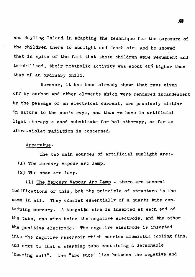
and Hayling Island In adapting the technique for the exposure of the children there to sunlight and fresh air, and he showed that in spite of the fact that these children were recumbent and immobilised, their metabolic activity was about 40# higher than that of an ordinary child.
However, it has been already shown that rays given off by carbon and other elements which were rendered incandescent by the passage of an electrical current, are precisely similar in nature to the sun's rays, and thus we have in artificial light therapy a good substitute for heliotherapy, as far as ultra-violet radiation is concerned.
Apparatus.The two main sources of artificial sunlight are:-
(1) The mercury vapour arc lamp.(2) The open arc lamp.
(1) The Mercury Vapour Arc Lamp - there are several modifications of this, but the principle of structure is the same in all. They consist essentially of a quartz tube containing mercury. A tungstfei wire is inserted at each end of the tube, one wire being the negative electrode, and the other the positive electrode. The negative electrode is inserted into the negative reservoir which carries aluminium cooling fins, and next to that a starting tube containing a detachable "heating coil”. The "arc tube" lies between the negative and

39.
positive reservoirs. A third tungstin wire is inserted into the positive reservoir.
---------- 4-Rluvkvin I u imCoolind Fins -■
JThivd Tonfreia W tR G -
rosl'rwtlxcstr\ro(v-Nt<xv:we._Teserv.ir — /' /
FiLo-ment-
Diagram of Quartz Mercury Vapour Lamp.
When the current is switched on, the heating coil boils the mercury it surrounds, and the hot mercury vapour divides the continuous liquid mercury at that end of the arc tube, creating two mercury electrodes: the current must span the gap of mercury vapour, and the arc is struck. The heat of the arc causes further expansion of the mercury vapour, and soon the heating coil and its connections are cut out because the vertical column of mercury causes a short circuit between E.Sc P. The current then flows directly through the lamp via mercury liquid, vapour arc^and liquid again.
There are two main types of mercury vapour lamp - the air-cooled and the water-cooled lamps, and these are used for both general and local treatments.
(2) The Open Arc Lamps are the other main sources of ultraviolet rays, and they are divided into two main groups, namely:-
(a) Lamps in which the electrodes consist of carbon, or carbon impregnated or cored with various metals.
59
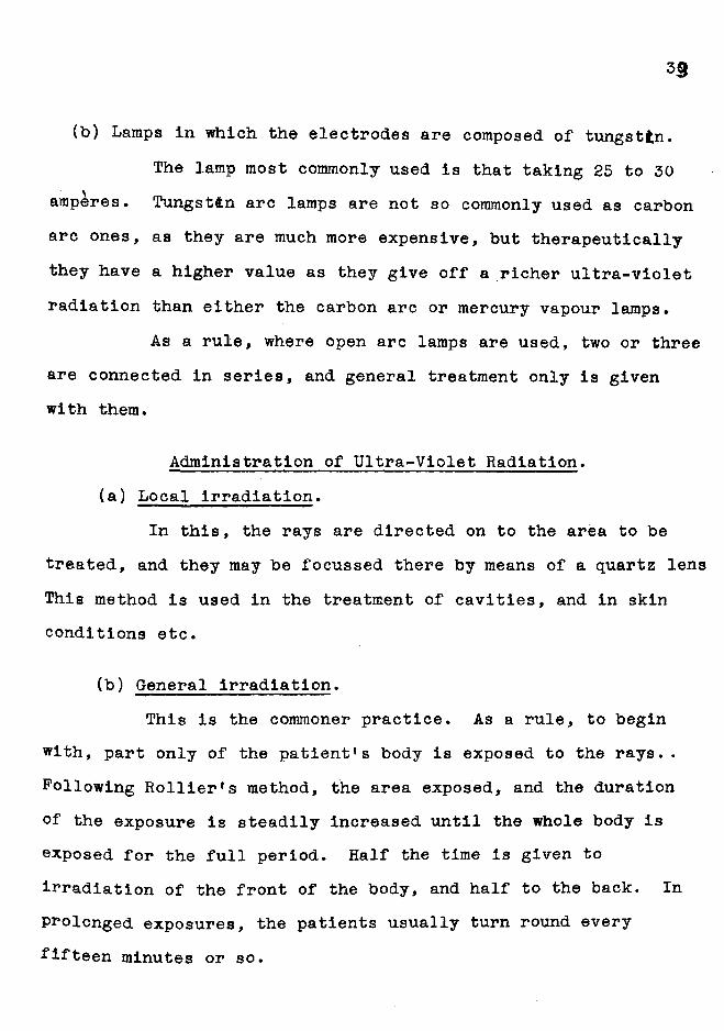
(b) Lamps in which the electrodes are composed of tungsten.The lamp most commonly used is that taking 25 to 30
amperes. Tungsten arc lamps are not so commonly used as carbonarc ones, as they are much more expensive, but therapeutically they have a higher value as they give off a richer ultra-violet radiation than either the carbon arc or mercury vapour lamps.
As a rule, where open arc lamps are used, two or three are connected in series, and general treatment only is given with them.
Administration of Ultra-Violet Radiation.(a) Local irradiation.
In this, the rays are directed on to the area to be treated, and they may be focussed there by means of a quartz lens This method is used in the treatment of cavities, and in skin conditions etc.
(b) General irradiation.This is the commoner practice. As a rule, to begin
with, part only of the patient's body is exposed to the rays.. Following Rollier*s method, the area exposed, and the duration of the exposure is steadily increased until the whole body is exposed for the full period. Half the time is given to irradiation of the front of the body, and half to the back. In prolonged exposures, the patients usually turn round every fifteen minutes or so.
3V
Another method of treatment is as followsUsing the mercury vapour arc lamp, the initial dose
is 6 minutes to the whole body - i.e. 3 minutes to the front and 3 to the back. This is gradually increased, according to the erythema reaction, to a maximum of 20 minutes to the whole body.
Using the open arc lamp, an initial dose of 20 minutes to the whole body is given - i.e. 10 minutes to the front and 10 minutes to the back, and this is increased till a total exposure of 2 to 4 hours is reached.
Patients treated by any of these methods wear a loin cloth, and also tinted spectacles to protect the eyes; otherwise the body is fully exposed.
As the intensity of the radiation varies inversely as the square of the distance of the patient from the lamp, it is important that patients do not sit too far away. Three feet is the usual distance, but may be considerably less, especially when using mercury vapour lamps. Patients must not be too close to carbon arcs on account of the heat, and the sparks that occasionally fall from the electrodes.
As regards the duration of each exposure and the total number of exposures that each patient should receive, the practice is far from uniform. The determination of the erythema dose for each individual, is, on the whole, the most useful guide to dosage, and should always be obtained before beginning

a series of treatment.With regard to the treatment of asthma, I give general
irradiation with the open carbon arc lamp. Treatment is given twice a week, and the dose is gradually increased till a mild erythema is obtained.
After a month's treatment, irradiation is given once a week; after two months once a fortnight, and so on. Treatment should be continued for 3 to 6 months if possible.
The treatment may be varied by using the mercury vapour and the open arc 1 simps successively or together, but it has been noted that cases which are resistant to one source of ultra- violet radiation, are equally resistant to the other.
V. (2) Technique of X-ray Therapy.(a) Apparatus.
In the Radiological Department at Edinburgh Royal Infirmary, where I treated a series of asthma cases, the equipment in the Deep Therapy room is of a special type, employing high potential transformers, condensers and rectifying valves of the hot cathode type.
An oscillographic record of the current curve of this type of high tension generator resulted in an almost straight line, which means that the voltage generated is as nearly as possible constant, and this current is therefore ideal for X-ray Therapy.

3è
The Deep Therapy apparatus is capable of an output of 10 milliamperes at 250,000 volts. A similar but smaller constant current generator supplies the superficial Therapy room, and it has an output of 10 milliamperes at 125,000 volts, Coolidge tubes are used.
(b ) Method of Treatment.In all these asthma cases, I gave Superficial Therapy
i.e., 2 milliamperes at 100,000 volts, as follows:- withI. Anticathode - skin Distance = 30 centimetres.
II. Filter - 3 millimetres of Aluminium. In addition, 4layers of felt covered the area to be treated.
III. Applicator - none was used, but the surrounding areaswere well protected by lead rubber to prevent any over-lapping of the dosage.
IV. Areas treated - 3 altogether i.e.,i. Anterior aspect of chest - hilar region,
ii. Posterior aspect of chest - hilar region, iii. Spleen.
V. Time - 5 minutes to each area i.e., f- Sabora&d pastille dose for the Coolidge tube used.VI. Duration of Treatment - I gave treatment
One serie/twice weekly for 3 weeks, i.e., of (each of the 3 areas received 2
treatmentvapplications. After that, I stopped treatment for 6 weeks, then resumed it again for 3 weeks as before, stopped for ô to 8 weeks, and so on, gradually decreasing the treatments and increasing the intervening time between them.
In every case, before beginning treatment, I made an X-ray examination of the chest. On screening, I noted the

îdiaphragmatic movements, and their limitation, if any, and also compared the radiability and expansion of the apices. Any increased or decreased density of the lungs was noted, and also the general size of the heart and the aorta.
Throughout the course of treatments, from time to time I made further screen examinations, to note any variations in the condition of the chest.

UtAucect PrirlT fvovn Film ofM' l 1) - iWows ■Root Sh»»<o
ovx lioTH 5ij>es .os
-To 38.
38
VI. Personal Experience and Notes of Eleven Asthma Cases.
In a year, I treated eleven cases of asthma at the Radiological Department, Edinburgh Royal Infirmary, and the following are the notes of these cases.
(1) D.R. act.16. Draper’s assistant. Family history negative for asthma, phthisis, neuroses. Predisposing causes - potatoes, fur. The attacks started in infancy and continued at intervals since then; the attacks were worse during the night, and often lasted till the next day; d&ppnoea was marked. He had had previous treatment of vaccines, which did good at first, but soon lost their effect. Before beginning X-ray treatment. X-ray examination of the chest revealed heavy root shadows on both sides.
Superficial X-ray Therapy was given to the chest and spleen as previously stated, and 5 series of exposures were given over a period of eleven months. No other treatment was given at the same time.
Result - a week after the first treatment, the spasm and d&ppnoea decreased and general improvement continued"from then onwards, the attacks becoming less in frequency and severity. Eventually definite improvement in his general condition was obtained.
À fortnight after commencing treatment, he complained of pain in the right side of his chest, but apart from rhonchi at

=9
both bases, no abnormality was detected on clinical examination; treatment was stopped for a fortnight, and there was no recurrence of the pain.
In this case, I noticed that an eczematous condition which was present on the face, cleared up rapidly during the routine treatment, no local application being made whatsoever.
(2) J.H. aet.58. Labourer. Family history negative for asthma, predisposing causes not known. Asthmatic attacks were of 5 months* duration and were spasmodic and irregular in character and frequency. He was treated for a short time with iodides before receiving superficial X-ray treatment; before treatment, no radiological abnormality of the chest was detected. He received altogether 4 weekly treatments, the chest and spleen being treated on each occasion; improvement after the 2nd. treatment was noticed. He wrote six months after his last treatment that he had had no recurrence of the asthma.
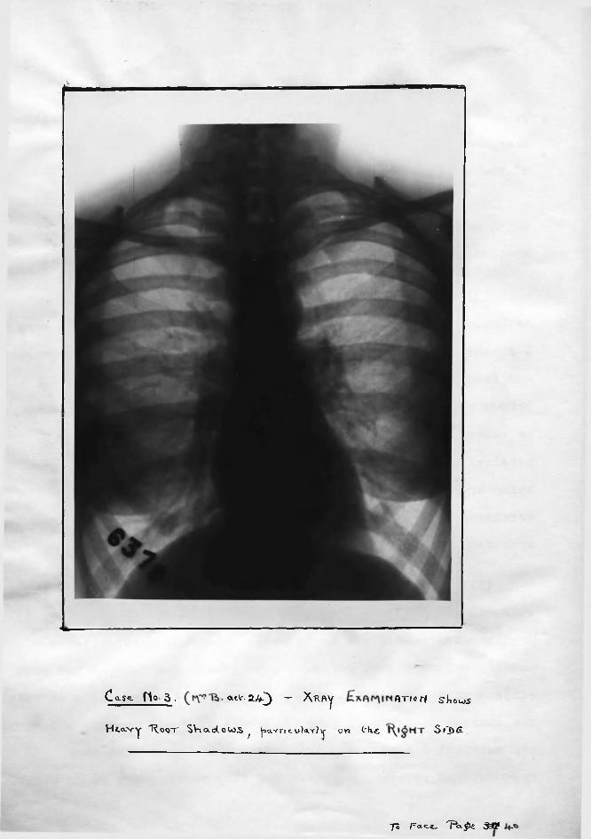
(3) Mrs.B. aet.24. Housewife. Her father and mother both suffered from asthma, but no other members of the family had it. Predisposing causes - a heavy meal; potatoes; dust; attacks usually occurred just before her periods. The patient had been subject to asthmatic attacks since she was 4 years old - at first, the attacks occurred at long intervals of 2 - 3 months, but since her marriage 3 years ago, the attacks have been much more frequent and severe. DÏppnoea was very marked, and cough also.
#
C a S ^ H o 3 ■ a -t l '-a /f-3 “ X h P>Y f lT io r< S^KowS
H ttx -v y 'R o o t S V icxd .oW ^^ |:>avTico)aT?\j- on ( r k t S i3 )ê .
70 pa.c.£_ n < pe jaSf

Previous treatment of vaccines and medicines caused temporary improvement. X-ray examination of the chest showed heavy root shadows, particularly on the right side.
Five series of Superficial X-ray treatment were given over a period of 7 months, and then the patient was transferred for ultra-violet radiation instead; there, general treatment with the open carbon arc lamp, three times a week, was given for 4 months.
Result - great improvement after the first and second series of X-ray treatment. The patient was able to do without any injections of adrtnatln, and the attacks ceased for a month, but recurred with;the onset of her period. After the recurrence of the attacks, there was no further improvement in her condition, but after transference to ultra-violet radiation, slight temporary improvement occurred. During the course of X-ray treatment, she lost weight slightly.
(4) D.R, aet.28. Family history negative for asthma.Attacks began when he was aged 7 following whooping cough and continued at irregular intervals. He was gassed during the war, and since then spasm and dï^pnoea have been more marked. Protein tests were negative: 3 years ago had operation on nose: previously, had 8 X-ray treatments the last one being 3 months before the commencement of the series here: he also had radiant heat, ultraviolet radiation and vaccines: none of these had any lasting

Ço.5& fjo- S , r iS3 3*^ - £x.b*t>ivi ciT/f'ia sKotoS
H fo -v y R o o t S h a d o w s ovx l3 o T H 5 fJ>^S L o it ‘ k
P e p i c i e n - T A # R £ n T R , y 1\ i g h t ^ T E x .
'Tl Facf 3% 4h*

36
effect. X-ray examination of the chest showed nothing definitely abnormal.
Three series of Superficial X-ray Therapy were given, with the result that improvement occurred after the first series, dï^pnoeaj»spasm being both considerably lessened. On reporting after the third series, he stated that the attacks had returned: X-ray examination of the chest after completion of the third series of treatment showed abnormality of the right apex, suggesting tuberculosis, so X-ray treatment was discontinued.
(5) Miss C. (aet.37). Housekeeper. Family history negative for asthma, though her father had "hay fever". The attacks of asthma were always worse during the summer months, and were brought on by the scent of flowers; milk; cheese; also if she went to hilly country. The attacks were of 16 years’ duration, and were very frequent during the summer months; d&ppnoea was marked, and sputum copious. Previous treatment - 7 years ago, she had an operation on her nose; 2 years ago and again one year ago, she had a series of Protein shocks, neither of which had the slightest effect on her condition. X-ray examination of the chest before X-ray treatment showed heavy root shadows, with deficient air entry at the right apex. X-ray treatment was begun in January, and 3 series of treatments were given over 5 months.
The patient was unable to attend for any more treatments.

but 6 months after her last treatment, she reported that the asthmatic attacks had been less in frequency and severity during the summer months, and she felt a slight improvement in her general condition.
(6) J.D. (aet.l5). Family history - negative for asthma, and predisposing causes of attacks not stated. The asthmatic attacks were only of several months duration, and occurred once or twice a week. "Medicines" had previously helped the condition. X-ray of the chest before X-ray treatment showedno radiological abnormality.
The patient received 2 series of Superficial X-ray Therapy in 2 months - improvement was observed after the first series, but the patient did not report to hospital again.
(7) G.H. aet 54. Shopkeeper. Family history negative for asthma, chest or nervous diseases. The attacks were brought on by any exertion or change in temperature, and were worse in summer. The attacks were of 3 years duration, were worse at night, and occurred at irregular intervals. Previous treatment consisting of medicines and vaccines caused slight relief at first, but these soon lost their effect. X-ray examination of the chest showed heavy root shadows on both sides, and quite a marked degree of emphystma was present. He was given 2 series of Superficial X-ray Therapy over 2 months. After the first series, he stated that he had slept much better - after the second series.

49
there was marked improvement in his condition, and he did not report for further treatment. Six months after the last treatment he reported that the improvement had been maintained for a month or so, but the attacks had again recurred, though they were not so severe as formerly.
(8) J.D. aet.25. Labourer. Family history negative for asthma; predisposing causes of attacks were sudden changes in temperature. The patient had suffered from asthma for 19 years; the attacks usually occurred once a week, and were worse during the day; pain in the chest before an attack was commonly present. Medicines, including adrenalin, special diet and exercises, previously given, had helped the attacks slightly. The patient was sent for X-ray treatment before having an operation for hernia, to diminish the asthmatic attacks if possible. Accordingly, one series of treatment to the chest and spleen was given before the operation, and a second series a month after the operation. Result - the treatment given before operation caused marked improvement in the asthmatic condition, but after the operation, the asthma recurred, and was not amenable to treatment; the general condition of the patient was very poor.
(9) Miss L. aet.28. No family history of asthma; predisposing causes of asthma not known; attacks were of several months duration, and were spasmodic and irregular in character and frequency. The patient had had no previous treatment, and on

4V
X-ray examination, the chest appeared clear. One series of X-ray treatment was given, with slight improvement in the condition.The patient did not report again for further treatment.
(10) Mrs.J.G. aet.28. Housewife. Family history - her father had asthma, and her mother had rheumatism. The patient’s asthmatic attacks were associated with the onset of her periods, colds, or changes in temperature. In this case, the asthma was of 2 years’ duration and occurred monthly. Previous treatment of vaccines had no effect whatsoever on her condition. X-ray examination of the chest showed numerous calcified glands in both hilar regions, with slight dulS^ess at the left apex.
Superficial Therapy was given, and she received two series of treatment in 4 months. Result - asthmatic attacks decreased considerably in number and severity, and her general health improved. She is still receiving X-ray treatments at long intervals.
(11) A.W. aet.46. Plasterer. Family history negative for asthma; predisposing causes - frequent soakings when employed on outside work: heavy meals also induced attacks. The asthma was of 8 months’ duration, and occurred at irregular intervals. Previous treatment - recent operation for antral disease. X-ray examination of the chest showed no radiological abnormality. One series of X-ray treatment was given; the asthmatic attacks considerably decreased, and the general health was very much

4Ç
improved. The patient did not return for further treatment, but reported some months later that the improvement was well maintained.
This concludes the series of cases which I personally treated. As, with one exception, all these cases were outpatients, I occasionally experienced difficulty in having them report for treatment at regular times.
In view of the marked results which usually follow immediately after the first series of treatment, I consider that, as a pre-operative measure in asthmatic subjects. X-ray treatment is of value, to decrease coughing and the general discomfort of the attacks after the operation.
It will be seen, from these cases, that X-ray treatment is more efficacious in young people than in older people. Improvement is also much more marked in cases with a recent history of asthma than in those with a long standing asthmatic history. If improvement is going to take place, it occurs either during or immediately after the first series of treatment, and remains for a variable period of weeks to several months. I also noticed that when the asthmatic attacks did recur, they were not so severe in character or frequency as previous to X-ray treatment. In all cases except one, there was an improvement in the general health, according to the patient’s own statements.

4&
Ultra-violet radiation had also a beneficial temporary effect, but it was not so prolonged as that of X-ray Therapy.
s
S'! :::
'■V 1;
r-:" f"
r :i i ^
t'-':>■m
I f ? V
r ,
-7 % C ,' 7.7 :-7 \ ;
g 7: % ;7 '3t C ' 8 f ? : Ü ' 0 *7 g ' j P- .7 r - ■''7 -7 TS ift a a >0 c r B T
n ;j r ï" 3;'- a r " i 7 7 ' : â 3-; S : j '-7 0 33 a B B ',e 7Ï> w - 3 a -S î"ï 0 a ? a 0 ■ B if- ■i -CÔ i
7T g a . g a 0 0 0 s v7 V,-?' ■0 V
a ï B ' 7 7 Ü 7 a î*3 ■~5 ' 3 7 - r, ! • t t* -# # g g 73 & ;7 . :-t 0 3 ;
g •? 37 .0 ' ( 7 ■■ -7 7:1 -tv I':': V"': 0 I
i?" ■ '77 iPs I " IT: a 77 -7 B iV..-- g "7 r r ;■--- . g g f t . i-p 7'" -i I
p ; & m ' A e 5-7 ,;7 ' V .0. Vr - ;s- g a X Ï 77 a 7;' :;£ ’
. <5 M - n t fr. a tl- 7 7 - :7 a'- 7 ; . : t iB O . - @ S t g 'r Ï Ï a IP: p i a - t -vP i 4 ^ . Ï 3 - fa C à': Ï.Î: 3:: P tp ! a % y :3 a 0 7: : .7 -
o - ,,-0. -s "7 ' v;w r--:. ;;; ~,.3 -.- --7
7 ;rt.
;. V 't;
: V-'::: v-t
tv'■f-
' -7; î 7
3- h fe -.7 a t * -'I? tv:g_ -3 f.y t r it;% 7: 7 -7 r f Î
' # ■ •:;- - ■® â - % m # a
g g 7 T w% ' g

g ^ y y (D y o y p
§ §• Sc+ O O M
pj _ etH* ca g 3 ® asO P H* et «-b ro My g <D et yy CD eto ® 00 13O P g 3 et H* »
*et 3 O 0 H* et P O raw 3 H* ® CDV 3 00 0
3O tJ O '•<0y
cMety
0
Ml O et p, P y 3 0 P<î O et et
P H* 3*y 3 i CP? P
H* '• '*3CR H* P P
Ml y IH* 0 < 3 3 H*et O O O 0 «t M) M*y 0< p p, et P 0 H t) Ml 3J W 0 H* Py 3 P*o g H» H* M) p et P
3 0 et et 0 H*
g S- P 3O 3'*
0 p,w p
3 X3* y0 o p 3 g«|P *3 et p y
y 0M 0 p 0 et P Ml g 0 p 0et < 3
O etP 3 '•_ yP p p 0 o' » Ml M H*<ûd 3
0 et3*
MM
PO (O 00 03 03 1 03 co p
S tçj tç) g S S S tçj s s• • • • • • • • •
4). ro M M 03 p 03 W W 03 po> 00 00 W 4S. 03 <3 00 00 CJi
— '4
00 to 0 p 03 0 p W0 co 0 Oî pg «i g < M g <o 0 G 0 M 0 G 03 P 3 y 0 P 3 yet y et p p y et p 3y 0 3* P y 0 3* P0 • 0 0 • 0
p w p co co CO 03 030 0 0 0 0 0 0 00 0 0 0 0 0 0 0y y y y y y y yp p p p p p p p0 0 0 0 0 0 0 00 0 0 0 0 0 0 0
0
O30H*P0y&p0
3•oy0 <1et
WO0
0
g cj üiG .3 < 0et » 03* y0 • H*• Ml 0 G 0y ••pM0G
0
Ü1 ifk333*0
X0R0
Cl00yM00
co p Sp 0 p g P et Pp 0 MtO et P ym y et y et P îsr3* p 0 G P 3 0et 0 y < O 0 pi0 0P G g 0 pg to 0 . Ml gto 0 3 G toy y et y yG p G< et P p <0 P Ml g
0g O et g0 3 0 G 03 y 3 3et et et
3 M) 3G py '* %P 0 et etg et 3 0to 0 yy 0 3o 0 co< y y 30 p 0 p.g 0 o •0 0 3 03 y 0et y y0 ypP 0 3 0Ml o G 0et 3 00 y gy y G0 M) Pco 3 p3 o 3p, 0 1
p Î25 Pg p o y g etto 3 0 to 0y Pi G y go m .0 3 G to< 0 M) y < G0 3 P y 0 yg 0 3 0 g p0 y p pi 0 y3 p et 3 ^etP 0 p et Ml PP G P et P gMl G g 0 MitOet 3 to y et y0 pi y 0 Gy p G 03 y <et <5 y 0p P 0 pi Ml g0 O g « p 0et 3 0 0 y 3« • 3 0 0 et0 et y et •0 py P 0 0p 3 0 00 . y0 P p0 0
1 0
1 petet0 PP Gp !sr0
Qmj 3 G y0 0 M) 0y Ml y o 0 yp 0 3 S'et t)G y yP et GH* c3 • y < . 0 0 0 p get 0
!z!Gety y
< 0 * yy H- g 3p 0 0 et3et p p p 0 M»p 0 H*et 3 H* P et 0 p y3 o et G et H ^ 3 P 0 H* et o et et H" p;' .ÿ 3 0 P
3 30 0 y P M p 0 H* • G DO%et h3 y •y 0 0
gg *3 p y0 0 H* G p et 3 <g y P g0 3 H* 0 3 3 3et G 0 et • Ml P
Pp Ml M) 0 G et
r jp Ml «<4 H*Ml 0 yG p 0 y y et
p 0yH*00
o>igpMlet0y
0Pi*GMlety0pIet
CO0X
0
IPetH»G3GMl
ety0p
I3et
%GGMl
00yH*00GM)
33003Met0

48
V I I I . B ib l io g r a p h y .
1. Electro*.Therapeutics and Rontgen Rays - Kassabian.2. Handbook of Medicine'^ - 1/lheeler & Jack.3. Heliotherapy - A.Rollier.4. International Survey of Roentgenology and Radiotherapy -
Hirsch and Petersen 1923.5. Journal of Roentgenology - Ramirez & Cole, 1925.
Tsuzuki, 1926.6. Journal of Actinotherapy - Biancani, 1927.7. Practical ultra-violet light Therapy - McKenzie Sc King.8. Radioactivity & Radioactive substances - Chadwick.9. Scottish Board of Health - Report on Artificial Light,
& X-ray Therapy, 1925.10. X-rays - Kaye.11. X-ray Diagnosis and Treatment - Knox.12. X-rays - their Origin, Dosage and Practical Application -
Schall.
Reproduced with permission of copyright owner. Further reproduction prohibited without permission.