thickness, cross-sectional areas depth invasion...
TRANSCRIPT
Thickness, Cross-Sectional Areas and Depth ofInvasion in the Prognosis of
Cutaneous Melanoma
ALEXANDER BRESLOW,* M.D.
From The George Washington University School of Medicine, Washington, D. C.
CuTANEous melanoma is a most unpre-dictable lesion. The marked variation inprognosis is probably a function of manyvariables, one of which is the size of thetumor. Though there is a roughly inverserelationship between the diameter of thelesion and survival,5 very small lesions haverecurred or metastasized. One possible rea-son for the lack of reliability of tumor sizein estimating prognosis may be that stud-ies to date have considered size in only twodiamensions and have neglected tumor vol-ume. Two melanomas can have the samediameter but differ greatly in thickness be-cause of variation in either depth of in-vasion or degree of protrusion from thesurface of the skin or both. A recent study 2has shown that prognosis correlates wellwith staging of the depth of invasion, butthere have been no studies relating survivalto tumor volume.To measure tumor volume it is necessary
to know the surface area of the tumor, butin this retrospective study we only knowthe maximal diameters of the lesions. Bymeasuring the maximal thickness of the le-sions we can calculate the maximal cross-sectional area, which should be roughlyproportional to the volume of the tumor.The depth of invasion was also studiedusing the criteria for staging of Clark et al.2
to see if maximal cross-sectional area, thick-ness, stage of invasion, or a combination ofthese can be of value in assessing the prog-nosis of cutaneous melanoma. A total of 98lesions were so studied.
Materials and MethodsThe 98 patients in this study were all
free of recurrent or metastatic disease andnone had satellite nodules when first seenat the George Washington University Hos-pital. None of the lesions were related toan antecedent lentigo malignum (melanoticfreckle of Hutchinson). Their ages rangedfrom 18 to 80 (average 47). Forty-six weremen and 52, women. Sixteen had lesions ofthe head or neck; 50, of an extremity; and32, of the trunk.
Following operation 71 remained free ofdisease for 5 or more years, while 27 de-veloped metastatic or recurrent disease. Pa-tients who died of unknown causes, whodied in less than 5 years without melanomaand those lost to follow-up were excluded.Follow-up was 92%o.The maximal diameters of the lesions
were measured after fixation in Bouin'ssolution. Sections were taken through thecenter of the lesion, and slides preparedand stained with hematoxylin-eosin in theusual manner. One to five slides were pre-pared from each lesion (usually two).By means of an ocular micrometer, the
maximal thickness of the lesion was mea-sured from the skin surface to the deepest
902
Submitted for publication December 19, 1969.Reprint requests to: George Washington Uni-
versity Hospital, 901 23rd Street, N. W., Wash-ington, D. C. 20037.

Volume 172Number 5
FIG. 1. Stage Imelanoma. Malignantmelanocytes are con-fined to the epidermis(H & E, 190X).
FIG. 2. S tageIemelanoma. The papil-lary dermis is widenedand densely infiamed.Malignant melanocytesinfiltrate the papillarydermis but do notreach the dense re-ticular dermis (H & E,185X).
THE PROGNOSIS OF CUTANEOUS MELANOMA
point of invasion. The maximal cross-sec-tional area of the lesion was calculated bymultiplying the thickness by the maximaldiameter of the lesion. The lesions were
903
also staged as to the depth of invasion.2 Bythis classification in situ lesions are stage I.In stage II the tumor is confined to theloose papillary dermis but does not form a
904 BRESLOW
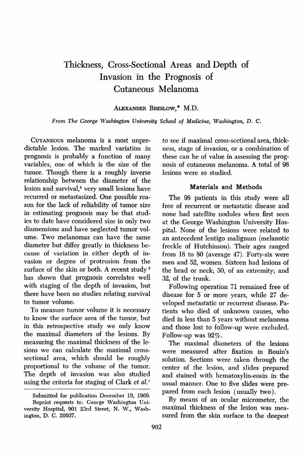
plaque at the junction of the loose papil-lary and the dense reticular dermis. Instage III the tumor forms such a plaqueand usually fills the papillary dermis. In
Annals of SurgeryNovember 1970
FIG. 3. Stage IIImelanoma. Malignantmelanocytes fill thepapillary dermis andabut on the reticulardermis (H & E, 42x).
FIG. 4. Stage IVmelanoma. Malignantmelanocytes infiltratethe reticular dermis(H & E, 250X).
stage IV there is invasion of the reticulardermis, and in V, the subcutaneous fat isinvolved. These are illustrated in Figures1-5.

Volume 172Number 5
FIG. 5. Stage Vmelanoma. Malignantmelanocytes infiltratesubcutaneous fat (H& E, 250X).
RTumor Size
THE PROGNOSIS OF CUTANEOUS MELANOMA
'esults
The distribution of the lesions accordingto size are seen in Figures 6-8. No patientwith a lesion less than 5 mm. in diameter,less than 0.76 mm. in thickness or less than6.01 mm.2 in maximal cross-sectional areasubsequently developed recurrent or meta-static disease. Tumor thickness was themost useful measurement, identifying 38 ofthe 71 patients who remained free of dis-ease. The smallest melanoma which re-curred or metastasized was 7 mm. wide,0.88 mm. thick with a cross-sectional areaof 6.16 mm.2 The incidence of recurrent ormetastatic disease appears to be a functionof all three variables and was 100%o for le-sions over 30 mm. wide or over 5 mm. thickand almost 100%o for lesions over 40 mm.2in maximal cross-sectional area. When thepatients whose lesions were less than 0.76mm., in thickness were compared withthose whose lesions were over 2.25 mm.thick, there were no significant differencesin age, sex or anatomic location of thetumors.
905
Stage of InvasionThe incidence of recurrent or metastatic
disease is also a function of the stage ofinvasion and our findings (Table 1) arein reasonably good agreement with thoseof Clark et al.° No stage I lesions were en-
I-
z I
< I
OL AU-0
Ilo
z
40 - [: FREE OF DISEASE5+ YEARS
36 -UM RECURRENCE ORMETASTASIS
32 -
28-
24-
20-
16-
12-
8-
4
<5 5-9 10-14 15-19 20+
DIAMETER (mm.)
FIG. 6. Diameter of melanomas. None less than5 mm. recurred or metastasized.

Annals of SurgeryNovember 1970
36- FREE OF DISEASE 32 -JFE
5+ YEARS 3FREODIAS~~~~~~~U)5+ YEARS32- ~~~RECURRENCEOR -28M RECURRENCE OR
METASTASIS Z28- METASTASIS
24 a.~~~~~~~~~~= 4
U. 20-
20- 0
lm 16-
16-12-
12-Z8
8 -4-
4-
6.01 6.01- 12.01- 18.01- > 24.000 -<0.76 0.76- 1.51- 2.26- ~3.00 12.00 18.00 24.00
1.50 2.25 3.00CROSS SECTIONAL AREA (mm?)
THICKNESS {mm.)
FIG. 7. Maximal thickness of melanomas. None lessthan 0.76 mm. recurred or metastasized.
countered. The prognosis for stage II tu-mors is excellent with only 1 of 39 patientsdeveloping a recurrence or a metastasis.
Simultaneous Evaluation of Size andStageIn general all measurements of tumor
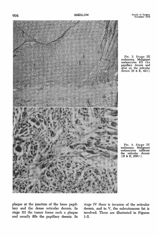
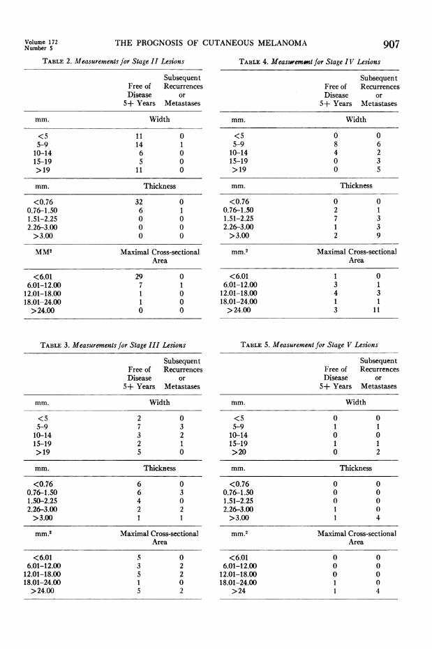
size increased with the stage of invasion,but there was a great deal of overlap (Ta-bles 2-5). For example one stage IV lesionhad a maximal cross-sectional area of 6.00mm.2 while one stage II lesion was 22.50mm. in maximal cross-sectional area.
Simultaneous evaluation of tumor thick-ness and stage of invasion is of greatervalue in assessing prognosis than is eitheralone. The prognosis for stage II lesions is
TABLE 1. Staging by Depth of Invasion
Subsequent Recur-Free of Disease rences or Metastases5+ Years
Stage (Number) Number %G
I 0 0 0II 38 1 2.6
III 19 6 24IV 12 16 57V 2 4 66
FIG. 8. Maximal cross-sectional area of mela-nomas. None less than 6.01 mm.2 recurred ormetastasized.
excellent, and the one lesion that metasta-sized was over 0.76 mm. in maximal thick-ness. For patients with stage III lesions sixof 13 whose lesions were over 0.76 mm.thick developed recurrence or metastaseswhile all six of those whose lesions wereless than 0.76 mm. survived 5 or more
years free of disease. Tumor thickness ap-pears to identify those stage II and IIIlesions with a good prognosis, and a testof partial association in 2 x 2 tables 1, 3 re-veals this to be significant at the 0.05 level.By combining stage II lesions with thosestage III lesions thinner than 0.76 mm. a
group of 45 lesions is identified only oneof which recurred or metastasized (2.2%o).All stage IV and V lesions were thickerthan 0.76 mm.
DiscussionPrognosis of cutaneous melanoma ap-
pears in part to be a function of bothtumor size and stage of invasion with tumorthickness the most significant measure ofsize. Stage II lesions and lesions less than0.76 mm. in maximal thickness are associ-ated with a favorable prognosis and eachidentified 38 of 71 patients who remained
906 BRESLOW
'I)
I-
'
z
I.
IL-0A
zi
THE PROGNOSIS OF CUTANEOUS MELANOMA
TABLE 2. Measurements for Stage II Lesions
SubsequentFree of RecurrencesDisease or5+ Years Metastases
mm. Width
<5 11 05-9 14 110-14 6 015-19 5 0>19 11 0
mm. Thickness
<0.76 32 00.76-1.50 6 11.51-2.25 0 02.26-3.00 0 0>3.00 0 0
MM2 Maximal Cross-sectionalArea
<6.01 29 06.01-12.00 7 112.01-18.00 1 018.01-24.00 1 0>24.00 0 0
TABLE 3. Measurements for Stage III Lesions
SubsequentFree of RecurrencesDisease or5+ Years Metastases
mm. Width
<5 2 05-9 7 310-14 3 215-19 2 1>19 5 0
mm. Thickness
<0.76 6 00.76-1.50 6 31.50-2.25 4 02.26-3.00 2 2>3.00 1 1
mm.2 Maximal Cross-sectionalArea
<6.01 5 06.01-12.00 3 2
12.01-18.00 5 218.01-24.00 1 0>24.00 5 2
TABLE 4. Measzremont for Stage IV Lesions
SubsequentFree of RecurrencesDisease or5+ Years Metastases
mm. Width
<5 0 05-9 8 610-14 4 215-19 0 3>19 0 5
mm. Thickness
<0.76 0 00.76-1.50 2 11.51-2.25 7 32.26-3.00 1 3>3.00 2 9
mm.2 Maximal Cross-sectionalArea
<6.01 1 06.01-12.00 3 112.01-18.00 4 318.01-24.00 1 1>24.00 3 1t
TABLE 5. Measurement for Stage V Lesions
SubsequentFree of RecurrencesDisease or5+ Years Metastases
mm. Width
<5 0 05-9 1 110-14 0 015-19 1 1>20 0 2
mm. Thickness
<0.76 0 00.76-1.50 0 01.51-2.25 0 02.26-3.00 1 0>3.00 1 4
mm.2 Maximal Cross-sectionalArea
<6.01 0 06.01-12.00 0 0
12.01-18.00 0 018.01-24.00 1 0
>24 1 4
Volume 172Number S 907

908 BRESLOW Annals of Surgery
free of disease for 5 or more years. Only 1stage II lesion, 1.00 mm. thick, subse-quently metastasized. When both criteriaare applied, 45 patients whose lesions wereeither stage II or were stage III but thin-ner then 0.76 mm. are identified and only1, the thick stage II lesion subsequentlymetastasized. These criteria are, of course,not absolute and one can expect on occa-sion to find a lesion less than 0.76 mm. inmaximal thickness which will recur or me-tastasize. I have seen such lesions at theNational Cancer Institute, but they arerare. Patients at the Cancer Institute arehighly selected, and these small lethalmelanomas must represent a very small percent of lesions operated upon.These criteria may be helpful in select-
ing patients for prophylactic lymph nodedissection. Though some reports stronglyadvocate prophylactic node dissectionwhenever possible,5 . 8 some question thevalue of this procedure for small super-ficial lesions.5' c It would seem reasonableto exclude all patients with stage II lesionsas well as those with stage III lesions thin-ner than 0.76 mm. from this procedure.Thirty-five of our 98 patients underwentprophylactic node dissection. All 12 whowere in this favorable group had negativenodes including the patient with the lethalstage II lesion who died of hematogenousmetastases.
SummaryFrom a retrospective study of 98 cutane-
ous melanomas it was found that bothtumor thickness and stage of invasion areof value in assessing prognosis. By combin-
ing these two criteria it was possible toidentify a group of 45 patients only one ofwhom developed recurrent or metastaticdisease. These criteria may be of value inselecting patients for prophylactic lymph-node dissection.
AcknowledgmentsThe author wishes to thank the members of
the Department of Surgery for their cooperationand Dr. L. Thomas and Dr. A. Rabson of theNational Institutes of Health for permission toexamine their material. Dr. C. T. Ireland of theGeorge Washington University Department ofStatistics provided the statistical analysis. Thisstudy would not have been possible without thehelp of Mrs. H. O'Brien of the George Washing-ton University Cancer Registry.
References1. Birch, M. W.: The Detection of Partial Asso-
ciation, II: The 2 X Case. J. Statist. Soc., B,26:313, 1964.
2. Clark, W. H., Jr., From, L., Bernardino, E. A.and Mihm, M. C.: The Histogenesis and Bi-ologic Behavior of Primary Human Malig-nant Melanomas of the Skin. Cancer Res.,29:705, 1969.
3. Cochran, W. G.: Some Methods for Strength-ening the Common Tests. Biometrics, 10:417,1954.
4. Lane, N., Lattes, R. and Malm, J.: Clinico-pathological Correlations in a Series of 117Malignant Melanomas of the Skin of Adults.Cancer, 11:1025, 1958.
5. Lehman, J. A., Jr., Cross, F. S. and Richey,W. G.: Clinical Study of Forty-nine Patientswith Malignant Melanoma. Cancer, 19:611,1966.
6. Lehr, H. B., Royster, H. P., Enterline, H. T.and Askovitz, S. I.: The Surgical Manage-ment of Patients with Melanoma. Plast. Re-constr. Surg., 40:475, 1967.
7. Lund, R. H. and Ihnen, M.: Malignant Mela-noma, Clinical and Pathologic Analysis of 93Cases. Surgery, 38:652, 1955.
8. Sylven, B.: Malignant Melanoma of the Skin,Report of 341 Cases Treated During theYears 1929-1943. Acta Radiol., 32:33, 1949.