thirty years of search and capture: the complex simplicity
TRANSCRIPT
JCB
JCB: Review
1103
The Rockefeller University Press $30.00J. Cell Biol. Vol. 211 No. 6 1103–1111www.jcb.org/cgi/doi/10.1083/jcb.201510015
The origin of “search and capture”In his seminal review of the mitotic cycle, Daniel Mazia boldly stated: “Nothing we have learned about mitosis since it was dis-covered a century ago is as dazzling as the discovery itself” (Mazia, 1987). This statement reflected Mazia’s (and the whole field’s) frustration with the lack of mechanistic understanding of cell division. Although voluminous phenomenological data had been gathered on the mitotic apparatus (spindle), the prin-ciples governing its assembly remained elusive. Interestingly, Mazia’s opus major was published merely a year after the orig-inal formulation of the “search and capture” (S&C) hypothesis (Kirschner and Mitchison, 1986). If Mazia had read this paper (it was not cited in his 1987 review), he might have changed his stance. Indeed, S&C offered the first plausible mechanism to drive spindle assembly in animal cells and signified the transi-tion from descriptions to molecular investigations of the process.
The S&C hypothesis stems from the discovery of micro-tubule dynamic instability (Mitchison and Kirschner, 1984). In sharp contrast to other cytoskeletal filaments, the plus ends of a typical microtubule oscillate between periods of growth and shrinkage caused by the addition and loss of αβ-tubulin subunits. The frequency of shrinkage events increases dramatically when
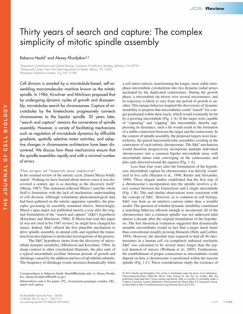
a cell enters mitosis, transforming the longer, more stable inter-phase microtubule cytoskeleton into two dynamic radial arrays nucleated by the duplicated centrosomes. During the growth phase, a microtubule tip moves over several micrometers, and its trajectory is likely to vary from one period of growth to an-other. This unique behavior inspired the discoverers of dynamic instability to propose that microtubules could “search” for a tar-get positioned within their reach, which would eventually be hit by a growing microtubule (Fig. 1 A). If the target were capable of “capturing” and “capping” this microtubule, thereby sup-pressing its dynamics, such a hit would result in the formation of a stable connection between the target and the centrosome. In the context of spindle assembly, the proposed targets were kine-tochores, the paired macromolecular assemblies residing at the centromere of each mitotic chromosome. The S&C mechanism would therefore progressively incorporate multiple individual chromosomes into a common bipolar microtubule array with microtubule minus ends converging on the centrosomes and plus ends directed toward the equator (Fig. 1 A).
Less than four years after the formulation of the hypoth-esis, microtubule capture by chromosomes was directly visual-ized in live cells (Hayden et al., 1990; Rieder and Alexander, 1990). These elegant studies established that the first step of a chromosome’s incorporation into the spindle involves a di-rect contact between the kinetochore and a single microtubule (Fig. 1 B). This and similar observations were consistent with the concept of S&C. However, as is common in cell biology, S&C was born as an intuitive cartoon rather than a testable model. The question of whether dynamic instability constituted a searching behavior efficient enough to incorporate all of the chromosomes into a common spindle was not addressed until almost a decade after the original formulation of the hypothe-sis. The first theoretical evaluation suggested that dynamically unstable microtubules would in fact find a target much faster than conventional steadily growing filaments (Holy and Leibler, 1994). However, the absolute time required to find all 46 chro-mosomes in a human cell via completely unbiased stochastic S&C was calculated to be several times longer than the typ-ical duration of mitosis (Wollman et al., 2005). Furthermore, the establishment of proper connections to microtubules would depend on how a chromosome is positioned within the nascent spindle (Fig. 1 C). These considerations imply the existence of
Cell division is enacted by a microtubule-based, self-as-sembling macromolecular machine known as the mitotic spindle. In 1986, Kirschner and Mitchison proposed that by undergoing dynamic cycles of growth and disassem-bly, microtubules search for chromosomes. Capture of mi-crotubules by the kinetochores progressively connects chromosomes to the bipolar spindle. 30 years later, “search and capture” remains the cornerstone of spindle assembly. However, a variety of facilitating mechanisms such as regulation of microtubule dynamics by diffusible gradients, spatially selective motor activities, and adap-tive changes in chromosome architecture have been dis-covered. We discuss how these mechanisms ensure that the spindle assembles rapidly and with a minimal number of errors.
Thirty years of search and capture: The complex simplicity of mitotic spindle assembly
Rebecca Heald1 and Alexey Khodjakov2,3
1Department of Molecular and Cellular Biology, University of California, Berkeley, Berkeley, CA 947202Wadsworth Center, New York State Department of Health, Albany, NY 122013Rensselaer Polytechnic Institute, Troy, NY 12180
© 2015 Heald and Khodjakov This article is distributed under the terms of an Attribution–Noncommercial–Share Alike–No Mirror Sites license for the first six months after the publication date (see http ://www .rupress .org /terms). After six months it is available under a Creative Commons License (Attribution–Noncommercial–Share Alike 3.0 Unported license, as described at http ://creativecommons .org /licenses /by -nc -sa /3 .0 /).
Correspondence to Rebecca Heald: [email protected]; or Alexey Khodja-kov: [email protected] used in this paper: CPC, chromosome passenger complex; S&C, search and capture.
TH
EJ
OU
RN
AL
OF
CE
LL
BIO
LO
GY
JCB • Volume 211 • NumBer 6 • 20151104
additional factors that facilitate spindle assembly, and a series of such mechanisms has been identified in recent years. A com-mon theme emerging from these studies is that S&C depends on spatially selective biochemical pathways that promote the formation of microtubules in the vicinity of kinetochores and also differentially engage molecular motors to position chro-mosomes in the areas with maximal exposure to spindle micro-tubules. Furthermore, proper attachment of chromosomes to the spindle is assisted by orderly changes in the shape of the cell and the adaptive architecture of kinetochores. Together, these facil-itating mechanisms ensure that stochastic encounters between microtubules and kinetochores result in a rapid yet low error incorporation of all chromosomes into the mitotic apparatus.
Guidance by gradientsIn the original formulation of S&C, radial arrays of microtu-bules nucleated by the duplicated centrosomes were expected to be the sole source of spindle microtubules. However, the den-sity of microtubule ends and therefore the probability of capture decreases rapidly as the distance between the centrosome and chromosomes increases, and a 200-nm small kinetochore has only a slight chance of being contacted by a microtubule orig-inating from a centrosome positioned 15–20 µm away within the 10–15-min period typical of spindle assembly (Wollman et al., 2005). This problem can be overcome if the density of mi-crotubules near kinetochores is increased, and multiple mecha-nisms have been identified that selectively promote microtubule nucleation and stabilize microtubule plus ends near the chro-mosomes, leading to the formation of kinetochore-attached mi-crotubule bundles termed kinetochore fibers (K-fibers).
The role of chromosome arms in mitosis was once com-pared with that of a “corpse at a funeral”; the DNA comprising
the bulk of the genome provides the reason for the proceedings, but does not actively participate in the event (Mazia, 1961). However, this view was challenged by the demonstration that viral DNA injected into a frog egg, or plasmid DNA-coated beads incubated in metaphase egg extract, induce the forma-tion of spindle-like structures in the absence of centrosomes or kinetochores (Karsenti et al., 1984; Heald et al., 1996). It is now understood that mitotic chromatin exudes “perfume” in the form of biochemical gradients that promote spin-dle microtubule assembly.
The most prominent gradient is of RanGTP (discussed extensively in Forbes et al., 2015). RanGTP is produced by the guanine nucleotide exchange factor for Ran, regulator of chromosome condensation 1 (RCC1), which ubiquitously dec-orates chromatin. In contrast, Ran’s GTPase-activating factor (RanGAP) is cytoplasmic. The opposing activities of RCC1 and RanGAP result in a steep gradient of RanGTP centered on chromosomes (Kalab et al., 2002, 2006). Similar to its function in nucleocytoplasmic transport, RanGTP releases cargoes from nuclear transport receptors called importins (Gruss et al., 2001; Nachury et al., 2001; Wilde et al., 2001). Among these cargoes are many proteins that function in microtubule polymerization and organization, such as TPX2 (Gruss and Vernos, 2004), the spindle microtubule cross-linking kinesin XCTK2, which re-quires a RanGTP gradient for proper localization and motility (Weaver et al., 2015), and components of the nonspecific lethal (NSL) complex, which binds to and stabilizes K-fiber minus ends (Meunier and Vernos, 2011; Meunier et al., 2015). An im-portant source of RanGTP-induced spindle microtubules that serves to increase the density of microtubule plus ends in the vicinity of chromosomes is microtubule-templated nucleation mediated by the Augmin complex, which requires the micro-
Figure 1. Assembly of the mitotic spindle by microtubule S&C. (A) Sequence of events envisioned in the classic formulation of the S&C hypothesis. Microtubules nucleated at the duplicated centrosomes form two radial arrays (blue and orange). Dynamically unstable microtubules explore space until a growing plus end encounters a kinetochore (magenta). This encounter results in the capture and partial stabilization of the microtubule (denoted by a color change of both the microtubule and kinetochore to green). Captured microtubules connect individual kinetochores to the centrosomes (spindle poles). In this scenario, each kinetochore ultimately becomes attached to microtubules, although the duration of spindle assembly varies significantly as a result of the stochastic nature of the process. (B) Direct visualization of microtubule capture by kinetochores in a newt lung cell (adapted with modifications from Rieder and Alexander, 1990). Because of the large cell size, individual chromosomes are often positioned in areas with a low density of microtubules and remain motionless for extended periods before suddenly moving poleward (arrows) at a velocity that often exceeds 30 µm/min. Immunofluorescence of the assembling spindle fixed immediately after initiation of the poleward movement reveals a kinetochore interacting with a single microtubule (arrowheads). Note that the initial contact is not to the plus end but to the wall of the microtubule (i.e., lateral interaction). Time is given in minutes :seconds. (C) The efficiency and fidelity of S&C depends on kinetochore orientation and the distance from centrosomes. Chromosomes positioned near the intersection of the spindle equator and spindle axis would have a reasonable chance of forming proper amphitelic attachments (1). Chromosomes located at the periphery of the spindle have reduced chances of capturing microtubules (2 and 3). In contrast, chromosomes positioned near a centrosome are exposed to a high density of unipolar microtubules (4 and 5), which promotes either erroneous attachment of both sister kinetochores to the same spindle pole (syntelic attachment; 4) or attachment of only one kinetochore (monotelic attachment; 5).

mechanisms of spindle assembly • Heald and Khodjakov 1105
tubule nucleator γ-tubulin (Petry and Vale, 2015) and is stimu-lated by TPX2 (Petry et al., 2013). In addition to being liberated from importins by RanGTP, the inhibition of TPX2 is also re-lieved by the Golgi protein GM130, which sequesters impor-tin-α to Golgi membranes (Wei et al., 2015). Thus, the RanGTP gradient promotes both de novo nucleation of microtubules near kinetochores and amplification of microtubule growth toward chromosomes (Fig. 2 A). Under normal conditions, these mecha-nisms increase the probability of kinetochore capture. However, if the chromatin and kinetochores become spatially separated, the gradient can erroneously guide microtubules away from the kinetochores. This was directly observed in mitotic cells with unreplicated genomes, where the bulk of chromatin along with its associated RanGTP gradient resides in the cell periphery and astral microtubules extend from the centrosomes toward the chromatin, which becomes particularly prominent when K-fiber formation is inhibited (O’Connell et al., 2009).
RanGTP is thought to contribute to spindle assembly in all metazoan cells, but it is most crucial in the second meiotic division of vertebrate eggs (Kalab et al., 1999; Ohba et al., 1999; Wilde and Zheng, 1999; Dumont et al., 2007), when cen-trosomes are absent and the chromosomes and spindle are tiny relative to the size of the cell, making spindle assembly via un-biased S&C virtually impossible. However, eggs contain stock-piles of cellular material including Ran pathway components, and the RanGTP generator RCC1 is not significantly enriched or activated on chromosomes, begging the question of why a large unbound pool of RCC1 does not obscure a chromatin-centered RanGTP gradient. Using Xenopus laevis egg extracts, Zhang et al. (2014) found that the cytoplasmic pool of RCC1 is inactive
because of its association in a complex together with Ran and Ran binding protein 1 (RanBP1). This mechanism is critical for spatial control of the RanGTP gradient and spindle assembly.
Importantly, chromosomes still regulate microtubule nucleation and stability in the absence of a RanGTP gradient (Maresca et al., 2009) as a result of a second chromatin-in-duced pathway mediated by the chromosome passenger com-plex (CPC; Zierhut and Funabiki, 2015). The kinase subunit of the CPC, Aurora B, locally phosphorylates and inactivates microtubule-destabilizing proteins including MCAK (Andrews et al., 2004; Lan et al., 2004; Sampath et al., 2004) and Stath-min/Op18 (Kelly et al., 2007). Spatial activation of Aurora B in mitosis occurs through a kinase cascade initiated by the ki-nase Haspin, which phosphorylates histone H3. Phospho-H3 is bound directly by another CPC component, Survivin, enriching Aurora B and promoting its trans-autophosphorylation (Zier-hut and Funabiki, 2015). A powerful nucleosome depletion and add-back approach in Xenopus egg extracts demonstrated that histone H3 phosphorylation is the only target of Haspin import-ant for the spatial regulation of Aurora B (Zierhut et al., 2014).
By concentrating at the centromere that underlies each ki-netochore, the CPC is known to control and correct erroneous microtubule attachments to kinetochores, thereby promoting biorientation of chromosomes so that chromatids are attached to opposite spindle poles and poised for segregation (Lampson and Cheeseman, 2011). Active Aurora B may diffuse away and phosphorylate substrates at a distance (Wang et al., 2011), thus also regulating microtubule depolymerization within the spin-dle. Interestingly, another kinase of the Aurora family, Aurora A, is situated at the spindle poles, where erroneously attached
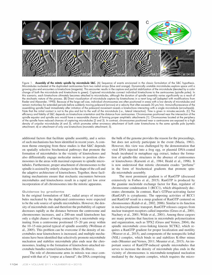
Figure 2. Chromatin gradients guide spindle assembly. (A) RCC1 localized to chromosomes increases the local concentration of RanGTP (pink) and promotes microtubule polymerization by liberating cargoes such as TPX2 (yellow) and the nonspecific lethal (NSL) complex (beige) from inhibitory interaction with importins (blue). Importin-α is also sequestered at the Golgi apparatus. Microtubules positioned near the kinetochore are likely to be rapidly captured, which initiates formation of a nascent K-fiber with a capped minus end (left side). At the kinetochore (red), microtubule nucleation is stimulated when RanGTP relieves inhibition of nucleoporins ELYS and Nup107–160 by transportin, allowing recruitment of γ-tubulin ring complexes (light green; right side). Within the spindle, templated microtubule nucleation is mediated by Augmin (dark green) and stimulated by TPX2. (B) Because of its rapid diffusion, RanGTP forms a gradient resulting in a differential regulation of microtubule nucleation/dynamics near versus away from the chromosomes. This, in turn, facilitates integration of chromosomes and their associated kinetochore microtubules into a common spindle. (C) A K-fiber formed via the kinetochore-mediated mechanism (arrows) grows via polymerization at plus ends, generating a larger target for astral microtubules. Direct contact (capture) between the elongating K-fiber and an astral microtubule (arrowheads) is followed by poleward transport and incorporation of the fiber’s free end into the spindle. Time is given in minutes :seconds.
JCB • Volume 211 • NumBer 6 • 20151106
chromosomes frequently accumulate, and generates a pole-cen-tered phosphorylation gradient that also contributes to error cor-rection (Chmátal et al., 2015; Ye et al., 2015).
In the context of S&C, microtubule growth induced in the vicinity of chromatin significantly accelerates spindle assembly (Fig. 2 B). The proximity of the freshly formed microtubules to the kinetochores facilitates their rapid capture. In addition, RanGTP appears to act directly at kinetochores, relieving in-hibition of a complex containing nuclear pore proteins and γ-tubulin (Bernis et al., 2014; Yokoyama et al., 2014) and ac-tivating TPX2 to generate kinetochore-associated microtubules (Fig. 2 A; Tulu et al., 2006). Once captured, the plus ends of microtubules that reside at the kinetochore tend to grow contin-uously, resulting in a steady elongation of the nascent K-fiber (Maiato et al., 2004). As these fibers extend outwards, they pro-vide large “antennae” that are rapidly discovered by the astral microtubules and are incorporated into the common spindle (Fig. 2, B and C). The minus end capture of preformed K-fibers is particularly evident when spindles transition from a monopo-lar to a bipolar configuration (Khodjakov et al., 2003).
Motor-mediated mechanismsIn the S&C hypothesis of 1986, the molecular nature of a capture event was not defined, but was assumed to dampen dynamics at the tip of the kinetochore-associated microtubule (Kirschner and Mitchison, 1986). However, observations of microtubule capture in cells revealed that kinetochores initially come in con-tact with the wall of a microtubule (Fig. 1 B) and that these lat-eral interactions are subsequently replaced by attachment to the microtubule plus end (Rieder and Alexander, 1990; Tanaka et al., 2005; Magidson et al., 2011; Kalinina et al., 2013). Indeed, direct single-step capture of the tip is significantly less probable than an encounter at a random point along the length of a mi-crotubule, particularly because microtubules are known to pivot in space, which significantly enhances their ability to search for kinetochores (Kalinina et al., 2013). Molecular mechanisms that govern conversion from lateral to end-on attachments re-main poorly understood and may occur as a direct transition of the same microtubule (Gandhi et al., 2011). Alternatively, lateral interactions may facilitate capture of other plus ends by orienting kinetochores favorably within the spindle (Magidson et al., 2011, 2015). The transition from lateral interaction to end-on attachment involves the coordinated activities of several molecular motors and microtubule depolymerases (Shrestha and Draviam, 2013). In fact, a second fundamentally important property of capture revealed by live-cell observations was the activity of molecular motors residing at the kinetochore. Kine-tochores were seen to initiate rapid movement toward the minus end of a captured microtubule immediately after lateral contact (Rieder and Alexander, 1990).
The S&C hypothesis predated the discovery of motor pro-teins in the spindle, and in its primitive form, the duplicated centrosomes were thought to dictate the bipolar configuration of the spindle. It is now recognized that cytoplasmic dynein and a large family of kinesin motor proteins normally act to drive microtubule self-organization and spindle bipolarity (Gatlin and Bloom, 2010). The fundamental role of molecular motors is best illustrated in egg extract systems that lack centrosomes or kinetochores (Heald et al., 1996), and upon elimination of the centrosome in vertebrate cells (Khodjakov et al., 2000; Basto et al., 2006). Spindle motors support S&C in a couple of ways. First, they generate the bipolar microtubule array, which
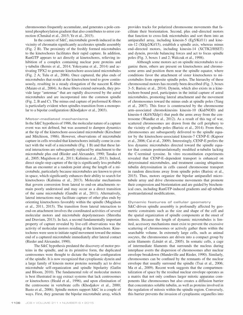
provides tracks for polarized chromosome movements that fa-cilitate their biorientation. Second, plus end–directed motors that function to cross-link microtubules and sort them into an antiparallel array, including kinesin-5 (Eg5/Kif11) and kine-sin-12 (Xklp2/Kif15), establish a spindle axis, whereas minus end–directed motors, including kinesin-14 (XCTK2/HSET) and dynein, provide balancing forces and act to focus spindle poles (Fig. 3, boxes 1 and 2; Walczak et al., 1998).
Although some motors act on spindle microtubules to or-ganize them, others are present on kinetochores and chromo-some arms and position them near the spindle equator, where conditions favor the attachment of sister kinetochores to mi-crotubules from opposite spindle poles. The hierarchy of these chromosomal motors has recently been described (Fig. 3, boxes 3–5; Barisic et al., 2014). Dynein, which also exists in a kine-tochore-bound pool, participates in the initial capture of astral microtubules, promoting lateral attachment and the movement of chromosomes toward the minus ends at spindle poles (Yang et al., 2007). This force is counteracted by the chromosome arm–associated chromokinesins kinesin-10 (Kid/NOD) and kinesin-4 (Kif4/Xklp1) that push the arms away from the cen-trosome (Wandke et al., 2012). As a result of this tug of war, scattered chromosomes are drawn from the cell periphery to the vicinity of spindle poles (Barisic et al., 2014). From there, chromosomes are subsequently delivered to the spindle equa-tor by the kinetochore-associated kinesin-7 CENP-E (Kapoor et al., 2006; Cai et al., 2009). Interestingly, CENP-E prefers the less dynamic microtubules directed toward the spindle equa-tor that contain posttranslationally modified α-tubulin lacking the C-terminal tyrosine. In vitro reconstitution experiments revealed that CENP-E–dependent transport is enhanced on detyrosinated microtubules, and treatment causing ubiquitous tubulin detyrosination in cells caused chromosome transport in random directions away from spindle poles (Barisic et al., 2015). Thus, motors organize the bipolar antiparallel micro-tubule array and drive chromosome movements that promote their congression and biorientation and are guided by biochem-ical cues, including RanGTP-induced gradients and αβ-tubulin posttranslational modifications.
Dynamic features of cellular geometryS&C-driven spindle assembly is profoundly affected by geo-metric constraints such as the size and shape of the cell and the spatial organization of spindle components at the onset of mitosis. Because the length of dynamic microtubules is lim-ited, accessory mechanisms must exist to prevent the excessive scattering of chromosomes or actively gather them within the searchable volume. In extremely large cells, such as animal oocytes, the chromosomes are driven into a compact group by actin filaments (Lénárt et al., 2005). In somatic cells, a cage of intermediate filaments that surrounds the nucleus during interphase averts the dispersion of chromosomes after nuclear envelope breakdown (Mandeville and Rieder, 1990). Similarly, chromosomes can be confined by the remnants of the nuclear envelope that usually surround the spindle (Tsai et al., 2006; Ma et al., 2009). Recent work suggests that the compartmen-talization of space by the residual nuclear envelope operates as a matrix that not only confines larger mitotic apparatus com-ponents like chromosomes but also creates a diffusion barrier that concentrates soluble tubulin, as well as proteins involved in the regulation of mitosis within the spindle region. Conversely, this barrier prevents the invasion of cytoplasmic organelles into

mechanisms of spindle assembly • Heald and Khodjakov 1107
the spindle compartment to avoid their steric interference that would impede interactions between kinetochores and microtu-bules (Schweizer et al., 2015). Intriguingly, the spindle matrix contains proteins such as BuGZ that harbor low-complexity hydrophobic sequences and undergo a temperature-dependent phase transition that promotes microtubule polymerization (Jiang et al., 2014). Therefore, the search for chromosomes normally takes place not throughout the cytoplasm but within a compact and biochemically distinct subcellular environment that promotes spindle assembly. Regulation that affects the size, shape, and composition of this compartment may indirectly yet profoundly affect the efficiency and fidelity of spindle assem-bly. For example, a common feature of animal cells is that they round up during mitosis. Preventing this morphological change either by perturbing cortical actin or by pure mechanical means impedes the gathering of chromosomes into a compact group near the geometric center of the cell. This in turn limits the effi-
ciency of S&C and leads to a higher probability of chromosome loss (Lancaster et al., 2013; Cattin et al., 2015).
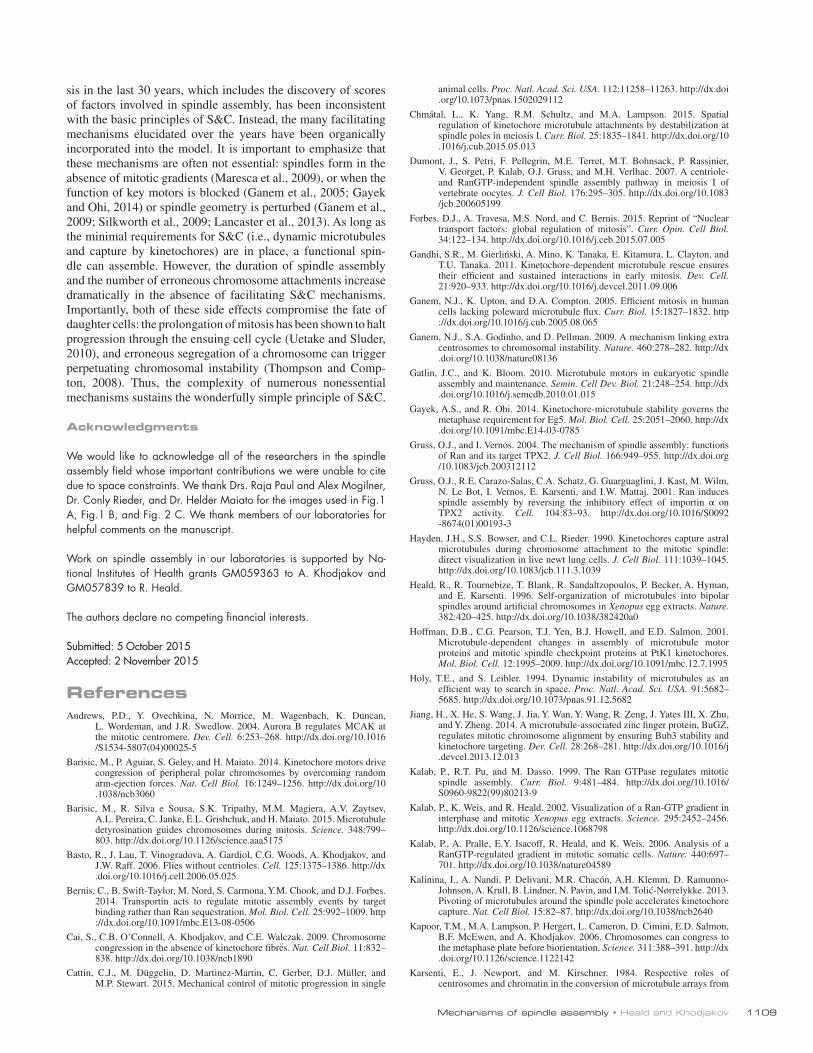
Although gathering the chromosomes within the reach of spindle microtubules is necessary, it also poses a problem for S&C-driven spindle assembly. In a crowded environment, many kinetochores become inaccessible to microtubules because of occlusion by chromosome arms (Fig. 4 A). The number of ki-netochores that are invisible to microtubules increases rapidly as the number of chromosomes grows. Computational analyses suggest that only ∼3% of kinetochores would be accessible to microtubules if 46 average-size chromosomes were randomly distributed within a typical-size spherical nuclear volume (Paul et al., 2009). To overcome this problem, cells have developed mechanisms that shape, orient, and distribute chromosomes into spatial patterns that actively present kinetochores to the searching microtubules during prometaphase (Kitajima et al., 2011; Magidson et al., 2011). These mechanisms involve the
Figure 3. Motor activities in spindle assembly. Microtubule (MT)-bound motors promote bipolar spindle formation, whereas chromosome-associated motors drive proper kinetochore orientation and chromosome movement to the equator. Box 1: Motor-dependent mechanisms establish bipolarity as Eg5 (kinesin-5) motors slide antiparallel microtubules apart with their minus ends leading and their plus ends directed toward the spindle equator. Box 2: Minus end–directed motors such as dynein move microtubules poleward with their minus ends leading, thereby incorporating K-fibers into the spindle and focusing spindle poles. Box 3: Kinetochore-associated dynein transports chromosomes along astral microtubules toward the spindle poles from the periphery. Box 4: Plus end–directed chromokinesins (kinesin-4 and -10) eject chromosome arms outward. Box 5: CENP-E (kinesin-7) transports unattached kinetochores toward the equator along spindle microtubules. MTOC, microtubule organizing center.

JCB • Volume 211 • NumBer 6 • 20151108
interplay between a chromokinesin-mediated ejection force on the arms (Rieder and Salmon, 1994; Vanneste et al., 2011) and inward-directed forces produced by the kinetochore-associated microtubule motors, which arrange the chromosomes into a to-roid around the nascent spindle. In this belt-like configuration (Fig. 4 B), kinetochores become exposed to a high density of microtubules, which promotes efficient capture. Subsequently, stronger and more stable end-on attachments allow chromo-somes to gradually repopulate the central part of the spindle. Conditions that prevent the formation of the chromosomal belt (e.g., the inactivation of chromokinesins) prolong spindle as-sembly and markedly increase the number of lagging chromo-somes that segregate improperly during the ensuing anaphase (Magidson et al., 2011, 2015).
Another set of geometric constraints that inevitably affect the efficiency and fidelity of S&C-based spindle assembly are the size and relative positions of sister kinetochores assembled at the centromere (Östergren, 1951). Intuitively, small sister ki-netochores positioned on opposite sides would ensure error-free spindle assembly, as they would be sterically shielded by the centromere from capturing microtubules that emanate from the same spindle pole. Indeed, when sister kinetochores become juxtaposed, the number of syntelic attachments increases dra-matically (Lončarek et al., 2007; Sakuno et al., 2009). How-ever, small kinetochores cannot capture microtubules very efficiently and would increase the time required for spindle assembly. Interestingly, recent computational analyses suggest that the intuitive reciprocal relationship between efficiency and fidelity in S&C-driven spindle assembly is incorrect. A model
that considers the formation of end-on attachments in a spin-dle environment dominated by lateral microtubule interactions predicts that the enlargement of kinetochores during prometa-phase would both accelerate spindle assembly and suppress the number of errors (Magidson et al., 2015). This unexpected syn-ergy is the result of rotational alignment of centromeres with respect to the spindle axis driven by opposing forces acting at the kinetochores versus chromosome arms. The extent of an-gular prealignment is less for smaller kinetochores, which are not capable of remaining in direct contact with microtubules during extensive rotations (Fig. 4 C). Indeed, enlargement of kinetochores during earlier stages of spindle assembly followed by their compaction upon the formation of end-on attachment (Fig. 4 D) has been directly observed in cells (Thrower et al., 1996; Hoffman et al., 2001; Magidson et al., 2015) as well as in egg extracts (Wynne and Funabiki, 2015). Interestingly, com-putational modeling suggests that once angular chromosome alignment is attained, efficient correction of erroneous attach-ments will be achieved simply because of the rapid turnover of microtubules at kinetochores (Zaytsev and Grishchuk, 2015). Thus, dynamic changes in chromosome architecture in the con-text of spatial cues and constraints, together with the high turn-over rate of microtubules within the spindle, promote the proper attachment of sister kinetochores to opposite spindle poles.
ConclusionCell biology is a rapidly advancing field, and new observa-tions frequently disprove mechanistic hypotheses after just a few short years. Yet, nothing that we have learned about mito-
Figure 4. Cellular geometry and dynamic kinetochores. (A) The efficiency of S&C is affected by the spatial organization of chromosomes. The arms of peripheral chromosomes (blue) shield kinetochores (red) positioned deeper inside the nucleus from astral microtubules (green). (B) Typical spatial patterns observed in mammalian cells at progressive stages of spindle assembly. At prophase, duplicated centrosomes (green) separate to opposite sides of the nucleus, and the distribution of kinetochores (orange) appears to be random. During early prometaphase, chromosomes form a toroid with most kineto-chores residing on the surface of the nascent spindle and chromosome arms pointing outwards. Upon formation of stable end-on kinetochore attachments, chromosomes repopulate the central part of the spindle, becoming uniformly distributed at the spindle equator at metaphase. (C) The ejection force of the arms (blue arrows) opposed by the inward forces generated at the kinetochore (red arrow) rotates the centromere so that sister kinetochores become prefer-entially oriented toward opposite spindle poles. Notice that larger kinetochores support a more significant rotation (top), whereas smaller kinetochores lose contact with microtubules, dampening the inward force (bottom). (D) Typical examples of chromosome architecture during various stages of mitosis. Note that chromosome arms (blue) are bent inside the prophase nucleus but become straightened by the ejection force (prometaphase and metaphase). Inner kinetochores (CENP-A; yellow) remain compact throughout mitosis, whereas the outer kinetochore (CENP-F; orange) encircles a large part of the centromere during prometaphase and compacts after the formation of end-on attachments (metaphase).
mechanisms of spindle assembly • Heald and Khodjakov 1109
sis in the last 30 years, which includes the discovery of scores of factors involved in spindle assembly, has been inconsistent with the basic principles of S&C. Instead, the many facilitating mechanisms elucidated over the years have been organically incorporated into the model. It is important to emphasize that these mechanisms are often not essential: spindles form in the absence of mitotic gradients (Maresca et al., 2009), or when the function of key motors is blocked (Ganem et al., 2005; Gayek and Ohi, 2014) or spindle geometry is perturbed (Ganem et al., 2009; Silkworth et al., 2009; Lancaster et al., 2013). As long as the minimal requirements for S&C (i.e., dynamic microtubules and capture by kinetochores) are in place, a functional spin-dle can assemble. However, the duration of spindle assembly and the number of erroneous chromosome attachments increase dramatically in the absence of facilitating S&C mechanisms. Importantly, both of these side effects compromise the fate of daughter cells: the prolongation of mitosis has been shown to halt progression through the ensuing cell cycle (Uetake and Sluder, 2010), and erroneous segregation of a chromosome can trigger perpetuating chromosomal instability (Thompson and Comp-ton, 2008). Thus, the complexity of numerous nonessential mechanisms sustains the wonderfully simple principle of S&C.
Acknowledgments
We would like to acknowledge all of the researchers in the spindle assembly field whose important contributions we were unable to cite due to space constraints. We thank Drs. Raja Paul and Alex Mogilner, Dr. Conly Rieder, and Dr. Helder Maiato for the images used in Fig.1 A, Fig.1 B, and Fig. 2 C. We thank members of our laboratories for helpful comments on the manuscript.
Work on spindle assembly in our laboratories is supported by Na-tional Institutes of Health grants GM059363 to A. Khodjakov and GM057839 to R. Heald.
The authors declare no competing financial interests.
Submitted: 5 October 2015Accepted: 2 November 2015
ReferencesAndrews, P.D., Y. Ovechkina, N. Morrice, M. Wagenbach, K. Duncan,
L. Wordeman, and J.R. Swedlow. 2004. Aurora B regulates MCAK at the mitotic centromere. Dev. Cell. 6:253–268. http ://dx .doi .org /10 .1016 /S1534 -5807(04)00025 -5
Barisic, M., P. Aguiar, S. Geley, and H. Maiato. 2014. Kinetochore motors drive congression of peripheral polar chromosomes by overcoming random arm-ejection forces. Nat. Cell Biol. 16:1249–1256. http ://dx .doi .org /10 .1038 /ncb3060
Barisic, M., R. Silva e Sousa, S.K. Tripathy, M.M. Magiera, A.V. Zaytsev, A.L. Pereira, C. Janke, E.L. Grishchuk, and H. Maiato. 2015. Microtubule detyrosination guides chromosomes during mitosis. Science. 348:799–803. http ://dx .doi .org /10 .1126 /science .aaa5175
Basto, R., J. Lau, T. Vinogradova, A. Gardiol, C.G. Woods, A. Khodjakov, and J.W. Raff. 2006. Flies without centrioles. Cell. 125:1375–1386. http ://dx .doi .org /10 .1016 /j .cell .2006 .05 .025
Bernis, C., B. Swift-Taylor, M. Nord, S. Carmona, Y.M. Chook, and D.J. Forbes. 2014. Transportin acts to regulate mitotic assembly events by target binding rather than Ran sequestration. Mol. Biol. Cell. 25:992–1009. http ://dx .doi .org /10 .1091 /mbc .E13 -08 -0506
Cai, S., C.B. O’Connell, A. Khodjakov, and C.E. Walczak. 2009. Chromosome congression in the absence of kinetochore fibres. Nat. Cell Biol. 11:832–838. http ://dx .doi .org /10 .1038 /ncb1890
Cattin, C.J., M. Düggelin, D. Martinez-Martin, C. Gerber, D.J. Müller, and M.P. Stewart. 2015. Mechanical control of mitotic progression in single
animal cells. Proc. Natl. Acad. Sci. USA. 112:11258–11263. http ://dx .doi .org /10 .1073 /pnas .1502029112
Chmátal, L., K. Yang, R.M. Schultz, and M.A. Lampson. 2015. Spatial regulation of kinetochore microtubule attachments by destabilization at spindle poles in meiosis I. Curr. Biol. 25:1835–1841. http ://dx .doi .org /10 .1016 /j .cub .2015 .05 .013
Dumont, J., S. Petri, F. Pellegrin, M.E. Terret, M.T. Bohnsack, P. Rassinier, V. Georget, P. Kalab, O.J. Gruss, and M.H. Verlhac. 2007. A centriole- and RanGTP-independent spindle assembly pathway in meiosis I of vertebrate oocytes. J. Cell Biol. 176:295–305. http ://dx .doi .org /10 .1083 /jcb .200605199
Forbes, D.J., A. Travesa, M.S. Nord, and C. Bernis. 2015. Reprint of “Nuclear transport factors: global regulation of mitosis”. Curr. Opin. Cell Biol. 34:122–134. http ://dx .doi .org /10 .1016 /j .ceb .2015 .07 .005
Gandhi, S.R., M. Gierliński, A. Mino, K. Tanaka, E. Kitamura, L. Clayton, and T.U. Tanaka. 2011. Kinetochore-dependent microtubule rescue ensures their efficient and sustained interactions in early mitosis. Dev. Cell. 21:920–933. http ://dx .doi .org /10 .1016 /j .devcel .2011 .09 .006
Ganem, N.J., K. Upton, and D.A. Compton. 2005. Efficient mitosis in human cells lacking poleward microtubule flux. Curr. Biol. 15:1827–1832. http ://dx .doi .org /10 .1016 /j .cub .2005 .08 .065
Ganem, N.J., S.A. Godinho, and D. Pellman. 2009. A mechanism linking extra centrosomes to chromosomal instability. Nature. 460:278–282. http ://dx .doi .org /10 .1038 /nature08136
Gatlin, J.C., and K. Bloom. 2010. Microtubule motors in eukaryotic spindle assembly and maintenance. Semin. Cell Dev. Biol. 21:248–254. http ://dx .doi .org /10 .1016 /j .semcdb .2010 .01 .015
Gayek, A.S., and R. Ohi. 2014. Kinetochore-microtubule stability governs the metaphase requirement for Eg5. Mol. Biol. Cell. 25:2051–2060. http ://dx .doi .org /10 .1091 /mbc .E14 -03 -0785
Gruss, O.J., and I. Vernos. 2004. The mechanism of spindle assembly: functions of Ran and its target TPX2. J. Cell Biol. 166:949–955. http ://dx .doi .org /10 .1083 /jcb .200312112
Gruss, O.J., R.E. Carazo-Salas, C.A. Schatz, G. Guarguaglini, J. Kast, M. Wilm, N. Le Bot, I. Vernos, E. Karsenti, and I.W. Mattaj. 2001. Ran induces spindle assembly by reversing the inhibitory effect of importin α on TPX2 activity. Cell. 104:83–93. http ://dx .doi .org /10 .1016 /S0092 -8674(01)00193 -3
Hayden, J.H., S.S. Bowser, and C.L. Rieder. 1990. Kinetochores capture astral microtubules during chromosome attachment to the mitotic spindle: direct visualization in live newt lung cells. J. Cell Biol. 111:1039–1045. http ://dx .doi .org /10 .1083 /jcb .111 .3 .1039
Heald, R., R. Tournebize, T. Blank, R. Sandaltzopoulos, P. Becker, A. Hyman, and E. Karsenti. 1996. Self-organization of microtubules into bipolar spindles around artificial chromosomes in Xenopus egg extracts. Nature. 382:420–425. http ://dx .doi .org /10 .1038 /382420a0
Hoffman, D.B., C.G. Pearson, T.J. Yen, B.J. Howell, and E.D. Salmon. 2001. Microtubule-dependent changes in assembly of microtubule motor proteins and mitotic spindle checkpoint proteins at PtK1 kinetochores. Mol. Biol. Cell. 12:1995–2009. http ://dx .doi .org /10 .1091 /mbc .12 .7 .1995
Holy, T.E., and S. Leibler. 1994. Dynamic instability of microtubules as an efficient way to search in space. Proc. Natl. Acad. Sci. USA. 91:5682–5685. http ://dx .doi .org /10 .1073 /pnas .91 .12 .5682
Jiang, H., X. He, S. Wang, J. Jia, Y. Wan, Y. Wang, R. Zeng, J. Yates III, X. Zhu, and Y. Zheng. 2014. A microtubule-associated zinc finger protein, BuGZ, regulates mitotic chromosome alignment by ensuring Bub3 stability and kinetochore targeting. Dev. Cell. 28:268–281. http ://dx .doi .org /10 .1016 /j .devcel .2013 .12 .013
Kalab, P., R.T. Pu, and M. Dasso. 1999. The Ran GTPase regulates mitotic spindle assembly. Curr. Biol. 9:481–484. http ://dx .doi .org /10 .1016 /S0960 -9822(99)80213 -9
Kalab, P., K. Weis, and R. Heald. 2002. Visualization of a Ran-GTP gradient in interphase and mitotic Xenopus egg extracts. Science. 295:2452–2456. http ://dx .doi .org /10 .1126 /science .1068798
Kalab, P., A. Pralle, E.Y. Isacoff, R. Heald, and K. Weis. 2006. Analysis of a RanGTP-regulated gradient in mitotic somatic cells. Nature. 440:697–701. http ://dx .doi .org /10 .1038 /nature04589
Kalinina, I., A. Nandi, P. Delivani, M.R. Chacón, A.H. Klemm, D. Ramunno-Johnson, A. Krull, B. Lindner, N. Pavin, and I.M. Tolić-Nørrelykke. 2013. Pivoting of microtubules around the spindle pole accelerates kinetochore capture. Nat. Cell Biol. 15:82–87. http ://dx .doi .org /10 .1038 /ncb2640
Kapoor, T.M., M.A. Lampson, P. Hergert, L. Cameron, D. Cimini, E.D. Salmon, B.F. McEwen, and A. Khodjakov. 2006. Chromosomes can congress to the metaphase plate before biorientation. Science. 311:388–391. http ://dx .doi .org /10 .1126 /science .1122142
Karsenti, E., J. Newport, and M. Kirschner. 1984. Respective roles of centrosomes and chromatin in the conversion of microtubule arrays from

JCB • Volume 211 • NumBer 6 • 20151110
interphase to metaphase. J. Cell Biol. 99:47s–54s. http ://dx .doi .org /10 .1083 /jcb .99 .1 .47s
Kelly, A.E., S.C. Sampath, T.A. Maniar, E.M. Woo, B.T. Chait, and H. Funabiki. 2007. Chromosomal enrichment and activation of the aurora B pathway are coupled to spatially regulate spindle assembly. Dev. Cell. 12:31–43. http ://dx .doi .org /10 .1016 /j .devcel .2006 .11 .001
Khodjakov, A., R.W. Cole, B.R. Oakley, and C.L. Rieder. 2000. Centrosome-independent mitotic spindle formation in vertebrates. Curr. Biol. 10:59–67. http ://dx .doi .org /10 .1016 /S0960 -9822(99)00276 -6
Khodjakov, A., L. Copenagle, M.B. Gordon, D.A. Compton, and T.M. Kapoor. 2003. Minus-end capture of preformed kinetochore fibers contributes to spindle morphogenesis. J. Cell Biol. 160:671–683. http ://dx .doi .org /10 .1083 /jcb .200208143
Kirschner, M., and T. Mitchison. 1986. Beyond self-assembly: From microtubules to morphogenesis. Cell. 45:329–342. http ://dx .doi .org /10 .1016 /0092 -8674(86)90318 -1
Kitajima, T.S., M. Ohsugi, and J. Ellenberg. 2011. Complete kinetochore tracking reveals error-prone homologous chromosome biorientation in mammalian oocytes. Cell. 146:568–581. http ://dx .doi .org /10 .1016 /j .cell .2011 .07 .031
Lampson, M.A., and I.M. Cheeseman. 2011. Sensing centromere tension: Aurora B and the regulation of kinetochore function. Trends Cell Biol. 21:133–140. http ://dx .doi .org /10 .1016 /j .tcb .2010 .10 .007
Lan, W., X. Zhang, S.L. Kline-Smith, S.E. Rosasco, G.A. Barrett-Wilt, J. Shabanowitz, D.F. Hunt, C.E. Walczak, and P.T. Stukenberg. 2004. Aurora B phosphorylates centromeric MCAK and regulates its localization and microtubule depolymerization activity. Curr. Biol. 14:273–286. http ://dx .doi .org /10 .1016 /j .cub .2004 .01 .055
Lancaster, O.M., M. Le Berre, A. Dimitracopoulos, D. Bonazzi, E. Zlotek-Zlotkiewicz, R. Picone, T. Duke, M. Piel, and B. Baum. 2013. Mitotic rounding alters cell geometry to ensure efficient bipolar spindle formation. Dev. Cell. 25:270–283. http ://dx .doi .org /10 .1016 /j .devcel .2013 .03 .014
Lénárt, P., C.P. Bacher, N. Daigle, A.R. Hand, R. Eils, M. Terasaki, and J. Ellenberg. 2005. A contractile nuclear actin network drives chromosome congression in oocytes. Nature. 436:812–818. http ://dx .doi .org /10 .1038 /nature03810
Lončarek, J., O. Kisurina-Evgenieva, T. Vinogradova, P. Hergert, S. La Terra, T.M. Kapoor, and A. Khodjakov. 2007. The centromere geometry essential for keeping mitosis error free is controlled by spindle forces. Nature. 450:745–749. http ://dx .doi .org /10 .1038 /nature06344
Ma, L., M.Y. Tsai, S. Wang, B. Lu, R. Chen, J.R. Iii, X. Zhu, and Y. Zheng. 2009. Requirement for Nudel and dynein for assembly of the lamin B spindle matrix. Nat. Cell Biol. 11:247–256. http ://dx .doi .org /10 .1038 /ncb1832
Magidson, V., C.B. O’Connell, J. Lončarek, R. Paul, A. Mogilner, and A. Khodjakov. 2011. The spatial arrangement of chromosomes during prometaphase facilitates spindle assembly. Cell. 146:555–567. http ://dx .doi .org /10 .1016 /j .cell .2011 .07 .012
Magidson, V., R. Paul, N. Yang, J.G. Ault, C.B. O’Connell, I. Tikhonenko, B.F. McEwen, A. Mogilner, and A. Khodjakov. 2015. Adaptive changes in the kinetochore architecture facilitate proper spindle assembly. Nat. Cell Biol. 17:1134–1144. http ://dx .doi .org /10 .1038 /ncb3223
Maiato, H., C.L. Rieder, and A. Khodjakov. 2004. Kinetochore-driven formation of kinetochore fibers contributes to spindle assembly during animal mitosis. J. Cell Biol. 167:831–840. http ://dx .doi .org /10 .1083 /jcb .200407090
Mandeville, E.C., and C.L. Rieder. 1990. Keratin filaments restrict organelle migration into the forming spindle of newt pneumocytes. Cell Motil. Cytoskeleton. 15:111–120. http ://dx .doi .org /10 .1002 /cm .970150207
Maresca, T.J., A.C. Groen, J.C. Gatlin, R. Ohi, T.J. Mitchison, and E.D. Salmon. 2009. Spindle assembly in the absence of a RanGTP gradient requires localized CPC activity. Curr. Biol. 19:1210–1215. http ://dx .doi .org /10 .1016 /j .cub .2009 .05 .061
Mazia, D. 1961. Mitosis and the physiology of cell division. In The Cell: Biochemistry, Physiology, Morphology. Vol. III. J. Brachet and A.E. Mirsky, editors. Academic Press, London. 77–412. http ://dx .doi .org /10 .1016 /B978 -0 -12 -123303 -7 .50008 -9
Mazia, D. 1987. The chromosome cycle and the centrosome cycle in the mitotic cycle. Int. Rev. Cytol. 100:49–92. http ://dx .doi .org /10 .1016 /S0074 -7696(08)61698 -8
Meunier, S., and I. Vernos. 2011. K-fibre minus ends are stabilized by a RanGTP-dependent mechanism essential for functional spindle assembly. Nat. Cell Biol. 13:1406–1414. http ://dx .doi .org /10 .1038 /ncb2372
Meunier, S., M. Shvedunova, N. Van Nguyen, L. Avila, I. Vernos, and A. Akhtar. 2015. An epigenetic regulator emerges as microtubule minus-end binding and stabilizing factor in mitosis. Nat. Commun. 6:7889. http ://dx .doi .org /10 .1038 /ncomms8889
Mitchison, T., and M. Kirschner. 1984. Dynamic instability of microtubule growth. Nature. 312:237–242. http ://dx .doi .org /10 .1038 /312237a0
Nachury, M.V., T.J. Maresca, W.C. Salmon, C.M. Waterman-Storer, R. Heald, and K. Weis. 2001. Importin β is a mitotic target of the small GTPase Ran in spindle assembly. Cell. 104:95–106. http ://dx .doi .org /10 .1016 /S0092 -8674(01)00194 -5
O’Connell, C.B., J. Loncarek, P. Kaláb, and A. Khodjakov. 2009. Relative contributions of chromatin and kinetochores to mitotic spindle assembly. J. Cell Biol. 187:43–51. http ://dx .doi .org /10 .1083 /jcb .200903076
Ohba, T., M. Nakamura, H. Nishitani, and T. Nishimoto. 1999. Self-organization of microtubule asters induced in Xenopus egg extracts by GTP-bound Ran. Science. 284:1356–1358. http ://dx .doi .org /10 .1126 /science .284 .5418 .1356
Östergren, G. 1951. The mechanism of co-orientation in bivalents and multivalents. The theory of orientation by pulling. Hereditas. 37:85–156. http ://dx .doi .org /10 .1111 /j .1601 -5223 .1951 .tb02891 .x
Paul, R., R. Wollman, W.T. Silkworth, I.K. Nardi, D. Cimini, and A. Mogilner. 2009. Computer simulations predict that chromosome movements and rotations accelerate mitotic spindle assembly without compromising accuracy. Proc. Natl. Acad. Sci. USA. 106:15708–15713. http ://dx .doi .org /10 .1073 /pnas .0908261106
Petry, S., and R.D. Vale. 2015. Microtubule nucleation at the centrosome and beyond. Nat. Cell Biol. 17:1089–1093. http ://dx .doi .org /10 .1038 /ncb3220
Petry, S., A.C. Groen, K. Ishihara, T.J. Mitchison, and R.D. Vale. 2013. Branching microtubule nucleation in Xenopus egg extracts mediated by augmin and TPX2. Cell. 152:768–777. http ://dx .doi .org /10 .1016 /j .cell .2012 .12 .044
Rieder, C.L., and S.P. Alexander. 1990. Kinetochores are transported poleward along a single astral microtubule during chromosome attachment to the spindle in newt lung cells. J. Cell Biol. 110:81–95. http ://dx .doi .org /10 .1083 /jcb .110 .1 .81
Rieder, C.L., and E.D. Salmon. 1994. Motile kinetochores and polar ejection forces dictate chromosome position on the vertebrate mitotic spindle. J. Cell Biol. 124:223–233. http ://dx .doi .org /10 .1083 /jcb .124 .3 .223
Sakuno, T., K. Tada, and Y. Watanabe. 2009. Kinetochore geometry defined by cohesion within the centromere. Nature. 458:852–858. http ://dx .doi .org /10 .1038 /nature07876
Sampath, S.C., R. Ohi, O. Leismann, A. Salic, A. Pozniakovski, and H. Funabiki. 2004. The chromosomal passenger complex is required for chromatin-induced microtubule stabilization and spindle assembly. Cell. 118:187–202. http ://dx .doi .org /10 .1016 /j .cell .2004 .06 .026
Schweizer, N., N. Pawar, M. Weiss, and H. Maiato. 2015. An organelle-exclusion envelope assists mitosis and underlies distinct molecular crowding in the spindle region. J. Cell Biol. 210:695–704. http ://dx .doi .org /10 .1083 /jcb .201506107
Shrestha, R.L., and V.M. Draviam. 2013. Lateral to end-on conversion of chromosome-microtubule attachment requires kinesins CENP-E and MCAK. Curr. Biol. 23:1514–1526. http ://dx .doi .org /10 .1016 /j .cub .2013 .06 .040
Silkworth, W.T., I.K. Nardi, L.M. Scholl, and D. Cimini. 2009. Multipolar spindle pole coalescence is a major source of kinetochore mis-attachment and chromosome mis-segregation in cancer cells. PLoS One. 4:e6564. http ://dx .doi .org /10 .1371 /journal .pone .0006564
Tanaka, K., N. Mukae, H. Dewar, M. van Breugel, E.K. James, A.R. Prescott, C. Antony, and T.U. Tanaka. 2005. Molecular mechanisms of kinetochore capture by spindle microtubules. Nature. 434:987–994. http ://dx .doi .org /10 .1038 /nature03483
Thompson, S.L., and D.A. Compton. 2008. Examining the link between chromosomal instability and aneuploidy in human cells. J. Cell Biol. 180:665–672. http ://dx .doi .org /10 .1083 /jcb .200712029
Thrower, D.A., M.A. Jordan, and L. Wilson. 1996. Modulation of CENP-E organization at kinetochores by spindle microtubule attachment. Cell Motil. Cytoskeleton. 35:121–133. http ://dx .doi .org /10 .1002 /(SICI)1097 -0169(1996)35 :2<121::AID-CM5>3.0.CO;2-D
Tsai, M.Y., S. Wang, J.M. Heidinger, D.K. Shumaker, S.A. Adam, R.D. Goldman, and Y. Zheng. 2006. A mitotic lamin B matrix induced by RanGTP required for spindle assembly. Science. 311:1887–1893. http ://dx .doi .org /10 .1126 /science .1122771
Tulu, U.S., C. Fagerstrom, N.P. Ferenz, and P. Wadsworth. 2006. Molecular requirements for kinetochore-associated microtubule formation in mammalian cells. Curr. Biol. 16:536–541. http ://dx .doi .org /10 .1016 /j .cub .2006 .01 .060
Uetake, Y., and G. Sluder. 2010. Prolonged prometaphase blocks daughter cell proliferation despite normal completion of mitosis. Curr. Biol. 20:1666–1671. http ://dx .doi .org /10 .1016 /j .cub .2010 .08 .018
Vanneste, D., V. Ferreira, and I. Vernos. 2011. Chromokinesins: localization-dependent functions and regulation during cell division. Biochem. Soc. Trans. 39:1154–1160. http ://dx .doi .org /10 .1042 /BST0391154

mechanisms of spindle assembly • Heald and Khodjakov 1111
Walczak, C.E., I. Vernos, T.J. Mitchison, E. Karsenti, and R. Heald. 1998. A model for the proposed roles of different microtubule-based motor proteins in establishing spindle bipolarity. Curr. Biol. 8:903–913. http ://dx .doi .org /10 .1016 /S0960 -9822(07)00370 -3
Wandke, C., M. Barisic, R. Sigl, V. Rauch, F. Wolf, A.C. Amaro, C.H. Tan, A.J. Pereira, U. Kutay, H. Maiato, et al. 2012. Human chromokinesins promote chromosome congression and spindle microtubule dynamics during mitosis. J. Cell Biol. 198:847–863. http ://dx .doi .org /10 .1083 /jcb .201110060
Wang, E., E.R. Ballister, and M.A. Lampson. 2011. Aurora B dynamics at centromeres create a diffusion-based phosphorylation gradient. J. Cell Biol. 194:539–549. http ://dx .doi .org /10 .1083 /jcb .201103044
Weaver, L.N., S.C. Ems-McClung, S.H. Chen, G. Yang, S.L. Shaw, and C.E. Walczak. 2015. The Ran-GTP gradient spatially regulates XCTK2 in the spindle. Curr. Biol. 25:1509–1514. http ://dx .doi .org /10 .1016 /j .cub .2015 .04 .015
Wei, J.H., Z.C. Zhang, R.M. Wynn, and J. Seemann. 2015. GM130 regulates golgi-derived spindle assembly by activating TPX2 and capturing microtubules. Cell. 162:287–299. http ://dx .doi .org /10 .1016 /j .cell .2015 .06 .014
Wilde, A., and Y. Zheng. 1999. Stimulation of microtubule aster formation and spindle assembly by the small GTPase Ran. Science. 284:1359–1362. http ://dx .doi .org /10 .1126 /science .284 .5418 .1359
Wilde, A., S.B. Lizarraga, L. Zhang, C. Wiese, N.R. Gliksman, C.E. Walczak, and Y. Zheng. 2001. Ran stimulates spindle assembly by altering microtubule dynamics and the balance of motor activities. Nat. Cell Biol. 3:221–227. http ://dx .doi .org /10 .1038 /35060000
Wollman, R., E.N. Cytrynbaum, J.T. Jones, T. Meyer, J.M. Scholey, and A. Mogilner. 2005. Efficient chromosome capture requires a bias in the ‘search-and-capture’ process during mitotic-spindle assembly. Curr. Biol. 15:828–832. http ://dx .doi .org /10 .1016 /j .cub .2005 .03 .019
Wynne, D.J., and H. Funabiki. 2015. Kinetochore function is controlled by a phospho-dependent coexpansion of inner and outer components. J. Cell Biol. 210:899–916. http ://dx .doi .org /10 .1083 /jcb .201506020
Yang, Z., U.S. Tulu, P. Wadsworth, and C.L. Rieder. 2007. Kinetochore dynein is required for chromosome motion and congression independent of the spindle checkpoint. Curr. Biol. 17:973–980. http ://dx .doi .org /10 .1016 /j .cub .2007 .04 .056
Ye, A.A., J. Deretic, C.M. Hoel, A.W. Hinman, D. Cimini, J.P. Welburn, and T.J. Maresca. 2015. Aurora A kinase contributes to a pole-based error correction pathway. Curr. Biol. 25:1842–1851. http ://dx .doi .org /10 .1016 /j .cub .2015 .06 .021
Yokoyama, H., B. Koch, R. Walczak, F. Ciray-Duygu, J.C. González-Sánchez, D.P. Devos, I.W. Mattaj, and O.J. Gruss. 2014. The nucleoporin MEL-28 promotes RanGTP-dependent γ-tubulin recruitment and microtubule nucleation in mitotic spindle formation. Nat. Commun. 5:3270. http ://dx .doi .org /10 .1038 /ncomms4270
Zaytsev, A.V., and E.L. Grishchuk. 2015. Basic mechanism for biorientation of mitotic chromosomes is provided by the kinetochore geometry and indiscriminate turnover of kinetochore microtubules. Mol. Biol. Cell. 26:3985–3998. http ://dx .doi .org /10 .1091 /mbc .E15 -06 -0384
Zhang, M.S., A. Arnaoutov, and M. Dasso. 2014. RanBP1 governs spindle assembly by defining mitotic Ran-GTP production. Dev. Cell. 31:393–404. http ://dx .doi .org /10 .1016 /j .devcel .2014 .10 .014
Zierhut, C., and H. Funabiki. 2015. Nucleosome functions in spindle assembly and nuclear envelope formation. BioEssays. 37:1074–1085. http ://dx .doi .org /10 .1002 /bies .201500045
Zierhut, C., C. Jenness, H. Kimura, and H. Funabiki. 2014. Nucleosomal regulation of chromatin composition and nuclear assembly revealed by histone depletion. Nat. Struct. Mol. Biol. 21:617–625. http ://dx .doi .org /10 .1038 /nsmb .2845