this lecture is part of the basic xrd course.jiam.utk.edu/facilities/diffraction/introduction...
TRANSCRIPT

Sample Preparation: Introduction
This lecture is part of the Basic XRD Course.
Basic XRD Course 1

Sample Preparation: Introduction
Basic XRD Course 2

Sample Preparation: Introduction
A measurement on a single sample should normally give the same result as an average measurement on a number of the same samples. Due to inadequate
Basic XRD Course 3
average measurement on a number of the same samples. Due to inadequate sample preparation, a significant variation in the results can occur. Therefore, sample preparation plays an important role.
Note:It is nearly impossible to correct for sample preparation errors afterwards.

Sample Preparation: Introduction
In the next slides we will discuss:
Basic XRD Course 4
• Sample displacement error
• Flat sample error
• Other positional errors

Sample Preparation: Introduction
The peak shift effect due to specimen displacement is maximal for low angles and zero at 180o 2θ.
Basic XRD Course 5
and zero at 180o 2θ.
An indication of the specimen displacement effect: ∆2θ ≈ 0.06o at 2θ = 30o for 100 µm displacement.

Sample Preparation: Introduction
This solution requires:
Basic XRD Course 6
a. Fix sample (surface) at proper height in sample holder
=> this is the responsibility of the user
b. Position the sample holder properly in the instrument (in sample stage)
=> included in design of instrument
A height positioning accuracy of ±10 µm will be sufficient.

Focusing Optics: Powder Diffractometers
Sample stages are used to allow the sample to be easily placed in the X-ray beam at the correct height and with the beam striking the middle of the
Basic XRD Course 7
beam at the correct height and with the beam striking the middle of the sample.
It is important when preparing and mounting the sample to ensure that it is positioned at the right height. Any displacement away from this height will result in a shift in the position of the peaks.
If the sample is positioned so that it is too low then the peaks will be observed at a lower angle than they should be.
The peak shift is not the same for every peak. Low angle reflections are shifted more than high angle peaks.
The change in peak position (∆2θ) is related to the sample height error (s), the instrument radius (R) and the diffraction angle θ by the formula:
At a peak position of 30 º2θ a sample height error of 100 micron results in a peak shift of 0.06 º2θ.

Sample Preparation: Introduction
The peak shift effect due to the flat sample error is maximal at 90o 2θ and zero for 0 and 180o 2θ.
Basic XRD Course 8
zero for 0 and 180o 2θ.
An indication of the flat sample error effect: ∆2θ ≈ -0.02o at 90o 2θ.

Sample Preparation: Introduction
Note:The radius of the focusing circle changes with the diffraction angle.
Basic XRD Course 9
The radius of the focusing circle changes with the diffraction angle.
To minimize the flat sample error one can reduce/minimize the irradiated length on the sample. This will cost intensity and hence a longer measurement time will be needed.

Sample Preparation: Introduction
• α1-2 = α1 shift due to α2 contribution
Basic XRD Course 10
The effect of peak shift due to overlap of α1 and α2 peak is maximal for intermediate angles.Solution: Apply profile fitting or α2-stripping.
• AD = axial divergence
At low diffraction angles, the peaks are asymmetric at the low angle side. The peak position shifts to lower angles.Solution: Use 0.02 rad Soller slits to minimize the axial divergence.
• T = 100 cm-1 transparency effect
At low angles, the penetration/information depth is small and therefore the transparency effect is also small. The same happens at high angles. The effect is maximal around 90o 2θ.Solution: For materials with a low linear absorption coefficient (between 1 and 10 cm-1 for organic materials like polymers and pharmaceuticals), it is advised to prepare a sample with a limited thickness (thin powder layer on a zero background holder, between foils or capillary loading) instead of a bulk powder sample.
Note 1: The transparency effect is not the same as specimen displacement effect.
Note 2:Some errors will more or less cancel each other out.
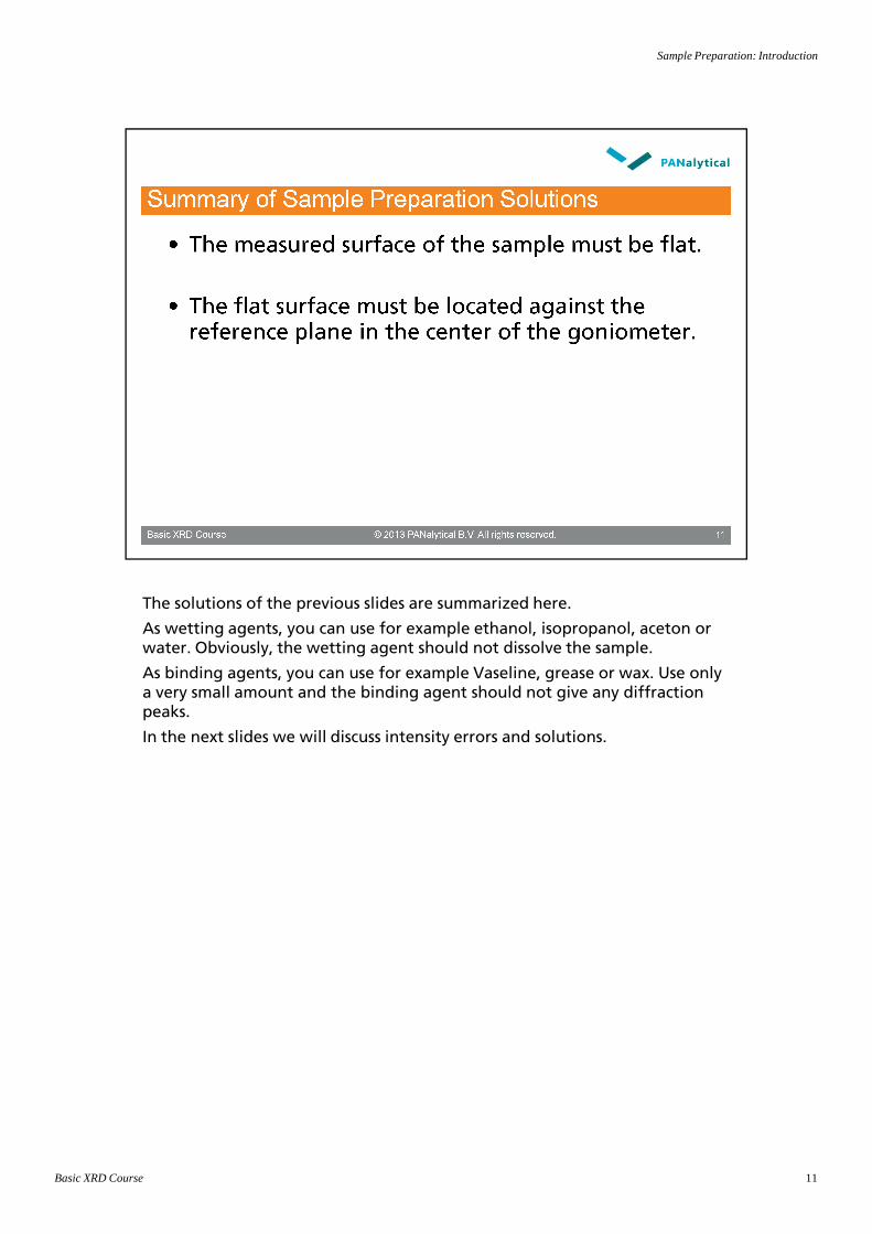
Sample Preparation: Introduction
The solutions of the previous slides are summarized here.
Basic XRD Course 11
As wetting agents, you can use for example ethanol, isopropanol, aceton or water. Obviously, the wetting agent should not dissolve the sample.
As binding agents, you can use for example Vaseline, grease or wax. Use only a very small amount and the binding agent should not give any diffraction peaks.
In the next slides we will discuss intensity errors and solutions.

Sample Preparation: Introduction
Another (better) name for particle statistics is ‘crystal statistics’.
Basic XRD Course 12
The external particle size can be larger than the so-called crystallite size.
Crystal statistics cause incorrect measured intensities. In case of severe crystal statistics, the shape of the peaks will be affected as well. In this case, the peaks shapes are not smooth but show a jagged (sawtooth-like) behavior. The peak position will also be affected in this case.
This can cause problems to identify and/or quantify the proper phases present in the sample.

Sample Preparation: Introduction
The graph shows the relation between the relative error in the measured intensity and the crystallite size for a constant irradiated volume.
Basic XRD Course 13
intensity and the crystallite size for a constant irradiated volume.
Warning: The above figure indicates the effect only.
Note: The relative error in the measured intensity is a statistical expected average error. The real occurring error is not predictable and varies from step to step. It can be zero, much larger or much smaller and it can be positive or negative. Hence, any correction afterwards is not possible.

Sample Preparation: Introduction
Measured on standard NIST SRM 640. This is a relatively coarse grained silicon standard powder.
Basic XRD Course 14
standard powder.
Number Intensity (counts/s)
# Stationary Spinning
1 2040.6 2006.7
2 2648.9 1991.9
3 1793.5 1985.9
4 1622.5 2020.9
5 1770.5 2002.5
6 1953.1 2003.6
7 2100.4 2010.6
8 2017.1 1996.7
9 1824.2 2003.6
10 1982.8 1990.5
Average 1975.4 2001.3
St.dev. 278.5 10.4
Rel.spread 14.1% 0.5%

Sample Preparation: Introduction
Note:The relative error in the measured intensity is a statistically expected average
Basic XRD Course 15
The relative error in the measured intensity is a statistically expected average error. The real occurring error is not predictable and varies from step to step. It can be zero, much larger or much smaller and it can be positive or negative. Hence, any correction afterwards is not possible.

Sample Preparation: Introduction
Two Phi-scans have been performed on a Si test sample using the 28.44 o2θ(111) peak.
Basic XRD Course 16
(111) peak.
One scan is performed with a ½o divergence slit (irradiated length = 11.4 mm) and the other scan with 1/16o divergence slit (irradiated length = 1.4 mm). The sample mask (illuminated width) was 20 mm for both scans.
The scan with the smaller irradiated area (blue) shows much larger statistical variation. This is because a smaller (average) number of particles contribute to the diffraction process.
Note 1:The two scans in the graph are scaled with respect to each other for better comparison.
Note 2:In a Phi-scan, the intensity is measured for a slowly rotating sample as a function of the rotation angle (phi) around the normal of the sample surface. The rotation cycle repeats after one complete revolution of the sample.

Sample Preparation: Introduction
Various Phi-scans have been performed on a Si test sample using the 28.44 o2θ (111) peak.
Basic XRD Course 17
o2θ (111) peak.
The divergence slit varied from 1o (irradiated length = 22.7 mm) to 1/32o
(irradiated length = 0.7 mm). The sample mask (illuminated width) was 20 mm for all scans.
Further reductions of the irradiated area have been achieved with a smaller beam mask (illuminated width): 10 and 5 mm combined with the smallest divergence slit (1/32o).
The average and standard deviation of the measured intensity has been calculated. The graph shows the errors on a relative basis.
The counting statistical error for these scans is indicated as well. In all situations, this error is much smaller than the crystal statistical error. If necessary, the counting statistical error can be reduced by counting longer (i.e. collecting more counts).
The crystal statistical error will not change for experiments with longer counting time!
Note 1:In a Phi-scan, the intensity is measured for a slowly rotating sample as a function of the rotation angle (phi) around the normal of the sample surface. The rotation cycle repeats after one complete revolution of the sample.
Note 2:A larger divergence slit to increase the illuminated area will reduce the resolution.

Sample Preparation: Introduction
The penetration depth can be influenced by choosing another X-ray wavelength (for example Cr or Co). Depending on the material, the
Basic XRD Course 18
wavelength (for example Cr or Co). Depending on the material, the difference in the penetration depth can be 3 to 5 times (for example for Fe samples).
For good particle statistics (see later), a large number of crystallites should contribute to the diffraction process. A way to achieve this is to reduce the average particle/crystallite size by grinding (if possible). One should investigate different grinding procedures (method, time, cooling, et cetera) for each sample type. There can be very different for example for minerals and organic materials (e.g. pharmaceuticals).

Sample Preparation: Introduction
Instrumental
Basic XRD Course 19
• Increasing the irradiated volume by increasing the incidence divergence and/or a larger beam mask is only possible if the sample size allows it. For powder samples, a larger sample can be made only if enough powder is available.
• Sample oscillation (linear movement) is usually done for stress or texture measurements.
• Sample rocking (angular movement) is implicitly done when you use line (1D) detectors.
• Sample rocking can be simulated by adding repeated scans each with a different omega-offset (usually up to 2 degrees) also called “wobble” scan.

Sample Preparation: Introduction
An example of a reactive phase is ‘free lime’ in cement clinker material. The amount of free lime influences the quality of the cement clinker. Due to
Basic XRD Course 20
amount of free lime influences the quality of the cement clinker. Due to reaction with moisture (and CO2) in the air, free lime disappears within a few days. Hence, the proper amount of this phase can only be obtained when you measure freshly prepared samples made from correctly stored material.
Free lime + Water => Portlandite: CaO + H2O => Ca(OH)2
Portlandite + Carbondioxide => Calcite + Water: Ca(OH)2 + CO2=> CaCO3 + H2O
Note 1: The water from the second reaction will be re-used in the first reaction.
Other example: Smectites (clay mineral) show a tremendous peak shift in relation to humidity.
Note 2:If the composition of the sample changes during a long measurement, the low and high angle regions do not represent the same average composition. A good average pattern for the whole angle range can be obtained by making multiple short scans (in time) and summing up the results afterwards.

Sample Preparation: Introduction
Glass capillaries can be used to shield the sample powder from the air (to avoid reaction with oxygen or moisture).
Basic XRD Course 21
avoid reaction with oxygen or moisture).
The sample surface can also be covered with Mylar foil to shield the sample powder from the air (for lightly reactive samples).
Highly reactive samples need to be prepared in a glass capillary inside a glove-box under a neutral atmosphere (like Nitrogen). The glass capillary must be sealed at both ends.
Note: In a so-called humidity chamber, you can also study the reaction of the sample with moisture in relation with a controlled humidity and/or temperature. This is especially interesting for pharmaceuticals. Clay minerals are also studied in humidity chambers (usually at room temperature).

Sample Preparation: Introduction
Basic XRD Course 22

Sample Preparation: Introduction
The options of powder sample preparation are presented here.
Basic XRD Course 23
As wetting agents (in case of small amounts of sample), you can use for example ethanol, isopropanol, aceton or water. Obviously, the wetting agent should not dissolve the sample.
As binding agents, you can use for example Vaseline, grease or wax. Use only a very small amount and the binding agent should not give any diffraction peaks.

Sample Preparation: Introduction
This solution requires:
Basic XRD Course 24
a. Fix sample (surface) at proper height in sample holder
=> this is the responsibility of the user
b. Position the sample holder properly in the instrument (in sample stage)
=> included in design of reflection-transmission spinner
A height positioning accuracy of ±10 µm will be sufficient.

Sample Preparation: Introduction
Sample preparation
Basic XRD Course 25
• Grinding powder sample should be done with care! Too much might result in too small particles or lattice damage and this will result in broadened peaks. Hence, do some testing in order to find out how much grinding is allowed. Start with manual grinding using mortar and pestle (the so called “grinding bowl”).
Certain materials might change their crystal structure due to the forces applied during grinding. Usually, the kind of change is from Alpha to Beta form and vice-versa. Especially for pharmaceuticals, this side effect is not wanted.

Sample Preparation: Introduction
Non-spherical particles are: flakes, needles, cubes, …
Basic XRD Course 26

Sample Preparation: Introduction
The random oriented sample is prepared by dusting onto a greased surface (of a silicon single crystal substrate).
Basic XRD Course 27
(of a silicon single crystal substrate).
For the preferred oriented sample, the sample surface is pressed (flattened) with a glass plate. The flat mica particles are pushed against the substrate surface. Hence, preferred orientation is introduced when one touches the sample surface.
The next sheets will show the differences in the measured scans.

Sample Preparation: Introduction
The sample is prepared by dusting onto a greased surface (of a silicon single crystal substrate).
Basic XRD Course 28
crystal substrate).
The sample shows some preferred orientation. The 00l reflections show relatively more intensity than the other reflections. However, the other reflections are still measurable. Let us assume that this example is close to random orientation.

Sample Preparation: Introduction
The sample is initially prepared by dusting onto a greased surface (of a Silicon single crystal substrate).
Basic XRD Course 29
single crystal substrate).
After that, the sample surface is pressed (flattened) with a glass plate. The flat mica particles are pushed against the substrate surface. Hence, the preferred orientation is much stronger.
The intensities of the 00l reflections have been increased (more particles with 00l orientation). The intensities of the other intensities have been decreased. Some are not measurable anymore. This is a sample with a strong preferred orientation.
The next slide shows zoomed parts of the two patterns.

Sample Preparation: Introduction
The dusted sample has a random orientation.
Basic XRD Course 30
The pressed sample has a preferred orientation (reflections of type hk0
disappear).

Sample Preparation: Introduction
Other solutions instead of randomizing:
Basic XRD Course 31
1. Deliberately create a sample with extreme (and known) preferred orientation.
2. Create samples with a known but constant preferred orientation.
For back loading: prepare the sample against a rough reference surface. For example, emery paper (fine sand-paper) covered with thin Mylar foil (against sticking) can be used as reference surface. Also a special tool with a roughened surface can be used.
Sometimes the quality of the measurement can be improved by dispersing a small amount of the powder on the sample surface (last step of the sample preparation). You can also brush very carefully.
Amorphous fillers could be anything from glass particles to instant coffee!

Sample Preparation: Introduction
These solutions have in common that only a relatively small amount of powder is used for preparing the XRD sample.
Basic XRD Course 32
powder is used for preparing the XRD sample.
Note: Another way of preparing a randomized powder sample with a small amount of powder is filling a glass capillary. However, this requires a different optical set-up.

Sample Preparation: Introduction
Be aware of the particle (crystal) statistics problem! Apply spinning!
Basic XRD Course 33
Note 1: If the sample surface is also small (micro-diffraction), it will be difficult to avoid crystal statistics.
Note 2:The single crystal substrate is also called zero-background holder.

Sample Preparation: Introduction
Clay minerals and paste-like samples can be measured on a porous ceramic filter plate. The material is deposited on the plate using the suction method
Basic XRD Course 34
filter plate. The material is deposited on the plate using the suction method [ref]. When designing the suction device, consideration should be given to the dimensions (diameter range of the disk is between 31.8 mm and 32.2 mm, with thicknesses between 3.3 mm and 3.7 mm).
The porous ceramic filter plate is made out of porous aluminium oxide, mean pore size 3.5 µm.
[Ref]: E.G. Kinter, S. Diamond, (1956) “A new method for preparation and treatment of oriented-aggregate specimens of soil clays for X-ray diffraction analysis”, Soil Sci. 81, 111-120.

Sample Preparation: Introduction
Environmental analysis
Basic XRD Course 35
XRD is playing a big role in this application area since it is used not only for analysis of filter samples taken from mining and refractory production sites, but also for research on soil additives for reclamation, quantitative analysis of asbestos in building demolition waste and identification of deposits found in automotive and boiler components.
A sample of respirable dust is collected on a membrane filter using a respirable dust sampler. The filter is then placed directly into the sample beam of an X-ray diffractometer. The mass of crystalline silica on the filter is determined from X-ray diffraction response, calibrated against filters loaded with known amounts of standard quartz or cristobalite. Since the volume of air sampled is known, the concentration of airborne crystalline silica is readily calculated.

Sample Preparation: Introduction
Basic XRD Course 36

Sample Preparation: Introduction
Solid objects can be fixed in the sample holder for non-standard sample sizes and shapes using modeling wax or plasticine.
Basic XRD Course 37
and shapes using modeling wax or plasticine.
The diameter of the cavity is 44 mm, the maximum sample height is 6.5 mm.
Flat samples can be first fixed on a glass slide using photo glue. In this case it is easy to remove the sample after the measurement is completed. Also positioning the sample on a flat glass slide helps avoiding tilt of the sample and creates a good contrast to the sample stage table.
Note: Extra care is needed for proper sample mounting for stress and texture measurements. In case of stress measurements, the sample height is even more critical. In case of texture measurements, the sample height is less critical. For both applications, avoid that a sample tilt is introduced with sample mounting.

Sample Preparation: Introduction
Thin films or thin solid objects as well as fiber samples can be easily fixed in frame-like sample holders such as rectangular powder holders or for example
Basic XRD Course 38
frame-like sample holders such as rectangular powder holders or for example a film strip holder.

Sample Preparation: Introduction
Position the sample holder properly in the instrument (in sample stage) is included in design of both reflection-transmission spinner and stage for flat
Basic XRD Course 39
included in design of both reflection-transmission spinner and stage for flat samples.

Sample Preparation: Introduction
In case of using stages with no reference surface placing of the sample at a proper height can be done in following ways:
Basic XRD Course 40
proper height can be done in following ways:
a. Alignment with a height sensor, which is controlled from Data Collector software. This device is used in combination with the stages, which have programmable Z axis (cradles, modular stage with programmable Z-adjustment).
b. Adjustment of the sample height using dial gauge. This option is used when the Z axis of the stage is controlled manually (multi-purpose sample stage, modular stage with manual Z-adjustment).
In the last case to fix the sample at the proper height is the responsibility of the user.
A height positioning accuracy of ±10 µm is sufficient.