this page intentionally blank. - nist center for neutron … c. stock, et al. (chrns) 14...
TRANSCRIPT


This page intentionally blank.

ON THE COVER
Doug Ogg pre-aligns new neutron guides for the NCNR guide hall expansion using a laser-tracking system.

2011 NIST Center for Neutron Research
Accomplishments and Opportunities
NIST Special Publication 1127
Robert M. Dimeo, Director
Ronald L. Cappelletti, Editor
December 2011
National Institute of Standards and TechnologyPatrick Gallagher, Director
U.S. Department of CommerceJohn Bryson, Secretary

DISCLAIMER Certain commercial entities, equipment, or materials may be identified in this document in order to describe an experimental procedure or concept adequately. Such identification is not intended to imply recommendation or endorsement by the National Institute of Standards and Technology, nor is it intended to imply that the entities, materials, or equipment are necessarily the best available for the purpose.
National Institute of Standards and Technology Special Publications 1127
Natl. Inst. Stand. Technol. Spec. Publ. 1127, 88 pages (December 2011)
CODEN: NSPUE2
U.S. GOVERNMENT PRINTING OFFICE-WASHINGTON: 2011
For sale by the Superintendent of Documents, U.S. Government Printing Office
Internet: bookstore.gpo.gov Phone: 1.866.512.1800 Fax: 202.512.2104 Mail: Stop SSOP, Washington, DC 20402-0001

Table of Contents
iv FOREWORD
1 THE NIST CENTER FOR NEUTRON RESEARCH
2 NIST CENTER FOR NEUTRON RESEARCH INSTRUMENTS
4 NCNR EXPANSION ACTIVITIES
6 NCNR IMAGES 2011
HIGHLIGHTS
CONDENSED MATTER
8 Structural collapse and high-temperature superconductivity in iron-pnictide CaFe2As2 at ambient pressure, S.R. Saha, et al.
10 Spin liquid state in the S = ½ triangular lattice Ba3CuSb3O9, H.D Zhou, et al. (CHRNS)
12 InterstitialirontuningofthespinfluctuationsinFe1+xTe, C. Stock, et al. (CHRNS)
14 Delta-doping of ferromagnetism in digitally-synthesized manganite superlattices, T. Santos, et al.
16 The magnetic structure of multiferroic BeFeO3, W. Ratcliff II, et al.
BIOLOGY
18 Quantifying disorder in lipid membranes, M. Mihailescu, et al.
20 NeutronreflectometrystudiesoftheParkinson’sdisease-related protein, α-synuclein, at the lipid bilayer interface,C.M.Pfefferkorn,et al.
22 SANSandmodelingrevealchangesinflexibilityofaprotein important for DNA replication, S. Krueger, et al. (CHRNS)
ENGINEERING
24 Residual stresses and mechanical damage in gas pipelines, T. Gnäupel-Herold, et al.
26 Nano-void volume linked to sensitivity of RDX explosive, C.S. Stoltz, et al. (CHRNS)
CHEMICAL PHYSICS
28 Densityhysteresisinnanoconfinedwater,Y.Zhang, et al. (CHRNS)
30 Cholesterol transport rates in model membranes, S. Garg, et al. (CHRNS)
32 Thefirstmixed-metalamidoborane:sodiummagnesium amidoborane, a good hydrogen storage candidate, H. Wu, et al.
34 Nature of CO2adsorptioninMg-MOF74:acombinedneutrondiffractionandfirst-principlesstudy,W.L.Queen, et al.
SOFT MATTER
36 Neutronscatteringtechniquesshednewlightongelformation,A.P.R.Eberle,et al. (CHRNS)
38 Interlayer distance dependence of thickness fluctuationsinaswollenlamellarphase,M.Nagao, et al. (CHRNS)
40 Morphological characterization of polymer-based solar cells,H.Lu,et al.
42 Nanoscale mixing of soft solids, S.-H. Choi, et al.44 Zonerefinementeffecttoremoveimpuritiesin
organic semiconductor polymer blends for printable electronics,V.M.Prabhu,et al.
NEUTRON PHYSICS
46 Limitonparity-violatingneutronspinrotationin4He, J.S. Nico, et al.
ADVANCES IN MEASUREMENT
48 Improving the measurement of trace selenium by neutron activation analysis, I.J. Kim, et al.
49 Directly probing anisotropy gradients using polarized neutronreflectometry,B.J.Kirby,et al.
50 Detection of dynamical transitions in hydrogenous materials by transmission of very cold neutrons, N. Verdal, et al. (CHRNS)
51 Uniformity of a large cross section neutron spin analyzer for SANS polarization analysis, W.C. Chen, et al. (CHRNS)
52 NEUTRON SOURCE OPERATIONS
53 FACILITY DEVELOPMENT
56 SERVING THE SCIENCE AND TECHNOLOGY COMMUNITY
59 THE CENTER FOR HIGH RESOLUTION NEUTRON SCATTERING (CHRNS)
62 AWARDS 2011
64 PUBLICATIONS
80 INSTRUMENTS AND CONTACTS
CONTACTS (inside back cover)

iv
Foreword
ItiswithpleasurethatIpresentthisyear’sannualreportfortheNISTCenterforNeutronResearch.Whenlookingbackoverthelastyear,theonlywordthatIcanusetodescribethis time period is dynamic.Useroperationsweresuccessfulandproductive,thereactoroperating for 142 days out of a scheduled 142 days. The cold source continued to operate reliablyanddeliverneutrons99%ofthescheduledoperatingtime.Thisperiodwasmarkedbyconsiderablescientificproductivityandimpactasthehighlightsinthisreportillustrate.Theproductivityisduetomanyfactors,butitisworthnotingthatthenumberofuser
experimentsontheMACSspectrometercontinuedtogrowastheamountoftimeavailabletotheusercommunitywasincreased,industrialresearchparticipationonmanyoftheneutroninstruments,especiallyuSANS,wasstrong,andresearchopportunitieswithotherlaboratoriesgrew,particularlyintheareaofbiologyandbiosciences.Moreover,significantprogresswasmadetowardsbringingtheisotopelabelinglaboratoryonlinewhichwillcertainly prove to be a valuable resource for NCNR users.
Thisyearwasalsomarkedbyaperiodofmanyfacilityenhancementsthatcanbestbedescribedashistoric. On April3,thereactorshutdown,asplanned,foraperiodof11months.AsIdescribedinlastyear’sannualreportthisoutagewasscheduledinordertoexecutemanytaskstowardstheexpansionofourcoldneutronmeasurementcapabilityandenhancethereliabilityofthereactor.Afewoftheactivitiesincludeinstallationofanewneutronguidesysteminourexpandedguidehallwhichwillholdnewandrelocatedinstruments,installationofanewcoldsourcededicatedtotheMACSinstrument,relocationofMACS,andinstallationofanewcoolingsystemforthereactor’sthermalshield.Inadditionsignificantconstructioninandaroundthesitehastakenplaceduringtheoutage,includingtheconstructionofanewsecondarycoolingsystem,theadditionofanewcoolingtowercell, and additional electrical capacity. The commencement and execution of this outage marks the culmination ofasignificantamountofplanningandcoordinationaswellasthehardworkanddiligenteffortsofourstaff,especiallythosefromReactorOperationsandEngineeringandResearchFacilityOperations.Youcanreadmoreabouttheprogresswehavemadeduringtheoutagewithinthisreportandonourwebsite:http://www.ncnr.nist.gov/expansion2/whereyouwillfindtechnicaldetailsandphotos.Iamalsopleasedtoinformyouofthereleasethisyearofaninformalhistory,“TheNISTCenterforNeutronResearch:Over40YearsServingNIST/NBSandtheNation”,byJackRushandRonCappelletti,NISTSpecialPublication1120.ContainedthereinisanaccountofmajoreventsoftheNCNRspanningtheperiodfromconceptionoftheNISTreactortotheNCNRExpansionProject.
Asofthiswritingwejustannouncedourfirstcallforproposalsforexperimentstoberunuponrestartofthereactor.InjustafewshortmonthsthereactorwillrestartanduserswillreturntotheNCNR.Thereasonfortheoutageandexpansionisforyou,ourusers.Iamlookingforwardtorestorationofuseroperationsandespeciallyofferingnewmeasurementcapabilitiesthatourexpansionprojectwillprovidethroughthecomingyear.Iinviteyoutotakealookattheresearchhighlightsfromthepastyearaswellasthedescriptionofwhatwehavebeenupto during the outage. As you read through this report, I hope you get the same sense of excitement and opportunity thatweareallfeelinghere.

1
The NIST Center for Neutron Researchfacilitiesmostofwhichareusedforneutronscatteringresearch. The subsequent pages provide a schematic
Neutrons provide a uniquely effective probe of the structure and dynamics of materials ranging from watermovingnearthesurfaceofproteinstomagneticdomains in memory storage materials. The properties ofneutrons(outlinedbelow)canbeexploitedusingavariety of measurement techniques to provide information nototherwiseavailable.Thepositionsofatomicnucleiin crystals, especially of those of light atoms, can be determined precisely. Atomic motion can be directly measured and monitored as a function of temperature or pressure. Neutrons are especially sensitive to hydrogen, sothathydrogenmotioncanbefollowedinH-storagematerialsandwaterflowinfuelcellscanbeimaged.Residualstressessuchasthosedeepwithinoilpipelinesorinhighwaytrussescanbemapped.Neutron-basedmeasurements contribute to a broad spectrum of activities including in engineering, materials development, polymer dynamics, chemical technology, medicine, and physics.
TheNCNR’sneutronsourceprovidestheintense,conditioned beams of neutrons required for these types of measurements. In addition to the thermal neutron beams fromtheheavywaterorgraphitemoderators,theNCNRhas a large area liquid hydrogen moderator, or cold source, thatprovideslongwavelengthguidedneutronbeamsforthemajorcoldneutronfacilityintheU.S.
Therearecurrently25experimentstations:fourprovidehighneutronfluxpositionsforirradiation,and21arebeam
description of our instruments. More complete descriptions canbefoundatwww.ncnr.nist.gov/instruments/.Constructionofasecondguidehallisnowcomplete(seepp.4-5)andfivenewinstrumentsareunderdevelopment.
The Center supports important NIST measurement needs,butisalsooperatedasamajornationaluserfacilitywithmerit-basedaccessmadeavailabletotheentireU.S. technological community. Each year, more than 2000 research participants from government, industry, and academia from all areas of the country are served by the facility (see pp. 56-58). Beam time for research to bepublishedintheopenliteratureiswithoutcosttotheuser, but full operating costs are recovered for proprietary research.Accessisgainedmainlythroughaweb-based,peer-reviewedproposalsystemwithusertimeallottedbyabeamtimeallocationcommitteetwiceayear.Fordetailsseewww.ncnr.nist.gov/beamtime.html.TheNationalScience Foundation and NIST co-fund the Center for High Resolution Neutron Scattering (CHRNS) that operates sixoftheworld’smostadvancedinstruments(seepp.59-61). Time on CHRNS instruments is made available through the proposal system. Some access to beam time for collaborativemeasurementswiththeNISTsciencestaffcanalso be arranged on other instruments.
Why Neutrons?
Neutrons reveal properties not readily probed by photons or electrons. They are electrically neutral and therefore easily penetrate ordinary matter. They behave like microscopic magnets,propagateaswaves,cansetparticlesintomotion,losing or gaining energy and momentum in the process, and theycanbeabsorbedwithsubsequentemissionofradiationtouniquelyfingerprintchemicalelements.WAVELENGTHS−inpracticerangefrom≈0.01nm(thermal)to≈1.5nm(cold)(1nm=10Å),allowingtheformationofobservableinterferencepatternswhenscatteredfromstructuresas small as atoms to as large as biological cells. ENERGIES−ofmillielectronvolts,thesamemagnitudeas atomic motions. Exchanges of energy as small as nanoelectronvolts and as large as tenths of electronvolts can be detectedbetweensamplesandneutrons,allowingmotionsinfolding proteins, melting glasses and diffusing hydrogen to be measured.
SELECTIVITY−inscatteringpowervariesfromnucleustonucleussomewhatrandomly.Specificisotopescanstandoutfromotherisotopesofthesamekindofatom.Specificlightatoms,difficulttoobservewithx-rays,arerevealedbyneutrons.Hydrogen, especially, can be distinguished from chemically equivalentdeuterium,allowingavarietyofpowerfulcontrasttechniques.MAGNETISM−makestheneutronsensitivetothemagneticmomentsofbothnucleiandelectrons,allowingthestructureand behavior of ordinary and exotic magnetic materials to be detailed precisely. NEUTRALITY−oftheunchargedneutronsallowsthemtopenetratedeeplywithoutdestroyingsamples,passingthroughwallsthatconditionasample’senvironment,permittingmeasurements under extreme conditions of temperature and pressure. CAPTURE−characteristicradiationemanatingfromspecificnuclei capturing incident neutrons can be used to identify and quantify minute amounts of elements in samples as diverse as ancientpotteryshardsandlakewaterpollutants.
NCNR 2011 Accomplishments and Opportunities
A distorted composite view of the new guide hall, looking through the removed columns towards its center from within the old guide hall (east wing). At the extreme left is the backscattering spectrometer. A portion of the shield assembly for the new guides can be seen in the center of the picture. Far away to the right is all that remains of the wall separating the east and west wings of the guide hall.

2
NC
NR
Instruments
NIST Center for Neutron Research Instruments (as of Feb. 2012)

3NCNR 2011 Accomplishments and Opportunities

4
NC
NR
Expansion
Facility Expansion Activities

5NCNR 2011 Accomplishments and Opportunities

6
NC
NR
Images 2011
NCNR Images 2011
North American neutron facility representatives meet at the NCNR: L. to R.: Sean O’Kelly, Ian Anderson (SNS), Richard Ibberson, Alan Hurd (LANL), Jim Rhyne (LANL), John Root (Chalk River), Dan Neumann, and Rob Dimeo.
Kathryn Krycka describes contrast matching on a tour of the NG-7 SANS facility.
Cool RFO guys: George Baltic, Bill Clow, Doug Johnson, and Dan Ogg move the MACS analyzer to temporary storage in the new guide hall.
Sammi Jo Cooper, a teacher at Briggs-Chaney MS, checks out crystal growth during the Summer Institute.
Summer Highschool Intern Program (SHIP) students andNCNR personnel take a break from research activity for a group photo on the beautiful grounds in front of the facility.
Pop goes the nucleus! in Juscelino Leão’s vivid chain reaction demonstration to a middle school tour.
Jeff Lynn encourages Roberto Clemente Middle School students to do violence! Smash it! Smash that frozen rose into shards!
Yun Liu expounds on the Spin-Echo spectrometer from the “dance floor” (or is he demonstrating the latest dance step?)

7NCNR 2011 Accomplishments and Opportunities
Paul Brand and Tony Norbedo enjoy the success ofthe thermal shield upgrade, which moves cooling waterwhile maintaining the pressure below atmospheric.
And it didn’t come tumbling down: removing the final columns between the guide hall east and west wings.
Pee Wee (new cold source for MACS) goes in...it’s all connected...and Bob Williams says, Look!...no leaks!
New guides assembled at various stages. Upper right: beam shutters partially installed at the C100 wall.Lower right: John Copley and Jeff Zeigler survey recently arrived guide components.
L. to R.: Nancy Hadad, Bill Clow, Dan Adler, Dan Ogg, Mike Rinehart, George Baltic and Don Pierce watch intently as a guide shutter is set into place.

8
Condensed M
atter
8
Structural collapse and high-temperature superconductivity in iron-pnictide CaFe2As2 at ambient pressureS.R. Saha1, N.P. Butch1, T. Drye1, J. Magill1, S. Ziemak1, K. Kirshenbaum1, P.Y. Zavalij2, J.W. Lynn3, and J. Paglione1
1Center for Nanophysics and Advanced Materials, Department of Physics, University of Maryland, College Park, MD 207422Department of Chemistry and Biochemistry, University of Maryland, College Park, MD 207423NIST Center for Neutron Research, National Institute of Standards and Technology, Gaithersburg, MD 20899
Thediscoveryofthenewfamilyofiron-basedhightransition temperature (Tc) superconductors has
offeredanewroutetohightransitiontemperatures[1], breaking the copper-oxide based high Tc superconductor monopoly. The highest Tcof≈55Khas been found in the oxypnictide series RFeAsO1-xFx,whiletheoxygen-free“122”intermetallic series A1-xKxFe2As2 (A = Ba, Sr, Ca), crystallizing in the ThCr2Si2-type tetragonal structure,showsamaximumTc of 38 K [1] and is the most popular series of the Fe-based superconductors. The 122 parent compounds are tetragonal at room temperature but undergo an orthorhombic structural distortionthatisassociatedwiththeonsetofantiferromagnetic (AFM) order. Tuning the system by elemental substitution or by external pressure suppresses the magnetic and structural ordering temperatures and induces superconductivity. CaFe2As2 is a unique member of this family; upon pressure application the magnetostructural transition is suppressed, but is abruptly interrupted by a structural collapse of the tetragonal unit cell, whichwasidentifiedbyneutronmeasurementsina He-gas pressure cell on BT-1 and BT-7 at NIST [4]. Intriguingly, the appearance of superconductivity in CaFe2As2underpressurebelow12Konlyoccurswhennon-hydrostaticpressureisapplied[2],whilenotraceofsuperconductivitywasfoundwhenusinga truly hydrostatic He gas pressure environment [3].
This contrast and sensitivity to pressure application encouraged us to use chemical substitution as an effective hydrostatic pressure on the unit cell to induce the structural collapsed tetragonal phase in this compound. Single crystals of CaFe2As2weregrownwithlightrareearths(R=La,Ce,Pr,andNd)substituted into the Ca position to form Ca1-xRxFe2As2 , in order to investigate the structural andsuperconductingproperties[4].Wefindthatelectron-doping via aliovalent substitution of trivalent R3+ ions for divalent Ca2+ acts to suppress AFMorderandinducesuperconductivity,withTc
surprisingly as high as 47 K, by far the highest value found in 122 FeAs-based compounds [6]. Moreover, the close matchbetweenionicradiiofLa,Ce,Pr,andNdwiththat ofCaallowsustoselectivelytunethestructuralparametersby using the slightly decreasing variation in rare earth size withf-electroncount,knownasthelanthanidecontraction,to tune the unit cell volume in a controllable manner.
FIGURE 1: Temperature dependence of the a- and c-axis lattice parameters of Ca1−xRxFe2As2. The color-intensity plots highlight the structural collapse of the tetragonal unit cell induced by rare earth substitution upon both warming (red arrows) and cooling (blue arrows) that is present in Pr0.075Ca0.925Fe2As2 (a-d), but absent in La0.19Ca0.81Fe2As2 (e-f). The first-order structural transition is strongly hysteretic in nature.

9NCNR 2011 Accomplishments and Opportunities
Single-crystaldiffractionstudieswerecarriedouttostudythe Ca1-xRxFe2As2 structure, revealing dramatic changes in the structure that include not only an unprecedented thermal expansion, but also a structural collapse of the tetragonal unit cell akin to that previously found under pressure. Figure 1 presents the temperature dependence of lattice parameters determined by single crystal thermal neutron diffraction measurements of the (110) and (002) typenuclearreflections,yielding,respectively,a- and c-axis parameters. Thesubstitutionofonly7.5%PrintoCaFe2As2 isenoughtodrivethecollapsedtetragonaltransition,withthe a-axis and c-axis lattice parameters undergoing adiscontinuousjump.Incontrast,thesubstitutionof 19%LadoesnotdrivethesystemtowardanyobservabletransitionbecausethelargerionicradiusofLaisnotamenable to inducing positive chemical pressure.
FIGURE 2: Comparison of c-axis lattice parameters in the Ca1−xRxFe2As2 family for different rare earth substitutions at ambient pressures. The collapse of the c-axis in Pr- and Nd-substituted Ca1−xRxFe2As2 is directly comparable to that in undoped CaFe2As2 under applied hydrostatic pressure (black squares; from Ref. 4), while La- and Ce-substituted samples do not undergo a collapse in the substitution range investigated.
Figure 2 presents the temperature dependence of the c-axis unit cell dimension upon cooling, determined from neutron diffractionmeasurementsofthe(006)nuclearreflectionforundoped CaFe2As2 [4] and several Ca1−xRxFe2As2 crystals at ambient pressure. A dramatic collapse in the structure is
evident,withNdsubstitutionactinginasimilarmannerasPrtoachieveanalmostidenticalcollapsetransition.Bothcasesshowanabsolutechangeinthec-axis through the collapse transition that is nearly identical to that observed in CaFe2As2underpressure[4].Incontrast,La-andCe-substitutedcrystalsfailtoundergoacollapsetothelowestmeasuredtemperatures,evenforthecaseofCewherethe c-axisdimensionreachesbelowthevaluewherethecollapse occurs in the lighter rare earth-doped samples. This is understood to be the effect of the interlayer As-As distance,whichwebelieveistheimportantparametercontrollingthecollapseoftheunitcell[5],whichdoesnotreachthecriticalvalueof3Åinthesecrystals.However,asis evident by an extremely large c-axis thermal expansion of 180 x 10-6/K(thelargestknownvalueforanymetal),the As-As bonding tendency is extremely strong in these crystals,whichisliterallyonthevergeofcollapse.
High Tcsuperconductivitywasfoundtobepresentinboththe collapsed and uncollapsed structures [5], suggesting that the unconventional superconductivity of the iron-based superconductor family appears to occur in this system regardless of the nature of the interlayer bonding. Given the absence of superconductivity in the collapsed phase of undoped CaFe2As2 under hydrostatic pressure [3], this is quite surprising and may originate from the subtle but important addition of charge doping. The insensitivity of the observed high-Tc superconducting phase to this boundary is unusual, raising important questions regardingwhichfeaturesofthechemical,electronicandmagnetic structure are important to Cooper pairing.
References
[1] J.PaglioneandR.L.Greene,NaturePhys.6, 645 (2010).[2] M.S.Torikachvili,S.L.Bud’ko,N.Ni,andP.C.Canfield,Phys.
Rev.Lett.101, 057006 (2008).[3] W.Yu,A.A.Aczel,T.J.Williams,S.L.Bud’ko,N.Ni,P.C.
Canfield,andG.M.Luke,Phys.Rev.B79, 020511R (2009).[4] A.Kreyssig,M.A.Green,Y.Lee,G.D.Samolyuk,P.Zajdel,
J.W.Lynn,S.L.Bud’ko,M.S.Torikachvili,N.Ni,S.Nandi,J.B.Leão,S.J.Poulton,D.N.Argyriou,B.N.Harmon,R.J.McQueeney,P.C.Canfield,andA.I.Goldman,Phys.Rev.B78, 184517 (2008).
[5] S.R.Saha,N.P.Butch,T.Drye,J.Magill,S.Ziemak,K.Kirshenbaum,P.Y.Zavalij,J.W.Lynn,andJ.Paglione,arXiv:1105.4798v1.

10
Condensed M
atter
10
Spin liquid state in the S = 1/2 triangular lattice Ba3CuSb2O9
H.D. Zhou1, E.S. Choi1, G. Li1, L. Balicas1, C.R. Wiebe1,2,3, Y. Qiu4,5, J.R.D. Copley4, and J.S. Gardner4,6
1National High Magnetic Field Laboratory, Florida State University, Tallahassee, FL 32306-40052University of Winnipeg, Winnipeg, MB, R3B 2E9 Canada3University of Manitoba, Winnipeg, MB, R3T 2N2 Canada4NIST Center for Neutron Research, National Institute of Standards and Technology, Gaithersburg, MD 20899-61025University of Maryland, College Park, MD 207426Indiana University, Bloomington, IN 47408
One of the current thrusts of modern condensed matter science has been the realization of an
importantmodelcompoundknownasthequantumspinliquid(QSL).Theexistenceofthesematerials,inwhichmagneticspinsremaindisorderedbyquantumfluctuationsinthelimitofzeroKelvin,underpinsmuchofmoderncondensedmattertheory.PreviousstudieshaveshownthatQSLgroundstatestendtoemerge in the geometrically frustrated materials, in whichtheinteractionsamongthelimitedmagneticdegrees of freedom lead to a strong enhancement ofquantumfluctuations.WhilethestudyoftheQSLstateintheorganiccompoundsremainsahottopic,thereareveryfewinorganicmaterialsidentifiedasmodelsystemsforQSLgroundstates.Many efforts to synthesize spin liquids on triangular latticesininorganicmaterialshavefailed.Here,weunveilanewcandidateforaspinliquidcompound– Ba3CuSb2O9–inwhichCu2+ species form a geometrically frustrated triangular lattice.
The crystal structure of Ba3CuSb2O9, a 6-H perovskite-relatedmaterialwitha = b =5.8090Åand
c =14.3210Å,canberepresentedasaframeworkconsistingof corner-sharing SbO6 octahedra and face-sharing CuSbO9 bi-octahedra,asshowninFig.1(a,b).Inthebi-octahedra,theCuandSbcationsarewell-ordered.TheCuionsoccupy the 2b Wyckoff site of space group P63mc, and this site forms the triangular lattice in the ab plane (Fig. 1(c)). Therefore,thestructurecanbeseenasatwo-dimensionaltriangular magnet, i.e., the Cu magnetic triangular lattices aremagneticallyseparatedbythetwonon-magneticSblayers (Fig. 1(b)).
The temperature dependence of the DC magnetic susceptibilitymeasuredwithm0H = 0.5 T for Ba3CuSb2O9 showsnosignatureforamagnetictransitionabove1.8K,as indicated in Fig. 2(a). The temperature dependence of theACmagneticsusceptibility(Fig.2(b))furthershowsnosignofamagnetictransitiondownto0.2K.Theneutronpowderdiffractionpatternobtainedat0.2Kwithl =1.8ÅontheDCSatNIST(Fig.2(c))showsnointensitychangenoradditionalpeaksfromthe4Kdata(notshownhere),indicating that there is no magnetic transition nor structural distortiondownto0.2K.ThepossibilityofmixingbetweentheCuandSbsiteshasbeentestedbyrefinementoftheneutrondiffractiondata,whichshowafulloccupancyof Sb(1) on the corner-sharing SbO6 octahedron sites and Cu on one of the ordered bi-octahedra sites. There is a slightamountofsitemixing(refinedfromtheneutrondata to be 5.1(4) %) of the other ordered bi-octahedra sites - Sb(2) sites are replaced by (orphan) Cu ions. The DC magnetic susceptibility contribution from this 5.1 % Cu2+ orphan spins has been calculated by a simple Curie law.Thesusceptibility,aftersubtractingthiscontribution,isshownasopencirclesinFig.2(a).TheCurie-Weissfitof this linear behavior at high temperature gives a qCW = -55 K and an effective moment meff = 1.79 mB/Cu,whichisconsistentwiththeexpectedvalueforCu2+ (S=1/2)ions.The exchange interaction J is estimated to be J/kB = 32 K by fittingthedatabetween150Kand300Ktothecalculationforthespin1/2triangularlatticeusingahigh-temperature-series expansion (HTSE).
FIGURE 1: (a) Schematic crystal structure for Ba3CuSb2O9; (b) The layer structure along the c-axis; (c) The triangular lattice of Cu2+ in the ab plane.

11NCNR 2011 Accomplishments and Opportunities
Themagneticspecificheat(CM, Fig. 3) of the Cu2+ triangular lattice is obtained by subtracting the lattice contribution (CP of the non-magnetic Ba3ZnSb2O9), and the Schottky anomaly from the Cu2+ orphan spins (detail, seeRef.[1]).AsshowninFig.3(b)withthelog-logscale,between1.4Kand4K,CMcanbefittedasCM = bTα withb = 37.0 mJ K-3mol-1 and α = 1.83(2). This α value is near 2.0,showingquadratictemperaturedependence.Atlowertemperatures,between0.35Kand1.4K,CMcanbefitasCM = gTα withg = 43.4 mJ K-2mol-1 and α = 0.99(2), giving a linear temperature dependence. The integrated magnetic entropyvariationbelow30Kis1.7JK-1mol-1,whichisaround 30 % of Rln(2) = 5.76 J K-1mol-1 for a S=1/2system,whereRisthegasconstant,asshowninFig.3(a).Thisfeatureindicatesahighdegeneracyoflow-energystatesatlowtemperatures.
Thesusceptibilityandspecificheatbothshownomagneticorderingdownto0.2KforBa3CuSb2O9 despite moderately strong nearest neighbor antiferromagnetic interactions withJ ≈ 32K,whichclearlyplacesthiscompoundinthehighlyfrustratedregime.Thefield-independentCM iscommoninspinliquidcandidates,whichshouldbeinsensitivetomoderateappliedfields.Ontheotherhand,the linear dependence of CM is unusual for 2D frustrated lattices,whichshouldhaveaquadraticdependenceforlinearlydispersivelowenergymodes.However,alinearT dependenceatlowtemperatureshasbeenpredictedbythe resonating-valence-bond model of Anderson, due to the pairing of spins into singlets [2]. One current mode of thought is that a Fermi surface of spinon excitations is formedwithintheliquidstate,whichgivesrisetoalinearterm even for Mott insulators [3].
In summary, the synthesis and characterization of Ba3CuSb2O9,whichhasalayeredarrayofCu2+ spins in a triangular lattice, are reported. The magnetic susceptibility andneutronscatteringexperimentsofthismaterialshownomagneticorderingdownto0.2KwithaqCW = -55 K. ThemagneticspecificheatrevealsalineardependenceonTwithg = 43.4 mJ K-2mol-1below1.4K.Theseobservationssuggest that Ba3CuSb2O9isanewquantumspinliquidcandidatewithaS=1/2triangularlattice.
References
[1] H.D. Zhou, et al.,Phys.Rev.Lett.106, 147204 (2011).[2] P.W.Anderson,Science,235 1196, (1987).[3] S.S.Lee,P.A.Lee,andT.Senthil,Phys.Rev.Lett.98, 067006
(2007).
FIGURE 2: (a) The temperature dependences of the DC magnetic susceptibility (c). The red open squares represent c as measured, the green dashed line, c of orphan spins, and the blue open circles, represent c after deleting orphan spins. The solid curve on c data above 150 K represents a fit to the high-temperature-series expansion. Inset: 1/c – after deleting the orphan spin contribution (open triangles). The solid line on the 1/c data represents a Curie-Weiss fit. (b) The temperature dependence of the real part of the AC magnetic susceptibility (c’). (c) Neutron diffraction pattern (red crosses) at 0.2 K. The solid blue curve is the best fit from a Rietveld refinement using FullProf. The vertical marks indicate the position of Bragg peaks, and the bottom curve shows the difference between the observed and calculated intensities. The error bars ± s are too small to be visible on the figures.
FIGURE 3: (a) The temperature dependences of the reduced heat capacity CM/T and the magnetic entropy SM; (b) The temperature dependence of CM (open symbols). The solid lines are fits as described in the text.

12
Condensed M
atter
12
Interstitial iron tuning of the spin fluctuations in Fe1+xTeC. Stock1,2, E.E. Rodriguez2, M. Green1,3, P. Zavalij4, and J. Rodriguez-Rivera1,3
1Indiana University, Bloomington, IN 474042NIST Center for Neutron Research, National Institute of Standards and Technology, Gaithersburg, MD 208993Department of Materials Science and Engineering, University of Maryland, College Park, MD 207424Department of Chemistry, University of Maryland, College Park, MD 20742
Magnetism is directly related to superconductivity in several heavy fermion
and d-transition metal-ion systems. Most notably, localized magnetism is believed to bedirectlycoupledwithhigh-temperaturesuperconductivity in the cuprates as evidenced through studies as a function of charge concentration. More recently, the discovery of superconductivity in the iron-based compounds has revealed a seriesofmaterialsweresuperconductivityand magnetism coexist. Magnetism and superconductivity are strongly intertwinedinthesesystemsasillustratedby neutron inelastic-scattering experiments, whichhaveshownadistinctchangeinthespinfluctuationsoncoolingthroughthesuperconducting transition temperature [1].
Usingneutroninelasticscattering,weinvestigatethelow-energyspinfluctuationsinFe1+xTe as a function of both temperature and interstitial iron concentration. For Fe1.057(7)Te, the magnetic structure isdefinedbyacommensuratewavevectorof(1/2,0,1/2).Thespinfluctuationsaregappedwithasharp onset at 7 meV and are three dimensional in momentumtransfer,becomingtwodimensionalathigher-energytransfers.Ondopingwithinterstitialiron,wefind,inFe1.141(5)Te,theorderingwavevectoris located at the incommensurate (0.38, 0, ½) position andthefluctuationsaregaplesswiththeintensitypeaked at an energy transfer of 4 meV. These results showthatthespinfluctuationsintheFe1+xTe system can be tuned not only through selenium doping, but also interstitial iron.
Arguably, the simplest iron-based superconductor is the single layered Fe1+xTe1-ySeysystemwheresuperconductivityhasbeenobservedwithamaximum transition temperature of 14 K for y ≈ 0.5. ThestructureisshowninFig.1illustratingboth the cation (iron) and anion (selenium and tellurium) sites. While selenium doping has proven
to be the most direct route for inducing superconductivity inthissystem,recentstudieshaveshownthattheselenium concentration is directly tied to the amount ofinterstitialironbetweentheFeTelayers.Thiswasdemonstrated in a study of Fe1+xTe0.7Se0.3whichprovedthat the superconductivity and the magnetic excitations weredirectlylinkedwiththelevelofinterstitialiron,independent of the selenium concentration [2]. The magnetic structure of the parent non-superconducting Fe1+xTe, in the absence of selenium doping, has been investigated using neutron diffraction illustrating a dramatic change of the magnetic and crystalline structure withchangingtheinterstitialironconcentration[3].Forsmall amounts of interstitial iron, a commensurate double-stripespin-densitywavephaseexistswhichabruptlychanges to a spiral magnet for x > 0.12.
The superconducting variants of Fe1+xTe1-ySey have been investigated and neutron inelastic scattering has reported the static magnetic order observed in the parent material tobereplacedbyshort-rangecorrelationsnearthe(1/2,1/2,L) position. This position in reciprocal space matches anestingwavevectorproposedfromphotoemissionmeasurementsindicatingpossiblecouplingbetweenelectronic and magnetic properties. Notably, in the
FIGURE 1: Left: crystalline structure of the parent Fe1+xTe compound: the gold/white spheres represent possible interstitial positions of the +x Fe ions, gold spheres represent Fe, and green spheres represent Te. Right: double-stripe magnetic structure on the Fe sublattice at low interstitial Fe concentration.

13NCNR 2011 Accomplishments and Opportunities
superconductingphase,aresonancepeakat≈7meVhasbeen observed in the half-doped (y≈0.5)systems[4].Thepeakappearsbelowthesuperconductingtransitionandissharp in energy. Unlike its counterpart in heavy-fermion superconductors,themomentumdependenceisverytwodimensional in nature, forming a rod along the c* direction.
The microscopic nature of the magnetism and its relation to superconductivity in the FeAs-based high temperature superconductors is a matter of current debate and research. While the reduced-ordered moment may indicate itinerant effectsareimportant,therearetwokeydifferenceswiththeFe1+xTesystemwhichmaypointtowardstronglocalizedmagnetism in this system. Firstly, the ordered moments aresignificant.Secondly,theorderingwavevectorisnotconsistentwiththenestingwavevectorreportedfromphotoemission.LittleattentionhasbeenplacedontheroleofexcessFeonthespinfluctuationsandtransportpropertieswithmuchactivityfocusedontheroleofSedoping.
UsingtheMACScoldtriple-axisspectrometeratNIST,wehavemeasuredthemagneticfluctuationsasafunctionofinterstitial iron doping in the parent Fe1+xTe compound. In particular,wehavefocusedontwodifferentconcentrationsof x = 0.057(7) and x = 0.141(5) in the commensurate double-stripe and spiral phase respectively. Single crystal x-ray diffraction on crystals cleaved from the larger crystals wasperformedtocharacterizetheamountofinterstitialiron.Constantenergyplaneswereprobedbyfixingthefinalenergyto3.6meVandscanningmomentumtransferwiththe20double-bouncePG(002)analyzingcrystalsand
detectorsandvaryingtheincidentenergydefinedbyadouble-focusedPG(002)monochromator.
The inelastic scattering measured on MACS is summarized at T = 2 K in Fig. 2 through a series of constant-Q cuts taken by integrating over L=[-1.55,-1.45].Panels(a)and(b)comparetheexcitationsintheorderedstateatlowtemperatures for Fe1+xTewithx = 0.057(7) and 0.141(5), respectively. The excitations in the x = 0.057(7) sample aregappedwithavalueof7meV(panel(c))whilethosein the interstitial Fe-rich x = 0.141(5) concentration are gapless,yetwiththespectrumpeakedataround4meV(panel (d) ). The excitation spectrum for x = 0.057(7) is broadlyconsistentwiththemagneticstructurewithalargeanisotropy, stabilizing the double stripe commensurate structureshowninFig.1.Oncethisenergyscaleistunedtozerowithinterstitialiron,e.g., x = 0.141(5), the gapless excitation is the required phason for a spiral structure. Theseresultsshowthatthecharacteristicenergyscaleofthemagneticfluctuationscanbetunedwithinterstitialirondopingaswellasthepreviouslyestablishedchargedopingthrough selenium substitution.
One of the most important discoveries of this study isthefindingofawell-definedspin-gapinthenon-superconductingparentcompoundwiththesameenergy as the resonance peak found in superconducting concentrations. While the energy is very similar, there aresomenoteworthydifferencesbetweentheexcitationsin the parent and superconducting concentrations. Firstly,thewavevectorintheparentcompounddoesnotmatchaFermisurfacenestingwavevectorasitdoesinsuperconductingconcentrations.Secondly,thelow-energyfluctuationsnearthegapvaluearehighlythreedimensional in the parent compound investigated here whiletheyaretwodimensionalinsuperconductingconcentrations of Fe1+xTe1-ySey. This difference is broadly consistentwiththeoriesthatsuggestthattwodimensionalmagneticfluctuationsareimportantforunconventionalhigh temperature superconductivity. Our results might also suggest that the resonance peak found in superconducting concentrationsmayoriginatefromspinfluctuationsconsistentwithseveraltheoriesproposedfortheresonancepeakinthecupratesuperconductors.Thisworkisfurtherdescribed in Ref. [5].
References
[1] A.D. Christianson et al., Nature 456, 930 (2008).[2] E.E. Rodriguez et al., Chem. Sci. 2, 1782 (2011).[3] E.E. Rodriguez et al., Phys.Rev.B 84, 064403 (2011).[4] Y.Qiuet al., Phys.Rev.Lett.103, 067008 (2009).[5] C. Stock et al., Phys.Rev.B84, 045124 (2011).
FIGURE 2: Constant-Q slices (a,b) and constant –Q scans (c,d) obtained using the MACS cold triple-axis spectrometer. Note the gapless spectrum for x = 0.141.

14
Condensed M
atter
14
Delta-doping of ferromagnetism in digitally-synthesized manganite superlatticesT. S. Santos1, B.J. Kirby2, S. Kumar3, S. May4, J.A. Borchers2, B.B. Maranville2, S.G.E. te Velthuis5, J. Zarestky6, J. van den Brink3 and A. Bhattacharya1,5
1Center for Nanoscale Materials, Argonne National Laboratory, Argonne, IL 604392NIST Center for Neutron Research, National Institute of Standards and Technology, Gaithersburg, MD 208993Institute for Theoretical Solid State Physics, IFW Dresden, 01171 Dresden, Germany4Drexel University, Philadelphia, PA 191045Materials Science Division, Argonne National Laboratory, Argonne, IL 604396Ames Laboratory and Iowa State University, Ames, IA 50011
Advancesinstate-of-the-artthinfilmdepositiontechniques have enabled the synthesis of
complexoxideheterostructureswithsingle,atomiclayer precision (delta-doping). With this high level ofcontroloverorderingofatomsinthelattice,wecan also tailor the magnetic exchange interactions thatoperatebetweennearestneighbors.Startingwithantiferromagnetic(AF)La0.5Sr0.5MnO3,whichhasacompositionclosetoaferromagnetic(F)/AFphaseboundary[1],weusedadelta-dopingstrategy to locally introduce regions of enhanced Fdouble-exchangebetweenspinsontheMnions, relative to AF superexchange. Altering the chargeprofileinthismannerproducedahighly-modulated magnetization. We used polarized neutronreflectometry(PNR)attheNCNRtoexaminethemagneticdepthprofileandthelengthscaleoverwhichthedelta-dopedchargesextendedinthiscorrelatedmaterial,whichisaquantityoffundamental interest analogous to the Thomas-Fermi length [2].
The AF host material is a superlattice (SL) comprised of alternating LaMnO3 (LMO) and SrMnO3 (SMO) perovskite unit cell layers (each ≈ 0.4 nm thick), epitaxially grown on a SrTiO3 substrate by ozone-assisted molecular beam epitaxy [3] at Argonne National Laboratory. By periodically inserting an extra LMO unit cell, as shown schematically in the inset of Fig. 1a, we delta-doped the structure with a layer of electrons on the MnO2 planes, yielding a nominal composition La0.56Sr0.44MnO3. The total SL thickness was ≈ 30nm.
A-type AF order, consisting of F coupling within the MnO2 planes and AF coupling between adjacent planes, was confirmed by measuring a (0 0 ½) neutron diffraction peak. However, SQUID magnetometry measurements showed that the SL also had a net F moment. This was the first clue that the spins were actually canted.
FIGURE 1: (a) Non spin flip PNR measured in 675 mT applied field at 120 K. Inset: schematic of the SL structure. (b) Magnetic depth profile determined by the fit to the PNR spectra. Location of the LMO (pink) and SMO (green) layers are shown. (c) Spin-flip intensity, showing the AF peak and satellite peak. Inset: Non spin flip intensity in the same range.
InordertoexamineifthenetmomentvariedwiththedopantprofileoftheSL,weperformedPNRmeasurementsusingtheNG1reflectometer.Becausethenuclearscattering

15NCNR 2011 Accomplishments and Opportunities
lengthdensitiesofLMOandSMOarenearlyidentical,onlymagneticvariationsinthelayerprofileweredetectable.Indeed,aBraggpeakwasmeasuredinthePNRspectra,showninFig.1b,revealingthatthemagnetizationismodulated, and the period of the magnetic modulation matchestheperiodoftheSLstructure.Infact,thesizeofthe modulation is quite large, from 0.7 μB up to 2.2 μB per Mn ion, and the maxima coincide with the extra LaMnO3 layer,asshowninFig.1b.Thehighmomentregionsextend across 6 unit cells. This implies that the delta-doped charges spread out to three unit cells from both sides of the delta-doped layer. Spin frustration typical ofF/AFinterfacesisabsenthere.Rather,thetransitionsbetweenhighestandlowestmomentsintheprofileoccurvia a continuous change in canting angle. In comparison, thePNRspectraforanalloyfilmhavingauniformcompositionprofile(nodelta-doping)showsaFmomentbut no Bragg peak.
Toinvestigatethismagneticprofileevenfurther,wecarriedoutneutrondiffractionmeasurementswithpolarized neutrons and polarization analysis (also on NG1 reflectometer)intheqrangeoftheAFpeak,asshowninFig.1c.TheAFpeakappearsinthespinflipchannelsof the spectra because the AF spins have a component ofmagnetizationorientedawayfromtheappliedfielddirection,withaperiodicityoftwicetheout-of-planelatticeparameter. Furthermore, because the amount of canting ismodulatedwithaperiodmatchingtheSLstructure,a satellite peak appears to the side of the AF peak. This spin-flipdiffractionmeasurementconfirmsthecanted, modulated spinstructureoftheSL,shownschematicallyinFig.2.Inagreementwithourexperimentaldeterminationofthisspinstructure,atwo-orbitaltight-bindingmodelshowedastrongdependenceofcantingangleondopantconcentrationthroughtheF/AFphasetransition.
Inhispioneeringworkin1960,deGenneshadpredictedthatthecantedAFstateresultswhenmobilecarriersareaddedtoanAFmanganitehost[4].However,experimentalobservation of the canted AF state had been obscured by tendenciesofthematerialtoformamixedF/AFphase.Byusing a digital synthesis technique to mitigate the effects ofdisorderandusingpolarizedneutronreflectometryanddiffractiontodirectlyobservethespincanting,wehave
indeed realized the canted AF state in the manganites for thefirsttime.ExploitingthecompetitionbetweenFdouble-exchange and AF superexchange interactions to tailor themagneticstructureaswehaveshownhere,isunique to the correlated manganites and cannot be realized in conventional semiconductors or metals.
References
[1] J. Hemberger et al.,Phys.Rev.B66, 094410 (2002).[2] T.S. Santos et al.,Phys.Rev.Lett.107, 167202 (2011).[3] T.S.Santos,S.J.May,J.L.Robertson,A.Bhattacharya,Phys.
Rev. B 80, 155114 (2009).[4] deGennes,Phys.Rev. 118, 141 (1960).
FIGURE 2: Spin structure within 4 supercells (9 unit cells each) of the SL. Pink and green arrows represent the F spin alignment within the MnO2 planes of the LMO and SMO layers, respectively. The dotted blue lines are the projection of the spins onto the yz-plane and signify the magnetic potential measured by neutron diffraction. The dotted purple lines are the projection of the spins onto the xz-plane and signify the magnetic potential measured by PNR. The dotted red lines show the canting angle θz. The black lines aid the eye in identifying the spin modulation.

16
Condensed M
atter
16
The magnetic structure of multiferroic BiFeO3 epitaxial filmsW. Ratcliff II1, D. Kan2,3, W.C. Chen1, S. Watson1, S. Chi1, R. Erwin1, G.J. McIntyre4, S.C. Capelli4, and I. Takeuchi2
1NIST Center Neutron Research, National Institute of Standards and Technology, Gaithersburg, MD 20899 2University of Maryland, College Park, MD 207423Kyoto University, Uji, Kyoto, 611-0011, (Japan)4Institut Laue-Langevin BP 156, 38042 Grenoble Cedex 9, France
Multiferroics are materials having both ferroelectric and magnetic order. To be
ferroelectric, materials must break spatial inversion symmetry. To be magnetic, they must break time reversal symmetry. Materials possessing the requisitesymmetriesarerare.Materialswhichbreak both symmetries at room temperature are rarer still. BiFeO3 (BFO) is the only material thus far to be both multiferroic and to demonstrate strong couplingbetweenmagnetismandferroelectricityat room temperature. BiFeO3 is a cubic perovskite whichordersferroelectricallyat820°C[1]andantiferromagneticallyat370°C[2].Ithasarhombohedral distortion along the [111] direction, and in the ferroelectric state can be described by the R3cspacegroup.Ithasbeenshownthattheapplicationofanelectricfieldcanchangethemagneticdomainpopulationsinbothfilmsandsingle crystals [3,4].
The magnetic structure of BiFeO3wasfirstprobedinpowders[4].Thesestudieswereabletodemonstratethatthemagneticstructurewasmodulated,butwereunabletodeterminethephaseofthemagneticordering.Suchadeterminationwouldhavetoawaittheavailabilityofstudiesonsinglecrystalswhichrevealedthatthemagneticstructurewasalongwavelengthspiralofperiodicityof620Å,inwhichspins rotate in an easy-plane containing both the ferroelectric polarization and the propagation vector describingthespiral[4].However,inverythinfilms,the spiral collapses to a G-type antiferromagnetic order, evinced by a single peak at the (0.5 0.5 0.5) position [5]. Unfortunately, the direction of the momentsinthefilmisstillundetermined.
Inourstudy,wemeasureneutrondiffractionfrom1μmthickepitaxialfilmsofBFOgrownbypulsedlaser deposition on (110) and (111) oriented SrTiO3 (STO) substrates. A thin SrRuO3layerwasgrownbetweentheBFOandSTOlayers.MeasurementswereperformedontheBT9andBT7thermaltriple-axis instruments. 3Hecells[21]wereplacedbefore and after the sample for the polarized beam
measurements.Theexperimentswereperformedinairatambient temperatures.
Figure1ashowsareciprocalspacemapofthe(110)oriented BiFeO3filmintheHHLzoneusingunpolarizedneutrons.Weseethatforthickfilms,themodulationisrecovered,shownbythepresenceoftwodistinctreflections.However,fromthismeasurementaloneitis not possible to determine the magnetic structure, so weturntopolarizedbeammeasurements.InFig.1bweshowmeasurementsofSpinFlip(SF)andNonSpinFlip(NSF)scatteringwiththeneutronpolarizationparallelto the scattering vector, Q = (0.5 0.5 0.5). We observe that the SF scattering is much stronger than NSF. Thus, a component of the moment is perpendicular to (0.5 0.5 0.5) and is either normal to the scattering plane or along the
FIGURE 1: Neutron diffraction measurements of the (110) oriented BiFeO3 film. (a) Reciprocal space map taken in the HHL zone using unpolarized neutrons; (b,c) intensity profiles taken in HHL zone with (b) P(neutron polarization)║Q(scattering vector) and (c) P⊥Q; (d) changes in scattering intensities with the polarization rotated by an angle θ within the scattering plane relative to the scattering wave vector direction (i.e., θ = 0 corresponds to the neutron polarization lying along the scattering vector). The red and black squares represent spin-flip and non-spin-flip scattering, respectively. The data have been corrected for spin transport.

17NCNR 2011 Accomplishments and Opportunities
[11-2]direction.Hence,whilefromthismeasurementwelearnthatmagneticscatteringispresent,thedirectionisnotdetermined.Next(Fig.1c),weplacetheneutronpolarization normal to the scattering plane and again measure SF and NSF scattering. We observe that SF scattering is stronger than NSF scattering. Therefore, the moment has a component along the [1 1 -2] direction, one of the crystallographic hexagonal axes. To further constrain thestructure,wemeasuredtheintensityoftherightpeakinFig.1baswerotatedtheneutronpolarizationwithinthescatteringplane.InFig.1d,weshowhowtheintensityvarieswiththeangleofthepolarizationasitdeviatesfromthe scattering vector, Q. The measurement is also consistent withamagneticmomenthavingacomponentalongthe [11-2]direction.Thus,weconcludethatinthe(110)orientedfilm,wehaveamodulatedmagneticstructurealthoughwearenotabletoresolvewhetheritisaspiral or other modulated structure, due to our lack of sensitivity to a component of the moment along the [111] direction. However,weareabletorestrictthemomenttothe HHLplane.
InFig.2,weshowmeasurementsofBFOgrownonthe(111)orientedsubstrate.InFig.2a,wecanclearlyseethat the unpolarized diffraction results are markedly differentfromthoseobservedinthe(110)orientedfilm.Tolearnmore,weagainturntopolarizedbeamdiffraction
measurements.Here,weorientthefilminthezonedefinedbythe(111)and(1-10)reflections.FromFig.2b,wesee that SF dominates NSF scattering and observe that only a single peak is present. We note from Fig. 2c that for P⊥Q,NSFscatteringnowdominatesSFscattering.We also observe that, unlike the unpolarized neutron measurements,weclearlyseetwopeaksinthesescans,whichisindicativeofamodulatedstructure.ThesethreemeasurementsareconsistentwithapictureinwhichG-typeorderintheHHLzonecoexistswithamodulatedstructure(whichisobscuredbythe[11-2]componentof the G-type order in the unpolarized diffraction measurements).InFig.2d,weagainrotatetheneutronpolarizationwithinthescatteringplaneandconfirmthatthe G-type ordering has a strong component along the [1 1 -2] crystallographic hexagonal axis. These measurements do not completely determine the modulated structure. We can say that it is comparable in magnitude totheG-typephaseandthatitlieswithintheplanecontaining [1 -1 0] and [1 1 1].
OurmeasurementsshowthatitispossibletorecoveramodulatedstructureinBFOthinfilms.Thenatureofthismagnetic structure can be tailored through the use of strain (forexample,bygrowingthefilmondifferentsubstratesurfaces). Recently it has been found that the magnetic easy axis of permalloy deposited on BFO single crystals can becontrolledbyanelectricfield[8].Thus,re-establishingmodulatedmagneticstructuresinthinfilmsopensthedoorwaytonewdevices.
References[1] R.T. Smith, G.D. Achenbac, R. Gerson, W.J. James, J. Appl.
Phys.39, 70 (1968).[2]G.SmolenskiiandI.Chupis,Sov.Phys.Usp.25, 475 (1982);
Y.VenevtsevandV.Gagulin,Ferroeletrics162, 23 (1994).[3]T.Zhao,A.Scholl,F.Zavaliche,K.Lee,M.Barry,A.Doran,
M.P.Cruz,Y.H.Chu,C.Ederr,N.A.Spaldin,R.R.Das,D.M.Kim, S.H. Baek, C. B. Eom, and R. Ramesh, Nature Materials 5, 823 (2006).
[4]I.Sonowska,T.Peterlin-Neumaier,E.Steichele,J.Phys.C.15,4835(1982);R.Przenioslo,M.Regulski,I.Sosnowska,J.Phys.Soc.Jpn75,084718(2006);S.Lee,T.Choi,W.RatcliffII,R.Erwin,S-W.Cheong,V.Kiryukhin,Phys.Rev.B78, 100101 (2008);D.Lebeugle,D.Colson,A.Forget,M.Viret,A.M.Bataille,A.Gukasov,Phys.Rev.Lett.100,227602(2008);S.Lee,W.Ratcliff,S-W.Cheong,V.Kiryukhin,Appl.Phys.Lett.92, 192906 (2008).
[5]H.Bea,M.Bibes,S.Petit,J.Kreisel,A.Barthelemy,Philos.Mag.Lett,87, 165 (2007).
[6]W.RatcliffII,D.Kan,W.C.Chen,S.Watson,S.Chi,R.Erwin,G. J. McIntyre, S. Capelli, I.Takeuchi, Advanced Functional Materials, 21, 1567 (2011).
[7]W.C.Chen,R.Erwin,J.W.McIver,S.Watson,C.B.Fu,T.R.Gentile,J.A.Borchers,J.W.Lynn,andG.L.Jones,PhysicaB 404, 2663 (2009).
[8]D.Lebeugle,A.Mougin,M.Viret,D.Colson,andL.Ranno,Phys.Rev.Lett.103, 257601 (2009).
FIGURE 2: Neutron diffraction measurements of the (111) oriented BiFeO3 film. (a) Reciprocal space map taken in the HHL zone using unpolarized neutrons; (b,c) intensity profiles taken with (b) P(neutron polarization)║Q(scattering vector), and (c) P⊥Q; (d) changes in scattering intensities with the polarization rotated by an angle θ within the scattering plane relative to the scattering wave vector direction. The red and black squares represent spin-flip and non-spin-flip scattering, respectively. The data have been corrected for spin transport.

18
Biology
18
Quantifying disorder in lipid membranesM. Mihailescu1,2, R.G. Vaswani3, E. Jardón-Valadez3,4, F. Castro-Román5, J. Alfredo Freites6,3,4, D.L. Worcester6,2,7, A.R. Chamberlin3,8, D.J. Tobias3,4, and S.H. White6,2
1Institute of Bioscience and Biotechnology Research, University of Maryland, Rockville, MD 208502NIST Center for Neutron Research, National Institute of Standards and Technology, Gaithersburg, MD 208993Department of Chemistry, University of California at Irvine, Irvine, CA 926974Institute for Surface and Interface Science, University of California at Irvine, Irvine, CA 926975Centro de Investigación y de Estudios, Avanzados Instituto Politécnico National, México DF, 07360, México6Department of Physiology and Biophysics, Center for Biomembrane Systems, University of California at Irvine, Irvine, CA 926977University of Missouri, Columbia, MO 652118Department of Pharmaceutical Sciences, University of California at Irvine, Irvine, CA 92697
Twoopposinglayersoflipidmoleculespackedtightlytogetherwithpolarsidesfacingaqueous
regions and oily hydrocarbon chains inside, out of contactwithwater,createalipidbilayer,thebasicelement of all cellular membranes. The complex architecture and many essential functions of eukaryotic cells rely on lipid bilayer membranes. About1/3oftheproteinscodedbythehumangenomeareassociatedwithlipidmembranes.Theseproteins are essential to many cellular functions, suchasion,metaboliteandwatertransport,signal transduction, muscle contraction, and cell-cell recognition. Despite the importance of these proteins,littleisknownabouttheirassociationwithlipid environments. A lipid membrane is normally highlyfluidwithlipidsdiffusingrapidlyalongthebilayer surface and hydrocarbon chains disordered, in constant motion due to rotations about carbon-carbonbonds.Butthisfluiditycanbegreatlyaffectedby factors such as composition and temperature.
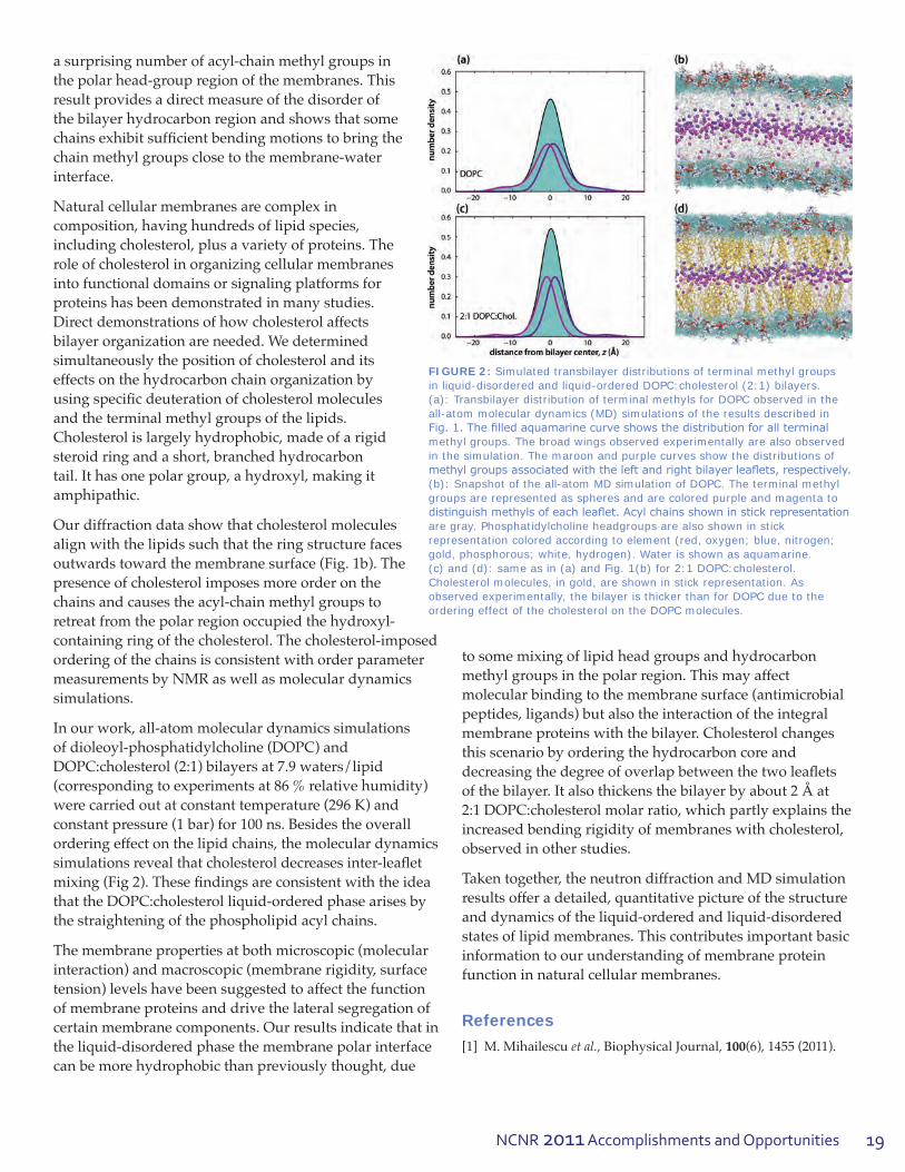
We use neutron diffraction as a tool for resolving the time-average distribution of atoms in a thermally disorderedlipidbilayerbyemployingspecificdeuterium labeling. Bragg diffraction from oriented lipid multilayers is converted, via Fourier analysis, intoone-dimensionaldensityprofilesofthebilayeralong the z-axis, normal to the bilayer surface. Thedensityprofilesrevealthebilayer’smoleculararchitecture. There are peaks for the phosphate-carbonyl groups, and a trough at z = 0, corresponding to the terminal methyl groups of the acyl chains (blackcurve,Fig.1a).Specificdeuterationoftheacylchain terminal methyl groups is used as a direct and quantitative method to determine the extent of chain disorder in the bilayer [1]. Hydration of the lipid in H2O and D2Oallowsfordeterminingthewaterdistributionassociatedwiththepolarhead-groups.Deuteriumdifferenceprofiles(lipiddeuteratedatthe terminal methyl groups vs. natural lipid) reveal
FIGURE 1: (a): Neutron diffraction results on the transbilayer distribution of lipid (black), terminal methyl groups (orange) and water (blue) in a DOPC bilayer. The scattering-length density profiles are on an absolute scale, up to an unknown factor (S), the area per lipid. (b): Transbilayer distribution of the terminal methyl groups in the liquid-ordered phase (2:1 DOPC:cholesterol) (brown, central peak) and the position and distribution of the first steroid ring of cholesterol (brown, side peaks). The brown curve was collected from a single lipid sample that contained cholesterol deuterated near the hydroxyl group and DOPC with deuterated terminal methyl groups. The black curves show the overall lipid profile in a bilayer without cholesterol (dashed) and with cholesterol (solid). Temperature: 21 °C, relative humidity: 86 %. The inset illustrates the disposition of the lipid molecules and cholesterol in the bilayer. Orange spheres indicate the deuterium sites.
19NCNR 2011 Accomplishments and Opportunities
a surprising number of acyl-chain methyl groups in the polar head-group region of the membranes. This result provides a direct measure of the disorder of thebilayerhydrocarbonregionandshowsthatsomechainsexhibitsufficientbendingmotionstobringthechainmethylgroupsclosetothemembrane-waterinterface.
Natural cellular membranes are complex in composition, having hundreds of lipid species, including cholesterol, plus a variety of proteins. The role of cholesterol in organizing cellular membranes into functional domains or signaling platforms for proteins has been demonstrated in many studies. Directdemonstrationsofhowcholesterolaffectsbilayer organization are needed. We determined simultaneously the position of cholesterol and its effects on the hydrocarbon chain organization by usingspecificdeuterationofcholesterolmoleculesand the terminal methyl groups of the lipids. Cholesterol is largely hydrophobic, made of a rigid steroid ring and a short, branched hydrocarbon tail. It has one polar group, a hydroxyl, making it amphipathic.
Ourdiffractiondatashowthatcholesterolmoleculesalignwiththelipidssuchthattheringstructurefacesoutwardstowardthemembranesurface(Fig.1b).Thepresence of cholesterol imposes more order on the chains and causes the acyl-chain methyl groups to retreat from the polar region occupied the hydroxyl-containing ring of the cholesterol. The cholesterol-imposed orderingofthechainsisconsistentwithorderparametermeasurementsbyNMRaswellasmoleculardynamicssimulations.
Inourwork,all-atommoleculardynamicssimulationsofdioleoyl-phosphatidylcholine(DOPC)andDOPC:cholesterol(2:1)bilayersat7.9waters/lipid(corresponding to experiments at 86 % relative humidity) werecarriedoutatconstanttemperature(296K)andconstant pressure (1 bar) for 100 ns. Besides the overall ordering effect on the lipid chains, the molecular dynamics simulationsrevealthatcholesteroldecreasesinter-leafletmixing(Fig2).ThesefindingsareconsistentwiththeideathattheDOPC:cholesterolliquid-orderedphasearisesbythe straightening of the phospholipid acyl chains.
The membrane properties at both microscopic (molecular interaction) and macroscopic (membrane rigidity, surface tension) levels have been suggested to affect the function of membrane proteins and drive the lateral segregation of certain membrane components. Our results indicate that in the liquid-disordered phase the membrane polar interface can be more hydrophobic than previously thought, due
to some mixing of lipid head groups and hydrocarbon methyl groups in the polar region. This may affect molecular binding to the membrane surface (antimicrobial peptides, ligands) but also the interaction of the integral membraneproteinswiththebilayer.Cholesterolchangesthis scenario by ordering the hydrocarbon core and decreasingthedegreeofoverlapbetweenthetwoleafletsofthebilayer.Italsothickensthebilayerbyabout2Åat2:1DOPC:cholesterolmolarratio,whichpartlyexplainstheincreasedbendingrigidityofmembraneswithcholesterol,observed in other studies.
Taken together, the neutron diffraction and MD simulation results offer a detailed, quantitative picture of the structure and dynamics of the liquid-ordered and liquid-disordered states of lipid membranes. This contributes important basic information to our understanding of membrane protein function in natural cellular membranes.
References
[1] M. Mihailescu et al., Biophysical Journal, 100(6), 1455 (2011).
FIGURE 2: Simulated transbilayer distributions of terminal methyl groups in liquid-disordered and liquid-ordered DOPC:cholesterol (2:1) bilayers. (a): Transbilayer distribution of terminal methyls for DOPC observed in the all-atom molecular dynamics (MD) simulations of the results described in Fig. 1. The filled aquamarine curve shows the distribution for all terminal methyl groups. The broad wings observed experimentally are also observed in the simulation. The maroon and purple curves show the distributions of methyl groups associated with the left and right bilayer leaflets, respectively. (b): Snapshot of the all-atom MD simulation of DOPC. The terminal methyl groups are represented as spheres and are colored purple and magenta to distinguish methyls of each leaflet. Acyl chains shown in stick representation are gray. Phosphatidylcholine headgroups are also shown in stick representation colored according to element (red, oxygen; blue, nitrogen; gold, phosphorous; white, hydrogen). Water is shown as aquamarine. (c) and (d): same as in (a) and Fig. 1(b) for 2:1 DOPC:cholesterol. Cholesterol molecules, in gold, are shown in stick representation. As observed experimentally, the bilayer is thicker than for DOPC due to the ordering effect of the cholesterol on the DOPC molecules.

20
Biology
20
Neutron reflectometry studies of the Parkinson’s disease-related protein, α-synuclein, at the lipid bilayer interface C. M. Pfefferkorn1, F. Heinrich2,3, J. C. Lee1
Parkinson’sdisease(PD)isaprevalentage-relatedneurodegenerativediseasetypifiedby
restingtremors,slownessofmovement,rigidity,and impaired balance in patients. Intensive research is beginning to shed light on its pathological underpinnings. One clue into the molecular causes ofPDisaneuronalproteincalledα-synuclein (α-syn).HallmarksofPDincludethepresenceofinclusions(Lewybodies)inthecellsofbraintissues,composed mostly of aggregated or misfolded α-syn, and the loss of neurons that produce dopamine; moreover,geneticfindingslinkmutantsandover-expression of α-syntoearly-onsetPD[1].Sinceα-syn is unstructured in solution but converts to a β-sheet enriched,amyloidforminLewybodies,theselargepolypeptide structural rearrangements are associated inthecausesofPD.Inourwork,weinvestigatedthebinding of α-syntoalipidbilayeranditsinfluencesonmembranepropertiesusingneutronreflectometry(NR) [2].
The role of membranes in mediating α-syn (mis)folding is of particular interest because membranes are ubiquitous in vivo and the binding of α-syn to membranes in vitro both induces formation of secondary structure (unstructured → α-helix) and modulates the rate of α-syn aggregation. Notably, PDmutationsinα-syn can affect membrane binding, and oligomeric α-synhasbeenshowntodisruptmembraneintegrity,aprocesswhichcouldinitiatecell death [1]. Despite substantial research efforts regarding the interaction of α-synwithmodelmembranes,themolecularmechanismbywhichtheyinfluenceα-syn aggregation remains unclear. While ourpriorworkusingresidue-specificfluorescencespectroscopy examined the interaction of α-syn withsmallunilamellarvesiclesfromtheperspectiveoftheprotein[3],togaininsightsintohowα-syn protein association affects membrane properties, a simultaneous probe of both α-syn protein and membranestructureisrequired.Hence,wehaveemployed NR and a surface-stabilized sparsely tetheredbilayerlipidmembrane(stBLM)[4]towards
thisobjective.UsingNR,thedepthprofileofthedifferentinterface layers (e.g., lipid headgroups and α-syn) normal tothebilayersurfacecanbereadilymeasured.Herein,wedescriberesultsinwhichNRwasexploitedtoelucidateboth membrane-bound α-syn and bilayer structure.
Figure 1 provides a schematic diagram that highlights some of the key features of α-syn primary sequence. There are seven N-terminal imperfect membrane binding amino acid amphiphatic repeats (i.e., containing both hydrophobic and hydrophilic ends). Also, there is the highly hydrophobic and aggregation-prone central region called the non-amyloid β component (NAC).
ThestBLMwascomposedofa1:1molarratioof1-palmitoyl-2-oleoyl-sn-glycero-3-phosphate(POPA):1-palmitoyl-2-oleoyl-sn-glycero-3-phosphocholine(POPC).ThisPOPA:POPClipidcompositionwaschosenbothtofacilitatecomparisontoourpriorwork[3]andbecauseα-synhaspreferentialaffinityforanionic(POPA)lipidheadgroups.UsingthisstBLM,NRdatawerecollectedinthe presence and absence of α-syn using the NCNR NG1 reflectometer[5].
ThestBLMwasfirstmeasuredinaqueousbuffer(10mmol/Lsodiumphosphate,100mmol/LNaCl,pH7.4)containing three isotopically different compositions (100 % D2O, 100 % H2O,and2:1H2O:D2O),followedbyaqueousbuffers containing α-syn(3μmol/Lin10mmol/Lsodiumphosphate,100mmol/LNaCl,pH7.4ineitherH2O or D2O).Becauseoftheuniqueflowcellsamplechamberdesign,allowingforinsitusolventexchange,itispossibleto perform successive measurements on the same sample (bilayer) area.
FIGURE 1: Schematic representation of α-syn, with the seven imperfect amino acid repeats in green (KXKEGV) containing both hydrophobic and hydrophilic (amphiphatic) residues, the non-amyloid beta component region in orange, (NAC), and the three disease related α-syn mutations in purple, (A30P, E46K, A53T).
1National Institutes of Health, Bethesda, MD 208922Carnegie Mellon University, Pittsburgh, PA 152133NIST Center for Neutron Research, National Institute of Standards and Technology, Gaithersburg, MD 20899

21NCNR 2011 Accomplishments and Opportunities
UsingbestfitstoNRdatabasedonacompositionspacemodel[6],across-sectionalprofileofthedifferentinterfacelayersalongwithotherfitparameterssuchasscatteringlengthdensity(nSLD)andα-synvolumeoccupancywereextractedandareshowninFig.2.AschematicviewofthestBLMwithα-syn superimposed is also provided in Fig. 2 for reference. In the absence of α-syn protein, values for theinnerandouterbilayerleafletthicknessesfallwithinexpected values.
Upon α-syn addition, NR data indicate that it indeed bindstothestBLMatanoccupancylevelof≈15%volumefractioninboththeouterleafletheadgroupsandhydrocarbons. Additionally, α-synwasfoundtoextendinto the bulk solvent region, indicating that the protein is partiallyanchoredtothestBLM.Thisisanewobservation,and one plausible explanation for it is that some portion of the α-syn polypeptide inserts into the hydrocarbon region whiletheremainingsegmentisexposedtothesolvent.Amodelconsistentwithpublisheddatawouldbethat
thehighaffinitymembranebindingsite,theN-terminus,serves as the membrane anchor [3]. Interestingly, the thicknessoftheproteinregionintheouterleafletofthestBLM(≈13Å)isconsistentwiththemembrane-boundα-syn adopting an α-helicalconformation,whichisinaccordwithsolutionstudiesconductedwithvesicles.Finally, the binding of α-syntothestBLMalsoresultedinsizeablereduction(>1Å)inmembranebilayerthickness.WhilethecompletenessofthestBLMwasmaintainedduringthecourseofourexperiments,weproposethatmembranethinningcouldrepresentafirststepleadingtodisruptionorevenpermeabilizationofthebilayer,whichhavebeenlinkedtoPDpathogenesis.
By successfully collecting high resolution NR data simultaneously for both membrane-bound α-syn and the stBLM,wehavegainednewinsightsintotheorientationand conformation of α-syn at the membrane interface as wellasapotentialclueintohowmembrane-boundα-syn initiates membrane perturbation. It is plausible that the observed membrane induced α-syn conformation and subsequentmembranethinningcouldsetthestageforPDprogression. In our continued effort to gain a molecular understanding of the role of membranes and α-syn in PD,weaimtoelucidatetheeffectsofdifferentsolutionconditions on α-syn membrane-bound structure and membrane induced perturbations.
References [1] K. Beyer, Cell Biochem. Biophys. 47, 285 (2007).[2] C.M.Pfefferkorn,F.Heinrich,J.C.Lee,(tobesubmitted).[3] C.M.Pfefferkorn,J.C.Lee,J.Phys.Chem.B114, 4615 (2010).[4] D. J. McGillivray, G. Valincius, D. J. Vanderah, W. Febo-Ayala,
J.T.Woodward,F.Heinrich,J.J.Kasianowicz,M.Lösche,Biointerphases 2, 21 (2007).
[5] J.A.Dura,D.J.Pierce,C.F.Majkrzak,N.C.Maleszewskyj,D.J.McGillivray,M.Lösche,K.V.O’Donovan,M.Mihailescu,U.Perez-Salas,D.L.Worcester,S.H.White,Rev.Sci.Instrum.77, 074301 (2006).
[6] M.Schalke,P.Kruger,M.Weygand,M.Losche,Biochim.Biophys. Acta, Biomembr. 1464, 113 (2000).
FIGURE 2: Simplified molecular distributions for organic interface layers of the 1:1 POPA:POPC stBLM in the presence of α-syn (3 μmol/L in 10 mmol/L sodium phosphate, 100 mmol/L NaCl, pH 7.4) obtained from the best-fit of reflectivity data to the composition space model. A reference value for the area per lipid is indicated by the dotted line. Data for Si substrate and the SiOx, Cr, and Au layer have been partially omitted. A corresponding schematic of the stBLM is also provided for reference.

22
Biology
22
SANS and modeling reveal changes in flexibility of a protein important for DNA replicationS. Krueger1, J.-H. Shin2, S. Raghunandan1, J.E. Curtis1, Z. Kelman3
1NIST Center for Neutron Research, National Institute of Standards and Technology, Gaithersburg, MD 208992Kyungpook National University, Daegu 702-701, Republic of Korea3Cell Biology and Molecular Genetics, University of Maryland, College Park, MD 20742 and Institute for Bioscience and Biotechnology Research, University of Maryland, Rockville, MD 20850
Duringreproduction,DNAisseparatedintotwostrandstobereplicated.Howisthisdone?There
are special proteins (helicases) that carry out this task, butthedetailsoftheprocessremaintobeworkedout. One type of helicase is the minichromosome maintenance(MCM)proteinwhichhasmanymovingparts(domains).InthishighlightwedescribeSANS measurements and computer modeling studies on the N-terminal domain of a MCM protein from the microorganism Methanothermobacter thermautotrophicus (N-mtMCM).Thisworkprovidesstructural information that supports the previously reported biochemical observations that large domain motions are required for the activation of the MCM helicase.
A high-resolution x-ray crystal structure [1] of N-mtMCMshowsthatitisaproteinwitha12subunit(dodecamer)structure,witheachmonomericsubunit consisting of three domains. Domain A is suggested to play a role in regulating helicase activity, domain B participates in DNA binding, and domain C is involved in the assembly of the proteinsubunits(multimerization)aswellasDNAbinding and is necessary for helicase activity [2]. An unstructured loop region consisting of 20 amino acid residues(89to108)existsinN-mtMCMbetweendomains A and C. A second unstructured region consisting of 43 amino acid residues (244 to 286) is located at the C-terminus of N-mtMCM. This regionwouldbeconnectedtothecatalyticdomainin the full mtMCM molecule. Electron microscopy (EM) studies of N-mtMCM revealed a six subunit (hexameric)structure[3],whichisdifferentfromthedodecamer suggested by the x-ray crystal structure and by biochemical characterization of the protein in solution [4]. Since neither EM nor crystal structure determination reveals the structure of a protein in solutionwhereitisatfullhydrationunderconditionsclosertoitsnaturalstate,theSANSstudieswereundertaken to provide complementary structural information.
SANSdatawereobtainedfromN-mtMCMintheabsenceof DNA and in the presence of a single-stranded DNA molecule consisting of 50 nucleotides (nt). Fits to the data for N-mtMCM in the absence of DNA resulted in a radius of gyration, Rg,valueof78ű1Å.TheRg value for the N-mtMCM/DNAcomplexwas69ű1Å,indicatingthatthereisasignificantdecreaseinthesizeoftheN-mtMCMmoleculewhenthe50ntDNAisbound.Asastartingpointfor the modeling, an all-atom structure of the N-mtMCM dodecamermoleculewasbuiltbasedonthex-raycrystalstructure,withresidues244to286addedtotheC-terminusofeachmonomer.TheSASSIEsoftwarepackage[5]wasused to generate ensembles of structures for comparison to SANS data by randomly varying amino acid residue backbonedihedralangleswithinchosenregionsoftheprotein.Aftereachrandomlychosenanglewasrotatedbyagivenvalue,thefinalvaluewascheckedtodetermineifitwasenergeticallyprobable.Thenewconfigurationwascheckedforoverlapofbasisatoms,usuallychosentobealpha-carbonatoms.Ifbothchecksweremet,thenewstructurewasaccepted.
ThemeasuredSANSdatawerecomparedtotheoreticalSANScurvescalculatedfromtwodifferentseriesofaccepted structures using the c2testforgoodnessoffit.AnexampleofeachofthesestructuresisshowninFig.1.Inbothcases,domainAisshowninyellowanddomainsBand C are in blue. The unstructured loop region (residues 89to108)betweendomainsAandCineachmonomerisshowningreenandtheunstructuredregionattheC-terminusofeachmonomer(244to286)isshowninpurple.Inthefirstseriesofstructures(Fig.1a),residues244to286weretreatedasunstructuredflexibleregionsinwhichthedihedralangleswerevariableasdescribedabove.Thesecondseriesofstructures(Fig.1b)definethissameregiontobeflexible,aswellasresidues89to108betweendomainsAandC.
Abest-fitfamilyofstructureswaschosenforeachofthesamplesbasedonthelowest≈20%ofthec2 values for eachseries.Thelowestc2valuesrangebetween0.73and1.5,exceptfortheMCMsamplewithoutDNAcomparedto

23NCNR 2011 Accomplishments and Opportunities
thefirstseriesofstructures,wherethevaluesarebetween3.6and3.9.Figure2showstheSANSdatafromtheMCMsampleswithandwithoutDNA,alongwiththreerepresentativebest-fittheoreticalcurvesfromthefirstseriesofstructures.ByinspectionitcanbeseenthatworstpartofthefittothedatafromthesampleswithoutDNAoccursforq>0.07Å-1. This leads to the conclusion that structures that donotallowresidues89to108tobeflexiblearenotagoodrepresentationoftheMCMsamplewithoutDNA.Ontheother hand, the c2analysisshowsthatequallygoodfitscanbeobtainedfortheDNA-boundsamplewhetherornotthisregionismodeledasflexible.
From the comparison of the theoretical SANS curves for eachofthetwoseriesofstructurestotheSANSdata,itisclear that the MCM sample in the absence of DNA must havebothflexibleC-terminalandN-terminalregionsinordertofittheSANSdata.Thisflexibility,whileincontrasttothemorerigidcrystalstructure,isconsistentwiththe
EM and biochemical studies of the MCM proteins, suggesting thatsignificantmovementofdomain A is required prior to the initiation of MCM activity and DNA replication. The fact that equallygoodfitsoftheDNA-bound sample can be obtained whethertheN-terminalresiduesareflexibleornotissignificant,asthismeansthattheMCM/DNAcomplex can have this more rigid structure,whiletheunboundsample cannot. The data presented here demonstrate that SANSanalysiscombinedwithenergetically relevant all-atom ensemble structure modeling isapowerfultoolthatallowsthe assessment of the degree
offlexibilityofproteinsinsolution,evenincaseswherean ensemble of structures is likely present. The methods developed here for N-mtMCM solution structure modeling can likely be utilized for other large, multimeric protein complexeswithunstructuredflexibleregions.
References[1] R.J.Fletcher,B.E.Bishop,R.P.Leon,R.A.Sclafani,C.M.Ogata,
X.S. Chen, Nature Struct. Biol. 10, 160 (2003).[2] N.Sakakibara,L.M.Kelman,Z.Kelman,Mol.Microbiol.72,
286 (2009).[3] Y.-J.Chen,X.Yu,R.Kasiviswanathan,J.-H.Shin,Z.Kelman,
E.H. Egelman, J. Mol. Biol. 346, 389 (2005).[4] R.Kasiviswanathan,J.-H.Shin,E.Melamud,Z.Kelman,
J. Biol. Chem. 279, 28358 (2004).[5] J.E. Curtis, S. Raghunandan, H. Nanda, S. Krueger, Computer
PhysicsComm.,accepted(2011);http://www.smallangles.net/sassie/SASSIE/SASSIE_HOME.html.
FIGURE 1: Examples of two series of model structures for N-mtMCM. Domain A is shown in yellow and domains B and C are in blue. An unstructured loop region between domains A and C is shown in green and a second unstructured region at the C-terminus is shown in purple. a) Only the C-terminus unstructured region (purple) is allowed to be flexible. b) Both unstructured regions are allowed to be flexible.
FIGURE 2: log(I) vs log(q) plots of the measured SANS data for the samples without and with DNA bound, along with representative examples of best fit theoretical SANS curves from the ensemble of structures in which only the C-terminus unstructured region is allowed to be flexible (Fig. 1a.)

24
Engineering
24
Residual stresses and mechanical damage in gas pipelinesT. Gnäupel-Herold1,2, R. Batisse3, L. Clapham4
1University of Maryland, College Park, MD 207422NIST Center for Neutron Research, National Institute of Standards and Technology, Gaithersburg, MD 20899-61023GDF SUEZ, Saint-Denis la Plaine, France4Queen’s University, Kingston, Ontario, K7L 3N6 , Canada
Pipeline failure comes into the focus of public attention because of loss of human life or from
great environmental damage, be it from release of crude oil or from natural gas. These events appear to be rare because large, catastrophic releases > 104 m3ofcrude(orgas:× 103) of hydrocarbons are rare compared to the much more frequent (> 102/year in Canada alone) small to medium releases [1]. Inherent ruptures tend to be the main cause of large ruptures, but third party damage is the cause of 40 % of pipeline ruptures. Mechanical damage withoutimmediateruptureduetoconstructionor agricultural activity often occurs, and it raises acrucialquestion:Howcriticalisthedamageandwhatactionsneedtobetaken?Inordertoaddressthisquestiondamagehastobequantifiedinsomemanner,andMagneticFluxLeakage(MFL)isoneofthe tools the industry uses for that purpose. In this methodaMFLproberecordschangesinthemagneticfieldbetweentwomagneticpolesthatareinclosecontactwiththepipewall.Dentsandgougescreatechanges in geometry, plastic strains and residual stresses;allofwhichseverelyaltertheMFLsignalinwaysthatarenotcompletelyunderstood.Geometrychangesaremorereadilyquantifiablebuttheresidualstresscontributionisdifficulttodeterminedue to the lack of experimental data and reliable modeling results. Some progress has been made by annealing the sample, thus eliminating the residual stresscontribution[2].However,thiscannotaddressquestions about the role of the multiaxiality of stresses, sign changes of stresses and stress gradients, both laterally and through the thickness [3]. Thus, it is necessary to measure the residual stresses non-destructively, and the only technique capable of handling large pipe sections due to problems of weight,dimensionsandwallthicknessisneutrondiffraction. It is obvious that, in order to put such measurementsonasoundfooting,onecannotwaitfor such damage to occur and then remove that pipe section, and perform the necessary measurements. Instead, the various type of gouges and dents that are encountered on real pipelines need to be produced
in a controlled and repeatable fashion. By varying the testing speed, the deformation path, and the tool shape itispossibletocoverawiderangeofreal-worlddamagescenarios(Fig.1)thatcombinethetwomajorelementsofgouging(whichremovesmaterialandhassomeplasticdeformation)anddenting(whichhasplasticdeformationonly).
Figure 1 illustrates the unique challenges to the stress measurementpresentedbythisapproach:First,thedamagedregionswith300mmdentlength(seecircled30 cm mark in Fig. 1, upper right corner), and 130 mm gouge length, respectively, are unusually large. Second, the pipe should stay intact circumferentially, and retain sufficientlengthaxiallyasmuchaspossibleinordertopreservethestressfield.Theresultingsampleswerepipesections 650 mm in diameter, 500 mm in length, and 8 mmwallthickness.Thewallthicknessisquitelow,andinordertosamplethestressesinsufficientdetailthroughthe thickness the spatial resolution of the measurement –inotherwords,thesizeoftheneutronbeam–mustbechosenappropriately(here:1mm).Asidefromsomebasicsymmetry considerations – e.g.,thestressfieldispresumedto be laterally symmetric – there is not much one can assume about the stresses, thus creating the necessity for
FIGURE 1: Left: testing equipment at GDF SUEZ, France. Right: types of damage. Denting merely deforms but does not remove material as gouging does. BEA161 (gouge) in the lower right corner was investigated here; BEA182 is similar to the dent investigated here.

25NCNR 2011 Accomplishments and Opportunities
measurements at numerous locations in the defect area and through the thickness.
AsshowninFig.2,measurementsalongthelineofsymmetryshowthatthestressescreatedbythetwodefecttypesexhibitsignificantchangesinmagnitude,particularlynear the start and end of the defects. The high tensile hoop stressesneartheendofthedentarearedflagbecausethetotal stress in a pressurized pipeline– residual stress plus stress from pressure – may reach the tensile strength of the material. The danger of pipe rupture arises if the high stresslevelsarecombinedwithaweakenedcrosssectionat the location of the peak stress due to large strains (dent) ormaterialremoval(gouge).However,thestressesmustalsobeconsideredinthecontextofMFLmeasurements,particularlyinviewofthelateralresolutionofMFLprobeswhichcanbeoftheorderof1mm.TheMFLsignalisaweightedaveragethroughthethickness,whichiswhyitisdifficulttoassignadepthlocationtoasub-surfacedefect.Rather, the interplay of gradients in different directions createsacumulativeeffectwheredifferentcombinationsofstressescanproducethesimilarMFLsignals.Thus,thecontribution of different through-thickness stress gradients toMFLsignalsisofparticularinterest.AsshowninFig.3 stress gradients near the surface are most severe in the lateraldirectionawayfromthedefectwhereoveralengthof5mmstresseschangebymorethan300MPa.Thestressdifferential in the radial direction (through the thickness) is of similar magnitude.
Aside from aiding the interpretation of signals from MFLprobesthesefindingsalsohelpinaddressingsafetyconcerns.Pressureinapipelinesuperimposesastresson the residual stresses, and the total stresses should not exceedcertainlimitssetbythesafetyfactor(between1.5 and 10). What problems could arise if creating the mechanical damage in the pipes did not cause immediate
piperupture?Ononehand,theselimitsareexceededinpipelineswithlowsafetyfactorsbothinregionsofpeaktensilestressesandwherethecrosssectionisreducedthemost. On the other hand, the mechanical damage creates workhardeningwhichraisesthestrengthlocally.Theendresultcouldbeviewedasalocallyreducedsafetyfactorthat in extreme cases may require the reduction of the operating pressure in the pipeline. Another damage-related problem to consider is the increased risk of stress corrosion cracking assisted both by high tensile stresses and coating failureinthedent/gougeregion.
These measurements are part of an ongoing international projectinwhichdifferentpipelinedefectsareproducedunderrealisticconditions,andthencharacterizedwithrespecttoresidualstressesandMFLsignaldistribution.Theneutrondiffractionmeasurementsarethefirstofthis type and they revealed complex stress patterns both through the thickness and in the lateral directions. It is one of the goals of these efforts to use the neutron data in ordertocreateafunctioningmagneticmodelthatallowsthe separation of the geometry effect and the stress effect onthemagneticfluxleakagesignal,thusgreatlyenhancingthescopeofMFLprobesasanin-lineinspectiontool.
References
[1] PipelinePerformanceinAlberta1980-1997,AlbertaEnergyandUtilitiesBoard1998,availableat:http://www.eub.gov.ab.ca
[2] L.Claphamet al.,Proc.2004Int.PipelineConference,Calgary,983 (2004).
[3] P.A.Ivanovet al., IEEE Transactions on Magnetics, 34, 3020 (1998).
FIGURE 2: Comparison of hoop residual stresses in the gouge and the dent. Dots represent measurements through the thickness. Note the high tensile stresses outside near the end of the dent. Additional tensile stresses are superimposed in pressurized pipelines.
FIGURE 3: Stresses in the center region of a plate sample that was cut from a pipe to allow for measurements in greater number and detail.
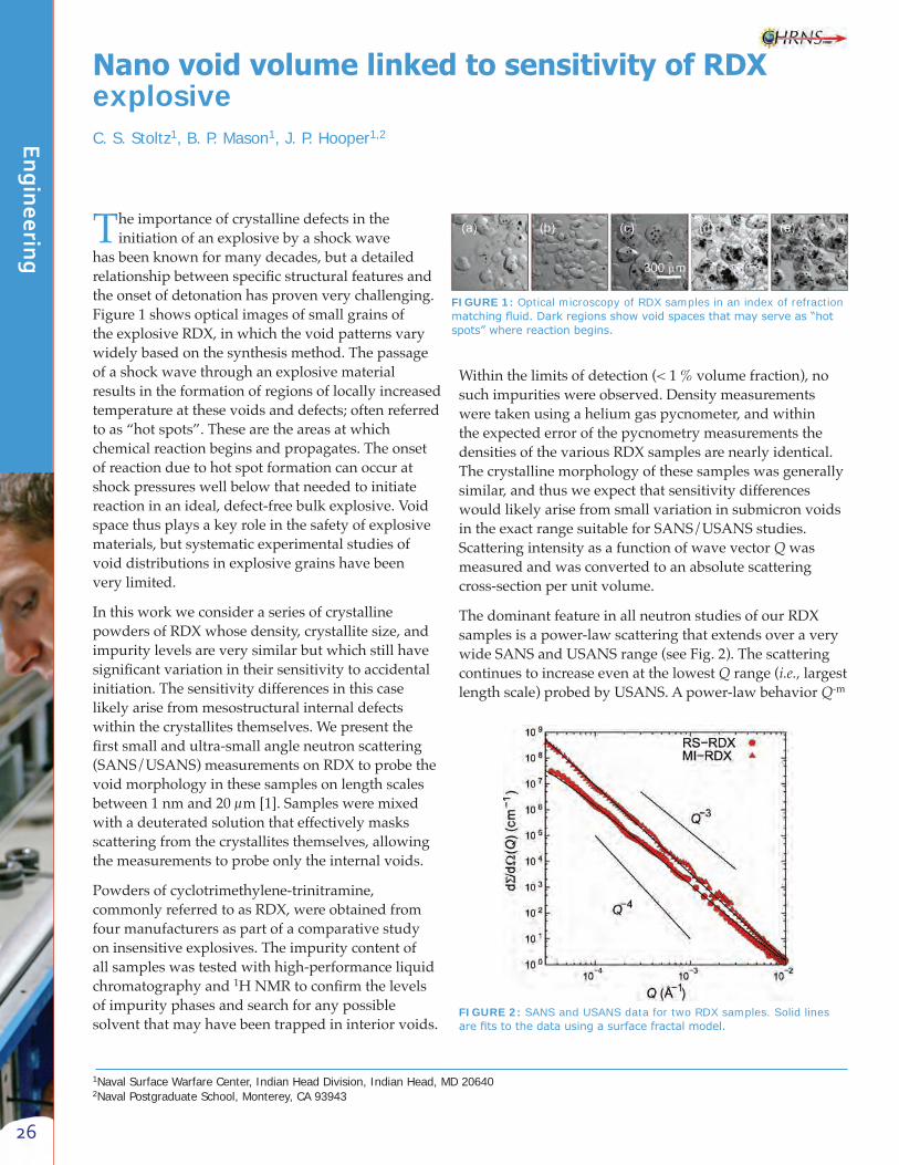
26
Engineering
26
Nano void volume linked to sensitivity of RDX explosive C. S. Stoltz1, B. P. Mason1, J. P. Hooper1,2
1Naval Surface Warfare Center, Indian Head Division, Indian Head, MD 206402Naval Postgraduate School, Monterey, CA 93943
The importance of crystalline defects in the initiationofanexplosivebyashockwave
hasbeenknownformanydecades,butadetailedrelationshipbetweenspecificstructuralfeaturesandthe onset of detonation has proven very challenging. Figure1showsopticalimagesofsmallgrainsoftheexplosiveRDX,inwhichthevoidpatternsvarywidelybasedonthesynthesismethod.Thepassageofashockwavethroughanexplosivematerialresults in the formation of regions of locally increased temperature at these voids and defects; often referred toas“hotspots”.Thesearetheareasatwhichchemical reaction begins and propagates. The onset of reaction due to hot spot formation can occur at shockpressureswellbelowthatneededtoinitiatereaction in an ideal, defect-free bulk explosive. Void space thus plays a key role in the safety of explosive materials, but systematic experimental studies of void distributions in explosive grains have been very limited.
InthisworkweconsideraseriesofcrystallinepowdersofRDXwhosedensity,crystallitesize,andimpuritylevelsareverysimilarbutwhichstillhavesignificantvariationintheirsensitivitytoaccidentalinitiation. The sensitivity differences in this case likely arise from mesostructural internal defects withinthecrystallitesthemselves.Wepresentthefirstsmallandultra-smallangleneutronscattering(SANS/USANS)measurementsonRDXtoprobethevoid morphology in these samples on length scales between1nmand20μm[1].Samplesweremixedwithadeuteratedsolutionthateffectivelymasksscatteringfromthecrystallitesthemselves,allowingthe measurements to probe only the internal voids.
Powdersofcyclotrimethylene-trinitramine,commonlyreferredtoasRDX,wereobtainedfromfour manufacturers as part of a comparative study on insensitive explosives. The impurity content of allsampleswastestedwithhigh-performanceliquidchromatography and 1HNMRtoconfirmthelevelsof impurity phases and search for any possible solvent that may have been trapped in interior voids.
Within the limits of detection (< 1 % volume fraction), no suchimpuritieswereobserved.Densitymeasurementsweretakenusingaheliumgaspycnometer,andwithinthe expected error of the pycnometry measurements the densities of the various RDX samples are nearly identical. Thecrystallinemorphologyofthesesampleswasgenerallysimilar,andthusweexpectthatsensitivitydifferenceswouldlikelyarisefromsmallvariationinsubmicronvoidsintheexactrangesuitableforSANS/USANSstudies.ScatteringintensityasafunctionofwavevectorQ wasmeasuredandwasconvertedtoanabsolutescatteringcross-section per unit volume.
The dominant feature in all neutron studies of our RDX samplesisapower-lawscatteringthatextendsoveraverywideSANSandUSANSrange(seeFig.2).ThescatteringcontinuestoincreaseevenatthelowestQ range (i.e., largest lengthscale)probedbyUSANS.Apower-lawbehaviorQ-m
FIGURE 1: Optical microscopy of RDX samples in an index of refraction matching fluid. Dark regions show void spaces that may serve as “hot spots” where reaction begins.
FIGURE 2: SANS and USANS data for two RDX samples. Solid lines are fits to the data using a surface fractal model.

27NCNR 2011 Accomplishments and Opportunities
isobserved,wherem variesbetween3.1and3.6.Exponentsin this range generally correspond to fractal scattering from surfaceswithadimensionalityD =6-mbetween2and3[2]. The data suggests internal fractal surfaces are present inRDXoverawiderangeoflengthscales,from ≈ 50 nm upto≈20μm.NoregionofQ-4 behavior is observed in any sample;suchapowerlawwouldindicatescatteringfromthesurfaceofasmooth3DobjectofdimensionalityD = 2.
Akeyquestionintheareaofexplosivesensitivityiswhatsize a void must be to become a hot spot during initiation. PreviousworkonpowdersoftheexplosiveHMXhadsuggested that polydisperse spherical voids on the order of 3 nm to 60 nm may be present and important for sensitivity [3];wefindnoevidenceofsuchfeaturesinourRDXsamples. Around 10 nm to 30 nm the intensity of the fractal surface scattering becomes comparable to the background, and there is no evidence of any features arising from spherical scatterers.
Tocomparebetweenthedifferentexplosivesamples,wehavecalculatedtheneutronscatteringinvariant,whichinvolves an integration of the cross section over Q and is related to the overall volume fraction of void space. In Fig.3weplotacorrelationbetweenthesensitivityoftheexplosivemeasuredbythelarge-scalegaptest(LSGT)andtheneutronscatteringinvariant.Wefindthat,thoughthe pycnometry densities of the samples are similar, the invarianttrackswiththeshocksensitivityvaluesderivedfromthelarge-scalegaptests.Thus,withothermaterialproperties held constant to the extent possible, the internal void space on length scales from 60 nm to 20 μm appearstobewellcorrelatedwithmacroscopicchangesinsensitivityoftheexplosiveRDX.Whileonewouldexpect this trend based on general theories of hot spot formationinexplosives,directexperimentallinksbetweenspecificmesostructuralcrystalfeaturesandaccurateshocksensitivity tests have been limited.
What is the origin of the fractal scattering in the explosive samples?Onepossibilityisthatthesefractalregionsarisefromacrystalgrowthprocessthatoccursonthesurface of interior voids as solvent diffuses out of the RDX crystals.Thecommercialsamplesusedweresubjectedto thermal cycling during the manufacturing and post-productionprocessesthatwouldencouragediffusionofany solvent remaining in the void space. Small amounts ofRDXdissolvedwithintheinternalsolventwouldgraduallyagglomerateonthevoidswalls,similartootheraggregationprocessesleadingtofractalgrowthpatterns.RecentobservationsbyMelingerandcoworkers[4]ofacomplex dendritic crystal structure formed during drop-castgrowthofmicrogramquantitiesofRDXonanAusurface may provide support for this hypothesis.
Insummary,SANSandUSANSmeasurementswereperformedonRDXpowderstocharacterizetheinternalvoidfeaturesonlengthscalesbetween1nmand20μm.ExperimentswereperformedonfiveRDXsampleswitharangeofsensitivitiestoaccidentalshockinitiation. The neutron scattering for all samples exhibits afractalbehaviorindicativeofsurfacefractalswithadimensionalitybetween2.4and2.9.Thefractalbehaviorextends up to the maximum length scales measured in our USANS experiments, indicating a fractal correlation length on the order of 30 μm or higher. No evidence of distinct nanometer-scale void distributions is observed. The neutron data are interpreted in terms of surface scattering frominternalvoids,whosefractalcharactermayarisefromcrystallitegrowthonthevoidwallsassolventdiffusesoutfromthesystem.Thescatteringinvariant,whichservesasa relative comparison of the overall amount of scattering as wellasageneralmeasureofthevolumefractionofinternalvoids,differswidelybetweenthedifferentRDXsamplesandtrackswellwiththemeasuredshocksensitivities.Despite similarities in density and particle distribution, neutronscatteringrevealscleardifferencesbetweensamplesthatofferadirectlinkbetweenstructuralfeaturesand sensitivity testing.
References
[1] C.S.Stoltz,B.P.Mason,andJ.Hooper.J.Appl.Phys.107 (10) 103527 (2010).
[2] J. Teixeira, J. Appl. Cryst. 21, 781 (1988).[3] J. T. Mang et al., J. Mater. Res. 15, 1199 (2000).[4] J. S. Melinger et al., Appl.Phys.Lett.93, 011102 (2008).
FIGURE 3: The susceptibility to accidental initiation as measured by the large-scale gap test (LSGT) is plotted versus the neutron scattering invariant calculated from the SANS and USANS data. The invariant is a general measure of the volume fraction of void space. As void space in the 60 nm to 20 μm range increases, less shock pressure is needed to induce the explosive to detonate.
Biology
Chem
ical Physics
28
Density hysteresis in nanoconfined waterY. Zhang1,2, A. Faraone3,4, W.A. Kamitakahara3, K-H. Liu5, C-Y. Mou5, J.B. Leão3, S. Chang3, S-H. Chen1
1Massachusetts Institute of Technology, Cambridge, MA 021392Oak Ridge National Laboratory, Oak Ridge, TN 378313NIST Center for Neutron Research, National Institute of Standards and Technology, Gaithersburg, MD 208994University of Maryland, College Park, MD 207425National Taiwan University, Taipei 106, Taiwan
Thepropertiesofwaterhavebeenexploredbymany techniques, including neutron scattering.
Despite a huge research effort, basic gaps in our understanding persist, especially in the supercooled regionwherewaterremainsintheliquidstatebelowthemeltingpoint.Aparticularlyinterestingand controversial hypothesis posits that, across a rangeoftemperaturesandpressures,bulkwateriscomposedofamixtureoftwostructurallydistinctliquids:alow-densityliquid(LDL)andahigh-densityliquid(HDL).Thelatteriswaterofoureverydayexperience,whereastheformerischaracterized by a local intermolecular bonding environment much more like that of solid ice, whichleadstoalowerdensity.Acentralquestioniswhetherafirst-orderliquid-liquidphasetransitionbetweenLDLandHDLexists,asathermodynamicextensionofthefirst-ordertransitionestablishedintheamorphoussolidwaters[1-3].Anexperimentaldeterminationofwater’sdensityinthedeeplysupercooledregionisperhapsthemostdirectwayofinvestigating the hypothesized liquid-liquid phase transition.Byundertakingsuchmeasurements,weareseekingevidenceofafirst-orderliquid-liquidphasetransitionofwaterthatwouldexistinthemacroscopicsystemifitwerepossibletoavoidcrystallization [4].
An experimental challenge arises because the hypothesized transition exists in a region of the phasediagraminwhichbulkwatercannotexistina liquid state [1]. One method of overcoming this difficultyistotakeadvantageofconfinement.Byconfiningwaterinananoporoussilicamatrix,MCM-41-Swith15Åporediameter,thehomogeneousnucleation process (crystallization) can be avoided, allowingustoenterthisregion.Thereisstillmuchdebateonthedifferencesandsimilaritiesbetweenbulkandconfinedwater.However,evenifthesilicamatrix,withitshydrophilicsurfaces,mightchangebasicpropertiesofwaterbeyondsuppressing
crystallization,thepropertiesoftheconfinedwaterareimportantintheirownright,sincetheyarerepresentativeof many environments of interest in biology and geology wheresimilarhydrophilicinterfacesarepresent.
The contrast of the neutron coherent scattering length density(SLD)ofheavywateragainstthatofthesilicamatrixgivesrisetoawell-defineddiffractionpeakarisingfromthe(10)planeofa2-Dhexagonallatticeofwatercylinders in the grains of MCM-41-S silica matrix. Once the data are corrected for the temperature-independent background arising from the fractal packing of the MCM-41-S crystallites and the incoherent scattering, the only temperature-dependent quantity is the height of the Bragg peak,whichisproportionaltothesquareofthedifferenceofSLDbetweentheheavywaterandthesilicamatrix,andtherefore a sensitive indicator of the average mass density oftheconfinedwater.
Figure1showsthehypothesizedphasediagramsoflowtemperaturewaterinthepresenceofafirst-orderHDL-LDLtransition.Whentheequilibriumphaseboundaryis crossed, due to the metastability or the kinetics of the phase transition, the phase separation may take a very long timetohappen,especiallyinconfinement[5].However,afirst-orderlineshouldstillmanifestitselfwithasignificanthysteresiswhenitiscrossedfromoppositedirectionsofthetransitionline,showninFig.1(b).Accordingly,weperformedaseriesofwarmingandcoolingscansoverarange of pressures.
Figure2showsthemeasureddensityofconfinedD2Owithbothcoolingandwarmingscansataseriesofpressures.Thefactthatthewarmingandcoolingcurvesjoinatbothhighandlowtemperatureendsimpliesthattheexpansion-contractionprocessesarereversible.Upto100MPa,thedensitydifferencebetweenthecoolingandwarmingscansissmall,whichcouldbeattributedtothetemperaturelagwhenrampingthetemperaturecontinuously.Thedensity difference due to this reason is small and relatively independentofpressure.However,above100MPathedensity difference (hysteresis) opens up progressively

NCNR 2011 Accomplishments and Opportunities 29
as the pressure is increased. We expect the magnitude of the density difference might depend on the temperature ramping rate. Considering the feasibility of neutron scatteringexperiments,wechosearampingrateof0.2K/min.Withsuchaslowrate,aratheruniformtemperaturedistribution over the sample is assured, but the system may require much longer times to reach physical equilibrium after crossing a phase boundary.
Notethattheaccuracyoftheabsolutedensitywedetermined depends on the background subtraction and thescaling.However,therelativeshapeofthedensityprofilesisindependentoftheanalysis.Theobserveddensityhysteresis,althoughitisnotadefinitiveproof,isconsistentwiththehypothesisofthecoexistenceoftwoliquidphasesofconfinedheavywaterwithdifferentdensities,atpressureslargerthan100MPa.Wealsoobserved an unexpected sudden change of slope in the densityprofiles(a“kink”)atthetemperaturesofthemaximumslopeatandbelow100MPa.Thekinkmightbe related to the previously observed fragile-to-strong dynamiccrossoverofconfinedH2O [6].
UsingtheSPINStriple-axisinstrument,wewereableto obtain sensitive measurements of the density of D2OconfinedinMCM-41-S.Weobservedaprominenthysteresisphenomenoninthemeasureddensityprofilesbetweenwarmingandcoolingscansabove100MPa.Weinterpret this observation as support, although not proof, oftheproposedpresenceofafirst-orderliquid-liquidphasetransitionofwaterthatwouldexistinthemacroscopicsystem if crystallization could be avoided in the relevant phase region.
References
[1] O. Mishima and H. Eugene Stanley, Nature 396, 329 (1998).[2] P.G.Debenedetti,J.Phys.Condens.Mat.15, R1669 (2003).[3] C. A. Angell, Science 319, 582 (2008).[4] Y.Zhang,A.Faraone,W.A.Kamitakahara,K-H.Liu,C-Y.
Mou,J.B.Leão,S.Chang,andS-H.Chen,PNAS108, 12206 (2011).
[5] L.D.Gelb,K.E.Gubbins,R.Radhakrishnan,andM.Sliwinska-Bartkowiak,Rep.Prog.Phys.62, 1573 (1999).
[6] A.Faraone,L.Liu,C-Y.Mou,C-W.Yen,andS-H.Chen,J.Chem.Phys.121, 10843 (2004).
FIGURE 1: The hypothesized (a) T-P and (b) ρ-T phase diagrams of water in the presence of a first-order liquid-liquid phase transition. If a first-order HDL-LDL transition exists at high pressures, a hysteresis phenomenon should be observed, as shown by the difference between the red and blue curves in panel (b). At low pressures, when no first-order line is crossed, there should be no hysteresis (green curve).
FIGURE 2: The density profiles of confined D2O in a hydrophilic substrate MCM-41-S are measured in both warming and cooling scans. The data are shifted by 0.05 g/cm3 between adjacent pressures for clarity. A hysteresis phenomenon becomes prominent above 100 MPa. The two horizontal arrows indicate the locations of the sudden change of slope in the density profiles (“kink”) at 0.1 MPa and 100 MPa.

Biology
Chem
ical Physics
30
Cholesterol transport rates in model membranesS. Garg1, A.C. Woodka2, P.D. Butler2, U. Perez-Salas1,3, L. Porcar4,5
1Argone National Laboratory, Argonne, IL 604392NIST Center for Neutron Research, National Institute of Standards and Technology, Gaithersburg, MD 20899-61023University of Illinois at Chicago, Chicago, IL 606074Institut Laue Langevin, Grenoble Cedex 9, France5University of Delaware, Newark, DE 19716
Cholesterol is an essential structural component of most cell membranes
whicharecomposedofphospholipids,sterols,andproteins.Phospholipidsarethe main component of the membrane, actingasthescaffoldinwhichtheother components embed. Cholesterol contributes to the organization, dynamics, and bending modulus of the lipid membrane; helps regulate the membrane interactionswiththecytoskeleton;andin some cases controls the functions of membrane proteins crucial for several trans-membrane signaling processes. Healthy cells maintain a large cholesterol gradientwithonly0.05%to1%ofthecellularcholesterolresidingwhereitisgeneratedintheinteriorofthecell,while60%to70%residesintheplasmamembraneorcellwall.Thus,propercholesterolmovementwithinthecell,andindeedthroughoutthebody, is critical. Not only does cholesterol movement play a key role in atherosclerosis and heart disease, the leading causes of death in the US, it has been implicated indementias(suchasAlzheimer’s),diabetes,cancer,andseveral rare but fatal diseases.
Despitetheessentialroleofcholesteroltransportwithinthe cell, progress has been hampered by confusion associatedwiththewidevariationintransferratesreported in the literature. In particular, the transfer of cholesterol from one side of the lipid bilayer to the other, knownasflipping, has reported half-lives that range from severalhourstoafewmilliseconds.Apotentialsourcefor these discrepancies is that most techniques used to measuretheseratesrequirelabelingthecholesterolwithafluorescentdye.Theassumptionisthatsuchchemicallabelswillnotaffecttheratesbeingmeasured.Similarly,additives such as cyclodextrin and cholesterol oxidase areoftenintroducedtomeasurecholesterolflippingratesassumingthat,whiletheywillacceleratetheejectionofcholesterolfromtheouterlayerofthebilayer,theywillnotaffecttheflippingprocessfromtheinnerlayertothe outer layer. Finally, most of the studies of cholesterol exchange kinetics involve mixing cholesterol donor and acceptor vesicles requiring a periodic isolation step to assay the amount of cholesterol exchanged, introducing additional uncertainties.
Inthisstudy[1]wecircumventthesepotentiallimitations by using Time-Resolved Small Angle Neutron Scattering (TR-SANS) as an in situ probe of the transfer rate of the particular component of interest in the absence
of chemical tags or extraneous compounds. In this method, cholesterol enriched donor vesicles and cholesterol free acceptor vesicles are added together and the total scattered neutron intensity of the solution is measured as a function of time to quantitativelytracktheredistributionofcholesterolbetweenvesicles.Lipidcontrastmatchedsolventisusedtorenderthelipid component invisible to neutrons and to ensure that all the observed scattering originates from the cholesterol alone, asshownschematicallyinFig.1.Undertheseexperimentalconditions the overall intensity decreases as cholesterol moves from donor to acceptor vesicles, reaching half the original intensity once fully equilibrated.
Thesuccessofthisquantitativeapproachreliesontwocriticalassumptions:thatthepurelipidvesiclesareinvisibleandthatthe integrity of the vesicles is maintained throughout the entire cholesterol transfer process. The data in Fig. 2a demonstrate the validity of these assumptions. Our contrast matched, cholesterol-freevesicles,whicharemadeofdeuterated1-palmitoyl-2-oleoyl-sn-glycero-3-phosphocholine(dPOPC)showno scattering above background. From time t = 0 min to t = 600 mintheshapeofthecurvesdoesnotchangewhiletheintensitydecreasesbyafactoroftwo,indicatingthatthevesiclesareintactandfullequilibriumhasbeenachieved.Figure2bshowsthatnormalizeddecayratesincreasewithtemperature.
While the normalized intensity decay data of Fig. 2b and Fig. 3canbemodeledasasingledecayprocess,thefitisnotverysatisfactoryandatwo-processmodelprovidesamuchbetterdescription.Twolocationsofcholesterolareconsidered:onein the inner layer and the other in the outer layer. Cholesterol intheouterlayerisdirectlyavailableforexchange,whilecholesterolintheinnerlayercanonlyexchangeafterflippingto the outer layer, thereby limiting the exchange rate. Modeling thedataastheresultoftwokineticprocessesleadstoasetofdifferentialequationswhichcanbesolvedtoyieldtwo
FIGURE 1: Cholesterol-rich (donor) and cholesterol-free (acceptor) vesicles made of dPOPC in a contrast matched solvent.

NCNR 2011 Accomplishments and Opportunities 31
effective rate constants, kf and kex,forflippingandexchange,respectively.
Applying the above analysis leads to excellent fitsofthe50°CdecaycurvesofFig.3whosefittingparametersaregiveninTable1.Weestimatetheflippinghalf-lifeofcholesteroltobe3hand20min(±13min)andtheexchangehalf-lifetobe1hand28min(±2min).Ourinter-vesicleexchangehalf-lifeisontheslowerend of previously reported exchange half-lives whichvaryfromseveralhoursdowntotensof minutes. Our half-life for intra-membrane cholesterolflippingisordersofmagnitudelongerthanmostrecentlyreportedhalf-liveswhichrangefromseconds to milliseconds (though several older studies do report flippinghalf-livesofafewhours),raisingquestionsregardingthe validity of our model.
Inordertobettertestthistwo-processmodel,wesetouttomoreclearlydecoupleflippingandexchangebyreplacingcholesterolwithcholesterolsulfate.Thecholesterol-sulfatemolecule is structurally very similar to cholesterol but the sulfate group makes the head group far more hydrophilic. Thustheflippingrate,requiringthehydrophilicheadgrouptotransition through the hydrophobic interior of the membrane, shouldbeinhibited,whiletheexchangerate,requiringthemolecule to transit through the aqueous phase, should be enhanced. The decay curve for pure cholesterol-sulfate (Fig. 3a) clearlyshowstwodistinctdecays,validatingtheuseofatwo-process model. The break point in the decay curve occurs after 11min,whileittakesnearly17htoachievefullequilibrium.Fromthenormalizedintensityatthebreakpointweknowthat
exactly 25 % of the cholesterol is transferred from the donor to theacceptorvesiclesinthefirstprocess.Thisprovidesstrongsupportforourmodelinwhichthecholesterolintheouterlayerofthedonorvesicles(50%)fullyequilibratesbetweenvesicle populations long before the 50 % in the inner layer can fliptotheouterlayerandthusbecomeavailabletoparticipatein the exchange.
Apotentialsourceforthefastflippingratesreportedintheliteratureistheuseoffluorescentcholesterolsubstitutesandadditives to extract transport properties. To test this concept weprobedtwosystemscommonlyusedtomeasurecholesteroltransferrates:cholesterolwithcyclodextrinadded,andpureDHE.Cyclodextrinincreasescholesterol’saqueoussolubilityandisknowntoaccelerateinter-membraneexchange,whileDHEisaself-fluorescentcholesterolanaloguewithsimilaritiesto cholesterol in structure, organization, and dynamics. The assumptions are that cyclodextrin does not affect the inner layerandthushasnoeffectoncholesterolflippingrates,and
thatDHEissufficientlysimilartotruecholesterolthatithasnoeffect on either inter or intra-membrane transfer rates. Using ourexistingprotocol,wemeasuredcholesteroltransferratesinthepresenceofcyclodextrinataconcentrationof0.002mol/Ltypically used in such studies. We also measured the transfer rates of DHE by substituting it for the cholesterol. The data for pureDHEandforcholesterolwithcylcodextrinarecomparedto pure cholesterol in Fig. 3a.
As expected, the cyclodextrin causes an increase in inter-membraneexchangerates.However,theflippingratesalsoincrease6fold.IntheDHEsystembothflippingandexchangerates are much faster than for normal cholesterol by factors of 10 and 4 times, respectively. These results clearly challenge the assumptions underlying the use of cyclodextrin and DHE for accurate measurements of cholesterol transfer kinetics.
The cell membrane is a rich and complex environment and it is easy to see, given this exquisite sensitivity of cholesterol transporttothedetailsofthatenvironment,howorganismsmightbeabletoveryfinelytunethecholesterolbalancewithintheir cellular structures. Our results, using non-invasive in situ measurements clearly challenge many of the assumptions made in previous studies and more generally highlight the difficultyinutilizingchemicaltagsoradditivestounderstandthe transport properties and energetics of self-assembled systems.Theysuggestthatcholesterolflippingratesmaybemuchslowerthancurrentlybelievedandshedlightonthewidevariabilityinreportedvalues.Theseresultsshouldsignificantlyimpactourunderstandingofcholesteroltransportand regulation.
References
[1]S.Garg,L.Porcar,A.C.Woodka,P.D.Butler,andU.Perez-Salas, Biophys. J. 101, 370 (2011).
FIGURE 2: (a) Scattering curves for the dPOPC contrast matched sample (green), and a sample with donor (cholesterol + dPOPC) and acceptor (dPOPC) vesicles mixed together at t = 0 (red) and t = 600 min (blue). (b) Decay rates increase with temperature.
FIGURE 3: Normalized intensity decay curves for cholesterol exchange between dPOPC vesicles at 50 ºC; pure cholesterol (red) is shown for reference. (a) pure cholesterol-sulfate (black) (inset expands the view at the breakpoint), and (b) cholesterol with 0.002 mol/L cyclodextrin (blue) and pure DHE (green). Kinetic constants for the fits are compared in Table 1.
kf (min-1) T1/2_f (min) kex (min-1) T1/2_ex (min)cholesterol 0.0035±0.0002 200±13 0.0079±0.0002 88±2cholesterol-sulfate 0.0038±0.0005 182±25 0.15±0.03 4.6±0.8DHE 0.03±0.01 22±7 0.033±0.003 21±2cholesterolwith β-cyclodextrin
0.021±0.002 33±3 0.055±0.003 12.5±0.6
Table 1: Intra- and inter-membrane diffusion rates and half-lives for cholesterol transport from the decay curves in Fig. 3.

Biology
Chem
ical Physics
32
The first mixed-metal amidoborane: sodium magnesium amidoborane, a good hydrogen storage candidate H. Wu1,2, W. Zhou1,2, Q. Yao4, S. Gadipelli1,3, T. J. Udovic1, T. Yildirim1, J. J. Rush1, F. E. Pinkerton5 and M. S. Meyer5
1NIST Center for Neutron Research, National Institute of Standards and Technology, Gaithersburg, MD 20899-61022University of Maryland, College Park, MD 20742-2115 3University of Pennsylvania, Philadelphia, PA 19104-62724Guilin Electronic University, Guilin, China5General Motors Research and Development Center, Warren, MI 48090-9055
Suitablehydrogenstoragematerialswithhighhydrogen contents and superior properties are
essential for the development of a hydrogen-based energy economy. Metal amidoboranes M(NH2BH3)x (orMAB)withtherarelyseenNH2BH3- anion group [1,2,3]showpropertiessuperiortoammoniaborane(NH3BH3, AB) in terms of reduced dehydrogenation temperatures, accelerated H2 release kinetics, and minimized borazine ((BH)3(NH)3)release.However,the purity of hydrogen released has been found to suffer from the simultaneous formation of ammonia for almost all MAB compounds. Moreover, due to the limited number of metal amidoboranes reported to date, the optimization of this class of compounds is far from complete, and information on the nature of NH2BH3- and related ligands is thus far inadequate. Therefore, it is of great interest to discover and developnewMM’(NH2BH3)x containing cheap, abundant,light-weightmetals.Introducingmultiplemetalspecieswithvarioussizes,charges,electronegativities, and coordination preferences could increase the array of possible bonding interactionswiththeNH2BH3- group, ultimately allowingonetotailormorefavorablematerialsproperties compared to monometallic amidoboranes.
Inourrecentstudy[4],wereportthesynthesis,crystal structure, and dehydrogenation properties ofthefirstexampleofamixed-metalamidoborane,Na2Mg(NH2BH3)4. Unlike the monometallic amidoboranes,wherethecationinteractswithbothN- and BH3 units, in Na2Mg(NH2BH3)4, each cation coordinatessolelywithonetypeoftheendunitinthe NH2BH3-group.Suchastructurewithdistinctcation-anion bonding environment ordering is found to have a great impact on the dehydrogenation properties. As a result, Na2Mg(NH2BH3)4 can release 8.4 % mass fraction pure hydrogen starting at 65 ºC withlittlecontaminationfromtoxicgasessuchasborazine, ammonia, or diborane.
Figure 1 illustrates the structure of Na2Mg(NH2BH3)4. The divalent Mg2+connectsexclusivelywithfourNH2BH3- units, forming a Mg[NH2BH3]4 tetrahedron. The monovalent Na+octahedrallycoordinatesonlywithsix BH3unitswiththeNa-Bseparationsintherangeof2.900Åto3.634Å,resultinginadistortedoctahedralenvironment. As a result, Na+ and Mg2+ are linked through interactionswithtwodifferentendunitsofthebridgingNH2BH3- groups. Another manifest distinction compared to the monometallic amidoboranes is that the shortest BHd-…d+HNdistancebetweentheneighboringNH2BH3-’sin Na2Mg(NH2BH3)4 is2.107Å,slightlylongerthanthatinAB(2.02Å),butmuchshorterthanthoseobservedinthemonometallic amidoboranes. Therefore, the NH2BH3- units establish a strong intermolecular dihydrogen bonding network,whichtogetherwiththecation-anioninteractions,is responsible for the stabilization of the structure of Na2Mg(NH2BH3)4.
The structure and the bonding environments of Na2Mg(NH2BH3)4 are further investigated by neutron vibrational(NV)spectroscopy(Fig.2).Thefirst-principlescalculatedspectrumisalsoshowntofacilitatetheinterpretation of the observed spectrum. Most of the observed phonon bands can be assigned to the modes of
FIGURE 1: Crystal structure and bonding environments of Na2Mg(NH2BH3)4. Each Mg2+ is tetrahedrally bonded to N– of four NH2BH3
– groups, forming a Mg(NH2BH3)4 tetrahedron, and each Na+ is octahedrally coordinated to BH3 unit of six NH2BH3
- groups. Na, pink; Mg, yellow; N, blue; B, orange; H, white. BHd-…d+HN interactions are indicated in dotted lines. Distances are measured in angstroms.

NCNR 2011 Accomplishments and Opportunities 33
B-H, N-H, and B-N in the NH2BH3- groups. In particular, twovibrationalmodesareobservedat49meVand59meV,whichcorrespondtotheMg-Nstretchingmodesandthe deformation modes of the Mg[NH2BH3]4 tetrahedra, indicating strong and directional Mg-N ionic bonding. In general,thecalculatedspectraagreewellwiththeobservedNV spectra, and thus further support the validity of our determined structure.
Figures3showthethermaldecompositionofNa2Mg(NH2BH3)4 obtained using volumetric temperature programmeddesorption(TPD)analysisandindependentthermogravimetrywithmassspectroscopy(TGA-MS).Na2Mg(NH2BH3)4 startedtoreleasehydrogenaslowas65 ºC for a total of 8.4 % mass fraction hydrogen released below200ºCwitharesidualproductofNa2Mg(NBH)4. The observed ammonia amounts to < 1 % mole fraction of
the evolved gas, and the trace of borazine in the leading edgeofthethirdpeaktotals≈200mg/g.Theammoniaissignificantlysuppressedcomparedtothemonometallicamidoboranes, e.g., ≈ 15.4 % mole fraction NH3wasobserved in NaNH2BH3.Theammoniadetected(aswellas the trace borazine) may also be from a small amount of unreacted AB left during the synthesis, as suggested recently.
Compared to almost all monometallic amidoboranes, Na2Mg(NH2BH3)4 showssignificantlyreducedammoniarelease. The minimization of NH3 in Na2Mg(NH2BH3)4 is likely related to its unique structural motif and the ionic characterofthecations,whichfavorsthereleaseofH2. According to the previous proposed dehydrogenation mechanismofMAB,anintermediateM-Hclusterwouldform involving the M-N bond and B-H bond breaking [5]. Such an intermediate M-H cluster is believed to play a catalytic role in the hydrogen release process. In Na2Mg(NH2BH3)4, Na+ cations do not bond to N- but only to BH3 units. Therefore, the intermediate catalytic NaH canformwithoutanyM-Nbondbreaking.Inaddition,the calculation predicted that of all alkali and alkaline earth metal amidoboranes, NaNH2BH3showsthemostaccelerated hydrogen release rate [5]. It also suggested thatmagnesiumamidoboranewouldreleasehydrogenviaa“direct”pathwayinsteadofbyanN-Bchainoligomerizationpathway.ThelatterwasfoundintheMABsystemswithsignificantNH3 detected. Therefore, the co-existing Na+ and Mg2+ cations accentuate the H2 release, whichwillsurpasstheformationandreleaserateofNH3. As a result, the gas released from Na2Mg(NH2BH3)4 is predominantly H2.
Insummary,thefirstexampleofmixedmetalamidoborane,Na2Mg(NH2BH3)4,wassuccessfullysynthesizedandcrystalstructure determined. Na2Mg(NH2BH3)4 releases a 8.4 % massfractionpurehydrogenwithminimaltoxicgasescontamination. Our study demonstrates that hydrogen release properties of amidoboranes can be rationally and significantlyimprovedbytuningtheatomicinteractionsthrough the formation of mixed-metal amidoboranes.
References
[1] Z.Xiong,C.K.Yong,G.Wu,P.Chen,W.Shaw,A.Karkamkar,T.Autrey,M.O.Jones,S.R.Johnson,P.P.Edwards,W.I.F.David, Nature Mater. 7, 138 (2008).
[2] H.Wu,W.Zhou,T.Yildirim,J.Am.Chem.Soc.130, 14834 (2008).
[3] H.V.K.Diyabalanage,T.Nakagawa,R.P.Shrestha,B.L.Davis,A.K.Burrell,W.I.F.David,M.O.Jones,P.P.Edwards,J.Am.Chem. Soc. 132, 11836 (2010).
[4] H.Wu,W.Zhou,F.E.Pinkerton,M.S.Meyer,Q.Yao,S.Gadipelli,T.J.Udovic,T.Yildirim,J.J.Rush,Chem.Comm.47, 4102 (2011).
[5] D.Y.Kim,H.M.Lee,J.Seo,S.K.Shin,K.S.Kim,Phys.Chem.Chem.Phys.12, 5446 (2010).
FIGURE 2: Observed and calculated neutron vibrational spectra of Na2Mg(NH2BH3)4.
FIGURE 3: (Left) TPD results of hydrogen release from Na2Mg(NH2BH3)4 with 2 ºC/min heating rate to 300 ºC. The amount of H2 gas released (top panel) has been normalized as n (H2 gas)/ mol NH3BH3. The hydrogen release rates are shown in the lower panels. (Right) TGA weight loss (upper panel) and the accompanying MS partial pressures (lower panel) measured at 1.7 ºC/min to 400 ºC. The rate of mass loss dM/dt (black curve) is also shown. Note that the NH3 signal has been multiplied by a factor of 10 and the (NH)3(BH)3 signal has been multiplied by 100 to be visible on the same scale as H2. The green crosses are the total mass contribution H2+ NH3 + borazine to the evolved gas, scaled to compare with dM/dt.

Biology
Chem
ical Physics
34
Nature of CO2 adsorption in Mg-MOF 74: A combined neutron diffraction and first- principles studyW.L. Queen1, C.M. Brown1, M.R. Hudson1,2, D.K. Britt3, W. Zhou1,2, T. Yildirim1,4 and O.M. Yaghi3
One of the most pressing issues facing the energy sector is the mitigation of CO2, a greenhouse
gasimplicatedinglobalwarming,throughpost-combustion capture applications. While methods exist to separate CO2 from the other components of a post-combustionstream,primarilyscrubbingwithliquidamine solutions, their implementation on an industrial scale has not been possible due to the massive energy requirementsofsuchprocesses[1].Efficientmethodsfor effecting such a separation are urgently sought. Here wehighlightourworkonMg-MOF74,acandidateCO2 separation compound.
Metal-organicframeworks(MOFs)areaversatileclassofcompoundswithaverylargenumberofbuilding block combinations. Their high internal surfaceareascoupledwithchemicaltunabilitymakethem particularly attractive for the selective capture of CO2.Recentworkhasshownthattheincorporationof coordinatively unsaturated metal centers (UMCs) intotheframeworksprovideenhancedbindingenergy and an increase in surface packing density of adsorbates,duetothehighlyreactive,electrondeficientnature of the sites [2]. While adsorption capacity is important, high selectivity and facile regeneration are pertinent properties for post-combustion capture. MOFs containing UMCs provide interactions that are strong enough to achieve CO2 adsorption close toroomtemperatureandslightlyabove;however,theseinteractionsareweakrelativetotheformationofchemicalbonds,alsoallowingfacileCO2 release during regeneration processes. Furthermore, MOFs canbetunedtoalterselectivity,apropertywhichhas been attributed to a number of phenomena includingexclusionduetothesize/shapeofthepores,adsorbent/adsorbatesurfaceinteractions,andvaryingdiffusionratesbetweendifferentcomponentsinagasmixture.
Mg-MOF 74, a candidate for selective adsorption of CO2, has unsaturated 5-coordinate Mg-ions (upon solvent removal) decorating the inside of 1-dimensional hexagonalchannels,asshownschematicallyin Fig.1.Thismaterialwasrecentlyfoundtoexhibitanexceptionally high CO2uptakeatlowpressure(< 0.1 bar) and room temperature, and easy, reversible adsorption/desorptionofCO2 [3-4]. In order to obtain a
betterunderstandingofhowstructuredictatesfunctionduringadsorption/desorptionprocesseswehaveperformeddetailedneutronpowderdiffraction(NPD)experimentsusingthehighresolutiondiffractometer,BT1,whilesystematicallyincreasingtemperatureandamountofadsorbedgas.Inaddition,firstprinciple calculations based on density functional theory (DFT)wereperformedtoprovidemoreinsightintositespecificbindingpropertiesoftheadsorbate[5-6].
NPDdata(20K)obtainedfromloading,≈0.25CO2 per Mg, reveals that CO2 only populates a single site, I, located at the UMC. The Mg-CO2 interaction exhibits “end on” coordination, Fig.1,withahighlyangularMg-O(2b)-C(2a)orientation,125(7)°,whichcanberationalizedbasedontwoshort (≈3.1Å)lateralinteractionsbetweentheelectrondeficientC(2a) of the CO2molecule,andtheframeworkatoms,O(2)andC(3).TherelativelyshortMg-O(2b)distance,2.39(6)Å,isconsistentwiththereportedhighinitialisostericheatofadsorption,≈−39kJ/mol.ThenextclosestCO2/frameworkdistance,approximately2.9Å,isfoundbetweenO(2b)andtheO(2)atom,whichisequatoriallycoordinatedtotheMg-ion.This distance, only slightly shorter than the sum of the van der WaalsradiifortwoOatoms(3.04Å)islikelytobealimitingfactor in the approach of the CO2moleculetotheframeworksurfaceaselectrostaticrepulsionwouldbegintodestabilizethe CO2 adsorption.
FIGURE 1: Ball and stick model derived from neutron diffraction of Mg-MOF-74 loaded with 1.75 CO2 per Mg viewed along the crystallographic c axis. The highlighted region reveals nearest neighbor interactions (drawn as dashed black lines) found between the CO2 sites I and II (shown in yellow) and the framework. Oxygen atoms of the second CO2 site were refined anisotropically to illustrate the disorder found in the nuclear scattering density. The ellipsoids are drawn at 50% probability.
1NIST Center for Neutron Research, National Institute of Standards and Technology, Gaithersburg, MD 208992University of Maryland, College Park, MD 20742 3University of California, Los Angeles, Los Angeles, CA 900954University of Pennsylvania, Philadelphia, PA 19104
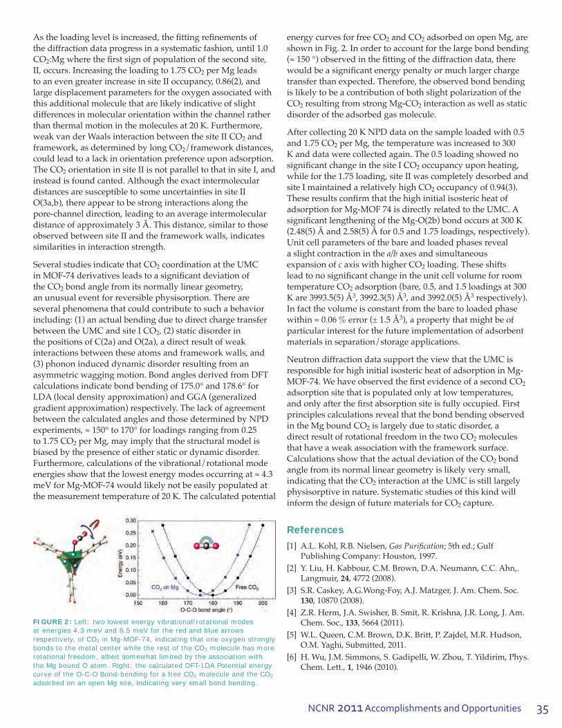
NCNR 2011 Accomplishments and Opportunities 35
Astheloadinglevelisincreased,thefittingrefinementsofthe diffraction data progress in a systematic fashion, until 1.0 CO2:Mgwherethefirstsignofpopulationofthesecondsite,II, occurs. Increasing the loading to 1.75 CO2 per Mg leads to an even greater increase in site II occupancy, 0.86(2), and largedisplacementparametersfortheoxygenassociatedwiththis additional molecule that are likely indicative of slight differencesinmolecularorientationwithinthechannelratherthan thermal motion in the molecules at 20 K. Furthermore, weakvanderWaalsinteractionbetweenthesiteIICO2 and framework,asdeterminedbylongCO2/frameworkdistances,could lead to a lack in orientation preference upon adsorption. The CO2 orientation in site II is not parallel to that in site I, and instead is found canted. Although the exact intermolecular distances are susceptible to some uncertainties in site II O(3a,b), there appear to be strong interactions along the pore-channel direction, leading to an average intermolecular distanceofapproximately3Å.Thisdistance,similartothoseobservedbetweensiteIIandtheframeworkwalls,indicatessimilarities in interaction strength.
Several studies indicate that CO2 coordination at the UMC inMOF-74derivativesleadstoasignificantdeviationofthe CO2 bond angle from its normally linear geometry, an unusual event for reversible physisorption. There are several phenomena that could contribute to such a behavior including:(1)anactualbendingduetodirectchargetransferbetweentheUMCandsiteICO2, (2) static disorder in thepositionsofC(2a)andO(2a),adirectresultofweakinteractionsbetweentheseatomsandframeworkwalls,and(3) phonon induced dynamic disorder resulting from an asymmetricwaggingmotion.BondanglesderivedfromDFTcalculationsindicatebondbendingof175.0°and178.6°forLDA(localdensityapproximation)andGGA(generalizedgradient approximation) respectively. The lack of agreement betweenthecalculatedanglesandthosedeterminedbyNPDexperiments,≈150°to170°forloadingsrangingfrom0.25to 1.75 CO2 per Mg, may imply that the structural model is biased by the presence of either static or dynamic disorder. Furthermore,calculationsofthevibrational/rotationalmodeenergiesshowthatthelowestenergymodesoccurringat≈4.3meVforMg-MOF-74wouldlikelynotbeeasilypopulatedatthe measurement temperature of 20 K. The calculated potential
energy curves for free CO2 and CO2 adsorbed on open Mg, are showninFig.2.Inordertoaccountforthelargebondbending(≈150°)observedinthefittingofthediffractiondata,therewouldbeasignificantenergypenaltyormuchlargerchargetransfer than expected. Therefore, the observed bond bending is likely to be a contribution of both slight polarization of the CO2 resulting from strong Mg-CO2interactionaswellasstaticdisorder of the adsorbed gas molecule.
Aftercollecting20KNPDdataonthesampleloadedwith0.5and 1.75 CO2perMg,thetemperaturewasincreasedto300Kanddatawerecollectedagain.The0.5loadingshowednosignificantchangeinthesiteICO2 occupancy upon heating, whileforthe1.75loading,siteIIwascompletelydesorbedandsite I maintained a relatively high CO2 occupancy of 0.94(3). Theseresultsconfirmthatthehighinitialisostericheatofadsorption for Mg-MOF 74 is directly related to the UMC. A significantlengtheningoftheMg-O(2b)bondoccursat300K(2.48(5)Åand2.58(5)Åfor0.5and1.75loadings,respectively).Unit cell parameters of the bare and loaded phases reveal a slight contraction in the a/b axes and simultaneous expansion of caxiswithhigherCO2 loading. These shifts leadtonosignificantchangeintheunitcellvolumeforroomtemperature CO2 adsorption (bare, 0.5, and 1.5 loadings at 300 Kare3993.5(5)Å3,3992.3(5)Å3,and3992.0(5)Å3 respectively). In fact the volume is constant from the bare to loaded phase within≈0.06%error(±1.5Å3), a property that might be of particular interest for the future implementation of adsorbent materialsinseparation/storageapplications.
NeutrondiffractiondatasupporttheviewthattheUMCisresponsible for high initial isosteric heat of adsorption in Mg-MOF-74.WehaveobservedthefirstevidenceofasecondCO2 adsorptionsitethatispopulatedonlyatlowtemperatures,andonlyafterthefirstabsorptionsiteisfullyoccupied.Firstprinciples calculations reveal that the bond bending observed in the Mg bound CO2 is largely due to static disorder, a directresultofrotationalfreedominthetwoCO2 molecules thathaveaweakassociationwiththeframeworksurface.CalculationsshowthattheactualdeviationoftheCO2 bond angle from its normal linear geometry is likely very small, indicating that the CO2 interaction at the UMC is still largely physisorptiveinnature.Systematicstudiesofthiskindwillinform the design of future materials for CO2 capture.
References
[1] A.L.Kohl,R.B.Nielsen,Gas Purification; 5th ed.; Gulf PublishingCompany:Houston,1997.
[2] Y.Liu,H.Kabbour,C.M.Brown,D.A.Neumann,C.C.Ahn,.Langmuir,24, 4772 (2008).
[3] S.R. Caskey, A.G.Wong-Foy, A.J. Matzger, J. Am. Chem. Soc. 130, 10870 (2008).
[4] Z.R.Herm,J.A.Swisher,B.Smit,R.Krishna,J.R.Long,J.Am.Chem. Soc., 133, 5664 (2011).
[5] W.L.Queen,C.M.Brown,D.K.Britt,P.Zajdel,M.R.Hudson,O.M.Yaghi,Submitted,2011.
[6] H.Wu,J.M.Simmons,S.Gadipelli,W.Zhou,T.Yildirim,Phys.Chem.Lett.,1, 1946 (2010).
FIGURE 2: Left: two lowest energy vibrational/rotational modes at energies 4.3 meV and 8.5 meV for the red and blue arrows respectively, of CO2 in Mg-MOF-74, indicating that one oxygen strongly bonds to the metal center while the rest of the CO2 molecule has more rotational freedom, albeit somewhat limited by the association with the Mg bound O atom. Right: the calculated DFT-LDA Potential energy curve of the O-C-O Bond-bending for a free CO2 molecule and the CO2 adsorbed on an open Mg site, indicating very small bond bending.

36
Soft M
atter
36
Neutron scattering techniques shed new light on gel formation A.P.R. Eberle1,2, N.J. Wagner2 and R. Castañeda-Priego2,3
Colloidal dispersions are used in many household products ranging from paints to shaving cream
to gelatin and even to foods such as salad dressings or mayonnaise. The stability and processing of these and similar products highly depend on the state of the material. Of particular interest is the phase transitionfromfluidtoasolid,calledagelatlowparticle volume fractions, f < ≈ 0.40, and a glass at higher volume fractions. While the phase behavior for such systems has been studied for decades, theboundaryseparatingafluidfromagelisstilldebated.
Inthisworkwestudygelationusingamodelnanoparticlesystemwiththermoreversibleinteractions. The system consists of nanometer spheres of silica dioxide (≈30nm)coatedwithashortcarbon chain (C14), and suspended in tetradecane. At high temperatures T > ≈ 32 °C the particles are stable and the system behaves like a simple hard spherefluid.Uponquenchingthesystem,particlesaggregateinconnectionwithaphasetransitionofthe molecular brush [1]. This in-turn causes a short-rangeattractionbetweenparticles,andiftheparticlevolumefractionissufficient,f≥0.05,thesystemwillform a stress-supporting gel. Wedefinegelationanddetermine the gel temperature (Tgel )towithin±0.1°C using a combination of rheological experiments and an experimental study of the dynamics using fiber-opticquasi-elasticlightscattering(FOQELS).Finally,westudythestructureofthedispersionat and around the gel transition using small-angle neutron scattering (SANS).
Small-amplitude oscillatory shear (SAOS) rheological measurements are used to identify the transition fromthefluidstatetothegelstateemployingawell-definedcriterionestablishedforcrosslinking polymer resins [2]. The SAOS results for a temperature ramp experiment for one dispersion, f = 0.12, can be seen in Fig. 1. At high temperatures (T > ≈ 31 °C),inthefluidstate,thesuspensionexhibits a negligible storage modulus G’,whichdefinestheamountofelasticenergythefluid can store.
1NIST Center for Neutron Research, National Institute of Standards and Technology, Gaithersburg, MD 208992University of Delaware, Newark, DE 19716, USA3Universidad de Guanajuato, 37150 León, Mexico
FIGURE 1: Storage, G′ (closed symbols), and loss, G′′ (open
Asthesystemisquenchedtolowertemperaturesfromthefluidstate,theelasticmodulusincreasesoverfiveordersofmagnitudeinthenarrowtemperaturerangeof29°C to 27 °C.Thisshowsthesuspensionundergoesatransitionto a strong viscoelastic solid similar to cooled Jello. Frequencysweepmeasurementsprovideadeterminationofthestrengthofthegel.Frequencysweepexperimentson the same sample can be seen in the inset to Fig. 1 as a function of temperature near the gel point. At the highest temperatureshown,28.4°C, G′′ (the loss modulus) is larger than G′overthewholefrequencyrangetested.Asthe temperature is decreased to 28.2 °C, G′ and G′′ become comparableathighfrequencies.Atlowertemperaturesthe sample has a greater elastic modulus, characteristic of solid-like behavior. At 28.2 °C, G′ and G′′ are nearly equal withpowerlawslopesof½,overmorethanadecadeoffrequency,whichisacharacteristicfeatureofgelation[2].ThegeltemperatureisconfirmedusingFOQELS(notshown)whichprovidesameasureoftheparticles’dynamics.FromFOQELSmeasurementsitcanbeshownthat the dynamics decrease and exhibit an entire relaxation spectrum at the gel temperature determined from rheology.
symbols), moduli vs. temperature in a small-amplitude oscillatory (stress amplitude, so = 17.68 mPa, frequency, ω = 2π rad/s) temperature ramp experiment (ramp rate 0.2 °C/min). Inset: G′ and G′′ from frequency sweep measurements at temperatures around the gel-point. Data are offset vertically for clarity by factors 0.03, 0.05, 0.08, and 0.2 for temperatures (28.4, 28.2, 28.1, and 28) °C, respectively.

37NCNR 2011 Accomplishments and Opportunities
Asthesystemisquenchedtolowertemperaturesfromthefluidstate,theelasticmodulusincreasesoverfiveordersofmagnitudeinthenarrowtemperaturerangeof29°C to 27 °C.Thisshowsthesuspensionundergoesatransitionto a strong viscoelastic solid similar to cooled Jello. Frequencysweepmeasurementsprovideadeterminationofthestrengthofthegel.Frequencysweepexperimentson the same sample can be seen in the inset to Fig. 1 as a function of temperature near the gel point. At the highest temperatureshown,28.4°C, G′′ (the loss modulus) is larger than G′overthewholefrequencyrangetested.Asthe temperature is decreased to 28.2 °C, G′ and G′′ become comparableathighfrequencies.Atlowertemperaturesthe sample has a greater elastic modulus, characteristic of solid-like behavior. At 28.2 °C, G′ and G′′ are nearly equal withpowerlawslopesof½,overmorethanadecadeoffrequency,whichisacharacteristicfeatureofgelation[2].ThegeltemperatureisconfirmedusingFOQELS(notshown)whichprovidesameasureoftheparticles’dynamics.FromFOQELSmeasurementsitcanbeshownthat the dynamics decrease and exhibit an entire relaxation spectrum at the gel temperature determined from rheology.
SANSmeasurementswereperformedoverarangeoftemperaturesspanningthetransitionfromthefluid-statethroughgelation.Thelow-Q scattering intensity systematically evolves for decreasing temperature in the rangewhereaggregationoccursuntilTgel,wherestructureon the length scales accessible by SANS becomes arrested. Figure2showstheSANSspectraatTgel,asidentifiedfromrheology, for the entire concentration range.
The spectra at the gel point evolve from fractal-like scatteringatlowf to a glass-like spectra at high f. The scatteringintensityisfitted(solidlines),usingwellestablishedtheoriesassumingasquare-wellinteractionpotentialfortheparticles.Fromthefitsweextractthestrengthofattractiondefinedbyareducedtemperature, τ = (e/12)exp(–U/kBT), called the Baxter temperature, whereUisthesquare-welldepth,e is a perturbation parameter e = D/(a + D),whereDisthesquare-wellwidthand a is the particle diameter, and kBT is the thermal energy. As τ decreases, the strength of attraction increases.
Thestatediagramwithourexperimentallydeterminedgel transition for the AHS system is presented in Fig. 3. At intermediatetolowf and high attractive strength there is a liquid/vaporcoexistenceregion.Conversely,athighf and lowattractivestrengththereisaliquid/crystalcoexistenceregion.
This is in addition to a repulsive driven glass (RDG) line whichintersectstheattractivedrivenglass(ADG)linewithmodecouplingtheory(MCT)singularity,A3. As is typically done, the location of the RDG and ADG lines calculated from MCT are shifted to higher particle densities usingknownexperimentaldata,sinceMCTunder-predictstheRDGline[6,7].ThepercolationlinefortheAHSfluidcanbeseenextendingfromtheliquid/vaporcoexistenceregionatlowftotheliquid/crystalcoexistenceregionathigh f.Theexperimentallydeterminedpointsdefiningthegeltransition(triangles)agreewellwithpercolation
theory up to f ≈ 0.41. For concentrations f > ≈ 0.41 the boundaryfordynamicalarresttendstowardandjoinstheADGline.Thus,themajorconclusionsofthisworkshowthatforthisAHSnanoparticlesystem:The dynamical arrest transition extends from the dilute particle concentration side of the liquid/vapor coexistence above the critical point following predictions of percolation theory until at sufficiently high particle concentrations it subtends the predictions and joins the MCT ADG line.
Weexperimentallydeterminethefluid-to-geltransitionofamodelnanoparticledispersionwithshort-rangedattractions. Our experiments demonstrate a continuous boundary that extends from the gas-liquid phase separationregionatlowconcentrationstotheattractivedriven glass line at high concentrations. The conclusions presentedhereinhaveimplicationsinawiderangeof colloidal systems that interact via a short range interaction.Theanticipationisthatthisworkwilldirectthe development of advanced theory and simulation efforts.Thesewillinturnhelpustofurtherunderstandtheunderlying mechanisms that drive gelation and ultimately allowfortheoptimizationofproductsthroughpredictivetools rather than guess and check formulation.
References
[1] A.P.R.Eberle et al.,Langmuir26, 3003 (2010).[2] H.H. Winter, and F. Chambon, J. Rheol. 30, 367 (1986).[3] M.A.Miller,andD.Frenkel,Phys.Rev.Lett.90, 135702 (2003).[4] Y.C.Chiew,andE.D.Glandt,J.Phys.A-MathGen.16, 2599
(1983).[5] D.W.Marr,andA.P.Gast,J.Chem.Phys.99, 2024 (1993).[6] J.Bergenholtz,andM.Fuchs,Phys.Rev.E59, 5706 (1999).[7] P.N.Pusey,andW.vanMegen,Phys.Rev.Lett.59, 2083 (1987).
FIGURE 2: SANS measurements at the gel point for various volume fractions f and the corresponding model fits.
FIGURE 3: State diagram for the model adhesive hard sphere system (AHS). (▬) Is the liquid/vapor coexistence region with critical point (star, τ = 0.1133 and f = 0.266) as determined by Monte Carlo (MC) simulations [3]; broken line is the analytical solution to the percolation line [4]; grey shaded region is the liquid/crystal coexistence region from the modified weighted-density approximation (MWDA) theory [5]; (—) repulsive and attractive driven glass (RDG and ADG) lines from model coupling theory (MCT) with point singularity, A3 (circle) [6]. The triangles are the experimentally determined dynamical arrest transition.

38
Soft M
atter
38
Interlayer distance dependence of thickness fluctuations in a swollen lamellar phaseM. Nagao1,2, S. Chawang3, T. Hawa3
1NIST Center for Neutron Research, National Institute of Standards and Technology, Gaithersburg, MD 208992Indiana University, Bloomington, IN 474083The University of Oklahoma. Norman, OK 73019
Surfactants are one of the basic building blocks of nanoscale self-assembling structures, such
as micelles, microemulsions, and biological membranes. In particular, surfactant membranes play important roles from household (e.g., detergents) and pharmaceutical (e.g., drugs) products to biological (e.g., drug delivery and virus infections) and industrial (e.g., oil recovery plants) applications. Understanding the behaviors of surfactant membranes and their properties is thus of both fundamental and industrial relevance. One of the unique features of the membranes is collective dynamics of surfactant molecules near room temperature. Membrane bending motion has been established both theoretically and experimentally, whilemembranethicknessfluctuationsremainthesubjectofresearch.Thethicknessscaleofsurfactantmembranes in solution is on the order of nanometers, and thus the neutron spin echo (NSE) technique is wellsuitedtoprobethedynamics[1,2].Apreviousinvestigationofthedynamics[2]showedthatthethicknessfluctuationswereclearlyseenbyNSEinthe nonionic surfactant pentaethylene glycol dodecyl ether (C12E5)mixedwithdeuteratedwater(D2O) and deuterated octane (C8D18). In the study highlighted here,weexaminedthesamesystemwhilechangingthe effective membrane thickness, dm, as controlled by the ratio of the volume fractions of oil, fo, to surfactant, fs,definedasR=fo/fs. In addition totheNSEmeasurement,wehaveemployedmolecular dynamics (MD) simulations to calculate the dynamics since the time scale covered by NSE matches that covered by MD. The combined use of these techniques provides further insight into the dynamics of membranes.
Figure1(a)showsthesmall-angleneutronscattering(SANS)profilesatT =(59.6±0.2)°C for R = 0.0 and at T=(31.6±0.1)°Cfor0.3≤R≤2.0.Inthelow-q region, a broad scattering peak originating from theBraggreflectionofthemembranestackingisobserved at the same q(≈0.08nm-1) for R =1.5 and R=2.0asshownbyaverticalstraightlineinthefigure.Althoughthepeakpositionofthelamellaeis not clear for samples for R≤1.2,itisconsidered
to be almost independent of R. On the other hand, in the high-qregion,anotherpeakprofileoriginatingfromtheform factor of the membranes is observed. With increasing R, the characteristic q value, qdip,shiftstowardlowerq. Utilizing a model scattering function for isotropic lamellar structures, membrane thickness, dm,whichfollowstheidealswellinglaw,isestimatedfromdm = 3.1 nm at R = 0.0 to dm = 9.2 nm at R = 2.0. A constant inter-lamellar distance dwithchangingdmisconfirmedfromtheSANSprofiles,schematicallyshowninFig.1(c)[3].
Thestructurewasalsoconfirmedusingcourse-grained(CG) MD simulations [3] by calculating the intermediate scattering function, , wherer→i is a position vector for particle i, t is a time, N is the number of surfactant molecules, and + , represents an ensemble average.Thesimulationswereperformedwithanintegrationtimestepof25fsandwererunfor200nsat30 °C (1 bar) to equilibrate the system, and for 50 ns to calculate I(q,t).Figure1(b)showsI(q,t = 0) calculated after the MD simulation, corresponding to the respective SANS profile.ThebehaviorofqdipwithR, similar to the SANS result,confirmsthattheMDcalculationreconstructsasimilar structure in the computational space.
Figure2(a)showsI(q,t) normalized by I(q,0) observed by NSE at R = 0.3 and T = 30 °C [3]. I(q,t)/I(q,0)followsa
FIGURE 1: (a) SANS profiles show that the interlamellar mean repeat distance d is constant (vertical line) while the dip position qdip, which relates to the membrane thicknesses dm, is changing with R. (b) MD calculation of the SANS equivalent I(q,t = 0), shows a similar dependence of qdip on R. (c) Schematic illustrating constant d while dm increases with R, as confirmed by SANS and MD.
1 N N → → →I(q,t)= — S S +exp[iq •{ri(t)–rj(0)}],
N i=1 j=1

39NCNR 2011 Accomplishments and Opportunities
stretched exponential function withthedecayrateΓ. The lines obtained from the MD
I(q,t)——– = exp[–(Γt)2/3] I(q,0)simulationsalsodecaywithtimeas
showninFig.2(b).Thedecayfunctioniswellexplainedwiththeaboveequationexceptfortheintermediateq range, e.g., q = 1.4 nm-1 in Fig. 2(b). This result suggests that a better analysis in the intermediate q-range may require multiple exponential functions to account for multiple modes.
Figures2(c)and2(d)showtheq-dependence of Γ/q3 at various values of R for NSE and MD, respectively [3]. Tocharacterizethedynamics,aLorentzfunction Γ ΓTF 1 ΓBEND— = ––– —————— + ——— q3 q03 1+(q–q0)2 x–2 q3
is utilized. The lines in Fig. 2(c) and 2(d) are the
resultsoffittingtothisequation.Inthisprocedure,twoterms of membrane dynamics, the bending and thickness fluctuations,areconsidered.Thecontributionsfromthesemotions are characterized by decay rates ΓBEND and ΓTF , respectively. Each parameter relates to the thickness fluctuations:ΓTF/q03 and x-1 are proportional to the damping frequency of the mode and the mode amplitude, respectively, and q0isthecenterofthemotion,whichrelates to the membrane thickness. It is noted that the thicknessfluctuationsareobservedevenfortheR = 0.0
sample, corresponding to the bilayers. On the other hand, the R = 1.37 (NSE data) and R = 1.14 (MD data) samples showasmallenhancementofthedynamics.Thissuggeststhatalargeamountofoil,whichswellsthebilayers,suppressesthethicknessfluctuations.
Thethicknessfluctuationamplitudesaregivenby dmx-1/q0wheretheparameters,bothfromNSEandMD,areobtainedfromreciprocalspaceinformationandtheLorentzfunctionfit.ThevaluesobtainedfromthisexpressionarecomparedtothedistributionoflocalfluctuationsofsurfactantsinrealspacebyMDsimulation,asshowninFig.3(a).Excellentagreementwasobtained,especiallybetweenNSE(reciprocal)andMD(real)data.ThisresultconfirmsthatthemotionobservedbyNSEisthethicknessfluctuationsofsurfactantmembranesandthatthethicknessfluctuationamplitudeincreaseslinearlywithR.
ThemembranebendingmotioniswellpredictedbytheZilman-Granek(ZG)singlemembranefluctuationmodel[4]. Utilizing the ZG theory, the bending modulus of the membrane κ is estimated from the values of ΓBEND, as showninFig.3(b).Theκ at R = 0.3 is larger than that at R = 0.0 (bilayer), indicating more rigid membranes around R=0.3thanthebilayers.Twopossiblescenariosaretheincrease of κ due to the change of the bilayer thickness and that the increase of κ originates from the enhanced thicknessfluctuations.Whenoilthicknessissmallenough,thefluctuationsofthetwomonolayersaresynchronizedand they behave as a single thick membrane (pseudo bilayers). With increasing R at R > 0.3 the value of κ becomessmaller,andfinallyκ≈1kBT at R = 1.37. In this regimethesynchronizationbetweentwomonolayersbecomessmallerandeachmonolayerfluctuationdominatesthesystemdynamicswhendm is large enough.
ThepresentstudyshowsthatcombineduseofNSEand MD gives an excellent agreement of the thickness fluctuationsofsurfactantmembranes.Theimportanceof intra-membrane dynamics to the elastic properties oftheoverallmembraneisalsoconfirmed.Therigidityof membranes is controlled by the interactions among moleculesaswellasbytheenergydissipationmechanismswithinthemembranes.Thepresentresultsandthemethods described here are very useful in understanding the dynamics of surfactant membranes, in particular, bilayerlipidmembranes,interactionofdrugswithbiological membranes, and molecular transport across membranes.
Reference
[1] B. Farago, M. Monkenbusch, K. D. Goecking, D. Richter, J. S. Huang,PhysicaB213&214,712(1995).;B.Farago,PhysicaB226, 51 (1996).
[2] M.Nagao,Phys.Rev.E80, 031606 (2009).[3] M.Nagao,S.Chawang,andT.Hawa,SoftMatter7, 6598
(2011).[4] A.G.Zilman,R.Granek,Phys.Rev.Lett.77, 4788 (1996).
FIGURE 2: I(q,t)/I(q,0) measured by (a) NSE and (b) calculated by MD show good agreement in membrane dynamics. Both results by (c) NSE and (d) MD show that the decay rate Γ is no longer growing with q3, which indicates that the current theory is not adequate to explain the membrane dynamics. Τhe widths of the peaks are related to the thickness fluctuation amplitudes.
FIGURE 3: (a) NSE and MD data show excellent agreement in predictions of thickness fluctuation amplitude. (b) An unexpected enhancement of the bending modulus κ is observed near R = 0.3.
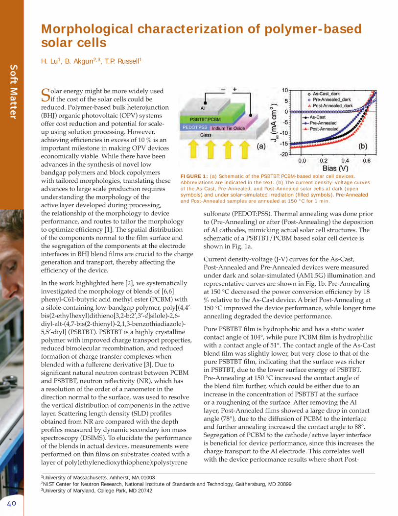
40
Soft M
atter
40
Morphological characterization of polymer-based solar cellsH. Lu1, B. Akgun2,3, T.P. Russell1
Solarenergymightbemorewidelyusedif the cost of the solar cells could be
reduced.Polymer-basedbulkheterojunction(BHJ)organicphotovoltaic(OPV)systemsoffer cost reduction and potential for scale-upusingsolutionprocessing.However,achievingefficienciesinexcessof10%isanimportantmilestoneinmakingOPVdeviceseconomically viable. While there have been advancesinthesynthesisofnovellowbandgap polymers and block copolymers withtailoredmorphologies,translatingtheseadvances to large scale production requires understanding the morphology of the active layer developed during processing, the relationship of the morphology to device performance, and routes to tailor the morphology tooptimizeefficiency[1].Thespatialdistributionofthecomponentsnormaltothefilmsurfaceandthe segregation of the components at the electrode interfacesinBHJblendfilmsarecrucialtothechargegeneration and transport, thereby affecting the efficiencyofthedevice.
Intheworkhighlightedhere[2],wesystematicallyinvestigated the morphology of blends of [6,6]phenyl-C61-butyricacidmethylester(PCBM)withasilole-containinglow-bandgappolymer,poly[(4,4′-bis(2-ethylhexyl)dithieno[3,2-b:2′,3′-d]silole)-2,6-diyl-alt-(4,7-bis(2-thienyl)-2,1,3-benzothiadiazole)-5,5′-diyl](PSBTBT).PSBTBTisahighlycrystallinepolymerwithimprovedchargetransportproperties,reduced bimolecular recombination, and reduced formationofchargetransfercomplexeswhenblendedwithafullerenederivative[3].DuetosignificantnaturalneutroncontrastbetweenPCBMandPSBTBT,neutronreflectivity(NR),whichhasa resolution of the order of a nanometer in the directionnormaltothesurface,wasusedtoresolvethe vertical distribution of components in the active layer.Scatteringlengthdensity(SLD)profilesobtainedfromNRarecomparedwiththedepthprofilesmeasuredbydynamicsecondaryionmassspectroscopy (DSIMS). To elucidate the performance oftheblendsinactualdevices,measurementswereperformedonthinfilmsonsubstratescoatedwithalayerofpoly(ethylenedioxythiophene):polystyrene
sulfonate(PEDOT:PSS).Thermalannealingwasdonepriorto(Pre-Annealing)orafter(Post-Annealing)thedepositionof Al cathodes, mimicking actual solar cell structures. The schematicofaPSBTBT/PCBMbasedsolarcelldeviceisshowninFig.1a.
Current density-voltage (J-V) curves for the As-Cast, Post-AnnealedandPre-Annealeddevicesweremeasuredunder dark and solar-simulated (AM1.5G) illumination and representativecurvesareshowninFig.1b.Pre-Annealingat150°Cdecreasedthepowerconversionefficiencyby18%relativetotheAs-Castdevice.AbriefPost-Annealingat150°Cimprovedthedeviceperformance,whilelongertimeannealing degraded the device performance.
PurePSBTBTfilmishydrophobicandhasastaticwatercontactangleof104°,whilepurePCBMfilmishydrophilicwithacontactangleof51°.ThecontactangleoftheAs-Castblendfilmwasslightlylower,butveryclosetothatofthepurePSBTBTfilm,indicatingthatthesurfacewasricherinPSBTBT,duetothelowersurfaceenergyofPSBTBT.Pre-Annealingat150°Cincreasedthecontactangleoftheblendfilmfurther,whichcouldbeeitherduetoanincreaseintheconcentrationofPSBTBTatthesurfaceor a roughening of the surface. After removing the Al layer,Post-Annealedfilmsshowedalargedropincontactangle(78°),duetothediffusionofPCBMtotheinterfaceandfurtherannealingincreasedthecontactangleto88°.SegregationofPCBMtothecathode/activelayerinterfaceisbeneficialfordeviceperformance,sincethisincreasesthechargetransporttotheAlelectrode.ThiscorrelateswellwiththedeviceperformanceresultswhereshortPost-
1University of Massachusetts, Amherst, MA 01003 2NIST Center for Neutron Research, National Institute of Standards and Technology, Gaithersburg, MD 20899 3University of Maryland, College Park, MD 20742
FIGURE 1: (a) Schematic of the PSBTBT:PCBM-based solar cell devices. Abbreviations are indicated in the text. (b) The current density–voltage curves of the As-Cast, Pre-Annealed, and Post-Annealed solar cells at dark (open symbols) and under solar-simulated irradiation (filled symbols). Pre-Annealed and Post-Annealed samples are annealed at 150 °C for 1 min.

41NCNR 2011 Accomplishments and Opportunities
Annealing times improved the device performance but longerPost-Annealingtimesdegradedtheperformance.
NRdataoftheAs-Cast,Pre-Annealed,andPost-AnnealedfilmsandtheircorrespondingSLDprofilesareshowninFig.2.TheAs-CastandPre-AnnealedfilmshaveverysimilarSLDprofiles,showinganenrichmentofPSBTBTanddepletionofPCBMattheairsurface,andPre-Annealingcausesthisdepletionlayertobeslightlythicker(Fig.2a).IntheAs-Castfilm,athinlayerclosesttothePEDOT:PSSlayercorrespondstotheactivelayerinwhichthevolumefractionofPCBM(fPCBM) is 0.28. In the bulk of the As-Cast active layer, fPCBMis0.57whichisclosetothevolumefractionofthetwocomponentsintheblend solution (fPCBM = 0.53 and fPSBTBT=0.47).WhilePre-Annealing did not change the distribution in the bulk, it didincreasetheSLDofthePEDOT:PSSlayer,indicatingthediffusionofPCBMintothePEDOT:PSSlayer.
ThePost-Annealedsample,ontheotherhand,showedmarkedlydifferentNRandSLDprofiles(Fig.2b).Tosimplifythefittingprocedure,theactivelayerwasspin-castdirectlyontoaUV-ozone-treatedSisubstrate,followedbyevaporationofanAlelectrodeontop.PCBMandPSBTBTaredistributedmoreuniformlythanintheAs-CastandPre-Annealedsamples.ThedropintheSLDatthesubstratesuggestsadepletionofPCBM(≈4nmthick)atthe substrate. The layer located at z≈110nmto160nmwasascribedtoaluminumoxide.Theroughinterfacebetweentheactivelayerandthecathode(rmsroughness≈7.7nm)
indicated a diffusion of Al into the active layer during thermalannealingorthermalevaporationofAl,whichisalsoconfirmedbyhigh-resolutiontransmissionelectronmicroscopy.
The depth distribution obtained by NR is in good agreementwiththatmeasuredbyDSIMS,showingthedepletionofPCBMandenrichmentofPSBTBTattheairinterfaceforAs-CastandPre-AnnealedfilmsandanincreaseofPCBMconcentrationatthecathodeinterfaceafterPost-Annealing.SegregationofthedonorattheanodeandacceptoratthecathodeinterfacesinOPVdevicesisbeneficialforchargetransportandcollection,and,assuch,Post-Annealingleadstobetterdeviceperformance.
DiffusionofPCBMintoPSBTBTwasmeasuredusingNRinabilayersystem.ShowninFig.3areNRcurvesandthecorrespondingSLDprofilesforPCBM/PSBTBTbilayerfilmsunderdifferentannealingconditions(PSBTBTfilmswerepre-annealedat150°Cfor5minbeforetheywerefloatedontoPCBMlayers.)ThesharpinterfacebetweenthetwolayersforAs-CastfilmindicatesthattherewasnointerdiffusionbetweenPCBMandPSBTBTatroomtemperature.Evenamildannealingat100°Cfor1minwasenoughtoinitiatediffusionofPCBMintothePSBTBTlayer.AtthebeginningPCBMdidnotcompletelydiffuseintotheentirepolymerlayerbutafterannealingat150°Cfor5minPCBMwasuniformlydistributedinthepolymerlayerexceptaverythinlayerattheairsurface.TheSLDofthepolymerlayerdidnotshowobviouschangesafter5min,suggestingasaturationofPCBM(fPCBM = 0.19) in thePSBTBTlayer.TheslightincreaseintheSLDofthePCBMfilmuponannealingat150°CcanbeattributedtoadensificationofthePCBM.ThediffusionbehaviorofPCBMwithinthePCBM/PSBTBTbilayersisquitesimilartothatseeninthePCBM/P3HTsystemwherePCBMwasseentorapidlydiffuseintotheamorphousregionsofP3HTwithoutperturbingtheorderingofP3HT[4].
In summary, a systematic investigation on the morphology of the active layer and the structure at the interfaces underdifferenttreatmentswasaccomplishedin situ usingneutronreflectivityemployingthenaturalneutroncontrastbetweenthecomponentsintheactivelayer.ThisstudyindicateshowPost-Annealingimprovesdeviceperformance by enhancing segregation of the donor at the anode and acceptor at the cathode interfaces. Such studies are important for improving the performance of the polymer-based solar cells.
References
[1]J.Peet,A.J.Heeger,G.C.Bazan,Acc.Chem.Res.42, 1700 (2009).
[2]H.Lu,B.Akgun,T.P.Russell,Adv.EnergyMater.ASAP(DOI:10.1002/aenm.201100128).
[3]H.Y.Chen,J.H.Hou,A.E.Hayden,H.Yang,K.N.Houk,Y.Yang,Adv.Mater.22, 371 (2010).
[4]D.Chen,F.Liu,C.Wang,A.Nakahara,T.P.Russell,NanoLett.11, 2071 (2011).
FIGURE 2: Neutron reflectivity profiles of (a) Si/SiOX/PEDOT:PSS/PSBTBT:PCBM films and (b) Si/SiOX/PSBTBT:PCBM/Al films. The solid lines indicate the best fits for the reflectivity profiles. The insets are the corresponding profiles of SLD as a function of z, where z = 0 is the Si substrate.
FIGURE 3: (a) Neutron reflectivity profiles for the PCBM/PSBTBT bilayer films (PSBTBT film was Pre-Annealed at 150 °C for 5 min) before and after annealing. The solid lines indicated the best fits for the reflectivity profiles. (b) The corresponding SLD profiles as a function of z (where z = 0 is the Si wafer). SLD profiles do not overlap along the horizontal axis due to the varying thickness of each sample.

42
Soft M
atter
42
Nanoscale mixing of soft solidsS-H. Choi,1 S. Lee,1 H.E. Soto,3 T.P. Lodge,1,2 and F.S. Bates1
1Department of Chemical Engineering and Materials Science, University of Minnesota, Minneapolis, MN 554552Department of Chemistry, University of Minnesota, Minneapolis, MN 554553University of Texas-Pan America, Edinburg, TX 78541
Mixing of materials on the molecular scale represents a fundamental and demanding
facet of modern chemical technology. Blending thermodynamicallycompatibleliquidssuchaswaterandmethanolistrivial,whereascombininginorganicsolidsatlowtemperaturesisvirtuallyimpossible.Soft solids such as colloids, biological materials, andpolymermeltsshowcomplicatedanddelicatethermodynamicanddynamicpropertieswithinbothlimits of behavior, necessitating innovative mixing processes. While the properties of blends and alloys are highly dependent on the precise arrangement of the constituents, assessing the state of mixing at the molecular scale is extremely challenging. In this highlight,wedemonstratethatstatisticallyperfectnanoscale mixing of ordered block copolymer micellar solutions can be achieved using a simple laboratory-scale mixer, as characterized by SANS measurements (see Fig. 1)
Poly(styrene-b-ethylene-alt-propylene)(PS-PEP)diblockcopolymersweredispersedinamixtureofnormal and deuterated squalane (C30H62 and C30D62), resultinginsphericalmicellescontainingglassyPScoresandswollenPEPcoronas.Thesemicellesformliquid-likesolutionsatlowconcentrations(<7%mass fraction), but soft solid body-centered cubic (bcc) ordered arrangements at higher concentrations. Nearlychemicallyidenticalnormal(PS(H)-PEP)and
selectivelyperdeuterated(PS(D)-PEP)blockcopolymersweresynthesizedforthisstudy,thenblendedwiththesolventintwoways.Premixedsolutionscontainmicellecoreswith50%PS(D)and50%PS(H)molarfractions,designed to perfectly contrast match the isotopically labelledsqualanesolvent.PostmixedsolutionswereformedbymechanicallymixingorderedblendsofPS(D)-PEPandPS(H)-PEPmicelles.
Complete mixing of liquid micelle solutions (e.g., 1 % volume fraction) can be achieved by simple stirring at room temperature [1].However,mixingorderedmicellarsolutions is considerably more challenging because these aresoftyetsolid-likematerials.Hereweemployedasimple MiniMax mixer (CSI Co., see Fig. 2) to postmix 15 %volumefractionofPS(H)-PEPandPS(D)-PEPcopolymersolutionsatroomtemperature.SincethePScoresareglassy(Tg≈70°C) at room temperature, copolymer chains are notabletoexchangebetweenmicellesduringmechanicalblending.
SANSmeasurementswereperformedwiththeNG-730minstrumentatNIST,withaneutronwavelengthof7Å.SAXSmeasurementswereconductedwiththeequipmentmaintainedbytheDuPont-Northwestern-DowCollaborativeAccessTeamatArgonneNational
FIGURE 1: Illustration of PS(H) (blue) and PS(D) (red) spheres ordered on a body-centered cubic lattice and immersed in a contrast-matched matrix of squalane (C30H62 + C30D62) (purple). SANS from both (a) and (b) results in Bragg diffraction, while random placement of blue and red spheres (c) extinguishes interparticle interference, thus exposing single-sphere scattering.
FIGURE 2: Schematic illustration of the MiniMax mixer (CSI Co.) employed to blend ordered block copolymer micelles at room temperature. The specimen is held in a cup, along with three balls that facilitate mixing, confined by a rotor that applies a macroscopic shear stress.

43NCNR 2011 Accomplishments and Opportunities
Laboratory,withanx-raywavelengthof0.729Å.Figure3 displays SANS and SAXS (inset) data obtained from the 15%volumefractionPS(H)-PEPandPS(D)-PEPsolutionsprior to blending at room temperature. Bragg peaks areevident,consistentwithabcclatticewithunitcellparameters abcc = 62 nm and abcc=68nmforPS(H)-PEPandPS(D)-PEPmicelles,respectively.Weattributethesmalldifference in lattice parameter to the minor variations in the molecular characteristics of the block copolymers.
For a blend containing equal numbers (NH = ND = N/2)of perfectly randomly mixed and identically sized normal and perdeuterated spheres, the neutron scattering intensity canbedescribedbythesumofintraparticle,P(q) and interparticle Q(q)contributions:
I(q) (DrH)2+(DrD)2 DrH + DrD 2
—— =[——————–]P(q)+[—————] NQ(q), (1) A 2 2
whereAisthesystem-specificconstant.HereDrH and DrD aretheexcessscatteringlengthdensityofPS(H)andPS(D),respectively,determinedwiththeisotopicsqualanesolvent.InthiscaseweignorescatteringfromthesolvatedPEPblocks,whichcanbeshowntobeinsignificantrelativetothemicellecorescattering.Critically,Eq.(1)issatisfiedonlyif the normal and deuterated spheres are randomly distributedwithinthespecimen,eitheronadisorderedorordered basis. When the scattering length density of the isotopic solvent is designed to be (rH + rD)/2,theinterparicle contribution to Eq (1) is exactly eliminated leaving only the interparticle contribution. An isotopic mixture containing 42 % volume fraction of normal squalane and 58 % volume fraction of deuterated squalane meets this criterion.
Figure4showsSANSdataobtainedfrompostmixedand premixed solutions. Clearly, the Bragg peaks are
completely suppressed for the scattering pattern obtained from the postmixed solution (blended at room temperature using the MiniMax mixer) and the resulting data are quantitatively accounted for using a hard-sphere form factor(solidcurve)withacoreradiusof11.0nm.AnindependentSAXSmeasurementverifiesthatthisblendretainsawell-definedbccstructure(seeinsettofigure).ThisresultconfirmsthattheblendcontainsuniformlydistributedPS(D)-andPS(H)-PEPmicellesonabcclattice,as illustrated in Fig. 1. As anticipated by Eq. 1, the SANS profileobtainedfromthepremixedsolutionshowsalmostno coherent scattering intensity.
Inconclusion,wehavedemonstratedcompletemixingof bcc ordered block copolymer micelles using a simple commercialmixer.RealizationofnanoscalemixingwasconfirmedbySANSandSAXSmeasurements.Thiswork
also highlights the application of small-angle neutron scattering using the contrast matching technique to investigate particle form factors under circumstances whereordering(i.e., interparticle interference) generally dominates the overall scattering intensity.
References
[1] S.Choi,T.P.Lodge,F.S.Bates,Phys.Rev.Lett.104, 047802 (2010).
[2] S.Choi,F.S.Bates,T.P.Lodge,Macromolecules44, 3594 (2011).
[3] S.Choi,T.P.Lodge,F.S.Bates,Phys.Rev.Lett.104, 047802 (2010).
[4] S.Choi,F.S.Bates,T.P.Lodge,Macromolecules44, 3594 (2011).
FIGURE 3: SANS profiles obtained from 15 % volume fraction PS(D)-PEP (red) and PS(H)-PEP (blue) in an isotopic squalane mixture (42 % C30H62 and 58 % C30D62 volume fractions) at 30 ºC. Inset shows the corresponding SAXS patterns.
FIGURE 4: SANS pattern obtained from postmixed (purple) and premixed (black) solutions. The postmixed solutions were prepared by blending PS(H)-PEP and PS(D)-PEP micelle solutions (each 15 % volume fraction in contrast-matched squalane) at 30 ºC. The solid curve represents a best fit to the hard-sphere form factor. A synchrotron SAXS pattern (inset) obtained from the same mixtures confirms a bcc crystal structure.

44
Soft M
atter
44
Zone refinement effect to remove impurities in organic semiconductor polymer blends for print-able electronicsV. M. Prabhu1, D. M. DeLongchamp1, R. J. Kline1, D.Y. Yoon2, S.K. Satija3, J. E. Anthony4
A probleminmakingthin-filmprintableorganicsemiconductor devices is the appearance of impuritieswithinthecharge-transportregion,limitingdeviceperformance.Herewehighlightourworkonaprocessingstrategyformanufacturethatimprovestheuniformityofthefilmsandthatsignificantlyreducestheeffectoftrapimpurities.
Organic semiconductors are the most critical elementofanewgenerationofoptoelectronicdevicesthatincludesprintableflexibleelectronicsand photovoltaic devices. The vertical distribution and orientation of crystalline semiconductors in anorganicthinfilmfield-effecttransistor(OTFT)must be controlled by processing to achieve optimalswitchingspeedandcurrentflow,oftenforaspecificOTFTarchitecture.CommonOTFTarchitecture variations involve different placements of the contacts and gate dielectric. For large-area flexibledisplays,manytechnologydevelopersprefer a bottom-gated OTFT to a top-gated one. Gate placement is critical to device optimization because chargecarriersareformedwithinthesemiconductorlayeronlyatitsinterfacewiththegatedielectric.Chargetransportoccurswithinthe2nmregionadjacenttothegatedielectric,andthecompositionand orientation of the semiconductor layer at this interface is critical to device performance.
Formulation and processing are key contributors to the structure of the organic semiconductor layer at the dielectric gate interface. Since organic semiconductors can be processed from solution, they offer advantages over inorganic semiconductors such as amorphous silicon that requires processing under vacuum. While synthetic advances in recent yearshaveproducedorganicsemiconductorswithperformance characteristics that match amorphous silicon, there remain challenges in manufacturing processdevelopmentformethodssuchasinkjetdrop-on-demand printing, roll-casting, and spin coating. A processing challenge affecting some of the highest-performing organic semiconductors is that theneatsolutionviscosityistoolowforcommon
coating methods, preventing the formation of high-quality films.
An attractive approach to organic semiconductor formulationwasproposedbyBrownet al.,whoblendeda high-performance organic semiconductor in solution withaninsulatingpolymerbinder[1].Duetothepolymerbinder, the blend solution forms high-quality conformal films,andthefilmshadchargecarriermobilitygreaterthan 0.3 cm2/Vsintop-gatedOTFTs.Becausethetop-gatedmobilitywasquitehigh,Brownet al. postulated that the semiconductingsmallmoleculeswereverticallysegregatedtothetopsurfaceofthethinblendfilm.Althoughthisapproachrepresentedasignificantadvanceinformulation, the top-gated architecture is not preferred for manufacturing electronic devices, such as displays because the active layer can be damaged by subsequent fabrication stepssuchasgateelectrodepatterningandwiring.Instead,the bottom-gate architecture is favored.
BuildinguponthepioneeringworkofBrownetal.,the NIST-SNU team developed a blend formulation approach to ensure that the semiconducting molecules can also segregate to a bottom gate interface [2]. Neutron reflectivityexperimentsperformedontheNG7horizontalreflectometerprovidedthecriticaldataprovingthat blends of deuterium-labeled 6,13-bis(triisopropylsilylethy-nyl)pentacene(TIPS-pentacene)withhighmolecularweightpoly(α-methylstyrene)(PaMS)formanon-uniformverticalcompositionprofileinfilmsspin-coatedontosiliconwafersfromthehomogeneousmixturesolutions.Theseexperimentsshowedconclusivelythattheorganicsemiconductorwaspreferentiallysegregatedtothegate-dielectricinterfacewithsegregationcharacteristicscontrolled by the number-average relative molecular mass ofthePaMS.
Infollow-upwork,theNIST-SNUteaminvestigatedthesurprisingrelationshipbetweenverticalsegregationintheseblendfilmsandthepresenceofimpuritiesinthesemiconductor.Itisgenerallyknownthatorganicsemiconductors can contain or develop impurities that act as charge carrier traps; they can be formed on exposure to ultraviolet (UV) light, for example. The NIST-SNU
1Polymers Division, Material Measurement Laboratory, National Institute of Standards and Technology, Gaithersburg, MD 208992Seoul National University, Seoul, Korea3NIST Center for Neutron Research, National Institute of Standards and Technology, Gaithersburg, MD 208994University of Kentucky, Lexington, KY 40506

45NCNR 2011 Accomplishments and Opportunities
team investigated impurities [3] in the semiconductor triethylsilylethynyl anthradithiophene (TESADT) (Fig. 1a) that forms an endo peroxide impurity (Fig. 1c) upon prolonged exposure to UV light.
ImpureTESADThaddrasticallyreducedfield-effectmobility (< 10-5 cm2/Vs)andlostitshigh-temperaturecrystalphase.WhenmixedwithPaMSbinderpolymer,however,theimpureTESADTrecoveredcompletely,toexhibitperformancelikethatofhighlypurifiedTESADT (0.040 cm2/Vs).Theexpectedphasetransitioncharacteristicsalsoreturnedinpartofthefilm.Itisclear that the impurities must be removed by the binder polymer,atleastwithinthe2nmatthegatedielectricinterface.Neutronreflectivityprovidedaroutetomeasuretheverticalcompositionprofileofdeuterium-labeledTESADTblendfilms.Figure2showsthespecularneutronreflectivitydatafortheblendfilms(30/70bymass) of d-TESADTwithPaMSof number-average relative molecular mass (Mr,n) ≈ 420 000 g·mol-1, spin-cast on a thick silicon substrate.Ingeneral,reflectivityarisesfromthe interference of the reflectedneutronsfromtheair and
the substrate interfaces. Fits to the data provide scattering-lengthdensity(SLD)profilesthatwere interpreted as d-TESADTvolumefractiondepthprofilesusingknownpurecomponentSLDvalues.For this blend, the phase-segregated structure forms a d-TESADT-rich layer not only attheairsurface,butalsoattheblend/siliconsubstrateinterface (Fig. 2 inset). The central polymer-rich layer surprisingly appears to collect and isolate the impure TESADT.The“purified”TESADT-richsegregated surface layerwascharacterizedbynear-edgex-rayabsorptionfinestructure(NEXAFS)spectroscopy,whichshowedthe absence of endo peroxide type impurity at the critical bottom-gateinterface.Bottom-gatedOTFTsshowchargecarrier mobility that is completely recovered. The blend formulationstrategyisthereforevaluableforatleasttworeasons:itaidscoatingbyenhancingviscosity,anditalsoincreases shelf stability by purifying the semiconductor at the charge-transporting interface.
Theself-purificationofTESADTimpuritiesduringthefilmformingandsemiconductorcrystallizationprocessis reminiscent of an inorganic semiconductor process calledzonerefinement.Thismethodologyisusedinthemanufacturing of defect-free inorganic semiconductors and plays an analogous role during the phase segregation and crystallization process to remove impurity species that are detrimental to devices from the critical charge-transporting interface region.
Ourapproachofusingspecularneutronreflectivitytomeasuretheverticalcompositionprofileinsemiconductingblend formulations provides a general paradigm for formulation and process development in organic electronics.Directmeasurementofthecompositionprofileidentifiesprocessingstrategiesthatpermitadvantageoussegregationtothedesireddeviceinterface.NR,whencombinedwithothermethods,providesaroutetocorrectlyassign the impacts of process and formulation variables to differences in segregation, crystallinity, or nanoscale morphology. Application of this measurement strategy can accelerate formulation and manufacturing process developmentandhastentheintroductionofnewproductsfromprinted,flexibleelectronics.
References
[1] B.A.Brown,J.Veres,R.M.Anemian,R.T.Williams,S.D.Ogier,S.W.Leeming,InInternationalApplicationPublishedUnderthePatentCooperationTreaty,WO2005/055248A2,2005.
[2] J.Kang,N.Shin,D.Y.Jang,V.M.PrabhuandD.Y.Yoon.J.Am.Chem. Soc. 130, 12273 (2008).
[3] Y.S.Chung,N.Shin,J.Kang,Y.Jo,V.M.Prabhu,S.K.Satija,R.J.Kline,D.M.DeLongchamp,M.F.Toney,M.A.Loth,B.Purushothaman,J.E.Anthony,D.Y.Yoon,J. Am. Chem. Soc. 133, 412 (2011).
FIGURE 1: Chemical structures of (a) TESADT, (b) the impurity in light-exposed TESADT, (c) poly(α-methylstyrene) (PαMS).
FIGURE 2: Neutron reflectivity profile and fitted concentration profile (in the top inset) of d-TESADT in 7:3 (by mass) blend films of d-TESADT and high molecular-mass PαMS. The experimental and fitted reflectivity curves are shown by circles and line, respectively. The lower inset shows the geometry of a bottom-gated OTFT. Colored regions in the active layer correspond to the top inset.

Biology
Ne
utro
n P
hy
sics
46
Limit on parity-violating neutron spin rotation in 4HeJ.S. Nico1, C.D. Bass1, T.D. Bass1, B.E. Crawford2, K. Gan3, B.R. Heckel4, D. Luo5, D.M. Markoff6, A.M. Micherdzinska3, H.P. Mumm1, A.K. Opper3, M. Sarsour7, E.I. Sharapov8, W.M. Snow5, H.E. Swanson4, S.B. Walbridge5, and V. Zhumabekova9
Theweakinteractionbetweennucleons (protons and neutrons) remains one of the most
poorly-understood areas of the Standard Model. Ithasprovendifficultbothexperimentallyandtheoretically to test the fundamental nucleon-nucleon (NN)weakinteraction.Measurementsusinglow-energyneutrons,however,haveleadtosignificantexperimental progress and have the potential to illuminatepreviouslyunknownfeaturesofthetheory of quantum chromodynamics. A collaboration involving several institutions recently completed an experiment on the NG6 beamline at the NIST Center for Neutron Research (NCNR) to study the strong interactionusingweakinteractionpropertiesofthe neutron. The neutron spin-rotation experiment operated by measuring the extremely small rotation angle of polarized neutrons traversing a target of liquid helium [1].
Inthe19thcenturyLouisPasteurdiscoveredthattheplaneofpolarizationoflightoftencorkscrewsas it moves through a material even if there is no obviousaxisdefinedbythematerialstructure.Later,itwasdiscoveredthatmanysubstances(biologicalmolecules, in particular) have an internal structure whichpossessesahandedness-violatingmirrorsymmetry, and the electromagnetic interaction of the lightwiththeelectronicstructureofamaterialwithhanded structures causes the rotation of the plane of polarization. The present collaboration searched for a neutron analogue of this phenomenon (neutron spin rotation) in liquid helium. In this case, the violation of mirror symmetry that causes the neutron plane of polarization to rotate comes not from any mirror-asymmetric internal structure but rather from the parity-violating(PV)weakinteractionbetweentheneutron beam and the neutrons and protons in the helium nucleus. Although the degree of violation of mirrorsymmetryininteractionsbetweenthequarksinsidethenucleonsisknown,thestronginteractionswhichconfinesthequarksinnucleonsandbindsthenucleonsintheheliumnucleusmodifiesthesizeoftheeffect.Thepurposeofthisworkistomeasurethe
neutron optical activity (i.e., spin rotation angle per unit length of the medium) in helium and use it to learn about quark dynamics in the nucleon.
Theweakinteractionbetweentheneutronsandthemedium causes the amplitudes of the positive and negative neutron helicity states of polarized neutrons to accumulate different phases. The difference fPVbetweenthephaseshifts of the helicity states leads to a rotation of the neutron polarizationvectoraboutitsmomentum,whichmanifestlyviolates parity [2]. The rotation angle per unit length of aneutronofwavevectork in a medium of density r is dfPV/dz=4πrfPV/|k|,wherefPVistheforwardlimitoftheparity-oddp-wavescatteringamplitude.BecausefPV is proportional to the parity-odd correlation σn·k,withσn the neutron spin vector and k the neutron momentum, df/dz is constant as k → 0 in the absence of resonances [3].
Theoretically, the NN interaction can be described in terms of the Desplanques, Donoghue, and Holstein (DDH) nucleon-mesonweakcouplingamplitudes[4]and,morerecently,effectivefieldtheorycouplingparameters[5].Within the DDH parameterization, the calculation for spin rotation in the n-4Hesystemintermsoftheweakcouplingsf and h df
–— = –0.97fp –0.22hºw + 0.22hw1 –0.32hºr + 0.11hr
1 , dzyieldswhere
subscripts p, r, and w are the meson couplings at the NN vertex and the superscripts indicate the isospin change. The possiblenumericalvaluesspanabroadrangeof±1.5H10–6 rad/mandaredominatedbytheuncertaintiesintheweakcouplings. Because the values of these couplings are not well-constrainedbytheoryorexperiment,aprecisionmeasurement of the spin rotation in liquid helium can providesignificantconstraintsontherangeofvalues.
The neutron spin rotation apparatus is fundamentally a neutron polarimeter consisting of a crossed polarizer and analyzer pair. It measured the count rate asymmetry of neutronsthatpassedthroughtwoliquidheliumtargetchambersimmediatelyupstreamanddownstreamofacentrally-locatedsolenoid(π-coil),asshowninFig.1. To measure such a small rotation angle, one must shieldmagneticfieldsandtheirfluctuations,because
1National Institute of Standards and Technology, Gaithersburg, MD 208992Gettysburg College, Gettysburg, PA 173253The George Washington University, Washington, DC 200524University of Washington/CENPA, Seattle, WA 981955Indiana University/CEEM, Bloomington, IN 474086North Carolina Central University/TUNL, Durham, NC 277077Georgia State University, Atlanta, GA 303038Joint Institute for Nuclear Research, 141980 Dubna, Russia9Al-Farabi Kazakh National University, 050038 Almaty, Kazakhstan

NCNR 2011 Accomplishments and Opportunities 47
straymagneticfieldscangiverisetoverylargeparity-conserving rotations that are orders of magnitude larger thanthePVsignal.Thus,theapparatusisburiedinthreelayers of mumetal. What may not be so obvious is that for ameasurementofthistypeinwhich(duetothehighrateinthedirectbeam)wecanonlymeasurethecurrentoftheneutronsasopposedtocountingindividualneutrons,wearesusceptibletoreactorintensityfluctuations(whichendupfollowingthenear-universal1/fnoisespectrum).Wethereforesplittheentireexperimentdownthemiddleandsimultaneouslyperformedtwomeasurementsinparalleltoeliminate this common-mode source of noise.
x-component of the neutron polarization by π/2inthex-y plane. This rotated spin component produced an asymmetryinthefluxtransmittedthroughthepolarizationanalyzer given by N+ – N–
———–– = +PAsin f,wherePA is the N+ + N–
product of the neutron polarization P and the analyzing powerA, N+ and N–arethecountratesinthetwostatesofthe output coil, and f is the measured rotation angle.
Datawereacquiredinfivemonthsofrunningin2008.Possiblefalseeffectsfromslowdriftsinthepolarimetryweresuppressedbyanalyzingthetimesequenceofasymmetrieswithanalgorithmthatcanceledlinearandquadratic time-dependent effects. Systematic effects associatedwithbeamparametersandneutronpolarimetrythatcouldcauseafalseasymmetrywerestudiedindetail[7].Additionalmeasurementsamplifiedpotentialsystematiceffects,suchasthemagneticfieldamplitudeandmagneticfieldgradient,thatallowedonetoplaceupper boundsontheirmagnitude.Figure2showsthedistributionofspinrotationangleswiththeπ-coil on for the complete data set. The result for neutron spin rotation in
liquid
dfPV–——=[1.7±9.1(stat)±1.4(sys)]H10–7rad/m , dzheliumiswheretheoveralluncertaintyisdominated
by statistics [8]. This experiment represents the most sensitivesearchforneutronweakopticalactivityeverperformedandyieldsasignificantadvanceinprecisionincomparison to past measurements in heavy nuclei. Regardless, it is necessary to improve the overall precision tofurtherconstraintheNNweakamplitudes.Asecondphase of the experiment is planned at a more intense beam lineunderconstructionattheNCNR,whereitisexpectedthatanupgradedapparatuswouldachieveaprecisionof≤2 H 10–7rad/monthen-4He rotation angle.
References
[1] B.Heckel,NBSSP711, 90 (1986).[2] F.C.Michel,Phys.Rev.133, B329 (1964).[3] L.Stodolsky,Nucl.Phys.B197, 213 (1982). [4] B.Desplanques,J.F.Donoghue,andB.R.Holstein,Ann.Phys.
124, 449 (1980). [5] M.J.Ramsey-MusolfandS.A.Page,Ann.Rev.Nucl.Part.Sci.
56, 1 (2006).[6] C.D. Bass et al., Nucl. Instrum. Meth. A 612, 69 (2009).[7] A. Micherdzinska et al., Nucl. Instrum. Meth. A 631, 80 (2011).
[8] W.M.Snowet al.,Phys.Rev.C83, 022501(R) (2011).
Whilepassingthroughthefilledtarget,theneutronsexperience a parity-violating spin rotation transverse to their momentum direction (z),whichisproportionaltotheir path length in the liquid helium target. They also experience a parity-conserving rotation proportional to both their path length and the strength of the residual magneticfieldsthroughouttheentiretargetregion(Fig.1).Theserotationswerereducedsignificantlybypassivemagneticshieldingandtheimplementationoftheπ-coil.Thefieldoftheπ-coilwasalignedinthedirectionoftheinitialneutronpolarizationandconstructedsothatwhenneutronspassthroughit,theirspinvectorprecessed180°.Oneofthetargetpositionswasfilledwithliquidheliumandtheotherwasempty;thecontentsofeachtargetcouldberemotelyfilledoremptied,sothattwotargetstateswerepossible [6]. An output coil adiabatically rotated the
FIGURE 1: Overview of the apparatus to search for parity-violating spin rotation in liquid helium.
FIGURE 2: Distribution of measured parity non-conserving (PNC) rotation angles per meter. The solid line is a fit to a Gaussian distribution with a constant background.

Advances in M
easurement
48
Improving the measurement of trace selenium by neutron activation analysisI.J. Kim1, R.P. Watson2, and R.M. Lindstrom2
1Korea Research Institute of Standards and Science, Daejeon, Republic of Korea 2Analytical Chemistry Division, Material Measurement Laboratory, National Institute of Standards and Technology, Gaithersburg, MD, USA 20899
Selenium is an essential element in humans, but it is also a potential toxin, making the reliable
determination of trace selenium in the environment a critical need. The Analytical Chemistry Division at NIST therefore provides Standard Reference Material (SRM) 3149, a standard selenium solution, as a primary calibration standard to industrial and academic institutionsaroundtheworldwhowishtovalidatetheirownmethodsforelementalanalysesofselenium.The goal of any elemental analysis is to return a value as close as possible to the actual amount of the element in the sample, i.e.,providehighaccuracy,whileminimizing the uncertainty in the measurement itself, thus also providing high precision. In order to certify withhighaccuracyandprecisionthelevelofseleniuminSRM3149,wemeasureditsseleniumcontentbyinstrumental neutron activation analysis (INAA). In INAA,asampleisirradiatedwithneutronstoproduceradionuclideswhosecharacteristicdecayradiation,typically g-rays, may be used to identify the elements present and, more importantly, quantify the amounts of these elements.
The use of INAA in determining selenium content presentsseveraladvantages:Thereisminimalsample preparation; pure elemental selenium may be conveniently used as an exceptionally accurate standard; the target isotope of naturally occurring selenium, 74Se, has an adequate natural abundance and thermal neutron capture cross section for the measurement; the activated radioisotope, 75Se, emits several g-rays,eachofwhichmayprovideindependentcorroboration of the measurement; and 75Se is long-lived (half-life of ca.120days),allowingforcarefulg-ray counting.
While the use of elemental selenium in our analysis improved the accuracy of the measurement, it also introduced a potential obstacle to high precision in that the uncertainty in the measurement could be increased by the neutron self-shielding of the selenium fragments used as standards. We employed a semi-empirical solutiontothisproblembymeasuringthespecificactivities of several selenium fragments of differing massesandapplyingamodelthatfollowedthedecreaseinspecificactivityasafunctionofmasscausedbytheself-shieldingphenomenon.AsshowninFig.1,thedependenceofspecificactivityonmassisnonlinearandismodeledbythecurvedescribedinEq.(1),wherefn(x)
and fg(x) are idealized predictions of the neutron and gamma-rayself-shieldingbehaviorsinafragmentwithmassx, p is the empirical correction factor reconciling the idealized behavior withtheobservedtrend,andN(x) is the observed activity. The extrapolated N0, being the theoretical activity of a selenium standard exhibiting no self-shielding, is the value used in calculating the selenium content in the SRM samples. The useofEq.(1)allowedustotreattheuncertaintyintheself-shieldingcorrectionwithconventionalstatisticalanalysesthatloweredtheoveralluncertaintyandimprovedtheprecisionofthe measurement.
N(x) —— = 1– p[1– fn(x ) f g(x )] (1) N0
In addition to the neutron self-shielding factor, several other aspectsofthemeasurementwerecarefullymonitoredtominimize the measurement uncertainty. The most important ofthesewerethecountingstatisticaluncertainty,therepeatabilityofthemeasurement,theneutronfluencegradientpresent during irradiation, the uncertainty due to differences in counting geometry, and the pile-up correction of the counting system. The INAA-determined selenium content in eachsampleofSRM3149wasfoundtobe10.09mg/g±0.06mg/g.Theuncertaintyof0.06mg/grepresents0.5%ofthemeasured value and demonstrates the role INAA can have in providing excellent accuracy and precision in elemental analyses.
Reference
I.J.Kim,R.P.Watson,R.M.Lindstrom,Anal.Chem.83 (9), 3493 (2011).
FIGURE 1: INAA data and curve of normalized specific activity fitted by the empirical model of Eq. (1). Uncertainties propagated from 1 s counting statistics are indicated by error bars.

NCNR 2011 Accomplishments and Opportunities 49
Directly probing anisotropy gradients using polarized neutron reflectometryB. J. Kirby1, J. E. Davies2,3, Peter Greene4, Kai Liu4, S. M. Watson1, G. T. Zimanyi4, R. D. Shull2, P. A. Kienzle1, and J. A. Borchers1
For magnetic recording media, it is desirable to reduce energyconsumptionbywritingmagneticbitsintothe
intendedupordownstateusingthesmallestpossiblemagneticfield.However,easeofwritingmustbebalancedwiththermalstability–ifthebitsaretooloose,unintentionalflipscanoccur,degradingorerasingstoreddata.Recentwork[1]predictsthatanoptimalbalancebetweeneaseofswitchingand thermal stability can be achieved by gradually varying the magnetic anisotropy throughout a stack of magnetic multilayers. In principle, high anisotropy “hard” layers anchor thestackagainstthermalfluctuations,whilelowanisotropy“soft”layersassistthehardlayersduringswitching.
However,definitiveexperimentalevidenceofananisotropygradientisdifficulttoobtainwithstandardmagnetometrytechniqueswhichmeasurethecollectivemagneticresponseof the entire multilayer structure. Even if layers in the stack areknowntobeharderorsofterwhenseparated,suchindividualcharacteristicsaresubjecttochangeinamultilayerstructure due to interlayer coupling. Therefore, a spatially sensitive magnetic probe is ideal for characterizing an anisotropygradient.Polarizedneutronreflectometry(PNR)isjustsuchatechnique,sensitivetothenuclearcompositionandmagnetizationdepthprofiles,andwehaveusedPNRto unambiguously identify anisotropy gradients in magnetic nanostructures [2].
ForaCo/Pdmultilayer,magneticanisotropyshoulddecreasewithincreasedColayerthickness.Thustoachieveananisotropygradient,wedeposited15Co/PdbilayerswheretheCothicknessprogressivelyincreasedwitheachbilayer(from0.3nmto1.1nm)whilethePdlayerthickness(0.9nm)remainedfixed.Thisgradedmultilayerstructurewasdeposited on top of a constant structure of 15 repeats of 0.3 nm Co/0.9nmPd.WeusedtheNG-1ReflectometerattheNCNRtoperformPNRmeasurementsanddeterminethenuclearandfield-dependentmagneticprofilesofthissample.Figure1showsanexampleoffittednonspin-flip,scatteringvector(Q)dependentreflectivities,takenat660mT.Clear,spin-dependent oscillations are observed, indicating sensitivity to themagneticdepthprofile.
TheprofilesdeterminedfromfitstothedataareshowninFig.2withrespecttoz, the depth into the sample. The nuclearprofile(panela)givesastructuralreferenceforthemagnetizationprofilesshownbelowit(panelsb-c).Whilepronouncedmagnetizationgradientsareobservedatallfields(panel b), this alone does not prove an anisotropy gradient, as the variation in magnetization is partially due to an increase in Co moments near the free surface (i.e., the individual Co layers get thicker). Thus, to illuminate anisotropy variations, Fig.2cshowseachofthemagnetizationprofilesinFig.2bnormalized by the respective maximum values. If the sample weretoexhibituniformanisotropy,themagnetizationatalldepthsofthesampleshouldrespondtofieldatthesamerate,meaningalltheprofilesinFig.2cwouldbeidentical.Instead,thenormalizedprofilesshowthatthelowCothickness portion of the sample (high z) becomes progressively moresimilartothehigherCothicknessportion(lowz) as magneticfieldisincreased.Thisshowsthatthetopofthesampleismagneticallysofterthanthebottom,adefinitivedemonstration of graded anisotropy.
Withananisotropygradientfirmlyestablished,magnetometrywasusedtocorrelatethegradientwitharealreductioninwritefield,demonstratingtheadvantageofgradedanisotropymedia.
References
[1] D.Suess,Appl.Phys.Lett.89, 189901 (2006).[2] B. J. Kirby, et al.,Phys.Rev.B81, 100405(R) (2010).
1NIST Center for Neutron Research, National Institute of Standards and Technology, Gaithersburg, MD 208992Metallurgy Division, National Institute of Standards and Technology, Gaithersburg, MD 208993Advanced Technology Group, NVE Corporation, Eden Prairie, MN 55344University of California at Davis, Davis, CA 95616
FIGURE 1: Fitted spin-down (blue) and spin-up (red) non spin-flip reflectivities, measured at 660 mT. Data and fits are multiplied by Q4 for clarity.
FIGURE 2: Nuclear (a), magnetization (b), and normalized magnetization (c) depth profiles, determined from fits to the PNR data. That the normalized profiles are different demonstrates graded anisotropy.

Advances in M
easurement
50
Detection of dynamical transitions in hydrogenous materials by transmission of very cold neutronsN. Verdal1, T. J. Udovic1, J.R.D. Copley1 and J.J. Rush1,2
1NIST Center for Neutron Research, National Institute of Standards and Technology, Gaithersburg, MD 20899-61022University of Maryland, College Park, MD 20742-2115
For over half a century, neutron scattering, aided by the large scattering cross section for hydrogen,
has been an invaluable tool to study vibrational, rotational, and diffusive motions in hydrogenous materials. Many of these studies involve measurements toprobetransitionsindynamicalbehaviorwhichoccur as a function of temperature, pressure, or molecular adsorption. The extensive use of neutron spectrometersaroundtheworldinsuchresearchoften relies on previous thermal scans, NMR, or diffractionexperimentstodecideatwhattemperatureor pressure to carry out scattering measurements to probedynamicaldetails.Here,wedemonstratethatsimple and rapid measurements of the transmission of very cold neutrons are a highly sensitive in situ probe of such dynamical changes, and can be a useful guide forcomplexscatteringexperimentswhichultimatelydetermine the details of the motional changes.
Itwasdemonstrateddecadesagothatcrosssectionmeasurementsatlongneutronwavelength(l) are very sensitive to total inelastic scattering from energy-gain scattering processes involving large amplitude vibrational or rotational modes in hydrogenous materials [1,2]. If the velocity of the incident neutron is much less than that of the scattered neutrons, the total inelastic scattering, and thus the scattering cross section, increaseslinearlywith1/vi(orwithli). A key point is thatthecrosssectioncontinuestoincreasewithli, so that the sensitivity to the dynamics of the hydrogenous species is greatly enhanced at very long li.
To test the method, representative neutron transmission measurementswereperformedontheDiskChopperSpectrometer.Sampleswith≈50%transmissionat30Åwerepreparedofseveralmaterials,includingammonium iodide (NH4I), hexamethylbenzene (HMB), and dicesium dodecahydro-closo-dodecaborate (Cs211B12H12),allofwhichundergotransitionsinvolvingdynamicalchanges.Sampleswereplacedinacryostatcapable of controlled temperature ramps and centered ina30Åneutronbeamemergingfromtheneutronguide after the monochromating chopper array. A “black” 3Hedetectorarraywasplaced40cmbeyondthe sample. The resulting neutron transmission intensitiesvs.temperatureprimarilyreflectchangesinenergy-gain scattering.
Figure1showsthechangeintransmittedintensityvs.temperature for each material. NH4I exhibits a phase transition from a bcc to an fcc structure at 280 K [3], atwhichtheNH4+ motion changes from torsional vibrationtoquasi-freerotation.Amajordropinneutronintensityisobservednear280K,whichreflectstheremovalof30Åincidentneutronsbyasharpincrease
in rotational inelastic scattering. Moreover, the transmission techniqueshowshysteresiseffects,i.e., the intensities obtained oncoolingshowanenormousshiftofthetransitiondown to≈200K.
HMB undergoes a less dramatic transition at 118 K [3] that has beenshowntoinvolvethemethyltorsionalmode,duetoachangeinintermolecularinteractionsbetweenmethylgroups.The vibrational spectra for HMB in the inset, as measured withFANS,showasmearingofthetorsionalbandabovethetransition due to dynamical changes. These changes are easily observed as a decrease in transmitted intensity, due primarily to an increase in energy-gain scattering from the increased population of CH3 torsional levels.
The transmission for Cs211B12H12 clearly changes at the subtle second-order phase transition near 529 K to a more disordered orientational state for the large B12H122- ions.Thisconfirmsthesensitivity of the method to even relatively small dynamical changes of larger molecular species. Thus the method could be appropriate for dynamical transformations of larger molecules or polymers.
Finally,wewouldpointoutthatthethicknessofCs2B12H12 providesa≈90%transmissionat(5to6)Å,whichminimizesmultiple scattering effects. Therefore, the same sample in a cryostat, furnace, pressure device, etc.canbeusedfirstforaquick transmission scan (over several hours) for searching foradynamicaltransitionandthen,withoutremoval,forscattering measurements.
FIGURE 1: The transmitted, reduced neutron intensities (I/Imax) through HMB, NH4I, and Cs2
11B12H12 as functions of temperature. The blue data points for NH4I were measured upon cooling from room temperature, demonstrating a hysteresis in its structural phase transition. The HMB inset shows the vibrational density of states below and above its transition. Uncertainties are commensurate with the scatter in the data.
References
[1] J.J.Rush,T.I.Taylor,W.W.Havens,Phys.Rev.Lett.5, 507 (1960).
[2] P.S.Leung,J.J.Rush,T.I.Taylor,J.Chem.Phys.57, 175 (1972).
[3] N. Verdal, T. J. Udovic, J. R.D. Copley, J. J. Rush, J. Solid State Chem.(2011),doi:10.1016/j.jssc.2011.07.046
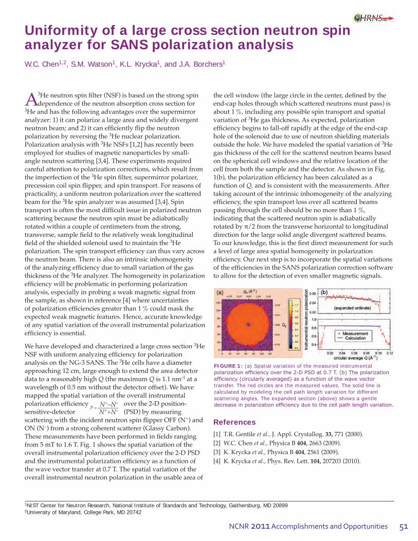
NCNR 2011 Accomplishments and Opportunities 51
Uniformity of a large cross section neutron spin analyzer for SANS polarization analysisW.C. Chen1,2, S.M. Watson1, K.L. Krycka1, and J.A. Borchers1
1NIST Center for Neutron Research, National Institute of Standards and Technology, Gaithersburg, MD 208992University of Maryland, College Park, MD 20742
A 3Heneutronspinfilter(NSF)isbasedonthestrongspindependence of the neutron absorption cross section for 3Heandhasthefollowingadvantagesoverthesupermirroranalyzer:1)itcanpolarizealargeareaandwidelydivergentneutronbeam;and2)itcanefficientlyfliptheneutronpolarization by reversing the 3He nuclear polarization. Polarizationanalysiswith3He NSFs [1,2] has recently been employed for studies of magnetic nanoparticles by small-angle neutron scattering [3,4]. These experiments required carefulattentiontopolarizationcorrections,whichresultfromthe imperfection of the 3Hespinfilter,supermirrorpolarizer,precessioncoilspinflipper,andspintransport.Forreasonsofpracticality, a uniform neutron polarization over the scattered beam for the 3Hespinanalyzerwasassumed[3,4].Spintransportisoftenthemostdifficultissueinpolarizedneutronscattering because the neutron spin must be adiabatically rotatedwithinacoupleofcentimetersfromthestrong,transverse,samplefieldtotherelativelyweaklongitudinalfieldoftheshieldedsolenoidusedtomaintainthe3He polarization.Thespintransportefficiencycanthusvaryacrossthe neutron beam. There is also an intrinsic inhomogeneity oftheanalyzingefficiencyduetosmallvariationofthegasthickness of the 3He analyzer. The homogeneity in polarization efficiencywillbeproblematicinperformingpolarizationanalysis,especiallyinprobingaweakmagneticsignalfromthesample,asshowninreference[4]whereuncertaintiesofpolarizationefficienciesgreaterthan1%couldmasktheexpectedweakmagneticfeatures.Hence,accurateknowledgeof any spatial variation of the overall instrumental polarization efficiencyisessential.
We have developed and characterized a large cross section 3He NSFwithuniformanalyzingefficiencyforpolarizationanalysis on the NG-3 SANS. The 3He cells have a diameter approaching 12 cm, large enough to extend the area detector data to a reasonably high Q (the maximum Q is 1.1 nm-1 at a wavelengthof0.5nmwithoutthedetectoroffset).Wehavemapped the spatial variation of the overall instrumental polarizationefficiency N+–N–
P = ––—––––– N++N–
over the 2-D position-sensitive-detector (PSD)bymeasuringscatteringwiththeincidentneutronspinflipperOFF(N+) and ON (N–) from a strong coherent scatterer (Glassy Carbon). Thesemeasurementshavebeenperformedinfieldsrangingfrom5mTto1.6T.Fig.1showsthespatialvariationoftheoverallinstrumentalpolarizationefficiencyoverthe2-DPSDandtheinstrumentalpolarizationefficiencyasafunctionofthewavevectortransferat0.7T.Thespatialvariationoftheoverall instrumental neutron polarization in the usable area of
thecellwindow(thelargecircleinthecenter,definedbytheend-capholesthroughwhichscatteredneutronsmustpass)isabout 1 %, including any possible spin transport and spatial variation of 3He gas thickness. As expected, polarization efficiencybeginstofall-offrapidlyattheedgeoftheend-caphole of the solenoid due to use of neutron shielding materials outside the hole. We have modeled the spatial variation of 3He gas thickness of the cell for the scattered neutron beams based onthesphericalcellwindowsandtherelativelocationofthecellfromboththesampleandthedetector.AsshowninFig.1(b),thepolarizationefficiencyhasbeencalculatedasafunction of Q,andisconsistentwiththemeasurements.Aftertaking account of the intrinsic inhomogeneity of the analyzing efficiency,thespintransportlossoverallscatteredbeams passing through the cell should be no more than 1 %, indicating that the scattered neutron spin is adiabatically rotatedbyπ/2fromthetransversehorizontaltolongitudinaldirection for the large solid angle divergent scattered beams. Toourknowledge,thisisthefirstdirectmeasurementforsucha level of large area spatial homogeneity in polarization efficiency.OurnextstepistoincorporatethespatialvariationsoftheefficienciesintheSANSpolarizationcorrectionsoftwaretoallowforthedetectionofevensmallermagneticsignals.
References
[1] T.R. Gentile et al., J. Appl. Crystallog. 33, 771 (2000).[2] W.C. Chen et al.,PhysicaB404, 2663 (2009). [3] K. Krycka et al.,PhysicaB404, 2561 (2009). [4] K. Krycka et al.,Phys.Rev.Lett.104, 207203 (2010).
FIGURE 1: (a) Spatial variation of the measured instrumental polarization efficiency over the 2-D PSD at 0.7 T. (b) The polarization efficiency (circularly averaged) as a function of the wave vector transfer. The red circles are the measured values. The solid line is calculated by modeling the cell path length variation for different scattering angles. The expanded section (above) shows a gentle decrease in polarization efficiency due to the cell path length variation.

52
Neutron S
ource Operations
Neutron Source Operations
The NCNR Source (NBSR) operated for 142 days this past year providing 99 % availability before shutting down,asscheduled,fortheOutage.TheExpansionOutage that began on April 3 has provided many opportunitiesforminorandmajorupgradestothe reactor systems for enhanced reliability of operations.
TominimizeinterferencewiththenewneutronguidesthatwillbeinstalledduringthecurrentOutageitwasnecessarytomovethemajorityoftheNCNRsecondarycoolingsystem.Thenewsystemwillreplacethesix,singlespeedMainSecondaryPumpswithfourenergy-efficient,variablespeedpumpsandwillprovideequivalentcoolingandflowrates.Inaddition,anewcoolingtowercellwillbeinstalledtosupplementthe22MWcoolingcapacityoftheexistingtowersduringthesummerwhenheatandhumiditylevelsreducethetowercoolingefficiency.Themajorityoftheinstallationof the upgrade to the secondary cooling system has been performed under an American Recovery and ReinvestmentAct(ARRA)projectbuttheReactorOperations/Engineering(ROE)grouppreparedthefull system for long-term dry layup of the piping and willberesponsibleforinterfacingthenewsystemto the reactor control room and bringing the system back into full service.
TheThermalShieldcoolingsystem’sfunctionisto cool a radiation shield that protects the concrete surrounding the NCNR source by circulating waterthroughcoolingtubes.Overthelifetimeofthefacility,thetubeshavedevelopedsmallwaterleaks that had become operationally inconvenient. PatchingtheleakswasonlyatemporaryrepairsotheNCNRdevelopedasystemthatutilizedlowvacuum
ratherthanpressure,tomovecoolingwateraroundtheThermal Shield. The strategy has been successfully tested in prototypes and full-scale mockups. During the current Outage, a complete upgrade of the Thermal Shield cooling systemhasbeeninstalledthatwillmovewaterbyusingaspirators to maintain the highest pressure in the system justbelowoneatmosphere,thuspreventingoutleaks.
ROE is performing a phased-upgrade to the reactor controlconsole.PhaseIinvolvesbuildinganinformationacquisitionsystemthatwillstoredataroutinelyavailabletothereactoroperators.Thisdatawillbeacquiredinreal-timeduringandusedtodevelopanewdisplayandcontrolsystemforthecontrolroomthatwillusecontemporaryhuman-machineinterface(HMI)best-practiceforPhaseII.PhaseIhasprogressedthroughouttheOutageandmanyof the older displays, alarm indicators, and controllers havebeenreplacedwithmodernsystemsthatwilloutputdirectly to the Console data-acquisition system. Initially, the systemwillprovideonlyaninternaldisplayofdataintheNCNR control room and an auxiliary HMI development laboratory but the data could be eventually provided to experimenters or other areas of the NCNR that might need to monitor the system remotely.
The installation of the second NCNR liquid hydrogen cold-source in BT-9 is the culmination of years of design anddevelopmentwork.ThesuccessfulsourceinstallationwasdelayedbecausebothBT-9andthefabricatedcoldsource required machining to compensate for distortions in the experimental tube and the small tolerance differences betweenthecoldsourceandthetubedimensions.ThesourcewasfullyinstalledinthefirstweekofSeptemberbutthereisstillagreatdealofworkbeforethesystemwillbefilledwithhydrogen.Completeinstallationandtestingis expected to occur in early 2012.Paul Liposky and Jim Moody work on the RT-4 pneumatic
transfer system.
Tony Norbedo examines the new stainless-steel lines on the upgraded Thermal Shield cooling system.

53NCNR 2011 Accomplishments and Opportunities
Facility Development
Facility support staff have taken advantage of the scheduled outage period of the Expansion Initiative programtoprogressdesignandconstructionofnewinstruments—scheduled for installation in the post-outageperiod—whiledevelopingandrefurbishingexistinginstrumentation,software,andsampleenvironment equipment.
Instrument Development
The past year has seen the preassembly and staging of the10mSANSinstrumenttobeinstalledonthenewNG-Bguide.Thediffractometerhasbeenoutfittedwithmotioncontrolsandotherancillaryelectronics,and adaptation of the 30 m SANS instrument control softwareisunderway.Thepre-staged10mSANSinstrumentwillalsoprovideatestbedforthenewInstrumentControlEnvironment(NICE)softwaresystem;thealphareleaseofthismajorrefactoringanddevelopmentprojectisscheduledearlyin2012.
TheFacilitysupportsagrowingprogramtodevelopandcommissionnewdetectorsystemstoaddresstheneedsofnewinstrumentsintheExpansionInitiativeandbeyond.ThedetectorprojectfortheCANDORwhite-beamreflectometerhasrecentlybeenawardedNIST intramural funding and these additional funds willexpeditedevelopmentofthedetectorandspeedtheprocessofbringingthefinaldesignintoproduction.Thecurrentdesignisbasedon6LiF/ZnS(Ag)impregnatedplasticscintillatortechnologiescoupledviawavelengthshiftingfiberstosiliconphotomultiplierdevices(SiPMs).The promising early results from a prototype detector haveledtorefinementsoftheneutronconverterportionofthedetectorthatwillbetestedinthenearterm.
The design of the Very Small Angle Neutron Scattering (vSANS)diffractometercallsfortwotypesofdetectors:a high-resolution area detector typical of most instruments of this type and arrays of linear position sensitivetubes.IndiscussionswithGEReuterStokes,adesignhasbeenfinalizedforeightpanelsconsistingof
“8-pack” linear position sensitive proportional counters for incorporation into the vSANS spectrometer. These panelarrayswillalsofeaturereadoutelectronicsbasedon the SNS data acquisition standard. Fabrication of the panelassembliesisunderwayanddeliveryisprojectedfor December 2011.
The primary detector for the MAGIK spectrometer, soontobeinstalledonthenewNG-Dguide,hasbeenprocured and integrated into data acquisition for the instrument. This detector, a DENEX 200TN, is a 20 cm × 20 cm 3Hegas-filledareadetectorwith1mm× 2 mm spatial resolution.
Data Analysis Software Development
The Data Analysis and Visualization Environment (DAVE)projectprovidessoftwaresolutionsfordatareduction,viewing,andinterpretationcoveringawiderange of inelastic neutron scattering instruments at the NCNRandelsewhere.DAVEisbasedontheIDLiToolparadigm and has been under development for close to adecade.Itisnowinitssecondmajordevelopmentalphase realizing improvements in many aspects of the applicationsuiteespeciallywithregardstousabilityandvisualization functionality.
FIGURE 1: A backlit picture of an assembly in which are embedded wavelength shifting fibers in a neutron detecting 6LiF-ZnS(Ag) scintillator matrix. The scintillation pulses travel via the fibers to silicon photomultiplier devices.

54
Facility Developm
ent
Coincidentwiththeextendedoutageperiod,workhasprogressedtoaddresssomelong-standingissuesandaddnewfeaturescoveringall aspects of DAVE functionality from planning tools, reduction and visualization to basic data analysis.Anewreductioncomponentdesignedfor the multi-channel detector system on BT-7 is being developed using an identical interface to the recently completed FANS reduction module. When complete,itwillreadilyenableloadingofmultiplerawdatasets,applynormalizationandotherrawdatacorrections,andcalculatethewavevectorandenergy transfer taking into account the variation in angle and energy across the multichannel detector.Diffractiondatafrompowdersamplesand diffraction or inelastic data from single crystal samples are also supported. In many cases, the reduced data are volumetric (multi-dimensional) and can thus be analyzed (sliced and cut) further using the DAVE Mslice tool.
SupportforMACSdatareductionwasintroducedlast year and since then users have had the opportunity to provide much feedback that has resultedinasignificantrevisionoftheinterfacealongwiththeadditionofnewfunctionality.The data reduction package for Neutron Spin Echohasbeenupgradedtoallowtreatmentofmultiplesampleandbackgrounddatasets,whichisespeciallyusefulwhenemployingpartialdeuteration techniques.
Anewreflectometryfittingpackage,Refl1D,has been released. Developed as part of the NSF fundedDANSEprojectforneutronscatteringsoftware,thispackageprovidesstateoftheartglobal optimization and uncertainty analysis softwaretothereflectometrycommunity.Themodelingengineallowsuserstocombinefreeformandslablayersintoprofilesandsimultaneouslyrefineprofilesformultipledatasets.Magnetismis fully supported, including polarized and unpolarizedmeasurementsfromtimeofflightand steady state sources. Users are free to extend theirmodelsinarbitraryways,includingsupportfor different measurement techniques; results
can be combined for a robust uncertainty assessment incorporating information from all measurements on thesample.DANSEhasalsoreleasedDIReflfordirectinversionanalysisonsurroundvariationreflectometrydata,yieldingprofileswithoutfitting,andOsReflforanalysis on in-plane scattering data from patterned magnetic and non-magnetic samples. Numerous publicationswithintheNCNRandfromthebroadercommunityincluderesultsfromDANSEreflectometrysoftwarepackages.(http://reflectometry.org/danse/software.html)
Sample Environment
TheNCNRprovidesawiderangeofstate-of-the-artsample environment equipment and technical expertise to support user research programs. The outage period has afforded time for a number of development andrefurbishmentprojects,inparticulartosupportoperationalcapacityandreliabilityoflow-temperaturehighmagneticfieldoperations.The10Tcryogen-freemagnethasbeenoutfittedwithacryogen-freevariable-temperatureinsertandisnowundertest.The7Tverticalfieldmagnetisnowredeployedwithits3He low-temperatureinsertandthe11.5Tverticalfieldmagnetwithdilutionrefrigeratorhasundergonerepairsand is being re-commissioned. Maintenance and repairs totheentirefleetofcryostatsandofbottom-loadingclosed-cyclerefrigeratorswillbecompletedintimeforthe re-start of Facility operations.
Neutron Spin Filters
NCNRsupportsaworld-classuser-drivenprogramof polarized beam experiments and demand for 3He neutronspinfilters(NSFs)continuestobestrongand increasing. The Facility program to support and developNSFsisrunincollaborationwiththePhysicalMeasurementLaboratoryatNISTandduringthepastyear has serviced 37 user experiments totaling over 130 days of beam time. Experiments are routinely carried out on the thermal neutron triple-axis spectrometer BT-7 and the suite of small-angle neutron scattering instruments andreflectometers.Theprogramwasexpandedthisyearwithproof-of-principleexperimentsperformedonMACSforwide-anglepolarizationanalysis.

55NCNR 2011 Accomplishments and Opportunities
3He gas is polarized by spin-exchange optical pumping (SEOP),inwhichalkali-metalatomsarepolarizedbyoptical pumping and this electronic polarization is transferred to 3He nuclei in spin-exchange collisions. EffortscontinuetodeveloptheSEOPsystemsand3He cells yielding improve to the overall 3He polarization performance.TheNSFteamhasbuilttwostate-of-the-artSEOPsystemsdedicatedfortheNCNRuserprograminpolarizedneutronscattering.EachSEOPsystemisequippedwithtwo100Wspectrallynarrowedlaserstoilluminate the gas cell from opposite sides, and this year sawimplementationofachirpvolumeBragggratingforeachlaser,whichhasdeliveredhigherpowerandimproved suppression of broadband background. The lasersnowroutinelyoperatecontinuouslyfordayswithonlyoccasionalneedforminorre-tuningofthewavelength.Thebestperformanceforlargevolumecells is achieved by the use of potassium-rubidium mixtures, but this requires careful control of the mix ratio in sealed cells and a standard protocol to optimize thisratiohasnowbeendeveloped.ThisenhancedSEOPcapability,combinedwithanadvancedcellfabricationfacility,allowsthepolarizationof1Lcellsto80%3He polarizationwithinadayortwo.Moreover,thisyearsawrecordbreakinglevelsof3He polarizations from 82 % to 85 % obtained for user experiments on both ANDR and BT-7.
There has been a continued focus on developing user-friendlyhardwareandsoftwaretocontrol3He NSFs that isreadilyincorporatedintotheinstrumentsoftware.Softwareisnowimplementedtopredictandmonitorthepolarizedneutronicperformanceandflipthe3He polarizationwhentakingdataoneachinstrument.Thesoftwareprovidesauser-friendlyinterfaceandallowstheinstrumentcomputertoperformNMRmeasurements,flipthe3He polarization and control the neutron spin. An alternative method for 3He, the polarization-flippingschemehasbeenimplemented
thatusesanamplitude-modulatedradio-frequencyfieldwhenperformingafrequency-sweepbasedadiabaticfastpassageNMR.Thenewschemepromisestoreducethe 3Hepolarizationlossperflipbyafactorofatleast4andwillbetestedonthebeamlineswhenfollowingthe source re-start in 2012. User-friendly polarization efficiencycorrectionsoftware,whichincludesthe3He polarization decay, is being implemented on the triple-axis spectrometer and SANS.
Progresshasbeenmadetoadvancepolarizedneutroninstrumentationforeachspecificinstrumentclass.OnBT-7,oneofthemostimportantadvanceswastodevelop polarized inelastic neutron instrumentation. A polarizedbeamsetupwithaconventionalprecessioncoilspinflipperwasnoteffectiveforelasticexperimentsat large scattering angles and inelastic experiments duetotheimpactofspaceconstraints.However,thesuccessful development of automated 3Hespinflippingcapabilityonthebeamlinenowenablesefficientflippingandtransporttheneutronspinwithouttheconventionalflipper.Thisenhancedmeasurementcapabilityhasmadethefirstsuccessfulpolarized-beaminelasticexperimentonBT-7withanenergytransferup to 35 meV. The successful development of a high-fieldmagicboxhasledtoimprovements—byafactorx3—in the 3He polarization relaxation enabling reliable operationwitharelaxationtimeofupto100h.
Development activity for SANS experiments has includedworktoincreasethemaximumsamplefieldavailablefrom1.2Tupto1.6T.Thiswasachievedbythemodificationofanexistinghighfieldelectromagnettominimizeitsstrayfieldusingsoft-ironplatesandenablethepassageofapolarizedbeam.Polarizedneutrontestshaveshowntheneutronspintransportwiththismagnetisnotanissueandtestsoftheeffectonthe relaxation of the 3He polarization are on-going.

56
Serving
Serving the Science and Technology Community
The mission of the NIST Center for Neutron Research is to assure the availability of neutron measurement capabilities to meet the needs of U.S. researchers from industry, academia and from other U.S. government agencies. To carry out this mission, the NCNRusesseveraldifferentmechanismstoworkwithparticipantsfromoutsideNIST,includinga competitive proposal process, instrument partnerships,andcollaborativeresearchwithNIST.
Proposal System
Most of the time on NCNR instruments is made availablethroughacompetitive,peer-reviewproposal process. The NCNR issues calls for proposalsapproximatelytwiceayear.Proposalsarereviewedatseveraldifferentlevels.First,expertexternal referees evaluate each proposal on merit and provideuswithwrittencommentsandratings.Thisisaverythoroughprocesswhereseveraldifferentrefereesrevieweachproposal.Second,theproposalsare evaluated on technical feasibility and safety byNCNRstaff.Third,weconveneourBeamTimeAllocationCommittee(BTAC)toassessthereviewsand to allocate the available instrument time. Using theresultsoftheexternalpeerreviewandtheirownjudgment,theBTACmakesrecommendationstothe NCNR Director on the amount of beam time to allocate to each approved experiment. Approved experiments are scheduled by NCNR staff members inconsultationwiththeexperimenters.
ThecurrentBTACmembersare:
• AndrewAllen(NISTCeramicsDivision)• JeffreyAllen(MichiganTechnologicalUniversity)• CollinBroholm(JohnsHopkinsUniversity)• LeslieButler(LouisianaStateUniversity)• KalinaHristova(JohnsHopkinsUniversity)• RamananKrishnamoorti(UniversityofHouston)• ValeryKiryukhin(RutgersUniversity)• RaulLobo(UniversityofDelaware)• LeeMagid(UniversityofTennessee,Knoxville)• JannaMaranas(ThePennsylvaniaState
University) • AlanNakatani • StephanRosenkranz(ArgonneNationalLaboratory)
• LynnWalker(Carnegie-MellonUniversity)
Partnerships
TheNCNRmayformpartnershipswithotherinstitutionsto fund the development and operation of selected instruments.Thesepartnerships,or“ParticipatingResearch Teams”, may have access to as much as 75 % of the available beam time on the instrument depending on the share of total costs borne by the team. A minimum of 25%oftheavailablebeamtimeisalwaysmadeavailablethrough the NCNR proposal program to all users. Partnershipsarenegotiatedforafixedperiod(usuallythreeyears)andmayberenewedifthereismutualinterestand a continued need. These partnerships have proven to beanimportantandeffectivewaytoexpandtheresearchcommunity’saccesstoNCNRcapabilitiesandhavebeenverysuccessfulindevelopingnewinstruments.
Collaboration with NIST
Some time on all instruments is available to NIST staff in supportofourmission.ThistimeisusedtoworkonNISTresearch needs, instrument development, and promoting thewidespreaduseofneutronmeasurementsinimportantresearchareas,particularlybynewusers.Asaresultoftheseobjectives,asignificantfractionofthetimeavailableto NIST staff is used collaboratively by external users, whooftentaketheleadintheresearch.Accessthroughsuchcollaborationsismanagedthroughwrittenbeamtime requests. In contrast to proposals, beam time requests arereviewedandapprovedinternallybyNCNRstaff.We encourage users interested in exploring collaborative
FIGURE 1: Research participants and publications at the NCNR 1986- 2011.
Dow ChemicalCompany)(The

57NCNR 2011 Accomplishments and Opportunities
research opportunities to contact an appropriate NCNR staff member.
Research Participation and Productivity
The NCNR continued its strong record of serving the U.S. research community this year. Over the 2011 reporting year,2265researchparticipantsbenefitedfromuseoftheNCNR.(ResearchparticipantsincludeuserswhocometotheNCNRtousethefacilityaswellasactivecollaborators,including co-proposers of approved experiments, and co-authorsofpublicationsresultingfromworkperformedattheNCNR.)Asthenumberofparticipantshasgrown,the number of publications per year has increased in proportion. The quality of the publications has been maintainedataveryhighlevel.Thetrendofthepastfewyears,however,suggeststhatthenumberofparticipantsand publications is beginning to saturate, as one might expect as the capacity of the facility is reached. The Expansion Initiative, currently in progress, promises increasing activity and productivity.
2011 NCNR Proposal Program
Onlyonecallforproposals,ratherthantheusualtwo,wasissued in the past year, because of the outage that began in April 2011 for the NCNR Expansion Initiative. We received 357proposals,ofwhich170wereapprovedandreceivedbeam time. The oversubscription, i.e., the ratio of days requestedonallproposalstothedaysavailable,was2.9ontheaverage,butashighas6forspecificinstruments.ProposaldemandhasgrownconstantlysincetheNCNRfirstbeganacceptingproposalsin1991,andhasdoubledinthepasteightyears.Thefollowingtableshowsthedataforseveral instrument classes.
Instrument class Proposals Days requested Days allocatedSANS and USANS 119 458 175Reflectometers 56 417 156Spectrometers 163 1081 341Diffraction 7 27 12Imaging 12 60 26Total 357 2043 710
User Group Conducts Survey
The NCNR Users Group (NUG) provides an independent forum for all facility users to raise issues to NCNR management,workingthroughitsexecutiveofficerstocarryoutthisfunction.ThecurrentofficersareMarkDadmun (University of Tennessee, chair), John Katsaras (OakRidgeNationalLaboratory),MichelKenzelmann(PaulScherrerInstitute,Switzerland),DespinaLouca(University of Virginia), Dale Schaefer (University of Cincinnati),LynnWalker(Carnegie-MellonUniversity),and Erik Watkins (University of California, Davis). In
December 2011, the NUG conducted a comprehensive survey of user satisfaction and concerns, addressing areas suchassupportfromNCNRstaff,proposalreview,sampleenvironments,instrumenthardwareandsoftware,dataanalysis, and user laboratories. Many useful comments werereceivedinthe327responses.Asaresult,severalpolicychangesareplanned,beginningwitharevampingofour radiation safety training process for incoming users.
Panel of Assessment
ThemajororganizationalcomponentsofNISTareevaluated annually for quality and effectiveness by the National Research Council (NRC), the principal operating agency of both the National Academy of Sciences and the National Academy of Engineering. A panel appointed by the NRC reported on the NIST Center of Neutron Research inJanuary2011.Theirfindingsforthepreviousyeararesummarizedinadocumentthatmaybeviewedonlineathttp://www.nist.gov/director/nrc/upload/NR-Panel-2010-Report-Final-9-23-10.pdf The 2011 panel members included Tonya Kuhl of the University of California, Davis (chair), Meigan Aronson , Stony Brook University, Frank Bates, University of Minnesota, Donald Engelmann, YaleUniversity,PaulFleury,YaleUniversity,ChristopherGould,NorthCarolinaStateUniversity,AlanHurd,LosAlamosNationalLaboratory,JamesLee,SandiaNationalLaboratory,JohnParise,StonyBrookUniversity,andSunilSinha, University of California, San Diego.
The Center for High Resolution Neutron Scattering (CHRNS)
CHRNSisanationaluserfacilitythatisjointlyfundedby the National Science Foundation and the NCNR. Its primary goal is to maximize access to state-of-the-art neutron scattering instrumentation for the research community. It operates seven neutron scattering
FIGURE 2: User ratings from the survey; the top rating is 5.

58
Serving
instruments at the NCNR, enabling users from around the nation to observe dynamical phenomena involvingenergiesfrom≈30neVto≈100meV,andto obtain structural information on length scales from 1nmto≈10μm.AmoredetailedaccountofCHRNSactivities may be found on pp 59-61 of this report.
Partnerships for Specific Instruments NG-7 SANS Consortium
A consortium that includes NIST, the Industrial PartnershipforResearchinInterfacialandMaterialsEngineering(IPRIME)ledbytheUniversityofMinnesota, and the ExxonMobil Research and Engineering Company, operates, maintains, and conducts research at the NG-7 30 m SANS instrument. The consortium uses 57 % of the beam timeonthisinstrument,withtheremaining43%allocatedtothegeneralscientificcommunitythroughtheNCNR’sproposalsystem.Consortiummembersconduct independent research programs primarily in the area of large-scale structure in soft matter. For example, ExxonMobil has used this instrument to deepen their understanding of the underlying nature ofExxonMobil’sproductsandprocesses,especiallyinthefieldsofpolymers,complexfluids,andpetroleum mixtures.
NIST/General Motors – Neutron Imaging
AnongoingpartnershipandcollaborationbetweenGeneral Motors and NIST continues to yield exciting results using neutron imaging. Neutron imaging has been employed to visualize the operation of fuel cells and lithium-ion batteries for automotive vehicle applications. Neutron imaging is an ideal method for visualizing both hydrogen and lithium, the fuel of electric vehicles engines. These unique, fundamental measurements, provide valuable materialcharacterizationsthatwillhelpimprovethe
performance, increase the reliability, and reduce the time to market introduction of the next generation electric car engines.TheGM/NISTpartnershipisentitledto25%ofthe time on the BT-2 Neutron Imaging Facility.
Interagency Collaborations
TheSmithsonianInstitution’sNuclearLaboratoryforArcheological Research is part of the Anthropology Department at the National Museum of Natural History. Ithashadaproductive33yearpartnershipwiththeNCNR,duringwhichtimeithaschemicallyanalyzedover42,500 archaeological artifacts by Instrumental Neutron ActivationAnalysis(INAA),drawingextensivelyonthecollectionsoftheSmithsonian,aswellasonthoseofmany other institutions in this country and abroad. Such chemical analyses provide a means of linking these diverse collections together in order to study continuity and change involved in the production of ceramic and other artifacts.
The Center for Food Safety and Applied Nutrition, U.S. Food and Drug Administration (FDA), directs and maintains a multi-laboratory facility at the NCNR thatprovidesagency-wideanalyticalsupportforfoodsafety and food defense programs. Neutron activation (instrumental, neutron-capture prompt-gamma, and radiochemical),x-rayfluorescencespectrometry,andlow-level gamma-ray detection techniques provide diverse multi-element and radiological information about foods and related materials. Current studies include preparation ofswordfishin-housereferencematerial,revalidationofcocoapowderin-housereferencematerial,measuringcesium levels in soil to study cesium uptake behavior into food products, screening food products for dangerous levels of cadmium, lead, and mercury in beverages and residual bromine in baked goods, and evaluation of procedural losses of mercury, arsenic, selenium, and bromine from foods.

59NCNR 2011 Accomplishments and Opportunities
The Center for High Resolution Neutron Scattering (CHRNS)
The Center for High Resolution Neutron Scattering (CHRNS)isanationaluserfacilitythatisjointlyfundedby the National Science Foundation through its Division of Materials Research (grant number DMR-0944772), and by the NCNR. The primary purpose of this partnership is to maximize access to state-of-the-art neutron scattering instrumentation for the research community using the NCNR’sproposalsystem.ProposalstousetheCHRNSinstrumentsarecriticallyreviewedonthebasisofscientificmeritand/ortechnologicalimportance.
ThecoremissionofCHRNSisfourfold:(i)todevelopandoperateneutronscatteringinstrumentation,withbroadapplication in materials research, for use by the general scientificcommunity;(ii)topromotetheeffectiveuseoftheCHRNSinstrumentsbyhavinganidentifiablestaffwhoseprimary function is to assist users; (iii) to conduct research that advances the capabilities and utilization of CHRNS facilities; and (iv) to contribute to the development of human resources through educational and outreach efforts.
Scattering Instruments and Research
CHRNSsupportsoperationofthefollowinginstruments:
• theNG-330mSmallAngleNeutronScattering(SANS)instrument,
• theUltra-SmallAngleNeutronScattering(USANS)instrument,
• theSpin-PolarizedInelasticNeutronScattering(SPINS)spectrometer,
• theMulti-AngleCrystalSpectrometer(MACS),• theDiskChopperSpectrometer(DCS),• theHighFluxBackscatteringSpectrometer(HFBS),and• theNeutronSpin-Echo(NSE)spectrometer.
The small angle scattering instruments provide structural informationoverlengthscalesfrom≈1nmto≈10μm.Thespectrometers collectively yield dynamical information overtimescalesfrom≈3×10-14sto≈10-7 s (energy scalesfrom≈100meVto≈30neV).Thesewiderangesof accessible distances and times support a very diverse scientificprogram,allowingresearchersinmaterialsscience, chemistry, biology, and condensed matter physics to investigate materials such as polymers, metals, ceramics, magneticmaterials,porousmedia,fluidsandgels,andbiological molecules.
InthemostrecentCallforProposals(call27),258proposalsrequestedCHRNSinstruments,ofwhich106receivedbeam time. Of the 1359 days requested for the CHRNS instruments,453wereawarded.Thecorrespondingnumbersforallinstrumentswere357proposalsreceived,171 proposals approved, 2043 days requested, and 712 days awarded.Roughlyhalfoftheusersofneutronscatteringtechniques at the NCNR use CHRNS-funded instruments, and more than one third of NCNR publications (see the “Publications”sectiononp.65),overthecurrentone-year period, are based on research performed using these instruments.In2010morethan25Ph.D.dissertationswerecompletedusingresultsfromCHRNS-sponsoredinstruments.
Scientific Support Services
An important ingredient of the CHRNS operation is itssupportofstaffwhoseprincipalresponsibilityisto provide services that are essential to the success of neutron scattering investigations. One such service is the provision and operation of an expanding range of sample
FIGURE 1: Participants in the NCNR 2011 Summer School “Methods and Applications of High Resolution Neutron Spectroscopy and Small Angle Neutron Scattering.”

60
CH
RN
S
environmentequipment,coveringlowandhightemperatures, elevated pressures, gas handling, magneticfields,motionandtemperaturecontrolandelectronics. This past year sample environment team members concentrated their efforts on providing service to users so that they could successfully run their experiments before the scheduled long outage. Duringtheoutagetheyworkedonavarietyofprojectsdesignedtoensurethattheequipmentworksproperlyandmeetstheusers’needs.
Theseprojectshaveincluded
• installationandtestingofanewvariabletemperature insert for the 10 T magnet,
• deploymentofa3He refrigerator for the 7 T magnet,
• re-commissioningofthe11.5Tmagnetwithdilution refrigerator,
• maintenanceandrefurbishmentoftheentireheliumcryostatfleet,
• maintenanceofthebottom-loadingclosed-cyclerefrigerators, including testing and deployment of swivelfittingstoaidinhosemanagement.
Other CHRNS-supported services include the maintenance ofseveralwell-equippeduserlaboratories, and the development, documentation and maintenance of data reduction, visualization, andanalysissoftwarethroughtheDAVEproject.
TheDAVEsoftwarecontinuesto be maintained and developed tokeepabreastwithfeaturerequestsandwithimprovementsin instrumentation. DAVE is
distributed as a desktop application and is available free of charge from thewebsitehttp://www.ncnr.nist.gov/dave/,either as a binary executablewithanembeddedruntime license or as the complete source code. Our development efforts over the past year have been focused on improving and extending the planning, reduction and analysis of data collected on supported instruments, especially the triple axis instruments MACS and BT-7. Both instruments require sophisticated data treatment to handle the large datasets accumulated using multi-detector systems.
Education and Outreach
One of the principal missions of the CHRNS program is to contribute to the development of human resources through education and outreach, thereby fostering future generations of scientists and neutron scatterers.
The CHRNS summer schools are very effective in educatingpotentialnewusersofneutronscatteringsincetypically more than 70 % of the participants return to theNCNRfortheirownresearch.Thereactorshutdownin 2011 provided a unique opportunity to enhance and expand the summer school offerings. A school entitled “Methods and Applications of High Resolution Neutron Spectroscopy and Small Angle Neutron Scattering” washeldonJune12-16,andaschoolon“MethodsandApplications of Cold and Thermal Neutron Spectroscopy” washeldthefollowingweek,fromJune19-23.Togetherthe schools served 62 students, almost double the number servedinatypicalyear.Lectures,researchseminars,and hands-on instruction in data reduction and analysis techniqueswerehighlightedforawidevarietyofneutronmeasurement techniques from spin echo to triple-axis
FIGURE 2: The 2011 SURF students, NCNR director Rob Dimeo (left), and CHRNS director Dan Neumann (second from right).
FIGURE 3: Participants in the NIST Summer High School Internship Program, flanked by Dan Neumann (left) and Rob Dimeo (right).

61NCNR 2011 Accomplishments and Opportunities
spectroscopy.Thestudenttostaffratiowaslow,asinprevious years, and the students had many opportunities toconsultwiththeguestlecturers.Anewfeatureofthisyear’sschoolswasanoralsessioninwhichthestudentsintroducedthemselvesandgavetwominutepresentationsdescribing their current research. NCNR staff members agreed that the introductory session enabled them to better tailortheirinstructiontothespecificbackgroundsofthestudents.
Another school, entitled “Modeling Neutron Data of BiologicalSystems”,washeldattheNCNRonJuly11-15,2011.The21studentattendeesweretaughtmodern atomisticapproachestomodelingSANS,reflectivity,andbackscattering data. The modeling methods included molecular dynamics and molecular Monte Carlo simulations of proteins and protein-nucleic acid systems. The school also featured an introductory tutorial prepared byPrasadGerardfromPoolesvilleHighSchool,whoworkedwiththeschoolorganizersduringthesummeraspart of the RET (Research Experience for Teachers) program at the NCNR.
The “Second School on Representational Analysis and MagneticStructures”washeldatGeorgetownUniversityon July 31 – August 5. The school highlighted guest lectures on representational analysis of inherent symmetries in complexmagneticstructures.Therewere21participants,manyofwhomweretreatedtoatouroftheNCNRfacilitiesearlierintheweek.
In2011CHRNSonceagainparticipatedinNIST’sSummerUndergraduateResearchFellowship(SURF)program,thistimehostingtenstudentswhoworkedwithstaffmembersstudyingtopicssuchasthin-filmdeposition,x-raycharacterization,biologicalsampledeuteration,web-baseddata reduction, and reactor control systems development. TwoofthestudentswerestationedattheInstituteforBioscience and Biotechnology Research. The students‘accomplishmentswerehighlightedin oral presentations that they gave during the SURF colloquium in early August.
The NCNR also took part in the NIST Summer HighSchoolInternshipProgram,knownasSHIP.SixstudentsfromfourlocalhighschoolsperformedresearchwithNCNRstaffmembers.Threeofthemworkedasateamtodevelopweb-baseddataanalysiscapabilitiesforSANS,reflectometry,andtripleaxismeasurements.Theothershadprojectsthatinvolvedfabricationofmagnetic refrigerants, the analysis of magnetic reflectionsincrystaldiffractiondata,andthe
extensionofNIST-authoredFiPysoftwaretotheanalysisofthinfilmsofphotomagneticmaterials.Thestudentspresented their results in an oral session at the NCNR and ataNIST-widepostersession.
The NCNR once again participated in the annual “Summer InstituteforMiddleSchoolScienceTeachers.”Thistwoweekprogram,sponsoredbyNIST,providesmiddleschoolteacherswithanenhancedunderstandingofthescientificprocess.Thisyeartheprogramhostedtwenty-oneteachersfrom Maryland, Virginia, Washington DC, Illinois, North Carolina, and South Carolina. On July 25 the group toured the NCNR and attended lectures and demonstrations thatdescribedhowneutronsareusedtoprobematter.TheywerealsoprovidedtheopportunitytointeractwithSURFstudents,SHIPstudents,andthescientificstaff.Thehighlightofthepresentationasalwayswasthemousetrapand ping pong ball demonstration of a chain reaction. In their evaluations, the teachers indicated that they had gainedagreaterappreciationofwhatcanbeaccomplishedusing neutron scattering.
Other education and outreach activities during the year included“TakeYourDaughtersandSonstoWorkDay”in April. Every year NIST hosts more than 250 elementary andmiddleschoolstudents.Aspartofthissite-wideyearly program, the NCNR provides tours of the facility, lectures, and specially designed demonstrations that showmiddleschoolagechildrenhowneutronsareusedto probe matter. In addition, the NCNR provided tours to elementary, middle and high school students from private and public schools. The students from the magnet programsatRobertoClementeandTakomaParkmiddleschoolswereparticularlyimpressedwiththeinteractivedemonstration of liquid nitrogen and its applications in sample environment.
FIGURE 4: Middle school science teachers and NCNR/CHRNS staff members.

62
Aw
ards 2011
Awards 2011
NCNR’s Hui Wu is the 2010 recipient of the (Professor Surhain) Sidhu Award of the Pittsburgh Diffraction Society “for her exceptional contribution
to the structural investigation of new materials for energy storage applications.” The award goes to an outstanding scientist who is within 6 years of having earned the Ph.D. or its equivalent. The prize, bestowed biannually, carries a cash prize of $2000. Dr. Wu received the award at the Society conference where she also presented the Sidhu Award Lecture.
Daniel Hussey, an instrument scientist for the Neutron Imaging Facility of the NCNR, received a 2010 Presidential Early Career Awards for Scientists and Engineers (PECASE) for showing scientific excellence at an early stage of his career. In the recent
past, he has played a key role in the development of neutron imaging methods for hydrogen fuel cell research for automotive applications. He is also involved in a project aimed at the improvement of Li-ion batteries, as well as in several other projects relevant to the structure of materials and the fundamental properties of the neutron. Dr. Hussey is currently designing an advanced cold-neutron phase imaging instrument for neutron radiography.
NCNR’s Craig Brown received a Department of Commerce Silver Medal. He is being recognized “For critical advances in our understanding of new materials for hydrogen energy storage in next-generation clean automobiles.”
Reactor Health Physicist Jim Clark received a Department of Commerce Silver Medal.The citation reads as follows: “For leadership in managing the recovery and restoration of the NIST Boulder laboratories that were contaminated as a result of the Plutonium Spill Incident of June 9, 2008.”
NCNR’s Daniel Dender and Julia K. Scherschligt have won the 2010 NIST Safety Award. The purpose of this award is “to recognize NIST
employees and organizations for substantial contributions to improving safety at NIST.” Julia and Dan are being recognized for their “innovative design, used nationwide, of a robust safety system to prevent accidental build-up of dangerous over-pressure conditions in cryostats.”
NCNR’s Matt Hudson was the co-winner of Sigma Xi’s annual NIST Post-doc Poster Presentation in the area of Materials for his poster entitled “CO2 Separation and Hydrogen Storage Properties of Zeolites for Clean Energy
Applications.”
NCNR’s Efrain Rodriguez was named a runner-up of Sigma Xi’s annual NIST Post-doc Poster Presentation in the area of Materials, for his poster entitled “Neutron Scattering of New Iron-Based Superconductors.”

63
NCNR’s Alan Munter has received a Special Act Award from the NIST Safety Office for his “significant role in the chemical inventory migration of all NIST Organizational Units.”
NCNR 2011 Accomplishments and Opportunities
Matt Helgeson has been awarded the 2011 Victor K. LaMer award from the Division of Colloid & Surface Chemistry of the American Chemical Society. Helgeson received his Ph.D. in chemical engineering from the University of Delaware in 2009 making many of his
dissertation measurements at the NCNR. He is currently a postdoctoral associate in the Novartis-MIT Center for Continuous Manufacturing.
In March 2011, Robert J. Cava of Princeton University received the ACS Award in Inorganic Chemistry from the American Chemical Society. He has also received the Gutenberg Lecture
Award worth €10,000 from the MAINZ Graduate School for his research on superconductivity. Cava, who was a NIST post-doc in the late 1970’s, has 500 publications which have been cited 30,000 times. 100 of these papers are in collaboration with NCNR scientists.
The Canadian Association of Physicists and the Division of Condensed Matter and Materials Physics has awarded the 2011 CAP/DCMMP Brockhouse Medal to Bruce D. Gaulin, McMaster University, for his internationally recognized
contributions to the field of collective phenomena in magnetic, superconducting and structural systems using x-ray and neutron scattering techniques. Gaulin is a frequent facility user at the NCNR.
In the student paper competition at the 2010 Canadian Association of Physicists congress, McMaster University graduate students Bruce Gaulin, Patrick Clancy and Kate Ross, won 1st and 3rd place, respectively, in the division of Condensed Matter and Materials Physics. Ross and Clancy carried out many of their neutron measurements at the NCNR. Patrick Clancy went on to be awarded one of 70 inaugural $140,000 (Canadian $) two-year Banting postdoctoral fellowships.
Ioanna Bakaimi of the Institute of Electronic Structure & Laser (IESL), Foundation for Research & Technology - Hellas (FORTH) won an award at the XXVI Panhellenic Conference on Solid State Physics and Materials Science
for the best oral presentation: “Frustration and Induced Magnetodielectric Coupling in NaMgO2 Polymorphs.” The work was largely carried out while Bakaimi was guest scientist at the NCNR.

64
Publications
Publications: August 1, 2010 to July 31, 2011
Aaron,D.S.,Borole,A.P.,Hussey,D.S.,Jacobson,D.L.,Yiacoumi,S.,Tsouris,C.,“QuantifyingtheWaterContent in the Cathode of Enzyme Fuel Cells via NeutronImaging,”J.PowerSources196(4), 1769 (2011).
Akcora,P.,Kumar,S.K.,GarciaSakai,V.,Li,Y.,Benicewicz,B.C.,Schadler,L.S.,“SegmentalDynamicsinPMMA-GraftedNanoparticleComposites,” Macromol. 43(19), 8275 (2010). [CHRNS]
Angelova,L.V.,Terech,P.,Natali,I.,Dei,L.,Carretti,E.,Weiss, R.G., “Cosolvent Gel-like Materials from PartiallyHydrolyzedPoly(vinylacetate)sandBorax,”Langmuir,inpress.
Armstrong,C.L.,Kaye,M.D.,Zamponi,M.,Mamontov,E., Tyagi, M., Jenkins, T., Rheinstädter, M.C., “DiffusioninSingleSupportedLipidBilayersStudied by Quasi-Elastic Neutron Scattering,” Soft Matter 6, 5864 (2010). [CHRNS]
Aronson,M.,Kim,M.,Bennett,M.,Janssen,Y.,Sokolov,D.A.,Wu,L.,“FieldTuningandQuantumCriticalityinYtterbiumBasedHeavyElectronCompounds,”J.LowTemp.Phys.161, 98 (2010). [CHRNS]
Asada,M.,Gin,P.,Endoh,M.K.,Satija,S.K.,Taniguchi,T., Koga, T., “Directed Self-Assembly of NanoparticlesatthePolymerSurfacebyHighlyCompressible Supercritical Carbon Dioxide,” Soft Matter, in press.
Babcock, E., Boag, S., Beecham, C., Chupp, T.E., Gentile, T.R.,Jones,G.L.,Petukhov,A.K.,Walker,T.G.,“Effects of High Intensity Neutron Flux on In-situ Spin-exchangeOpticalPumpingof3He,”J.Phys.:Conf. Series 294, 012011 (2011).
Bai,Z.,Xie,Y.,Chen,C.,Cosgrove,T.,Qiu,D.,“A Small-Angle Neutron Scattering Study of Poly(ethyleneoxide)MicrostructureinAqueousPoly(styrenesulfonateSodium)Solutions,” J. Colloid Interface Sci. 358, 226 (2011).
Balzar, D., “Coherency Strain and Dislocations in Copper-Rich-PrecipitateEmbrittledA710FerriticSteels,” Metall. Mater. Trans. A, in press.
Bao, W., Huang, Q., Chen, G.F., Green, M.A., Wang, D.M.,He,J.B.,Wang,X.Q.,Qiu,Y.,“ANovelLargeMoment Antiferromagnetic Order in K0.8Fe1.6Se2 Superconductor,”ChinesePhys.Lett.28(8), 086104-1 (2011).
Bianoco,C.L.,Schneider,C.S.,Santonicola,M.,Lenhoff,A.M., Kaler, E.W., “Effects of Urea on the Microstructure andPhaseBehaviorofAqueousSolutionsofPoly(oxyethylene)Surfactants,”Ind.Eng.Chem.Res.50, 85 (2011). [CHRNS]
Bloch,E.D.,Murray,L.J.,Queen,W.,Chavan,S.,Maximoff,S.N.,Bigi,J.P.,Krishna,R.,Peterson,V.K.,Grandjean,F.,Long,G.J.,Smit,B.,Bordiga,S.,Brown,C.M.,Long,J.R.,“Selective Binding of O2 over N2 in a Redox-Active Metal-OrganicFrameworkwithOpenIron(II)CoordinationSites,” J. Am. Chem. Soc., in press.Borreguero, J.M., He, J., Meilleur,F.,Weiss,K.,Brown,C.M.,Myles,D.A.,Herwig,K.W.,Agarwal,P.K.,“Redox-PromotingProteinMotionsin Rubredoxin,” J. Am. Chem. Soc. 115, 8925 (2011).
Borreguero,J.M.,He,J.,Meilleur,F.,Weiss,K.,Brown,C.M.,Myles,D.A.,Herwig,K.W.,Agarwal,P.K.,“Redox-PromotingProteinMotionsinRubredoxin,”J.Am.Chem.Soc. 115, 8925 (2011). [CHRNS]
Braisted,J.,Schneider,E.,O’Kelly,S.,VanderHoeven,C.,“DesignofanIrradiationFacilitywithaReal-TimeRadiation Effects Monitoring Capability,” Nucl. Instrum. Meth. A, in press.
Braunagel, J., Junghans, A., Koper, I., “Membrane-Based Sensing Approaches,” Australian J. Chem. 64(1), 54 (2011).
Burgess,I.J.,“ApplicationsofNeutronReflectivityinBiochemistry,” in “Advances in Electrochemical Science and Engineering, Volume 13 – Bioelectrochemistry,” editedbyAlkire,R.C.,Kolb,D.M.,Lipkowski,J.(Wiley-VCH, Weinheim, Germany), Chap. 3, 143 (2011).
Burress, J., Srinivas, G., Ford, J., Simmons, J.M., Zhou, W., Yildirim,T.,“GrapheneOxideFrameworkMaterials:TheoreticalPredictionsandExperimentalResults,”Angew.Chem.Int.Edit.49, 8902 (2010).
Butch,N.P.,Jeffries,J.R.,Chi,S.X.,Leão,J.B.,Lynn,J.W.,Maple,M.Brian,“AntiferromagneticCriticalPressurein URu2Si2UnderHydrostaticConditions,”Phys.Rev.B82(6), 060408-1 (2010).
Butch,N.P.,Jeffries,J.R.,Evans,W.J.,Chi,S.X.,Leão,J.B.,Lynn,J.W.,Sinogeikin,S.V.,Hamlin,J.,Zocco,D.A.,Maple,M.B.,“ThePressure-TemperaturePhaseDiagramof URu2Si2 Under Hydrostatic Conditions” in “Basic Actinide Science and Materials for Nuclear Applications,” edited by Gibson, J.K., McCall, S.K., Bauer, E.D., Soderholm,L.,Fanghaenel,T.,Devanathan,R.,Misra,A.,Trautmann,C.,Wirth,B.D.,(Mater.Res.Soc.Symp.Proc.,CambridgeUniv.Press,April2010,SanFrancisco,CA)1264, Z10-03 (2010).

65NCNR 2011 Accomplishments and Opportunities
Cappelletti,R.L.,editor,“2010NISTCenterforNeutronResearchAccomplishmentsandOpportunities,”NISTSP1110 (2010).
Chan,A.S.W.,Groves,M.N.,Li,X.,Malardier-Jugroot,C., “Conformation of pH Responsive Alternating Copolymers and Self-assembly in Nanoarchitectures” in “TechConnectWorld2010Proceedings:Nanotechnology2010Volume1-AdvancedMaterials,CNTs,Particles,Films and Composites,” (NSTI, June 2010, Anaheim, CA) Chap. 1, 41 (2010).
Chang,L.J.,Schweika,W.,Kao,Y.-J.,Chou,Y.Z.,PerBon,J.,Bruckel,T.,Yang,H.C.,Chen,Y.Y.,Gardner,J.S.,“Magnetic Correlations in HoxTb2-xTi2O7,”Phys.Rev.B83(14), 14413-1 (2011).
Chen,L.,Hu,F.X.,Wang,J.,Shen,J.,Sun,J.R.,Shen,B.G.,Yin,J.H.,Pan,L.Q.,Huang,Q.,“EffectofPost-AnnealingonMartensitic Transformation and Magnetocaloric Effect in Ni45Co5Mn36.7In13.3Alloys,”J.Appl.Phys.109(7), 07A939-1 (2011).
Chen,S.,Zhang,Y.N.,“DynamicCrossoverPhenomenoninConfinedWaterandItsRelationtotheLiquid-LiquidCriticalPoint:ExperimentsandMDSimulations”in“ProceedingsoftheInternationalSchoolofPhysics‘EnricoFermi’,CourseCLXXVI,”editedbyMallamace,F.,Stanley,E.,(SIFandIOSPress,June2010,Varenna,Italy),in press. [CHRNS]
Chen,W.,Gentile,T.R.,Fu,C.B.,Watson,S.,Jones,G.L.,McIver,J.W.,Rich,D.R.,“Polarized3He Cell Development andApplicationatNIST,”J.Phys.:Conf.Series294, 012003 (2011).
Chen,W.,Wang,J.,Wei,X.,Balazs,A.C.,Matyjaszewski,K.,Russell,T.P.,“UV-EnhancedOrderinginAzobenzene-ContainingPolystyrene-block-Poly(n-ButylMethacrylate)Copolymer Blends,” Macromol. 44, 278 (2011).
Chen,W.,Wei,X.,Balazs,A.C.,Matyjaszewski,K.,Russell,T.P.,“PhaseBehaviorandPhotoresponseofAzobenzene-ContainingPolystyrene-block-poly(n-butylmethacrylate)Block Copolymers,” Macromol. 44(5), 1125 (2011).
Cheng,G.,Liu,Z.,Murton,J.K.,Jablin,M.,Dubey,M.,Majewski,J.,Halbert,C.,Browning,J.,Ankner,J.F.,Akgun,B.,Wang,C.,Esker,A.R.,Sale,K.L.,Simmons,B.,Kent,M.S.,“NeutronReflectometryandQCM-DStudyoftheInteractionofCellulaseswithFilmsofAmorphousCellulose,” Biomacromol. 12, 2216 (2011).
Chi,S.X.,Lynn,J.W.,Chen,Y.,Ratcliff,II,W.,Ueland,B.,Butch,N.P.,Saha,S.R.,Kirshenbaum,K.,Paglione,J.,“Nitrogen Contamination in Elastic Neutron Scattering,” Meas. Sci. Tech. 22, 047001-1 (2011).
Choi,S.,Bates,F.S.,Lodge,T.P.,“MolecularExchangeinOrdered Diblock Copolymer Micelles,” Macromol. 44, 3594 (2011).
Choi,S.,Lee,S.,Soto,H.E.,Lodge,T.P.,Bates,F.S.,“NanoscaleMixing of Soft Solids,” J. Am. Chem. Soc. 133, 1722 (2011).
Chu,X.,Liu,K.,Tyagi,M.,Mou,C.,Chen,S.,“Low-TemperatureDynamicsofWaterConfinedinaHydrophogicMesoporousMaterial,”Phys.Rev.E82(2), 020501-1 (2010). [CHRNS]
Chuang,A.C.,Liu,Y.,Udovic,T.J.,Liaw,P.K.,Yu,G.,Huang,J., “Inelastic Neutron Scattering Study of Hydrogenated (Zr55Cu30Ni5Al10)99Y1BulkMetallicGlass,”Phys.Rev.B83(17), 174206-1 (2011).
Chung,J.-H.,Song,Y.,Yoo,T.,Chung,S.,Lee,S.,Kirby,B.J.,Liu,X.,Furdyna,J.K.,“InvestigationofWeakInterlayerExchangeCouplingintheGaMnAs/GaAsSuperlatticewithInsulatingNonmagneticSpacers,”J.Appl.Phys.110(1), 013912 (2011).
Chung,S.,Lee,S.,Chung,J.,Yoo,T.,Lee,H.,Kirby,B.J.,Liu,X.,Furdyna,J.K.,“GiantMagnetoresistanceandLong-Range Antiferromagnetic Interlayer Exchange Coupling in(Ga,Mn)As/GaAs:BeMultilayers,”Phys.Rev.B82(5), 054420-1 (2010).
Chung,Y.S.,Shin,N.,Kang,J.,Jo,Y.,Prabhu,V.M.,Satija,S.K.,Kline,R.J.,DeLongchamp,D.M.,Toney,M.F.,Loth,M.A.,Purushothaman,B.,Anthony,J.E.,Yoon,D.Y.,“Zone-RefinementEffectinSmall-Molecule-PolymerBlendSemiconductors for Organic Thin-Film Transistors,” J. Am. Chem. Soc. 133, 412 (2011).
Cicerone, M.T., Zhong, Q.N., Johnson, J., Aamer, K.A., Tyagi, M., “Surrogate for Debye-Waller Factors from Dynamic StokesShifts,”J.Phys.Chem.Lett.2, 1464 (2011). [CHRNS]
Climent-Pascual,E.,Ni,N.,Jia,S.,Huang,Q.,Cava,R.J.,“PolymorphisminBaPb1-xBixO3 at the Superconducting Composition,”Phys.Rev.B83(17), 174152-1 (2011).
Couet,A.,Motta,A.T.,Comstock,R.J.,Paul,R.L.,“ColdNeutronPromptGammaActivationAnalysis,aNon-destructiveTechniqueforHydrogenLevelAssessmentinZirconium Alloys,” J. Nucl. Mater., in press.
Curtis,J.,Bates,F.S.,Fultz,B.,Harmon,B.N.,Herwig,K.W.,Myles,D.A.A.,Rez,P.,Trouw,F.,Fultz,B.,“Nanostructures:StructureandDynamics,”in“Workshop on Computational Scattering Science,” edited byFultz,B.,Herwig,K.W.,Long,G.G.(WorkshopRep.,July2010,ArgonneNationalLaboratory,Argonne,IL),24(2010).
DaLuz,M.S.,Neumeier,J.J.,dosSantos,C.A.M.,White,B.D.,IzarioFilho,H.J.,Leão,J.B.,Huang,Q.,“NeutronDiffractionStudyofQuasi-one-dimensionalLithiumPurpleBronze:PossibleMechanismforDimensionalCrossover,”Phys.Rev.B84(1), 014108-1 (2011).
Das,N.C.,Sokol,P.E.,“HybridPhotovoltaicDevicesfromRegioregularPolythiopheneandZnONanoparticlesComposites,”RenewableEnergy35, 2683 (2010).

66
Publications
Datta, S., Heinrich, F., Raghunandan, S., Krueger, S., Curtis, J., Rein, A., Nanda, H., “HIV-1 Gag Extension:ConformationalChangesRequireSimultaneousInteractionwithMembraneandNucleic Acid,” J. Mol. Biol. 406, 205 (2011). [CHRNS]
DeCaluwe,S.C.,Grass,M.E.,Zhang,C.,ElGabaly,F.,Bluhm,H.,Liu,Z.,Jackson,G.S.,McDaniel,A.H.,McCarty,K.F.,Farrow,R.L.,Linne,M.A.,Hussain,Z., Eichhorn, B.W., “In Situ Characterization of Ceria Oxidation States in High-Temperature ElectrochemicalCellswithAmbientPressureXPS,”J.Phys.Chem.C114, 19853 (2010).
Diallo, S.O., Azuah, R.T., Glyde, H.R., “The Quest for Bose-Einstein Condensation in Solid 4He,”J.LowTemp.Phys.162, 449 (2010).
Dufour, C., Fitzsimmons, M.R., Borchers, J.A., Krycka, K.L.,Dumesnil,K.,Watson,S.M.,Chen,W.,Won,J., Singh, S., “Nanometer-size Magnetic Domains and Coherent Magnetization Reversal in a Giant Exchange-biasSystem,”Phys.Rev.B84(6), 064420-1 (2011).
Dumas,R.K.,Fang,Y.,Kirby,B.J.,Zha,C.,Bonanni,V.,Nogues,J.,Akerman,J.,“ProbingVerticallyGradedAnisotropyinFePtCuFilms,”Phys.Rev.B84(5), 054434-1 (2011).
Dura,J.A.,Kelly,S.T.,Kienzle,P.A.,Her,J.-H.,Udovic,T.J.,Majkrzak,C.F.,Chung,C.,Clemens,B.M.,“PorousMgFormationUponDehydrogenationofMgH2ThinFilms,”J.Appl.Phys.109(9), 093501-1 (2011).
Dutton, S.E., Huang, Q., Tchernyshyov, O., Broholm, C.L.,Cava,R.J.,“SensitivityoftheMagneticPropertiesoftheZnCr2O4 and MgCr2O4 Spinels toNonstoichiometry,”Phys.Rev.B83(6), 064407-1 (2011).
Eberle,A.P.R.,Wagner,N.J.,Castañeda-Priego,R.,“Dynamical Arrest Transition in Nanoparticle DispersionswithShort-RangeInteractions,”Phys.Rev.Lett.106(10), 105704-1 (2011). [CHRNS]
Eckert,J.,Trouw,F.R.,Mojet,B.,Forster,P.,Lobo,R.,“InteractionofHydrogenwithExtraframeworkCationsinZeoliteHostsProbedbyInelasticNeutron Scattering Spectroscopy,” J. Nanosci. Nanotech. 10, 49 (2010).
El-Khatib,S.,Bose,S.,He,C.,Kuplic,J.,Laver,M.,Borchers,J.A.,Huang,Q.,Lynn,J.W.,Mitchell,J.F.,“Spontaneous Formation of an Exchange Spring CompositeviaMagneticPhaseSeparationin Pr1-xCaxCoO3,”Phys.Rev.B82(10), 100411-1 (2010). [CHRNS]
Fagan,J.A.,Bauer,B.J.,Becker,M.L.,HightWalker,A.R.,Simpson,J.,Chun,J.,Obrzut,J.,Phelan,F.R.,Simien,D.,Huh,J.Y.,Migler,K.,“CarbonNanotubes:MeasuringDispersionandLength,”Adv. Mater. 23, 338 (2011).
Fagan,J.A.,Lin,N.J.,Zeisler,R,Hight,W.A.R.,“Effectsof Gamma Irradiation for Sterilization on Aqueous DispersionsofLengthSortedCarbonNanotubes,” Nano Res. 4(4), 393 (2011).
Fahey,A.J.,Zeissler,C.J.,Newbury,D.E.,Lindstrom,R.M.,“PostdetonationNuclearDebrisforAttribution,” Proc.Natl.Acad.Sci.USA107(47), 20207 (2010).
Faraone,A.,Hagaman,E.W.,Han,K.S.,Fratini,E.,“ALow-Temperature Crossover in Water Dynamics in an Aqueous LiClSolution:DiffusionProbedbyNeutronSpin-EchoandNuclearMagneticResonance,”J.Phys.Chem.B114(50), 16737 (2010).
Ferguson,P.P.,Todd,A.D.W.,Dahn,J.R.,“ImportanceofNanostructure for High Capacity Negative Electrode MaterialsforLi-ionBatteries,”Electrochem.Commun.12, 1041 (2010).
Feygenson,M.,Teng,X.,Inderhees,S.E.,Yiu,Y.,Du,W.,Han,W.,Wen,J.,Xu,Z.,Podlesnyak,A.A.,Niedziela,J.L.,Hagen,M.,Qiu,Y.,Brown,C.M.,Zhang,L.,Aronson,M.C.,“Low-EnergyMagneticExcitationsinCo/CoOCore/ShellNanoparticles,”Phys.Rev.B83(17), 174414-1 (2011). [CHRNS]
Finnerty,P.,MacMullin,S.,Back,H.O.,Henning,R.,Long,A.,Macon,K.T.,Strain,J.,Lindstrom,R.M.,Vogelaar,R.B.,“Low-BackgroundGammaCountingattheKimballtonUnderground Research Facility,” Nucl. Instrum. Meth. A 642, 65 (2011).
Fisher, B.M., Abdurashitov, J.N., Coakley, K.J., Gavrin, V.N., Gilliam, D.M., Nico, J.S., Shikhin, A.A., Thompson, A.K., Vecchia,D.F.,Yants,V.E.,“FastNeutronDetectionwith6Li-LoadedLiquidScintillator,”Nucl.Instrum.Meth.A646, 126 (2011).
Fratini,E.,Faraone,A.,Todea,A.M.,Baglioni,P.,“HydratedMo72Fe30Clusters:Low-FrequencyHydrogenModesandSelf-Aggregation,” Inorg. Chim. Acta 363, 4234 (2010). [CHRNS]
Freedman,D.E.,Han,T.H.,Prodi,A.,Muller,P.,Huang,Q.,Chen,Y.,Webb,S.M.,Lee,Y.S.,McQueen,T.M.,Nocera,D.G.,“SiteSpecificX-rayAnomalousDispersionof the Geometrically Frustrated Kagomé Magnet, Herbertsmithite, ZnCu3(OH)6Cl2,” J. Am. Chem. Soc. 132, 16185 (2010).
Freitas,R.S.,Gardner,J.S.,“TheMagneticPhaseDiagramofGd2Sn2O7,”J.Phys.:Condens.Matter23, 164215-1 (2011).
Frick, B., Neumann, D.A., “Neutron Backscattering,” in “Neutrons in Soft Matter,” edited by Imae, T., Kanaya,T., Furusaka, M., and Torikai, N. (John Wiley & Sons, Inc., May 2011, Hoboken, NJ), Chap. II.3.2, 183 (2011).
Fu,C.B.,Gentile,T.R.,Jones,G.L.,Chen,W.,Erwin,R.W.,Watson,S.,Broholm,C.L.,Rodriguez,J.A.,Scherschligt,J.K., “A Wide Angle Neutron Spin Filter System Using Polarized3He,”PhysicaB406, 2419 (2011). [CHRNS]

67NCNR 2011 Accomplishments and Opportunities
Fu,C.B.,Gentile,T.R.,Snow,W.M.,“LimitsonPossibleNewNucleon Monopole-Dipole Interactions from the Spin RelaxationRateofPolarized3HeGas,”Phys.Rev.D83(3), 031504-1 (2011).
Fu,R.S.,Preston,J.S.,Pasaogullari,U.,Shiomi,T.,Miyazaki,S.,Tabuchi,Y.,Hussey,D.S.,Jacobson,D.L.,“WaterTransportAcrossaPolymerElectrolyteMembraneUnderThermal Gradients,” J. Electrochem. Soc. 158(3), B303 (2011).
Gao,Y.,VanNguyen,T.,Hussey,D.S.,Jacobson,D.L.,“In-situImagingofWaterDistributioninaGasDiffusionLayerby Neutron Radiography,” ECS Trans. 33(1), 1435 (2010).
Gardner,J.S.,“GeometricallyFrustratedMagnetism,”J.Phys.:Condens. Matter 23, 160301 (2011).
Gardner,J.S.,Ehlers,G.,Fouquet,P.,Farago,B.,Stewart,J.R.,“SlowandStaticSpinCorrelationsinDy2+c Ti2-c O7-d,” J.Phys.:Condens.Matter23(16), 164220 (2011).
Gardner,J.S.,Rule,K.C.,Ruff,J.P.C.,Clancy,J.P.,Gaulin,B.D., “Cold Neutron Scattering Studies of Frustrated PyrochloreAntiferromagnets,”Nucl.Eng.Tech.43(1), 7 (2011).
Garg,S.,Porcar,L.,Woodka,A.C.,Butler,P.,Perez-Salas,U., “Noninvasive Neutron Scattering Measurements RevealSlowerCholesterolTransportinModelLipidMembranes,” Biophys. J. 101, 370 (2011). [CHRNS]
Geissler, E., Hecht, A.-M., Horkay, F., “Scaling Behavior of HyaluronicAcidinSolutionwithMono-andDivalentIons,” Macromol. Symp. 291-292, 362 (2010). [CHRNS]
Gentile, T.R., Bass, C.D., Nico, J.S., Breuer, H., Farrell, R., “MagneticFieldEffectsonLargeAreaAvalanchePhotodiodesatCryogenicTemperatures,”Nucl.Instrum.Meth. A, in press.
Gentile,T.R.,Crawford,C.B.,“NeutronChargeRadiusandtheNeutronElectricFormFactor,”Phys.Rev.C83(5), 055203-1 (2011).
Gericke,M.T.,Alarcon,R.,Balascuta,S.,Barron-Palos,L.,Blessinger,C.,Bowman,J.D.,Carlini,R.D.,Chen,W.,Chupp,T.E.,Crawford,C.,Covrig,S.,Dabaghyan,M.,Fomin, N., Freedman, S.J., Gentile, T.R., Gillis, R.C., Greene,G.L.,Hersman,F.W.,Ino,T.,Jones,G.L.,Lauss,B.,Leuschner,M.,Lozowski,W.R.,Mahurin,R.,Masuda,Y.,Mei,J.,Mitchell,G.S.,Muto,S.,Nann,H.,Page,S.A.,Penttila,S.I.,Ramsay,W.D.,Salas-Bacci,A.,Santra,S.,Sharma,M.,Seo,P.,Sharapov,E.I.,Smith,T.B.,Snow,W.M.,Wilburn,W.S.,Yuan,V.,“MeasurementofParity-Violating g-rayAsymmetryintheCaptureofPolarizedColdNeutronsonProtons,”Phys.Rev.C83(1), 015505-1 (2011).
Ghosh,S.,Zhou,H.D.,Balicas,L.,Hill,S.,Gardner,J.S.,Qiu,Y.,Wiebe,C.R.,“ShortRangeOrderingintheModifiedHoneycombLatticeCompoundSrHo2O4,”J.Phys.:Condens. Matter 23, 164203 (2011). [CHRNS]
Ginley,T.P.,Borchers,J.A.,Kirby,B.J.,Dennis,C.L.,Carey,M.J.,Childress, J.R., “Magnetization Reversal Mechanisms in HeuslerAlloySpinValves,”J.Appl.Phys.109(7), 07B110 (2011).
Glinka, C.J., “Incoherent Neutron Scattering from Multi-Element Materials,” J. Appl. Crystallogr. 44, 618 (2011).
Glyde, H.R., Diallo, S.O., Azuah, R., Kirichek, O., Taylor, J.W.,“Bose-EinsteinCondensationinLiquid4He Under Pressure,”Phys.Rev.B83(10), 100507-1 (2011).
Gnäupel-Herold, T.H., Creuziger, A.A., “Diffraction Study of theRetainedAusteniteContentinTRIPSteels,”Mater.Sci. Eng. A 528, 3594 (2011).
GomezdelRio,J.,Hayes,D.G.,Urban,V.S.,“PartitioningBehavior of an Acid-Cleavable, 1,3-Dioxolane Alkyl Ethoxylate, Surfactant in Single and Binary Surfactant Mixturesfor2-and3-PhaseMicroemulsionSystemsAccording to Ethoxylate Head Group Size,” J. Colloid Interface Sci. 352, 424 (2010).
Greenberg,R.R.,Bode,P.,DeNadaiFernandes,E.A.,“NeutronActivationAnalysis:APrimaryMethodofMeasurement,”Spectrochim.ActaB:Atom.Spectr.66, 193 (2011).
Grieshaber,S.E.,Nie,T.,Yan,C.,Zhong,S.,Teller,S.S.,Clifton,R.J.,Pochan,D.J.,Kiick,K.L.,Jia,X.,“AssemblyPropertiesofanAlanine-Rich,Lysine-ContainingPeptideandtheFormationofPeptide/PolymerHybridHydrogels,”Macromol.Chem.Phys.212, 229 (2011).
Guidi, T., “Neutron Spectroscopy of Molecular Nanomagnets,” in “Molecular Cluster Magnets,” edited by Winpenny, R.(WorldScientificSeriesinNanoscienceandNanotechnology), in press. [CHRNS]
Guo,Y.,Wu,H.,Zhou,W.,Yu,X.,“DehydrogenationTuningofAmmine Borohydrides using Double-Metal Cations,” J. Am. Chem. Soc. 113, 4690 (2011).
Guo,Z.,Wu,H.,Srinivas,G.,Zhou,Y.,Xiang,S.,Chen,Z.,Yang,Y.,Zhou,W.,O’Keeffe,M.,Chen,B.,“AMetal-OrganicFrameworkwithOptimizedOpenMetalSitesandPoreSpacesforHighMethaneStorageatRoomTemperature,”Angew.Chem.Int.Edit.50(14), 3178 (2011).
Ham, M., Choi, J.H., Boghossian, A.A., Jeng, E.S., Graff, R.A., Heller, D.A., Chang, A.C., Mattis, A., Bayburt, T.H., Grinkova,Y.V.,Zeiger,A.S.,VanVliet,K.,Hobbie,E.K.,Sligar,S.,Wraight,C.,Strano,M.S.,“PhotoelectrochemicalComplexes for Solar Energy Conversion That Chemically and Autonomously Regenerate,” Nat. Chem. 2, 929 (2010).
Hammouda,B.,“ANewGuinier-PorodModel,”J.Appl.Crystallogr. 43, 716 (2010). [CHRNS]
Hammouda, B., “Analysis of the Beaucage Model,” J. Appl. Crystallogr. 43, 1474 (2010).
68
Publications
Hammouda,B.,“ClusteringinPolarMedia,”J.Chem.Phys.133(8), 084901-1 (2010). [CHRNS]
Hammouda,B.,“SANSfromPluronicP85ind-water,”Eur.Polym.J.46, 2275 (2010). [CHRNS]
Han,T.H.,Helton,J.S.,Chu,S.,Prodi,A.,Singh,D.K.,Mazzoli,C.,Muller,P.,Nocera,D.G.,Lee,Y.S.,“Synthesis and Characterization of Single Crystals oftheSpin-½Kagome-LatticeAntiferromagnetsZnxCu4-x(OH)6Cl2,”Phys.Rev.B83(10), 100402-1 (2011). [CHRNS]
Hanson,H.A.,Wang,X.,Dimitrov,I.K.,Shi,J.,Ling,X.S.,Maranville,B.B.,Majkrzak,C.F.,Laver,M.,Keiderling, U., Russina, M., “Structural Evidence foranEdge-ContaminatedVortexPhaseinaNbCrystalUsingNeutronDiffraction,”Phys.Rev.B84(1), 014506-1 (2011).
Harrower,C.,Poirier,E.,Fritzsche,H.,Kalisvaart,P.,Satija,S.K.,Akgun,B.,Mitlin,D.,“EarlyDeuterationStepsofPd-andTa/Pd-CatalyzedMg70Al30 Thin Films Observed at Room Temperature,” Int. J. Hydrogen Energy 35, 10343 (2010).
Hatzell, M.C., Turhan, A., Kim, S., Hussey, D.S., Jacobson,D.L.,Mench,M.M.,“QuantificationofTemperatureDrivenFlowinaPolymerElectrolyteFuel Cell Using High-Resolution Neutron Radiograpy,” J. Electrochem. Soc. 158(6), B717 (2011).
He, F., Wang, W., Chen, W., Xu, T., Darling, S.B., Strzalka,J.,Liu,Y.,Yu,L.,“Tetrathienoanthracene-BasedCopolymersforEfficientSolarCells,”J.Am.Chem. Soc. 133, 3284 (2011).
Helgeson,M.E.,Porcar,L.,Lopez-Barron,C.,Wagner,N.J.,“DirectObservationofFlow-ConcentrationCouplinginaShear-BandingFluid,”Phys.Rev.Lett.105(8), 084501-1 (2010). [CHRNS]
Helgeson, M.E., Wagner, N.J., “Colloidal Interactions MediatedbyEnd-adsorbingPolymer-LikeMicelles,”J.Chem.Phys.,inpress.
Helton, J., Singh, D., Nair, H.S., Elizabeth, S., “Magnetic Order of the Hexagonal Rare Earth Manganite Dy0.5Y0.5MnO3,”Phys.Rev.B,inpress.[CHRNS]
Hong,T.,Kim,Y.H.,Hotta,C.,Takano,Y.,Tremelling,G.,Turnbull,M.M.,Landee,C.P.,Kang,H.,Christensen,N.,Lefmann,K.,Schmidt,K.P.,Uhrig,G.S., Broholm, C., “Field-Induced Tomonaga-LuttingerLiquidPhaseofaTwo-LegSpin-1/2LadderwithStrongLegInteractions,”Phys.Rev.Lett.105, 137207-1 (2010). [CHRNS]
Hong,T.,Poulton,S.J.,Stock,C.,Cabrera,I.,Broholm,C.L.,Qiu,Y.,Leão,J.B,Copley,J.R.D.,“NeutronScatteringStudyofaQuasi-two-DimensionalSpin-1/2DimerSystem:PiperaziniumHexachlorodicuprateUnderHydrostaticPressure,”Phys.Rev.B82(18), 184424-1 (2010).
Hopkins,A.R.,Tomczak,S.J.,Vij,V.,Jackson,A.J.,“SmallAngle Neutron Scattering (SANS) Characterization of ElectricallyConductingPolyanilineNanofiber/PolyimideNanocomposties,” Thin Solid Films, in press. [CHRNS]
Horkay, F., “Ionic Effects and Self-Assembly in the Solution of theBiopolymerAggrecan,”Polym.Prepr.51, 301 (2010).
Horkay, F., Magda, J., Alcoutlabi, M., Atzet, S., Zarembinski, T.,“Structural,MechanicalandOsmoticPropertiesofInjectableHyaluronan-BasedCompositeHydrogels,”Polymer51, 4424 (2010). [CHRNS]
Hould,N.D.,Lobo,R.F.,Wagner,N.J.,“IndirectFourierTransform and Model Fitting of Small Angle Neutron ScatteringfromSilicaNanoparticles,”Part.Part.Sys.Charact., in press. [CHRNS]
Hu, N., Borkar, N., Kohls, D., Schaefer, D.W., “Characterization ofPorousMaterialsUsingCombinedSmall-AngleX-rayand Neutron Scattering Techniques,” J. Membr. Sci. 379, 138 (2011). [CHRNS]
Hu,Y.,Liu,X.P.,Ma,X.,Rawal,A.,Prozorov,T.,Akinc,M.,Mallapragada, S.K., Schmidt-Rohr, K., “Biomimetic Self-Assembling Copolymer-Hydroxyapatite NanocompositieswiththeNanocrystalSizeControlledbyCitrate,” Chem. Mater. 23, 2481 (2011).
Huang,E.,Liu,Y.,Ren,Y.,Porcar,L.,Kai,J.,Liaw,P.K.,Smith,G.S., Chen, W., “Evolution of Microstructure in a Nickel-based Superalloy as a Function of Ageing Time,” Phil.Mag.Lett.91(7), 483 (2011).
Hucker, M., Zimmermann, M.v., Gu, G.D., Xu, Z.J., Wen, J.S., Xu, G., Kang, H.J., Zheludev, A., Tranquada, J.M., “StripeOrderinSuperconductingLa2-xBaxCuO4 (0.095≤x≤0.155),”Phys.Rev.B83(10), 104506-1 (2011).
Hughes,P.P.,Coplan,M.A.,Thompson,A.K.,Vest,R.E.,Clark, C.W., “Far-Ultraviolet Signatures of the 3He(n, tp) ReactioninNobleGasMixtures,”Appl.Phys.Lett.97(23), 234105-1 (2010).
Hussey,D.S.,Baltic,E.,Jacobson,D.L.,“WaterContentMeasurement of Gas Diffusion Media and Membranes by Neutron Radiography,” ECS Trans. 33(1), 1385 (2010).
Hussey,D.S.,Jacobson,D.L.,Baltic,E.M.,“ChangingOpticalAxis due to Reactor Operation,” Nucl. Instrum. Meth. A, in press.
Hyland,L.L.,Taraban,M.B.,Hammouda,B.,Yu,Y.B.,“MutuallyReinforcedMulti-ComponentPolysaccharideNetworks,”Macromol.,inpress.[CHRNS]

69NCNR 2011 Accomplishments and Opportunities
Iida,K.,Qiu,Y.,Sato,T.J.,“Dzyaloshinsky-MoriyaInteractionandLongLifetimeoftheSpinStateintheCu3 Triangular Spin Cluster by Inelastic Neutron Scattering Measurements,”Phys.Rev.B,inpress.[CHRNS]
Iqbal, U., Albaghadadi, H., Nieh, M., Tuor, U.I., Mester, Z., Stanimirovic, D., Katsaras, J., Abulrob, A., “Small UnilamellarVesicles:APlatformTechnologyforMolecular Imaging of Brain Tumors,” Nanotech. 22, 195102-1 (2011).
Ishii,R.,Tanaka,S.,Onuma,K.,Nambu,Y.,Tokunaga,M.,Sakakibara,T.,Kawashima,N.,Maeno,Y.,Broholm,C.L.,Gautreaux,D.P.,Chan,J.Y.,Nakatsuji,S.,“SuccessivePhaseTransitionsandPhaseDiagramsfortheQuasi-two-dimensionalEasy-AxisTriangularAntiferromagnetRb4Mn(MoO4)3,”Europhys.Lett.94(1), 17001-p1 (2011).
Jackson,A.J.,McGillivray,D.J.,“ProteinAggregateStructureUnderHighPressure,”Chem.Comm.47, 487 (2011). [CHRNS]
Jacobson,D.L.,Hussey,D.S.,Baltic,E.M.,Udovic,T.J.,Rush,J.J.,Bowman,J.R.C.,“NeutronImagingStudiesofMetal-Hydride Storage Beds,” Int. J. Hydrogen Energy 35, 12837 (2010).
Jacques,D.A.,Langley,D.B.,Hynson,R.M.G.,Whitten,A.E.,Kwan,A.,Guss,J.M.,Trewhella,J.,“ANovelStructureof an Antikinase and its Inhibitor,” J. Mol. Biol. 405, 214 (2011). [CHRNS]
Jacquin,M.,Muller,P.,Cottet,H.,Theodoly,O.,“Self-Assembly of Charged Amphiphilic Diblock Copolymers withInsolubleBlocksofDecreasingHydrophobicity:From Kinetically Frozen Colloids to Macrosurfactants,” Langmuir24(24), 18681 (2010). [CHRNS]
Jeong,Y.,Chung,T.C.Mike,“TheSynthesisandCharacterization of a Super-Activated Carbon Containing Substitutional Boron (BCx) and its Applications in Hydrogen Storage,” Carbon 48, 2526 (2010).
Jiang,P.,Li,J.,Sleight,A.W.,Subramanian,M.A.,“NewOxidesShowinganIntenseOrangeColorBasedonFe3+ in Trigonal-Bipyramidal Coordination,” Inorg. Chem. 50, 5858 (2011).
Jiang,Y.,Huq,A.,Booth,C.H.,Ehlers,G.,Greedan,J.E.,Gardner,J.S.,“OrderandDisorderintheLocalandLong-RangeStructureoftheSpin-GlassPyrochlore,Tb2Mo2O7,” J.Phys.:Condens.Matter23, 164214 (2011).
Jin,L.,Rother,G.,Cole,D.R.,Mildner,D.F.R.,Duffy,C.J.,Brantley,S.L.,“CharacterizationofDeepWeatheringandNanoporosity Development in Shale - a Neutron Study,” Am. Mineral. 96(4), 498 (2011). [CHRNS]
Johansen,D.,Jeffries,C.M.,Hammouda,B.,Trewhella,J.,Goldenberg,D.P.,“MacromolecularCrowdingofanIntrinsicallyDisorderedProteinCharacterizedbySmallAngle Neutron Scattering,” Biophys. J. 100, 1120 (2011). [CHRNS]
Ju, H., Heuser, B.J., Abernathy, D., Udovic, T.J., “Comparison of FANS and ARCS Incoherent Inelastic Neutron Scattering Measurements of Hydrogen Trapped at DislocationsinDeformedPd,”Nucl.Instrum.Meth.A, in press.
Junghans,A.,Champagne,C.,Cayot,P.,Loupiac,C.,Koper,I.,“ProbingProtein-MembraneInteractionsUsingSolidSupportedMembranes,”Langmuir27, 2709 (2011).
Kalinichev, A., Iskrenova-Tchoukova, E., Faraone, A., Kirkpatrick,R.,“MolecularMechanismsoftheLibrationalMotions of Water in the Interlayers of Hydrocalumite,” Geochim. Cosmochim. Acta 74(12), A489 (2010). [CHRNS]
Kamazawa,K.,Nozkai,H.,Harada,M.,Mukai,K.,Ikedo,Y.,Iida,K.,Sato,T.J.,Qiu,Y.,Tyagi,M.,Sugiyama,J.,“InterrelationshipAmongLi+ Diffusion, Charge, and Magnetism for 7LiMn2O4 and 7Li1.1Mn1.9O4:Elastic,Inelastic,andQuasielasticNeutronScattering,”Phys.Rev.B 83(9), 094401-1 (2011).
Kang,H.J.,Lee,Y.S.,Lynn,J.W.,Shiryaev,S.V.,Barilo,S.N.,“SoftPhononsandStructuralPhaseTransitioninSuperconducting Ba0.59K0.41BiO3,”PhysicaC471, 303 (2011).
Karhu,E.A.,Kahwaji,S.,Robertson,M.D.,Fritzche,H.,Kirby,B.J.,Majkrzak,C.F.,Monchesky,T.L.,“HelicalMagneticOrderinMnSiThinFilms,”Phys.Rev.B,inpress.
Kassi,E.,Loizou,E.,Porcar,L.,Patrickios,C.S.,“Di(n-butyl) ItaconateEnd-functionalizedPolymers:SynthesisbyGroupTransferPolymerizationandSolutionCharacterization,”Eur.PolymerJ.47, 816 (2011).
Katayama,N.,Ji,S.,Louca,D.,Lee,S.L.,Fujita,M.,Sato,T.J.,Wen,J.,Xu,Z.,Gu,G.,Xu,G.,Lin,Z.,Enoki,M.,Chang,S.,Yamada,K.,Tranquada,J.M.,“InvestigationoftheSpin-GlassRegimeBetweentheAntiferromagneticandSuperconductingPhasesinFe1+ySexTe1-x,”J.Phys.Soc.Jpn. 79(11), 113702 (2010). [CHRNS]
Kelley,E.G.,Smart,T.P.,Jackson,A.,Sullivan,M.O.,Epps,III,T.H., “Structural Changes in Block Copolymer Micelles Induced by Cosolvent Mixtures,” Soft Matter, in press.
Kent,M.S.,Cheng,G.,Murton,J.K.,Carles,E.L.,Dibble,D.C.,Zendejas,F.,Rodriquez,M.A.,Tran,H.,Holmes,B.,Simmons,B.A.,Knierim,B.,Auer,M.,Banuelos,J.L.,Urquidi,J.,Hjelm,R.P.,“StudyofEnzymaticDigestionof Cellulose by Small Angle Neutron Scattering,” Biomacromol. 11(2), 357 (2010). [CHRNS]
Kent,M.S.,Murton,J.K.,Sasaki,D.Y.,Satija,S.K.,Akgun,B.,Nanda,H.,Curtis,J.,Majewski,J.,Morgan,C.R.,Engen,J.R.,“NeutronReflectometryStudyoftheConformationofHIVNefBoundtoLipidMembranes,”Biophys.J.99, 1940 (2010).
Khan,A.S.,Pandey,A.,Gnäupel-Herold,T.H.,Mishra,R.K.,“Mechanical Response and Texture Evolution of AZ31 AlloyatLargeStrainsforDifferentStrainRatesandTemperatures,”Int.J.Plast.27, 688 (2011).
70
Publications
Khodadadi,S.,Curtis,J.,Sokolov,A.P.,“NanosecondRelaxationDynamicsofHydratedProteins:WaterVersusProteinContributions,”J.Phys.Chem.B115, 6222 (2011). [CHRNS]
Kiel,J.W.,Eberle,A.P.R.,Mackey,M.E.,“NanoparticleAgglomerationinPolymer-BasedSolarCells,”Phys.Rev.Lett.105, 168701 (2010). [CHRNS]
Kiel, J.W., Mackay, M.E., Kirby, B.J., Maranville, B.B.,Majkrzak,C.F.,“Phase-SensitiveNeutronReflectometryMeasurementsAppliedintheStudyofPhotovoltaicFilms,”J.Chem.Phys.133(7), 074902-1 (2010).
Kim,F.S.,Penumadu,D.,Hussey,D.S.,“WaterDistributionVariationinPartiallySaturatedGranular Materials Using Neutron Imaging,” J. Geotech. Geoenviron. Eng., in press.
Kim,I.J.,Watson,R.P.,Lindstrom,R.M.,“AccurateandPreciseMeasurementofSeleniumbyInstrumentalNeutron Activation Analysis,” Anal. Chem. 83, 3493 (2011).
Kim, J., van der Vegte, M.A., Scaramucci, A., Artyukhin, S.,Chung,J.,Park,S.,Cheong,S.-W.,Mostovoy,M.,Lee,S.-H.,“MagneticExcitationsintheLow-TemperatureFerroelectricPhaseofMultiferroicYMn2O5 Using Inelastic Neutron Scattering,” Phys.Rev.Lett.107(9), 097401-1 (2011).
Kim,J.M.,Castañeda-Priego,R.,Liu,Y.,Wagner,N.J.,“On the Importance of Thermodynamic Self-Consistency for Calculating Stable Clusters in Hard-CoreDoubleYukawaFluids,”J.Chem.Phys.134, 064904-1 (2011).
Kim,S.-H.,Halasyamani,P.S.,Melot,B.C.,Seshadri,R., Green, M.A., Sefat, A.S., Mandrus, D., “Experimental and Computational Investigation of thePolarFerrimagnetVOSe2O5,” Chem. Mater. 22, 5074 (2010).
Kim,S.Y.,Park,M.J.,Balsara,N.P.,Jackson,A.,“ConfinementEffectsonWateryDomainsin Hydrated Block Copolymer Electrolyte Membranes,” Macromol. 43, 8128 (2010). [CHRNS]
Kim,S.Y.,Zukoski,C.F.,“RoleofPolymerSegment-ParticleSurfaceInteractionsinControllingNanoparticleDispersionsinConcentratedPolymerSolutions,”Langmuir,inpress.
Kirby,B.J.,Lau,J.W.,Williams,D.V.,Bauer,C.A.,Miller, C.W., “Impact of Interfacial Magnetism onMagnetocaloricPropertiesofThinFilmHeterostructures,”Appl.Phys.Lett.109(6), 063905-1 (2011).
Kjallman,T.H.,Nelson,A.J.,James,M.,Dura,J.A.,Travas-Sejdic,J.,McGillivray,D.,“ANeutronReflectivityStudyoftheInterfacialandThermalBehaviour of Surface-Attached Hairpin DNA,” Soft Matter 7, 5020 (2011).
Klimczuk,T.,Wang,C.H.,Lawrence,J.M.,Xu,Q.,Durakiewicz,T.,Ronning,F.,Llobet,A.,Trouw,F.,Kurita,N.,Tokiwa,Y.,Lee,H.,Booth,C.H.,Gardner,J.S.,Bauer,E.D., Joyce, J.J., Zandbergen, H.W., Movshovich, R., Cava, R.J., Thompson, J.D., “Crystal Fields, Disorder, and AntiferromagneticShort-RangeOrderinYb0.24Sn0.76Ru,” Phys.Rev.B84(7), 075152-1 (2011).
Koga,T.,Gin,P.,Yamaguchi,H.,Endoh,M.K.,Asada,M.,Sendogdular,L.,Kobayashi,M.,Takahara,A.,Akgun,B.,Satija,S.K.,Sumi,T.,“GeneralityofAnomalousExpansionofPolymerChainsinSupercriticalFluids,”Polymer52, 4331 (2011).
Kohlmann, H., Skripov, A.V., Soloninin, A.V., Udovic, T.J., “TheAnti-PerovskiteTypeHydrideInPd3H0.89,” J. Solid State Chem. 183, 2461 (2010).
Kopetka,P.,Middleton,M.,Williams,R.E.,Rowe,J.M., “ASecondLiquidHydrogenColdSourcefortheNISTReactor,”in“Proceedingsofthe12thMeetingoftheInternational Group on Research Reactors,” (IGORR-12, October2009,Beijing,China),Session3,Paper6(2009).
Kravtsov, E.A., Haskel, D., te Velthuis, S.G.E., Jiang, J.S., Kirby, B.J.,“ApplicationofPolarizedNeutronReflectometryandX-RayResonantMagneticReflectometryforDeterminingtheInhomogeneousMagneticStructureinFe/GdMultilayers,”Bull.RussianAcad.Sci.:Phys.74(10), 1471 (2010).
Kruk,I.,Zajdel,P.,VanBeek,W.,Bakaimi,I.,Lappas,A., Stock, C., Green, M., “Coupled Commensurate Cation and Charge Modulation in the Tunneled Structure, Na0.40(2)MnO2,” J. Am. Chem. Soc. 133, 13950 (2011).
Krycka, K., Jackson, A.J., Borchers, J.A., Shih, J., Briber, R.M., Ivkov,R.,Gruttner,C.,Dennis,C.L.,“InternalMagneticStructure of Dextran Coated Magnetite Nanoparticles inSolutionusingSmallAngleNeutronScatteringwithPolarizationAnalysis,”J.Appl.Phys.109(7), 07B513-1 (2011). [CHRNS]
Kucerka,N.,Nieh,M.,Katsaras,J.,“FluidPhaseLipidAreas and Bilayer Thickness of Commonly used PhosphatidylcholinesasaFunctionofTemperature,”Biochim. Biophys. Acta, in press. [CHRNS]
LaFollette,T.A.,Walker,L.M.,“StructuralandMechanicalHysteresis at the Order-Order Transition of Block CopolymerMicellarCrystals,”Polymers3, 281 (2011).
Lavery,K.A.,Prabhu,V.,Satija,S.K.,Wu,W.,“LateralUniformity in Chemical Composition along a Buried ReactionFrontinPolymersusingOff-SpecularReflectivity,”J.Phys.:Condens.Matter22, 474001-1 (2010).
LeBrun,A.P.,Holt,S.A.,Shah,D.S.,Majkrzak,C.F.,Lakey,J.H.,“TheStructuralOrientationofAntibodyLayersBoundto Engineered Biosensor Surfaces,” Biomaterials 32, 3303 (2011).

71NCNR 2011 Accomplishments and Opportunities
Lee,D.,Gutowski,I.A.,Bailey,A.E.,Rubatat,L.,deBruyn,J.R., Frisken, B.J., “Investigating the Microstructure of a Yield-StressFluidbyLightScattering,”Phys.Rev.E83(3), 031401-1 (2011).
Lee,H.,Diehn,K.K.,Ko,S.W.,Tung,S.-H.,Raghavan,S.R.,“CanSimpleSaltsInfluenceSelf-AssemblyinOil?MultivalentCationsasEfficientGelatorsofLecithinOrganosols,”Langmuir26(17), 13831 (2010). [CHRNS]
Lee,N.,Choi,Y.J.,Ramazanoglu,M.,Ratcliff,II,W.,Kiryukhin,V., Cheong, S.W., “Mechanism of Exchange Striction of Ferroelectricity in Multiferroic Orthorhombic HoMnO3 SingleCrystals,”Phys.Rev.B84(2), 020101-1 (2011).
Lee,S.,Schomer,M.,Peng,H.N.,Page,K.A.,Wilms,D.,Frey,H.,Soles,C.L.,Yoon,D.Y.,“CorrelationsBetweenIonConductivityandPolymerDynamicsinHyperbranchedPoly(ethyleneoxide)ElectrolytesforLithium-IonBatteries,” Chem. Mater. 23, 2685 (2011). [CHRNS]
Lee,W.-T.,Tong,X.,Fleenor,M.,Ismaili,A.,Robertson,J.L.,Chen, W., Gentile, T.R., Hailemariam, A., Goyette, R., Parizzi,A.,Lauter,V.,Klose,F.,Kaiser,H.,Lavelle,C.M.,Baxter,D.V.,Jones,G.L.,Wexler,J.,McCollum,L.,Pierce,J.,“In-SituPolarized3He-BasedNeutronPolarizationAnalyzerforSNSMagnetismReflectometer,”J.Phys.:Conf. Series 251, 012086 (2010).
Leiner,J.,Lee,H.,Yoo,T.,Lee,S.,Kirby,B.J.,Tivakornsasithorn,K.,Liu,X.,Furdyna,J.K.,Dobrowolska,M.,“ObservationsofAntiferromagneticInterlayer Exchange Coupling in a Ga1-xMnxAs/GaAs:Be/Ga1-xMnxAsTrilayerStructure,”Phys.Rev.B82(19), 195205-1 (2010).
Leiner,J.,Tivakornsasithorn,K.,Liu,X.,Furdyna,J.K.,Dobrowolska,M.,Kirby,B.J.,Lee,H.,Yoo,T.,Lee,S.,“AntiferromagneticExchangeCouplingBetweenGaMnAsLayersSeparatedbyaNonmagneticGaAsBespacer,”J.Appl.Phys.109(7), 07C307-1 (2011).
Lerbret,A.,Lelong,G.,Mason,P.E.,Saboungi,M.,Brady,J.W.,“Molecular Dynamics and Neutron Scattering Study of GlucoseSolutionsConfinedinMCM-41,”J.Phys.Chem.B 115, 910 (2011). [CHRNS]
Leventis,N.,Sotiriou-Leventis,C.,Chandrasekaran,N.,Mulik,S.,Larimore,Z.J.,Lu,H.,Churu,G.,Mang,J.T.,“MultifunctionalPolyureaAerogelsFromIsocyanatesandWater.AStructure-PropertyCaseStudy,”Chem.Mater. 22, 6692 (2010).
Levin,I.,Tucker,M.G.,Wu,H.,Provenzano,V.,Dennis,C.L.,Karimi,S.,Comyn,T.P.,Stevenson,T.J.,Smith,R.I.,Reaney,I.M.,“DisplacivePhaseTransitionsandMagneticStructures in Nd-Substituted BiFeO3,” Chem. Mater. 23(8), 2166 (2011).
Li,H.,Broholm,C.,Vaknin,D.,Fernandes,R.M.,Abernathy,D.L.,Stone,M.B.,Pratt,D.K.,Tian,W.,Qiu,Y.,Ni,N.,Diallo,S.O.,Zarestky,J.L.,Bud’ko,S.L.,Canfield,P.C.,McQueeney, R., “Anisotropic and Quasipropagating Spin Excitations in Superconducting Ba(Fe0.926Co0.074)2As2,” Phys.Rev.B82(14), 140503-1 (2010).
Li,H.,Chiang,W.,Fratini,E.,Ridi,F.,Bausi,F.,Baglioni,P.,Tyagi,M.,Chen,S.,“DynamicCrossoverinHydrationWaterofCuringCementPaste:TheEffectofSuperplasticizer,”J.Phys.:Condens.Matter,inpress.[CHRNS]
Li,J.,Siritanon,T.,Stalick,J.K.,Sleight,A.W.,Subramanian,M.A., “Electronic Conduction Mechanism in Mixed-ValentTelluratePyrochlores,”Inorg.Chem.50, 5747 (2011).
Li,J.,Siritanon,T.,Stalick,J.K.,Sleight,A.W.,Subramanian,M.A.,“StructuralStudiesandElectricalPropertiesofCs/Al/Te/OPhaseswiththePyrochloreStructure,”Inorg.Chem. 50, 5747 (2011).
Li,S.,Lu,X.,Wang,M.,Luo,H.,Wang,M.,Zhang,C.,Faulhaber,E.,Regnault,L.,Singh,D.,Dai,P.,“Effectof the In-plane Magnetic Field on the Neutron Spin Resonance in Optimally Doped FeSe0.4Te0.6 and BaFe1.9Ni0.1As2Superconductors,”Phys.Rev.B84(2), 024518-1 (2011). [CHRNS]
Li,X.,Buttrey,D.J.,Blom,D.A.,Vogt,T.,“ImprovementoftheStructuralModelfortheM1PhaseMo-V-Nb-Te-OPropane(Amm)oxidationCatalyst,”Top.Catal.54, 614 (2011).
Li,X.,Hong,K.,Liu,Y.,Shew,C.,Liu,E.,Herwig,K.W.,Smith,G.S.,Chen,W.,“WaterDistributionsinPolystyrene-block-poly[styrene-g-poly(ethylene oxide)] Block Grafted Copolymer System in Aqueous Solutions Revealed by Contrast Variation Small Angle Neutron Scattering Study,”J.Chem.Phys.133, 144912 (2010).
Li,X.,Shew,C.-Y.,Liu,Y.,Pynn,R.,Liu,E.,Herwig,K.W.,Smith,G.S.,Robertson,J.L.,Chen,W.,“ProspectforCharacterizing Interacting Soft Colloidal Structures Using Spin-Echo Small Angle Neutron Scattering,” J. Chem. Phys.134(9), 094504-1 (2011).
Li,X.,Zamponi,M.,Hong,K.,Porcar,L.,Shew,C.-Y.,Jenkins,T.,Liu,E.,Smith,G.S.,Herwig,K.W.,Liu,Y.,Chen,W.,“pHResponsivenessofPolyelectrolyteDendrimers:ADynamicalPerspective,”SoftMatter7(2), 618 (2011). [CHRNS]
Li,Z.,Cheng,H.,Li,J.,Hao,J.,Zhang,L.,Hammouda,B.,Han,C.C.,“Large-ScaleStructuresinTetrahydrofuran-WaterMixturewithaTraceAmountofAntioxidantButylhydroxytoluene(BHT),”J.Phys.Chem.B115, 7887 (2011). [CHRNS]
Lin,L.Y.,Lee,N.S.,Zhu,J.,Nystrom,A.M.,Pochan,D.J.,Dorshow,R.B.,Wooley,K.L.,“TuningCorevs.ShellDimensionstoAdjustthePerformanceofNanoscopicContainersfortheLoadingandReleaseofDoxorubicin,”J. Controlled Release 152, 37 (2011). [CHRNS]
Lindstrom,R.M.,“HydrogenMeasurementinSteel: A Query,” J. Radioanal. Nucl. Chem., in press.

72
Publications
Lindstrom,R.M.,Fischbach,E.,Buncher,J.B.,Greene,G.L.,Jenkins,J.H.,Krause,D.E.,Mattes,J.J.,Yue,A., “Study of the Dependence of 198Au Half-life on Source Geometry,” Nucl. Instrum. Meth. A 622, 93 (2010).
Linton,P.,Rennie,A.R.,Alfredsson,V.,“EvolutionofStructure and Composition During the Synthesis of Mesoporous Silica SBA-15 Studied by Small-Angle Neutron Scattering,” Solid State Sci. 13, 793 (2011).
Liu,L.J.,Liu,D.M.,Huang,Q.,Zhang,T.L.,Zhang,L.,Yue,M.,Lynn,J.W.,Zhang,J.X.,“NeutronDiffraction Study of the Magnetic Refrigerant Mn1.1Fe0.9P0.76Ge0.24,”PowderDiffr.25(S1), S25 (2010).
Liu,T.J.,Hu,J.,Qian,B.,Fobes,D.,Mao,Z.Q.,Bao,W.,Reehuis,M.,Kimber,S.A.J.,Prokes,K.,Matas, S., Argyriou, D.N., Hiess, A., Rotaru, A., Pham,H.,Spinu,L.,Qiu,Y.,Thampy,V.,Savici,A.T.,Rodriguez,J.A.,Broholm,C.L.,“From(π,0) MagneticOrdertoSuperconductivitywith(π,π)Magnetic Resonance in Fe1.02Te1-xSex,” Nat. Mater. 9(9), 718 (2010). [CHRNS]
Liu,Y.,Porcar,L.,Chen,J.,Chen,W.-R.,Falus,P.,Faraone,A.,Fratini,E.,Hong,K.,Baglioni,P.,“LysozymeProteinSolutionwithanIntermediateRangeOrderStructure,”J.Phys.Chem.B115, 7238 (2010). [CHRNS]
Lu,H.,Akgun,B.,Russell,T.P.,“MorphologicalCharacterizationofaLow-BandgapCrystallinePolymer:PCBMBulkHeterojunctionSolarCells,”Adv. Energy Mater., in press.
Lucas,G.E.,“AnEvolutionofUnderstandingofReactorPressureVesselSteelEmbrittlement,”J.Nucl.Mater. 407, 59 (2010). [CHRNS]
Luzin,V.,Spencer,K.,Zhang,M.-X.,“ResidualStressandThermo-MechanicalPropertiesofColdSprayMetal Coatings,” Acta Mater. 59, 1259 (2011).
Lynn,J.W.,“MagneticNeutronScattering,”in“Methodsin Materials Research,” edited by Kaufmann, E.N. (John Wiley & Sons), in press.
Maitland,C.F.,Buckley,C.E.,O’Connor,B.H.,Butler,P.D.,Hart,R.D.,“CharacterizationofthePoreStructure of Metakaolin-derived Geopolymers by Neutron Scattering and Electron Microscopy,” J. Appl. Crystallogr. 44(4), 697 (2011). [CHRNS]
Majkrzak,C.F.,Carpenter,E.,Heinrich,F.,Berk,N.F.,“When Beauty is Only Skin Deep; Optimizing theSensitivityofSpecularNeutronReflectivityforProbingStructureBeneaththeSurfaceofThinFilms,”J.Appl.Phys.,inpress.
Mallia,V.A.,Butler,P.B.,Sarkar,B.,Holman,K.T.,Weiss,R.G.,“ReversiblePhaseTransitionsWithinSelf-AssembledFibrillarNetworksof(R)-18-(n-alkylamino)octadecan-7-ols in their Carbon Tetrachloride Gels,” J. Am. Chem. Soc. 133(38), 15045 (2011). [CHRNS]
Manahan,M.P.,Hatzell,M.C.,Kumbur,E.C.,Mench,M.M.,“LaserPerforatedFuelCellDiffusionMedia.PartI:RelatedChangesinPerformanceandWaterContent,” J.PowerSources196, 5573 (2011).
Marques,C.,Janssen,Y.,Kim,M.,Wu,L.,Chi,S.X.,Lynn,J.W., Aronson, M., “HfFeGa2 and HfMnGa2:Transition-Metal-BasedItinerantFerromagnetswithLowCurieTemperatures,”Phys.Rev.B83(18), 184435-1 (2011). [CHRNS]
Matan,K.,Bartlett,B.M.,Helton,J.S.,Sikolenko,V.,Mat’aš,S.,Prokeš,K.,Chen,Y.,Lynn,J.W.,Grohol,D.,Sato,T.J.,Tokunaga,M.,Nocera,D.G.,Lee,Y.S.,“Dzyaloshinskii-Moriya Interaction and Spin Re-Orientation Transition in theFrustratedKagomeLatticeAntiferromagnet,” Phys.Rev.B83(21), 214406-1 (2011).
Matan,K.,Ibuka,S.,Morinaga,R.,Chi,S.X.,Lynn,J.W.,Christianson,A.D.,Lumsden,M.D.,Sato,T.J.,“DopingDependence of Spin Dynamics in Electron-doped Ba(Fe1-xCox)2As2,”Phys.Rev.B82(5), 05415-1 (2010).
McMullan, J.M., Wagner, N.J., “Directed Self-Assembly of Colloidal Crystals by Dielectrophoretic Ordering ObservedwithSmallAngleNeutronScattering(SANS),”Soft Matter 6, 5443 (2010). [CHRNS]
Melnichenko,Y.B.,He,L.,Sakurovs,R.,Kholodenko,A.L.,Blach,T.P.,Mastalerz,M.,Radlinski,A.P.,Cheng,G.,Mildner,D.F.R.,“AccessibilityofPoresinCoaltoMethane and Carbon Dioxide,” Fuel, in press. [CHRNS]
Melot,B.,Goldman,A.,Darago,L.E.,Furman,J.D.,Rodriguez, E., Seshadri, R., “Magnetic Ordering and MagnetodielectricPhenomenainCoSeO4,”J.Phys.:Condens. Matter 22, 506003 (2010).
Micherdzinska, A.M., Bass, C.D., Bass, T.D., Gan, K., Luo,D.,Markoff,D.M.,Mumm,H.P.,Nico,J.S., Opper,A.K.,Sharapov,E.I.,Snow,W.M.,Swanson,H.E.,Zhumabekova,V.,“PolarizedNeutronBeamPropertiesforMeasuringParity-ViolatingSpinRotationinLiquid4He,” Nucl. Instrum. Meth. A 631, 80 (2011).
Mihailescu, M., Soubias, O., Worcester, D., White, S.H., Gawrisch,K.,“StructureandDynamicsofCholesterol-ContainingPolyunsaturatedLipidMembranesStudiedby Neutron Diffraction and NMR,” Journal of Membrane Biology 239, 63 (2011).
Mihailescu,M.,Vaswani,G.R.,Jardon-Valadez,E.,Castro-Roman,F.,Freites,J.A.,Worcester,D.L.,Chamberlin,A.R., Tobias, D.J., White, S.H., “Acyl-Chain Methyl DistributionsofLiquid-Orderedand-DisorderedMembranes,” Biophys. J. 100, 1455 (2011).

73NCNR 2011 Accomplishments and Opportunities
Mildner, D.F.R., Gubarev, M.V., “Wolter Optics for Neutron Focusing,” Nucl. Instrum. Meth. A 634, S-7 (2011).
Mishler,J.,Wang,Y.,Borup,R.,Hussey,D.S.,Jacobson,D.L.,“In-situInvestigationofWaterDistributioninPolymerElectrolyte Fuel Cell Using Neutron Radiography,” ECS Trans. 33(1), 1443 (2010).
Moyer,Jr.,R.O.,Gilson,D.F.R.,Toby,B.H.,“NeutronPowderDiffraction, and Solid-State Deuterium NMR Analyses ofYb2RuD6 and Spectroscopic Vibrational Analysis of Yb2RuD6andYb2RuH6,” J. Solid State Chem. 184, 1895 (2011).
Mukundan,R.,Davey,J.R.,Fairweather,J.D.,Spernjak,D.,Spendelow,J.S.,Hussey,D.S.,Jacobson,D.L.,Wilde,P.,Schweiss,R.,Borup,R.L.,“EffectofHydrophilicTreatmentofMicroporousLayeronFuelCellPerformance,”ECSTrans.33(1), 1109 (2010).
Nagao, M., “Temperature and Scattering Contrast Dependences of Thickness Fluctuations in Surfactant Membranes,”J.Chem.Phys.,inpress.[CHRNS]
Nagao,M.,Chawang,S.,Hawa,T.,“InterlayerDistanceDependenceofThicknessFluctuationsinaSwollenLamellarPhase,”SoftMatter7, 6598 (2011).
Nagpure,S.C.,Downing,R.G.,Bhushan,B.,Babu,S.S.,Cao,L.,“NeutronDepthProfilingTechniqueforStudyingAginginLi-ionBatteries,”Electrochim.Acta56, 4735 (2011).
Nambu,Y.,Zhao,L.L.,Morosan,E.,Kim,K.,Kotliar,G.,Zajdel,P.,Green,M.A.,Ratcliff,II,W.,Rodriguez-Rivera,J.,Broholm,C.L.,“IncommensurateMagnetisminFeAsStrips:NeutronScatteringfromCaFe4As3,”Phys.Rev.Lett.106(3), 037201-1 (2011). [CHRNS]
Nanda,H.,Datta,S.,Heinrich,F.,Loesche,M.,Rein,A.,Krueger, S., Curtis, J., “Electrostatic Interactions and Binding Orientation of HIV-1 Matrix Studied by Neutron Reflectivity,”Biophys.J.99, 2516 (2010).
Nedoma,A.J.,Lai,P.,Jackson,A.,Robertson,M.L.,Balsara,N.P.,“PhaseBehaviorofOff-CriticalA/B/A-CBlends,”Macromol. 43, 7852 (2010). [CHRNS]
Nedoma,A.J.,Lai,P.,Jackson,A.,Robertson,M.L.,Wanakule,N.S.,Balsara,N.P.,“PhaseDiagramsofBlendsofPolyisobutyleneandDeuteratedPolybutadieneasaFunctionofChainLength,”Macromol.44, 3077 (2011). [CHRNS]
Neeraj,T.,Gnäupel-Herold,T.H.,Prask,H.J.,Ayer,R.,“ResidualStressesinGirthWeldsofCarbonSteelPipes:AComparisonbetweenFerriticandAusteniticWeldMetals,” Sci. Tech. Weld. Join. 16(3), 249 (2011).
Newbloom,G.M.,Kim,F.S.,Jenekhe,S.A.,Pozzo,D.,“Mesoscale Morphology and Charge Transport in ColloidalNetworksofPoly(3-hexylthiophene),”Macromol. 44, 3801 (2011). [CHRNS]
Ni,N.,Jia,S.,Huang,Q.,Climent-Pascual,E.,Cava,R.J.,“Structural, Transport, Thermodynamic, and Neutron DiffractionStudiesofLayeredR2O3Fe2Se2 (R=Ce,Pr,Nd,andSm),”Phys.Rev.B83(22), 24403-1 (2011).
Nieh, M., Kucerka, N., Katsaras, J., “Formation Mechanism of Self-AssembledUnilamellarVesicles,”CanadianJ.Phys.88, 735 (2010). [CHRNS]
Nolte,M.W.,Liberatore,M.W.,“ViscosityofBiomassPyrolysisOils from Various Feedstocks,” Energy Fuels 24, 6601 (2010).
Ofer,O.,Keren,A.,Gardner,J.S.,Ren,Y.,MacFarlane,W.A.,“OriginofMagneticFreezinginPyrochloreY2Mo2O7,” Phys.Rev.B82(9), 092403-1 (2010).
Ortony,J.H.,Chatterjee,T.,Garner,L.E.,Chworos,A.,Mikhailovsky, A., Kramer, E.J., Bazan, G.C., “Self-AssemblyofanOpticallyActiveConjugatedOligoelectrolyte,” J. Am. Chem. Soc. 133, 8380 (2011). [CHRNS]
Ospinal-Jimenez,M.,Pozzo,D.,“StructuralAnalysisofProteinComplexeswithSodiumAlkylSulfatesbySmall-AngleScatteringandPolyacrylamideGelElectrophoresis,”Langmuir27(3), 928 (2011). [CHRNS]
Pafiti,K.S.,Philippou,Z.,Loizou,E.,Porcar,L.,Patrickios,C.S.,“End-LinkedPoly[2-(dimethylamino)ethylMethacrylate]-Poly(methacrylicAcid)PolyampholyteConetworks:SynthesisbySequentialRAFTPolymerizationandSwellingandSANSCharacterization,”Macromol.44, 5352 (2011).
Palsson,G.K.,Kapaklis,V.,Dura,J.A.,Jacob,J.,Jayanetti,S.,Rennie,A.R.,Hjorvarsson,B.,“Deuterium-InducedVolume Expansion in Fe0.5V0.5/VSuperlattices,”Phys.Rev.B82(24), 24524-1 (2010).
Patel,A.J.,Rappl,T.J.,Balsara,N.P.,“SimilarityoftheSignaturesoftheInitialStagesofPhaseSeparationinMetastableandUnstablePolymerBlends,”Phys.Rev.Lett.106(3), 035702-1 (2011). [CHRNS]
Paul,R.L.,“EvaluationofRadiochemicalNeutronActivationAnalysis Methods for Determination of Arsenic in Biological Materials,” Anal. Chem. 83(1), 152 (2010).
Peri,S.R.,Habersberger,B.,Akgun,B.,Jiang,H.,Enlow,J.,Bunning,T.J.,Majkrzak,C.F.,Foster,M.D.,“VariationsinCross-linkDensitywithDepositionPressureinUltrathinPlasmaPolymerizedBenzeneandOctafluorocyclobutaneFilms,”Polymer51, 4390 (2010).
Peri,S.R.,Kim,H.,Akgun,B.,Enlow,J.,Jiang,H.,Bunning,T.J.,Li,X.,Foster,M.D.,“StructureofCopolymerFilmsCreatedbyPlasmaEnhancedChemicalVaporDeposition,”Polymer51, 3971 (2010).
Peterson,V.K.,Brown,C.M.,Liu,Y.,Kepert,C.J.,“StructuralStudyofD2WithintheTrimodalPoreSystemofaMetalOrganicFramework,”J.Am.Chem.Soc.115, 8851 (2011).

74
Publications
Petit,C.,Burress,J.,Bandosz,T.J.,“Cu-BasedMOF/GrapheneComposites:SynthesisandSurfaceCharacterization,” Carbon 49(2), 563 (2011).
Petorak,C.,Ilavsky,J.,Wang,H.,Porter,W.,Trice,R.,“MicrostructuralEvolutionof7wt.%Y2O3-ZrO2 Thermal Barrier Coatings due to Stress Relaxation at Elevated Temperatures and the Concomitant Changes in Thermal Conductivity,” Surf. Coat. Technol. 57, 205 (2010).
Phelan,D.P.,Long,X.,Xie,Y.,Ye,Z.,Glazer,A.M.,Yokota,H.,Thomas,P.A.,Gehring,P.M.,“SingleCrystal Study of Competing Rhombohedral and MonoclinicOrderinLeadZirconateTitanate,”Phys.Rev.Lett.105(20), 207601-1 (2010). [CHRNS]
Phelan,D.P.,Millican,J.,Gehring,P.M.,“DissimilarityofPolarDisplacementsinBariumandLeadBasedRelaxors,”Appl.Phys.Lett.97(7), 072903-1 (2010).
Pochan,D.J.,Zhu,J.,Zhang,K.,Wooley,K.L.,Miesch, C., Emrick, T., “Multicompartment and Multigeometry Nanoparticle Assembly,” Soft Matter 7, 2500 (2011).
Porcar,L.,Pozzo,D.,Langenbucher,G.,Moyer,J.,Butler,P.D.,“Rheo–Small-AngleNeutronScatteringat the National Institute of Standards and Technology Center for Neutron Research,” Rev. Sci. Instrum., in press. [CHRNS]
Prabhu,V.M.,Kang,S.,Kline,R.J.,DeLongchamp,D.M.,Fischer,D.A.,Wu,W.,Satija,S.K.,Bonnesen,P.V.,Sha, J., Ober, C.K., “Characterization of the Non-uniformReactioninChemicallyAmplifiedCalix[4]resorcinarene Molecular Resist Thin Films,” Australian J. Chem., in press.
Prabhu,V.M.,Kang,S.,VanderHart,D.L.,Satija,S.K.,Lin,E.K.,Wu,W.,“PhotoresistLatentandDeveloperImagesasProbedbyNeutronReflectivityMethods,”Adv.Mater.23, 388 (2011).
Preston,J.S.,Fu,R.S.,Pasaogullari,U.,Hussey,D.S.,Jacobson,D.L.,“ConsiderationoftheRoleofMicro-PorousLayeronLiquidWaterDistributioninPolymerElectrolyteFuelCells,”J.Electrochem.Soc. 158(2), B239 (2010).
Prisk,T.,Tyagi,M.,Sokol,P.E.,“DynamicsofSmall-MoleculeGlassFormersConfinedinNanopores,”J.Chem.Phys.134(11), 114506-1 (2011). [CHRNS]
Quan,P.,Lai,M.,Hussey,D.S.,Jacobson,D.L.,Kumar,A., Hirano, S., “Time-Resolved Water Measurement inaPEMFuelCellUsingHigh-ResolutionNeutronImaging Technique,” J. Fuel Cell Sci. Technol. 7(5), 051009-1 (2010).
Ramachandran, R., Beaucage, G., McFaddin, D., Merrick-Mack, J., Galiatsatos, V., Mirabella, F., “BranchLengthDistributioninTREFFractionatedPolyethylene,”Polymer52(12), 2661 (2011).
Ramazanoglu,M.,Ratcliff,II,W.,Choi,Y.J.,Lee,S.,Cheong,S.,Kiryukhin,V.,“Temperature-DependentPropertiesoftheMagnetic Order in Single-Crystal BiFeO3,”Phys.Rev.B83(17), 174434-1 (2011).
Ramazanoglu,M.,Ratcliff,II,W.,Yi,H.T.,Sirenko,A.A.,Cheong, S.W., Kiryukhin, V., “Giant Effect of Uniaxial PressureonMagneticDomainPopulationsinMultiferroicBismuthFerrite,”Phys.Rev.Lett.107(6), 067203-1 (2011).
Ratcliff,II,W.,Kan,D.,Chen,W.,Watson,S.,Chi,S.,Erwin,R.W., McIntyre, G.J., Capelli, S., Takeuchi, I., “Neutron Diffraction Investigations of Magnetism in BiFeO3 Epitaxial Films,” Adv. Func. Mater. 21(9), 1567 (2011).
Révay,Z.,Lindstrom,R.M.,Mackey,E.A.,Belgya,T.,“Neutron-InducedPromptGammaActivationAnalysis(PGAA)”in“HandbookofNuclearChemistry,”editedbyVértes,A.,Nagy,S.,Klencsár,Z.,Lovas,R.G.,Rösch,F.(Springer Science and Business Media), Vol. 3, Chap. 30, 1619 (2011).
Richter, A.G., Dergunov, S.A., Ganus, B., Thomas, Z., Pingali,S.V.,Urban,V.,Liu,Y.,Porcar,L.,Pinkhassik,E., “Scattering Studies of Hydrophobic Monomers inLiposomalBilayers:AnExpandingShellModelofMonomerDistribution,”Langmuir27(7), 3792 (2011).
Riley,G.V.,Hussey,D.S.,Jacobson,D.L.,“InSituNeutronImagingofAlkalineandLithiumBatteries,”ECSTrans.25(35), 75 (2009).
Ripley,E.M.,Li,C.,Moore,C.H.,Elswick,E.R.,Maynard,J.B.,Paul,R.L.,Sylvester,P.,Seo,J.H.,Shimizu,N.,“Analytical Methods for Sulfur Determination in Glasses, Rocks, Minerals and Fluid Inclusions,” Rev. Mineralogy Geochem. 73(1), 9 (2011).
Roberts,N.J.,Jones,L.N.,Wang,Z.,Liu,Y.,Wang,Q.,Chen,X.,Luo,H.,Rong,C.,Kralik,M.,Park,H.,Choi,K.O.,Pereira,W.W.,daFonseca,E.S.,Cassette,P.,Dewey,M.S., Moiseev, N.N., Kharitonov, I.A., “International Key Comparison of Measurements of Neutron Source Emission Rate (1999-2005)-CCRI(III)-K9.AmBe,” Metrologia 48, 06018 (2011).
Rodriguez,E.E.,Poineau,F.,Llobet,A.,Kennedy,B.J.,Avdeev,M.,Thorogood,G.J.,Carter,M.L.,Seshadri,R.,Singh,D.J.,Cheetham, A.K., “High Temperature Magnetic Ordering in the 4d PerovskiteSrTcO3,”Phys.Rev.Lett.106(6), 067201-1 (2011).
Rodriguez,E.E.,Poineau,F.,Llobet,A.,Thompson,J.D.,Seshadri,R.,Cheetham,A.K.,“Preparation,Magnetismand Electronic Structures of Cadmium Technetates,” J. Mater. Chem. 21, 1496 (2011).
Rodriguez,E.E.,Stock,C.,Hsieh,P.,Butch,N.P.,Paglione,J.,Green,M.,“ChemicalControlofInterstitialIronLeadingto Superconductivity in Fe1+xTe0.7Se0.3,” Chem. Sci. 2, 1782 (2011). [CHRNS]

75NCNR 2011 Accomplishments and Opportunities
Rodriguez,E.E.,Stock,C.,Krycka,K.,Majkrzak,C.F.,Zajdel,P.,Kirshenbaum,K.,Butch,N.P.,Saha,S.R.,Paglione,J.,Green,M.A.,“NoncollinearSpin-density-waveAntiferromagnetisminFeAs,”Phys.Rev.B83(13), 134438-1 (2011).
Rodriguez,E.E.,Stock,C.,Zajdel,P.,Krycka,K.L.,Majkrzak,C.F.,Zavalij,P.,Green,M.A.,“Magnetic-CrystallographicPhaseDiagramoftheSuperconductingParentCompound Fe1+xTe,”Phys.Rev.B84(6), 064403-1 (2011). [CHRNS]
Romanyukha, A., Minniti, R.N., Moscovitch, M., Thompson, A.K.,Trompier,F.,Colle,R.N.,Sucheta,A.,Voss,S.P.,Benevides,L.A.,“EffectofNeutronIrradiationonDosimetricPropertiesofTLD-600H(6LiF:Mg,Cu,P),”Radiat. Meas., in press.
Ruff,J.P.C.,Gaulin,B.D.,Rule,K.C.,Gardner,J.S.,“SuperlatticeCorrelations in Tb2Ti2O7 Under the Application of [110] MagneticField,”Phys.Rev.B82(10), 100401-1 (2010). [CHRNS]
Rush,J.J.,Cappelletti,R.L.,“TheNISTCenterforNeutronResearch:Over40YearsServingNIST/NBSandtheNation,”NISTSP1120(2011).
Russo,D.,Teixeira,J.,Kneller,L.,Copley,J.R.D.,Ollivier,J.,Perticaroli,S.,Pellegrini,E.,Gonzalez,M.A.,“VibrationalDensity of States of Hydration Water at Biomolecular Sites:HydrophobicityPromotesLowDensityAmorphousIce Behavior,” J. Am. Chem. Soc. 133, 4882 (2011). [CHRNS]
Sadakane, K., Iguchi, N., Nagao, M., Endo, H., Melnichenko, Y.B.,Seto,H.,“2D-Ising-likeCriticalBehaviorinMixturesof Water and 3-Methylpyridine Including Antagonistic Salt or Ionic Surfactant,” Soft Matter 7, 1334 (2011).
Şahin,Ĭ.,“RandomLines:ANovelPopulationSet-BasedEvolutionary Global Optimization Algorithm” in “LectureNotesinComputerScience–Volume6621,”editedbySilva,S.,Foster,J.A.,Nicolau,M.,Machado,P.,Giacobini, M., (Springer-Verlag, April 2011, Torino, Italy), 97 (2011).
Sakuma,Y.,Urakami,N.,Ogata,Y.,Nagao,M.,Komura,S.,Kawakatsu,T.,Imai,M.,“DiffusionofDomainsonNanometerSizedVesicle,”J.Phys.:Conf.Series251, 012036 (2010). [CHRNS]
Schwieters,C.D.,Suh,J.,Grishaev,A.,Ghirlando,R.,Takayama,Y.,Clore,G.M.,“SolutionStructureofthe128kDa Enzyme Dimer from Escherichia coli and its 146kDa ComplexwithHPrUsingResidualDipolarCouplingsand Small and Wide-Angle X-ray Scattering,” J. Am. Chem. Soc. 132, 13026 (2010). [CHRNS]
Segawa,K.,Kofu,M.,Lee,S.,Tsukada,I.,Hiraka,H.,Fujita,M.,Chang,S.,Yamada,K.,Ando,Y.,“Zero-DopingStateand Electron-Hole Asymmetry in an Ambipolar Cuprate,” Nat.Phys.6, 579 (2010). [CHRNS]
Senkov, O.N., Wilks, G.B., Scott, J.M., Miracle, D.B., “MechanicalPropertiesofNb25Mo25W25 and V20Nb20Ta20W20 Refractory High Entropy Alloys,” Intermetallics 19, 698 (2011).
Shekhar,P.,Nanda,H.,Lösche,M.,Heinrich,F.,“ContinousDistribution Model for the Investigation of Complex MolecularArchitecturesNearInterfaceswithScatteringTechniques,”J.Appl.Phys.,inpress.
Shenoy, S., Moldovan, R., Fitzpatrick, J., Vanderah, D.J., Deserno,M.,Loesche,M.,“In-PlaneHomogeneityandLipidDynamicsinTetheredBilayerLipidMembranes(tBLMs),”SoftMatter6, 1263 (2010).
Shi,X.,Yang,J.,Salvador,J.R.,Chi,M.,Cho,J.Y.,Wang,H.,Bai,S.,Yang,J.,Zhang,W.,Chen,L.,“Multiple-FilledSkutterudites:HighThermoelectricFigureofMeritThrough Separately Optimizing Electrical and Thermal Transports,” J. Am. Chem. Soc. 133, 7837 (2011). [CHRNS]
Shiomi,T.,Fu,R.S.,Pasaogullari,U.,Tabuchi,Y.,Miyazaki,S., Kubo, N., Shinohara, K., Hussey, D.S., Jacobson, D.L.,“EffectofLiquidWaterSaturationonOxygenTransportinGasDiffusionLayersofPolymerElectrolyteFuelCells,”in“ProceedingsoftheASME2010EighthInternational Fuel Cell Science, Engineering and TechnologyConference:Volume1,”(ASME,June2010,Brooklyn,NY)Paperno.FuelCell2010-33225,667(2010).
Siegel,J.B.,Bohac,S.V.,Stefanopoulou,A.G.,Yesilyurt,S.,“NitrogenFrontEvolutioninPurgedPolymerElectrolyteMembraneFuelCellwithDead-EndedAnode,”J.Electrochem. Soc. 157(7), B1081 (2010).
Siegel,J.B.,Lin,X.,Stefanopoulou,A.G.,Gorsich,D.,“NeutronImagingofLithiumConcentrationinBatteryPouchCells,” in “2011 American Control Conference,” (IEEE Conf.Proc.,June2011,SanFrancisco,CA)376(2011).
Silvera-Batista,C.A.,Ziegler,K.J.,“SwellingtheHydrophobicCore of Surfactant-Suspended Single-Walled Carbon Nanotubes:ASANSStudy,”Langmuir27, 11372 (2011). [CHRNS]
Simmons,J.M.,Wu,H.,Zhou,W.,Yildirim,T.,“CarbonCaptureinMetal-OrganicFrameworks-AComparativeStudy,” Energy Environ. Sci. 4, 2177 (2011).
Singh,D.K.,Thamizhavel,A.,Chang,S.,Lynn,J.W.,Joshi,D.A., Dhar, S.K., Chi, S.X., “Coupling of Field-Induced Spin Fluctuations and Spin Density Wave in Intermetallic CeAg2Ge2,”Phys.Rev.Lett.84(5), 052401-1 (2011).
Singh,D.K.,Tuominen,M.T.,“TheRealizationofArtificialKondoLatticesinNanostructuredArrays,”Phys.Rev.B83(1), 014408-1 (2011).
Sinha, K., Maranas, J.K., “Segmental Dynamics and Ion AssociationinPEO-BasedSingleIonConductors,”Macromol. 44, 5381 (2011). [CHRNS]

76
Publications
Siritanon,T.,Li,J.,Stalick,J.K.,Macaluso,R.T.,Sleight,A.W., Subramanian, M.A., “CsTe2O6-x:NovelMixed-ValenceTelluriumOxideswithFramework-DeficientPyrochlore-RelatedStructure,”Inorg.Chem. 50(17), 8494 (2011).
Skripov,A.V.,Mushnikov,N.V.,Terent’ev,P.B.,Gaviko,V.S., Udovic, T.J., Rush, J.J., “Hydrogen Dynamics in Ce2Fe17H5:InelasticandQuasielasticNeutronScatteringStudies,”J.Phys.:Condens.Matter23, 405402-1 (2011). [CHRNS]
Skripov, A.V., Udovic, T.J., Rush, J.J., Uimin, M., “A Neutron Scattering Study of Hydrogen Dynamics in Coarse-Grained and Nanostructured ZrCr2H3,” J.Phys.:Condens.Matter23, 065402 (2011). [CHRNS]
Snow,W.M.,Bass,C.D.,Bass,T.D.,Crawford,B.E.,Gan,K.,Snow,W.M.,Bass,C.D.,Bass,T.D.,Crawford,B.E.,Gan,K.,Heckel,B.R.,Luo,D.,Markoff,D.M.,Micherdzinksa,A.M.,Mumm,H.P.,Nico,J.S.,Opper,A.K.,Sarsour,M.,Sharapov,E.I.,Swanson,H.E., Walbridge, S.C., Zhumabekova, V., “Upper BoundonParity-ViolatingNeutronSpinRotationin 4He,”Phys.Rev.C83(2), 022501-1 (2011).
Spernjak,D.,Mukherjee,P.P.,Mukundan,R.,Davey,J.R.,Hussey,D.S.,Jacobson,D.L.,Borup,R.L.,“MeasurementofWaterContentinPolymerElectrolyte Membranes Using High Resolution Neutron Imaging,” ECS Trans. 33(1), 1451 (2010).
Srinivas,G.,Burress,J.,Ford,J.,Yildirim,T.,“PorousGrapheneOxideFrameworks:SynthesisandGasSorptionProperties,”J.Mater.Chem.21, 11323 (2011).
Srinivas, G., Ford, J., Zhou, W., Wu, H., Udovic, T.J., Yildirim,T.,“NanoconfinementandCatalyticDehydrogenation of Ammonia Borane by Magnesium-Metal–OrganicFramework-74,” Chem. Eur. J. 17, 6043 (2011).
Srinivas,G.,Ford,J.,Zhou,W.,Yildirim,T., “Zn-MOF Assisted Dehydrogenation of Ammonia Borane:EnhancedKineticsandCleanHydrogenGeneration,” Int. J. Hydrogen Energy, in press.
Stock,C.,Chapon,L.C.,Schneidewind,A.,Su,Y.,Radaelli,P.G.,McMorrow,D.F.,Bombardi,A.,Lee,N.,Cheong,S.W.,“HelicalSpin-waves,MagneticOrder,andFluctuationsintheLangasiteCompound Ba3NbFe3Si2O14,”Phys.Rev.B83(10), 104426-1 (2011).
Stock,C.,Cowley,R.A.,Buyers,W.J.L.,Frost,C.D.,Taylor,J.W.,Peets,D.,Liang,R.,Bonn,D.,Hardy,W.N.,“EffectofthePseudogaponSuppressingHigh Energy Inelastic Neutron Scattering in SuperconductingYBa2Cu3O6.5,”Phys.Rev.B82(17), 174505-1 (2010).
Stock,C.,Cowley,R.A.,Taylor,J.W.,Bennington,S.M.,“Replyto “Comment on ‘High-Energy Neutron Scattering from HydrogenusingaDirectGeometrySpectrometer’,”Phys.Rev. B, in press.
Stock,C.,Rodriguez,E.,Green,M.,Zavalij,P.,Rodriguez,J.A.,“Interstitial Iron Tuning of the Spin Fluctuations in the NonsuperconductingParentPhaseFe1+xTe,”Phys.Rev.B84(4), 045124-1 (2011). [CHRNS]
Sumida,K.,Brown,C.M.,Herm,Z.R.,Chavan,S.,Bordiga,S.,Long,J.R.,“HydrogenStoragePropertiesandNeutronScattering Studies of Mg2(dobdc) - A Metal-Organic FrameworkwithOpenMg2+ Adsorption Sites,” Chem. Comm. 47, 1157 (2010). [CHRNS]
Sumida,K.,Her,J.,Dinca,M.,Murray,L.J.,Schloss,J.M.,Pierce,C.J.,Thompson,B.A.,FitzGerald,S.A.,Brown,C.M.,Long,J.R.,“NeutronScatteringandSpectroscopicStudies of Hydrogen Adsorption in Cr3(BTC)2 - A Metal-OrganicFrameworkwithExposedCr2+Sites,”J.Phys.Chem. C 115, 8414 (2011).
Sumida, K., Horike, S., Kaye, S.S., Herm, Z.R., Queen, W., Brown,C.M.,Grandjean,F.,Long,G.J.,Dailly,A.,Long,J.R., “Hydrogen Storage and Carbon Dioxide Capture in anIron-BasedSodalite-TypeMetal-OrganicFramework(Fe-BTT) Discovered via High-Throughput Methods,” Chem. Sci. 1, 184 (2010).
Tanimura,S.,Dieregsweiler,U.M.,Wyslouzil,B.E.,“BinaryNucleationRatesforEthanol/WaterMixturesinSupersonicLavalNozzles,”J.Chem.Phys.133, 174305 (2010).
Tassel,C.,Kang,J.,Lee,C.,Hernandez,O.,Qiu,Y.,Paulus,W.,Collet,E.,Lake,B.,Guidi,T.,Whangbo,M.,Ritter,C.,Kageyama,H.,Lee,S.-H.,“FerromagneticallyCoupledShastry-Sutherland Quantum Spin Singlets in (CuCl)LaNb2O7,”Phys.Rev.Lett.105(16), 167205-1 (2010). [CHRNS]
Thompson,D.G.,Brown,G.W.,Olinger,B.,Mang,J.T.,Patterson,B.,DeLuca,R.,Hagelberg,S.,“TheEffectsofTATBRatchetGrowthonPBX9502,”PropellantsExplos.Pyrotech.35(6), 507 (2010). [CHRNS]
Thompson, S.M., Ma, H.B., Wilson, C., “Investigation of a Flat-PlateOscillatingHeatPipewithTesla-TypeCheckValves,” Exp. Therm. Fluid Sci., in press.
Tian,W.,Li,J.,Li,H.,Lynn,J.W.,Zarestky,J.L.,Vaknin,D.,“NeutronScatteringStudiesofLiCoPO4&LiMnPO4,” J.Phys.:Conf.Series251, 012005 (2010). [CHRNS]
Tian,W.,Ratcliff,II,W.,Kim,M.G.,Yan,J.-Q.,Kienzle,P.A.,Huang, Q., Jensen, B., Dennis, K.W., McCallum, R.W., Lograsso,T.A.,McQueeney,R.,Goldman,A.I.,Lynn,J.W.,Kreyssig,A.,“InterplayBetweenFeandNdMagnetisminNdFeAsOSingleCrystals,”Phys.Rev.B82(6), 060514-1 (2010).

77NCNR 2011 Accomplishments and Opportunities
Tigelaar,D.M.,Palker,A.E.,He,R.,Scheiman,D.A.,Petek,T.,Savinelli,R.,Yoonessi,M.,“SynthesisandPropertiesofSulfonatedandUnsulfonatedPoly(aryleneethertriazine)s withPendantDiphenylamineGroupsforFuelCellApplications,” J. Membr. Sci. 369, 455 (2011).
Tomiyasu,K.,Crawford,M.K.,Adroja,D.,Manuel,P.,Tominaga, A., Hara, S., Sato, H., Watanabe, T., Ikeda, S.I.,Lynn,J.W.,Iwasa,K.,Yamada,K.,“MolecularSpinExcitations in the Orbitally Frustrated Spinel GeCo2O4,” Phys.Rev.B84(5), 054405-1 (2011).
Tong,P.,Huang,Q.,Kofu,M.,Lehman,M.C.,Yu,J.,Louca,D.,“A Complex Magnetic Structure in the Magnetoresistive La0.82Ba0.18CoO3,”J.Phys.:Conf.Series251(1), 012015 (2010).
Tong,P.,Yu,J.,Huang,Q.,Yamada,K.,Louca,D.,“PossibleLinkofaStructurallyDrivenSpinFlipTransition andtheInsulator-MetalTransitioninthePerovskite La1-xBaxCoO3,”Phys.Rev.Lett.106(15), 156407-1 (2011).
Tong,X.,Pierce,J.,Lee,W.T.,Fleenor,M.,Chen,W.,Jones,G.L.,Robertson,J.L.,“ElectricalHeatingforSEOP-basedpolarized 3Hesystem,”J.Phys.:Conf.Series251, 012087 (2010).
Top,A.,Zhong,S.,Yan,C.,Roberts,C.J.,Pochan,D.J.,Kiick,K.L.,“ControllingAssemblyofHelicalPolypeptidesvia PEGylationStrategies,”SoftMatter,inpress.
Torija,M.A.,Sharma,M.,Gazquez,J.,Varela,M.,He,C.,Schmitt, J., Borchers, J.A., El-Khatib, S., “Chemically DrivenNanoscopicMagneticPhaseSeparationattheSrTiO3(001)/La1-xSrxCoO3 Interface,” Adv. Mater. 23, 2711 (2011). [CHRNS]
Trinkle, D.R., Ju, H., Heuser, B.J., Udovic, T.J., “Nanoscale HydrideFormationatDislocationsinPalladium:Ab initio Theory and Inelastic Neutron Scattering Measurements,” Phys.Rev.B83(17), 174116-1 (2011).
Tsao,C.,Li,M.,Zhang,Y.,Leão,J.B.,Chiang,W.,Chung,T.,Tzeng,Y.,Yu,M.,Chen,S.,“ProbingtheRoomTemperature Spatial Distribution of Hydrogen in Nanoporous Carbon by Use of Small-Angle Neutron Scattering,”J.Phys.Chem.C114, 19895 (2010).
Tun,Z.,Noel,J.J.,Bohdanowicz,T.,Cao,L.R.,Downing,R.G.,Goncharova,L.V.,“Cold-NeutronDepthProfilingasaResearch Tool for the Study of Surface Oxides on Metals,” CanadianJ.Phys.88, 751 (2010).
Vaknin,D.,Garlea,V.O.,Demmel,F.,Mamontov,E.,Nojiri,H.,Martin,C.,Chiorescu,I.,Qiu,Y.,Kögerler,P.,Fielden,J.,Engelhardt,L.,Rainey,C.,Luban,M.,“LevelCrossingsandZero-fieldSplittinginthe{Cr8}-Cubane Spin-cluster by Inelastic Neutron Scattering and Magnetization Studies,”J.Phys.:Condens.Matter22, 466001-1 (2010). [CHRNS]
Verdal, N., Udovic, T.J., Copley, J.R.D., Rush, J.J., “Detection of Dynamical Transitions in Hydrogenous Materials Using TransmissionMeasurementswithVeryColdNeutrons,” J. Solid State Chem. 184, 2635 (2011). [CHRNS]
Verdal,N.,Udovic,T.J.,Rush,J.J.,Cappelletti,R.L.,Zhou,W., “Hydrogen Dynamics of the Dodecahydro-closo-dodecaborateCrystals,”Prepr.Pap.Am.Chem.Soc.,Div.Fuel Chem. 56(2), 181 (2011). [CHRNS]
Verdal,N.,Udovic,T.J.,Rush,J.J.,Cappelletti,R.L.,Zhou,W.,“Reorientational Dynamics of the Dodecahydro-Closo-Codecaborate Anion in Cs2B12H12,”J.Phys.Chem.A115(14), 2933 (2011). [CHRNS]
Verdal, N., Udovic, T.J., Rush, J.J., Stavila, V., Wu, H., Zhou, W., Jenkins,T.,“Low-TemperatureTunnelingandRotationalDynamics of the Ammonium Cations in (NH4)2B12H12,” J.Chem.Phys.135(9), 094501-1 (2011). [CHRNS]
Verdal,N.,Zhou,W.,Stavila,V.,Her,J.-H.,Yousufuddin,M.,Yildirim,T.,Udovic,T.J.,“AlkaliandAlkaline-EarthMetalDodecahydro-Closo-Dodecaborates:ProbingStructuralVariations via Neutron Vibrational Spectroscopy,” J. Alloys Compd., in press.
Vogt,B.D.,O’Brien,B.O.,Allee,D.R.,Loy,D.,Akgun,B.,Satija,S.K.,“DistributionofHydrogeninLowTemperaturePassivatedAmorphousSilicon(a-Si:H)FilmsfromNeutronReflectivity,”J.Non-Cryst.Solids357, 1114 (2011).
Wacklin,H.P.,“NeutronReflectionfromSupportedLipidMembranes,” Curr. Opin. Colloid Interface Sci. 15, 445 (2010).
Walker,M.L.,Vanderah,D.J.,Rubinson,K.A.,“In-situ Characterization of Self-Assembled Monolayers of Water-Soluble Oligo(ethylene oxide) Compounds,” Colloids Surf. B 82(2), 450 (2011).
Wang,C.H.,Christianson,A.D.,Lawrence,J.M.,Bauer,E.D.,Goremychkin,E.A.,Kolesnikov,A.I.,Trouw,F.,Ronning,F.,Thompson,J.D.,Lumsden,M.D.,Ni,N.,Mun,E.D.,Jia,S.,Canfield,P.C.,Qiu,Y.,Copley,J.R.D.,“NeutronScattering and Scaling Behavior in URu2Zn20 and YbFe2Zn20,”Phys.Rev.B82, 184407 (2010). [CHRNS]
Wang,C.H.,Lawrence,J.M.,Bauer,E.D.,Kothapalli,K.,Gardner, J.S., Ronning, F., Gofryk, K., Thompson, J.D.,Nakotte,H.,Trouw,F.,“UnusualSignaturesofthe Ferromagnetic Transition in the Heavy Fermion Compound UMn2Al20,”Phys.Rev.B82(9), 094406-1 (2010).
Wang,C.H.,Lawrence,J.M.,Christianson,A.D.,Chang,S.,Gofryk, K., Bauer, E.D., Ronning, F., Thompson, J.D., McClellan,K.J.,Rodriguez,J.A.,Lynn,J.W.,“QuantumCritical Behavior in the Heavy Fermion Single Crystal Ce(Ni0.935Pd0.065)2Ge2,”J.Phys.:Conf.Series273, 012018 (2011). [CHRNS]
Wang,M.,Luo,H.,Wang,M.,Chi,S.X.,Rodriguez,J.A.,Singh,D.,Chang,S.,Lynn,J.W.,Dai,P.,“MagneticFieldEffect on the Static Antiferromagnetic Order and Spin Excitations in Underdoped Iron Arsenide Superconductor BaFe1.92Ni0.08As2,”Phys.Rev.B83(9), 094516-1 (2011). [CHRNS]
78
Publications
Wang,M.,Wang,X.C.,Abernathy,D.L.,Harriger,L.W.,Luo,H.Q.,Zhou,Y.,Lynn,J.W.,Liu,Q.Q.,Jin,C.Q.,Fang,C.,Hu,J.,Dai,P.,“AntiferromagneticSpin Excitations in Single Crystals of NonsuperconductingLi1-xFeAs,” Phys.Rev.Lett.83(22), 220515-1 (2011).
Wang,S.-C.,Mirarefi,P.,Faraone,A.,Lee,C.T.,“Light-ControlledProteinDynamicsObservedwithNeutron Spin Echo Measurements,” Biochemistry, in press. [CHRNS]
Wang,S.-K.,Mamontov,E.,Bai,M.,Hansen,F.Y.,Taub,H., Copley, J.R.D., Garcia, S.V., Gasparovic, G., Jenkins,T.,Tyagi,M.,Herwig,K.W.,Neumann,D.A.,Montfrooij,W.,Volkmann,U.,“LocalizedDiffusiveMotiononTwoDifferentTimeScalesinSolidAlkaneNanoparticles,”Europhys.Lett.91, 66007 (2010). [CHRNS]
Wang,X.,Hanson,H.A.,Ling,X.S.,Majkrzak,C.F.,Maranville, B.B., “Three-Dimensional Spatially Resolved Neutron Diffraction from a Disordered VortexLattice,”J.Appl.Crystallogr.44, 414 (2011).
Wang,X.,Huang,Q.,Deng,J.,Yu,R.,Chen,J.,Xing,X.,“PhaseTransformationandNegativeThermalExpansion in TaVO5,” Inorg. Chem. 50, 2685 (2011).
Wang,X.,VanNguyen,T.,Hussey,D.S.,Jacobson,D.L.,“AnExperimentalStudyofRelativePermeabilityofPorousMediaUsedinProtonExchangeMembrane Fuel Cells,” J. Electrochem. Soc. 157(12), B1777 (2010).
Wang,X.,VanNguyen,T.,Hussey,D.S.,Jacobson,D.L.,“ExperimentalStudyofRelativePermeabilityofPorousMediaUsedinPEMFuelCells,”ECSTrans.33(1), 1151 (2010).
Waters,D.J.,Engberg,K.,Parke-Houben,R.,Ta,C.N.,Jackson, A., Toney, M.F., Frank, C.W., “Structure and Mechanism of Strength Enhancement in InterpenetratingPolymerNetworksHydrogels,”Macromol. 44, 5576 (2011).
Weck,P.F.,Kim,E.,Poineau,F.,Rodriguez,E.E.,Sattelberger,A.P.,Czerwinski,K.R.,“Structuraland Electronic Trends in Rare-Earth Technetate Pyrochlores,”DaltonTrans.39, 7207 (2010). [CHRNS]
West,D.V.,Davies,P.K.,“TriclinicandMonoclinicStructuresofSrLaCuNbO6andSrLaCuTaO6 DoublePerovskites,”J.Appl.Crystallogr.45, 595 (2011).
White,II,G.V.,Kitchens,C.L.,“Small-AngleNeutronScattering of Silver Nanoparticles in Gas-Expanded Hexane,”J.Phys.Chem.C114, 16285 (2010).
White,II,G.V.,Mohammed,F.S.,Kitchens,C.L., “Small-Angle Neutron Scattering Investigation of GoldNanoparticleClusteringandLigandStuctureUnderAntisolventConditions,”J.Phys.Chem.C115, 18397 (2011).
Wilking,J.N.,Chang,C.B.,Fryd,M.M.,Porcar,L.,Mason,T.G., “Shear-Induced Disruption of Dense Nanoemulsion Gels,”Langmuir27, 5204 (2011).
Wilson,S.D.,Yamani,Z.,Rotundu,C.R.,Freelon,B.,Valdivia,P.N.,Bourret-Courchesne,E.,Lynn,J.W.,Chi,S.,Hong,T.,Birgeneau, R.J., “Antiferromagnetic Critical Fluctuations in BaFe2As2,”Phys.Rev.B82, 144502-1 (2010).
Wolff,M.,Gutfreund,P.,Rühm,A.,Akgun,B.,Zabel,H.,“NanoscaleDiscontinuitiesattheBoundaryofFlowingLiquids:ALookintoStructure,”J.Phys.:Condens.Matter23, 184102 (2011).
Wong-Ng,W.,Espinal,L.,Allen,A.J.,Kaduk,J.A.,Suchomel,M.R., Wu, H., “High-Resolution Synchrotron X-ray PowderDiffractionStudyofbis(2-methylimidazolyl)-zincC8H10N4Zn(ZIF-9),”PowderDiffr.26(3), 234 (2011).
Wong-Ng,W.,Lu,T.,Xie,W.,Tang,W.H.,Kaduk,J.A.,Huang,Q.,Yan,Y.N.,Chattopadhyay,S.,Tang,X.,“PhaseDiagram,CrystalChemistryandThermoelectricPropertiesofCompoundsintheCa-Co-Zn-OSystem,” J. Solid State Chem. 184, 2159 (2011).
Wu,H.,Zhou,W.,Pinkerton,F.E.,Meyer,M.S.,Srinivas,G.,Yildirim,T.,Udovic,T.J.,Rush,J.J.,“ANewFamilyofMetalBorohydrideAmmoniaBoraneComplexes:Synthesis,Structures,andHydrogenStorageProperties,”J. Mater. Chem. 20, 6550 (2010).
Wu,H.,Zhou,W.,Pinkerton,F.E.,Meyer,M.S.,Yao,Q.,Srinivas,G.,Udovic,T.J.,Yildirim,T.,Rush,J.J.,“SodiumMagnesiumAmidoborane:TheFirstMixed-MetalAmidoborane,” Chem. Comm. 47, 4102 (2011).
Xie,T.,Page,K.A.,Eastman,S.A.,“Strain-BasedTemperatureMemoryEffectforNafionandItsMolecularOrigins,”Adv. Func. Mater. 21, 2057 (2011).
Xie,W.,He,J.,Tang,X.,Zhu,S.,Laver,M.,Wang,S.,Copley,J.R.D.,Brown,C.M.,Zhang,Q.,Tritt,T.M.,“IdentifyingtheSpecificNanostructuresResponsiblefortheHighThermoelectricPerformanceof(Bi,Sb)2Te3 Nanocomposites,”NanoLett.10, 3283 (2010). [CHRNS]
Xu,J.,Yang,B.,Hammouda,B.,“ThermalConductivityandViscosityofSelf-AssembledAlcohol/PolyalphaolefinNanoemulsionFluids,”NanoscaleRes.Lett.6, 274-1 (2011).
Xu, Z., Wen, J., Xu, G., Chi, S., Ku, W., Gu, G.D., Tranquada, J.M.,“Local-MomentMagnetisminSuperconductingFeTe0.35Se0.65 as Seen via Inelastic Neutron Scattering,” Phys.Rev.B84(5), 052506-1 (2011).
Xu,Z.,Wen,J.,Xu,G.,Jie,Q.,Lin,Z.,Li,Q.,Chi,S.X., Singh, D.K., Gu, G.D., Tranquada, J.M., “Disappearance of Static Magnetic Order and Evolution of Spin Fluctuations in Fe1+d SexTe1-x,” Phys.Rev.B82(10), 104525-1 (2010).
Xu,Z.,Wen,J.,Xu,G.,Stock,C.,Gardner,J.S.,Gehring,P.M.,“ATwo-ComponentModeloftheNeutronDiffuseScatteringintheRelaxorFerroelectricPZN-4.5%PT,”Phys.Rev.B82(13), 134124-1 (2010). [CHRNS]

79NCNR 2011 Accomplishments and Opportunities
Yamamuro,O.,Yamada,T.,Kofu,M.,Nakakoshi,M.,Nagao,M., “Hierarchical Structure and Dynamics of an Ionic Liquid1-Octyl-3-methylimidazoliumChloride,”J.Chem.Phys.,inpress.[CHRNS]
Yan,C.,Altunbas,A.,Yucel,T.,Nagarkar,R.P.,Schneider,J.P.,Pochan,D.J.,“InjectableSolidHydrogel:MechanismofShear-ThinningandImmediateRecoveryofInjectableb-HairpinPeptideHydrogels,”SoftMatter6, 5143 (2010).
Yang,K.X.,Kitto,M.E.,Orsini,J.P.,Swami,K.,Beach,S.E.,“EvaluationofSamplePretreatmentMethodsforMultiwalledandSingle-walledCarbonNanotubesfortheDeterminationofMetalImpuritiesbyICPMS,ICPOES,and Instrument Neutron Activation Analysis,” J. Anal. At. Spectrom. 25, 1290 (2010).
Yang,M.,Li,K.,Su,J.,Huang,Q.,Bao,W.,You,L.,Li,Z.,Wang,Y.,Jiang,Y.,Liao,F.,Lin,J.,“StudyontheCrystalStructureoftheRareEarthOxyborateYb26B12O57 from PowderX-rayandNeutronDiffraction,”J.AlloysCompd.509, 4707 (2011).
Yang,Y.,Brown,C.M.,Zhao,C.,Chafee,A.L.,Nick,B., Zhao,D.,Webley,P.A.,Schalch,J.,Simmons,J.M.,Liu,Y.,Her, J., Buckley, C.E., Sheppard, D.A., “Micro-Channel DevelopmentandHydrogenAdsorptionPropertiesinTemplatedMicroporousCarbonsContainingPlatinumNanoparticles,” Carbon 49, 1305 (2011). [CHRNS]
Yang,Y.,Wang,T.,Li,G.N.,Huang,Q.,Gao,Q.Q.,Li,J.B., Liu,G.Y.,Rao,G.H.,“MagneticStructureandPreferentialOccupation of Fe in the Composite Compound Nd2Co6Fe,”PhysicaB406, 1995 (2011).
Yi,Z.,Nagao,M.,Bossev,D.P.,“HowLidocaineInfluencesthe Bilayer Thickness and Bending Elasticity of Biomembranes,”J.Phys.:Conf.Series251, 012037 (2010). [CHRNS]
Yin,L.,Hillmyer,M.A.,“DisklikeMicellesinWaterfromPolyethylene-ContainingDiblockCopolymers,”Macromol. 44, 3021 (2011).
Yoonessi,M.,Gaier,J.R.,“HighlyConductiveMultifunctionalGraphenePolycarbonateNanocomposites,”ACSNano4(12), 7211 (2010). [CHRNS]
Yuan,G.,Li,C.,Satija,S.K.,Karim,A.,Douglas,J.F.,Han,C.C.,“ObservationofaCharacteristicLengthScaleintheHealingofGlassyPolymerInterfaces,”SoftMatter6, 2153 (2010).
Zajdel,P.,Hsieh,P.,Rodriguez,E.E.,Butch,N.P.,Magill,J.D.,Paglione,J.,Zavalij,P.,Suchomel,M.R.,Green,M.A.,“PhaseSeparationandSuppressionoftheStructuralandMagnetic Transitions in Superconducting Doped Iron Tellurides, Fe1+xTe1-ySy,” J. Am. Chem. Soc. 132, 13000 (2010).
Zavalij,P.,Bao,W.,Wang,X.F.,Ying,J.J.,Chen,X.H.,Wang,D.M.,He,J.B.,Wang,X.Q.,Chen,G.F.,Hsieh,P.,Huang,Q., Green, M.A., “Structure of Vacancy-Ordered Single-CrystallineSuperconductingPotassiumIronSelenide,”Phys.Rev.B83(13), 132509-1 (2011).
Zeisler,R.,Oflaz,R.,Paul,R.L.,Fagan,J.A.,“UseofNeutronActivation Analysis for the Characterization of Single-Wall Carbon Nanotube Materials,” J. Radioanal. Nucl. Chem., in press.
Zeisler,R.,Paul,R.L.,OflazSpatz,R.,Yu,L.L.,Mann,J.L., Kelly,W.R.,Lang,B.E.,Leigh,S.D.,Fagan,J.,“ElementalAnalysis of a Single-Wall Carbon Nanotube Candidate Reference Material,” Anal. Bioanal. Chem. 399, 509 (2010).
Zeisler,R.L.,Vajda,N.,Kennedy,G.J.,Lamaze,G.P.,Molnár,G., “Activation Analysis,” in “Handbook of Nuclear Chemistry,” edited by Vértes, A., Nagy, S., Klencsár, Z., Lovas,R.G.,Rösch,F.(SpringerScienceandBusinessMedia), Vol. 3, Chap. 30, 1553 (2011).
Zhang,C.,Grass,M.E.,McDaniel,A.H.,DeCaluwe,S.C., ElGabaly,F.,Liu,Z.,McCarty,K.F.,Farrow,R.L.,Linne,M.A., Hussain, Z., Jackson, G.S., Bluhm, H., Eichhor, B.W.,“MeasuringFundamentalPropertiesinOperatingSolid Oxide Electrochemical Cells by Using in Situ X-ray PhotoelectronSpectroscopy,”Nat.Mater.9, 944 (2010).
Zhang,W.,Lin,M.,Winesett,A.,Dhez,O.,Kilcoyne,L.A., Ade,H.,Rubinstein,M.,Shafi,K.V.P.M.,Ulman,A.,Dersappe, D., Tenne, R., Rafailovich, M., Sokolov, J., Frisch,H.L.,“TheUseofFunctionalizedNanoparticlesasNon-SpecificCompatibilizersforPolymersBlends,”Polym.Adv.Tech.22, 65 (2011).
Zhang,Y.,Faraone,A.,Kamitakahara,W.,Liu,K.,Mou,C.,Leão,J.B.,Chang,S.,Chen,S.,“DensityHysteresisofHeavyWaterConfinedinaNanoporousSilicaMatrix,”Proc.Natl.Acad.Sci.USA108(30), 12206 (2011).
Zhang,Y.,Tyagi,M.,Mamontov,E.,Chen,S.,“Quasi-ElasticNeutronScatteringStudiesoftheSlowDynamicsofSupercooledandGlassyAspirin,”J.Phys.:Condens.Matter, in press. [CHRNS]
Zhou,H.D.,Barlas,Y.,Wiebe,C.R.,Qiu,Y.,Copley,J.R.D.,Gardner, J.S., “Inter-and Intratrimer Excitations in the Multiferroic Ba3NbFe3Si2O14,”Phys.Rev.B82(13), 132408-1 (2010). [CHRNS]
Zhou,H.D.,Choi,E.S.,Li,G.,Balicas,L.,Wiebe,C.R.,Qiu,Y.,Copley,J.R.D.,Gardner,J.S.,“SpinLiquidStateinthe S=1/2TriangularLatticeBa3CuSb2O9,”Phys.Rev.Lett.106(14), 147204-1 (2011). [CHRNS]
Zhou,S.,Zhang,J.,Liu,D.,Lin,Z.,Huang,Q.,Bao,L.,Ma,R.,Wei,Y.,“SynthesisandPropertiesofNanostructuredDenseLaB6CathodesbyArcPlasmaandReactiveSparkPlasmaSintering,”ActaMater.58, 4978 (2010).
Zhou,W.,Wu,H.,Yildirim,T.,“StructuralStabilityandElasticPropertiesofPrototypicalCovalentOrganicFrameworks,”Chem.Phys.Lett.499, 103 (2010).
Ziserman,L.,Lee,H.,Raghavan,S.R.,Mor,A.,Danino,D.,“Unraveling the Mechanism of Nanotube Formation by Chiral Self-Assembly of Amphiphiles,” J. Am. Chem. Soc. 133, 2511 (2011)

80
Instruments and C
ontacts
Instruments and Contacts
•T.J.Udovic,6241,[email protected]• J.Leao,8867,[email protected]
DCS, Disk-chopper time-of-flight spectrometer (NG-4) (CHRNS):• J.R.D.Copley,5133,[email protected]•Y.Qiu.,3274,[email protected]•C.M.Brown,5134,[email protected]
HFBS, High-flux backscattering spectrometer (NG-2) (CHRNS): •M.Tyagi,2046,[email protected]•T.Jenkins,8396,[email protected]
NSE, Neutron spin echo spectrometer (NG-5) (CHRNS): •A.Faraone,5254,[email protected]• J.S.Gardner,8396,[email protected]•M.Nagao,5505,[email protected]
MACS, Multi-angle crystal spectrometer (BT-9) (CHRNS): • J.Rodriguez,6019,[email protected]•C.Stock,6422,[email protected]
Cold-neutron prompt-gamma neutron activation analysis (NG-D): •R.L.Paul,6287,[email protected]
Thermal-neutron prompt-gamma activation analysis (VT-5): •G.Downing,3782,[email protected]
Other activation analysis facilities: •G.Downing,3782,[email protected]
Cold neutron depth profiling (NG-1):•G.Downing,3782,[email protected]
Neutron Imaging Station (BT-2)•D.Jacobson,6207,[email protected]•D.Hussey,6465,[email protected]•M.Arif,6303,[email protected]
Neutron interferometer (NG-7):•M.Arif,6303,[email protected]•D.Jacobson,6207,[email protected]•D.Hussey,6465,[email protected]
Fundamental neutron physics station (NG-6): •NG-6M:M.S.Dewey,4843,[email protected]•NG-6U:H.P.Mumm,8355,[email protected]•NG-6:J.Nico,4663,[email protected]
Theory and modeling:• J.E.Curtis,3959,[email protected]•T.Yildirim,6228,[email protected]•W.Zhou,8169,[email protected]
Instruments and Contacts: (name, tel. 301-975-xxxx, email)
High resolution powder diffractometer (BT-1):• J.K.Stalick,6223,[email protected]•Q.Z.Huang,6164,[email protected]•M.Green,4297,[email protected]•H.Wu,2387,[email protected]
Residual stress diffractometer (BT-8):•T.Gnaeupel-Herold,5380,thomas.gnaeupel-herold@nist.
gov
30-m SANS instrument (NG-7): •YunLiu,6235,[email protected]•GilToombes,8396,[email protected]•PaulButler,2028,[email protected]• JeffKrzywon,6650,[email protected]
30-m SANS instrument (NG-3) (CHRNS):•BoualemHammouda,3961,[email protected]• SteveKline,6243,[email protected]• SusanKrueger,6734,[email protected]•CedricGagnon,2020,[email protected]
USANS, Perfect Crystal SANS (BT-5) (CHRNS):•DavidMildner,6366,[email protected]•PaulButler,2028,[email protected]
Polarized Beam Reflectometer (NG-D):•B.J.Kirby,8395,[email protected]• J.A.Borchers,6597,[email protected]•C.F.Majkrzak,5251,[email protected]
MAGIk, Off-Specular Reflectometer (NG-D):•B.B.Maranville,6034,[email protected]• J.A.Dura,6251,[email protected]
Neutron reflectometer-horizontal sample (NG-7): • S.K.Satija,5250,[email protected]•B.Akgun,6469,[email protected]
Double-focusing triple-axis Spectrometer (BT-7) •Y.Zhao,2164,[email protected]• J.Helton,4899,[email protected]• J.W.Lynn,6246,[email protected]
SPINS, Spin-polarized triple-axis spectrometer (NG-5) (CHRNS): •P.Gehring,3946,[email protected]•D.Singh,4863,[email protected]
Triple-axis spectrometer (BT-4):•W.Ratcliff,4316,[email protected]
FANS, Filter-analyzer neutron spectrometer (BT-4):

NIST CENTER FOR NEUTRON RESEARCH CONTACTS
Copies of annual reports, facility information, user information,And research proposal guidelines are available electronically.
Pleasevisitourwebsite:http://www.ncnr.nist.gov
For a paper copy of this report:Ron [email protected]
For general information on the facility:Rob [email protected]
For information on visiting the facility and/or user access questions:Julie [email protected]
Mary Ann [email protected]
For information on performing research at the facility:Bill [email protected]
Facility address:NIST Center for Neutron ResearchNational Institute of Standards and Technology100 Bureau Drive, Mail Stop 6100Gaithersburg, MD 20899-6100 USA
