this work has been submitted to chesterrep – the ...eckersley.pdf · author(s): deborah eckersley...
TRANSCRIPT

Malnutrition, enteral nutrition and the use ofthe percutaneous endoscopic gastrostomy
Item Type Thesis or dissertation
Authors Eckersley, Deborah
Publisher University of Chester
Download date 18/08/2018 12:44:15
Link to Item http://hdl.handle.net/10034/345816

This work has been submitted to ChesterRep – the University of Chester’s
online research repository
http://chesterrep.openrepository.com
Author(s): Deborah Eckersley
Title: Malnutrition, enteral nutrition and the use of the percutaneous endoscopic gastrostomy Date: 2014 Originally published as: University of Chester MSc dissertation Example citation: Eckersley, D. (2014). Malnutrition, enteral nutrition and the use of the percutaneous endoscopic gastrostomy. (Unpublished master’s thesis). University of Chester, United Kingdom. Version of item: Submitted version Available at: http://hdl.handle.net/10034/345816

1
MSc Exercise & Nutrition Science
XN7519
Malnutrition, enteral nutrition and the use of the percutaneous endoscopic
gastrostomy.
Deborah Eckersley
@chester.ac.uk
Word Count: 5435
Key Words: Malnutrition, enteral feeding, percutaneous endoscopic gastrostomy, care
pathway.
2
Contents
Page; 2 Contents;
Pages; 3‐4 Abbreviations;
Page; 5 Acknowledgements;
Page; 6 Introduction;
Pages; 7‐15 Malnutrition, definition, consequences and identification;
Pages; 15‐18 Treatment of malnutrition;
Pages; 18 Enteral nutrition support;
Pages; 19‐20 Orogastric and nasogastric feeding;
Pages; 21‐23 Gastrostomy feeding;
Pages; 24‐30 Nasogastric feeding versus percutaneous endoscopic
gastrostomy;
Pages; 30‐35 Care pathways and the multidisciplinary team in relation to
percutaneous endoscopic gastrostomy;
Pages; 36‐37 Trends in home enteral tube feeding;
Page; 38 Summary;
Pages; 39‐44 References.

3
Abbreviations
ANS Artificial nutrition support
BMI Body Mass Index
BANS British artificial nutrition survey
BAPEN British association of parenteral and enteral nutrition
EN Enteral nutrition
ETF Enteral tube feeding
HETF Home enteral tube feeding
GP General Practitioner
LOS Length of stay
‘MUST’ Malnutrition Universal Screening Tool
MAC Mid arm circumference
MND Motor neuron Disease
MDT Multidisciplinary team
MS Multiple sclerosis
NHS National Health Service

4
NICE National Institute for Health and Clinical Excellence
NBM Nil by mouth
NG Nasogastric
PN Parenteral nutrition
PEG Percutaneous endoscopic gastrostomy
PIG/PIGG/PRG Per‐oral image‐guided gastrostomy
RCTs Randomized control trials
RIG Radiologically inserted gastrostomy
RTT Referral to treatment
TST Tricep skinfold thickness
UK United Kingdom

5
Acknowledgements
I would like to thank my academic supervisors Stephen Fallows and Christine
Wolfendale, for the guidance and support showed to me throughout my study writing.
I am sure it would not have been possible without their help. I would like to thank my
husband, friends and work colleagues who have boosted me morally and provided the
inspiration for me to complete my study.
Deborah Eckersley.
15 June 2014.
6
Introduction
Context of the Exploratory Study
This review will critically appraise the literature on issues surrounding percutaneous
endoscopic gastrostomy (PEG) placement for patients who need enteral nutrition (EN)
support. The following sections outline the nature of related studies which have been
conducted in the past, helping in the establishment of a framework in which the
exploratory study can be located and thus providing rationale for the study. There are
a number of distinct subject areas which are as follows:
Malnutrition; definition, consequences and identification;
Treatment of malnutrition; EN support; orogastric and nasogastric (NG) tube
feeding;
NG versus PEG;
Care pathways and the multidisciplinary (MDT) team in relation to PEG;
Trends in home enteral tube feeding (HETF).

7
Malnutrition; definition, consequences and identification.
There is no universally accepted term to define malnutrition and as a result the term is
used interchangeably in literature (Azam, 2007). The term malnutrition for the
purpose of this review will be referred to as under‐nutrition; “a deficiency of energy,
protein and other nutrients that causes adverse effects on the body (shape, size and
composition), on the way it functions and on clinical outcomes” (Elia, 2003).
Malnutrition has a number of clinical consequences which include:
Impaired immune response;
Reduced muscle strength;
Impaired wound healing;
Impaired recovery from illness and surgery;
Impaired psycho‐social function;
Poorer clinical outcomes.
(Brotherton et al. 2012).
8
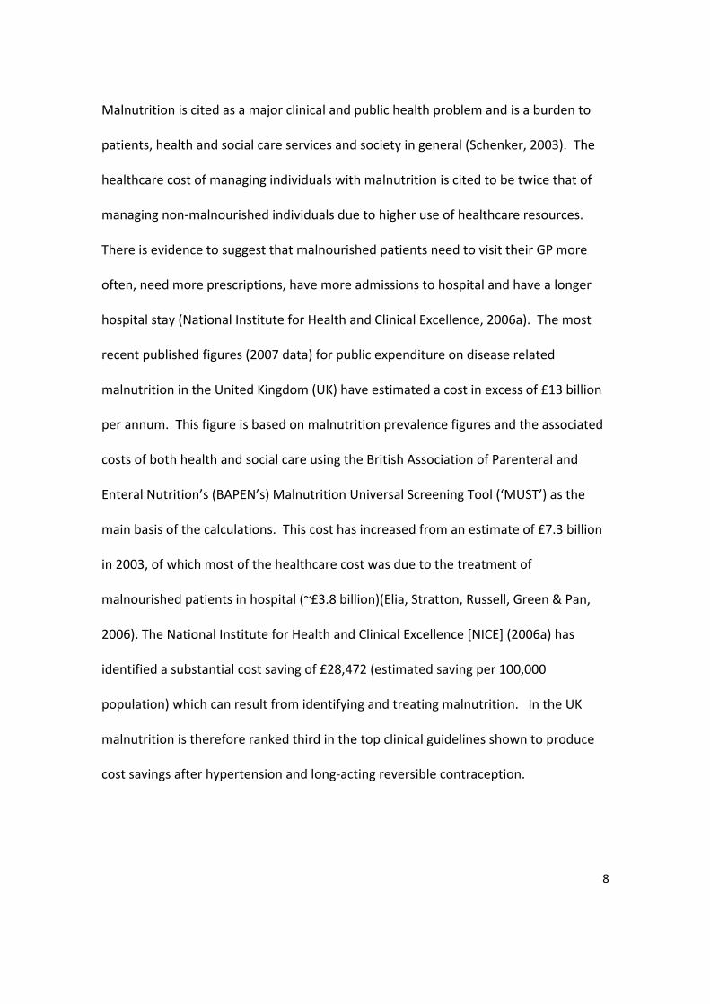
Malnutrition is cited as a major clinical and public health problem and is a burden to
patients, health and social care services and society in general (Schenker, 2003). The
healthcare cost of managing individuals with malnutrition is cited to be twice that of
managing non‐malnourished individuals due to higher use of healthcare resources.
There is evidence to suggest that malnourished patients need to visit their GP more
often, need more prescriptions, have more admissions to hospital and have a longer
hospital stay (National Institute for Health and Clinical Excellence, 2006a). The most
recent published figures (2007 data) for public expenditure on disease related
malnutrition in the United Kingdom (UK) have estimated a cost in excess of £13 billion
per annum. This figure is based on malnutrition prevalence figures and the associated
costs of both health and social care using the British Association of Parenteral and
Enteral Nutrition’s (BAPEN’s) Malnutrition Universal Screening Tool (‘MUST’) as the
main basis of the calculations. This cost has increased from an estimate of £7.3 billion
in 2003, of which most of the healthcare cost was due to the treatment of
malnourished patients in hospital (~£3.8 billion)(Elia, Stratton, Russell, Green & Pan,
2006). The National Institute for Health and Clinical Excellence [NICE] (2006a) has
identified a substantial cost saving of £28,472 (estimated saving per 100,000
population) which can result from identifying and treating malnutrition. In the UK
malnutrition is therefore ranked third in the top clinical guidelines shown to produce
cost savings after hypertension and long‐acting reversible contraception.

9
Malnutrition can be identified by screening patients using a screening tool and this is
therefore the first step in identifying those at risk of malnutrition (Nutricia, 2009). In
2003 ‘MUST’ was launched ‐ a five step, validated screening tool that can be used
across healthcare settings to identify adults who are malnourished or at risk of
malnutrition (Brotherton et al. 2012). The ‘MUST’ screening tool measures height,
weight, body mass index (BMI) and any unplanned/unintentional weight loss. An
unintentional weight loss of 10% of body weight is usually associated with poorer
clinical outcomes (McKinlay, 2008). The presence of an acute disease effect resulting
in no dietary intake for more than five days is also considered and an overall risk score
for malnutrition obtained. Tricep skinfold thickness (TST) and mid upper arm
circumference (MAC) are not included. The ‘MUST’ can be viewed as tracing the
clinical journey of a patient from the past history (history of unintentional weight
change) to the present (current weight status and BMI) and into the future (likely
effect of underlying condition) (Elia, 2004). Subtle nutritional deficiency states are
therefore not identified with ‘MUST’ but it will potentially identify patients at risk of
malnutrition (McKinlay, 2008). A copy of the ‘MUST’ screening tool is shown in Figure 1
(BAPEN, 2013a, p.3).

10
Figure 1: A copy of the ‘MUST’ screening tool.
BAPEN (2013a, p.3).
11
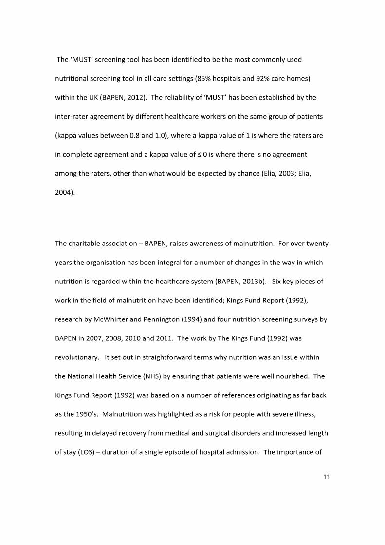
The ‘MUST’ screening tool has been identified to be the most commonly used
nutritional screening tool in all care settings (85% hospitals and 92% care homes)
within the UK (BAPEN, 2012). The reliability of ‘MUST’ has been established by the
inter‐rater agreement by different healthcare workers on the same group of patients
(kappa values between 0.8 and 1.0), where a kappa value of 1 is where the raters are
in complete agreement and a kappa value of ≤ 0 is where there is no agreement
among the raters, other than what would be expected by chance (Elia, 2003; Elia,
2004).
The charitable association – BAPEN, raises awareness of malnutrition. For over twenty
years the organisation has been integral for a number of changes in the way in which
nutrition is regarded within the healthcare system (BAPEN, 2013b). Six key pieces of
work in the field of malnutrition have been identified; Kings Fund Report (1992),
research by McWhirter and Pennington (1994) and four nutrition screening surveys by
BAPEN in 2007, 2008, 2010 and 2011. The work by The Kings Fund (1992) was
revolutionary. It set out in straightforward terms why nutrition was an issue within
the National Health Service (NHS) by ensuring that patients were well nourished. The
Kings Fund Report (1992) was based on a number of references originating as far back
as the 1950’s. Malnutrition was highlighted as a risk for people with severe illness,
resulting in delayed recovery from medical and surgical disorders and increased length
of stay (LOS) – duration of a single episode of hospital admission. The importance of

12
recognising and treating malnutrition before hospital admission was noted as the ideal
by nutritional assessment; monitoring of height and weight in general practice and
further monitoring within the hospital environment. Therefore the recognition of
malnutrition originating from the community was established; with the emphasis for
detecting malnutrition being predominantly focused within the hospital setting by
improving the education and awareness of malnutrition by doctors and nurses and a
hospital nutrition team offering services to the community health team in the area of
artificial nutrition support (ANS). The establishment of a care plan was highlighted as
an important step after the detection of malnutrition where a plan of treatment and
ongoing monitoring was acknowledged. The Kings Fund Report (1992) has been a
basis for ongoing research in the field of malnutrition in the UK.
The paper by Mc Whiter and Pennington (1994) is one of the first seminal pieces of
published work in the UK to determine the incidence of malnutrition of patients on
admission to hospital from the community. One hundred elderly patients were
recruited into the study from consecutive admissions into hospital. Each patient’s
nutritional status was determined from anthropometric data and weight loss before
illness; height and weight were recorded and used to determine BMI, MAC and TST.
Values were compared to those in tables of normal values for MAC and TST
measurements (standardised for sex and age, 16‐64 years drawn from data published
in the United States and data drawn directly from the elderly population in the UK). It

13
was noted that 43% of elderly patients admitted into hospital were malnourished of
which 4% suffer from mild malnutrition (BMI <20 kg/m2 ,TST or MAC below 15th
centile), 20% moderate malnutrition (BMI < 18kg/m2 ,TST or MAC below 5th centile)
and 19% severe malnutrition (BMI <16 kg/m2, TST or MAC below 5th centile). No
statistical details were included in the study by McWhirter and Pennington (1994) to
determine the significance of the results. It was acknowledged that BMI by itself is not
a sensitive indicator of protein energy malnutrition as it does not distinguish between
the depletion of fat or muscle which can introduce error; a low BMI will include
individuals who normally weigh less than usual for their height. As a result of the
potential misinterpretation when using BMI, other measurements of fat and muscle
mass were used; TST to estimate fat reserves and MAC as a measure of muscle protein
stores and unintentional weight loss before illness as a measure of nutritional status to
identify those at risk of complications as a result of getting thinner The results provide
a “snap shot” picture of the percentage of elderly admitted to hospital with
malnutrition. Implications highlighted for clinical practice as a result of the study
included the need to improve the recognition of malnutrition in hospital by doctors,
nurses and other health care staff (to identify malnourished elderly patients from the
community) and to heighten the awareness of nutrition within the community, as
there are potentially a number of individuals within the community who are in a
suboptimal nutritional state – high risk category of malnutrition.

14
Between 2007 and 2011, four nutrition screening week surveys by BAPEN aimed to
establish the prevalence of malnutrition on admission or recent admission to different
care settings, to document screening practice and provide feedback to improve
practice. The results based on April 2011 data and the ‘MUST’ criteria, established that
malnutrition affected one in four adults on admission to hospital, more than one in
three adults admitted to care homes in the previous six months and up to one in five
adults on admission to mental health units in the UK. Most of those affected by
malnutrition were found to be in the high risk category (‘MUST’ Score 2 or more).
These results were found to be similar to those obtained in the other three screening
surveys with the exception of a lower prevalence of malnutrition on admission to
hospital, and the prevalence of malnutrition found on recent admission to care homes
similar to the 2008 survey which was higher than in both 2010 and 2007. It was
highlighted that nutritional screening policies and practices vary between, and within,
healthcare settings and malnutrition continues to be under‐recognised and under‐
treated with much of the malnutrition present on admission to institutions cited to
originate in the community (Russell & Elia, 2012). Participation within the screening
surveys was voluntary and therefore potential problems with selection bias could
result. Higher values of malnutrition could potentially be expected for settings with a
screening policy in place; as a result of using a screening tool malnutrition is being
detected. In settings where no screening tool is in place malnutrition is not being
detected and therefore not treated. This assumption supports the statement that
malnutrition is often under‐recognised and under‐treated, an implication for practice.

15
No detail has been included in the nutrition screening surveys about the formal
determination of a sample size calculation, however with four surveys in total being
completed to date this helps to ensure that the surveys are large enough to help
determine clinically important results. The four surveys provide the most up to date
data on malnutrition within the UK to date.
Treatment of malnutrition.
In most cases malnutrition is a treatable condition that can be managed using first line
dietary advice to optimise food intake. Oral nutritional supplements can be used in
addition to first line dietary advice where dietary advice alone is unable to prevent and
treat malnutrition. ANS in the form of enteral tube feeding (ETF) or parenteral feeding
is indicated when oral intake is insufficient or unsafe in the case of swallowing
disturbances, unintentional weight loss (>10% of body weight), partial failure of
intestinal function requiring supplementary nutrition support and oncology disorders
for example (NICE 2006b; Murphy, 2010; Macmillan Cancer Support, 2014). The route
and level of feeding is decided on an individual basis according to the clinical
indications, treatment plan and nutritional status of the individual. Feeding via a tube
into the stomach is considered unless there is upper gastrointestinal dysfunction.
(NICE, 2006b). Previous work by Stratton, Green and Elia (2003) concluded that ETF
used within the hospital setting, can increase nutritional intake (98% of trials reviewed,
all random control trials (RCTs)), significantly reduce mortality (11% Vs. 22%, with

16
meta‐analysis, suggested odds ratio 0.48, 95% CI 0.30‐0.78) and significantly lower
complication rates including sepsis, wound and urinary infections and pneumonia (33%
vs. 48% with meta‐analysis, suggested odds ratio 0.5, 95% CI 0.35‐0.70). This study
has been noted by the European Society for Clinical Nutrition and Metabolism (2006)
as a comprehensive and extensive systematic review and meta‐analysis concerning the
benefit of ETF in the hospital setting and has been the basis of supporting the use of
nutritional support as a therapeutic intervention in clinical practice. The study is cited
within the American Society for Parenteral and Enteral Nutrition, Disease‐Related
Malnutrition and Enteral Nutrition Therapy (2010). It is noted however that out of the
seventy four trials reviewed as part of the study (n = 2769) only 46% of the trials were
randomized (n = 33) and that poor study designs due to low Jadad scores and small
sample sizes were limitations of the data set (80% of the trials had a Jadad score of 2
or less, the higher the score the better the study design, highest score 5). Therefore
future larger RCTs to assess the clinical effectiveness of ETF is noted as a requirement
for future practice.
The NICE Nutrition Support Guidelines (2006) provide recommendations for clinical
practice and were the result of the examination of systematic reviews, meta‐analyses
and RCTs in ETF. The Guidelines concluded that ETF does increase nutritional intake
however the evidence to support outcomes such as a reduced LOS or mortality is not
clear. The 2006 Guidelines were reviewed in 2012 and the Nutrition Support in Adults,

17
Quality Standards, QS24 issued (NICE, 2012). The quality standards are based upon the
2006 Guidelines and define clinical best practice within the area of nutrition support
including ETF. An integrated MDT approach to the provision of services is cited by
NICE (2012) as fundamental to the delivery of high‐quality care to adults who need
nutrition support. Five quality statements are included within the table below.
Table 1: NICE quality standard statements for nutrition support
Statement number Quality statement
Statement 1 A validated screening tool (‘MUST’ for example) is used to screen people for the risk of malnutrition in care settings.
Statement 2 People who are malnourished or at risk of malnutrition have a management care plan.
Statement 3 People who are screened for the risk of malnutrition have their screening results and nutrition support goals documented and communicated between care settings.
Statement 4 People managing their own ANS and/or their carers are trained to manage their nutrition delivery system and monitor their wellbeing.
Statement 5 People receiving nutrition support are “offered a review of the indicators, route, risks, benefits and goals of nutrition support at planned intervals”
(NICE, 2012, p. 7).
The quality standard for nutrition support in adults (2012) provides the most up to
date recommendations for current clinical practice.
Parenteral nutrition (PN) is administered intravenously and is usually used when a
patient is unable to have enteral nutrition. This may result from small bowel surgery

18
or obstruction for example where insertion of a tube for EN would be difficult or
contra‐indicated (Macmillan, 2014). For the purpose of the review PN has not been
considered as participants included within the author’s exploratory study had no
clinical indication for PN.
Enteral nutrition support.
There are a number of ways of administering EN. EN may be delivered either;
Directly into the stomach via an orogastric or NG tube, gastrostomy; PEG, Per‐
oral image‐guided gastrostomy (PIG/PIGG/ PRG), or radiologically inserted
gastrostomy (RIG);
Beyond the stomach (postpyloric) via a nasoduodenal/nasojejunal tube,
gastrojejunostomy or jejunostomy.
(Thomas & Bishop, 2007).
For the purpose of the review EN beyond the stomach has not been considered.
Participants included within the author’s exploratory study had no clinical indication
for postpyloric feeding. The postpyloric route is considered where there is upper
gastrointestinal dysfunction for example delayed gastric emptying.

19
Orogastric and NG feeding.
Orogastric and NG feeding are considered for short term feeding; four weeks or less
(Parenteral and Enteral Nutrition Group of the British Dietetic Association, 2011).
Orogastric feeding may be used in adults with basal skull fractures (NG feeding is
associated with patient death due to potential intracranial placement) (Spurrier, 2008)
and tends to be used extensively in the neonatal unit setting (infants are reported to
be obligatory nose breathers and the presence of a NG tube can increase airway
resistance and should therefore be used in ventilated infants only) (National Patient
Safety Agency, 2005; Rogahn, 1998). The orogastric procedure involves the insertion
of a thin flexible NG tube through the mouth directly into the stomach. NG feeding
involves the insertion of a NG tube through the nostril, down the back of the throat
and oesophagus and directly into the stomach as shown in Figure 2 (Macmillan Cancer
Support 2014).

20
Figure 2: Diagram to show the position of a NG feeding tube.
(Macmillan Cancer Support, 2014, p. 3).
Harm caused by misplaced NG tubes and NG tubes for orogastric feeding, tend to be
documented simultaneously as the type of tube used and insertion route is almost
identical. With both NG and orogastric feeding tubes there is a reported risk that the
tube can become misplaced into the lungs during insertion, or move out of the
stomach at a later stage. Patients may experience problems tolerating their enteral
feeding regime due to gastro‐oesophageal reflux or delayed gastric emptying which
could result in aspiration pneumonia (National Collaborating Centre for Acute Care,
2006). The tube is visible and some patients may find initial insertion of a NG tube
uncomfortable (Macmillan Cancer Support, 2014).

21
Gastrostomy feeding.
Gastrostomy feeding is indicated for patients with a functioning gastrointestinal tract
and should be considered for long‐term feeding (greater than four weeks) (NICE,
2006b). A gastrostomy feeding tube is a narrow tube that is placed directly through
the abdominal wall into the stomach shown in Figure 3. A gastrostomy feeding tube
can be inserted a number of ways shown in the Table 2 overleaf. The gastrostomy tube
is used to deliver nutrition, fluid and medications directly into the stomach (Abbott
Nutrition, 2006; White, 1998) and therefore results in an improved nutritional,
hydration or clinical status (Draper, 2011). Feeding via a gastrostomy tube permits the
maintenance of tissue metabolism and structure even though a patient cannot eat
anything, or enough to regain health (Clinical Resource Efficiency Support Team, 2004).

22
Figure 3: Diagram to show the position of a PEG tube in the stomach.
(Macmillan Cancer Support, 2014, p.5).
Table 2: Types of gastrostomies, methods of insertion and tube characteristics.
Type of gastrostomy Method of gastrostomy insertion
Gastrostomy tube characteristics
PEG
Endoscopic placement (use of a thin flexible optic camera)
Held in place with internal retention disc/bumper/dome
RIG X‐rays and other imaging techniques used
Held in place with internal retention balloon (water filled)
PIG/PIGG/PRG) X‐ray and other imaging techniques used
Held in place with internal retention disc/bumper/dome
(Abbott Nutrition, 2006; Great Ormond Street, 2014).

23
There have been a number of studies conducted and evaluated around the
effectiveness of NG, PEG, RIG and PIG/PIGG/PRG feeding. NG feeding is cited as a
classic, time‐proven technique (Gomes et al. 2011). The NG tube can be inserted at
the patient’s bedside and is inexpensive (Murphy, 2010). The first published report of
a PEG was in 1980 by Gauderer, Ponsky and Izant, and was cited as an innovative
technique. The technique has become widely accepted and was widely utilised and
favoured over the alternative laparotomy gastrostomy of the time. As a result, the
majority of research conducted and evaluated around the effectiveness of NG feeding
compared to gastrostomy feeding is based upon the PEG technique. There have now
been advancements in radiological imaging techniques permitting the use of the
fluoroscopically guided version of the PEG techniques; the RIG and PIG/PIGG/PRG
(Dickinson, 2013). For the purpose of the review the author will concentrate on the
PEG technique. Placement of RIG and PIG/PIGG/PRG are not technically facilitated
within the hospital setting where the study was conducted and therefore not included
in the review and subsequent exploratory study.

24
NG versus PEG.
PEG feeding tubes are increasingly used for EN in adults where patients cannot
maintain adequate nutrition with oral intake. Indications for PEG feeding include
obstruction of the gastrointestinal tract where the passing of a NG tube would be
difficult. In 2006 the National Collaborating Centre for Acute Care reviewed NG versus
PEG feeding. Four studies were included and reviewed; Baeten and Hoefnagels (1992),
Norton, Homer‐Ward, Donnelly, Long and Holmes (1996), Park et al. (1992) and The
FOOD Trial Collaboration (2005). A number of methodological problems were noted;
there was a greater proportion of sick patients in the PEG group when compared to
the NG group in the Baeten and Hoefnagels (1992) study and most of the patients in
the NG group in the study by Park et al. (1992) transferred to PEG feeding during the
study. The study by Norton et al. (1996) cited a significantly lower mortality in the
gastrostomy group (2 deaths, 12%) compared to the NG group (8 deaths, 57%), (P <
0.05). NG patients received significantly (P < 0.001) less of their feed (78%, 95% CI 63%
to 94%) compared with the gastrostomy group (100%). It is noted that the study
focused on patients with a diagnosis of acute dysphagic stroke; therefore the results
may not necessarily be significant when compared to other diagnoses. Results from
three large (n = 859), randomized multicentre FOOD collaboration trials conducted
between November 1996 and July 2003 with stroke patients highlighted that early ETF
was associated with a reduction in the risk of death of 5.8% (95% CI 0.8‐12.5, p=0.09)
and a reduction in death or poor outcome of 1.2% (95% CI ‐4.2 to 6.6, p=0.7). In the
NG versus PEG trial (n = 321), PEG feeding was associated with an increase in risk of

25
death by 1.0% (95% CI ‐10 to 11.9, p=0.9) and an increased risk of death or poor
outcome of 7.8% (95% CI 0.0 to 15.5, p=0.05). As a result the data was reported not to
support the use of PEG feeding (within 30 days) in the dysphagic stroke patient.
Research by Lockett, Templeton, Byrne and Norcross (2002) and a study by Longcroft‐
Wheaton et al. (2009) (summarised in Table 3 overleaf) suggests that PEG insertion
does no harm, supports the view that deaths observed were due to underlying
comorbidities and supports the opinion that nutrition support is associated with a
better outcome.

26
Table 3: Summary of Studies conducted by Lockett et al. (2002) and Longcroft‐Wheaton et al. (2009).
Lockett et al. (2002) Longcroft‐Wheaton et al. (2009)
Study design n = 166 Conducted over 2 years Indications for surgery, complications and antibiotic use determined
n = 44 (women n = 22), mean age 75 Retrospective Conducted over 1 year Reviewed mortality Based on previous study by Levine, Sachs, Jin and Meltzer (2007); study included a survival curve which the Longcroft‐Wheaton et al. (2009) study was compared Retrospective study by Levine et. al (2007) n = 6382, conducted over 4 years, model initially developed on n = 2739 patients, mean age 78 years, 68% female, internally validated n = 3643 from same sample, mean age 78 years, 68% female
Results 1 death directly related to PEG Insertion (peritonitis), 27 complications (16.3%). Mortality 30 days post PEG placement = 13 (7.8%) 30 patients died within 30 days post PEG insertion Wound infections occurred in 9 patients 4/153 patients with prophylactic antibiotics developed infections
Noted comorbidities (84% neurological, 23% cardiovascular) Mortality at 1, 3, 6 and 12 months = 16%, 20%, 25% and 28% respectively Risk factors from Levine et al. (2007) obtained from administrative data, including comorbidities. Risk factor assigned a weight based on odds ratios. Study not PEG specific, neurological comorbidity not included, 81% sample African American, therefore query comparable.
Conclusions PEG safe in establishing enteral access in most patients Antibiotics prevent wound infection
PEG does no harm; study supports opinion that nutrition support is associated with a better outcome Higher deaths initially related to poor pre morbid state
Observations Further comparison study to include PEG and comorbidities would be beneficial Should alternative feeding tube be used initially such as an NG until prognosis more predictable.

27
A review conducted by Gomes et al. on behalf of the Cochrane Collaboration (2011)
evaluated the effectiveness and safety of PEG as compared to NG for adults with
swallowing disturbances. Nine randomised control studies were included. It was
highlighted that research has been conducted in this field previously; Langmore,
Kasarkis, Manca and Olney (2007), Bath, Kerr and Collins (2009), Dennis, Lewis,
Cranswick and Forbes (2005), McClave et al. (2005), Douzinas et al. (2006) and
Hamidon et al. (2006). The studies by Langmore et al. (2007) and McClave et al. (2005)
were excluded from the review as the study by Langmore et al. (2007) was a meta‐
analysis of which the author was unable to find any controlled or randomized studies
and the study by McClave et al. (2005) contained no interventions of interest for the
purpose of the Cochrane Review; the study investigated the use of gastric residual
values as a marker for aspiration in critically ill patients, not the benefits of NG versus
PEG. There are a number of variations noted between the nine studies; follow‐up
times, age range and range of diagnoses including neurological disease, stroke and
neoplasia of the ear, nose and throat. It is noted that some of the authors in these
studies failed to report the methods used in sequence generation during
randomisation of participants into the NG verses PEG groups and failed to note
allocation concealment; a technique used to prevent bias by the researchers
(unconsciously or otherwise) influencing which participants are assigned to a given
intervention group. The main results were: intervention failure occurred in 19/156
patients in then PEG group and 63/158 patients in the NGT group (95% CI 0.08 to 0.76,
p = 0.01) in favour of PEG. No statistically significant difference between comparison

28
groups in complications (risk ratio 1.00) or mortality (relative risk 0.96) were observed
(95% CI 0.91 to 1.11, p = 0.93) and (95% CI 0.64 to 1.44, p = 0.64) respectively. No
specific details on the types of intervention failure and complications were noted in
the review. Intervention failure could be interpreted as unsuccessful tube placement
or even tube misplacement but this cannot be determined.
Complications in the region of 8‐30% are cited following the endoscopic placement of
enteral feeding tubes (Löser et al. 2005). Two important patient outcomes in regard to
PEG placement are improving mortality and quality of life (Plonk, 2005). Thirty day
mortality rates have been cited to vary in previous studies. In the National
Confidential Enquiry into Patient Outcome and Death [NCEPOD] (2004), mortality rates
of 6% (986 deaths) were noted out of a total of 16,648 PEG procedures obtained from
UK hospital episode statistics during 2002/2003. A degree of under reporting was
believed due to PEG procedures being undertaken in the outpatient setting which
would not have been covered by the dataset. Deaths post hospital discharge was not
included, potentially underestimating total mortality figures. NCEPOD (2004)
conducted a review of mortality in three data sets collected retrospectively by
questionnaire during 2002‐2003, including deaths within thirty days of PEG insertion.
In relation to PEG, 719 cases were identified for review; 76% had respiratory
complications and 26% had cardiovascular disease implicated in the causes of death,

29
indicating that present co‐morbidities may be the cause of death, as opposed to
mortality directly associated with PEG insertion.
Although some evidence suggests that PEG placement is preferable, patient selection
should be timely and appropriate (Ponsky & Gauderer, 1989) and should only be
performed if its benefits clearly outweigh its risks and burdens (Plonk, 2005). The
General Medical Council reinforce this viewpoint in the document “Treatment and care
towards the end of life: good practice in decision making” (2010). It highlights that the
most challenging decision is generally about withdrawing or not starting artificial
nutrition and hydration when it has the potential to prolong the patient’s life. Overall
there is a lack of information on the assessment of benefits with invasive procedures
such as PEG and ethical issues associated with randomised controlled trials. The
studies raise the question of what is acceptable mortality associated with PEG and
does low mortality merely indicate that patients who are sick have been excluded.
Table 4 overleaf details some of the major and minor complications associated with
PEG placement. Major complications can be defined as requiring the need for surgery,
non‐prophylactic antibiotics, or blood transfusion or procedure related death
(Vervloessem et al. 2009).
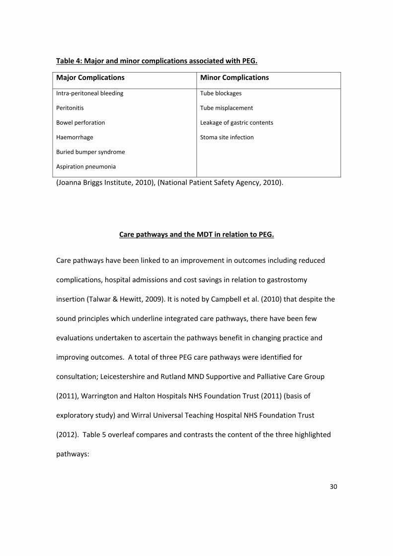
30
Table 4: Major and minor complications associated with PEG.
Major Complications Minor Complications
Intra‐peritoneal bleeding
Peritonitis
Bowel perforation
Haemorrhage
Buried bumper syndrome
Aspiration pneumonia
Tube blockages
Tube misplacement
Leakage of gastric contents
Stoma site infection
(Joanna Briggs Institute, 2010), (National Patient Safety Agency, 2010).
Care pathways and the MDT in relation to PEG.
Care pathways have been linked to an improvement in outcomes including reduced
complications, hospital admissions and cost savings in relation to gastrostomy
insertion (Talwar & Hewitt, 2009). It is noted by Campbell et al. (2010) that despite the
sound principles which underline integrated care pathways, there have been few
evaluations undertaken to ascertain the pathways benefit in changing practice and
improving outcomes. A total of three PEG care pathways were identified for
consultation; Leicestershire and Rutland MND Supportive and Palliative Care Group
(2011), Warrington and Halton Hospitals NHS Foundation Trust (2011) (basis of
exploratory study) and Wirral Universal Teaching Hospital NHS Foundation Trust
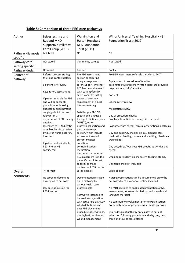
(2012). Table 5 overleaf compares and contrasts the content of the three highlighted
pathways:
31
Table 5: Comparison of three PEG care pathways
Author Leicestershire and Rutland MND Supportive Palliative Care Group (2011)
Warrington and Halton Hospitals NHS Foundation Trust (2011)
Wirral Universal Teaching Hospital NHS Foundation Trust (2012)
Pathway diagnosis specific
Yes, MND No No
Pathway care setting specific
Not stated Community setting Not stated
Pathway design Flowchart Booklet Booklet
Content of pathway
Referral process stating MDT and contact details Biochemistry review Respiratory assessment If patient suitable for PEG and willing consent; procedure for booking endoscopy appointment, copying of clinic letters to relevant MDT, organisation of EN training detailed. Discharge to HEN dietetic care, biochemistry review by district nurse post PEG insertion If patient not suitable for PEG; RIG or NG considered.
Pre PEG assessment section considering living arrangements, carer support, whether PEG has been discussed with patient/family/ carer, capacity, lasting power of attorney, requirement of a best interest meeting Detailed pre PEG GP, speech and language therapist, dietitian (uses ‘MUST’), other professional section and gastroenterology section, which include assessment around current medical condition, contraindications, medication, biochemistry, whether PEG placement is in the patient’s best interest, capacity to make decision re PEG insertion
Pre PEG assessment referrals checklist to MDT Explanation of procedure offered to patient/relatives/carers. Written literature provided on procedure, risks/benefits Consent Biochemistry review Medication review Day of procedure checks; prophylactic antibiotics, analgesia, transport, Post procedure checks; clinical observations, analgesia Day one post PEG checks; clinical, biochemistry, medication, feeding, nausea and vomiting, diarrhoea, wound site, Day two/three/four post PEG checks; as per day one checks Ongoing care; daily, biochemistry, feeding, stoma, Discharge checklist included
Overall comments
A4 format No scope to document directly on to pathway Day case admission for PEG insertion
Large booklet Documentation straight on to pathway by various health care professionals Pathway is intended to be used In conjunction with acute PEG pathway which details pre and post PEG placement procedure observations, prophylactic antibiotics, wound management
Large booklet Nursing observations can be documented on to the pathway directly, variance section included No MDT sections to enable documentation of MDT assessments, for example dietitian and speech and language therapist No community involvement prior to PEG insertion. Potentially more appropriate as an acute pathway Query design of pathway anticipates in patient admission following procedure with day one, two, three and four checks detailed.

32
It is noted by Westaby, Young and O’Toole (2010) that a defined referral pathway
should be completed for all cases considered for PEG. Care pathways are noted in
research undertaken by Norris (2005) to enable the seamless delivery of care to
patients across the primary and secondary care interface of the NHS. The content of
the three care pathways reviewed varied considerably, however content which is
reflected across all three of the pathways include the MDT, consent, biochemistry and
some mention of discharge. Physicians are noted to inform patients and carers poorly
regarding PEG benefits, burdens and alternatives (Plonk, 2005), with patients and
carers not always aware of the long term implications of PEG (Rickman, 1998). In a
study by Todd, Van Rosendaal, Duregon, and Verhoef (2005) it was observed that
individuals were insufficiently informed regarding the PEG and the ramifications of the
PEG placement. HETF is noted to place a considerable burden on family and carers
(Best & Hitchings, 2010) with PEG feeding noted as time consuming and interfering
with daily life (Martin, Blomberg & Lagergren, 2012). Research by McKee (1999)
highlights that patients must understand what a procedure involves and how the
procedure may or may not affect them in the future in order to provide informed
consent. In respect to consent and decision making and PEG placement, a team‐
orientated approach and more active dialogue with regard to care planning among
health care professionals and carers has been highlighted to promote effective
communication (Todd, Van Rosendaal, Duregon & Verhoef, 2005). In both the
Warrington and Halton Hospitals NHS Foundation Trust (2011) and the Wirral
33
University Teaching Hospitals NHS Trust (2012) pathways, specific reference to the
explanation of the PEG procedure with the patient/family and carers are noted.
Integrated care pathways are defined as care plans that detail the essential steps with
a specific clinical problem (Campbell, Hotchkiss, Bradshaw & Porteous, 1998). The care
pathways by Warrington and Halton Hospitals NHS Foundation Trust (2011) and the
Wirral University Teaching Hospital NHS Foundation Trust (2012) could be considered
to meet the integrated care pathway definition; they are presented in a structured
care plan format, describe the expected progress and variance of the patient’s clinical
condition and contribute towards positive outcomes. The pathways act as a resource
and allow users to access recommendations on PEG insertion and monitoring. This is
comparable to a NICE pathway for example (NICE, 2014), however links to other
relevant topics to create a network of information are not included.
The pathways reviewed were noted to be suited to either the acute or community
setting. The primary placement of a PEG is undertaken in the acute hospital setting
(Best & Hitchings, 2010). Pressure on hospital beds often leads to early discharge of EF
patients. In some instances, patients will attend the hospital for the insertion of their
tube as a day case and be discharged home on the same day (Thomas & Bishop, 2007)
as per the Leicestershire and Rutland MND Supportive Palliative Care Group (2011)
pathway. It is stated by Kurien et al. (2012) that complications are reported to “often

34
occur” following discharge in to the community post‐PEG insertion and it is
acknowledged that there is limited research in the evaluation of community teams in
the management of PEG complications. It has been documented by Madigan (2003)
and Lowry and Johnston (2007) that community follow up is often inadequate for
patients on HETF with only a small number of patients and carers feeling that they
receive sufficient information to enable them to manage their feeding tube prior to
leaving hospital. This situation poses potential problems if the patient is in hospital for
a shortened LOS and then discharged back into the care of community services. The
literature published by Madigan (2003), was recorded to reflect a conducted
symposium on EF and therefore reflects the views of participants in the symposium
and not necessarily documented research. In the study by Lowry and Johnston (2007) a
postal questionnaire was used to assess if patients/carers were trained in the care of
PEG tube pre‐discharge and to ascertain whether appropriate follow up was in place.
Sixty six patients were identified for inclusion in the study and a response rate of 44%
(29 patients) was noted. Out of the twenty nine respondents, twenty one (72%)
reported that they had been taught how to manage the tube, feeds and feeding
pumps prior to discharge and 24 had been reviewed by the dietitian post discharge.
Fifteen patients had encountered PEG problems, fourteen of whom knew who to
contact in the event of a problem. The main problems encountered were the PEG tube
falling out (n =7), PEG site infection (n = 2), migration/loosening of the PEG (n = 2) and
tube blockage (n = 1). In order to resolve the majority problem of the PEG falling out
and migration/loosening of the PEG, the situation would need to be potentially

35
addressed in the acute setting to facilitate tube re‐insertion/review, not the
community setting.
Work by Ditchburn (2005) highlights a number of patient benefits following the
implementation of a care pathway in the secondary care setting:
‐ Improved consistency of advice from pre‐PEG assessment to post‐PEG
community follow up;
‐ Improved continuity of care;
‐ Provision of information booklets on PEG;
‐ Involvement of patients/carers with education reducing tube related problems
and complications;
‐ Quicker referrals.
The study was observational; it documented the development of a care pathway to
support patients and maintain standards of PEG feeding.
There would appear to be a lack of PEG pathways dedicated for specific diagnoses. For
example in the diagnosis of head and neck cancer it is noted that there is no specific
published care pathway to coordinate prophylactic gastrostomy insertion before
treatment (Talwar & Hewitt, 2009). The use of pathways for a specific diagnosis can be
useful. For example in the MND pathway by the Leicestershire and Rutland MND
Supportive Palliative Care Group (2011), a respiratory assessment is highlighted as an

36
example of a diagnosis specific assessment. An alternatively placed gastrostomy tube
would need to be considered if the patient failed the respiratory assessment as a result
of sedation used during PEG insertion.
Trends in home enteral tube feeding.
PEG is reported to be one of the most commonly performed gastrointestinal
procedures (Potack, 2008) with a low incidence of complications (Draper, 2011). For
this reason, home enteral tube feeding (HETF) is an expanding area of nutritional
support with an increasing number of patients receiving HETF via a gastrostomy
(Kurien et al. 2012). The British Artificial Nutrition Survey (BANS) was established in
1996 and has been reporting the trends in ANS for more than 10 years. In 1998 it was
estimated that there were more than 12,000 patients on HETF (BANS, 1998). Data
from the 2011 British Artificial Nutrition Survey (BANS) suggests that the number of
HETF episodes increased by 44% from 12,190 episodes in 2010 to 17,550 episodes in
2011 (BAPEN, 2012). A reduction in reporting rates was noted due to the Health and
Social Care Act (2006) (Health Service, 2007), where a requirement for reporters to
obtain consent from patients prior to submitting data to BANS was imposed. In 2010 it
was confirmed that BANS reporters were no longer required to obtain consent from
patients and therefore a subsequent increase in reporting rates was observed. Data
suggests that the gastrostomy is the primary route of feeding (75%), as it has been
over the last 10 years (BAPEN, 2011). A number of factors have been quoted to have

37
contributed to the growth of HETF including the reduction in the number of hospital
beds and increase in the elderly population (Best & Hitchings, 2010), with 63% of new
registrations in 2010 being aged over 60 years and 41% over 70 years of age (BANS,
2011). Neurological conditions including progressive conditions such as MND, MS,
bulbar palsy and Parkinson’s disease are reported to be the most common indication
for PEG (Draper, 2011) (Department of Health, 2005). BANS data from 2008 highlights
that 48% of new patient HETF registrations suffered from neurological conditions.
Recent BANS data from 2011 notes a decrease in this overall figure from 58% in 2000
compared to 46% in 2010 of which 12% and 8% accounting for MND and MS primary
diagnoses respectively. Head and neck cancer accounts for 77% of new HETF
registrations.

38
Summary
In conclusion this review has been useful in considering how to approach PEG
placement and the services that should be provided throughout primary and
secondary care and across the multidisciplinary teams.
It has established a vision of what the priorities are in relation to PEG insertion in light
of the current research evidence available in this field:
Early and appropriate patient assessment;
Care of PEG tubes;
Awareness of complications.
Staff education and the provision of standardised guidelines, care planning and care
pathways should be standard practice in the management of PEG placement. The
importance of patients and relatives understanding PEG placement, together with the
implications for the long term have been highlighted. Assessment of patient suitability
for PEG placement should be made in collaboration with the patient and relatives
whenever possible.

39
Reference List
Abbott Nutrition. (2006). What is a gastrostomy feeding tube? Maidenhead: Author. Azam, P. (2007). Managing under‐nutrition in a nursing home setting. Nursing Older
People, 19(3), 33‐36. Baeten, C. & Hoefnagels, J. (1992). Feeding via a nasogastric tube or percutaneous
endoscopic gastrostomy. A comparison. Scandinavian Journal of Gastroenterology Supplement, 194, 95‐8.
Bath, P.M.W., Kerr, J. & Collins, M. (1997). Factorial trial of swallowing versus conventional therapy, and PEG versus nasogastric tube feeding, in dysphagic patients with recent stroke. Cochrane Database of Systematic Reviews, Issue 1.
Best, C. & Hitchings, H. (2010) Day case gastrostomy placement for patients in the community. British Journal of Community Nursing, 15( 6), 272‐278. British Association for Parenteral and Enteral Nutrition. (2007). Nutrition Screening
Survey in the UK in 2007. Redditch: BAPEN. British Association for Parenteral and Enteral Nutrition. (2008). Nutrition Screening
Survey in the UK in 2008. Redditch: BAPEN. British Association for Parenteral and Enteral Nutrition. (2011b). Annual BANS report, 2011. Artificial Nutrition Support in the UK 2000‐2010. A Report by the British
Artificial Nutrition Survey (BANS), a committee of BAPEN (The British Association for Parenteral and Enteral Nutrition. Retrieved December 20, 2013, from the British Association for Parenteral and Enteral Nutrition Web site: http://www.bapen.org.uk
British Association for Parenteral and Enteral Nutrition. (2011). Key points from BAPEN’s fourth Nutrition screening Week Survey. Retrieved December 30, 2013 from the British Association for Parenteral and Enteral Nutrition Web site: http://www.bapen.org.uk/screening‐for‐malnutrition/nutrition‐screening‐week
British Association for Parenteral and Enteral Nutrition (2012, March). E‐bans reporters’ digest. Retrieved October 30, 2012 from the British Association for Parenteral and Enteral Nutrition e‐bans Web site: http://www.e‐bans.co.uk
British Association for Parenteral and Enteral Nutrition. (2013a). ‘MUST’ toolkit(UK)2013. Retrieved January 8, 2014, from the British Association for Parenteral and Enteral Nutrition Web site:
http://www.bapen.org.uk British Association for Parenteral and Enteral Nutrition. (2013b). 21 ways to Make a
Difference. Retrieved December 30, 2013, from the British Association for Parenteral and Enteral Nutrition Web site: www.bapen.org.uk
Brotherton, A. , Holodoway, A., Mason, P., McGregor, I., Parsons. B. & Pryke, R. 2012). Managing Adult Malnutrition in the Community, Including a pathway for the appropriate use of oral nutritional supplements (ONS). Produced by a multi
40
professional consensus panel. Trowbridge: Nutricia Advanced Medical Nutrition.
Clinical Resource Efficiency Support Team. (2004). Guidelines for the management of enteral tube feeding in adults. Stormont: Author. National Confidential Enquiry into Patient Outcome and Death. (2004). Scoping Our
Practice. London: Author. Dennis, M., Lewis, S., Cranswick, G., Forbes, J. & FOOD Trial Collaboration. (2005).
FOOD: a multicentre randomised trial evaluating feeding policies in patients admitted to hospital with a recent stroke. Health Technology Assessment, 10(2), 1‐120.
Department of Health. (2005). Long term Neurological Conditions National Service Framework. Retrieved April 16, 2013, from the National Archives Web site:
http://webarchive.nationalarchives.gov.uk/+/www.dh.gov.uk/en/Healthcare/Longtermconditions/Long‐termNeurologicalConditions
Department of Health. (2007). Long term conditions. Retrieved April 16, 2013, from the National Archives Web site:
http://webarchive.nationalarchives.gov.uk/+/www.dh.gov.uk/en/Healthcare/Longter mconditions/DH_4128521
Department of Health. (2012a). Health Profile 2012 Halton. Retrieved December 07, 2012, from the Health Profiles Web site: http://www.apho.org.uk/resouce/item.aspx?RID=105209
Department of Health. (2012b). Health Profile 2012 Warrington. Retrieved December, 07, 2012, from the Health Profiles Web site:
http://www.apho.org.uk/resource/item.aspx?RID=117038 Department of Health. (2012c). Statistical Press Notice – NHS Referral to Treatment
(RTT) Waiting Times Data – November 2012. Retrieved March 02, 2013 from the Media Centre Web site: http://mediacentre.dh.gov.uk/2013/01/17/statistical‐press‐notice‐nhs‐referral‐to‐treatment‐rttwaiting‐times‐data‐september‐2012
Ditchburn, L. (2005). Joint primary‐secondary care design of PEG care pathways. Nursing Times, 101(18), 34‐36. Dickinson, 0. (2013). Reasoning and Undertaking of Percutaneous Image Guided Gastrostomies (PIGGS). NHD The Dietitians Magazine, pp.23‐25. Douzanas, E.E., Tsapalos, A., Dimitrakopoulos, A., Diamantri‐Kandarkis, E., Rapidis, A.D.
& Roussos, C. (2006). Effect of percutaneous endoscopic gastrostomy on gastro‐oesophageal reflux in mechanically‐ ventilated patients. World Journal of Gastroenterology, 12(1), 114‐118.
Draper, R. (2011). PEG Feeding Tubes – Indications and Management. Retrieved May, 04, 2013 from the Patient Web site:
http://www.patient.co.uk/doctor/PEG‐Feeding‐Tubes‐Indications‐and Management.htm
Eckersley, D. & Morris, M. (2010). Guidelines for the Treatment of Undernutrition in Adults in the Community. NHS Knowsley, NHS Halton and St Helens, Warrington and Halton Hospitals NHS Foundation Trust: Author.

41
Elia, M. (2003). Screening for malnutrition: a multidisciplinary responsibility. Development and use of the ‘Malnutrition Universal Screening Tool’ (‘MUST’) for adults. Redditch: Author.
Elia, M. (2004). Background to the development of the “Malnutrition Universal Screening Tool” (“MUST”). Clinical Nutrition Update, 8(3), 3‐11. Elia, M. & Russell, C.A. (2009). Combating Malnutrition: Recommendations for Action.
Report from the advisory group on malnutrition, led by BAPEN. Redditch: BAPEN.
Elia, M., Stratton, R., Russell, C., Green, C., & Pan, F. (2006). The cost of disease‐related malnutrition in the UK and economic considerations for the use of oral nutritional supplements (ONS) in adults. Redditch: BAPEN.
Elia, M. & Stratton, R.J. (2009). Calculating the cost of disease‐related malnutrition in The UK in 2007 (public expenditure only) in: Combating malnutrition: Recommendations for Action. Report from the advisory group on malnutrition, led by BAPEN. Redditch: BAPEN.
European Society for Clinical Nutrition and Metabolism. (2006). Evidence supports nutritional support. Clinical Nutrition, 25, 177‐179.
FOOD Trial Collaboration. (2005). Effect of timing and method of enteral tube feeding for dysphagic stroke patients (FOOD): a multicentre randomised controlled trial. Lancet, 365(9461), 764‐72.
Gauderer, M.W.L., Ponsky, J.L. & Izant, R.J. (1980). Gastrostomy without laparotomy: a percutaneous endoscopic technique. Journal of Paediatric Surgery, 15, 1206‐10.
General Medical Council (2010). Treatment and care towards the end of life: good practice in decision making. London: Author.
Gomes jr, C.A.R., Lustosa, S.A.S., Matos, D., Andriolo, R.B, Waisberg, D.R. & Waisberg, J. (2010). Percutaneous endoscopic gastrostomy versus nasogastric tube feeding for adults with swallowing disturbances (Review).The Cochrane Collaboration.
Hamidon, B.B., Abdullah, S.A., Zawawi, M.F., Sukumar, N., Aminuddin, A. & Raymond, A.A. (2006). A prospective comparison of percutaneous endoscopic gastrostomy and nasogastric tube feeding in patients with acute dysphagic stroke. Medical Journal of Malaysia, 61(1), 59‐66.
Health Service. (2007). National Health Service Act 2006. London:HMSO. Joanna Briggs Institute. (2010). The Prevention and Management of Complications
Associated with PEG Tubes in Adults. Best Practice. 14(10), 1‐4. Kings Fund Centre. (1992). A Positive approach to Nutrition as Treatment. London: Author. Kurien, M., White, S., Simpson, G., Grant, J., Sanders, D.S. & McAlindon, M.E. (2012). Managing patients with gastrostomy tubes in the community: Can a dedicated enteral feed dietetic service reduce hospital readmissions? European Journal of Clinical Nutrition, 66, 757‐760. Langmore, S.E., Kasarskis, E.J., Manca, M.L, Olney & R.K. (2007). Enteral tube feeding
for amyotrophic lateral sceloris/motor neurone disease. Cochrane Database of

42
Systematic Reviews, Issue 4. Levine, S.K., Sachs, G.A, Jin, L. & Meltzer, D. (2007). A prognostic model of 1‐year
mortality in older adults after hospital discharge. American Journal of Medicine, 120, 455‐460.
Lockett, M.A., Templeton, M.L., Byrne, T.K and Norcross, E.D. (2002). Percutaneous endoscopic gastrostomy in a tertiary care center. American Surgery, 68(2), 117‐20.
Longcroft‐Wheaton, G., Marden, P., Colleypriest, B., Gavin, D., Taylor, G., & Farrant, M. (2009). Understanding Why Patients Die After Gastrostomy Tube Insertion: A Retrospective Analysis of Mortality. Journal of Parenteral and Enteral Nutrition, 33(4), 375‐379.
Löser, Chr., Aschl, G., Hébuterne, X., Mathus‐Vliegen, E.M.H., Muscaritoli, M., Niv, Y., Rollins, H., Singer, P., Skelly, R.H. (2005). Consensus Statement. ESPEN guidelines on artificial enteral nutrition – Percutaneous endoscopic gastrostomy (PEG). Clinical Nutrition, 24, 848‐861.
Lowry, S., Johnston, S.D. (2007). Who follows up patients after PEG tube insertion? Ulster Medical Journal, 76(2), 88‐90.
Macmillan Cancer Support. (2014). Nutritional Support (artificial feeding). Retrieved April 16, 2014, from the Macmillan Web site: http://www.macmillan.org.uk/Cancerinformation/Livingwithandaftercancer/Eatingwell/Nutritionalsupport
Madigan, S.M. (2003). Home enteral feeding: the changing role of the dietitian. Proceedings of the Nutrition Society, 62(3), 761‐3.
Marshall, M.N. (1996) Sampling for qualitative research. Family Practitioner, 13, 522‐ 565. Martin, L., Blomberg, J. & Lagergren, P. (2012). Patients’ perspectives of living with a
percutaneous endoscopic gastrostomy (PEG). BMC Gastoenterology. 12(126), 1‐8.
Mayre‐Chilton, K.M., Talwar, B.P. & Goff, L.M. (2011). Different experiences and perspectives between head and neck cancer patients and their care‐givers on their daily impact of a gastrostomy tube. Journal of Human Nutrition and Dietetics, 24, 449‐459.
McClave, S.A., Lukan, J.K., Stefater, J.A., Lowen, C.C., Looney, S.W., Matheson, P.J (2005). Poor validity of residual volumes as a marker for risk of aspiration in critically ill patients. Critical Care Medicine, 33(2), 324‐330.
McWhirter, J.P. & Pennington, C.R. (1994). Incidence and recognition of malnutrition in hospital. British Medical Journal, 308 945‐948. McKee, D. (1999). The legal framework for consent. Professional Nurse, 14(10), 688‐ 690. McKinlay, A.W. (2008). Malnutrition:the spectare at the feast. Journal of the Royal
College of Physicians of Edinburgh. 38, 317‐21. Murphy, P. (2010). Enteral nutrition. In A.Payne & H.Barker (Ed.), Advancing Dietetics and Clinical Nutrition (pp. 109‐126). London:Elsevier. National Collaborating Centre for Acute Care. (2006). Nutrition support in adults Oral

43
nutrition support, enteral tube feeding and parenteral nutrition. London: Author.
National Health Service Direct Business Plan, (2011/12 to 2015/16). Retrieved April 16, 2013 from the NHS Direct Web site:
http://www.nhsdirect.nhs.uk/about/freedomofinformation/foipublicationscheme/͂/media/Files?freedomOfInformationDocument
National Health Service Institute for Innovation and Improvement. (2008). Length of Stay – Reducing Length of Stay. Retrieved February, 22, 2013 from the NHS Institute for Innovation and Improvement Web site:
http://www.institute.nhs.uk/quality_and_service_improvement_tools/length_of_stay.html
National Institute for Health and Clinical Excellence. (2006a). Costing template. Nutriton support in adults:oral supplements, enteral and parenteral feeding. Retrieved April 16, 2013, from the NICE Web site: http://guidance.org.uk/CG32/CostingTemplate/xls/English National Institute for Health and Clinical Excellence. (2006b). Nutrition support in
adults: Oral nutrition support, enteral tube feeding and parenteral nutrition. Clinical Guideline 32. London: Author.
National Institute for Health and Clinical Excellence. (2012). Quality standard for nutrition support in adults. NICE quality standard 24. Manchester: Author. National Institute for Health and Clinical Excellence. (2014). NICE Pathways. Retrieved
June 15, 2014, from the NICE Web site: http://www.pathways.nice.org.uk
National Patient Safety Agency. (2005). Reducing the harm caused by misplaced naso and orogastric feeding tubes in babies under the care of neonatal units. Retrieved December 30, 2013 from the National Patient Safety Agency Web site: http://nrls.npsa.nhs.uk
National Patient Safety Agency. (2010). Rapid Response Report. Early detection of complications after gastrostomy. NPSA:UK.
Norris, A.C. (1998). Care pathways and the new NHS. Journal of Integrated Care, 2, 78‐ 83. Norton, B., Homer‐Ward, M., Donnelly, M.T., Long, R.G & Homes G.K. (1996). A
randomised prospective comparison of percutaneous endoscopic gastrostomy and nasogastric tube feeding after acute dysphagic stroke. British Medical Journal, 312(7022), 6‐13.
Nutricia. (2009). Appropriate Use of Oral Nutritional Supplements in Older People: Good Practice Examples and Recommendations for Practical Implementation. Trowbridge: Author. Parenteral and Enteral Nutrition Group of the British Dietetic Association. (2011). A
Pocket Guide to Clinical Nutrition. (4th ed.). London:Author. Park, R.H., Allison, M.C., Lang, J., Spence, E., Morris, A.J & Danesh, B.J. (1992).
Randomised comparison of percutaneous endoscopic gastrostomy and nasogastric tube feeding in patients with persisting neurological dysphagia.

44
British Medical Journal, 304(6839), 1406‐9. Plonk, W.M. (2005). To PEG or not to PEG. Practical Gastroenterology. 29(7), 19‐31. Ponsky, J.L. & Gauderer M.W. (1989). Percutaneous Endoscopic Gastrostomy
Indications, Limitations, Techniques and Results. World Journal of Surgery, 13(2), 165‐170.
Potack, J.Z. (2008). Complications of and Controversies Associated With Percutaneous Endoscopic Gastrostomy: Report of a Case and Literature Review. Medscape Journal of Medicine 10(6), 142.
Rickman, J. (1998). Percutaneous endoscopic gastrostomy: psychological effects. British Journal of Nursing, 7(12), 723‐729. Russell, C.A. & Elia, M. (2012). Nutrition Screening Survey in the UK and Republic of Ireland in 2011. A report by BAPEN, 2012. Redditch:BAPEN. Stratton, R.J., Green, C.J. & Elia, M. (2003). Disease‐Related Malnutrition: an Evidence‐ Based approach to treatment. Wallingford:CABI Shenker, S. (2003). Undernutrition in the UK. British Nutrition Foundation Briefing Paper. Nutrition Bulletin. 28, 87‐120. Spurrier, E.J. (2008). Use of nasogastric tubes in trauma patients – a review. JR Army Med Corps, 154(1), 3‐10. Talwar, B.P. & Hewitt, R. (2009). Nutrition and dietetic‐led gastrostomy pathway of Care for insertion and removal contributes to outcomes. Proceedings of the
Nutrition Society, 68 (OCE1), E65. The American Society for Parenteral and Enteral Nutrition. (2010). Disease‐related
Malnutrition and Enteral Nutrition Therapy. Silver Spring:ASPEN. The King’s Fund. (2011). Transforming our health care system. Ten priorities for commissioners. London: Author. The NHS Constitution. (March, 2012). The Handbook To The NHS Constitution. London:
Department of Health. Thomas, B. & Bishop, J. (2007) Manual of Dietetic Practice. (4th ed.). Oxford: Blackwell Publishing. Todd, V., Van Rosendaal, G., Duregon, K. & Verhoef, M. (2005). Percutaneous
endoscopic gastrostomy (PEG): The role and perspective of nurses. Journal of Clinical Nursing, 14(2), 187‐194.
Thuraisingham, A.J. & Cairns, S.A. (2002). Percutaneous endoscopic gastrostomy: prospective clinician review appropriately decreases insertion. Gut, 50 (suppl 11), A86.
Vervloessem, D., Van Leersum, F., Boer, D., Hop, W.C., Escher, J.C, Madem, G.C., De Ridder, L. & Bax, K.N. (2009). Percutaneous endoscopic gastrostomy (PEG) in children is not a minor procedure: risk factors for major complications. Seminars in Pediatric Surgery, 18(2), 93‐7.
Westaby, D., Young, A. & O’Toole, P. (2010). The provision of a percutaneously placed enteral tube feeding service. Gut, 59, 1592‐1605. White, S. (1998). Percutaneous Endoscopic Gastrostomy (PEG). Nursing Standard, 12(28), 41‐47.

1
MSc Nutrition & Exercise Science
XN7519
Implementation of a pilot community percutaneous endoscopic gastrostomy
placement care pathway: An exploratory study
Deborah Eckersley
@chester.ac.uk
Word Count: 3863
Key Words: Enteral feeding, length of stay, referral to treatment, waiting time.

2
The journal appropriate for study submission is the Journal of Human Nutrition and
Dietetics, an international peer reviewed journal which publishes papers in applied
nutrition and dietetics. The following study would potentially be included in the
category of clinical nutrition and the practice of therapeutic dietetics.

3
Abstract
Background: The number of adult patients in the community receiving enteral feeding
via a percutaneous endoscopic gastrostomy (PEG) is increasing. Identified problems in
relation to PEG were highlighted by a community multidisciplinary team including
delayed referrals and discharges. The study aimed to explore retrospectively outcomes
in relation to PEG insertion following the implementation of a pilot community PEG
placement care pathway.
Methods: Data were analysed for a sample of participants over 18 years of age in
three communities, served by a district general hospital in the North West of England.
Group 1; ten participants managed on the community PEG placement care pathway
and Group 2; ten participants who were not managed on community PEG placement
care pathway with a similar primary diagnosis to Group 1. PEG insertion required to
maintain nutritional status, hydration and/or medication administration for greater
than fourteen days. Group 1 data for referral to treatment (RTT) waiting time was
compared with the National Health Service (NHS) RTT waiting times for
gastroenterology. Group 1 data for length of stay (LOS) following PEG insertion was
compared to Group 2 data by conducting an Independent t‐test to analyse LOS
between the two groups. A measure of central tendency obtained for LOS for Group 1
and Group 2 data was used in the calculation to estimate treatment cost. Group 1 data
to estimate treatment cost was compared to Group 2 data by conducting an
Independent t‐test to analyse treatment cost between the two groups. Data
4
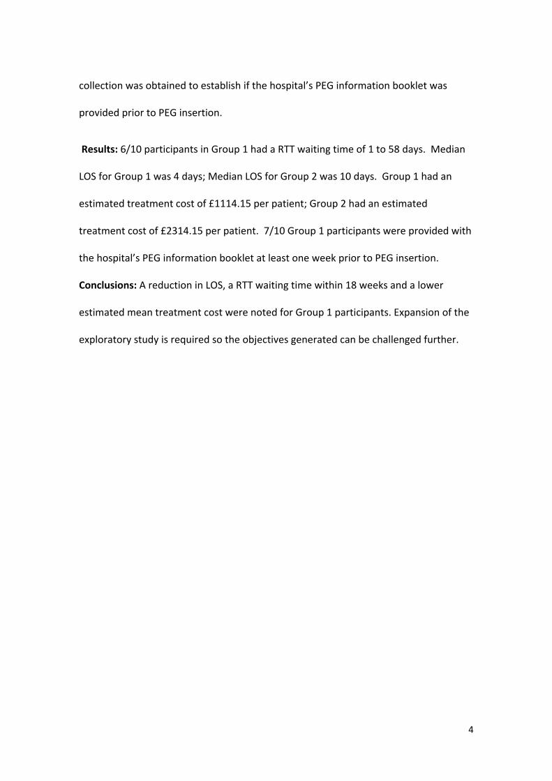
collection was obtained to establish if the hospital’s PEG information booklet was
provided prior to PEG insertion.
Results: 6/10 participants in Group 1 had a RTT waiting time of 1 to 58 days. Median
LOS for Group 1 was 4 days; Median LOS for Group 2 was 10 days. Group 1 had an
estimated treatment cost of £1114.15 per patient; Group 2 had an estimated
treatment cost of £2314.15 per patient. 7/10 Group 1 participants were provided with
the hospital’s PEG information booklet at least one week prior to PEG insertion.
Conclusions: A reduction in LOS, a RTT waiting time within 18 weeks and a lower
estimated mean treatment cost were noted for Group 1 participants. Expansion of the
exploratory study is required so the objectives generated can be challenged further.

5
Introduction
A gastrostomy feeding tube is a narrow tube that is placed directly through the
abdominal wall in to the stomach. The gastrostomy tube is generally kept in place with
a soft spongy balloon or plastic disc internally and a fixation device externally.
Percutaneous endoscopic gastrostomy (PEG) is a technique used to put a gastrostomy
tube in place using an endoscope (Abbott Nutrition, 2006). The first published report
of a PEG was in 1980 by Gauderer, Ponsky and Izant (1980). PEG is reported to be one
of the most commonly performed gastrointestinal procedures (Potack, 2008). The
gastrostomy tube is used to deliver nutrition, fluid and medications directly into the
stomach (Abbott Nutrition, 2006). Gastrostomy tube feeding should be considered for
long‐term (4 weeks or more) enteral tube feeding (National Institute for Health and
Clinical Excellence, 2006). Feeding via a gastrostomy tube mitigates malnutrition and
permits the maintenance of tissue metabolism and structure even if the patient cannot
eat anything or sufficient to regain health (Clinical Resource Efficiency Support Team,
2004).
The primary placement of a gastrostomy is undertaken in the acute hospital setting
(Best & Hitchings, 2010). Pressure on hospital beds often leads to early discharge of
enterally fed patients. Home enteral tube feeding (HETF) is an expanding area of
nutritional support. In some instances, patients will attend the hospital for the
insertion of their tube as a day case, so reducing costs compared with other placement
methods and discharged home on the same day (Thomas & Bishop, 2007). To ensure

6
the safe transfer of care and the appropriate monitoring of patients clear
communication needs to be established between health professionals in both primary
and secondary care (Best & Hitchings 2010). It has been documented that community
follow up is often inadequate for patients on HETF with only a small number of
patients and carers feeling that they receive sufficient information to enable them to
manage their feeding tube prior to leaving hospital (Best & Hitchings 2010). This
situation potentially poses problems if the patient is in hospital for a shortened length
of stay (LOS) and then discharged back into the care of community services. HETF is
noted to place a considerable burden on family and carers (Best & Hitchings, 2010).
Adequate training and support to reduce problems and complications are required,
making coordination of patient care by a multidisciplinary team (MDT) paramount.
A community HETF service in the North West (NW) of England problems highlighted
the lack of formal community assessment procedure prior to PEG insertion, increased
referral to treatment (RTT) waiting time from community referral to PEG insertion,
increased LOS post insertion and standardised information for PEG insertion was
available but not routinely used. A care pathway was developed (Appendix 1) to
address the identified issues. The concept of the care pathway is noted to have
originated in the United States. It is a “structured multidisciplinary care plan
developed to take in to account of current knowledge and best practice” (Ditchburn,
2005, p.34). It details the essential steps in the care of a patient with a specific clinical
problem” encouraging evidence based practice (Ditchburn, 2005, p.34). Therefore the
7
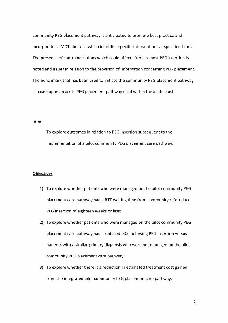
community PEG placement pathway is anticipated to promote best practice and
incorporates a MDT checklist which identifies specific interventions at specified times.
The presence of contraindications which could affect aftercare post PEG insertion is
noted and issues in relation to the provision of information concerning PEG placement.
The benchmark that has been used to initiate the community PEG placement pathway
is based upon an acute PEG placement pathway used within the acute trust.
Aim
To explore outcomes in relation to PEG insertion subsequent to the
implementation of a pilot community PEG placement care pathway.
Oblectives
1) To explore whether patients who were managed on the pilot community PEG
placement care pathway had a RTT waiting time from community referral to
PEG insertion of eighteen weeks or less;
2) To explore whether patients who were managed on the pilot community PEG
placement care pathway had a reduced LOS following PEG insertion versus
patients with a similar primary diagnosis who were not managed on the pilot
community PEG placement care pathway;
3) To explore whether there is a reduction in estimated treatment cost gained
from the integrated pilot community PEG placement care pathway.
8
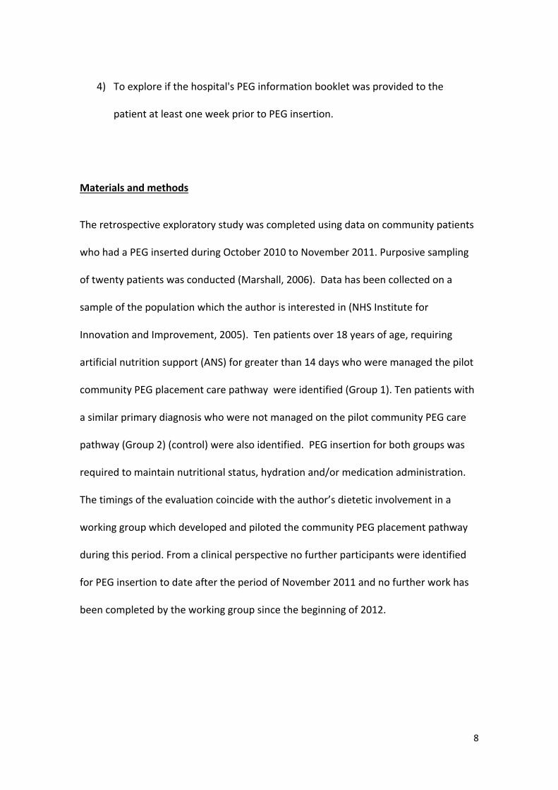
4) To explore if the hospital's PEG information booklet was provided to the
patient at least one week prior to PEG insertion.
Materials and methods
The retrospective exploratory study was completed using data on community patients
who had a PEG inserted during October 2010 to November 2011. Purposive sampling
of twenty patients was conducted (Marshall, 2006). Data has been collected on a
sample of the population which the author is interested in (NHS Institute for
Innovation and Improvement, 2005). Ten patients over 18 years of age, requiring
artificial nutrition support (ANS) for greater than 14 days who were managed the pilot
community PEG placement care pathway were identified (Group 1). Ten patients with
a similar primary diagnosis who were not managed on the pilot community PEG care
pathway (Group 2) (control) were also identified. PEG insertion for both groups was
required to maintain nutritional status, hydration and/or medication administration.
The timings of the evaluation coincide with the author’s dietetic involvement in a
working group which developed and piloted the community PEG placement pathway
during this period. From a clinical perspective no further participants were identified
for PEG insertion to date after the period of November 2011 and no further work has
been completed by the working group since the beginning of 2012.

9
Inclusion Criteria
ANS required for greater than 14 days;
Greater than 18 years of age;
PEG insertion required to facilitate the maintenance of nutritional status,
hydration and/or medication administration.
Exclusion Criteria
ANS required for less than 14 days;
Less than 18 years of age;
Diagnosed eating disorder;
Alternative feeding tube to PEG.
Exploratory Study Setting
The study has been conducted using patients from three communities served by one
district general hospital in the North West of England. The three communities were
selected as the author is employed within the dietetic department at the local district
general hospital which provides the service. Three towns are located in the area which
is split into two boroughs. Borough one consists of two towns and has a population of
119,000. The health of the population in borough one is reported to be worse than
the England average. Deprivation is a major issue where 21 out of the 79 ‘Super
Output Areas’ fall in 10% of the most deprived areas in England. Unemployment is
noted as a challenge where one in three adults in one ward is claiming an out‐of‐work

10
benefit (Department of Health, 2012a). Borough two consists of one town and has a
population of 199,000. At a glance, the health of the population in borough two is
reported to be mixed compared with the England average. (Department of Health,
2012b).
Sample Size
Data from the records relating to a total of twenty patients were purposively sampled
(Marshall, 1996). Group 1 consisted of ten patients who were managed on the pilot
community PEG placement care pathway and Group 2 consisted of ten patients who
were not managed on the pilot community PEG placement care pathway with a similar
primary diagnosis to Group 1. An activity analysis conducted by the district general
hospital’s finance department for the period of October 2010 to November 2011 for
endoscopic/open procedure codes enabled the identification of suitable Group 2
patients. The endoscopic/open procedure codes were clarified with the coding
department to capture total PEG insertion activity. All data obtained for inclusion in
the exploratory study was originally collected as part of routine care by professionals
involved in the patients overall care. It was necessary to conduct the exploratory
study including such patients as they were considered for PEG insertion during the
chosen retrospective study period. Patients were not specifically recruited from
vulnerable groups, however it is acknowledged by the author that some of the patients
included in the study were classed as vulnerable; elderly, adults with learning
disabilities and other vulnerable groups.

11
A community PEG placement pathway evaluation proforma (Appendix 2) was
developed to capture qualitative data from the patient’s medical notes.
Statistical Analysis
The exploratory study uses quantitative data collection. The quantitative approaches
used in the evaluation involve the collection of nominal data and the collection of
numerical data through statistics.
Objective One
For objective one, the data collected is nominal. The NHS RTT waiting time for
gastroenterology is “within a maximum of 18 weeks of referral”. Therefore the author
explored the proportion of patients managed on the pilot community PEG placement
care pathway who had a RTT within or beyond 18 weeks. The responses will be
summarised and commented upon in this report.
Objective Two
The dependent variable is LOS and the independent variable is the two treatment
regimes. An Independent t‐test was conducted. All data was analysed using the
Statistical Package for Social Sciences (SPSS) Version 21 (IBM software Limited, 2012).

12
The two data sets were analysed to test for any significant difference in LOS in days
following PEG insertion amongst patients in both groups.
Objective Three
The median LOS for Group 1 and Group 2 patients was used in the calculation of
estimated cost of treatment. An endoscopy examination profile (Appendix 3) and
subsequent PEG insertion cost analysis (Appendix 4) have been generated by the
hospital’s finance and endoscopy departments to provide a figure of £314.15 per PEG
insertion. The analysis includes staffing, consumables, equipment, coding, medical
records, cleaning and overhead costs. A figure of £200.00 for cost per day of in‐patient
stay has been provided by the hospital. This figure was multiplied with the respective
LOS data and added to the PEG insertion cost analysis to obtain an estimate of total
cost. The total cost data were analysed to test for any significant difference between
the two groups.
Objective Four
For objective four the data collected is nominal. Therefore the author explored
whether patients who were managed on the pilot community PEG placement care
pathway were provided with the hospital's PEG insertion information booklet at least
one week prior to PEG insertion.

13
Results
Table 1 below shows the primary diagnoses of the Group 1 and Group 2 patients;
Appendix 5 details the primary diagnoses, abbreviations and medical description of the
diagnoses.
Table 1: Primary diagnoses for Group 1 and Group 2 patients.
Primary Diagnosis Group 1 Group 2
CVA 1 2
CVA with DM 1 1
CVA with Dementia/Parkinson’s disease
1 1
CVA with Neurological disease
1 ‐
Friederich’s ataxia with DM ‐ 1
Huntington’s chorea ‐ 1
MND 3 2
Mitochondrial Cytopathy 1 ‐
PSP 2 1
Dementia ‐ 1
Where possible, patients for inclusion in Group 2 were selected with the same
diagnoses to patients in Group 1. When this was not possible the author selected
patients with a comparable diagnosis, however it is noted that this was not achievable
in all circumstances. Indications for PEG insertion for Group 1 and Group 2 patients
were mainly neurological (7/10) and (5/10) respectively. Difficulty swallowing
resulting in the inadequate maintenance of nutrition was indicated in all patients. PEG
was not required solely to maintain hydration and/or medication status.
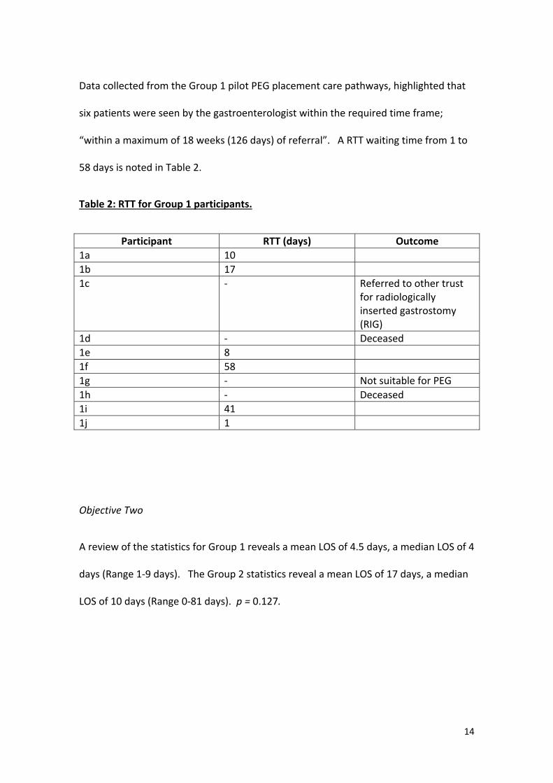
14
Data collected from the Group 1 pilot PEG placement care pathways, highlighted that
six patients were seen by the gastroenterologist within the required time frame;
“within a maximum of 18 weeks (126 days) of referral”. A RTT waiting time from 1 to
58 days is noted in Table 2.
Table 2: RTT for Group 1 participants.
Participant RTT (days) Outcome
1a 10
1b 17
1c ‐ Referred to other trust for radiologically inserted gastrostomy (RIG)
1d ‐ Deceased
1e 8
1f 58
1g ‐ Not suitable for PEG
1h ‐ Deceased
1i 41
1j 1
Objective Two
A review of the statistics for Group 1 reveals a mean LOS of 4.5 days, a median LOS of 4
days (Range 1‐9 days). The Group 2 statistics reveal a mean LOS of 17 days, a median
LOS of 10 days (Range 0‐81 days). p = 0.127.

15
Objective Three
Estimated calculated treatment costs for Group 1 and Group 2 patients are outlined in
Table 3;
Table 3: Estimated treatment cost of PEG insertion for Group 1 and Group 2 patients.
Cost of PEG + (cost per day of in‐patient stay x median LOS)
Total cost Range
Group 1 314.15 + (200.00 x 4)
£1114.15 £514.15 to £2114.15
Group 2 314.15 + (200.0 x 10)
£2314.15 £314.15 to £16,514.15
p = 0.127
Objective Four
Refer to Appendix 6 for a copy of the PEG information booklet. Data collected from
the Group 1 pilot PEG placement care pathways highlighted that seven patients/carers
were provided with the PEG information booklet at least one week prior to PEG
insertion. No pilot community PEG care pathways were recorded in three patients
medical notes; therefore the author was not able to establish whether the
patients/carers were provided with the PEG information booklet prior to PEG
insertion.

16
Discussion
Clinical conditions and diagnoses may be relevant in particular to “control” participants
as it is noted that some of these participants were admitted to hospital and had a PEG
inserted as the result of another condition. For example one participant had a
diagnosis of MND and was scheduled to have a routine PEG insertion as an out‐patient.
Unfortunately the participant sustained a fracture in the interim and was therefore
admitted in response to the fracture which could have increased the overall LOS,
rather than if the patient had been admitted solely for planned PEG insertion.
Therefore potentially the inclusion/exclusion criteria required modification.
The study was conducted over a thirteen month period, October 2010 – November
2011. It is noted by Clegg (1990) that using generalised data from a sample can
depend upon when the sampling took place. The exploratory study captured PEG
insertion activity over the four seasons of the year; a winter bed crisis can be a feature
in NHS hospitals, which can result in variability in bed occupancy (Fullerton &
Crawford, 1999). Public health concerns such as a flu outbreak can affect staffing, bed
occupancy and priority of use. For example there may be a need for the cancelling of
routine procedures during an outbreak to ensure the maintenance of other essential
NHS services. The study period was limited from a clinical perspective as no further
participants were identified for PEG insertion to date (at that time) after the period of
November 2011 and no further work has been completed by the working group since

17
the beginning of 2012. This could potentially influence RTT waiting time, LOS and the
provision of information pre PEG insertion.
In objective one, data collected from the Group 1 participants pilot PEG placement
care pathway highlighted that six patients were seen “within a maximum of 18 weeks
(126 days) of referral”, therefore patients managed on the pilot community PEG
placement care pathway did not have a RTT waiting time from community referral to
PEG insertion of 18 weeks or more. At first glance this appears acceptable that
patients experienced a RTT Waiting Time from 1 to 58 days. As part of the NHS
Constitution (March, 2012) patients have the right to access services within maximum
waiting times, or for the NHS to take all reasonable steps to offer a range of alternative
providers if this is not possible. Patients do have the option to choose to wait longer
or, if it is clinically appropriate, for the patient to wait longer. In order to interpret the
RTT waiting time values and use in practice, the measurements should not be
interpreted on the basis of numerical values alone but in the context of overall risk.
From a clinical perspective an increased RTT waiting time can have a detrimental effect
if a participant is unable to maintain their nutritional and hydration status resulting in
weight loss and a risk of malnutrition and dehydration. The retrospective extraction of
file data does not enable the author to capture the experiences of participants/carers
prior to PEG insertion. Data was not obtainable for four of the selected patients.
Unfortunately there were no other patient records that could be included in the study.
This may have affected analysis for objective two where it was noted there is no

18
significant difference in LOS between the two groups, despite the range of LOS in
Group 1 being between 1‐9 days, median LOS 4 days and Group 2; 0‐81 days, median
10 days; median LOS reduced for the pilot community PEG care pathway group.
Despite there being no significant difference in LOS a demonstrated reduction in LOS
would be a desirable outcome if confirmed. LOS is a measure to show the mean time
that a patient has stayed in hospital. Reducing LOS can free beds and staff time,
enabling more patients to be treated and therefore helping to reduce waiting times
resulting in an improved patient experience (NHS Institute for Innovation and
Improvement, 2008). In order to interpret the LOS values and use in practice, the
measurements cannot be interpreted on the basis of numerical values alone.
The estimated median treatment costs in objective three for Group 1 and Group 2
patients are £1114.15 and £2314.15 respectively. Based on the figures there would
appear to be a reduced cost of treatment episode based on the median LOS for Group
1 patients. Despite there being no significant difference in treatment cost, a
demonstrated cost reduction would be a desirable outcome to support the
implementation of the pilot community PEG placement care pathway.
In objective four, from the data which was available, the hospital's PEG insertion
information booklet was provided to seven patients/carers at least one week prior to
PEG insertion. At first glance this would appear to be acceptable. Some patients,
relatives and carers had commented to the author at review post PEG insertion (prior

19
to implementation of the pilot community PEG placement care pathway), that
information about PEG insertion, PEG/stoma aftercare and feeding options were not
discussed prior to PEG insertion. Standardised information on PEG was available but
wasn’t routinely being used. The literature available is suitable for use in both primary
and secondary care. It outlines what patients can expect before and after PEG
placement as well as identifying risks associated with PEG placement. In order to
interpret this information and use in practice a qualitative analysis to answer
subjective questions are required; was the information useful? Was it in the correct
format? Do patients/carers feel informed? Are the patients/carers able to understand
the information? Such information on the relevance of patient information could be
linked to further study outcomes and effectiveness of integrated care pathways.
Conclusions and recommendations
The investigation into the four objectives provided insight for the author. A reduction
in LOS, a RTT waiting time within 18 weeks and a lower estimated mean treatment
cost were associated with implementation of the care pathway. These are positive
outcomes. A number of recommendations have been noted by the author for further
development and evaluation of the service.
A larger sample size; possible inclusion of more hospitals. This potentially
would enable the author to include more participants with comparable
diagnoses and more data for RTT, LOS, cost of treatment episode and whether

20
the hospital’s PEG insertion information booklet was provided prior to PEG
insertion.
Prospective audit and evaluation of the service; would enable the
identification of a larger number of patients to include within the study.
Seasonal and specific public health concerns can also be monitored and
accounted for during the study.
Incorporation of qualitative data, evaluation from the patient/carer and staff
perspective; for example case studies and interviews would provide an in
depth description of the situation, noting patients/carers experiences and
expectations. Review of the MDT approach, completion of the pathway,
caseload management and the collaboration between primary and secondary
care could be reviewed. Case studies and interviews to provide feedback on the
PEG information booklet, noting patients/carers opinions on whether the
information provided is useful.
Study to assess morbidity and mortality post PEG insertion; such a study
would provide additional information to help ascertain if there was any effect
from an increased RTT Waiting Time and overall morbidity and mortality.
PEG insertion and improvement of patient outcomes; delivery of consistent
nutrition, hydration and medication as the result of PEG insertion.

21
Assessment of complications post PEG insertion; does frequency of
community follow result in a significant reduction of complications post PEG
insertion.
Review of quality of life post PEG placement; a study incorporating
appropriate markers to assess quality of life post PEG placement at specific
time intervals. Benefits and burdens of PEG tube and stoma maintenance, as
well as patients/carers opinions on feeding – method of administration and
frequency.
Non‐probability sampling was required for the exploratory study. The patients were
selected on the basis of the accessibility of their data/records and purposive personal
judgement of the author. The downside of this method is that an unknown proportion
of the entire population was not sampled and therefore may not represent the entire
population accurately; however the study conducted was merely exploratory (Non‐
Probability Sampling, 2013).
The pilot community PEG placement care pathway requires review and auditing to
assess and document its effectiveness in clinical practice. Problems identified as a
result of review and potential implementation would need to be addressed as they
arise.

22
References
Abbott Nutrition. (2006). What is a gastrostomy feeding tube? Maidenhead: Author. Best, C., & Hitchings, H. (2010) Day case gastrostomy placement for patients in the community. British Journal of Community Nursing, 15(6), 272‐278. Clinical Resource Efficiency Support Team. (2004). Guidelines for the management of enteral tube feeding in adults. Stormont: Author. Dickinson, 0. (2013, April). Reasoning and undertaking of percutaneous image guided gastrostomies (PIGGS). NHD The Dietitians Magazine, pp.23‐25. Ditchburn, L. (2005). Joint primary‐secondary care design of PEG care pathways. Nursing Times, 101(18), 34‐36. Gauderer, M.W.L., Ponsky, J.L. & Izant, R.J. (1980). Gastrostomy without laparotomy:
percutaneous endoscopic technique. Journal of Paediatric Surgery, 15, 1206‐10.
Hope, R.A., Longmore, J.M., Hodgetts, T.J., & Ramrakha, P.S. (1993). Oxford Handbook of Clinical Medicine. (3rd ed.). Oxford: Oxford University Press.
IBM Software Limited (2012). Statistical Package for the Social Sciences. Version 21.0. IBM Software Limited.
Marshall, M.N. (1996). Sampling for qualitative research. Family Practitioner, 13, 522‐ 565. Martin, E.A. (1994). Concise Medical Dictionary. (4th ed.). Oxford: Oxford University
Press National Health Service Institute for Innovation and Improvement. (2005).
Improvement Leaders’ Guide. Evaluating improvement. General improvement skills. Coventry: Author.
National Institute for Health and Clinical Excellence. (2006). Nutrition support in adults: Oral nutrition support, enteral tube feeding and parenteral nutrition. Clinical Guideline 32. London: Author.
Non‐Probability Sampling (2013). Retrieved August 22, 2013 from http://explorable.com/non‐probability‐sampling
Potack, J.Z. (2008). Complications of and Controversies Associated With Percutaneous Endoscopic Gastrostomy: Report of a Case and Literature Review. Medscape Journal of Medicine 10(6), 142. Thomas, B., & Bishop, J. (2007) Manual of Dietetic Practice. (4th ed.). Oxford: Blackwell Publishing.

23
Appendix 1: Community PEG placement care pathway Print copy only.
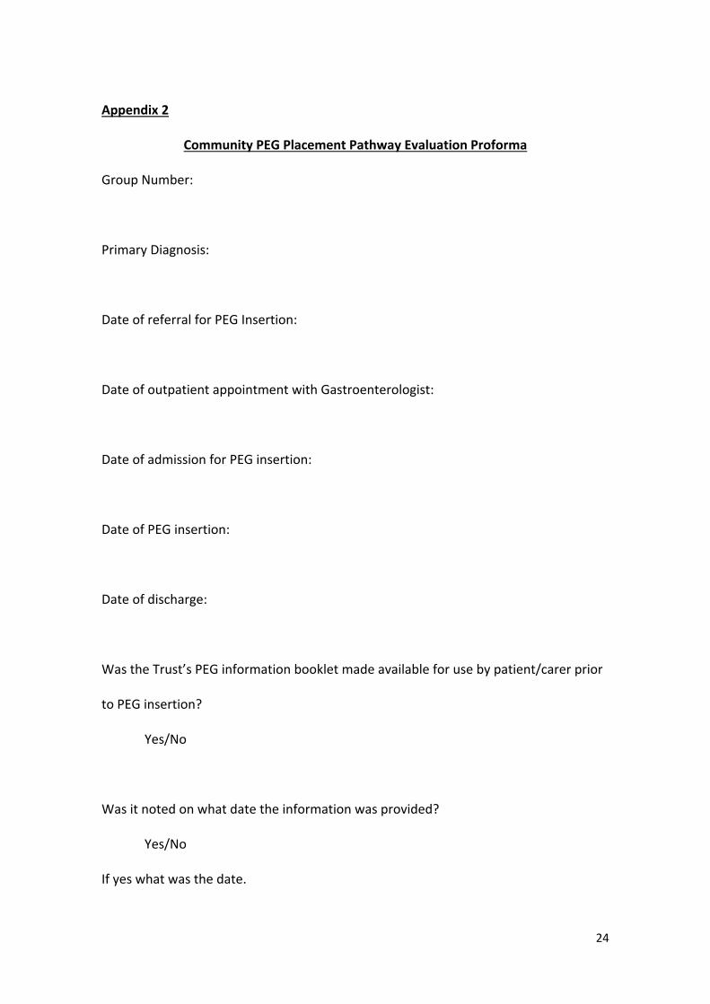
24
Appendix 2
Community PEG Placement Pathway Evaluation Proforma
Group Number:
Primary Diagnosis:
Date of referral for PEG Insertion:
Date of outpatient appointment with Gastroenterologist:
Date of admission for PEG insertion:
Date of PEG insertion:
Date of discharge:
Was the Trust’s PEG information booklet made available for use by patient/carer prior
to PEG insertion?
Yes/No
Was it noted on what date the information was provided?
Yes/No
If yes what was the date.

25
Appendix 3: Endoscopy Examination Profile.
Question Answer Comments
Examination type Therapeutic Gastroscopy – Insertion of PEG
PEG placement For therapeutic treatment and management of specific conditions
Performed by
Consultant/ Registrar
Nurse specialist
Consultant/ Registrar/Nurse Specialist
For PEG placement assistance needed;Nurse specialist for example
Staffing allocated in support of list x1 band 6 nurse and x1 band 2 carer
2 qualified nurses band 5 and above 2 Band 3 Carers (decontamination of Scopes)
Airway management Vital signs observation, Meditech documentation, specimen collection
Consumables routinely used for these cases drugs, guide wires, etc.
Lignocaine spray 6p Midazolam 60p Disposable mouth guard 49p PEG Tube kit £80 Drawing up needles, syringes, Chloroprep, cannula and venflon dressings, gloves, aprons £2.50
Appropriate consumables used to treat these various conditions
Average examination time +/‐ number of cases per standard (unmixed list)
This procedures can take 30 minutes and are best performed on an unmixed list due to the urgency and need of individual patients
Examination times are dependent on individual needs

26
Appendix 4: PEG Insertion Cost Analysis
Cost
Consultant £ 66.00
Registrar £ 18.00
Nursing (Procedure/Recovery) £ 32.00
Decontamination £ 5.00
Reception £ 4.00
Administration £ 11.00
Lignocaine £ 0.06
Midazolam £ 1.00
Disposable Mouth Guard £ 0.49
PEG Tube Kit £ 80.00
Drawing up needles, syringes, Chloroprep, cannula, gloves £ 3.00
Managed Service – Endoscopy equipment £ 35.00
Domestics £ 3.00
Overheads £ 57.00
TOTAL COST £ 314.55

27
Appendix 5
Primary Diagnosis Abbreviations and Definitions
Cerebrovascular Accident (CVA): Also known as stroke, resulting from infarction
of part of the brain, or from intracerebral haemorrhage (Hope, Longmore,
Hodgetts & Ramrakha, 1993).
Diabetes Mellitus (DM): A syndrome caused by the lack or diminished
effectiveness of endogenous insulin (Hope et al. 1993).
Dementia: A chronic or persistent disorder of brain process due to organic
brain disease. Symptoms include memory disorders, impaired reasoning ability
and disorientation (Martin, 1994).
Friederich’s ataxia: An inherited disorder resulting in the clumsiness of willed
movements, staggering when walking, unable to pronounce words
appropriately, rapid involuntary movements of the eyes and spasticity of the
limbs (Martin, 1994).
Huntington’s chorea: A hereditary disease caused by the defect of a single gene
inherited as a dominant characteristic. Symptoms include involuntary
movements accompanied by behavioural changes and progressive dementia
(Martin, 1994).
Motor Neurone Disease (MND): Progressive degenerative disease of unknown
cause, affects upper and lower neurones, causes muscle weakness and wasting,
no sensory abnormality present (Martin, 1994).
Mitochondrial cytoplasty: Genetic disease.
Neurological disease: Any disorder of the body’s nervous system. A range of
symptoms can be experienced which depends upon which nerves of the
various systems within the body are affected.
Parkinson’s disease: A progressive disease that may lead to dementia. The
disease is caused by the degeneration of dopaminergic neurones (Hope et al.
1993)
Progressive Supranuclear Palsy (PSP): Degenerative condition that affects
movement, personality, balance and later symptoms signs of dementia. Slurring
of speech, moving of eyes and difficulty swallowing.

28
Appendix 6
Hospital’s PEG information booklet
Print copy only.