thomson et al. 2014 – supplemental information...
TRANSCRIPT

Thomson et al. 2014 – Supplemental information
Methods
Mice and breeding
All animal experiments were approved by the Animal Care Committees at the Center for
Comparative Medicine of Northwestern University (Evanston Illinois, USA) and the Toronto
Centre for Phenogenomics (Toronto, Ontario, Canada). Animals housed at either center were
allowed unrestricted access to standard rodent chow (Harlan #7912) and water.
To create the conditional Angpt1, Angpt2 double knockout mouse line used in this study,
we have generated a new Angpt2Flox mouse which was crossed onto the inducible, whole-body
Angpt1 knockout line previously developed in our laboratory (1). An Angpt2Flox targeting
construct was obtained from the Sanger Institute knockout mouse project (clone
#PRPGS00100_B_CO2) and used to target mouse embryonic stem (ES) cells by homologous
recombination. This construct introduces loxp recombination sites flanking exon 4 of the Angpt2
gene, as well as a neomycin-resistance cassette flanked by frt recombination sites for flpe
recombinase (Supplemental figure 1 A). Morula aggregation was performed at the Toronto
Centre for Phenogenomics (Toronto, Canada), and chimeras were crossed to wild-type ICR
mice. F1 animals were screened by PCR and Southern blot, and those positive for the
Angpt1Flox(Neo in) allele were crossed to mice expressing FlpE recombinase (B6;SJL-
Tg(ACTFLPe)9205Dym/J, The Jackson laboratory, Bar Harbor, Maine) to excise the Neomycin
selection cassette used in ES cell cloning. FlpE excision was verified by Southern blot, and
Angpt2Flox(Neo out) mice were selected for all subsequent breeding. Newly created
Angpt2Flox mice were crossed onto the whole-body, inducible Angpt1Flox/ROSA-rtTA/Tet-
On-Cre mouse line (1) to create Angpt1/Angpt2/ROSA-rtTA/Tet-On-Cre line (A1A2FloxWB
mice). Angpt1 and Angpt2 deletion was induced by addition of 0.5% Doxycycline to the
drinking water of pregnant dams at day 16.5 of gestation to generate A1A2FloxWBΔE16.5 offspring
(Supplemental figure 1 B-C).
Tie2 conditional knockout mice were created using the COIN (Conditional by Inversion)
system as previously described (2). Briefly, The ex1COIN allele was engineered by inserting the
COIN intron into exon 1 of Tek (Tie2) close to the start of the ORF, at coordinate 94739653 in
the sense strand of chromosome 4 (in mouse genome’s Ensembl release 75 - February 2014),
effectively splitting the 393 bp exon into left (1L) and right (1R) exons of 354 and 39 bp
respectively (Supplemental figure 2). The point of insertion was chosen so as to conform to the
consensus sequence of the 5’ splice site (3). To generate the targeting vector, a BAC containing
mouse genomic DNA encompassing Tek was selected by PCR-screen from a BAC library of
129/Svj mouse genomic DNA (Incyte, Wilmington, Delaware, USA). BAC 352i20 was
modified by inserting the COIN module intron (eGFP; phase 1; hygro version) at the position
listed above, resulting in a BACVec with homology arms of 75 and 55 kb. For targeting, this
BACVec was digested with NotI and electroporated into F1H4 ES cells to generate
Tie2ex1COIN(hyg)/+ targeted ES cells. Targeted ES cells were genotyped by a Loss-of-Allele assay
as previously described (2); a list of the probes utilized for screening is appended below. 11 out
of 192 clones were targeted. Clone 1198E-A4 was selected for use and treated with FlpE
recombinase to remove the hygromycin selection cassette and generate the final Tie2COIN
allele. Tie2COIN mice were crossed with the ROSA-rtTA/Te-tOn-Cre mouse line as described
above to create Tie2COINWB mice for use in this study. Nursing Tie2COIN/ROSA-rtTA/Tet-On-

Cre dams and neonates were induced with doxycycline at postnatal day 0 (P0) to generate
Tie2COINWB-INVΔP0 offspring.
Knockout mice were genotyped by PCR, using the following primers: Angpt1Flox,
Forward 5’-CAATGCCAGAGGTTCTTGTGAA-3’; Reverse 5’-
TCAAAGCAACATATCATGTGCA-3’ (WT: 233bp product, Angpt1Flox: 328bp),
Angpt1Delete, Forward 5’-CAATGCCAGAGGTTCTTGTGAA-3’; Reverse 5’-
TGTGAGCAAAACCCCTTTC-3’ (431bp product), Angpt2Flox, Forward 5’-
GGGAAACCTCAACACTCCAA-3’; Reverse 5’-ACACCGGCCTCTAGACACAC-3’ (WT:
224bp product, Angpt2Flox: 258bp) and Angpt2Delete, Forward 5’-
AAGGCGCATAACGATACCAC-3’; Reverse 5’-TGAGAACTCTGCAGCCTTGA-3’
(Angpt2Flox: 1,372bp product, Angpt2Delete: 426bp). Tie2COIN, Forward 5’-
CTGAAGCACTGCACGCCGTAG-3’; Reverse 5’-CTCAGAGTATTTTATCCTCATCTC-3’
(470bp product). Tie2COININV, Forward 5’- GTTTTCAGGGTGTTGTTTAG-3’; Reverse 5’-
CTGAAGCACTGCACGCCGTAG-3’ (600bp product).
Tie2COIN targeted ES cells were screened using the following probes: 1198MD-Fwd 5’-
CCGGCTTAGTTCTCTGTGGAGT-3’, 1198MD-Probe FAM-AGCTTGCTCCTTTATG-MGB,
1198MD-Rev 5’-CACAAATAAACATCAAGCCAAAACTT-3’, 1198MU-Fwd 5’-
GAAGTATGGACTCTTTAGCCGGC-3’, 1198MU-Probe FAM-TTCTCTGTGGAGTCAGC-
MGB, 1198MU-Rev 5’-GCCAAAACTTACCATAAAGGAGCA-3’.

Live-animal studies
Littermate controls were used for all live-animal studies. Intraocular pressure was
measured at 10 weeks of age using a Tonolab rebound tonometer (iCare, Vantaa, Finland) (4).
Mice were restrained in a soft plastic cone and ocular pressure for each eye was averaged from
three sets of six recordings. Each mouse was measured on two subsequent days and the results
were averaged to obtain the reported IOP values. As no significant difference was found between
left and right eyes, both measurements were included in the final groups. Visual acuity was
measured using an Optomotor response test as previously described (5, 6). Briefly, animals were
placed on an elevated platform surrounded by four LCD monitors. Monitors displayed vertical
gratings as moving visual stimuli. Mice were observed and head movement following the
direction of moving gratings was scored as positive optomotor response. Spatial frequency of the
moving gratings was gradually increased, and visual acuity was scored as the highest frequency
triggering a response. Optomotor tests on each mouse were repeated on consecutive days, and
results for each mouse were averaged to obtain the final visual acuity value. The optomotor
response system used for these studies provides a maximum grating size of 0.042 cycles per
degree, and mice were scored as “no response” if they were unable to respond to this stimulus.
For statistical comparison, animals with no optomotor response were assigned a score of 0.042
c/d.
Tissue collection and Histology
A1A2FloxWB mice induced at E16.5 (A1A2FloxWBΔE16.5) and littermate controls were
aged 8 weeks before tissue harvest for histological studies. Mice were anesthetized by i.p.
injection with 2, 2, 2-Tribromoethanol. Tissues were cleared (PBS, 1mg/ml lidocaine, 10u/ml
heparin) and fixed (2.5% gluteraldehyde, 2% formaldehyde in 0.1M phosphate buffer pH 7.4) by
cardiac perfusion. Eyes were dissected and postfixed for an additional 8 hours at 4°C. Whole
eyes were embedded in Epon 812 and 0.5μM sections were prepared. Sections were stained with
Toluidine blue and imaged on a compound microscope. Optic nerve sections were embedded in
paraffin, sectioned and stained using haematoxylin and eosin following standard protocols.
Histological studies were performed using groups of 4 mice per genotype, and several sections
were examined from each animal.
Quantification of retinal ganglion cell layer nuclei
Eyes from A1A2FloxWBΔE16.5 mice were isolated and immersion fixed (10% formalin).
Whole eyes were then dehydrated and embedded in paraffin. 5 μm sagittal sections were
collected and stained using haematoxylin and eosin. Sections were examined using a compound
microscope, and 883 μm fields adjacent to the optic nerve were captured using a 40x objective
(supplemental figure 3 A). Fields from three consecutive sections were collected from each eye
and ganglion cell layer nuclei were counted. Counts from each field were averaged to obtain
mean nuclei per 100 μm. Representative fields from each animal are presented in supplemental
figure 3 B-I.
Immunofluorescence
A1A2FloxWBΔE16.5 mice and littermate controls were sacrificed following anesthesia by
i.p. injection of 2, 2, 2-Tribromoethanol. Eyes were dissected and immersion fixed (4%
formaldehyde, 0.1M phosphate buffer pH 7.4). For limbus whole mounts, fixed eyes were
bisected sagittally, retinas and optic nerves were removed and hemispheres stained as whole-
mounts. Flat mounted retina tissue was prepared similarly. The cornea and iris were removed
from fixed eyes, and intact retinas were collected for staining. Samples were blocked overnight
(5% Donkey serum, 0.5% Triton X100, TBS pH 7.4) before incubation with appropriate primary
(see below) and fluorochrome-labeled secondary antibodies (Invitrogen, Carlsbad, California).
Stained tissues were flat-mounted and imaged using a Nikon C2+ confocal microscope. For
images of Schlemm’s canal, the image plane was focused on the capillaries of the corneal limbus
and a 40 μm Z-stack towards the center of the eye was collected. Fiji software (4) was used to
depth-code slices, and a pseudo-colored three-dimensional projection was prepared. Due to the
thickness of whole-mount limbus tissue, 25 μm Z-stacks were collected and maximum intensity
projections were used to illustrate blood and lymphatic capillaries of the corneal limbus. Primary
antibodies used: goat anti-mouse Lyve-1 (R&D Systems, AF2125), rat anti-mouse CD31 (BD
Pharmingen, 550274) and rabbit anti-mouse Tuj1 (Covance, MRB-435P).
Fluorescent Activated Cell Sorting (FACS) and real-time PCR of mouse lymphatic endothelial
cells
Cells isolated from E15.5 control (Foxc2flox/flox) and lymphatic-specific Foxc2 knockout
(Foxc2flox/flox;Prox1CreERT2) mouse embryos were stained for Lyve-1 and CD31 and subjected
to FACS as previously described (7, 8). Briefly, E15.5 embryos were harvested in Hank’s
balanced salt solution (HBSS, Sigma-Aldrich) and then chopped for an overnight digestion with
collagenease I/II. The colleagenase-treated cell suspension was incubated with RBC (Red blood
cell) lysis buffer (StemCell Technologies,Vancouver, Canada). Following centrifugation, cell
pellets were incubated with anti-Lyve-1 antibody (Abcam) for 20 min at 4°C. After washing with

PBS, the cells were then stained with PE conjugated anti-CD31 antibody (BD Pharmingen) and
Alexa 488-conjugated donkey anti-rabbit secondary antibody (Invitrogen,Carlsbad, CA). After
gauze filtration with a cell strainer (40µm BD Biosciences) to obtain a single cell suspension,
Lyve-1+/CD31+ LECs were sorted using BD FacsAria SORP 4-Laser. RNA was extracted from
sorted LEC using Trizol (Invitrogen), cDNA was synthesized using the cDNA synthesis kit
(Biorad) according to manufacturer’s instructions. qPCR was run using an ABI 7500 real-time
thermocycler using the following primers: Tie2Fwd: 5’-ACACTGTCCTCCCAACAGCTTCTT-
3’, Tie2Rev: 5’-TGATTCGATTGCCATCCAACGCAC-3’, PpiaFwd: 5’-
CAAATGCTGGACCAAACACA-3’, PpiaRev: 5’- TGCCATCCAGCCATTCAGTC-3’.
Lymphatic Endothelial Cell Culture, siRNA Transfection, Protein Extract Preparation and
Western Blot
Human dermal lymphatic endothelial cells(9) were cultured in EBM media (LONZA,
Basel, Switzerland) with 10% fetal bovine serum, antibiotics and other supplements. The cells
were transfected with Foxc1, Foxc2 or control siRNAs (see below) using Lipofectamine
RNAiMAX (Invitrogen) and incubated for 48 h. At the end of incubation, cells were harvested
and whole cell lysates were prepared using RIPA buffer. Protein lysates were separated by SDS-
PAGE and blotted onto PVDF membranes (Bio-Rad). Membranes were blocked (TBS with 5%
donkey serum, 2.5% BSA, 0.05% Tween-20) and incubated with appropriate primary (See
below) and HRP-tagged secondary (Jackson Immunoresearch, West Grove PA) antibodies.
Signals were detected using ECL reagents (Bio-Rad). siRNAs: Scrambled (Qiagen, #1027281),
Foxc1 (Thermo Scientific, #L-009318-00-0005), Foxc2 (Thermo Scientific #ATTAA-004016).

Primary antibodies: Rabbit anti-mouse Tie2 (Santa Cruz #SC-324, reported by the supplier to
recognize Tie2 of human and mouse origin), Rabbit anti-mouse βActin (Abcam #ab8227), Sheep
anti-human FoxC2 (R&D Systems #AF5044).
Statistics and figures
Throughout this study, plotted values are shown as means +/- standard error (SEM).
Statistical comparisons were performed using Graphpad Prism 5.0 (GraphPad Software, La Jolla,
CA). Indicated P-values were obtained using a two-tailed Student’s t-test unless otherwise noted
in the manuscript. P-values were indicated in figures using the following notation: * P < 0.05, **
P < 0.01 and *** P<0.001. Figures were assembled using Graphpad Prism 5.0, Photoshop CS5
(Adobe Software, San Jose CA) and InDesign CS5 (Adobe Software).
1. Jeansson M, Gawlik A, Anderson G, Li C, Kerjaschki D, Henkelman M, and Quaggin
SE. Angiopoietin-1 is essential in mouse vasculature during development and in response
to injury. The Journal of Clinical Investigation. 2011;121(6):2278-89.
2. Economides AN, Frendewey D, Yang P, Dominguez MG, Dore AT, Lobov IB, Persaud
T, Rojas J, McClain J, Lengyel P, et al. Conditionals by inversion provide a universal
method for the generation of conditional alleles. Proceedings of the National Academy of
Sciences. 2013;110(34):E3179-E88.
3. Burge CB, Tuschl T, and Sharp AS. In: Gesteland RF, Cech TR, and Atkins JF eds. The
RNA World. New York: Cold Spring Harbor Press; 1999:525-60.
4. John SW, Hagaman JR, MacTaggart TE, Peng L, and Smithes O. Intraocular pressure in
inbred mouse strains. Investigative Ophthalmology & Visual Science. 1997;38(1):249-53.

5. Feng L, Zhao Y, Yoshida M, Chen H, Yang JF, Kim TS, Cang J, Troy JB, and Liu X.
Sustained Ocular Hypertension Induces Dendritic Degeneration of Mouse Retinal
Ganglion Cells That Depends on Cell Type and Location. Investigative Ophthalmology &
Visual Science. 2013;54(2):1106-17.
6. Prusky GT, Alam NM, Beekman S, and Douglas RM. Rapid Quantification of Adult and
Developing Mouse Spatial Vision Using a Virtual Optomotor System. Investigative
Ophthalmology & Visual Science. 2004;45(12):4611-6.
7. Fatima A, Culver A, Culver F, Liu T, Dietz WH, Thomson BR, Hadjantonakis A-K,
Quaggin SE, and Kume T. Murine Notch1 is required for lymphatic vascular
morphogenesis during development. Developmental Dynamics. 2014.
8. Sasman A, Nassano-Miller C, Shim KS, Koo HY, Liu T, Schultz KM, Millay M, Nanano
A, Kang M, Suzuki T, et al. Generation of conditional alleles for Foxc1 and Foxc2 in
mice. genesis. 2012;50(10):766-74.
9. Hirakawa S, Hong Y-K, Harvey N, Schacht V, Matsuda K, Libermann T, and Detmar M.
Identification of Vascular Lineage-Specific Genes by Transcriptional Profiling of
Isolated Blood Vascular and Lymphatic Endothelial Cells. The American Journal of
Pathology. 2003;162(2):575-86.
Supplemental figures
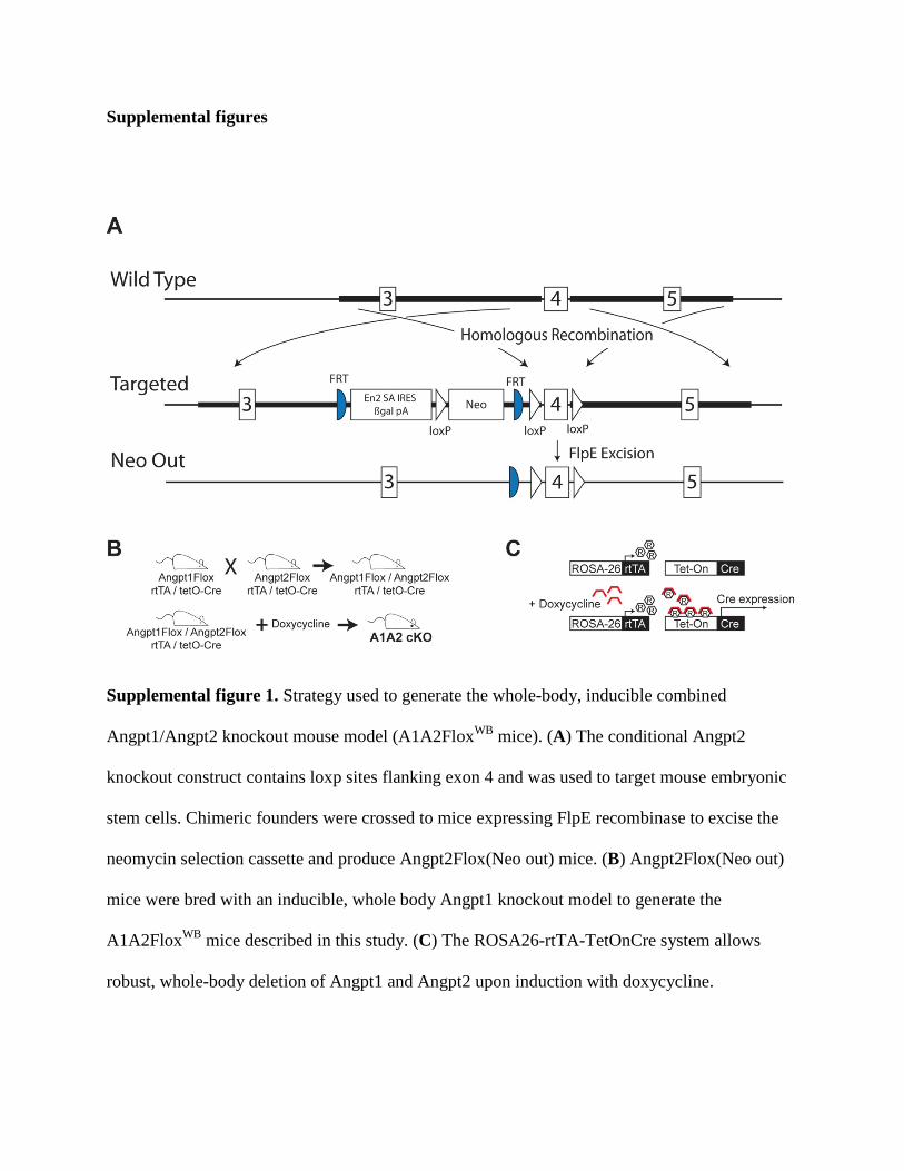
Supplemental figure 1. Strategy used to generate the whole-body, inducible combined
Angpt1/Angpt2 knockout mouse model (A1A2FloxWB mice). (A) The conditional Angpt2
knockout construct contains loxp sites flanking exon 4 and was used to target mouse embryonic
stem cells. Chimeric founders were crossed to mice expressing FlpE recombinase to excise the
neomycin selection cassette and produce Angpt2Flox(Neo out) mice. (B) Angpt2Flox(Neo out)
mice were bred with an inducible, whole body Angpt1 knockout model to generate the
A1A2FloxWB mice described in this study. (C) The ROSA26-rtTA-TetOnCre system allows
robust, whole-body deletion of Angpt1 and Angpt2 upon induction with doxycycline.
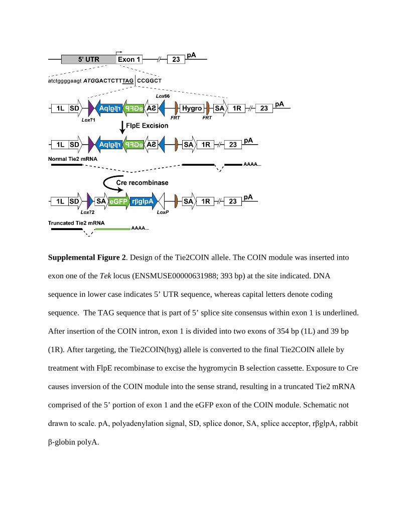
Supplemental Figure 2. Design of the Tie2COIN allele. The COIN module was inserted into
exon one of the Tek locus (ENSMUSE00000631988; 393 bp) at the site indicated. DNA
sequence in lower case indicates 5’ UTR sequence, whereas capital letters denote coding
sequence. The TAG sequence that is part of 5’ splice site consensus within exon 1 is underlined.
After insertion of the COIN intron, exon 1 is divided into two exons of 354 bp (1L) and 39 bp
(1R). After targeting, the Tie2COIN(hyg) allele is converted to the final Tie2COIN allele by
treatment with FlpE recombinase to excise the hygromycin B selection cassette. Exposure to Cre
causes inversion of the COIN module into the sense strand, resulting in a truncated Tie2 mRNA
comprised of the 5’ portion of exon 1 and the eGFP exon of the COIN module. Schematic not
drawn to scale. pA, polyadenylation signal, SD, splice donor, SA, splice acceptor, rβglpA, rabbit
β‐globin polyA.
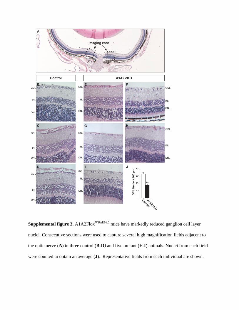
Supplemental figure 3. A1A2FloxWBΔE16.5 mice have markedly reduced ganglion cell layer
nuclei. Consecutive sections were used to capture several high magnification fields adjacent to
the optic nerve (A) in three control (B-D) and five mutant (E-I) animals. Nuclei from each field
were counted to obtain an average (J). Representative fields from each individual are shown.

Supplemental figure 4. Tuj1 staining shows marked reduction of nerve fibers in A1A2floxWBΔE16.5 mice. Full retinas from control (A) and mutant (B) mice were cropped to obtain panels in figure 2 C and I.