thorax and lungs assessment part two, lecture six 1
TRANSCRIPT

1
Thorax and Lungs Assessment
Part two, Lecture Six

Structure and Function
• Position and surface landmarks– Thoracic cage is a bony structure with a conical
shape, which is narrower at top • Defined by sternum, 12 pairs of ribs and 12 thoracic
vertebrae• Floor is the diaphragm.• First seven ribs attach to sternum by costal cartilages• Ribs 8, 9, and 10 attach to costal cartilage above• Ribs 11 and 12 are “floating,” with free palpable tips• Costochondral junctions are points at which ribs join
their cartilages; they are not palpable

Structure and Function (cont.)
– Anterior thoracic landmarks– Suprasternal notch: feel this hollow U-shaped
depression just above sternum between clavicles– Sternum: has three parts; manubrium, body, and
xiphoid process.– the sternal angle called “angle of Louis” at
articulation of manubrium and sternum, and continuous with second rib –Angle of Louis also marks site of tracheal
bifurcation into right and left main bronchi

Structure and Function (cont.)
• Position and surface landmarks (cont.)– Anterior thoracic landmarks (cont.)• Costal angle: the right and left costal margins form
an angle where they meet at xiphoid process• Usually 90 degrees or less, this angle increases
when rib cage is chronically overinflated, as in emphysema

Anterior Thoracic Cage
Slide 18-5

Structure and Function (cont.)
• Position and surface landmarks (cont.)– Posterior thoracic landmarks• Counting ribs and intercostal spaces on back is
harder due to muscles and soft tissue surrounding ribs and spinal column• Vertebra prominens: Start here; flex your head and
feel for most prominent bony spur protruding at base of neck• This is spinous process of C7; if two bumps seem
equally prominent, upper one is C7 and lower one is T1

Structure and Function (cont.)
• Position and surface landmarks (cont.)– Posterior thoracic landmarks (cont.)• Inferior border of scapula: scapulae are located
symmetrically in each hemithorax• Lower tip usually at seventh or eighth rib• Twelfth rib: palpate midway between spine and
person’s side to identify its free tip
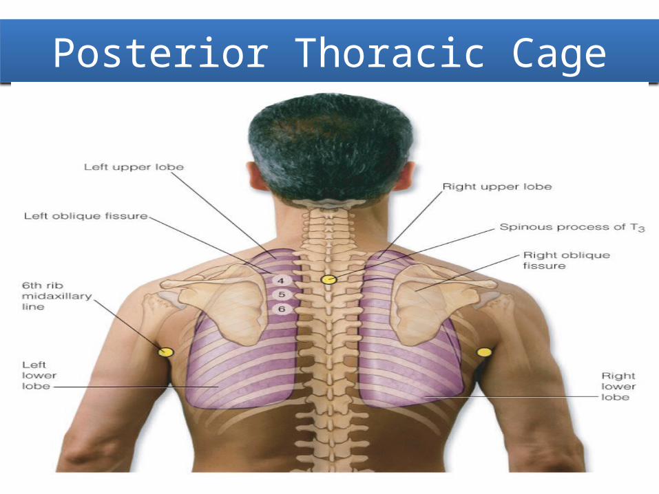
Posterior Thoracic Cage
Slide 18-8

Anterior Reference Lines
Slide 18-9

Posterior Reference Lines
Slide 18-10

Lateral Reference Lines
Slide 18-11

Structures of Respiratory System
Slide 18-12

Mechanics of Respiration
Slide 18-13
14
Subjective Data
• Cough• Shortness of breath• Chest pain with breathing• History of respiratory infections• Smoking history• Environmental exposure• Self-care behaviors
Subjective Data (cont.)
• Cough– Do you have a cough? When did it start? Gradual or
sudden?• How long have you had it?• How often do you cough? At any special time of day or
just on arising? Cough wake you up at night?• Do you cough up any sputum? How much? What color is
it?• Cough up any blood? Does the sputum have a foul odor?• How would you describe your cough: dry, barking,
hoarse, congested?
Subjective Data (cont.)
• Cough (cont.)– Cough seem to come with anything: activity,
position (lying), fever, congestion, talking, anxiety? • Activity make it better or worse?• What treatment have you tried? Does the cough
bring on anything such as chest pain or ear pain? Are you concerned about it?

Subjective Data (cont.)
• Shortness of breath– Ever had any shortness of breath or hard-
breathing spells?• What brings it on? How severe is it? How long does it
last?• Is it affected by position, such as lying down?• Occur at any specific time of day or night?• Shortness of breath episodes associated with night
sweats?• Or cough, chest pain, or bluish color around lips or
nails? Wheezing sound?

Subjective Data (cont.)
• Shortness of breath (cont.)– Do episodes seem to be related to food, pollen,
dust, animals, season, or emotion?• What do you do in a hard-breathing attack? Take a
special position, or use pursed-lip breathing? Do you use any oxygen, inhalers, or medications?
19
Subjective Data (cont.)
• Chest pain with breathing– Any chest pain with breathing? Please point to
exact location.• When did it start? Is it constant or does it come and
go?• Describe the pain: burning, stabbing?• Is it brought on by respiratory infection, coughing, or
trauma? Is it associated with fever, deep breathing, unequal chest inflation?
• What have you done to treat it? Have you tried medication or heat application?

20
Subjective Data (cont.)
• History of respiratory infections– Any past history of breathing trouble or lung diseases
such as bronchitis, emphysema, asthma, or pneumonia?• Any unusually frequent or unusually severe colds?• Any family history of allergies, tuberculosis, or asthma?
– Smoking history• Do you smoke cigarettes or cigars? At what age did you
start? How many packs per day do you smoke now? For how long? Do you live with someone who smokes?
• Have you ever tried to quit? Why do you think it did not work? What activities do you associate with smoking?

Subjective Data (cont.)
• Environmental exposure– Are there any environmental conditions that
may affect your breathing?• Where do you work? At a factory, chemical plant,
coal mine, farming, outdoors in a heavy traffic area?• Do you do anything to protect your lungs, such as
wear a mask or have ventilatory system checked at work?

Subjective Data (cont.)
• Self-care behaviors– When was your last TB skin test, chest x-ray
study, pneumonia or influenza immunization?
Objective Data (cont.)
• Inspect the posterior chest– Thoracic cage• Note shape and configuration of chest wall• Spinous processes should appear in a straight line;
thorax is symmetric, in an elliptical shape, with downward sloping ribs, about 45 degrees relative to spine; scapulae are placed symmetrically in each hemithorax• Anteroposterior diameter should be less than
transverse diameter; ratio of anteroposterior to transverse diameter is from 1:2

Objective Data (cont.)
• Inspect the posterior chest (cont.)– Thoracic cage (cont.)
• Note position person takes to breathe• Includes relaxed posture and ability to support one’s own
weight with arms comfortably at sides or in lap• Assess skin color and condition• Color should be consistent with person’s genetic
background, with allowance for sun-exposed areas on chest and back
• No cyanosis or pallor should be present• Note any lesions; inquire about any change in nevus on
back

25
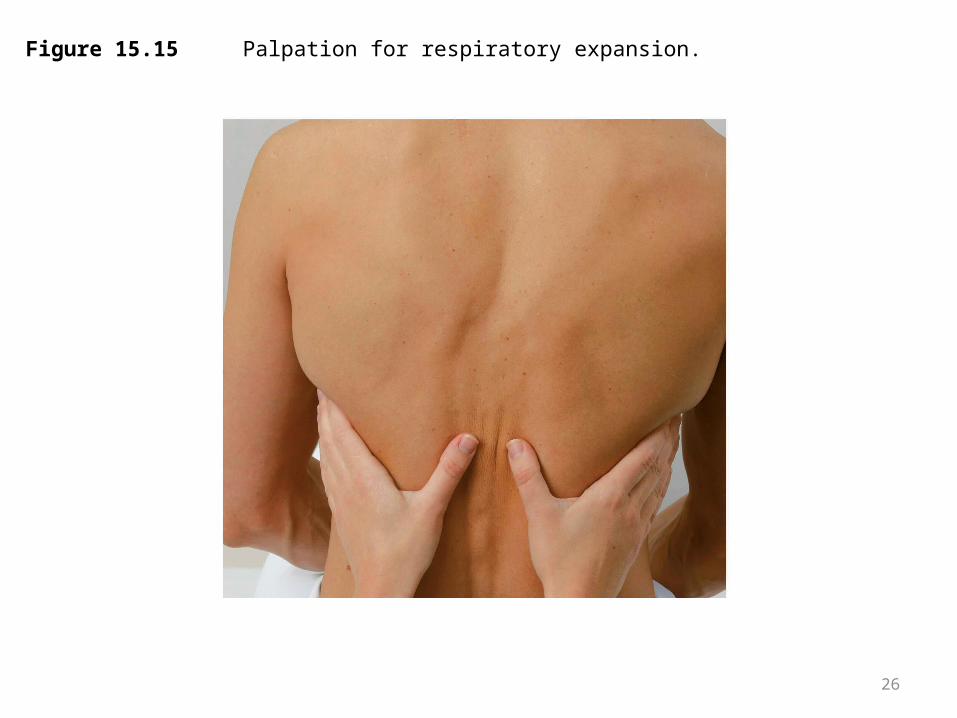
Objective Data (cont.)
• Palpate the posterior chest (cont.)– Symmetric expansion• Confirm symmetric chest expansion by placing your
warmed hands on posterolateral chest wall with thumbs at level of T9 or T10• Slide your hands medially to pinch up a small fold of
skin between your thumbs; ask person to take a deep breath• Your hands serve as mechanical amplifiers; as
person inhales deeply, your thumbs should move apart symmetrically; note any lag in expansion
26
Figure 15.15 Palpation for respiratory expansion.
Objective Data (cont.)
• Palpate the posterior chest (cont.)– Tactile fremitus• Fremitus is a palpable vibration• Use palmar base (ball) of fingers and touch person’s
chest while he or she repeats words “ninety-nine” or “blue moon” • These are resonant phrases that generate strong
vibrations
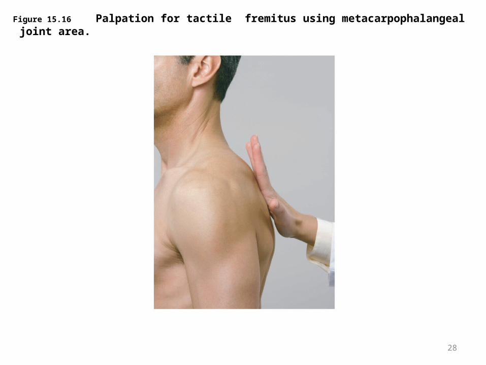
28
Figure 15.16 Palpation for tactile fremitus using metacarpophalangeal joint area.

Objective Data (cont.)
• Percuss the posterior chest (cont.)– Lung fields• Determine predominant note over lung fields; start
at apices and percuss band of normally resonant tissue across tops of both shoulders• Then, percussing in interspaces, make side-to-side
comparison all the way down lung region• Resonance is low-pitched, clear, hollow sound that
predominates in healthy lung tissue in adult

30
Figure 15.17 Pattern for percussion: Posterior thorax.

Objective Data (cont.)
• Percuss the posterior chest (cont.)– Diaphragmatic excursion
• Determine diaphragmatic excursion• Percuss to map out lower lung border, both in expiration
and in inspiration• First, ask the person to “exhale and hold it” briefly while
you percuss down scapular line until sound changes from resonant to dull on each side
• This estimates level of diaphragm separating lungs from abdominal viscera; may be somewhat higher on right side because of presence of liver
• Mark the spot
Slide 18-31

Objective Data (cont.)
• Percuss the posterior chest (cont.)– Diaphragmatic excursion (cont.)• Now ask person to “take a deep breath and hold it” • Continue percussing down from your first mark and
mark level where sound changes to dull on deep inspiration • Measure the difference; this diaphragmatic
excursion should be equal bilaterally and measure about 3 to 5 cm in adults, although it may be up to 7 to 8 cm in well-conditioned people

33
Figure 15.18B Diaphragmatic movement, measurement.

34
Objective Data (cont.)
• Auscultate the posterior chest (cont.)– Breath sounds• Evaluate presence and quality of normal breath
sounds• Instruct person to breathe through mouth, a little
bit deeper than usual• Use flat diaphragm endpiece of stethoscope and
hold it firmly on person’s chest wall

35
Objective Data (cont.)
• Auscultate the posterior chest (cont.)– Breath sounds (cont.)• You should expect to hear three types of normal
breath sounds in adult and older child– Bronchial, sometimes called tracheal or tubular– Bronchovesicular– Vesicular
• Adventitious sounds

36
Figure 15.19 Pattern for auscultation: Posterior thorax.
37
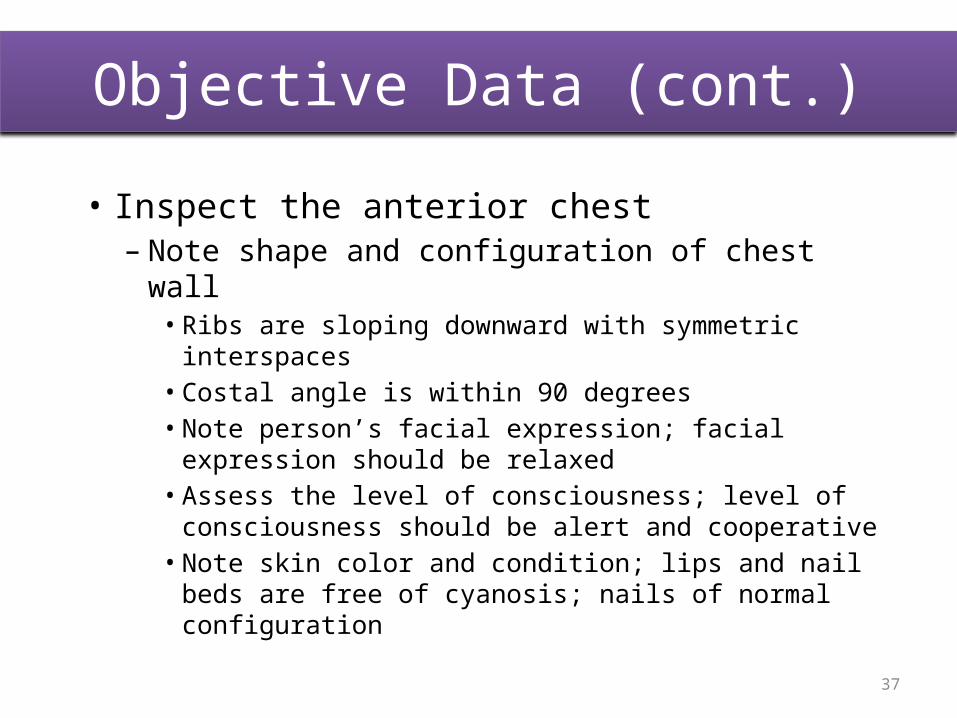
Objective Data (cont.)
• Inspect the anterior chest– Note shape and configuration of chest wall
• Ribs are sloping downward with symmetric interspaces
• Costal angle is within 90 degrees• Note person’s facial expression; facial expression
should be relaxed• Assess the level of consciousness; level of
consciousness should be alert and cooperative• Note skin color and condition; lips and nail beds are
free of cyanosis; nails of normal configuration
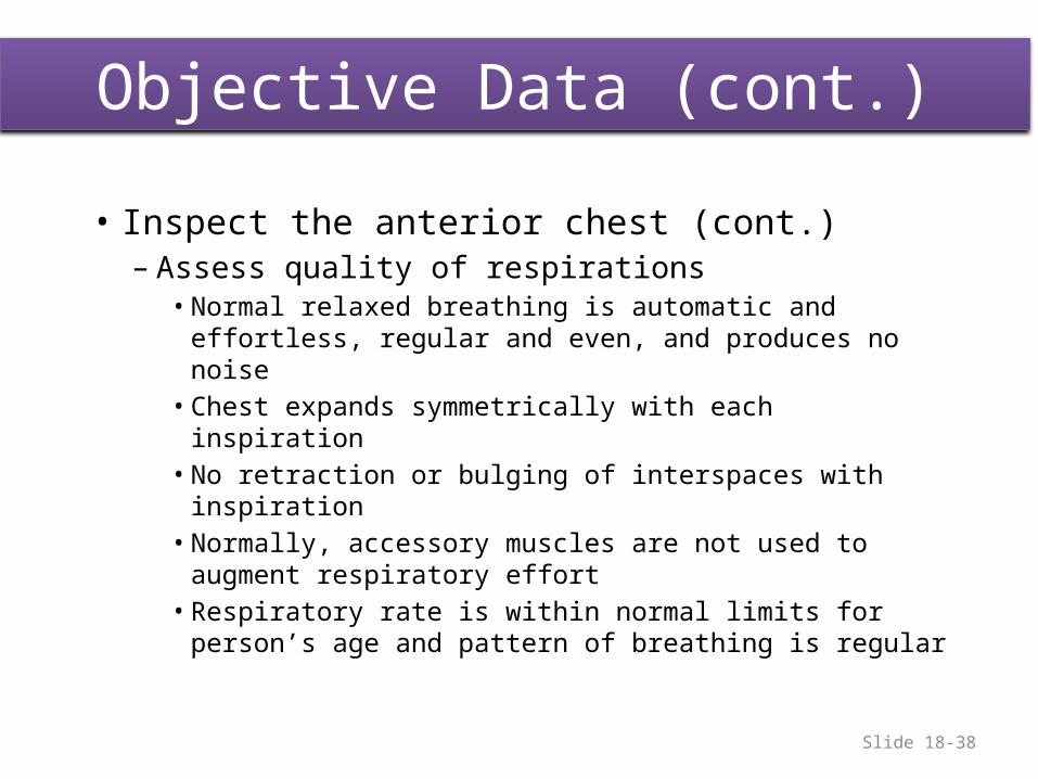
Objective Data (cont.)
• Inspect the anterior chest (cont.)– Assess quality of respirations
• Normal relaxed breathing is automatic and effortless, regular and even, and produces no noise
• Chest expands symmetrically with each inspiration• No retraction or bulging of interspaces with
inspiration• Normally, accessory muscles are not used to augment
respiratory effort• Respiratory rate is within normal limits for person’s
age and pattern of breathing is regularSlide 18-38

Objective Data (cont.)
• Palpate the anterior chest (cont.)– Palpate symmetric chest expansion• Place your hands on anterolateral wall with thumbs
along costal margins and pointing toward xiphoid process• Ask person to take a deep breath; watch thumbs
move apart symmetrically, and note smooth chest expansion with fingers
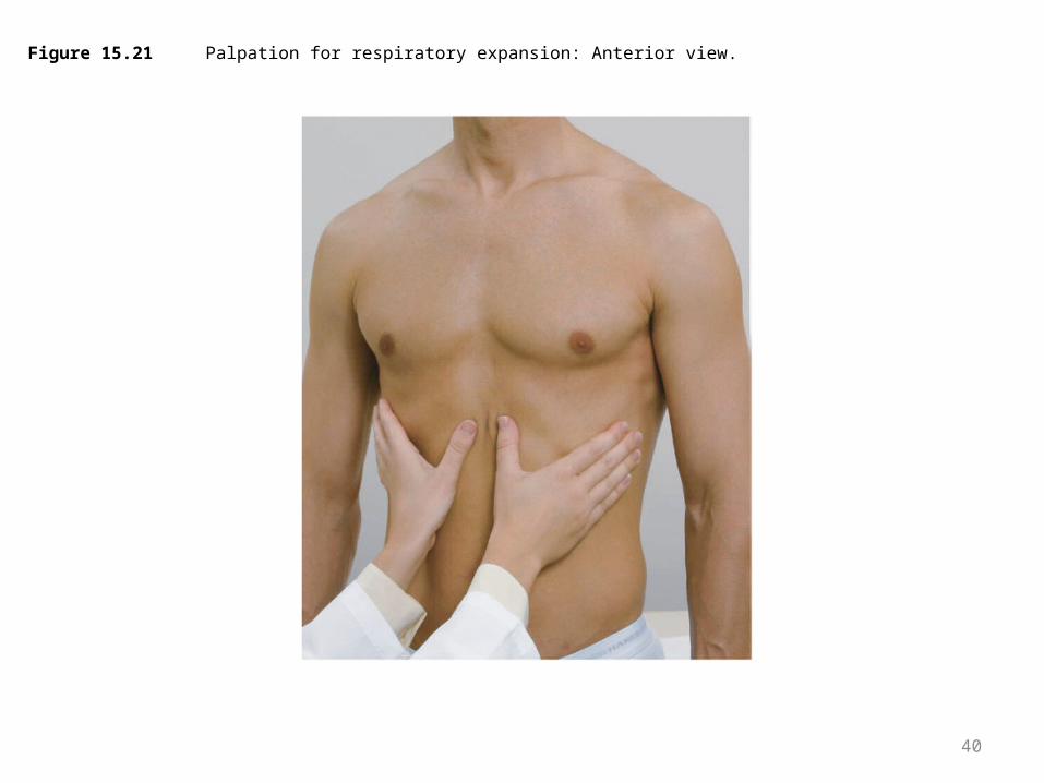
40
Figure 15.21 Palpation for respiratory expansion: Anterior view.
41
Objective Data (cont.)
• Palpate the anterior chest (cont.)– Assess tactile (vocal) fremitus– Palpate anterior chest wall • Note any tenderness; normally none is present• Detect any superficial lumps or masses, again,
normally none are present• Note skin mobility, turgor, temperature, and
moisture
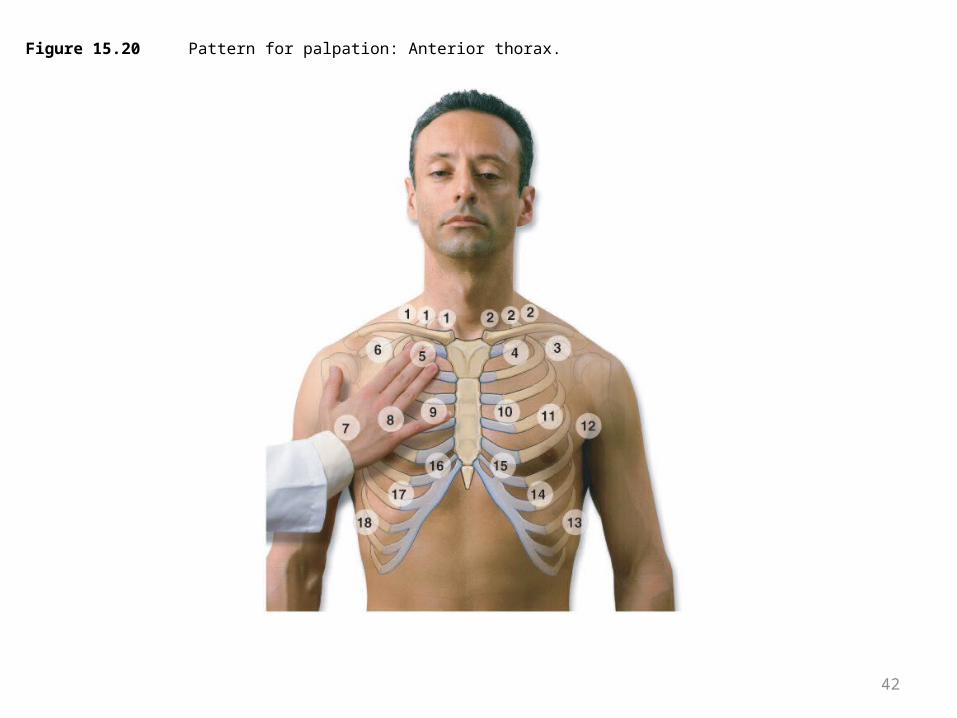
42
Figure 15.20 Pattern for palpation: Anterior thorax.

43
Objective Data (cont.)
• Percuss the anterior chest– Begin percussing apices in supraclavicular areas• Then, percussing interspaces and comparing one
side to other, move down anterior chest• Interspaces easier to palpate on anterior chest than
on back

44
Figure 15.23 Pattern for percussion: Anterior thorax.
Objective Data (cont.)
• Auscultate the anterior chest– Breath sounds• Auscultate lung fields over anterior chest from
apices in supraclavicular areas down to sixth rib• Progress from side to side as you move downward,
and listen to one full respiration in each location
Slide 18-45

46
Figure 15.24 Auscultatory sounds: Anterior thorax.

47
Table 15.1 Normal Breath Sounds
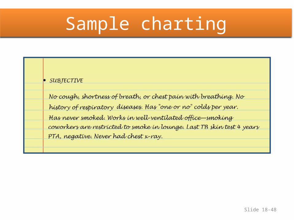
Sample charting
Slide 18-48
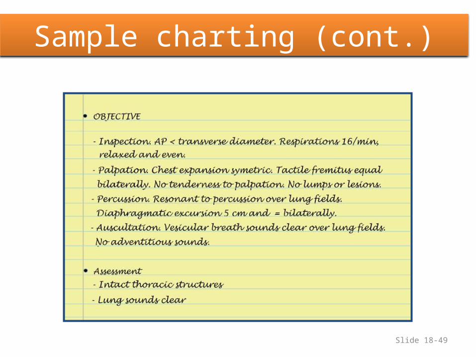
Sample charting (cont.)
Slide 18-49

Barrel Chest
Slide 18-50
51
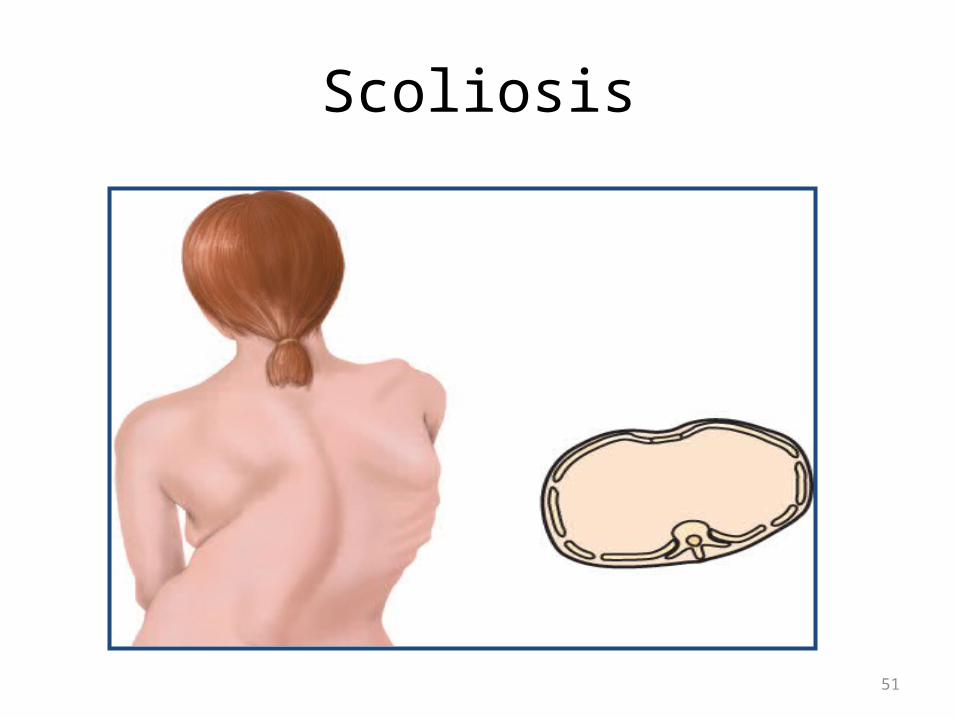
Scoliosis

52
Kyphosis