thumb ossification composite index (toci) for predicting peripubertal skeletal...
TRANSCRIPT

Thumb Ossification Composite Index (TOCI) forPredicting Peripubertal Skeletal Maturity and Peak
Height Velocity in Idiopathic ScoliosisA Validation Study of Premenarchal Girls with Adolescent Idiopathic Scoliosis
Followed Longitudinally Until Skeletal Maturity
Alec L.H. Hung, FRCSED(Ortho), W.W. Chau, MSc, B. Shi, MD, Simon K. Chow, PhD, Fiona Y.P. Yu, MPH,T.P. Lam, FRCSED(Ortho), Bobby K.W. Ng, FRCSED(Ortho), Y. Qiu, MD, and Jack C.Y. Cheng, FRCSED(Ortho)
Investigation performed at Prince of Wales Hospital, Shatin, Hong Kong
Background: Accurate skeletal maturity assessment is important to guide clinical evaluation of idiopathic scoliosis, butcommonly used methods are inadequate or too complex for rapid clinical use. The objective of the study was to propose anew simplified stagingmethod, called the thumb ossification composite index (TOCI), based on the ossification pattern of the2 thumb epiphyses and the adductor sesamoid bone; to determine its accuracy in predicting skeletal maturation whencomparedwith the Sanders simplified skeletal maturity system (SSMS); and to validate its interrater and intrarater reliability.
Methods: Hand radiographs of 125 girls, acquired when they were newly diagnosed with idiopathic scoliosis prior tomenarche and during longitudinal follow-up until skeletal maturity (a minimum of 4 years), were scored with the TOCI andSSMS. These scores were compared with digital skeletal age (DSA) and radius, ulna, and small hand bones (RUS) scores;anthropometric data; peak height velocity; and growth-remaining profiles. Correlations were analyzed with the chi-squaretest, Spearman and Cramer V correlation methods, and receiver operating characteristic curve analysis. Reliabilityanalysis using the intraclass correlation (ICC) was conducted.
Results: Six hundred and forty-five hand radiographs (average, 5 of each girl) were scored. The TOCI staging system washighly correlated with the DSA and RUS scores (r = 0.93 and 0.92, p < 0.01). The mean peak height velocity (and standarddeviation) was 7.43 ± 1.45 cm/yr and occurred at a mean age of 11.9 ± 0.86 years, with 70.1% and 51.4% of the subjectsattaining their peak height velocity at TOCI stage 5 and SSMS stage 3, respectively. The 2 systems predicted peak heightvelocity with comparable accuracy, with a strong Cramer V association (0.526 and 0.466, respectively; p < 0.01) and similarsensitivity and specificity on receiver operating characteristic curve analysis. The mean age at menarche was 12.57 ± 1.12years, with menarche occurring over several stages in both the TOCI and the SSMS. The growth remaining predicted by TOCIstage 8matched well with that predicted by SSMS stage 7, with amean of <2 cm/yr of growth potential over a mean of <1.7years at these stages. The TOCI also demonstrated excellent reliability, with an overall ICC of >0.97.
Conclusions: The new proposed TOCI could provide a simplified staging system for the assessment of skeletal maturityof subjects with idiopathic scoliosis. The index needs to be subjected to further multicenter validation in different ethnicgroups.
Accurate skeletal maturity assessment is important forprediction of curve progression and clinical man-agement of idiopathic scoliosis, including bracing
decisions and counseling about prognosis. Determinationof the timing of peak height velocity and growth remaining isof paramount importance for these purposes1-5. However,
Disclosure: There was no outside funding source for this study. The Disclosure of Potential Conflicts of Interest forms are provided with the onlineversion of the article (http://links.lww.com/JBJS/E323).
This is an open-access article distributed under the terms of the Creative Commons Attribution-Non Commercial-No Derivatives License 4.0 (CCBY-NC-ND), where it is permissible to download and share the work provided it is properly cited. The work cannot be changed in any way or used commerciallywithout permission from the journal.
1438
COPYRIGHT � 2017 THE AUTHORS. PUBLISHED BY THE JOURNAL OF BONE AND JOINT SURGERY, INCORPORATED. ALL RIGHTS RESERVED.
J Bone Joint Surg Am. 2017;99:1438-46 d http://dx.doi.org/10.2106/JBJS.16.01078

commonly used clinical or radiographic methods are inade-quate or too complex for rapid application in a busy clinicsetting6,7.
Sanders et al.6 demonstrated that the Tanner-Whitehouse-3(TW3)8 radius, ulna, and small bones of the hand (RUS) scoreshad the highest correlation with the curve acceleration phaseamong all skeletal maturity parameters in idiopathic scoliosis.The scoring system is, however, time-consuming and requiresthe use of an atlas with a substantial learning curve3,4,6,7. Sanderset al. simplified the RUS score into a digital skeletal age (DSA)score, by excluding the scoring of the distal aspect of the radiusand the ulnar epiphysis, and confirmed that the DSA score hadexcellent correlation with the curve acceleration phase as well6.
An ideal skeletal maturity assessment system would besimple and reliable with good predictability of peak heightvelocity and correlation with the standard DSA scoringsystem. We attempted to further simplify the currentskeletal maturity staging system by focusing on the mor-phology of a minimum number of essential hand ossifi-cation centers that we hoped would reliably predict theskeletal maturity information from the whole hand in aquicker and easier manner. Previous studies8,9 had revealedthat the epiphyses of the distal and proximal phalanges ofthe thumb demonstrated radiographic changes that weresimilar to those of the epiphyses of the ulnar 4 digits duringthe pubertal period. The adductor sesamoid has also been
Fig. 1
TOCI stages 1 through 8 and the corresponding ossification pattern and sequence of the adductor sesamoid, distal phalangeal epiphysis, and
proximal phalangeal epiphysis.
1439
THE JOURNAL OF BONE & JOINT SURGERY d J B J S .ORG
VOLUME 99-A d NUMBER 17 d SEPTEMBER 6, 2017TOCI FOR PREDICT ING PERIPUBERTAL SKELETAL MATURITY/PEAK
HEIGHT VELOCITY IN IDIOPATHIC SCOL IOS I S

extensively used to predict pubertal onset for decision-making regarding maxillofacial surgery9-12. After conducting avalidation study and confirming the very high concordancerate (71.3%) between TW staging of the thumb epiphyses andthat of the ulnar 4 digital epiphyses13, we developed a newthumb classification based on the epiphyses of the distaland proximal phalanges together with the adductor sesa-moid bone, which we call the “thumb ossification compositeindex (TOCI)” (Fig. 1). We hypothesized that this new sim-pler staging system could reliably predict when skeletal ma-turity would be reached by a subject in the early peripubertalperiod.
The objective of this study was to compare theTOCI with a current simplified system for prediction ofskeletal maturity and validate its interrater and intraraterreliability.
Materials and Methods
Hand radiographs were prospectively collected from a longitudinal cohortof girls with clinically and radiographically confirmed idiopathic scoliosis
recruited from a scoliosis clinic in a tertiary hospital. The inclusion criteria were(1) female sex, (2) premenarche, (3) an age of 8 to 12 years at the time of entryinto the study, (4) a diagnosis of idiopathic scoliosis, (5) no clinical evidenceof neurological abnormality, (6) no abnormalities of skeletal maturation, (7) aRisser sign of zerowith open physes in the hand, and (8) completion of at least 6follow-up visits over a minimum of 4 years. Demographic variables that werecollected included the ages when the radiographs were made, standing height,sitting height, body weight, and arm span. Institutional review board approvalwas obtained (ethics approval reference number 2016.045).
All patients were followed at an average of 6-month intervals startingprior to menarche, with more frequent visits during the early peripubertalgrowth period to capture accurately the timing of peak height velocity, untilskeletal maturity was confirmed by the radiographic appearance of physealclosure of the distal radial epiphysis. In accordance with the original TW3protocol, posteroanterior radiographs of the left hand were obtained at each
Fig. 2
Algorithm for TOCI staging. The words in red refer to the clinical decision-making algorithm. PP = proximal phalangeal and DP = distal phalangeal. Also see
the short training video on TOCI staging included with this article.
1440
THE JOURNAL OF BONE & JOINT SURGERY d J B J S .ORG
VOLUME 99-A d NUMBER 17 d SEPTEMBER 6, 2017TOCI FOR PREDICT ING PERIPUBERTAL SKELETAL MATURITY/PEAK
HEIGHT VELOCITY IN IDIOPATHIC SCOL IOS I S

visit8. All hand and wrist radiographs were independently reviewed by the
principal author and another experienced orthopaedic clinical fellow to es-tablish the stage of skeletal maturity according to (1) the proposed TOCImethod algorithm based on the 2 thumb epiphyses and the adductor sesamoid(Fig. 2) and (2) the standard TWmethod based on the epiphyses of the smallbones (distal phalanx, middle phalanx, proximal phalanx, and metacarpal) ofthe first, third, and fifth digits as well as the radial and ulnar epiphyses. Bothinvestigators were blinded to the chronological age and identity of the patients.The TW stages (E through I) and TOCI stages (1 through 8) were recorded onseparate data sheets, which were then collected by a third independent coin-vestigator for subsequent analysis. TW-derived RUS and DSA scores werecalculated. The Sanders simplified skeletal maturity scoring system (SSMS)
4
was included for comparison to determine the accuracy of TOCI staging inpredicting the timing of peak height velocity and menarche. Peak height ve-locity was calculated using the minimum 6-month interval method
8,14,15. To
define the accuracy in predicting the final growth remaining, the magnitude,time interval, and velocity of the growth occurring after the final TOCI stage (8)were compared with those parameters following a similar SSMS stage (7). AllTOCI and SSMS scores were tabulated against the chronological age when thescore was obtained and at menarche as well as peak height velocity.
Reliability TestThe principal investigator scored all radiographs using the TOCI method, andanother investigator, who was not involved in the study, grouped the radiographsby stage across the whole TOCI spectrum (stages 1 through 8). Nine radiographswere then selected randomly from each TOCI stage, to ensure an equal distri-bution of test radiographs demonstrating each stage, resulting in 63 radiographsfor testing (none were stage 1). One clinical fellow, 2 orthopaedic residents, and3 non-medical assistants were invited to serve as novices for reliability testing.
An interactive training session (see the instructional video on TOCIstaging included with this article [Video 1]) was provided to all 6 reviewers togive the details of the TOCI. For the reliability testing, the reviewers wereinstructed to view the radiographs on a high-resolution computer monitor andwere allowed to refer to the reference materials (Fig. 2). All reviewers wereblinded to the chronological age of the patients and performed the ratings
independently with no time limit. After 4 weeks, all reviewers repeated theratings using the same set of radiographs.
Statistical AnalysisDescriptive statistics and Spearman correlation coefficients were calculated forthe DSA and RUS scores in comparison with the TOCI staging system. Chartswere created to determine the timing of menarche and peak height velocity atdifferent maturity stages in the TOCI and SSMS systems. The chi-square testand Cramer V correlation were used to evaluate the accuracy of the 2 systemsin predicting the peak height velocity. The area under the receiver operatingcharacteristic curve was also used to determine the cutoff stages in the 2 systemswith the best sensitivity and specificity (highest percentages) to predict peakheight velocity. Reliability analysis was tested with Cronbach alpha values.Intraclass correlations (ICCs) in different kinds of comparisons were carriedout. The statistical analyses were done with SPSS version-20.0 software (IBM).The significance level was p < 0.05.
Results
In a 5-year period (2007 through 2011), 645 hand and wristradiographs were collected consecutively from 125 pre-
menarchal girls with adolescent idiopathic scoliosis followedlongitudinally until skeletal maturity, so that the patients hadan average of 5 follow-up radiographs each. The mean ages(and standard deviations [SDs]) when the first and last radio-graphs available for analysis were made were 10.9 ± 0.8 and13.7 ± 1.0 years, respectively. The last radiographwas defined asthe earliest radiograph that demonstrated TOCI stage 8. Noneof the patients in our cohort had TOCI stage 1.
Correlation of TOCI Staging with SSMS SystemThe TOCI stages were found to correlate strongly with both theDSA (r = 0.93, p < 0.01) and the RUS (r = 0.92, p < 0.01) score.
TABLE I Correlation of TOCI and SSMS Stages with Chronological Age and DSA and RUS Scores
Age (yr) DSA Scores RUS Scores
Mean (SD) Median Range Mean (SD) Median Range Mean (SD) Median Range
TOCIstage
2 10.9 (0.8) 10.7 10.2-12.6 306.2 (49.7) 332.0 221.0-363.0 455.3 (70.7) 469.0 332.0-573.0
3 11.4 (0.7) 11.1 10.6-13.3 344.7 (34.5) 353.5 293.0-397.0 549.6 (76.9) 556.0 441.0-683.0
4 11.7 (0.8) 11.6 10.1-13.8 385.4 (27.9) 390.5 293.0-429.0 640.9 (50.0) 641.0 481.0-748.0
5 12.2 (0.9) 12.1 10.1-15.0 417.0 (30.9) 415.0 351.0-542.0 716.6 (52.8) 706.0 608.0-857.0
6 12.8 (0.9) 12.8 10.5-15.5 505.3 (42.5) 508.0 392.0-571.0 840.9 (69.8) 836.5 562.0-962.0
7 13.2 (0.9) 13.2 11.1-16.0 570.4 (21.7) 571.0 513.0-609.0 938.5 (44.0) 947.0 786.0-1,000.0
8 13.7 (1.0) 13.8 11.6-16.4 602.6 (12.5) 609.0 540.0-609.0 988.9 (21.5) 1,000.0 909.0-1,000.0
SSMSstage
1 11.0 (0.9) 10.5 10.2-12.6 300.9 (61.9) 341.0 221.0-363.0 466.5 (74.2) 465.5 380.0-573.0
2 11.7 (0.8) 11.6 10.2-13.8 376.4 (36.2) 374.0 293.0-450.0 625.8 (83.6) 641.0 332.0-761.0
3 12.3 (1.0) 12.3 10.1-15.5 413.3 (23.2) 415.0 351.0-514.0 715.3 (52.6) 705.5 613.0-905.0
4 12.6 (1.0) 12.6 10.9-16.0 472.5 (42.2) 466.0 401.0-562.0 790.7 (67.3) 791.0 668.0-953.0
5 13.0 (0.8) 12.9 11.0-15.0 524.5 (29.0) 527.0 466.0-568.0 865.0 (76.1) 876.5 556.0-954.0
6 13.2 (1.0) 13.3 10.5-15.9 568.6 (31.6) 567.0 453.0-609.0 934.0 (60.0) 948.0 731.0-1,000.0
7 13.5 (0.9) 13.5 11.6-14.9 600.7 (19.1) 609.0 502.0-609.0 987.8 (31.5) 1,000.0 784.0-1,000.0
1441
THE JOURNAL OF BONE & JOINT SURGERY d J B J S .ORG
VOLUME 99-A d NUMBER 17 d SEPTEMBER 6, 2017TOCI FOR PREDICT ING PERIPUBERTAL SKELETAL MATURITY/PEAK
HEIGHT VELOCITY IN IDIOPATHIC SCOL IOS I S
The correlations of the TOCI and SSMS stages with chrono-logical age and the DSA and RUS scores are shown in Table Iand summarized in the integrated growth chart in Figure 3.
Comparison of TOCI and SSMS Staging of KeyGrowth ParametersTiming of Peak Height VelocityOne hundred and seven patients with clearly identifiablegrowth peaks were used for this analysis. The mean peak heightvelocity was 7.43 ± 1.45 cm/yr (range, 5 to 11.95 cm/yr) andoccurred at mean age of 11.93 ± 0.86 years (range, 9.73 to 13.99years).
Both the TOCI and the SSMS stages were strongly as-sociated with the peak height velocity as confirmed by strongCramer V correlations (0.526 and 0.466, respectively), withstatistical significance (p < 0.01).
As shown in Figure 4, 70.1% and 51.4% of the subjectsattained their peak height velocity at TOCI stage 5 ± 0.60 andSSMS stage 3 ± 0.99, respectively, both with a median DSAscore of 415.
The analysis of the area under the receiver operatingcharacteristic curve indicated that the cutoff stages thatpredicted the timing of peak height velocity with the bestsensitivity and specificity were TOCI stage 4.5, with asensitivity of 93.6% and a specificity of 72.2%, and SSMS stage2.5, with a sensitivity of 90.8% and a specificity of 75.0%.
Timing of MenarcheThe mean age at menarche was 12.57 ± 1.12 years (range, 9.4to 14.3 years), and menarche was found to occur over severalstages in both the TOCI and the SSMS (Fig. 5).
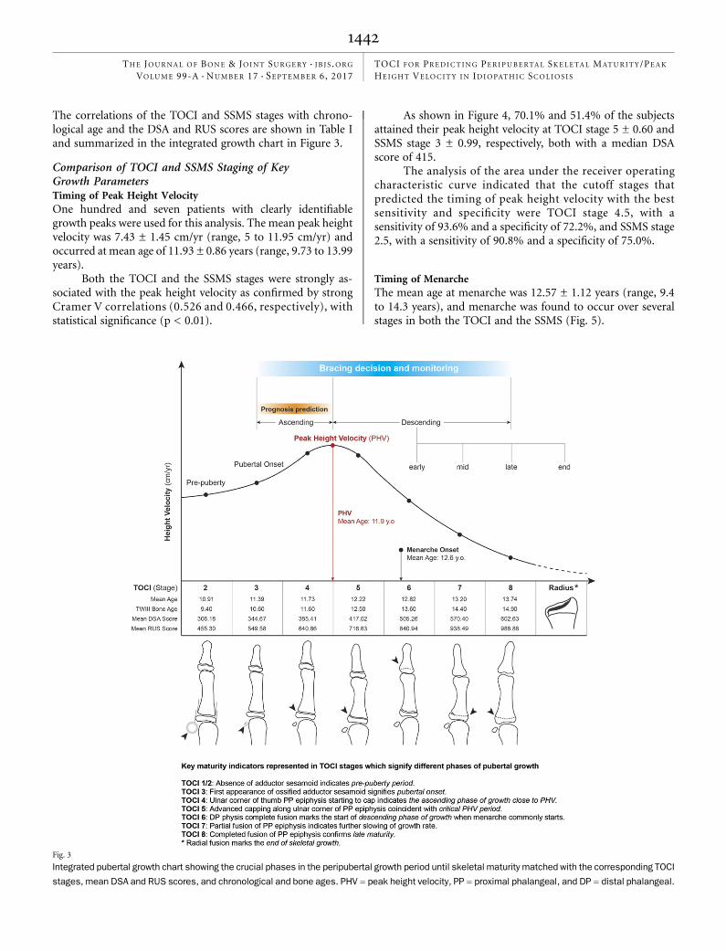
Fig. 3
Integrated pubertal growth chart showing the crucial phases in the peripubertal growth period until skeletal maturity matched with the corresponding TOCI
stages, mean DSA and RUS scores, and chronological and bone ages. PHV = peak height velocity, PP = proximal phalangeal, and DP = distal phalangeal.
1442
THE JOURNAL OF BONE & JOINT SURGERY d J B J S .ORG
VOLUME 99-A d NUMBER 17 d SEPTEMBER 6, 2017TOCI FOR PREDICT ING PERIPUBERTAL SKELETAL MATURITY/PEAK
HEIGHT VELOCITY IN IDIOPATHIC SCOL IOS I S

Fig. 4
Comparison of descriptive statistics, percentage distributions, and results of receiver operating characteristic (ROC) curve analysis of the TOCI and
SSMS to determine the timing of peak height velocity (PHV).
Fig. 5
Percentage distribution of menarche occurring at different TOCI and SSMS stages.
1443
THE JOURNAL OF BONE & JOINT SURGERY d J B J S .ORG
VOLUME 99-A d NUMBER 17 d SEPTEMBER 6, 2017TOCI FOR PREDICT ING PERIPUBERTAL SKELETAL MATURITY/PEAK
HEIGHT VELOCITY IN IDIOPATHIC SCOL IOS I S

Growth-Remaining ProfileWe found that 71.7% of the subjects at TOCI stage 8 were atSSMS stage 7. The median DSA score at both stages was 609.Overall, the mean amount of final growth remaining at these2 stages was <2 cm/yr over <1.7 years. The growth-remainingprofiles are summarized in Figure 6.
Reliability TestAll 6 clinical professionals showed excellent interrater andintrarater agreement for the TOCI classification (Table II).
Discussion
An ideal bone maturity assessment model should be bothsimple and reliable for clinical usage, with good corre-
lation to a standard scoring system. The excellent reliabilityof the TOCI demonstrated that it is easy to learn, with arelatively short learning curve even among novice users. Moreimportantly, it also has excellent correlation with DSA andRUS scores.
Sanders et al. proposed an important simplifiedskeletal maturity scoring system4 (SSMS) with 8 stagesbased on the ossification patterns of all digital epiphyses ofthe hand and using the same descriptors as utilized in theTW3 system. They reported that the SSMS had excellentcorrelation with the curve acceleration phase (r = 0.9) in 22female subjects and excellent reliability among senior or-thopaedic surgeons4. However, a subsequent validationstudy of 275 patients demonstrated a steep learning curvefor less experienced users16. Other recently reported sim-plified models using the olecranon apophysis17 or the distalpart of the radius and the ulnar epiphysis3 (DRU classifi-cation) were either not evaluated in strictly conductedlongitudinal studies or not fully validated through com-parison with the standard DSA and RUS scoring systems.The modified Risser grading system had low specificity andinterobserver reliability18.
As is the case for the existing systems3,4,9,17, any proposedsimplified system must be able to accurately predict the
Fig. 6
The percentage of patients at SSMS stage 7 who were at TOCI stage 8 and the growth-remaining profiles at SSMS stage 7 and TOCI stage 8.
TABLE II Interrater and Intrarater Reliability Test Results for Orthopaedic Surgeons and Non-Medical Assistants*
Junior Orthopaedic Surgeons Non-Medical Assistants
Interrater
First phase 0.976 (0.961-0.986) 0.985 (0.975-0.992)
Second phase, 4 wk later 0.990 (0.984-0.994) 0.988 (0.980-0.994)
Intrarater, first phase versus second phase 0.995 (0.992-0.997) 0.989 (0.981-0.994)
*The values are given as the reliability with the 95% confidence interval in parentheses.
1444
THE JOURNAL OF BONE & JOINT SURGERY d J B J S .ORG
VOLUME 99-A d NUMBER 17 d SEPTEMBER 6, 2017TOCI FOR PREDICT ING PERIPUBERTAL SKELETAL MATURITY/PEAK
HEIGHT VELOCITY IN IDIOPATHIC SCOL IOS I S
timing of peak height velocity and menarche as well as thegrowth-remaining period to inform the clinical decisionregarding bracing and prognostic counseling about curveprogression.
The mean age at peak height velocity (11.93 ± 0.86 years)in our study was comparable with that in most previousstudies3,6,14,17,19. The growth velocity magnitude was similar tothat in Asian studies3,19 and less than that in European17 andU.S. studies6,14. The age at peak height velocity in our study ofsubjects with idiopathic scoliosis was delayed (by a mean of1.47 years) compared with that of normal subjects20,21, signi-fying a longer period of peripubertal growth, which may havean effect on curve progression. Radiographically, the prepon-derance of capping of the epiphyses of the distal and middlephalanges (of the ulnar 4 digits) and of the proximal phalangesand metacarpal bones (of all 5 digits) at SSMS stage 3 wasfound to best represent the timing of peak height velocity4,5.This stage corresponds well to stage 5 in our TOCI classifica-tion. The median DSA score at TOCI stage 5 was 415 (Table I),which coincides exactly with the reported critical DSA range of400 to 425 (SSMS stage 3) that best matched with the timing ofthe peak height velocity and onset of the curve accelerationphase6. Our results showed that TOCI stage 5 had good ac-curacy for predicting peak height velocity compared withSSMS stage 3, as demonstrated by a better Cramer Vassociation(0.526 versus 0.466), a narrower standard deviation (0.60compared with 0.99), and similar sensitivity and specificity onreceiver operating characteristic curve analysis (Fig. 4). Moreimportantly, it is simpler to use TOCI stage 5 for this purposeas one only needs to pay attention to advanced capping overthe ulnar corner of the proximal phalangeal epiphysis in thethumb.
The mean age at menarche onset in our study was 12.6years, which was 8 months later than the mean age at peakheight velocity (11.9 years), and menarche consistently oc-curred between the median ages at TOCI stages 5 and 6,representing the early descending phase of the growth curve.The wide range of menarche age in this series (9.4 to 14.3years) indicated its limitation in accurately predicting re-maining growth. This finding echoed the observations byCharles et al.17 and Little et al.14 and was similar to that ina study of 2,196 Chinese girls with adolescent idiopathicscoliosis22. As the onset of menarche is affected by multiplefactors of growth, and because of the age spread and con-siderable overlapping with the timing of peak height velocity,menarche could not be matched with a single representativestage in either the SSMS or TOCI system (Fig. 5). In ourseries, 33.9% of the subjects had the onset of menarche atthe TOCI stage at which peak height velocity occurred(TOCI stage 5), 54.3% had it after that stage (menarche be-gan at stage 6, 7, or 8), and 11.8% had it before that stage(menarche began at stage 4). This pattern is similar to thatreported in longitudinal studies by Little et al.14, Tanner8, andBuckler15.
The growth-remaining prediction refers to the moreadvanced TOCI stages (6, 7, and 8), matched with the de-
scending phase of pubertal growth (Fig. 3). TOCI stage 8(completed fusion of the proximal phalangeal epiphysis ofthe thumb) is equivalent to SSMS stage 7 (completed fusionof all digital epiphyses) as evidenced by 71.7% concordancebetween these 2 systems and their comparable final growth-remaining profiles (Fig. 6). Radiographically, both theTOCI and the SSMS still rely on distal radial epiphysealfusion status to determine the end of growth, although it iscontroversial whether clinically relevant growth is stillpossible at the late radiographic stages of fusion of thedistal radial physis16. The SSMS may provide more clarityabout growth remaining at this stage since it shows thedistal radial physis.
The concept of using only the thumb distal phalanx,proximal phalanx, and adductor sesamoid to predict skel-etal maturity is novel but derived from precedent. Thedistal and proximal phalangeal epiphyses of the thumb havebeen used in traditional methods for determining ortho-paedic bone age (e.g., the Greulich and Pyle atlas23 and theTW3 method8). The adductor sesamoid is known to bealways present in adolescents24, is easily detectable, and hasbeen extensively used to estimate skeletal maturity in or-thodontic surgery (by determining the onset of puberty, itis possible to predict facial growth and the timing formandibular reconstruction10-12). In addition, the pronatedposition of the thumb proximal phalanx relative to theorientation of the 4 ulnar digits provides easier visualiza-tion of the capping of the epiphysis at a very early stage. Therelatively large black “cartilage gap” allows further sepa-ration into early and advanced capping stages through the
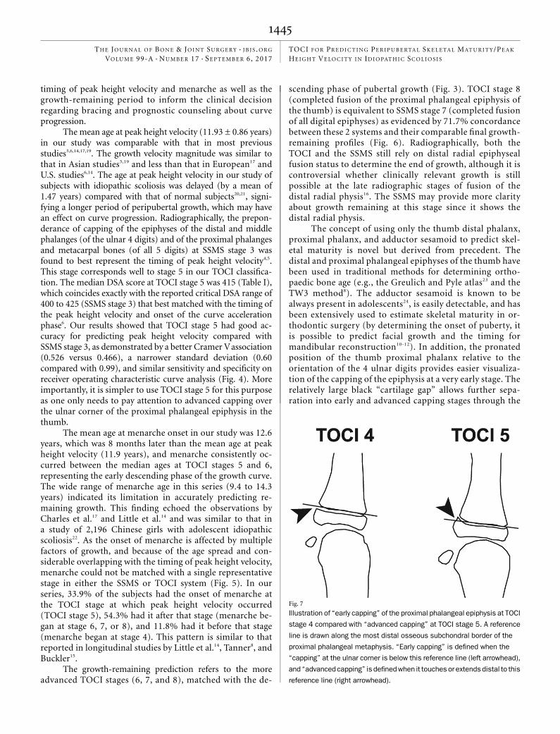
Fig. 7
Illustration of “early capping” of the proximal phalangeal epiphysis at TOCI
stage 4 compared with “advanced capping” at TOCI stage 5. A reference
line is drawn along the most distal osseous subchondral border of the
proximal phalangeal metaphysis. “Early capping” is defined when the
“capping” at the ulnar corner is below this reference line (left arrowhead),
and “advanced capping” is definedwhen it touches or extends distal to this
reference line (right arrowhead).
1445
THE JOURNAL OF BONE & JOINT SURGERY d J B J S .ORG
VOLUME 99-A d NUMBER 17 d SEPTEMBER 6, 2017TOCI FOR PREDICT ING PERIPUBERTAL SKELETAL MATURITY/PEAK
HEIGHT VELOCITY IN IDIOPATHIC SCOL IOS I S
use of a reference line (Fig. 7). The thumb adductor sesa-moid allows extra precision in predicting the onset of pu-berty9-12. The composite index score of the 2 thumbepiphyses and the adductor sesamoid corresponds well todifferent stages of pubertal growth curve as summarized inthe integrated growth chart in Figure 3. Most importantly,because the thumb epiphyses and the adductor sesamoid arelocated so close to each other, they can be easily imaged with amagnified view through the use of a very low-radiation mini-mobile x-ray machine in the clinic.
To our knowledge, this is the first study to investigatethe use of the thumb epiphyses to predict skeletal maturityin subjects with idiopathic scoliosis and to validate themethod with a large number of longitudinal hand radio-graphs made before menarche and at an average of 6-monthintervals in the peripubertal period. We believe that thenewly proposed thumb ossification composite index (TOCI)was shown to be simple to use and to have excellent relia-bility and accuracy in the prediction of skeletal maturitycomparable with the Sanders SSMS. It has the potential forapplication in a busy clinic setting. Further validation inlarger multicenter studies involving different ethnic groups,males, longitudinal correlation with normal adolescents
at different time points, and curve progression profiles areongoing. nNOTE: The authors thank Mr. Patrick Tsang for retrieval of information from the scoliosis database,Ms. Vivian Hung for technical support in the reliability testing material, Prof. Ben Yip for adviceregarding themethodology design of the reliability tests, Dr. Ronald Wong for dubbing the TOCI shorttraining video, and Ms. Huang Xueying for production and aftereffect of the TOCI short training video.
Alec L.H. Hung, FRCSED(Ortho)1
W.W. Chau, MSc1,2
B. Shi, MD1,2
Simon K. Chow, PhD1
Fiona Y.P. Yu, MPH1,2
T.P. Lam, FRCSED(Ortho)1,2
Bobby K.W. Ng, FRCSED(Ortho)1
Y. Qiu, MD2
Jack C.Y. Cheng, FRCSED(Ortho)1,2
1Department of Orthopaedics and Traumatology, The Chinese Universityof Hong Kong, Shatin, Hong Kong
2The Joint Scoliosis Research Centre of The Chinese University of HongKong and Nanjing University, Shatin, Hong Kong
E-mail address for J.C.Y. Cheng: [email protected]
References
1. Nachemson AL, Lonstein JE, Weinstein SL, editors. Report of the Prevalence andNatural History Committee of the Scoliosis Research Society. Denver; 1982.2. Song KM, Little DG. Peak height velocity as a maturity indicator for males withidiopathic scoliosis. J Pediatr Orthop. 2000 May-Jun;20(3):286-8.3. Luk KDK, Saw LB, Grozman S, Cheung KMC, Samartzis D. Assessment ofskeletal maturity in scoliosis patients to determine clinical management: a newclassification scheme using distal radius and ulna radiographs. Spine J. 2014 Feb1;14(2):315-25. Epub 2013 Nov 12.4. Sanders JO, Khoury JG, Kishan S, Browne RH, Mooney JF 3rd, Arnold KD,McConnell SJ, Bauman JA, Finegold DN. Predicting scoliosis progression fromskeletal maturity: a simplified classification during adolescence. J Bone Joint SurgAm. 2008 Mar;90(3):540-53.5. Sanders JO, Browne RH, Cooney TE, Finegold DN, McConnell SJ, Margraf SA.Correlates of the peak height velocity in girls with idiopathic scoliosis. Spine (Phila Pa1976). 2006 Sep 15;31(20):2289-95.6. Sanders JO, Browne RH, McConnell SJ, Margraf SA, Cooney TE, Finegold DN.Maturity assessment and curve progression in girls with idiopathic scoliosis. J BoneJoint Surg Am. 2007 Jan;89(1):64-73.7. Sanders JO. Maturity indicators in spinal deformity. J Bone Joint Surg Am.2007 Feb;89(Suppl 1):14-20.8. Tanner JM. Assessment of skeletal maturity and prediction of adult height(TW3 method). New York: WB Saunders; 2001.9. Fishman LS. Radiographic evaluation of skeletal maturation. A clinically orientedmethod based on hand-wrist films. Angle Orthod. 1982 Apr;52(2):88-112.10. Sidlauskas A, Zilinskaite L, Svalkauskiene V. Mandibular pubertal growth spurtprediction. Part one: method based on the hand-wrist radiographs. Stomatologija.2005;7(1):16-20.11. Pileski RC, Woodside DG, James GA. Relationship of the ulnar sesamoid boneand maximum mandibular growth velocity. Angle Orthod. 1973 Apr;43(2):162-70.12. Chapman SM. Ossification of the adductor sesamoid and the adolescent growthspurt. Angle Orthod. 1972 Jul;42(3):236-44.13. Hung A, Shi B, Chau W, Yip B, Yu F, Hung V, Lam T, Ng B, Cheng J. Canossification pattern of the thumb epiphyses reliably simplify the classical Tanner-Whitehouse hand skeletal maturity assessment? A study of 645 hand radiographsfrom 125 premature AIS girls followed longitudinally till skeletal maturity. Read at the
51st Annual Meeting of the Scoliosis Research Society; 2016 Sep 21-4; Prague,Czech Republic.14. Little DG, Song KM, Katz D, Herring JA. Relationship of peak height velocityto other maturity indicators in idiopathic scoliosis in girls. J Bone Joint Surg Am.2000 May;82(5):685-93.15. Buckler JMH. A longitudinal study of adolescent growth. London: Springer;1990.16. Verma K, Sitoula P, Gabos P, Loveland K, Sanders J, Verma S, Shah SA. Sim-plified skeletal maturity scoring system: learning curve and methods to improvereliability. Spine (Phila Pa 1976). 2014 Dec 15;39(26):E1592-8.17. Charles YP, Dimeglio A, Canavese F, Daures JP. Skeletal age assessment fromthe olecranon for idiopathic scoliosis at Risser grade 0. J Bone Joint Surg Am. 2007Dec;89(12):2737-44.18. Nault ML, Parent S, Phan P, Roy-Beaudry M, Labelle H, Rivard M. A modifiedRisser grading system predicts the curve acceleration phase of female adolescentidiopathic scoliosis. J Bone Joint Surg Am. 2010 May;92(5):1073-81.19. ChazonoM, Tanaka T, Marumo K, Kono K, Suzuki N. Significance of peak heightvelocity as a predictive factor for curve progression in patients with idiopathic sco-liosis. Scoliosis. 2015 Feb 11;10(Suppl 2):S5.20. Chae HW, Suh I, Kwon AR, Kim YJ, Kim YH, Kang DR, Kim HY, Oh SM, Kim HC,Kim DH, Kim HS. Longitudinal standards for height and height velocity in Koreanchildren and adolescents: the Kangwha study. [corrected]. J Korean Med Sci. 2013Oct;28(10):1512-7. Epub 2013 Sep 25.21. Lee TS, Chao T, Tang RB, Hsieh CC, Chen SJ, Ho LT. A longitudinal study ofgrowth patterns in school children in Taipei area I: growth curve and height velocitycurve. J Chin Med Assoc. 2004 Feb;67(2):67-72.22. Mao SH, Jiang J, Sun X, Zhao Q, Qian BP, Liu Z, Shu H, Qiu Y. Timing ofmenarche in Chinese girls with and without adolescent idiopathic scoliosis:current results and review of the literature. Eur Spine J. 2011 Feb;20(2):260-5.Epub 2010 Dec 14.23. Greulich WW, Pyle SI. Radiographic atlas of skeletal development of the handand wrist. Stanford: Stanford University Press; 1950.24. Yammine K. The prevalence of the sesamoid bones of the hand: asystematic review and meta-analysis. Clin Anat. 2014 Nov;27(8):1291-303. Epub2014 Feb 25.
1446
THE JOURNAL OF BONE & JOINT SURGERY d J B J S .ORG
VOLUME 99-A d NUMBER 17 d SEPTEMBER 6, 2017TOCI FOR PREDICT ING PERIPUBERTAL SKELETAL MATURITY/PEAK
HEIGHT VELOCITY IN IDIOPATHIC SCOL IOS I S