thumbnail - download.e-bookshelf.de · vided access to his photographic files, his illustrations,...
TRANSCRIPT

Essential Clinical Neuroanatomy
Essential Clinical Neuroanatomy
Thomas H ChampneyProfessor
Miller School of MedicineUniversity of MiamiMiami Florida USA
This edition first published 2016 copy 2016 by John Wiley amp Sons Ltd
Registered OfficeJohn Wiley amp Sons Ltd The Atrium Southern Gate Chichester West Sussex PO19 8SQ UK
Editorial Offices9600 Garsington Road Oxford OX4 2DQ UKThe Atrium Southern Gate Chichester West Sussex PO19 8SQ UK111 River Street Hoboken NJ 07030‐5774 USA
For details of our global editorial offices for customer services and for information about how to apply for permission to reuse the copyright material in this book please see our website at wwwwileycomwiley‐blackwell
The right of the author to be identified as the author of this work has been asserted in accordance with the UK Copyright Designs and Patents Act 1988
All rights reserved No part of this publication may be reproduced stored in a retrieval system or transmitted in any form or by any means electronic mechanical photocopying recording or otherwise except as permitted by the UK Copyright Designs and Patents Act 1988 without the prior permission of the publisher
Designations used by companies to distinguish their products are often claimed as trademarks All brand names and product names used in this book are trade names service marks trademarks or registered trademarks of their respective owners The publisher is not associated with any product or vendor mentioned in this book It is sold on the understanding that the publisher is not engaged in rendering professional services If professional advice or other expert assistance is required the services of a competent professional should be sought
The contents of this work are intended to further general scientific research understanding and discussion only and are not intended and should not be relied upon as recommending or promoting a specific method diagnosis or treatment by health science practitioners for any particular patient The publisher and the author make no representations or warranties with respect to the accuracy or completeness of the contents of this work and specifically disclaim all warranties including without limitation any implied warranties of fitness for a particular purpose In view of ongoing research equipment modifications changes in governmental regulations and the constant flow of information relating to the use of medicines equipment and devices the reader is urged to review and evaluate the information provided in the package insert or instructions for each medicine equipment or device for among other things any changes in the instructions or indication of usage and for added warnings and precautions Readers should consult with a specialist where appropriate The fact that an organization or Website is referred to in this work as a citation andor a potential source of further information does not mean that the author or the publisher endorses the information the organization or Website may provide or recommendations it may make Further readers should be aware that Internet Websites listed in this work may have changed or disappeared between when this work was written and when it is read No warranty may be created or extended by any promotional statements for this work Neither the publisher nor the author shall be liable for any damages arising herefrom
Library of Congress Cataloging‐in‐Publication Data
Champney Thomas authorEssential clinical neuroanatomy Thomas Champney p cm Includes bibliographical references and index ISBN 978-1-118-43993-7 (pbk)I Title [DNLM 1 Nervous Systemndashanatomy amp histology WL 101] QM451 611prime8ndashdc23
2014046419
A catalogue record for this book is available from the British Library
Wiley also publishes its books in a variety of electronic formats Some content that appears in print may not be available in electronic books
Cover image copy Science Photo LibraryPasiekaCover design by Visual Philosophy
Set in 1012pt Adobe Garamond Pro by SPi Global Pondicherry India
1 2016
v
Preface viAcknowledgments viiAbout the companion website viii
Part 1 Neuroanatomy of the Central Nervous System
1 Overview of the nervous system 3
2 Blood vessels meninges and ventricles 20
3 Neurodevelopment 39
4 Spinal cord 49
5 Medulla oblongata 61
6 Pons 77
7 Midbrain 95
8 Diencephalon 107
9 telencephalon 119
10 Cerebellum 131
11 Spinal tracts 145
Part 2 the Sensory Motor and Integration Systems
12 Visual system 175
13 auditory and vestibular system 192
14 Olfaction and taste 210
15 Central motor control 219
16 Limbic system 235
17 Cortical integration 245
18 Imaging essentials 259
Answers to study questions 268Answers to figures 275Glossary 278Index 301
Contents
Essential Clinical Neuroanatomy is the first neuroanatomy text that consistently illustrates and discusses the anatomy of the central nervous system from the clinical perspective All of the illustrations are provided in the clinical view (using the axial radiologic standard of computed tomography and magnetic resonance imaging) This provides consistency throughout the text and throughout the career of the reader In addi-tion the neural pathways are color coded for easier recognition and recall with green indicating sensory components red indicating voluntary motor components and purple indicating involuntary (autonomic) components The clinically relevant neuroanatomy is highlighted with case studies clinically‐oriented study questions and clinical boxes of interest Anatomic details that do not have direct clinical relevance are de‐emphasized
The text is divided into two main sections the first eleven chapters provide the neuroanatomy of the central nervous system while the last seven chapters provide descriptions of the sensory motor and inte-gration systems within the central nervous system Each chapter begins with objectives and an outline of the material to be covered Within each chapter highlighted clinical boxes are presented while case studies and clinically relevant multiple choice questions are found at the end of each chapter Each chapter contains a list of additional readings that include more detail‐oriented textbooks as well as current review articles
This text is designed for those in the health sciences who require a basic introduction to clinical neuro-anatomy This can include allied health students first‐year medical students dental students and neurosci-ence students Because of its essential nature it can be useful for those reviewing neuroanatomy for major licensing or competency examinations
Thomas H Champney
Preface
The development and production of Essential Clinical Neuroanatomy utilized the skills and strengths of numerous individuals First Dr Ron Clark my predecessor at the University of Miami generously pro-vided access to his photographic files his illustrations and his notes The foundation of the book is built on his many years of teaching neuroanatomy Second all of the clinical imaging (radiographs computed tomographs magnetic resonance images) were graciously provided by Dr Charif Sidani a neuroradiologist associated with the University of Miami He provided many hours of help in recommending and selecting images for use in the text
The editorial staff at Wiley Blackwell were extremely helpful in all phases of this project Specifically Elizabeth Johnston Karen Moore and Nick Morgan were professional and highly organized In addition the excellent illustrations by Jane Fallows and Roger Hulley are an integral part of this project Any anatomy text is only as good as its illustrations All of these individuals made the daunting task of this project much more manageable
Finally I express my gratitude to all of the students who have provided feedback on the neuroanatomy lectures that are at the center of this text Their comments and critiques on the lecture material provided the focus for teaching the essentials of clinical neuroanatomy
Thomas H Champney
Acknowledgments
About the companion website
Donrsquot forget to visit the companion website for this book
wwwwileyessentialcomneuroanatomy
There you will find valuable material designed to enhance your learning including
bullensp Moreenspmultiple‐choiceenspquestionsenspforenspself‐testing
bullensp Interactiveenspflashcardsenspwithensponoffensplabelenspfunctionality
bullensp PowerPointenspfiguresenspfromensptheenspbookenspforenspdownloading
Scan this QR code to visit the companion website
Neu
roan
atom
y of
the
Cen
tral
Ner
vous
Sys
tem
Par
t 1
Essential Clinical Neuroanatomy First Edition Thomas H Champney copy 2016 John Wiley amp Sons Ltd Published 2016 by John Wiley amp Sons Ltd Companion website wwwwileyessentialcomneuroanatomy
3
Divisions of the nervous system 5
Anatomic1 Central nervous system (CNS)
a) Brain and spinal cordb) Collection of nerve cell bodies = nucleus
2 Peripheral nervous system (PNS)a) Peripheral nervesb) Collection of nerve cell bodies = ganglia
Functional1 Sensory (afferent)
a) General ndash touchb) Special senses ndash sight sound taste smell balance
2 Motor (efferent)a) Voluntary (somatic) ndash skeletal muscleb) Involuntary (autonomic) ndash smooth and cardiac muscle
i Parasympathetic ndash craniosacral (III VII IX X S2ndashS4)ii Sympathetic ndash thoracolumbar (T1ndashL2)
3 Integrative ndash interneurons within the CNS
Components of the nervous system 8
Neurons1 Highly specialized excitable cells2 Morphologic diversity
Glia ndash supporting cells1 Schwann cells (neurolemmocytes) ndash myelin producing2 Oligodendrocytes ndash myelin producing
Overview of the nervous system
Chapter 1
Learning objectives
1 Describe the basic subdivisions of the human nervous system2 Understand basic neuroanatomical terminology3 Identify the major structures on the external surface of the gross brain4 Identify the major structures on the midsagittal surface of the brain5 Identify the cranial nerves
4 Chapter 1 Overview of the nervous system
3 Astrocytes ndash nutritional support4 Microglia ndash macrophages (immune support)
Neurons 8
Cellular structure1 Dendrites2 Axon
a) Axon hillockb) Terminal arborizationterminal boutonsc) Synapsesynaptic vesiclesd) Anterograderetrograde flow
3 Soma (perikaryon cell body)a) Nucleusnucleolusb) Nissl bodies (rough endoplasmic reticulum and
polyribosomes)c) Lipofuscin
4 Cell membrane (plasmalemma neurolemma)5 Types unipolar bipolar multipolar pseudounipolar
Glia ndash central nervous system 9
Oligodendrocytes ndash myelin production one oligodendrocyte for many axons
Astrocytes ndash support cells glial fibrillary acid protein (GFAP) end feet1 Fibrous astrocytes ndash white matter2 Protoplasmic astrocytes ndash gray matter
Microglia ndash macrophage‐like scavenging cells
Ependymal cells ndash columnar ciliated cells lining the ventricles
Central nervous system 9
Gray matter1 Nerve cell bodies (nuclei)2 Dendrites and axons3 Glia
White matter1 Nerve fibers (axons) ndash myelinated2 Glia
Brain neuroanatomy 11
Orientation of the brain ndash 90 degree rotation at midbrain flexure1 Superior ndash inferior2 Anterior ndash posterior3 Dorsal ndash ventral4 Rostral ndash caudal
Planes of the brain1 Sagittal plane
a) Midsagittalb) Parasagittal
2 Horizontal plane (transverse axial)3 Frontal plane (coronal)
Views of the brain1 Superior
a) Interhemispheric fissure (sagittal)b) Precentral gyrus (primary motor)c) Central sulcusd) Postcentral gyrus (primary somatosensory)
2 Inferiora) Interhemispheric fissure (sagittal)b) Lateral fissure (Sylvian)c) Midbrain ndash cerebral pedunclesd) Ponse) Medulla oblongata ndash pyramids inferior olivesf) Cerebellumg) Olfactory bulb and tracth) Optic chiasm and tracti) Infundibulum (pituitary stalk)j) Mammillary bodiesk) Cranial nerves (12)
i Olfactory nerve (I)ii Optic nerve (II)
iii Occulomotor nerve (III)iv Trochlear nerve (IV)v Trigeminal nerve (V)
vi Abducens nerve (VI)vii Facial nerve (VII)
viii Vestibulocochlear nerve (VIII)ix Glossopharyngeal nerve (IX)x Vagus nerve (X)
xi Spinal accessory nerve (XI)xii Hypoglossal nerve (XII)
3 Laterala) Lateral fissure (Sylvian)b) Brain stem (midbrain pons and medulla)c) Cerebellumd) Central sulcuse) Precentral gyrus (primary motor)f) Postcentral gyrus (primary sensory)g) Lobes of the brain
i Frontal lobeii Parietal lobe
iii Occipital lobe (vision)iv Temporal lobe (auditory)
h) Insular cortexi) Superior temporal gyrus (auditory)
4 Midsagittala) Frontal cortexb) Parietal cortex
4 Chapter 1 Overview of the nervous system
Chapter 1 Overview of the nervous system 5
c) Occipital cortexd) Cerebellume) Corpus callosumf) Hypothalamusg) Thalamush) Pineal glandi) Midbrainj) Ponsk) Medulla oblongatal) Cingulate gyrusm) Fornixn) Amygdalao) Hippocampus
Subdivisions of the brain and spinal cord1 Spinal cord
a) Central grey matteri Posterior (dorsal) horn ndash sensory (afferent)
ii Lateral horn ndash autonomiciii Anterior (ventral) horn ndash motor (efferent) ndash
alpha motor neuronsb) Peripheral white matterc) Reflexes and basic integrationd) Cervical (8 nerves)e) Thoracic (12 nerves)f) Lumbar (5 nerves)g) Sacral (5 nerves)h) Coccygeal (1 nerve)
2 Brain stema) Midbrainb) Ponsc) Medulla oblongata
3 Diencephalona) Thalamusb) Hypothalamusc) Epithalamus ndash pineal gland
4 Cerebrum ndash cerebral hemispheres ndash telencephalona) Frontal lobeb) Parietal lobec) Temporal lobe
d) Occipital lobee) Six histological layersf) Integration of afferent and efferent information
5 Cerebelluma) Three histological layers
i Molecular layerii Purkinje cell layer
iii Granule cell layerb) Coordinates balance and muscle tone
6 Cranial nerves (12)a) Olfactory nerve (I)b) Optic nerve (II)c) Occulomotor nerve (III)d) Trochlear nerve (IV)e) Trigeminal nerve (V)f) Abducens nerve (VI)g) Facial nerve (VII)h) Vestibulocochlear nerve (VIII)i) Glossopharyngeal nerve (IX)j) Vagus nerve (X)k) Spinal accessory nerve (XI)l) Hypoglossal nerve (XII)
Clinical considerations 16
Caveats1 Anterior (ventral) and posterior (dorsal)2 Ipsilateral and contralateral3 Anatomical axial view versus clinical axial view
Lesions
Neighborhood effects
Case studies 17
Multiple sclerosis
Age‐related cognitive decline
Study questions 18
Introduction
The nervous system is a remarkable communication system that can send a message from one part of the body to the brain react to that message and produce a response within seconds The goal of this chapter is to introduce the components and organization of the nervous system This may be a review for some but it will set a foundation on which the remainder of the text can be built
Nervous system organization
The nervous system can be described in two ways anatomically or functionally Anatomically the nervous system is divided into a central component (brain and spinal cord) and a peripheral component (cranial nerves and peripheral nerves) The central component called the central nervous system (CNS) is made of groups of neuronal cell bodies (called nuclei) their dendritic and axonal processes as well as many
Chapter 1 Overview of the nervous system 5
6 Chapter 1 Overview of the nervous system
supporting cells (glia) The peripheral component called the peripheral nervous system (PNS) is composed primarily of axonal neural processes but there are also small collections of neuronal cell bodies (called ganglia) and only one type of sup-porting cell (Schwann cells or neurolemmacytes) (Figure 11) It should be borne in mind that a collection of neural cell bod-ies has a different name depending on its anatomical location ganglia in the PNS and nuclei in the CNS These nuclei are collections of cell bodies and should not be confused with cel-lular nuclei that contain chromosomes Also to complicate matters there are some nuclei in the central nervous system that are referred to as the basal ganglia This is not the best nomenclature and can be confusing
Functionally the nervous system is divided into three com-ponents a sensory (afferent input) component an integra-tive decision‐making component and a motor (efferent output) component (Figure 11) The sensory portion contains peripheral receptors that respond to numerous factors (eg touch vibration pain chemical compounds (taste smell)
light (vision) sound (hearing) and position sense (balance)) These receptors interact with peripheral axons that propagate the signal towards the central nervous system The majority of these neurons have their cell bodies in the periphery (in gan-glia) and send a process into the central nervous system carry-ing the signal Within the central nervous system these processes can take a number of paths They can ascend to inform the brain (cortex and cerebellum) of the information and they can also synapse in the spinal cord to produce reflexes This information can be relayed by other neurons to numerous portions of the central nervous system where the information can be integrated with other inputs and a motor output can be executed As a simple example you could place your hand on a hot stove and feel the heat and pain associated with the stove You would then rapidly remove your hand by a reflex arc with the muscles in the hand forearm and arm This will actu-ally occur within the spinal cord and before you have any con-scious awareness of the pain If you want to continue to ldquocookrdquo your hand you can voluntarily override the reflex arc and force
Brain
Centralnervous system
(CNS)
Peripheralnervous system
Peripheralnervous system
Inputs Outputs
Salivary glandsSmooth muscleCardiac muscle
Autonomicganglion
Brain and spinal cordNerves and ganglia Nerves and ganglia
Voluntary
Involuntary
Motor nerveSensory ganglia
Spinalcord
Skeletal muscleSkin musclejoints viscera
Nose
Afferent neurons Efferent neurons
Receive and transmitinformation from the
environment to the CNS
Transmit informationgenerated in the CNS
to the periphery
Eye
Ear
Tastebuds
Figure 11 A diagrammatic representation of the central and peripheral nervous system with a collection of central cell bodies referred to as a nucleus (plural nuclei) while a collection of peripheral cell bodies is referred to as a ganglion (plural ganglia)
Chapter 1 Overview of the nervous system 7
Clinical box 11
The autonomic nervous system is particularly important in clinical medicine since dysfunction of the heart lungs and digestive systems can be linked to autonomic nerves Physicians modify a patientrsquos autonomic nerve function by prescribing drugs that stimulate or inhibit the firing of these nerves or their receptors For example individuals with heart problems can be prescribed ldquobeta blockersrdquo which are drugs that interact at the beta adrenergic receptors in the heart modifying the autonomic tone to the heart It is therefore quite important to know the organization and distribution of the autonomic nervous system
Both of the subdivisions of the autonomic nervous system (the parasympathetic and sympathetic systems) are a two neuron chain in which acetylcholine is released at the preganglionic synapse In the parasympathetic nervous system acetylcholine is also released at the postganglionic synapse with the target organ while in the sympathetic system norepinephrine is released at the postganglionic synapse (except for sweat glands which also use acetylcholine) Therefore cholinergic compounds can interact at both systems while noradrenergic compounds only affect sympathetic function
For further details on this system consult a basic anatomy textbook (Moore et al 2014) as well as a neuropharmacology textbook (Stahl 2013)
C1
2
3
45
6
7
8
2
2
3
4
5
6
78
9
1011
12
T1
L1
3
4
5
2
S1
Coccygeal 1
3
45
Sympathetic Parasympathetic
Sympatheticchain
Thoracic
Cervical Vagus nerve (X)
Glossopharyngeal nerve (IX)Facial nerve (VII)
Oculomotor nerve (III)
Ciliary ganglionPterygopalatine ganglion
Submandibular ganglionOtic ganglion
Celiac ganglion
Superior mesenteric ganglionInferior mesenteric ganglion
Lumbar
Sacral
Sweat glands andarrector pili muscles in skin
8 Chapter 1 Overview of the nervous system
your hand to stay on the stove This is using the integrative function of the brain to direct a willful activity
With the previous discussion the output used skeletal muscles to move an extremity This is called a voluntary (somatic) motor response There is also an involuntary (autonomic or visceral) motor response that drives smooth muscle and cardiac muscle For example you can see a por-tion of your favorite food hot and ready for you to eat and you will begin to salivate and your stomach may make sounds These are involuntary smooth muscle actions as you prepare to eat The autonomic motor system is further subdivided into two components a parasympathetic and a sympathetic portion The parasympathetic portion has its central neu-ronal cell bodies located in the brain stem or in a small por-tion of the sacral spinal cord (the second through fourth sacral nerves) The sympathetic system has its central neu-ronal cell bodies located in the spinal cord in the thoracic region and upper two lumbar segments These two systems generally produce opposite effects with the parasympathetic system stimulating the digestive system and decreasing heart rate and respiration (rest and digest) while the sympathetic system activates numerous systems (increases heart rate res-piration and blood flow to skeletal muscle flight or fright)
Components of the nervous system
The cells in the nervous system can be subdivided into the neurons that react to stimuli interact with each other and pro-duce outputs and the supporting cells that make sure the neu-rons can do their job Neurons are excitable cells with a high degree of specialization and a large morphological diversity The majority of neurons are unable to divide and cannot replenish themselves if damaged The supporting cells (glia) are smaller than most neurons can divide to replenish their numbers and have numerous roles in neuronal support
A typical neuron contains a large cell body (soma) and numerous processes One of the processes (the axon) is usu-ally quite long and is the main conduit for information from the cell body to other cells The remaining processes (den-drites) are usually much shorter and typically they bring information from other cells to the cell body where it is sum-mated for determination if the axon should fire The cell body of a neuron has distinguishing characteristics including 1) a large euchromatic nucleus (meaning it is highly active) sometimes referred to as a ldquobirdrsquos eyerdquo nucleus 2) a large quantity of rough endoplasmic reticulum and polyribsomes (called Nissl bodies or Nissl substance which stain a dark blue with typical histologic stains) and 3) accumulated cel-lular waste in the form of lipofuscin The axon exits the cell body from a clear staining region (the axon hillock) and ends at synapses with other cells (Figure 12) A single axon may branch at its end to supply a number of adjacent cells (a ter-minal arborization) and within these terminals small vesicles can be observed Because of the extreme length of some axons (from the spinal cord to the foot ndash over a meter in length)
there are mechanisms which allow for transport of materials to and from one end of the axon to the other Flow from the cell body to the terminals is termed anterograde axoplasmic transport while flow from the terminal to the cell body is termed retrograde axoplasmic transport As mentioned pre-viously the neuron is an excitable cell and this excitation is maintained and modified by the specific characteristics of the neuronal cell membrane (the plasmalemma or neurolemma) The membrane has numerous ion channels and ion pumps that can maintain an ionic gradient across the membrane producing the excitability
A typical neuron is described as a large cell body and nucleus with numerous small dendrites and one very long axon This is a multipolar neuron which is found in many parts of the nerv-ous system (Figure 12) There are however a number of other morphologically distinct neurons For example in the retina there are both unipolar and bipolar neurons The unipolar neurons are photoreceptors that do not have any dendrites and only a small axon The bipolar neurons have a single dendrite and a single axon The chapter on the visual system will describe these cells in greater detail Another unique neuron is the pseu-dounipolar cell that acts as the main cell for sensory input from the periphery into the central nervous system This neuron has its cell body ldquoparkedrdquo off to the side of the single axon which
Axon hillock
Axon fromanother neuron
NucleusNucleolus
Oligodendrocyte
Initial segmentof axon
Dendrites
Nissi substance
SynapseCell body
Central nervous system
Peripheral nervous system
Node of Ranvier
Schwann cell
Collateralbranch
Motorend-plates
Myelin sheath
Figure 12 A representation of a typical large motor multipolar neuron with a large euchromatic nucleus and prominent nucleolus dark staining Nissl substance in the cytoplasm numerous dendrites and a long axon extending into the periphery
Chapter 1 Overview of the nervous system 9
has a peripheral projection and a central projection This allows for rapid input of sensory information from the periphery to the central nervous system without the need to summate or interpret the information at the cell body
The glial supporting cells are found in both the peripheral and central nervous systems In the peripheral nervous system the Schwann cell (neurolemmacyte) protects the axons as they are distributed throughout the body In some cases the Schwann cells simply enclose the axons in their cell membrane provid-ing nutritional and mechanical support These are termed unmyelinated axons and there may be many of this type of axon supported by one Schwann cell In other cases a single Schwann cell will support only one portion of an axon and it will wrap its cell membrane around the axon numerous times insulating the axon This is termed a myelinated axon Since one Schwann cell only myelinates a small portion of an axon other Schwann cells are required to myelinate the remaining length of the axon Where the myelin layers of the adjacent Schwann cells meet there is a small gap between the cells a node of Ranvier While this myelin sheath insulates and pro-tects the axon it also modifies the ionic flow across the axon cell membrane producing a stronger faster and more reliable axonal signal (saltatory conduction) Therefore myelinated nerves are faster than their unmyelinated counterparts
In the central nervous system there are other glial cells that provide support The myelin producing cells in the central nervous system are the oligodendrocytes They produce mye-lin sheaths for a number of axons in the area The ability to myelinate neurons has important clinical aspects since there are diseases such as multiple sclerosis that can affect myelination and neural function Another glial cell within the central nerv-ous system is the astrocyte The astrocyte is a supporting cell that provides nutrition for neurons modifies neurotransmitter uptake and importantly has foot processes that surround the blood vessels in the brain producing a special immunologically privileged area the blood‐brain barrier There are two types of astrocytes the fibrous astrocyte associated with white matter and the protoplasmic astrocyte associated with gray matter
Astrocytes can be immunohistochemically identified by the presence of a particular intermediate filament glial fibrillary acidic protein (GFAP) This protein can also be used in clinical diagnosis of central nervous system tumors If GFAP is present in a tumor it means that the tumor was originally derived from astrocytes Another glial cell within the central nervous system is the microglia These are small macrophage‐like cells that act as a local immune response agent phagocytosing foreign mate-rials The microglia can be identified using similar immuno-logic techniques that would identify macrophages in the periphery An additional supporting cell within the central nervous system is the ependymal cell This is an epithelial sim-ple columnar ciliated cell that lines the ventricular system Modified versions of these cells are responsible for the produc-tion of cerebrospinal fluid
Central nervous system structure
When examining the central nervous system it is apparent that there are two distinct regions Historically these regions were named by their color One region looked white and was called the white matter This is due to the highly myelinated nerve fibers present in this region Myelin is made of numerous wrappings of cell membrane and therefore contains large quantities of choles-terol phospholipids and other lipid‐based compounds This produces the white appearance of the white matter The other region of the central nervous system contains both myelinated fibers and neuronal cell bodies This area is therefore less white and is referred to as gray matter Therefore the gray matter con-tains neuronal cell bodies axons and glia while the white matter contains only axons and their supporting glial cells (Figure 13)
Clinical box 12
There are numerous diseases associated with improper glial cell function Multiple sclerosis is an autoimmune disease in which a patient develops antibodies to their own myelin sheaths The antibodies damage the myelin sheath covering the axon and disrupt the neuronal firing There are different types of multiple sclerosis depending on the severity and progression of the disease There is also an interesting global distribution of the disease with more cases found in countries further away from the equator and more cases in industrialized nations The reason for this distribution is not known
Figure 13 A typical cross section of the spinal cord stained to indicate the presence of myelin with the outer white matter (containing axons and glia) more densely stained and the inner gray matter (containing neuronal cell bodies axons and glia) less densely stained Note the clinical orientation with the posterior aspect of the spinal cord at the bottom of the image
10 Chapter 1 Overview of the nervous system
Clinical box 13
There are numerous methods utilized to view the components of the central nervous system Clinically the use of plain films computed tomography (CT) magnetic resonance imaging (MRI) and ultrasound are used routinely to visualize a patientrsquos brain or spinal cord Chapter 18 on Imaging Essentials provides details on these and other techniques that are used clinically However the resolution of these techniques is presently not detailed enough to provide good differentiation of the nuclei and tracts within the central nervous system so neuroanatomists still rely on specific histologic methods to visualize the components of the nervous system
The simplest visualization of the nervous system is with the naked eye Fresh tissue sections of the brain and spinal cord can be viewed with white matter and gray matter easily distinguishable Structures such as the substantia nigra in the midbrain are easily observed in fresh tissue
These are fresh tissue sections of the midbrain and surrounding cortical tissues without any staining or treatment Note the two dark regions in the upper middle portion of the field (the substantia nigra) These areas are normally dark due to the presence of neuromelanin in the cells The white matter and gray matter can also be observed although the gray matter actually has a brown coloration in fresh tissue
Similar sections of midbrain and cortex can be histologically processed to indicate the presence of myelin (a myelin stain) This is a typical presentation that highlights the tracts and nuclei of the central nervous system
Chapter 1 Overview of the nervous system 11
Within the spinal cord the white matter is found on the periphery of the spinal cord and the gray matter is found cen-trally The gray matter forms a central ldquobutterflyrdquo shaped appearance while the white matter fills in these spaces around the butterfly (Figure 13) In the brain however the white matter is found centrally and the gray matter is found periph-erally Notice that this is opposite of the arrangement in the spinal cord and the mechanism of this transition will be described at a later point
Central nervous system orientation
When examining the central nervous system remember that the nervous system developed as a long tube with a central canal (see Chapter 3 for further details) The end of the neural tube closest to the nose is the rostral end while the end of the neural tube closest to the legs is the caudal end The region of the tube
nearest the back is the posterior portion (dorsal) while the area nearest the abdomen is the anterior portion (ventral)
During embryologic development this tube becomes more complex at the rostral (nasal) end due to the extensive development of the cerebrum cerebellum and the brain stem With this complex development the brain rotates 90 degrees anteriorly This also positions the eyes nose and mouth on the anterior (ventral) portion of the body as opposed to the rostral end This means that there is a change in orientation at the mid-brain causing an anterior rotation termed the midbrain flexure Therefore what was anterior (ventral) now becomes inferior and what was posterior (dorsal) now becomes superior (Figure 14) This rotation can be confusing and needs to be kept in mind when discussing the orientation of cerebral structures
When viewing the brain it is important to keep in mind the various planes that can be viewed especially with the advent of computed tomography (CT) and magnetic
These are myelin stained sections of the midbrain and cortex Note that the dark staining regions are the tracts of myelinated axons (white matter) while the light staining regions are the nuclei (gray matter) The lighter staining gray matter is variable in density depending on the quantity of cell bodies present and whether myelinated axons pass by or through the area In this presentation the substantia nigra is light staining due to the presence of many cell bodies with the neuromelanin not visualized
There are also special histologic stains and techniques that can be used to further identify specific types of neurons For example a typical hematoxylin and eosin stain can be used to identify the nucleus in a neuronal cell body while immunocytochemistry and fluorescence microscopy can be used to identify individual types of cells
Source Science Photo LibraryDr Gladden Willis Science Photo LibraryThomas DeerinckThese are histologic slides of the cerebellum with a standard hematoxylin and eosin presentation on the left and a
histofluorescence presentation on the right Note the ability to distinguish individual cells with both techniquesWhen examining figures it is important to determine the type of presentation used since it can be confusing when
comparing a fresh tissue section with a myelin stained section (compare the first four figures in this box) As clinical imaging develops in the future it may be possible to use these non‐invasive techniques to visualize all of the neuroanatomical tracts and nuclei without the need to use other histologic methods
12 Chapter 1 Overview of the nervous system
resonance imaging (MRI) If a cross section of the body occurs this would section the spinal cord the brain stem and various levels of the cerebrum in a horizontal (transverse) plane If a sagittal section of the body is continued into the brain this produces two hemispheres of the brain with a section through the interhemispheric fissure (sagittal plane) Parallel cuts in this orientation are parasagittal sections The third dimension for viewing sections of the brain is a frontal (coronal) plane This plane is perpendicular to a sagittal plane and can be thought of as the patient walking into a door (Figure 15)
When viewing cross sections of the brain or spinal cord the orientation of the cross section must be determined This is especially important in the spinal cord due to a difference between the clinical view and the anatomic view Historically anatomists oriented a cross section of the spinal cord with the posterior (dorsal) surface of the spinal cord at the top of the field and the anterior (ventral) surface at the bottom Neuroanatomists have used this orientation for decades With the development of cross‐sectional radiology radiologists set the standard orien-tation with the patient lying on his back being viewed from his
Midbrain exure(90 degree rotation)
Superior
Anterior Posterior
Inferior
Rostral
Anterior Posterior
Caudal
Figure 14 A drawing of the midbrain flexure indicating the brain rotation of 90 degrees anteriorly during development This produces a shift in describing positions in the brain compared to the spinal cord
All other planes are oblique
Horizontal plane(transverse axial)
Sagittal plane(midsagittalparasagittal)
Frontal plane(coronal)
Horizontal Sagittal Frontal
Figure 15 Anatomic planes of the nervous system that can be viewed by modern radiographic techniques
Chapter 1 Overview of the nervous system 13
feet In this orientation the posterior (dorsal) surface of the spi-nal cord would be at the bottom of the field while the anterior (ventral) surface of the spinal cord would be at the top of the field (opposite from the anatomic view) This has caused confu-sion in the past when examining cross sections of the spinal cord In this text the clinical view (CT view) will be used in order to maintain consistency and clinical relevance However other neuroanatomy texts may use the anatomic view and this requires the appropriate adjustment (Figure 16)
Views of the brain
When viewing the whole brain anatomically it can be seen from three distinct views ndash superior (dorsal) inferior ( ventral) and lateral An additional view a sagittal midline view can be described that involves cutting the brain in half through the interhemispheric fissure in the sagittal plane
In the superior view of the brain the lobes of the cortex can be identified along with the interhemispheric fissure Midway along the superior surface of the brain a primary sul-cus can be found the central sulcus On either side of the central sulcus reside two primary gyri the precentral gyrus which is the primary motor gyrus and the postcentral gyrus which is the primary somatosensory gyrus
On the lateral surface of the brain the most prominent feature is the lateral (Sylvian) fissure Above the fissure are the frontal and parietal lobes of the cortex while below the fissure the temporal lobe of the cortex is found Posterior to the fissure
the occipital cortex can be seen It is also possible to identify the central sulcus and the precentral and postcentral gyri If the lateral fissure is spread open a portion of cerebral cortex (the insular cortex) is observed within the fissure (Figure 17)
In the inferior view of the brain the lobes of the cortex can also be identified along with portions of the interhemi-spheric fissure and the lateral (Sylvian) fissure In addition the midbrain with the cerebral peduncles the pons with the middle cerebellar peduncles and the medulla oblongata with the pyramids can be observed The base of the brain and the brain stem also contain the roots of the twelve cranial nerves including the olfactory bulbs and the optic nerves and chi-asm Caudal to the optic chiasm is the infundibulum (pitui-tary stalk) and the mammillary bodies In this area the primary blood supply to the brain is also found ndash the cere-bral arterial circle (of Willis) It is important to be able to identify all of these components on the inferior surface of the brain (Figure 18)
On a midsagittal view of the brain portions of the lobes of the cerebral cortex can also be identified A large white arc of myelinated nerve fibers can be observed and is known as the corpus callosum Superior to the corpus callosum is the cin-gulate gyrus Inferior to the corpus callosum is the septum pellucidum separating the two lateral ventricles Inferior to the septum pellucidum is the diencephalon with the thala-mus hypothalamus and pineal gland easily identifiable In addition the midbrain pons medulla and cerebellum can be observed in sagittal section (Figure 19)
Posterior(dorsal)
root
Posterior(dorsal)
root
Anterior(ventral)
horn
(a) (b)
Anterior(ventral)
horn
Figure 16 Anatomical (a) versus clinical (b) view of the spinal cord The clinical view is the view that is used with CT MRI and other imaging systems and is described as the patient lying on their back (supine) in bed with the clinician viewing them from the foot of the bed (inferiorly) This view (b) will be consistently used throughout this text since it is the clinically relevant standard view The anatomical view may still be used in older textbooks and atlases
14 Chapter 1 Overview of the nervous system
Central nervous system subdivisions
The central nervous system can be subdivided anatomically Beginning caudally (or inferiorly) the spinal cord is found inside the vertebral column and extends from the base of the cranium to the second or third lumbar vertebrae The spinal cord continues into the cranium as the brain stem The brain stem is subdivided into three specific regions based on the relationship to the pons The structure caudal to the
pons is the medulla or more specifically the medulla oblongata The structure rostral to the pons is the mid-brain Extending from the midbrain is the diencephalon comprised of the hypothalamus thalamus and epithala-mus The diencephalon extends into the remainder of the skull as the telencephalon principally made up of the large cerebral cortex A unique structure located poste-rior to the brain stem and at the caudal end of the cortex is the cerebellum (Figure 19)
Central sulcusPostcentral gyrus
(primary somatosensory)
Temporal lobe
Lateral ssure(Sylvian)
Precentral gyrus(primary motor)
Frontal lobe
Parietal lobe
Occipital lobe
Cerebellum
Figure 17 A lateral view of the brain highlighting the major sulci and gyri including the lateral fissure (Sylvian) the central sulcus and the precentral and postcentral gyri The lobes of the brain can also be observed the frontal lobe parietal lobe temporal lobe and occipital lobe
Medullaoblongata
Lateral ssure
Pons
Cerebellum
Frontal lobe
Temporal lobe
Interhemisphericssure
Figure 18 A ventral view of the brain highlighting the interhemispheric fissure the lateral fissure (Sylvian) the brain stem with the prominent pons the roots of the cranial nerves and the cerebellum The frontal and temporal lobes of the brain can also be observed
Frontal cortex
Cinguate gyrus
Midbrain
Corpus callosum
PonsBrain stem
Medullaoblongata
Parietal cortex
Occipital cortex
Pineal glandThalamus
Hypothalamus
Cerebellum
Figure 19 A midsagittal section of the brain highlighting the diencephalon with the thalamus the brain stem with the prominent pons and the cerebellum The frontal parietal and occipital lobes of the brain can also be observed
Chapter 1 Overview of the nervous system 15
Spinal cord
As an introduction to the overall structure of the central nerv-ous system each region will be described briefly The spinal cord when viewed in cross section consists of an outer portion termed the white matter and an inner ldquobutterfly‐shapedrdquo region termed the gray matter These were named based on their coloration in fresh tissue the white matter containing only nerve fibers (many that are myelinated) and the gray mat-ter containing nerve cell bodies along with nerve fibers as mentioned previously The gray matter can be subdivided into a posterior (dorsal) region termed the posterior (dorsal) horn and an anterior (ventral) region termed the anterior (ventral) horn Generally each of these horns is responsible for a sepa-rate function the posterior horn is responsible for incoming sensory activity and the anterior horn is responsible for outgo-ing motor activity In some regions of the spinal cord there is a small lateral horn which contains nerve cell bodies of the autonomic nervous system Within the spinal cord incoming sensory nerves can be linked to outgoing motor nerves by small interneurons providing basic reflex actions (Figure 110)
The spinal cord is composed of 31 spinal segments producing 31 spinal nerves and subdivided by the region of the vertebral col-umn where each nerve exits The first eight segments are in the neck and are termed the cervical nerves (C1ndashC8) The second region of the spinal cord is the thoracic section containing 12 seg-ments (thoracic nerves) (T1ndashT12) The third region of the spinal cord is the lumbar section containing five segments (lumbar nerves) (L1ndashL5) The fourth region of the spinal cord is the sacral section containing five segments (sacral nerves) (S1ndashS5) The fifth and final region of the spinal cord is the coccygeal section containing just one segment (Cy1) (see chapter 4 for more details)
Brain stem
The upper cervical level of the spinal cord enters the cranium through the foramen magnum and expands to form the brain stem The sensory fibers ascending in the spinal cord continue into the
brain stem by passing through the medulla oblongata then into the pons and further into the midbrain (the three subdivisions of the brain stem) before reaching the diencephalon Likewise fibers descend from the diencephalon through the midbrain pons and medulla before reaching the spinal cord The pathways taken by these fibers and the nuclei present in these regions can be quite com-plex and will be discussed in greater detail in subsequent chapters
Diencephalon
The diencephalon contains a major relay region of the central nervous system the thalamus Virtually all information that is sent to the cerebral cortex passes through the thalamus The diencephalon also contains regions that maintain an individu-alrsquos homeostasis the metabolic and hormonal balance through-out the body These regions include the hypothalamus (that communicates with the pituitary gland) and the epithalamus (with its associated pineal gland) (Figure 19)
Telencephalon
The telencephalon is a large region that makes up the majority of the neural structures within the skull The telencephalon includes the cerebral cortex with its subdivided lobes the frontal lobe the parietal lobe the occipital lobe the temporal lobe and the insular cortex Generally these lobes have specific functions associated with them although each lobe has integrative aspects For example the temporal lobe is associated with hearing while the occipital lobe is associated with vision The parietal lobe has a major role in receiving primary sensory input as well as directing primary motor output The frontal lobe is associated with person-ality and decision‐making capacity (Figures 17 18 19)
The cerebral cortex when examined microscopically displays six cellular layers Each of these layers contain variable quantities of neurons and can be morphologically distinct The telencephalon also includes centrally located structures such as the basal ganglia and the components of the limbic system All of these structures will be discussed in more detail in subsequent chapters
Motor
Anteriorhorn
Lateralhorn
Posteriorhorn
Sensory
Interneuron
Figure 110 The regions of the spinal cord including the posterior (dorsal) horn lateral horn and anterior (ventral) horn with their general functions indicated (posterior horn = sensory = green lateral horn = autonomic motor function = purple anterior horn = somatic motor function = red) These colors will be used throughout the text to indicate the functional attributes associated with the labeled structures
16 Chapter 1 Overview of the nervous system
Cerebellum
The cerebellum is a small ldquobrainrdquo of its own that receives input from virtually all sources of the nervous system but most nota-bly the vestibular system It processes these inputs and provides balance localization and motor control information to the central nervous system The cerebellum has a simpler cortical structure than the cerebral cortex with only three layers the molecular layer Purkinje cell layer and the granule cell layer (see chapter 10 for more details)
Cranial nerves
Just as there are spinal nerves which exit from specific spinal segments there are twelve nerves which enter or exit the skull These nerves are termed cranial nerves and are denoted by a Roman numeral (IndashXII) A brief description of each cranial nerve follows with greater detail in subsequent chapters These cranial nerves can be observed leaving the inferior surface of the brain and are considered part of the peripheral nervous sys-tem at this point The cranial nerve cell bodies (nuclei) and the portion of their axons within the brain or brain stem are con-sidered part of the central nervous system
Cranial nerve one is the olfactory nerve (I) and it relays the sense of smell to the central nervous system The second cranial nerve is the optic nerve (II) and it relays vision to the central nervous system The third cranial nerve is the oculo-motor nerve (III) and it supplies five small optic muscles within the orbit which move the eye and raise the eyelid The oculomotor nerve also contains parasympathetic neurons which innervate muscles that constrict the pupil and modify the shape of the lens The fourth cranial nerve is the trochlear nerve (IV) and it supplies one optic muscle the superior oblique muscle The fifth cranial nerve is the trigeminal nerve (V) with three major branches (ophthalmic maxillary and mandibular) The trigeminal nerve (V) relays general sensation from the face to the central nervous system as well as supplying motor activity to the muscles of mastication and four other small muscles The sixth cranial nerve is the abducens nerve (VI) and it controls one optic muscle the lateral rectus muscle
The seventh cranial nerve is the facial nerve (VII) and it supplies the muscles of facial expression as well as supplying parasympathetic innervation to the lacrimal gland the nasal cavity the submandibular gland and the sublingual gland The facial nerve (VII) also relays the sense of taste from the anterior tongue to the central nervous system The eighth cranial nerve is the vestibulocochlear nerve (VIII) and it relays the sense of hearing and balance to the central nervous system The ninth cranial nerve is the glossopharyngeal nerve (IX) and it relays general sensation from the pharynx and the posterior tongue to the central nervous system The glossopharyngeal nerve (IX) also supplies one pharyngeal muscle The tenth cranial nerve is the vagus nerve (X) and it has a multitude of functions The vagus nerve (X) relays general sensation from the larynx as well as visceral sensation from the heart lungs and gastrointestinal
system to the central nervous system This nerve also provides the skeletal muscles of the larynx and pharynx as well as para-sympathetic innervation to the heart lungs and gastrointesti-nal system The eleventh cranial nerve is the spinal accessory nerve (XI) and it does not arise from the brain stem It arises from the upper five cervical segments ascends through the foramen magnum and exits the skull through the jugular fora-men to supply the sternocleidomastoid and trapezius muscles The twelfth cranial nerve is the hypoglossal nerve (XII) and it supplies the majority of muscles in the tongue
Caveats
It is important to highlight a few nomenclature issues that arise when studying neuroanatomy One such issue is the use of the terms dorsal and ventral instead of posterior and anterior Posterior and anterior are the more appropriate anatomical terms Historically neuroanatomists have however used the terms dorsal and ventral in place of posterior and anterior Some more recent texts have switched to the proper anatomical terminology and have replaced the term dorsal with posterior and the term ventral with anterior
Another important nomenclature issue is the use of the terms ipsilateral and contralateral These terms are used to describe the location of symptoms in a patient compared to the location of the nervous system lesion that produces the symp-toms For example if a lesion were to occur on the left side of the nervous system and the symptoms were displayed on the left side then this would be an ipsilateral effect On the other hand if a lesion were to occur on the left side of the nervous system and the symptoms were displayed on the right side of the body then this would be a contralateral effect This allows clinicians to refer to the location of a lesion in relation to the side of the symptoms without resorting to a right or left determination
The final caveat to be described was mentioned previously but is important to reiterate This is the difference between an anatomical cross‐sectional view (with the posterior (dorsal) surface at the top of the field) compared with a clinical cross‐sectional view (with the posterior (dorsal) surface at the bottom of the field) In order to maintain consistency and to present relevant clinical information the clinical view will be utilized throughout the text
Clinical considerations
One take home message from this chapter is that specific areas of the brain are responsible for specific functions Therefore a patientrsquos symptoms can be critical in determining the location and extent of a neural lesion Throughout this text numerous cases will be presented in which specific neural lesions have occurred While real estate agents may use the slogan ldquolocation location locationrdquo neuroanatomists use a variation on that slogan ndash ldquolesion lesion lesionrdquo These lesions could be due to vascular damage (ischemia or stroke) or they can be due to traumatic injury In either case it is especially important to understand the neuroanatomical location of specific structures
Chapter 1 Overview of the nervous system 17
and their functional attributes With this knowledge a clini-cian can pinpoint a lesion based on the presentation of symp-toms in the patient In addition it is important to understand which neural structures are located in close proximity to other
structures since a lesion may impact multiple structures in a small area This is referred to as the ldquoneighborhood effectrdquo since structures in the neighborhood can be damaged by a small localized lesion
Case 1
Mrs Daniela Jones is a 46-year-old mother of three with a catering business While preparing meals she notices a tingling in her hands and chalks it up to overuse A few weeks later she becomes quite fatigued and has muscle weakness in both her lower limbs Again she believes this is due to the stress of her job and her hectic schedule After a few daysrsquo bed rest the symptoms go away and she returns to her job One month later she has a spell of dizziness and vertigo as well as a loss of sensation from her right arm This concerns her and she schedules an appointment with her physician Dr Jill Jameson When she sees her physician two weeks later all of the symptoms have been resolved
Dr Jameson after taking a detailed history and hearing Daniela describe the neural signs and frequency of problems decides to schedule Daniela for further tests First she is scheduled for a magnetic resonance image (MRI) (see Chapter 18) and the results of the MRI suggest that small plaques are found in various white matter regions of the brain (Figure 111) With this information Daniela is then scheduled for a lumbar puncture (see Chapter 2) to gather a small amount of cerebrospinal fluid
The fluid is tested for the presence of oligoclonal bands of IgG ndash an inflammation marker When this test is positive Dr Jameson meets with Daniela and suggests that she may have multiple sclerosis an inflammatory autoimmune disease that targets the neuronal myelin sheaths in the central nervous system Recall that myelin is produced in the central nervous system by oligodendrocytes and in the peripheral nervous system by Schwann cells With a reduction in myelination of random nerves a variety of neural symptoms can occur
While there is no cure for multiple sclerosis Daniela is given guidance on managing the disease This includes avoiding stress (which can reduce the ability to fight inflammation) as well as avoiding inflammation provoking events She is given oral corticosteroids to use during an event (which may help lower the severity of the attack) and is told of other pharmacologic treatments that may be available if the number and severity of events increases She is asked to monitor her experiences so that a time course for the progression of the disease can be determined If she is lucky she can live a reasonably full life without serious complications from her multiple sclerosis
Figure 111 A sagittal and axial (horizontal) magnetic resonance image (MRI) of a patient with multiple sclerosis Note the hyperintensity near the ventricles and the small plaques located in white matter regions
18 Chapter 1 Overview of the nervous system
Study questions
1 When examining a CT of a patient with a spinal cord lesion and the symptoms occur on the same side as the lesion the ___________ surface would be at the top of the field and the lesion would produce ____________ effectsa) posterior (dorsal) ipsilateralb) posterior (dorsal) contralateral
c) anterior (ventral) ipsilaterald) anterior (ventral) contralateral
2 In order to determine where a patient has had a small cerebral cortical stroke it is important to equate structure with function If a patient presents with a primary motor deficit then a stroke could have occurred in thea) precentral gyrusb) postcentral gyrus
Case 2
Mr Timothy Woods is a 78‐year‐old retired truck driver who lives with his wife in a small single family home His wife notices that Timothy will forget where he placed small items like the car keys and that he becomes easily frustrated with simple memory tasks like remembering phone numbers or addresses During his yearly physical examination his physician Dr Bill Adams performs a simple neurological exam and notices that Timothy has some memory impairment Upon further questioning with Timothy and his wife Dr Adams suspects that Timothy may have symptoms of age‐related cognitive decline (Figure 112)
Dr Adams attempts to determine if there is a systemic reason for Timothyrsquos cognitive decline including drugs that he may be taking any vascular disorders (blood pressure
or atherosclerosis) or lifestyle issues (smoking vitamin deficiencies) While Timothy does not appear to have a systemic reason for his decline Dr Adams encourages him to increase his exercise (by a daily brisk walk) to take a daily vitamin supplement and to engage in daily mental activity (crossword puzzle) While there is still debate about the effectiveness of these life style changes many individuals can experience slight improvements or a reduction in the cognitive decline (perhaps due to a placebo effect) With these modest changes both Timothy and his wife notice that his memory while not perfect does improve slightly If his memory were to continue to decline Timothy could utilize pharmacologic treatments that can improve or prevent further memory loss
Figure 112 An axial (horizontal) computer tomograph (CT) and magnetic resonance image (MRI) of the same elderly patient with age‐related cognitive decline Note the enlarged ventricles and atrophy of the cortex (larger than normal sulci) in the brain
Chapter 1 Overview of the nervous system 19
c) cingulate gyrusd) superior temporal gyruse) calcarine gyrus
3 If a patient presents with multiple sclerosis (a central myelination disorder) which of the following cells would be primarily involveda) Schwann cellsb) astrocytesc) ependymal cellsd) microgliae) oligodendrocytes
4 The diencephalon contains the hypothalamus the thalamus and thea) cingulate gyrusb) pineal glandc) midbraind) pituitary glande) cerebellum
5 Create a table listing the twelve cranial nerves with their name and number then list whether they have a sensory role motor role or carry both functions describe how they leave the skull and if lesioned the general symptoms that would be present
For more self‐assessment questions visit the companion website at wwwwileyessentialcomneuroanatomy
FurThEr rEADING
For additional information on the general overview of neuroanatomy the following textbooks may be helpful
Blumenfeld H 2002 Neuroanatomy Through Clinical Cases Sinauer Associates Sunderland Massachusetts
Felten DL Shetty A 2009 Netterrsquos Atlas of Neuroscience 2nd edition Saunders Elsevier Philadelphia Pennsylvania
Haines DE 2012 Neuroanatomy An Atlas of Structures Sections and Systems 8th edition Lippincott Williams amp Wilkin Baltimore Maryland
Moore KL Dalley AF Agur AMR 2014 Moore Clinically Oriented Anatomy 7th edition Lippincott Williams amp Wilkins Philadelphia Pennsylvania
Patestas MA Gartner LP 2006 A Textbook of Neuroanatomy Blackwell Publishing Malden Massachusetts
Stahl SM 2013 Stahlrsquos Essential Psychopharmacology 4th edition Cambridge University Press Cambridge England
Essential Clinical Neuroanatomy First Edition Thomas H Champney copy 2016 John Wiley amp Sons Ltd Published 2016 by John Wiley amp Sons Ltd Companion website wwwwileyessentialcomneuroanatomy
20
Cranial meninges 21
Dura mater1 Falx cerebri2 Tentorium cerebelli3 Dural venous sinuses4 Epidural space5 Subdural space
Arachnoid mater1 Subarachnoid space2 Cerebrospinal fluid‐filled space
Pia mater
Cranial ventricular system 22
Choroid plexus1 Ependymal cells2 Cerebrospinal fluid production
Lateral ventricles1 Frontal (anterior) horn2 Body3 Occipital (posterior) horn4 Temporal (inferior) horn
Blood vessels meninges and ventricles
Chapter 2
Learning objectives
1 Identify the main blood vessels on the external surface of the brain2 Identify the main blood vessels on the sagittal surface of the brain3 Identify the main blood vessels in the cerebral arterial circle (of Willis)4 Describe the components of the blood‐brain barrier5 Identify the components of the ventricular system6 Discuss the embryological development of the ventricular system7 Describe the location of the choroid plexus8 Trace the flow of cerebrospinal fluid within the ventricular system and
subarachnoid space
20

Essential Clinical Neuroanatomy
Essential Clinical Neuroanatomy
Thomas H ChampneyProfessor
Miller School of MedicineUniversity of MiamiMiami Florida USA
This edition first published 2016 copy 2016 by John Wiley amp Sons Ltd
Registered OfficeJohn Wiley amp Sons Ltd The Atrium Southern Gate Chichester West Sussex PO19 8SQ UK
Editorial Offices9600 Garsington Road Oxford OX4 2DQ UKThe Atrium Southern Gate Chichester West Sussex PO19 8SQ UK111 River Street Hoboken NJ 07030‐5774 USA
For details of our global editorial offices for customer services and for information about how to apply for permission to reuse the copyright material in this book please see our website at wwwwileycomwiley‐blackwell
The right of the author to be identified as the author of this work has been asserted in accordance with the UK Copyright Designs and Patents Act 1988
All rights reserved No part of this publication may be reproduced stored in a retrieval system or transmitted in any form or by any means electronic mechanical photocopying recording or otherwise except as permitted by the UK Copyright Designs and Patents Act 1988 without the prior permission of the publisher
Designations used by companies to distinguish their products are often claimed as trademarks All brand names and product names used in this book are trade names service marks trademarks or registered trademarks of their respective owners The publisher is not associated with any product or vendor mentioned in this book It is sold on the understanding that the publisher is not engaged in rendering professional services If professional advice or other expert assistance is required the services of a competent professional should be sought
The contents of this work are intended to further general scientific research understanding and discussion only and are not intended and should not be relied upon as recommending or promoting a specific method diagnosis or treatment by health science practitioners for any particular patient The publisher and the author make no representations or warranties with respect to the accuracy or completeness of the contents of this work and specifically disclaim all warranties including without limitation any implied warranties of fitness for a particular purpose In view of ongoing research equipment modifications changes in governmental regulations and the constant flow of information relating to the use of medicines equipment and devices the reader is urged to review and evaluate the information provided in the package insert or instructions for each medicine equipment or device for among other things any changes in the instructions or indication of usage and for added warnings and precautions Readers should consult with a specialist where appropriate The fact that an organization or Website is referred to in this work as a citation andor a potential source of further information does not mean that the author or the publisher endorses the information the organization or Website may provide or recommendations it may make Further readers should be aware that Internet Websites listed in this work may have changed or disappeared between when this work was written and when it is read No warranty may be created or extended by any promotional statements for this work Neither the publisher nor the author shall be liable for any damages arising herefrom
Library of Congress Cataloging‐in‐Publication Data
Champney Thomas authorEssential clinical neuroanatomy Thomas Champney p cm Includes bibliographical references and index ISBN 978-1-118-43993-7 (pbk)I Title [DNLM 1 Nervous Systemndashanatomy amp histology WL 101] QM451 611prime8ndashdc23
2014046419
A catalogue record for this book is available from the British Library
Wiley also publishes its books in a variety of electronic formats Some content that appears in print may not be available in electronic books
Cover image copy Science Photo LibraryPasiekaCover design by Visual Philosophy
Set in 1012pt Adobe Garamond Pro by SPi Global Pondicherry India
1 2016
v
Preface viAcknowledgments viiAbout the companion website viii
Part 1 Neuroanatomy of the Central Nervous System
1 Overview of the nervous system 3
2 Blood vessels meninges and ventricles 20
3 Neurodevelopment 39
4 Spinal cord 49
5 Medulla oblongata 61
6 Pons 77
7 Midbrain 95
8 Diencephalon 107
9 telencephalon 119
10 Cerebellum 131
11 Spinal tracts 145
Part 2 the Sensory Motor and Integration Systems
12 Visual system 175
13 auditory and vestibular system 192
14 Olfaction and taste 210
15 Central motor control 219
16 Limbic system 235
17 Cortical integration 245
18 Imaging essentials 259
Answers to study questions 268Answers to figures 275Glossary 278Index 301
Contents
Essential Clinical Neuroanatomy is the first neuroanatomy text that consistently illustrates and discusses the anatomy of the central nervous system from the clinical perspective All of the illustrations are provided in the clinical view (using the axial radiologic standard of computed tomography and magnetic resonance imaging) This provides consistency throughout the text and throughout the career of the reader In addi-tion the neural pathways are color coded for easier recognition and recall with green indicating sensory components red indicating voluntary motor components and purple indicating involuntary (autonomic) components The clinically relevant neuroanatomy is highlighted with case studies clinically‐oriented study questions and clinical boxes of interest Anatomic details that do not have direct clinical relevance are de‐emphasized
The text is divided into two main sections the first eleven chapters provide the neuroanatomy of the central nervous system while the last seven chapters provide descriptions of the sensory motor and inte-gration systems within the central nervous system Each chapter begins with objectives and an outline of the material to be covered Within each chapter highlighted clinical boxes are presented while case studies and clinically relevant multiple choice questions are found at the end of each chapter Each chapter contains a list of additional readings that include more detail‐oriented textbooks as well as current review articles
This text is designed for those in the health sciences who require a basic introduction to clinical neuro-anatomy This can include allied health students first‐year medical students dental students and neurosci-ence students Because of its essential nature it can be useful for those reviewing neuroanatomy for major licensing or competency examinations
Thomas H Champney
Preface
The development and production of Essential Clinical Neuroanatomy utilized the skills and strengths of numerous individuals First Dr Ron Clark my predecessor at the University of Miami generously pro-vided access to his photographic files his illustrations and his notes The foundation of the book is built on his many years of teaching neuroanatomy Second all of the clinical imaging (radiographs computed tomographs magnetic resonance images) were graciously provided by Dr Charif Sidani a neuroradiologist associated with the University of Miami He provided many hours of help in recommending and selecting images for use in the text
The editorial staff at Wiley Blackwell were extremely helpful in all phases of this project Specifically Elizabeth Johnston Karen Moore and Nick Morgan were professional and highly organized In addition the excellent illustrations by Jane Fallows and Roger Hulley are an integral part of this project Any anatomy text is only as good as its illustrations All of these individuals made the daunting task of this project much more manageable
Finally I express my gratitude to all of the students who have provided feedback on the neuroanatomy lectures that are at the center of this text Their comments and critiques on the lecture material provided the focus for teaching the essentials of clinical neuroanatomy
Thomas H Champney
Acknowledgments
About the companion website
Donrsquot forget to visit the companion website for this book
wwwwileyessentialcomneuroanatomy
There you will find valuable material designed to enhance your learning including
bullensp Moreenspmultiple‐choiceenspquestionsenspforenspself‐testing
bullensp Interactiveenspflashcardsenspwithensponoffensplabelenspfunctionality
bullensp PowerPointenspfiguresenspfromensptheenspbookenspforenspdownloading
Scan this QR code to visit the companion website
Neu
roan
atom
y of
the
Cen
tral
Ner
vous
Sys
tem
Par
t 1
Essential Clinical Neuroanatomy First Edition Thomas H Champney copy 2016 John Wiley amp Sons Ltd Published 2016 by John Wiley amp Sons Ltd Companion website wwwwileyessentialcomneuroanatomy
3
Divisions of the nervous system 5
Anatomic1 Central nervous system (CNS)
a) Brain and spinal cordb) Collection of nerve cell bodies = nucleus
2 Peripheral nervous system (PNS)a) Peripheral nervesb) Collection of nerve cell bodies = ganglia
Functional1 Sensory (afferent)
a) General ndash touchb) Special senses ndash sight sound taste smell balance
2 Motor (efferent)a) Voluntary (somatic) ndash skeletal muscleb) Involuntary (autonomic) ndash smooth and cardiac muscle
i Parasympathetic ndash craniosacral (III VII IX X S2ndashS4)ii Sympathetic ndash thoracolumbar (T1ndashL2)
3 Integrative ndash interneurons within the CNS
Components of the nervous system 8
Neurons1 Highly specialized excitable cells2 Morphologic diversity
Glia ndash supporting cells1 Schwann cells (neurolemmocytes) ndash myelin producing2 Oligodendrocytes ndash myelin producing
Overview of the nervous system
Chapter 1
Learning objectives
1 Describe the basic subdivisions of the human nervous system2 Understand basic neuroanatomical terminology3 Identify the major structures on the external surface of the gross brain4 Identify the major structures on the midsagittal surface of the brain5 Identify the cranial nerves
4 Chapter 1 Overview of the nervous system
3 Astrocytes ndash nutritional support4 Microglia ndash macrophages (immune support)
Neurons 8
Cellular structure1 Dendrites2 Axon
a) Axon hillockb) Terminal arborizationterminal boutonsc) Synapsesynaptic vesiclesd) Anterograderetrograde flow
3 Soma (perikaryon cell body)a) Nucleusnucleolusb) Nissl bodies (rough endoplasmic reticulum and
polyribosomes)c) Lipofuscin
4 Cell membrane (plasmalemma neurolemma)5 Types unipolar bipolar multipolar pseudounipolar
Glia ndash central nervous system 9
Oligodendrocytes ndash myelin production one oligodendrocyte for many axons
Astrocytes ndash support cells glial fibrillary acid protein (GFAP) end feet1 Fibrous astrocytes ndash white matter2 Protoplasmic astrocytes ndash gray matter
Microglia ndash macrophage‐like scavenging cells
Ependymal cells ndash columnar ciliated cells lining the ventricles
Central nervous system 9
Gray matter1 Nerve cell bodies (nuclei)2 Dendrites and axons3 Glia
White matter1 Nerve fibers (axons) ndash myelinated2 Glia
Brain neuroanatomy 11
Orientation of the brain ndash 90 degree rotation at midbrain flexure1 Superior ndash inferior2 Anterior ndash posterior3 Dorsal ndash ventral4 Rostral ndash caudal
Planes of the brain1 Sagittal plane
a) Midsagittalb) Parasagittal
2 Horizontal plane (transverse axial)3 Frontal plane (coronal)
Views of the brain1 Superior
a) Interhemispheric fissure (sagittal)b) Precentral gyrus (primary motor)c) Central sulcusd) Postcentral gyrus (primary somatosensory)
2 Inferiora) Interhemispheric fissure (sagittal)b) Lateral fissure (Sylvian)c) Midbrain ndash cerebral pedunclesd) Ponse) Medulla oblongata ndash pyramids inferior olivesf) Cerebellumg) Olfactory bulb and tracth) Optic chiasm and tracti) Infundibulum (pituitary stalk)j) Mammillary bodiesk) Cranial nerves (12)
i Olfactory nerve (I)ii Optic nerve (II)
iii Occulomotor nerve (III)iv Trochlear nerve (IV)v Trigeminal nerve (V)
vi Abducens nerve (VI)vii Facial nerve (VII)
viii Vestibulocochlear nerve (VIII)ix Glossopharyngeal nerve (IX)x Vagus nerve (X)
xi Spinal accessory nerve (XI)xii Hypoglossal nerve (XII)
3 Laterala) Lateral fissure (Sylvian)b) Brain stem (midbrain pons and medulla)c) Cerebellumd) Central sulcuse) Precentral gyrus (primary motor)f) Postcentral gyrus (primary sensory)g) Lobes of the brain
i Frontal lobeii Parietal lobe
iii Occipital lobe (vision)iv Temporal lobe (auditory)
h) Insular cortexi) Superior temporal gyrus (auditory)
4 Midsagittala) Frontal cortexb) Parietal cortex
4 Chapter 1 Overview of the nervous system
Chapter 1 Overview of the nervous system 5
c) Occipital cortexd) Cerebellume) Corpus callosumf) Hypothalamusg) Thalamush) Pineal glandi) Midbrainj) Ponsk) Medulla oblongatal) Cingulate gyrusm) Fornixn) Amygdalao) Hippocampus
Subdivisions of the brain and spinal cord1 Spinal cord
a) Central grey matteri Posterior (dorsal) horn ndash sensory (afferent)
ii Lateral horn ndash autonomiciii Anterior (ventral) horn ndash motor (efferent) ndash
alpha motor neuronsb) Peripheral white matterc) Reflexes and basic integrationd) Cervical (8 nerves)e) Thoracic (12 nerves)f) Lumbar (5 nerves)g) Sacral (5 nerves)h) Coccygeal (1 nerve)
2 Brain stema) Midbrainb) Ponsc) Medulla oblongata
3 Diencephalona) Thalamusb) Hypothalamusc) Epithalamus ndash pineal gland
4 Cerebrum ndash cerebral hemispheres ndash telencephalona) Frontal lobeb) Parietal lobec) Temporal lobe
d) Occipital lobee) Six histological layersf) Integration of afferent and efferent information
5 Cerebelluma) Three histological layers
i Molecular layerii Purkinje cell layer
iii Granule cell layerb) Coordinates balance and muscle tone
6 Cranial nerves (12)a) Olfactory nerve (I)b) Optic nerve (II)c) Occulomotor nerve (III)d) Trochlear nerve (IV)e) Trigeminal nerve (V)f) Abducens nerve (VI)g) Facial nerve (VII)h) Vestibulocochlear nerve (VIII)i) Glossopharyngeal nerve (IX)j) Vagus nerve (X)k) Spinal accessory nerve (XI)l) Hypoglossal nerve (XII)
Clinical considerations 16
Caveats1 Anterior (ventral) and posterior (dorsal)2 Ipsilateral and contralateral3 Anatomical axial view versus clinical axial view
Lesions
Neighborhood effects
Case studies 17
Multiple sclerosis
Age‐related cognitive decline
Study questions 18
Introduction
The nervous system is a remarkable communication system that can send a message from one part of the body to the brain react to that message and produce a response within seconds The goal of this chapter is to introduce the components and organization of the nervous system This may be a review for some but it will set a foundation on which the remainder of the text can be built
Nervous system organization
The nervous system can be described in two ways anatomically or functionally Anatomically the nervous system is divided into a central component (brain and spinal cord) and a peripheral component (cranial nerves and peripheral nerves) The central component called the central nervous system (CNS) is made of groups of neuronal cell bodies (called nuclei) their dendritic and axonal processes as well as many
Chapter 1 Overview of the nervous system 5
6 Chapter 1 Overview of the nervous system
supporting cells (glia) The peripheral component called the peripheral nervous system (PNS) is composed primarily of axonal neural processes but there are also small collections of neuronal cell bodies (called ganglia) and only one type of sup-porting cell (Schwann cells or neurolemmacytes) (Figure 11) It should be borne in mind that a collection of neural cell bod-ies has a different name depending on its anatomical location ganglia in the PNS and nuclei in the CNS These nuclei are collections of cell bodies and should not be confused with cel-lular nuclei that contain chromosomes Also to complicate matters there are some nuclei in the central nervous system that are referred to as the basal ganglia This is not the best nomenclature and can be confusing
Functionally the nervous system is divided into three com-ponents a sensory (afferent input) component an integra-tive decision‐making component and a motor (efferent output) component (Figure 11) The sensory portion contains peripheral receptors that respond to numerous factors (eg touch vibration pain chemical compounds (taste smell)
light (vision) sound (hearing) and position sense (balance)) These receptors interact with peripheral axons that propagate the signal towards the central nervous system The majority of these neurons have their cell bodies in the periphery (in gan-glia) and send a process into the central nervous system carry-ing the signal Within the central nervous system these processes can take a number of paths They can ascend to inform the brain (cortex and cerebellum) of the information and they can also synapse in the spinal cord to produce reflexes This information can be relayed by other neurons to numerous portions of the central nervous system where the information can be integrated with other inputs and a motor output can be executed As a simple example you could place your hand on a hot stove and feel the heat and pain associated with the stove You would then rapidly remove your hand by a reflex arc with the muscles in the hand forearm and arm This will actu-ally occur within the spinal cord and before you have any con-scious awareness of the pain If you want to continue to ldquocookrdquo your hand you can voluntarily override the reflex arc and force
Brain
Centralnervous system
(CNS)
Peripheralnervous system
Peripheralnervous system
Inputs Outputs
Salivary glandsSmooth muscleCardiac muscle
Autonomicganglion
Brain and spinal cordNerves and ganglia Nerves and ganglia
Voluntary
Involuntary
Motor nerveSensory ganglia
Spinalcord
Skeletal muscleSkin musclejoints viscera
Nose
Afferent neurons Efferent neurons
Receive and transmitinformation from the
environment to the CNS
Transmit informationgenerated in the CNS
to the periphery
Eye
Ear
Tastebuds
Figure 11 A diagrammatic representation of the central and peripheral nervous system with a collection of central cell bodies referred to as a nucleus (plural nuclei) while a collection of peripheral cell bodies is referred to as a ganglion (plural ganglia)
Chapter 1 Overview of the nervous system 7
Clinical box 11
The autonomic nervous system is particularly important in clinical medicine since dysfunction of the heart lungs and digestive systems can be linked to autonomic nerves Physicians modify a patientrsquos autonomic nerve function by prescribing drugs that stimulate or inhibit the firing of these nerves or their receptors For example individuals with heart problems can be prescribed ldquobeta blockersrdquo which are drugs that interact at the beta adrenergic receptors in the heart modifying the autonomic tone to the heart It is therefore quite important to know the organization and distribution of the autonomic nervous system
Both of the subdivisions of the autonomic nervous system (the parasympathetic and sympathetic systems) are a two neuron chain in which acetylcholine is released at the preganglionic synapse In the parasympathetic nervous system acetylcholine is also released at the postganglionic synapse with the target organ while in the sympathetic system norepinephrine is released at the postganglionic synapse (except for sweat glands which also use acetylcholine) Therefore cholinergic compounds can interact at both systems while noradrenergic compounds only affect sympathetic function
For further details on this system consult a basic anatomy textbook (Moore et al 2014) as well as a neuropharmacology textbook (Stahl 2013)
C1
2
3
45
6
7
8
2
2
3
4
5
6
78
9
1011
12
T1
L1
3
4
5
2
S1
Coccygeal 1
3
45
Sympathetic Parasympathetic
Sympatheticchain
Thoracic
Cervical Vagus nerve (X)
Glossopharyngeal nerve (IX)Facial nerve (VII)
Oculomotor nerve (III)
Ciliary ganglionPterygopalatine ganglion
Submandibular ganglionOtic ganglion
Celiac ganglion
Superior mesenteric ganglionInferior mesenteric ganglion
Lumbar
Sacral
Sweat glands andarrector pili muscles in skin
8 Chapter 1 Overview of the nervous system
your hand to stay on the stove This is using the integrative function of the brain to direct a willful activity
With the previous discussion the output used skeletal muscles to move an extremity This is called a voluntary (somatic) motor response There is also an involuntary (autonomic or visceral) motor response that drives smooth muscle and cardiac muscle For example you can see a por-tion of your favorite food hot and ready for you to eat and you will begin to salivate and your stomach may make sounds These are involuntary smooth muscle actions as you prepare to eat The autonomic motor system is further subdivided into two components a parasympathetic and a sympathetic portion The parasympathetic portion has its central neu-ronal cell bodies located in the brain stem or in a small por-tion of the sacral spinal cord (the second through fourth sacral nerves) The sympathetic system has its central neu-ronal cell bodies located in the spinal cord in the thoracic region and upper two lumbar segments These two systems generally produce opposite effects with the parasympathetic system stimulating the digestive system and decreasing heart rate and respiration (rest and digest) while the sympathetic system activates numerous systems (increases heart rate res-piration and blood flow to skeletal muscle flight or fright)
Components of the nervous system
The cells in the nervous system can be subdivided into the neurons that react to stimuli interact with each other and pro-duce outputs and the supporting cells that make sure the neu-rons can do their job Neurons are excitable cells with a high degree of specialization and a large morphological diversity The majority of neurons are unable to divide and cannot replenish themselves if damaged The supporting cells (glia) are smaller than most neurons can divide to replenish their numbers and have numerous roles in neuronal support
A typical neuron contains a large cell body (soma) and numerous processes One of the processes (the axon) is usu-ally quite long and is the main conduit for information from the cell body to other cells The remaining processes (den-drites) are usually much shorter and typically they bring information from other cells to the cell body where it is sum-mated for determination if the axon should fire The cell body of a neuron has distinguishing characteristics including 1) a large euchromatic nucleus (meaning it is highly active) sometimes referred to as a ldquobirdrsquos eyerdquo nucleus 2) a large quantity of rough endoplasmic reticulum and polyribsomes (called Nissl bodies or Nissl substance which stain a dark blue with typical histologic stains) and 3) accumulated cel-lular waste in the form of lipofuscin The axon exits the cell body from a clear staining region (the axon hillock) and ends at synapses with other cells (Figure 12) A single axon may branch at its end to supply a number of adjacent cells (a ter-minal arborization) and within these terminals small vesicles can be observed Because of the extreme length of some axons (from the spinal cord to the foot ndash over a meter in length)
there are mechanisms which allow for transport of materials to and from one end of the axon to the other Flow from the cell body to the terminals is termed anterograde axoplasmic transport while flow from the terminal to the cell body is termed retrograde axoplasmic transport As mentioned pre-viously the neuron is an excitable cell and this excitation is maintained and modified by the specific characteristics of the neuronal cell membrane (the plasmalemma or neurolemma) The membrane has numerous ion channels and ion pumps that can maintain an ionic gradient across the membrane producing the excitability
A typical neuron is described as a large cell body and nucleus with numerous small dendrites and one very long axon This is a multipolar neuron which is found in many parts of the nerv-ous system (Figure 12) There are however a number of other morphologically distinct neurons For example in the retina there are both unipolar and bipolar neurons The unipolar neurons are photoreceptors that do not have any dendrites and only a small axon The bipolar neurons have a single dendrite and a single axon The chapter on the visual system will describe these cells in greater detail Another unique neuron is the pseu-dounipolar cell that acts as the main cell for sensory input from the periphery into the central nervous system This neuron has its cell body ldquoparkedrdquo off to the side of the single axon which
Axon hillock
Axon fromanother neuron
NucleusNucleolus
Oligodendrocyte
Initial segmentof axon
Dendrites
Nissi substance
SynapseCell body
Central nervous system
Peripheral nervous system
Node of Ranvier
Schwann cell
Collateralbranch
Motorend-plates
Myelin sheath
Figure 12 A representation of a typical large motor multipolar neuron with a large euchromatic nucleus and prominent nucleolus dark staining Nissl substance in the cytoplasm numerous dendrites and a long axon extending into the periphery
Chapter 1 Overview of the nervous system 9
has a peripheral projection and a central projection This allows for rapid input of sensory information from the periphery to the central nervous system without the need to summate or interpret the information at the cell body
The glial supporting cells are found in both the peripheral and central nervous systems In the peripheral nervous system the Schwann cell (neurolemmacyte) protects the axons as they are distributed throughout the body In some cases the Schwann cells simply enclose the axons in their cell membrane provid-ing nutritional and mechanical support These are termed unmyelinated axons and there may be many of this type of axon supported by one Schwann cell In other cases a single Schwann cell will support only one portion of an axon and it will wrap its cell membrane around the axon numerous times insulating the axon This is termed a myelinated axon Since one Schwann cell only myelinates a small portion of an axon other Schwann cells are required to myelinate the remaining length of the axon Where the myelin layers of the adjacent Schwann cells meet there is a small gap between the cells a node of Ranvier While this myelin sheath insulates and pro-tects the axon it also modifies the ionic flow across the axon cell membrane producing a stronger faster and more reliable axonal signal (saltatory conduction) Therefore myelinated nerves are faster than their unmyelinated counterparts
In the central nervous system there are other glial cells that provide support The myelin producing cells in the central nervous system are the oligodendrocytes They produce mye-lin sheaths for a number of axons in the area The ability to myelinate neurons has important clinical aspects since there are diseases such as multiple sclerosis that can affect myelination and neural function Another glial cell within the central nerv-ous system is the astrocyte The astrocyte is a supporting cell that provides nutrition for neurons modifies neurotransmitter uptake and importantly has foot processes that surround the blood vessels in the brain producing a special immunologically privileged area the blood‐brain barrier There are two types of astrocytes the fibrous astrocyte associated with white matter and the protoplasmic astrocyte associated with gray matter
Astrocytes can be immunohistochemically identified by the presence of a particular intermediate filament glial fibrillary acidic protein (GFAP) This protein can also be used in clinical diagnosis of central nervous system tumors If GFAP is present in a tumor it means that the tumor was originally derived from astrocytes Another glial cell within the central nervous system is the microglia These are small macrophage‐like cells that act as a local immune response agent phagocytosing foreign mate-rials The microglia can be identified using similar immuno-logic techniques that would identify macrophages in the periphery An additional supporting cell within the central nervous system is the ependymal cell This is an epithelial sim-ple columnar ciliated cell that lines the ventricular system Modified versions of these cells are responsible for the produc-tion of cerebrospinal fluid
Central nervous system structure
When examining the central nervous system it is apparent that there are two distinct regions Historically these regions were named by their color One region looked white and was called the white matter This is due to the highly myelinated nerve fibers present in this region Myelin is made of numerous wrappings of cell membrane and therefore contains large quantities of choles-terol phospholipids and other lipid‐based compounds This produces the white appearance of the white matter The other region of the central nervous system contains both myelinated fibers and neuronal cell bodies This area is therefore less white and is referred to as gray matter Therefore the gray matter con-tains neuronal cell bodies axons and glia while the white matter contains only axons and their supporting glial cells (Figure 13)
Clinical box 12
There are numerous diseases associated with improper glial cell function Multiple sclerosis is an autoimmune disease in which a patient develops antibodies to their own myelin sheaths The antibodies damage the myelin sheath covering the axon and disrupt the neuronal firing There are different types of multiple sclerosis depending on the severity and progression of the disease There is also an interesting global distribution of the disease with more cases found in countries further away from the equator and more cases in industrialized nations The reason for this distribution is not known
Figure 13 A typical cross section of the spinal cord stained to indicate the presence of myelin with the outer white matter (containing axons and glia) more densely stained and the inner gray matter (containing neuronal cell bodies axons and glia) less densely stained Note the clinical orientation with the posterior aspect of the spinal cord at the bottom of the image
10 Chapter 1 Overview of the nervous system
Clinical box 13
There are numerous methods utilized to view the components of the central nervous system Clinically the use of plain films computed tomography (CT) magnetic resonance imaging (MRI) and ultrasound are used routinely to visualize a patientrsquos brain or spinal cord Chapter 18 on Imaging Essentials provides details on these and other techniques that are used clinically However the resolution of these techniques is presently not detailed enough to provide good differentiation of the nuclei and tracts within the central nervous system so neuroanatomists still rely on specific histologic methods to visualize the components of the nervous system
The simplest visualization of the nervous system is with the naked eye Fresh tissue sections of the brain and spinal cord can be viewed with white matter and gray matter easily distinguishable Structures such as the substantia nigra in the midbrain are easily observed in fresh tissue
These are fresh tissue sections of the midbrain and surrounding cortical tissues without any staining or treatment Note the two dark regions in the upper middle portion of the field (the substantia nigra) These areas are normally dark due to the presence of neuromelanin in the cells The white matter and gray matter can also be observed although the gray matter actually has a brown coloration in fresh tissue
Similar sections of midbrain and cortex can be histologically processed to indicate the presence of myelin (a myelin stain) This is a typical presentation that highlights the tracts and nuclei of the central nervous system
Chapter 1 Overview of the nervous system 11
Within the spinal cord the white matter is found on the periphery of the spinal cord and the gray matter is found cen-trally The gray matter forms a central ldquobutterflyrdquo shaped appearance while the white matter fills in these spaces around the butterfly (Figure 13) In the brain however the white matter is found centrally and the gray matter is found periph-erally Notice that this is opposite of the arrangement in the spinal cord and the mechanism of this transition will be described at a later point
Central nervous system orientation
When examining the central nervous system remember that the nervous system developed as a long tube with a central canal (see Chapter 3 for further details) The end of the neural tube closest to the nose is the rostral end while the end of the neural tube closest to the legs is the caudal end The region of the tube
nearest the back is the posterior portion (dorsal) while the area nearest the abdomen is the anterior portion (ventral)
During embryologic development this tube becomes more complex at the rostral (nasal) end due to the extensive development of the cerebrum cerebellum and the brain stem With this complex development the brain rotates 90 degrees anteriorly This also positions the eyes nose and mouth on the anterior (ventral) portion of the body as opposed to the rostral end This means that there is a change in orientation at the mid-brain causing an anterior rotation termed the midbrain flexure Therefore what was anterior (ventral) now becomes inferior and what was posterior (dorsal) now becomes superior (Figure 14) This rotation can be confusing and needs to be kept in mind when discussing the orientation of cerebral structures
When viewing the brain it is important to keep in mind the various planes that can be viewed especially with the advent of computed tomography (CT) and magnetic
These are myelin stained sections of the midbrain and cortex Note that the dark staining regions are the tracts of myelinated axons (white matter) while the light staining regions are the nuclei (gray matter) The lighter staining gray matter is variable in density depending on the quantity of cell bodies present and whether myelinated axons pass by or through the area In this presentation the substantia nigra is light staining due to the presence of many cell bodies with the neuromelanin not visualized
There are also special histologic stains and techniques that can be used to further identify specific types of neurons For example a typical hematoxylin and eosin stain can be used to identify the nucleus in a neuronal cell body while immunocytochemistry and fluorescence microscopy can be used to identify individual types of cells
Source Science Photo LibraryDr Gladden Willis Science Photo LibraryThomas DeerinckThese are histologic slides of the cerebellum with a standard hematoxylin and eosin presentation on the left and a
histofluorescence presentation on the right Note the ability to distinguish individual cells with both techniquesWhen examining figures it is important to determine the type of presentation used since it can be confusing when
comparing a fresh tissue section with a myelin stained section (compare the first four figures in this box) As clinical imaging develops in the future it may be possible to use these non‐invasive techniques to visualize all of the neuroanatomical tracts and nuclei without the need to use other histologic methods
12 Chapter 1 Overview of the nervous system
resonance imaging (MRI) If a cross section of the body occurs this would section the spinal cord the brain stem and various levels of the cerebrum in a horizontal (transverse) plane If a sagittal section of the body is continued into the brain this produces two hemispheres of the brain with a section through the interhemispheric fissure (sagittal plane) Parallel cuts in this orientation are parasagittal sections The third dimension for viewing sections of the brain is a frontal (coronal) plane This plane is perpendicular to a sagittal plane and can be thought of as the patient walking into a door (Figure 15)
When viewing cross sections of the brain or spinal cord the orientation of the cross section must be determined This is especially important in the spinal cord due to a difference between the clinical view and the anatomic view Historically anatomists oriented a cross section of the spinal cord with the posterior (dorsal) surface of the spinal cord at the top of the field and the anterior (ventral) surface at the bottom Neuroanatomists have used this orientation for decades With the development of cross‐sectional radiology radiologists set the standard orien-tation with the patient lying on his back being viewed from his
Midbrain exure(90 degree rotation)
Superior
Anterior Posterior
Inferior
Rostral
Anterior Posterior
Caudal
Figure 14 A drawing of the midbrain flexure indicating the brain rotation of 90 degrees anteriorly during development This produces a shift in describing positions in the brain compared to the spinal cord
All other planes are oblique
Horizontal plane(transverse axial)
Sagittal plane(midsagittalparasagittal)
Frontal plane(coronal)
Horizontal Sagittal Frontal
Figure 15 Anatomic planes of the nervous system that can be viewed by modern radiographic techniques
Chapter 1 Overview of the nervous system 13
feet In this orientation the posterior (dorsal) surface of the spi-nal cord would be at the bottom of the field while the anterior (ventral) surface of the spinal cord would be at the top of the field (opposite from the anatomic view) This has caused confu-sion in the past when examining cross sections of the spinal cord In this text the clinical view (CT view) will be used in order to maintain consistency and clinical relevance However other neuroanatomy texts may use the anatomic view and this requires the appropriate adjustment (Figure 16)
Views of the brain
When viewing the whole brain anatomically it can be seen from three distinct views ndash superior (dorsal) inferior ( ventral) and lateral An additional view a sagittal midline view can be described that involves cutting the brain in half through the interhemispheric fissure in the sagittal plane
In the superior view of the brain the lobes of the cortex can be identified along with the interhemispheric fissure Midway along the superior surface of the brain a primary sul-cus can be found the central sulcus On either side of the central sulcus reside two primary gyri the precentral gyrus which is the primary motor gyrus and the postcentral gyrus which is the primary somatosensory gyrus
On the lateral surface of the brain the most prominent feature is the lateral (Sylvian) fissure Above the fissure are the frontal and parietal lobes of the cortex while below the fissure the temporal lobe of the cortex is found Posterior to the fissure
the occipital cortex can be seen It is also possible to identify the central sulcus and the precentral and postcentral gyri If the lateral fissure is spread open a portion of cerebral cortex (the insular cortex) is observed within the fissure (Figure 17)
In the inferior view of the brain the lobes of the cortex can also be identified along with portions of the interhemi-spheric fissure and the lateral (Sylvian) fissure In addition the midbrain with the cerebral peduncles the pons with the middle cerebellar peduncles and the medulla oblongata with the pyramids can be observed The base of the brain and the brain stem also contain the roots of the twelve cranial nerves including the olfactory bulbs and the optic nerves and chi-asm Caudal to the optic chiasm is the infundibulum (pitui-tary stalk) and the mammillary bodies In this area the primary blood supply to the brain is also found ndash the cere-bral arterial circle (of Willis) It is important to be able to identify all of these components on the inferior surface of the brain (Figure 18)
On a midsagittal view of the brain portions of the lobes of the cerebral cortex can also be identified A large white arc of myelinated nerve fibers can be observed and is known as the corpus callosum Superior to the corpus callosum is the cin-gulate gyrus Inferior to the corpus callosum is the septum pellucidum separating the two lateral ventricles Inferior to the septum pellucidum is the diencephalon with the thala-mus hypothalamus and pineal gland easily identifiable In addition the midbrain pons medulla and cerebellum can be observed in sagittal section (Figure 19)
Posterior(dorsal)
root
Posterior(dorsal)
root
Anterior(ventral)
horn
(a) (b)
Anterior(ventral)
horn
Figure 16 Anatomical (a) versus clinical (b) view of the spinal cord The clinical view is the view that is used with CT MRI and other imaging systems and is described as the patient lying on their back (supine) in bed with the clinician viewing them from the foot of the bed (inferiorly) This view (b) will be consistently used throughout this text since it is the clinically relevant standard view The anatomical view may still be used in older textbooks and atlases
14 Chapter 1 Overview of the nervous system
Central nervous system subdivisions
The central nervous system can be subdivided anatomically Beginning caudally (or inferiorly) the spinal cord is found inside the vertebral column and extends from the base of the cranium to the second or third lumbar vertebrae The spinal cord continues into the cranium as the brain stem The brain stem is subdivided into three specific regions based on the relationship to the pons The structure caudal to the
pons is the medulla or more specifically the medulla oblongata The structure rostral to the pons is the mid-brain Extending from the midbrain is the diencephalon comprised of the hypothalamus thalamus and epithala-mus The diencephalon extends into the remainder of the skull as the telencephalon principally made up of the large cerebral cortex A unique structure located poste-rior to the brain stem and at the caudal end of the cortex is the cerebellum (Figure 19)
Central sulcusPostcentral gyrus
(primary somatosensory)
Temporal lobe
Lateral ssure(Sylvian)
Precentral gyrus(primary motor)
Frontal lobe
Parietal lobe
Occipital lobe
Cerebellum
Figure 17 A lateral view of the brain highlighting the major sulci and gyri including the lateral fissure (Sylvian) the central sulcus and the precentral and postcentral gyri The lobes of the brain can also be observed the frontal lobe parietal lobe temporal lobe and occipital lobe
Medullaoblongata
Lateral ssure
Pons
Cerebellum
Frontal lobe
Temporal lobe
Interhemisphericssure
Figure 18 A ventral view of the brain highlighting the interhemispheric fissure the lateral fissure (Sylvian) the brain stem with the prominent pons the roots of the cranial nerves and the cerebellum The frontal and temporal lobes of the brain can also be observed
Frontal cortex
Cinguate gyrus
Midbrain
Corpus callosum
PonsBrain stem
Medullaoblongata
Parietal cortex
Occipital cortex
Pineal glandThalamus
Hypothalamus
Cerebellum
Figure 19 A midsagittal section of the brain highlighting the diencephalon with the thalamus the brain stem with the prominent pons and the cerebellum The frontal parietal and occipital lobes of the brain can also be observed
Chapter 1 Overview of the nervous system 15
Spinal cord
As an introduction to the overall structure of the central nerv-ous system each region will be described briefly The spinal cord when viewed in cross section consists of an outer portion termed the white matter and an inner ldquobutterfly‐shapedrdquo region termed the gray matter These were named based on their coloration in fresh tissue the white matter containing only nerve fibers (many that are myelinated) and the gray mat-ter containing nerve cell bodies along with nerve fibers as mentioned previously The gray matter can be subdivided into a posterior (dorsal) region termed the posterior (dorsal) horn and an anterior (ventral) region termed the anterior (ventral) horn Generally each of these horns is responsible for a sepa-rate function the posterior horn is responsible for incoming sensory activity and the anterior horn is responsible for outgo-ing motor activity In some regions of the spinal cord there is a small lateral horn which contains nerve cell bodies of the autonomic nervous system Within the spinal cord incoming sensory nerves can be linked to outgoing motor nerves by small interneurons providing basic reflex actions (Figure 110)
The spinal cord is composed of 31 spinal segments producing 31 spinal nerves and subdivided by the region of the vertebral col-umn where each nerve exits The first eight segments are in the neck and are termed the cervical nerves (C1ndashC8) The second region of the spinal cord is the thoracic section containing 12 seg-ments (thoracic nerves) (T1ndashT12) The third region of the spinal cord is the lumbar section containing five segments (lumbar nerves) (L1ndashL5) The fourth region of the spinal cord is the sacral section containing five segments (sacral nerves) (S1ndashS5) The fifth and final region of the spinal cord is the coccygeal section containing just one segment (Cy1) (see chapter 4 for more details)
Brain stem
The upper cervical level of the spinal cord enters the cranium through the foramen magnum and expands to form the brain stem The sensory fibers ascending in the spinal cord continue into the
brain stem by passing through the medulla oblongata then into the pons and further into the midbrain (the three subdivisions of the brain stem) before reaching the diencephalon Likewise fibers descend from the diencephalon through the midbrain pons and medulla before reaching the spinal cord The pathways taken by these fibers and the nuclei present in these regions can be quite com-plex and will be discussed in greater detail in subsequent chapters
Diencephalon
The diencephalon contains a major relay region of the central nervous system the thalamus Virtually all information that is sent to the cerebral cortex passes through the thalamus The diencephalon also contains regions that maintain an individu-alrsquos homeostasis the metabolic and hormonal balance through-out the body These regions include the hypothalamus (that communicates with the pituitary gland) and the epithalamus (with its associated pineal gland) (Figure 19)
Telencephalon
The telencephalon is a large region that makes up the majority of the neural structures within the skull The telencephalon includes the cerebral cortex with its subdivided lobes the frontal lobe the parietal lobe the occipital lobe the temporal lobe and the insular cortex Generally these lobes have specific functions associated with them although each lobe has integrative aspects For example the temporal lobe is associated with hearing while the occipital lobe is associated with vision The parietal lobe has a major role in receiving primary sensory input as well as directing primary motor output The frontal lobe is associated with person-ality and decision‐making capacity (Figures 17 18 19)
The cerebral cortex when examined microscopically displays six cellular layers Each of these layers contain variable quantities of neurons and can be morphologically distinct The telencephalon also includes centrally located structures such as the basal ganglia and the components of the limbic system All of these structures will be discussed in more detail in subsequent chapters
Motor
Anteriorhorn
Lateralhorn
Posteriorhorn
Sensory
Interneuron
Figure 110 The regions of the spinal cord including the posterior (dorsal) horn lateral horn and anterior (ventral) horn with their general functions indicated (posterior horn = sensory = green lateral horn = autonomic motor function = purple anterior horn = somatic motor function = red) These colors will be used throughout the text to indicate the functional attributes associated with the labeled structures
16 Chapter 1 Overview of the nervous system
Cerebellum
The cerebellum is a small ldquobrainrdquo of its own that receives input from virtually all sources of the nervous system but most nota-bly the vestibular system It processes these inputs and provides balance localization and motor control information to the central nervous system The cerebellum has a simpler cortical structure than the cerebral cortex with only three layers the molecular layer Purkinje cell layer and the granule cell layer (see chapter 10 for more details)
Cranial nerves
Just as there are spinal nerves which exit from specific spinal segments there are twelve nerves which enter or exit the skull These nerves are termed cranial nerves and are denoted by a Roman numeral (IndashXII) A brief description of each cranial nerve follows with greater detail in subsequent chapters These cranial nerves can be observed leaving the inferior surface of the brain and are considered part of the peripheral nervous sys-tem at this point The cranial nerve cell bodies (nuclei) and the portion of their axons within the brain or brain stem are con-sidered part of the central nervous system
Cranial nerve one is the olfactory nerve (I) and it relays the sense of smell to the central nervous system The second cranial nerve is the optic nerve (II) and it relays vision to the central nervous system The third cranial nerve is the oculo-motor nerve (III) and it supplies five small optic muscles within the orbit which move the eye and raise the eyelid The oculomotor nerve also contains parasympathetic neurons which innervate muscles that constrict the pupil and modify the shape of the lens The fourth cranial nerve is the trochlear nerve (IV) and it supplies one optic muscle the superior oblique muscle The fifth cranial nerve is the trigeminal nerve (V) with three major branches (ophthalmic maxillary and mandibular) The trigeminal nerve (V) relays general sensation from the face to the central nervous system as well as supplying motor activity to the muscles of mastication and four other small muscles The sixth cranial nerve is the abducens nerve (VI) and it controls one optic muscle the lateral rectus muscle
The seventh cranial nerve is the facial nerve (VII) and it supplies the muscles of facial expression as well as supplying parasympathetic innervation to the lacrimal gland the nasal cavity the submandibular gland and the sublingual gland The facial nerve (VII) also relays the sense of taste from the anterior tongue to the central nervous system The eighth cranial nerve is the vestibulocochlear nerve (VIII) and it relays the sense of hearing and balance to the central nervous system The ninth cranial nerve is the glossopharyngeal nerve (IX) and it relays general sensation from the pharynx and the posterior tongue to the central nervous system The glossopharyngeal nerve (IX) also supplies one pharyngeal muscle The tenth cranial nerve is the vagus nerve (X) and it has a multitude of functions The vagus nerve (X) relays general sensation from the larynx as well as visceral sensation from the heart lungs and gastrointestinal
system to the central nervous system This nerve also provides the skeletal muscles of the larynx and pharynx as well as para-sympathetic innervation to the heart lungs and gastrointesti-nal system The eleventh cranial nerve is the spinal accessory nerve (XI) and it does not arise from the brain stem It arises from the upper five cervical segments ascends through the foramen magnum and exits the skull through the jugular fora-men to supply the sternocleidomastoid and trapezius muscles The twelfth cranial nerve is the hypoglossal nerve (XII) and it supplies the majority of muscles in the tongue
Caveats
It is important to highlight a few nomenclature issues that arise when studying neuroanatomy One such issue is the use of the terms dorsal and ventral instead of posterior and anterior Posterior and anterior are the more appropriate anatomical terms Historically neuroanatomists have however used the terms dorsal and ventral in place of posterior and anterior Some more recent texts have switched to the proper anatomical terminology and have replaced the term dorsal with posterior and the term ventral with anterior
Another important nomenclature issue is the use of the terms ipsilateral and contralateral These terms are used to describe the location of symptoms in a patient compared to the location of the nervous system lesion that produces the symp-toms For example if a lesion were to occur on the left side of the nervous system and the symptoms were displayed on the left side then this would be an ipsilateral effect On the other hand if a lesion were to occur on the left side of the nervous system and the symptoms were displayed on the right side of the body then this would be a contralateral effect This allows clinicians to refer to the location of a lesion in relation to the side of the symptoms without resorting to a right or left determination
The final caveat to be described was mentioned previously but is important to reiterate This is the difference between an anatomical cross‐sectional view (with the posterior (dorsal) surface at the top of the field) compared with a clinical cross‐sectional view (with the posterior (dorsal) surface at the bottom of the field) In order to maintain consistency and to present relevant clinical information the clinical view will be utilized throughout the text
Clinical considerations
One take home message from this chapter is that specific areas of the brain are responsible for specific functions Therefore a patientrsquos symptoms can be critical in determining the location and extent of a neural lesion Throughout this text numerous cases will be presented in which specific neural lesions have occurred While real estate agents may use the slogan ldquolocation location locationrdquo neuroanatomists use a variation on that slogan ndash ldquolesion lesion lesionrdquo These lesions could be due to vascular damage (ischemia or stroke) or they can be due to traumatic injury In either case it is especially important to understand the neuroanatomical location of specific structures
Chapter 1 Overview of the nervous system 17
and their functional attributes With this knowledge a clini-cian can pinpoint a lesion based on the presentation of symp-toms in the patient In addition it is important to understand which neural structures are located in close proximity to other
structures since a lesion may impact multiple structures in a small area This is referred to as the ldquoneighborhood effectrdquo since structures in the neighborhood can be damaged by a small localized lesion
Case 1
Mrs Daniela Jones is a 46-year-old mother of three with a catering business While preparing meals she notices a tingling in her hands and chalks it up to overuse A few weeks later she becomes quite fatigued and has muscle weakness in both her lower limbs Again she believes this is due to the stress of her job and her hectic schedule After a few daysrsquo bed rest the symptoms go away and she returns to her job One month later she has a spell of dizziness and vertigo as well as a loss of sensation from her right arm This concerns her and she schedules an appointment with her physician Dr Jill Jameson When she sees her physician two weeks later all of the symptoms have been resolved
Dr Jameson after taking a detailed history and hearing Daniela describe the neural signs and frequency of problems decides to schedule Daniela for further tests First she is scheduled for a magnetic resonance image (MRI) (see Chapter 18) and the results of the MRI suggest that small plaques are found in various white matter regions of the brain (Figure 111) With this information Daniela is then scheduled for a lumbar puncture (see Chapter 2) to gather a small amount of cerebrospinal fluid
The fluid is tested for the presence of oligoclonal bands of IgG ndash an inflammation marker When this test is positive Dr Jameson meets with Daniela and suggests that she may have multiple sclerosis an inflammatory autoimmune disease that targets the neuronal myelin sheaths in the central nervous system Recall that myelin is produced in the central nervous system by oligodendrocytes and in the peripheral nervous system by Schwann cells With a reduction in myelination of random nerves a variety of neural symptoms can occur
While there is no cure for multiple sclerosis Daniela is given guidance on managing the disease This includes avoiding stress (which can reduce the ability to fight inflammation) as well as avoiding inflammation provoking events She is given oral corticosteroids to use during an event (which may help lower the severity of the attack) and is told of other pharmacologic treatments that may be available if the number and severity of events increases She is asked to monitor her experiences so that a time course for the progression of the disease can be determined If she is lucky she can live a reasonably full life without serious complications from her multiple sclerosis
Figure 111 A sagittal and axial (horizontal) magnetic resonance image (MRI) of a patient with multiple sclerosis Note the hyperintensity near the ventricles and the small plaques located in white matter regions
18 Chapter 1 Overview of the nervous system
Study questions
1 When examining a CT of a patient with a spinal cord lesion and the symptoms occur on the same side as the lesion the ___________ surface would be at the top of the field and the lesion would produce ____________ effectsa) posterior (dorsal) ipsilateralb) posterior (dorsal) contralateral
c) anterior (ventral) ipsilaterald) anterior (ventral) contralateral
2 In order to determine where a patient has had a small cerebral cortical stroke it is important to equate structure with function If a patient presents with a primary motor deficit then a stroke could have occurred in thea) precentral gyrusb) postcentral gyrus
Case 2
Mr Timothy Woods is a 78‐year‐old retired truck driver who lives with his wife in a small single family home His wife notices that Timothy will forget where he placed small items like the car keys and that he becomes easily frustrated with simple memory tasks like remembering phone numbers or addresses During his yearly physical examination his physician Dr Bill Adams performs a simple neurological exam and notices that Timothy has some memory impairment Upon further questioning with Timothy and his wife Dr Adams suspects that Timothy may have symptoms of age‐related cognitive decline (Figure 112)
Dr Adams attempts to determine if there is a systemic reason for Timothyrsquos cognitive decline including drugs that he may be taking any vascular disorders (blood pressure
or atherosclerosis) or lifestyle issues (smoking vitamin deficiencies) While Timothy does not appear to have a systemic reason for his decline Dr Adams encourages him to increase his exercise (by a daily brisk walk) to take a daily vitamin supplement and to engage in daily mental activity (crossword puzzle) While there is still debate about the effectiveness of these life style changes many individuals can experience slight improvements or a reduction in the cognitive decline (perhaps due to a placebo effect) With these modest changes both Timothy and his wife notice that his memory while not perfect does improve slightly If his memory were to continue to decline Timothy could utilize pharmacologic treatments that can improve or prevent further memory loss
Figure 112 An axial (horizontal) computer tomograph (CT) and magnetic resonance image (MRI) of the same elderly patient with age‐related cognitive decline Note the enlarged ventricles and atrophy of the cortex (larger than normal sulci) in the brain
Chapter 1 Overview of the nervous system 19
c) cingulate gyrusd) superior temporal gyruse) calcarine gyrus
3 If a patient presents with multiple sclerosis (a central myelination disorder) which of the following cells would be primarily involveda) Schwann cellsb) astrocytesc) ependymal cellsd) microgliae) oligodendrocytes
4 The diencephalon contains the hypothalamus the thalamus and thea) cingulate gyrusb) pineal glandc) midbraind) pituitary glande) cerebellum
5 Create a table listing the twelve cranial nerves with their name and number then list whether they have a sensory role motor role or carry both functions describe how they leave the skull and if lesioned the general symptoms that would be present
For more self‐assessment questions visit the companion website at wwwwileyessentialcomneuroanatomy
FurThEr rEADING
For additional information on the general overview of neuroanatomy the following textbooks may be helpful
Blumenfeld H 2002 Neuroanatomy Through Clinical Cases Sinauer Associates Sunderland Massachusetts
Felten DL Shetty A 2009 Netterrsquos Atlas of Neuroscience 2nd edition Saunders Elsevier Philadelphia Pennsylvania
Haines DE 2012 Neuroanatomy An Atlas of Structures Sections and Systems 8th edition Lippincott Williams amp Wilkin Baltimore Maryland
Moore KL Dalley AF Agur AMR 2014 Moore Clinically Oriented Anatomy 7th edition Lippincott Williams amp Wilkins Philadelphia Pennsylvania
Patestas MA Gartner LP 2006 A Textbook of Neuroanatomy Blackwell Publishing Malden Massachusetts
Stahl SM 2013 Stahlrsquos Essential Psychopharmacology 4th edition Cambridge University Press Cambridge England
Essential Clinical Neuroanatomy First Edition Thomas H Champney copy 2016 John Wiley amp Sons Ltd Published 2016 by John Wiley amp Sons Ltd Companion website wwwwileyessentialcomneuroanatomy
20
Cranial meninges 21
Dura mater1 Falx cerebri2 Tentorium cerebelli3 Dural venous sinuses4 Epidural space5 Subdural space
Arachnoid mater1 Subarachnoid space2 Cerebrospinal fluid‐filled space
Pia mater
Cranial ventricular system 22
Choroid plexus1 Ependymal cells2 Cerebrospinal fluid production
Lateral ventricles1 Frontal (anterior) horn2 Body3 Occipital (posterior) horn4 Temporal (inferior) horn
Blood vessels meninges and ventricles
Chapter 2
Learning objectives
1 Identify the main blood vessels on the external surface of the brain2 Identify the main blood vessels on the sagittal surface of the brain3 Identify the main blood vessels in the cerebral arterial circle (of Willis)4 Describe the components of the blood‐brain barrier5 Identify the components of the ventricular system6 Discuss the embryological development of the ventricular system7 Describe the location of the choroid plexus8 Trace the flow of cerebrospinal fluid within the ventricular system and
subarachnoid space
20

Essential Clinical Neuroanatomy
Thomas H ChampneyProfessor
Miller School of MedicineUniversity of MiamiMiami Florida USA
This edition first published 2016 copy 2016 by John Wiley amp Sons Ltd
Registered OfficeJohn Wiley amp Sons Ltd The Atrium Southern Gate Chichester West Sussex PO19 8SQ UK
Editorial Offices9600 Garsington Road Oxford OX4 2DQ UKThe Atrium Southern Gate Chichester West Sussex PO19 8SQ UK111 River Street Hoboken NJ 07030‐5774 USA
For details of our global editorial offices for customer services and for information about how to apply for permission to reuse the copyright material in this book please see our website at wwwwileycomwiley‐blackwell
The right of the author to be identified as the author of this work has been asserted in accordance with the UK Copyright Designs and Patents Act 1988
All rights reserved No part of this publication may be reproduced stored in a retrieval system or transmitted in any form or by any means electronic mechanical photocopying recording or otherwise except as permitted by the UK Copyright Designs and Patents Act 1988 without the prior permission of the publisher
Designations used by companies to distinguish their products are often claimed as trademarks All brand names and product names used in this book are trade names service marks trademarks or registered trademarks of their respective owners The publisher is not associated with any product or vendor mentioned in this book It is sold on the understanding that the publisher is not engaged in rendering professional services If professional advice or other expert assistance is required the services of a competent professional should be sought
The contents of this work are intended to further general scientific research understanding and discussion only and are not intended and should not be relied upon as recommending or promoting a specific method diagnosis or treatment by health science practitioners for any particular patient The publisher and the author make no representations or warranties with respect to the accuracy or completeness of the contents of this work and specifically disclaim all warranties including without limitation any implied warranties of fitness for a particular purpose In view of ongoing research equipment modifications changes in governmental regulations and the constant flow of information relating to the use of medicines equipment and devices the reader is urged to review and evaluate the information provided in the package insert or instructions for each medicine equipment or device for among other things any changes in the instructions or indication of usage and for added warnings and precautions Readers should consult with a specialist where appropriate The fact that an organization or Website is referred to in this work as a citation andor a potential source of further information does not mean that the author or the publisher endorses the information the organization or Website may provide or recommendations it may make Further readers should be aware that Internet Websites listed in this work may have changed or disappeared between when this work was written and when it is read No warranty may be created or extended by any promotional statements for this work Neither the publisher nor the author shall be liable for any damages arising herefrom
Library of Congress Cataloging‐in‐Publication Data
Champney Thomas authorEssential clinical neuroanatomy Thomas Champney p cm Includes bibliographical references and index ISBN 978-1-118-43993-7 (pbk)I Title [DNLM 1 Nervous Systemndashanatomy amp histology WL 101] QM451 611prime8ndashdc23
2014046419
A catalogue record for this book is available from the British Library
Wiley also publishes its books in a variety of electronic formats Some content that appears in print may not be available in electronic books
Cover image copy Science Photo LibraryPasiekaCover design by Visual Philosophy
Set in 1012pt Adobe Garamond Pro by SPi Global Pondicherry India
1 2016
v
Preface viAcknowledgments viiAbout the companion website viii
Part 1 Neuroanatomy of the Central Nervous System
1 Overview of the nervous system 3
2 Blood vessels meninges and ventricles 20
3 Neurodevelopment 39
4 Spinal cord 49
5 Medulla oblongata 61
6 Pons 77
7 Midbrain 95
8 Diencephalon 107
9 telencephalon 119
10 Cerebellum 131
11 Spinal tracts 145
Part 2 the Sensory Motor and Integration Systems
12 Visual system 175
13 auditory and vestibular system 192
14 Olfaction and taste 210
15 Central motor control 219
16 Limbic system 235
17 Cortical integration 245
18 Imaging essentials 259
Answers to study questions 268Answers to figures 275Glossary 278Index 301
Contents
Essential Clinical Neuroanatomy is the first neuroanatomy text that consistently illustrates and discusses the anatomy of the central nervous system from the clinical perspective All of the illustrations are provided in the clinical view (using the axial radiologic standard of computed tomography and magnetic resonance imaging) This provides consistency throughout the text and throughout the career of the reader In addi-tion the neural pathways are color coded for easier recognition and recall with green indicating sensory components red indicating voluntary motor components and purple indicating involuntary (autonomic) components The clinically relevant neuroanatomy is highlighted with case studies clinically‐oriented study questions and clinical boxes of interest Anatomic details that do not have direct clinical relevance are de‐emphasized
The text is divided into two main sections the first eleven chapters provide the neuroanatomy of the central nervous system while the last seven chapters provide descriptions of the sensory motor and inte-gration systems within the central nervous system Each chapter begins with objectives and an outline of the material to be covered Within each chapter highlighted clinical boxes are presented while case studies and clinically relevant multiple choice questions are found at the end of each chapter Each chapter contains a list of additional readings that include more detail‐oriented textbooks as well as current review articles
This text is designed for those in the health sciences who require a basic introduction to clinical neuro-anatomy This can include allied health students first‐year medical students dental students and neurosci-ence students Because of its essential nature it can be useful for those reviewing neuroanatomy for major licensing or competency examinations
Thomas H Champney
Preface
The development and production of Essential Clinical Neuroanatomy utilized the skills and strengths of numerous individuals First Dr Ron Clark my predecessor at the University of Miami generously pro-vided access to his photographic files his illustrations and his notes The foundation of the book is built on his many years of teaching neuroanatomy Second all of the clinical imaging (radiographs computed tomographs magnetic resonance images) were graciously provided by Dr Charif Sidani a neuroradiologist associated with the University of Miami He provided many hours of help in recommending and selecting images for use in the text
The editorial staff at Wiley Blackwell were extremely helpful in all phases of this project Specifically Elizabeth Johnston Karen Moore and Nick Morgan were professional and highly organized In addition the excellent illustrations by Jane Fallows and Roger Hulley are an integral part of this project Any anatomy text is only as good as its illustrations All of these individuals made the daunting task of this project much more manageable
Finally I express my gratitude to all of the students who have provided feedback on the neuroanatomy lectures that are at the center of this text Their comments and critiques on the lecture material provided the focus for teaching the essentials of clinical neuroanatomy
Thomas H Champney
Acknowledgments
About the companion website
Donrsquot forget to visit the companion website for this book
wwwwileyessentialcomneuroanatomy
There you will find valuable material designed to enhance your learning including
bullensp Moreenspmultiple‐choiceenspquestionsenspforenspself‐testing
bullensp Interactiveenspflashcardsenspwithensponoffensplabelenspfunctionality
bullensp PowerPointenspfiguresenspfromensptheenspbookenspforenspdownloading
Scan this QR code to visit the companion website
Neu
roan
atom
y of
the
Cen
tral
Ner
vous
Sys
tem
Par
t 1
Essential Clinical Neuroanatomy First Edition Thomas H Champney copy 2016 John Wiley amp Sons Ltd Published 2016 by John Wiley amp Sons Ltd Companion website wwwwileyessentialcomneuroanatomy
3
Divisions of the nervous system 5
Anatomic1 Central nervous system (CNS)
a) Brain and spinal cordb) Collection of nerve cell bodies = nucleus
2 Peripheral nervous system (PNS)a) Peripheral nervesb) Collection of nerve cell bodies = ganglia
Functional1 Sensory (afferent)
a) General ndash touchb) Special senses ndash sight sound taste smell balance
2 Motor (efferent)a) Voluntary (somatic) ndash skeletal muscleb) Involuntary (autonomic) ndash smooth and cardiac muscle
i Parasympathetic ndash craniosacral (III VII IX X S2ndashS4)ii Sympathetic ndash thoracolumbar (T1ndashL2)
3 Integrative ndash interneurons within the CNS
Components of the nervous system 8
Neurons1 Highly specialized excitable cells2 Morphologic diversity
Glia ndash supporting cells1 Schwann cells (neurolemmocytes) ndash myelin producing2 Oligodendrocytes ndash myelin producing
Overview of the nervous system
Chapter 1
Learning objectives
1 Describe the basic subdivisions of the human nervous system2 Understand basic neuroanatomical terminology3 Identify the major structures on the external surface of the gross brain4 Identify the major structures on the midsagittal surface of the brain5 Identify the cranial nerves
4 Chapter 1 Overview of the nervous system
3 Astrocytes ndash nutritional support4 Microglia ndash macrophages (immune support)
Neurons 8
Cellular structure1 Dendrites2 Axon
a) Axon hillockb) Terminal arborizationterminal boutonsc) Synapsesynaptic vesiclesd) Anterograderetrograde flow
3 Soma (perikaryon cell body)a) Nucleusnucleolusb) Nissl bodies (rough endoplasmic reticulum and
polyribosomes)c) Lipofuscin
4 Cell membrane (plasmalemma neurolemma)5 Types unipolar bipolar multipolar pseudounipolar
Glia ndash central nervous system 9
Oligodendrocytes ndash myelin production one oligodendrocyte for many axons
Astrocytes ndash support cells glial fibrillary acid protein (GFAP) end feet1 Fibrous astrocytes ndash white matter2 Protoplasmic astrocytes ndash gray matter
Microglia ndash macrophage‐like scavenging cells
Ependymal cells ndash columnar ciliated cells lining the ventricles
Central nervous system 9
Gray matter1 Nerve cell bodies (nuclei)2 Dendrites and axons3 Glia
White matter1 Nerve fibers (axons) ndash myelinated2 Glia
Brain neuroanatomy 11
Orientation of the brain ndash 90 degree rotation at midbrain flexure1 Superior ndash inferior2 Anterior ndash posterior3 Dorsal ndash ventral4 Rostral ndash caudal
Planes of the brain1 Sagittal plane
a) Midsagittalb) Parasagittal
2 Horizontal plane (transverse axial)3 Frontal plane (coronal)
Views of the brain1 Superior
a) Interhemispheric fissure (sagittal)b) Precentral gyrus (primary motor)c) Central sulcusd) Postcentral gyrus (primary somatosensory)
2 Inferiora) Interhemispheric fissure (sagittal)b) Lateral fissure (Sylvian)c) Midbrain ndash cerebral pedunclesd) Ponse) Medulla oblongata ndash pyramids inferior olivesf) Cerebellumg) Olfactory bulb and tracth) Optic chiasm and tracti) Infundibulum (pituitary stalk)j) Mammillary bodiesk) Cranial nerves (12)
i Olfactory nerve (I)ii Optic nerve (II)
iii Occulomotor nerve (III)iv Trochlear nerve (IV)v Trigeminal nerve (V)
vi Abducens nerve (VI)vii Facial nerve (VII)
viii Vestibulocochlear nerve (VIII)ix Glossopharyngeal nerve (IX)x Vagus nerve (X)
xi Spinal accessory nerve (XI)xii Hypoglossal nerve (XII)
3 Laterala) Lateral fissure (Sylvian)b) Brain stem (midbrain pons and medulla)c) Cerebellumd) Central sulcuse) Precentral gyrus (primary motor)f) Postcentral gyrus (primary sensory)g) Lobes of the brain
i Frontal lobeii Parietal lobe
iii Occipital lobe (vision)iv Temporal lobe (auditory)
h) Insular cortexi) Superior temporal gyrus (auditory)
4 Midsagittala) Frontal cortexb) Parietal cortex
4 Chapter 1 Overview of the nervous system
Chapter 1 Overview of the nervous system 5
c) Occipital cortexd) Cerebellume) Corpus callosumf) Hypothalamusg) Thalamush) Pineal glandi) Midbrainj) Ponsk) Medulla oblongatal) Cingulate gyrusm) Fornixn) Amygdalao) Hippocampus
Subdivisions of the brain and spinal cord1 Spinal cord
a) Central grey matteri Posterior (dorsal) horn ndash sensory (afferent)
ii Lateral horn ndash autonomiciii Anterior (ventral) horn ndash motor (efferent) ndash
alpha motor neuronsb) Peripheral white matterc) Reflexes and basic integrationd) Cervical (8 nerves)e) Thoracic (12 nerves)f) Lumbar (5 nerves)g) Sacral (5 nerves)h) Coccygeal (1 nerve)
2 Brain stema) Midbrainb) Ponsc) Medulla oblongata
3 Diencephalona) Thalamusb) Hypothalamusc) Epithalamus ndash pineal gland
4 Cerebrum ndash cerebral hemispheres ndash telencephalona) Frontal lobeb) Parietal lobec) Temporal lobe
d) Occipital lobee) Six histological layersf) Integration of afferent and efferent information
5 Cerebelluma) Three histological layers
i Molecular layerii Purkinje cell layer
iii Granule cell layerb) Coordinates balance and muscle tone
6 Cranial nerves (12)a) Olfactory nerve (I)b) Optic nerve (II)c) Occulomotor nerve (III)d) Trochlear nerve (IV)e) Trigeminal nerve (V)f) Abducens nerve (VI)g) Facial nerve (VII)h) Vestibulocochlear nerve (VIII)i) Glossopharyngeal nerve (IX)j) Vagus nerve (X)k) Spinal accessory nerve (XI)l) Hypoglossal nerve (XII)
Clinical considerations 16
Caveats1 Anterior (ventral) and posterior (dorsal)2 Ipsilateral and contralateral3 Anatomical axial view versus clinical axial view
Lesions
Neighborhood effects
Case studies 17
Multiple sclerosis
Age‐related cognitive decline
Study questions 18
Introduction
The nervous system is a remarkable communication system that can send a message from one part of the body to the brain react to that message and produce a response within seconds The goal of this chapter is to introduce the components and organization of the nervous system This may be a review for some but it will set a foundation on which the remainder of the text can be built
Nervous system organization
The nervous system can be described in two ways anatomically or functionally Anatomically the nervous system is divided into a central component (brain and spinal cord) and a peripheral component (cranial nerves and peripheral nerves) The central component called the central nervous system (CNS) is made of groups of neuronal cell bodies (called nuclei) their dendritic and axonal processes as well as many
Chapter 1 Overview of the nervous system 5
6 Chapter 1 Overview of the nervous system
supporting cells (glia) The peripheral component called the peripheral nervous system (PNS) is composed primarily of axonal neural processes but there are also small collections of neuronal cell bodies (called ganglia) and only one type of sup-porting cell (Schwann cells or neurolemmacytes) (Figure 11) It should be borne in mind that a collection of neural cell bod-ies has a different name depending on its anatomical location ganglia in the PNS and nuclei in the CNS These nuclei are collections of cell bodies and should not be confused with cel-lular nuclei that contain chromosomes Also to complicate matters there are some nuclei in the central nervous system that are referred to as the basal ganglia This is not the best nomenclature and can be confusing
Functionally the nervous system is divided into three com-ponents a sensory (afferent input) component an integra-tive decision‐making component and a motor (efferent output) component (Figure 11) The sensory portion contains peripheral receptors that respond to numerous factors (eg touch vibration pain chemical compounds (taste smell)
light (vision) sound (hearing) and position sense (balance)) These receptors interact with peripheral axons that propagate the signal towards the central nervous system The majority of these neurons have their cell bodies in the periphery (in gan-glia) and send a process into the central nervous system carry-ing the signal Within the central nervous system these processes can take a number of paths They can ascend to inform the brain (cortex and cerebellum) of the information and they can also synapse in the spinal cord to produce reflexes This information can be relayed by other neurons to numerous portions of the central nervous system where the information can be integrated with other inputs and a motor output can be executed As a simple example you could place your hand on a hot stove and feel the heat and pain associated with the stove You would then rapidly remove your hand by a reflex arc with the muscles in the hand forearm and arm This will actu-ally occur within the spinal cord and before you have any con-scious awareness of the pain If you want to continue to ldquocookrdquo your hand you can voluntarily override the reflex arc and force
Brain
Centralnervous system
(CNS)
Peripheralnervous system
Peripheralnervous system
Inputs Outputs
Salivary glandsSmooth muscleCardiac muscle
Autonomicganglion
Brain and spinal cordNerves and ganglia Nerves and ganglia
Voluntary
Involuntary
Motor nerveSensory ganglia
Spinalcord
Skeletal muscleSkin musclejoints viscera
Nose
Afferent neurons Efferent neurons
Receive and transmitinformation from the
environment to the CNS
Transmit informationgenerated in the CNS
to the periphery
Eye
Ear
Tastebuds
Figure 11 A diagrammatic representation of the central and peripheral nervous system with a collection of central cell bodies referred to as a nucleus (plural nuclei) while a collection of peripheral cell bodies is referred to as a ganglion (plural ganglia)
Chapter 1 Overview of the nervous system 7
Clinical box 11
The autonomic nervous system is particularly important in clinical medicine since dysfunction of the heart lungs and digestive systems can be linked to autonomic nerves Physicians modify a patientrsquos autonomic nerve function by prescribing drugs that stimulate or inhibit the firing of these nerves or their receptors For example individuals with heart problems can be prescribed ldquobeta blockersrdquo which are drugs that interact at the beta adrenergic receptors in the heart modifying the autonomic tone to the heart It is therefore quite important to know the organization and distribution of the autonomic nervous system
Both of the subdivisions of the autonomic nervous system (the parasympathetic and sympathetic systems) are a two neuron chain in which acetylcholine is released at the preganglionic synapse In the parasympathetic nervous system acetylcholine is also released at the postganglionic synapse with the target organ while in the sympathetic system norepinephrine is released at the postganglionic synapse (except for sweat glands which also use acetylcholine) Therefore cholinergic compounds can interact at both systems while noradrenergic compounds only affect sympathetic function
For further details on this system consult a basic anatomy textbook (Moore et al 2014) as well as a neuropharmacology textbook (Stahl 2013)
C1
2
3
45
6
7
8
2
2
3
4
5
6
78
9
1011
12
T1
L1
3
4
5
2
S1
Coccygeal 1
3
45
Sympathetic Parasympathetic
Sympatheticchain
Thoracic
Cervical Vagus nerve (X)
Glossopharyngeal nerve (IX)Facial nerve (VII)
Oculomotor nerve (III)
Ciliary ganglionPterygopalatine ganglion
Submandibular ganglionOtic ganglion
Celiac ganglion
Superior mesenteric ganglionInferior mesenteric ganglion
Lumbar
Sacral
Sweat glands andarrector pili muscles in skin
8 Chapter 1 Overview of the nervous system
your hand to stay on the stove This is using the integrative function of the brain to direct a willful activity
With the previous discussion the output used skeletal muscles to move an extremity This is called a voluntary (somatic) motor response There is also an involuntary (autonomic or visceral) motor response that drives smooth muscle and cardiac muscle For example you can see a por-tion of your favorite food hot and ready for you to eat and you will begin to salivate and your stomach may make sounds These are involuntary smooth muscle actions as you prepare to eat The autonomic motor system is further subdivided into two components a parasympathetic and a sympathetic portion The parasympathetic portion has its central neu-ronal cell bodies located in the brain stem or in a small por-tion of the sacral spinal cord (the second through fourth sacral nerves) The sympathetic system has its central neu-ronal cell bodies located in the spinal cord in the thoracic region and upper two lumbar segments These two systems generally produce opposite effects with the parasympathetic system stimulating the digestive system and decreasing heart rate and respiration (rest and digest) while the sympathetic system activates numerous systems (increases heart rate res-piration and blood flow to skeletal muscle flight or fright)
Components of the nervous system
The cells in the nervous system can be subdivided into the neurons that react to stimuli interact with each other and pro-duce outputs and the supporting cells that make sure the neu-rons can do their job Neurons are excitable cells with a high degree of specialization and a large morphological diversity The majority of neurons are unable to divide and cannot replenish themselves if damaged The supporting cells (glia) are smaller than most neurons can divide to replenish their numbers and have numerous roles in neuronal support
A typical neuron contains a large cell body (soma) and numerous processes One of the processes (the axon) is usu-ally quite long and is the main conduit for information from the cell body to other cells The remaining processes (den-drites) are usually much shorter and typically they bring information from other cells to the cell body where it is sum-mated for determination if the axon should fire The cell body of a neuron has distinguishing characteristics including 1) a large euchromatic nucleus (meaning it is highly active) sometimes referred to as a ldquobirdrsquos eyerdquo nucleus 2) a large quantity of rough endoplasmic reticulum and polyribsomes (called Nissl bodies or Nissl substance which stain a dark blue with typical histologic stains) and 3) accumulated cel-lular waste in the form of lipofuscin The axon exits the cell body from a clear staining region (the axon hillock) and ends at synapses with other cells (Figure 12) A single axon may branch at its end to supply a number of adjacent cells (a ter-minal arborization) and within these terminals small vesicles can be observed Because of the extreme length of some axons (from the spinal cord to the foot ndash over a meter in length)
there are mechanisms which allow for transport of materials to and from one end of the axon to the other Flow from the cell body to the terminals is termed anterograde axoplasmic transport while flow from the terminal to the cell body is termed retrograde axoplasmic transport As mentioned pre-viously the neuron is an excitable cell and this excitation is maintained and modified by the specific characteristics of the neuronal cell membrane (the plasmalemma or neurolemma) The membrane has numerous ion channels and ion pumps that can maintain an ionic gradient across the membrane producing the excitability
A typical neuron is described as a large cell body and nucleus with numerous small dendrites and one very long axon This is a multipolar neuron which is found in many parts of the nerv-ous system (Figure 12) There are however a number of other morphologically distinct neurons For example in the retina there are both unipolar and bipolar neurons The unipolar neurons are photoreceptors that do not have any dendrites and only a small axon The bipolar neurons have a single dendrite and a single axon The chapter on the visual system will describe these cells in greater detail Another unique neuron is the pseu-dounipolar cell that acts as the main cell for sensory input from the periphery into the central nervous system This neuron has its cell body ldquoparkedrdquo off to the side of the single axon which
Axon hillock
Axon fromanother neuron
NucleusNucleolus
Oligodendrocyte
Initial segmentof axon
Dendrites
Nissi substance
SynapseCell body
Central nervous system
Peripheral nervous system
Node of Ranvier
Schwann cell
Collateralbranch
Motorend-plates
Myelin sheath
Figure 12 A representation of a typical large motor multipolar neuron with a large euchromatic nucleus and prominent nucleolus dark staining Nissl substance in the cytoplasm numerous dendrites and a long axon extending into the periphery
Chapter 1 Overview of the nervous system 9
has a peripheral projection and a central projection This allows for rapid input of sensory information from the periphery to the central nervous system without the need to summate or interpret the information at the cell body
The glial supporting cells are found in both the peripheral and central nervous systems In the peripheral nervous system the Schwann cell (neurolemmacyte) protects the axons as they are distributed throughout the body In some cases the Schwann cells simply enclose the axons in their cell membrane provid-ing nutritional and mechanical support These are termed unmyelinated axons and there may be many of this type of axon supported by one Schwann cell In other cases a single Schwann cell will support only one portion of an axon and it will wrap its cell membrane around the axon numerous times insulating the axon This is termed a myelinated axon Since one Schwann cell only myelinates a small portion of an axon other Schwann cells are required to myelinate the remaining length of the axon Where the myelin layers of the adjacent Schwann cells meet there is a small gap between the cells a node of Ranvier While this myelin sheath insulates and pro-tects the axon it also modifies the ionic flow across the axon cell membrane producing a stronger faster and more reliable axonal signal (saltatory conduction) Therefore myelinated nerves are faster than their unmyelinated counterparts
In the central nervous system there are other glial cells that provide support The myelin producing cells in the central nervous system are the oligodendrocytes They produce mye-lin sheaths for a number of axons in the area The ability to myelinate neurons has important clinical aspects since there are diseases such as multiple sclerosis that can affect myelination and neural function Another glial cell within the central nerv-ous system is the astrocyte The astrocyte is a supporting cell that provides nutrition for neurons modifies neurotransmitter uptake and importantly has foot processes that surround the blood vessels in the brain producing a special immunologically privileged area the blood‐brain barrier There are two types of astrocytes the fibrous astrocyte associated with white matter and the protoplasmic astrocyte associated with gray matter
Astrocytes can be immunohistochemically identified by the presence of a particular intermediate filament glial fibrillary acidic protein (GFAP) This protein can also be used in clinical diagnosis of central nervous system tumors If GFAP is present in a tumor it means that the tumor was originally derived from astrocytes Another glial cell within the central nervous system is the microglia These are small macrophage‐like cells that act as a local immune response agent phagocytosing foreign mate-rials The microglia can be identified using similar immuno-logic techniques that would identify macrophages in the periphery An additional supporting cell within the central nervous system is the ependymal cell This is an epithelial sim-ple columnar ciliated cell that lines the ventricular system Modified versions of these cells are responsible for the produc-tion of cerebrospinal fluid
Central nervous system structure
When examining the central nervous system it is apparent that there are two distinct regions Historically these regions were named by their color One region looked white and was called the white matter This is due to the highly myelinated nerve fibers present in this region Myelin is made of numerous wrappings of cell membrane and therefore contains large quantities of choles-terol phospholipids and other lipid‐based compounds This produces the white appearance of the white matter The other region of the central nervous system contains both myelinated fibers and neuronal cell bodies This area is therefore less white and is referred to as gray matter Therefore the gray matter con-tains neuronal cell bodies axons and glia while the white matter contains only axons and their supporting glial cells (Figure 13)
Clinical box 12
There are numerous diseases associated with improper glial cell function Multiple sclerosis is an autoimmune disease in which a patient develops antibodies to their own myelin sheaths The antibodies damage the myelin sheath covering the axon and disrupt the neuronal firing There are different types of multiple sclerosis depending on the severity and progression of the disease There is also an interesting global distribution of the disease with more cases found in countries further away from the equator and more cases in industrialized nations The reason for this distribution is not known
Figure 13 A typical cross section of the spinal cord stained to indicate the presence of myelin with the outer white matter (containing axons and glia) more densely stained and the inner gray matter (containing neuronal cell bodies axons and glia) less densely stained Note the clinical orientation with the posterior aspect of the spinal cord at the bottom of the image
10 Chapter 1 Overview of the nervous system
Clinical box 13
There are numerous methods utilized to view the components of the central nervous system Clinically the use of plain films computed tomography (CT) magnetic resonance imaging (MRI) and ultrasound are used routinely to visualize a patientrsquos brain or spinal cord Chapter 18 on Imaging Essentials provides details on these and other techniques that are used clinically However the resolution of these techniques is presently not detailed enough to provide good differentiation of the nuclei and tracts within the central nervous system so neuroanatomists still rely on specific histologic methods to visualize the components of the nervous system
The simplest visualization of the nervous system is with the naked eye Fresh tissue sections of the brain and spinal cord can be viewed with white matter and gray matter easily distinguishable Structures such as the substantia nigra in the midbrain are easily observed in fresh tissue
These are fresh tissue sections of the midbrain and surrounding cortical tissues without any staining or treatment Note the two dark regions in the upper middle portion of the field (the substantia nigra) These areas are normally dark due to the presence of neuromelanin in the cells The white matter and gray matter can also be observed although the gray matter actually has a brown coloration in fresh tissue
Similar sections of midbrain and cortex can be histologically processed to indicate the presence of myelin (a myelin stain) This is a typical presentation that highlights the tracts and nuclei of the central nervous system
Chapter 1 Overview of the nervous system 11
Within the spinal cord the white matter is found on the periphery of the spinal cord and the gray matter is found cen-trally The gray matter forms a central ldquobutterflyrdquo shaped appearance while the white matter fills in these spaces around the butterfly (Figure 13) In the brain however the white matter is found centrally and the gray matter is found periph-erally Notice that this is opposite of the arrangement in the spinal cord and the mechanism of this transition will be described at a later point
Central nervous system orientation
When examining the central nervous system remember that the nervous system developed as a long tube with a central canal (see Chapter 3 for further details) The end of the neural tube closest to the nose is the rostral end while the end of the neural tube closest to the legs is the caudal end The region of the tube
nearest the back is the posterior portion (dorsal) while the area nearest the abdomen is the anterior portion (ventral)
During embryologic development this tube becomes more complex at the rostral (nasal) end due to the extensive development of the cerebrum cerebellum and the brain stem With this complex development the brain rotates 90 degrees anteriorly This also positions the eyes nose and mouth on the anterior (ventral) portion of the body as opposed to the rostral end This means that there is a change in orientation at the mid-brain causing an anterior rotation termed the midbrain flexure Therefore what was anterior (ventral) now becomes inferior and what was posterior (dorsal) now becomes superior (Figure 14) This rotation can be confusing and needs to be kept in mind when discussing the orientation of cerebral structures
When viewing the brain it is important to keep in mind the various planes that can be viewed especially with the advent of computed tomography (CT) and magnetic
These are myelin stained sections of the midbrain and cortex Note that the dark staining regions are the tracts of myelinated axons (white matter) while the light staining regions are the nuclei (gray matter) The lighter staining gray matter is variable in density depending on the quantity of cell bodies present and whether myelinated axons pass by or through the area In this presentation the substantia nigra is light staining due to the presence of many cell bodies with the neuromelanin not visualized
There are also special histologic stains and techniques that can be used to further identify specific types of neurons For example a typical hematoxylin and eosin stain can be used to identify the nucleus in a neuronal cell body while immunocytochemistry and fluorescence microscopy can be used to identify individual types of cells
Source Science Photo LibraryDr Gladden Willis Science Photo LibraryThomas DeerinckThese are histologic slides of the cerebellum with a standard hematoxylin and eosin presentation on the left and a
histofluorescence presentation on the right Note the ability to distinguish individual cells with both techniquesWhen examining figures it is important to determine the type of presentation used since it can be confusing when
comparing a fresh tissue section with a myelin stained section (compare the first four figures in this box) As clinical imaging develops in the future it may be possible to use these non‐invasive techniques to visualize all of the neuroanatomical tracts and nuclei without the need to use other histologic methods
12 Chapter 1 Overview of the nervous system
resonance imaging (MRI) If a cross section of the body occurs this would section the spinal cord the brain stem and various levels of the cerebrum in a horizontal (transverse) plane If a sagittal section of the body is continued into the brain this produces two hemispheres of the brain with a section through the interhemispheric fissure (sagittal plane) Parallel cuts in this orientation are parasagittal sections The third dimension for viewing sections of the brain is a frontal (coronal) plane This plane is perpendicular to a sagittal plane and can be thought of as the patient walking into a door (Figure 15)
When viewing cross sections of the brain or spinal cord the orientation of the cross section must be determined This is especially important in the spinal cord due to a difference between the clinical view and the anatomic view Historically anatomists oriented a cross section of the spinal cord with the posterior (dorsal) surface of the spinal cord at the top of the field and the anterior (ventral) surface at the bottom Neuroanatomists have used this orientation for decades With the development of cross‐sectional radiology radiologists set the standard orien-tation with the patient lying on his back being viewed from his
Midbrain exure(90 degree rotation)
Superior
Anterior Posterior
Inferior
Rostral
Anterior Posterior
Caudal
Figure 14 A drawing of the midbrain flexure indicating the brain rotation of 90 degrees anteriorly during development This produces a shift in describing positions in the brain compared to the spinal cord
All other planes are oblique
Horizontal plane(transverse axial)
Sagittal plane(midsagittalparasagittal)
Frontal plane(coronal)
Horizontal Sagittal Frontal
Figure 15 Anatomic planes of the nervous system that can be viewed by modern radiographic techniques
Chapter 1 Overview of the nervous system 13
feet In this orientation the posterior (dorsal) surface of the spi-nal cord would be at the bottom of the field while the anterior (ventral) surface of the spinal cord would be at the top of the field (opposite from the anatomic view) This has caused confu-sion in the past when examining cross sections of the spinal cord In this text the clinical view (CT view) will be used in order to maintain consistency and clinical relevance However other neuroanatomy texts may use the anatomic view and this requires the appropriate adjustment (Figure 16)
Views of the brain
When viewing the whole brain anatomically it can be seen from three distinct views ndash superior (dorsal) inferior ( ventral) and lateral An additional view a sagittal midline view can be described that involves cutting the brain in half through the interhemispheric fissure in the sagittal plane
In the superior view of the brain the lobes of the cortex can be identified along with the interhemispheric fissure Midway along the superior surface of the brain a primary sul-cus can be found the central sulcus On either side of the central sulcus reside two primary gyri the precentral gyrus which is the primary motor gyrus and the postcentral gyrus which is the primary somatosensory gyrus
On the lateral surface of the brain the most prominent feature is the lateral (Sylvian) fissure Above the fissure are the frontal and parietal lobes of the cortex while below the fissure the temporal lobe of the cortex is found Posterior to the fissure
the occipital cortex can be seen It is also possible to identify the central sulcus and the precentral and postcentral gyri If the lateral fissure is spread open a portion of cerebral cortex (the insular cortex) is observed within the fissure (Figure 17)
In the inferior view of the brain the lobes of the cortex can also be identified along with portions of the interhemi-spheric fissure and the lateral (Sylvian) fissure In addition the midbrain with the cerebral peduncles the pons with the middle cerebellar peduncles and the medulla oblongata with the pyramids can be observed The base of the brain and the brain stem also contain the roots of the twelve cranial nerves including the olfactory bulbs and the optic nerves and chi-asm Caudal to the optic chiasm is the infundibulum (pitui-tary stalk) and the mammillary bodies In this area the primary blood supply to the brain is also found ndash the cere-bral arterial circle (of Willis) It is important to be able to identify all of these components on the inferior surface of the brain (Figure 18)
On a midsagittal view of the brain portions of the lobes of the cerebral cortex can also be identified A large white arc of myelinated nerve fibers can be observed and is known as the corpus callosum Superior to the corpus callosum is the cin-gulate gyrus Inferior to the corpus callosum is the septum pellucidum separating the two lateral ventricles Inferior to the septum pellucidum is the diencephalon with the thala-mus hypothalamus and pineal gland easily identifiable In addition the midbrain pons medulla and cerebellum can be observed in sagittal section (Figure 19)
Posterior(dorsal)
root
Posterior(dorsal)
root
Anterior(ventral)
horn
(a) (b)
Anterior(ventral)
horn
Figure 16 Anatomical (a) versus clinical (b) view of the spinal cord The clinical view is the view that is used with CT MRI and other imaging systems and is described as the patient lying on their back (supine) in bed with the clinician viewing them from the foot of the bed (inferiorly) This view (b) will be consistently used throughout this text since it is the clinically relevant standard view The anatomical view may still be used in older textbooks and atlases
14 Chapter 1 Overview of the nervous system
Central nervous system subdivisions
The central nervous system can be subdivided anatomically Beginning caudally (or inferiorly) the spinal cord is found inside the vertebral column and extends from the base of the cranium to the second or third lumbar vertebrae The spinal cord continues into the cranium as the brain stem The brain stem is subdivided into three specific regions based on the relationship to the pons The structure caudal to the
pons is the medulla or more specifically the medulla oblongata The structure rostral to the pons is the mid-brain Extending from the midbrain is the diencephalon comprised of the hypothalamus thalamus and epithala-mus The diencephalon extends into the remainder of the skull as the telencephalon principally made up of the large cerebral cortex A unique structure located poste-rior to the brain stem and at the caudal end of the cortex is the cerebellum (Figure 19)
Central sulcusPostcentral gyrus
(primary somatosensory)
Temporal lobe
Lateral ssure(Sylvian)
Precentral gyrus(primary motor)
Frontal lobe
Parietal lobe
Occipital lobe
Cerebellum
Figure 17 A lateral view of the brain highlighting the major sulci and gyri including the lateral fissure (Sylvian) the central sulcus and the precentral and postcentral gyri The lobes of the brain can also be observed the frontal lobe parietal lobe temporal lobe and occipital lobe
Medullaoblongata
Lateral ssure
Pons
Cerebellum
Frontal lobe
Temporal lobe
Interhemisphericssure
Figure 18 A ventral view of the brain highlighting the interhemispheric fissure the lateral fissure (Sylvian) the brain stem with the prominent pons the roots of the cranial nerves and the cerebellum The frontal and temporal lobes of the brain can also be observed
Frontal cortex
Cinguate gyrus
Midbrain
Corpus callosum
PonsBrain stem
Medullaoblongata
Parietal cortex
Occipital cortex
Pineal glandThalamus
Hypothalamus
Cerebellum
Figure 19 A midsagittal section of the brain highlighting the diencephalon with the thalamus the brain stem with the prominent pons and the cerebellum The frontal parietal and occipital lobes of the brain can also be observed
Chapter 1 Overview of the nervous system 15
Spinal cord
As an introduction to the overall structure of the central nerv-ous system each region will be described briefly The spinal cord when viewed in cross section consists of an outer portion termed the white matter and an inner ldquobutterfly‐shapedrdquo region termed the gray matter These were named based on their coloration in fresh tissue the white matter containing only nerve fibers (many that are myelinated) and the gray mat-ter containing nerve cell bodies along with nerve fibers as mentioned previously The gray matter can be subdivided into a posterior (dorsal) region termed the posterior (dorsal) horn and an anterior (ventral) region termed the anterior (ventral) horn Generally each of these horns is responsible for a sepa-rate function the posterior horn is responsible for incoming sensory activity and the anterior horn is responsible for outgo-ing motor activity In some regions of the spinal cord there is a small lateral horn which contains nerve cell bodies of the autonomic nervous system Within the spinal cord incoming sensory nerves can be linked to outgoing motor nerves by small interneurons providing basic reflex actions (Figure 110)
The spinal cord is composed of 31 spinal segments producing 31 spinal nerves and subdivided by the region of the vertebral col-umn where each nerve exits The first eight segments are in the neck and are termed the cervical nerves (C1ndashC8) The second region of the spinal cord is the thoracic section containing 12 seg-ments (thoracic nerves) (T1ndashT12) The third region of the spinal cord is the lumbar section containing five segments (lumbar nerves) (L1ndashL5) The fourth region of the spinal cord is the sacral section containing five segments (sacral nerves) (S1ndashS5) The fifth and final region of the spinal cord is the coccygeal section containing just one segment (Cy1) (see chapter 4 for more details)
Brain stem
The upper cervical level of the spinal cord enters the cranium through the foramen magnum and expands to form the brain stem The sensory fibers ascending in the spinal cord continue into the
brain stem by passing through the medulla oblongata then into the pons and further into the midbrain (the three subdivisions of the brain stem) before reaching the diencephalon Likewise fibers descend from the diencephalon through the midbrain pons and medulla before reaching the spinal cord The pathways taken by these fibers and the nuclei present in these regions can be quite com-plex and will be discussed in greater detail in subsequent chapters
Diencephalon
The diencephalon contains a major relay region of the central nervous system the thalamus Virtually all information that is sent to the cerebral cortex passes through the thalamus The diencephalon also contains regions that maintain an individu-alrsquos homeostasis the metabolic and hormonal balance through-out the body These regions include the hypothalamus (that communicates with the pituitary gland) and the epithalamus (with its associated pineal gland) (Figure 19)
Telencephalon
The telencephalon is a large region that makes up the majority of the neural structures within the skull The telencephalon includes the cerebral cortex with its subdivided lobes the frontal lobe the parietal lobe the occipital lobe the temporal lobe and the insular cortex Generally these lobes have specific functions associated with them although each lobe has integrative aspects For example the temporal lobe is associated with hearing while the occipital lobe is associated with vision The parietal lobe has a major role in receiving primary sensory input as well as directing primary motor output The frontal lobe is associated with person-ality and decision‐making capacity (Figures 17 18 19)
The cerebral cortex when examined microscopically displays six cellular layers Each of these layers contain variable quantities of neurons and can be morphologically distinct The telencephalon also includes centrally located structures such as the basal ganglia and the components of the limbic system All of these structures will be discussed in more detail in subsequent chapters
Motor
Anteriorhorn
Lateralhorn
Posteriorhorn
Sensory
Interneuron
Figure 110 The regions of the spinal cord including the posterior (dorsal) horn lateral horn and anterior (ventral) horn with their general functions indicated (posterior horn = sensory = green lateral horn = autonomic motor function = purple anterior horn = somatic motor function = red) These colors will be used throughout the text to indicate the functional attributes associated with the labeled structures
16 Chapter 1 Overview of the nervous system
Cerebellum
The cerebellum is a small ldquobrainrdquo of its own that receives input from virtually all sources of the nervous system but most nota-bly the vestibular system It processes these inputs and provides balance localization and motor control information to the central nervous system The cerebellum has a simpler cortical structure than the cerebral cortex with only three layers the molecular layer Purkinje cell layer and the granule cell layer (see chapter 10 for more details)
Cranial nerves
Just as there are spinal nerves which exit from specific spinal segments there are twelve nerves which enter or exit the skull These nerves are termed cranial nerves and are denoted by a Roman numeral (IndashXII) A brief description of each cranial nerve follows with greater detail in subsequent chapters These cranial nerves can be observed leaving the inferior surface of the brain and are considered part of the peripheral nervous sys-tem at this point The cranial nerve cell bodies (nuclei) and the portion of their axons within the brain or brain stem are con-sidered part of the central nervous system
Cranial nerve one is the olfactory nerve (I) and it relays the sense of smell to the central nervous system The second cranial nerve is the optic nerve (II) and it relays vision to the central nervous system The third cranial nerve is the oculo-motor nerve (III) and it supplies five small optic muscles within the orbit which move the eye and raise the eyelid The oculomotor nerve also contains parasympathetic neurons which innervate muscles that constrict the pupil and modify the shape of the lens The fourth cranial nerve is the trochlear nerve (IV) and it supplies one optic muscle the superior oblique muscle The fifth cranial nerve is the trigeminal nerve (V) with three major branches (ophthalmic maxillary and mandibular) The trigeminal nerve (V) relays general sensation from the face to the central nervous system as well as supplying motor activity to the muscles of mastication and four other small muscles The sixth cranial nerve is the abducens nerve (VI) and it controls one optic muscle the lateral rectus muscle
The seventh cranial nerve is the facial nerve (VII) and it supplies the muscles of facial expression as well as supplying parasympathetic innervation to the lacrimal gland the nasal cavity the submandibular gland and the sublingual gland The facial nerve (VII) also relays the sense of taste from the anterior tongue to the central nervous system The eighth cranial nerve is the vestibulocochlear nerve (VIII) and it relays the sense of hearing and balance to the central nervous system The ninth cranial nerve is the glossopharyngeal nerve (IX) and it relays general sensation from the pharynx and the posterior tongue to the central nervous system The glossopharyngeal nerve (IX) also supplies one pharyngeal muscle The tenth cranial nerve is the vagus nerve (X) and it has a multitude of functions The vagus nerve (X) relays general sensation from the larynx as well as visceral sensation from the heart lungs and gastrointestinal
system to the central nervous system This nerve also provides the skeletal muscles of the larynx and pharynx as well as para-sympathetic innervation to the heart lungs and gastrointesti-nal system The eleventh cranial nerve is the spinal accessory nerve (XI) and it does not arise from the brain stem It arises from the upper five cervical segments ascends through the foramen magnum and exits the skull through the jugular fora-men to supply the sternocleidomastoid and trapezius muscles The twelfth cranial nerve is the hypoglossal nerve (XII) and it supplies the majority of muscles in the tongue
Caveats
It is important to highlight a few nomenclature issues that arise when studying neuroanatomy One such issue is the use of the terms dorsal and ventral instead of posterior and anterior Posterior and anterior are the more appropriate anatomical terms Historically neuroanatomists have however used the terms dorsal and ventral in place of posterior and anterior Some more recent texts have switched to the proper anatomical terminology and have replaced the term dorsal with posterior and the term ventral with anterior
Another important nomenclature issue is the use of the terms ipsilateral and contralateral These terms are used to describe the location of symptoms in a patient compared to the location of the nervous system lesion that produces the symp-toms For example if a lesion were to occur on the left side of the nervous system and the symptoms were displayed on the left side then this would be an ipsilateral effect On the other hand if a lesion were to occur on the left side of the nervous system and the symptoms were displayed on the right side of the body then this would be a contralateral effect This allows clinicians to refer to the location of a lesion in relation to the side of the symptoms without resorting to a right or left determination
The final caveat to be described was mentioned previously but is important to reiterate This is the difference between an anatomical cross‐sectional view (with the posterior (dorsal) surface at the top of the field) compared with a clinical cross‐sectional view (with the posterior (dorsal) surface at the bottom of the field) In order to maintain consistency and to present relevant clinical information the clinical view will be utilized throughout the text
Clinical considerations
One take home message from this chapter is that specific areas of the brain are responsible for specific functions Therefore a patientrsquos symptoms can be critical in determining the location and extent of a neural lesion Throughout this text numerous cases will be presented in which specific neural lesions have occurred While real estate agents may use the slogan ldquolocation location locationrdquo neuroanatomists use a variation on that slogan ndash ldquolesion lesion lesionrdquo These lesions could be due to vascular damage (ischemia or stroke) or they can be due to traumatic injury In either case it is especially important to understand the neuroanatomical location of specific structures
Chapter 1 Overview of the nervous system 17
and their functional attributes With this knowledge a clini-cian can pinpoint a lesion based on the presentation of symp-toms in the patient In addition it is important to understand which neural structures are located in close proximity to other
structures since a lesion may impact multiple structures in a small area This is referred to as the ldquoneighborhood effectrdquo since structures in the neighborhood can be damaged by a small localized lesion
Case 1
Mrs Daniela Jones is a 46-year-old mother of three with a catering business While preparing meals she notices a tingling in her hands and chalks it up to overuse A few weeks later she becomes quite fatigued and has muscle weakness in both her lower limbs Again she believes this is due to the stress of her job and her hectic schedule After a few daysrsquo bed rest the symptoms go away and she returns to her job One month later she has a spell of dizziness and vertigo as well as a loss of sensation from her right arm This concerns her and she schedules an appointment with her physician Dr Jill Jameson When she sees her physician two weeks later all of the symptoms have been resolved
Dr Jameson after taking a detailed history and hearing Daniela describe the neural signs and frequency of problems decides to schedule Daniela for further tests First she is scheduled for a magnetic resonance image (MRI) (see Chapter 18) and the results of the MRI suggest that small plaques are found in various white matter regions of the brain (Figure 111) With this information Daniela is then scheduled for a lumbar puncture (see Chapter 2) to gather a small amount of cerebrospinal fluid
The fluid is tested for the presence of oligoclonal bands of IgG ndash an inflammation marker When this test is positive Dr Jameson meets with Daniela and suggests that she may have multiple sclerosis an inflammatory autoimmune disease that targets the neuronal myelin sheaths in the central nervous system Recall that myelin is produced in the central nervous system by oligodendrocytes and in the peripheral nervous system by Schwann cells With a reduction in myelination of random nerves a variety of neural symptoms can occur
While there is no cure for multiple sclerosis Daniela is given guidance on managing the disease This includes avoiding stress (which can reduce the ability to fight inflammation) as well as avoiding inflammation provoking events She is given oral corticosteroids to use during an event (which may help lower the severity of the attack) and is told of other pharmacologic treatments that may be available if the number and severity of events increases She is asked to monitor her experiences so that a time course for the progression of the disease can be determined If she is lucky she can live a reasonably full life without serious complications from her multiple sclerosis
Figure 111 A sagittal and axial (horizontal) magnetic resonance image (MRI) of a patient with multiple sclerosis Note the hyperintensity near the ventricles and the small plaques located in white matter regions
18 Chapter 1 Overview of the nervous system
Study questions
1 When examining a CT of a patient with a spinal cord lesion and the symptoms occur on the same side as the lesion the ___________ surface would be at the top of the field and the lesion would produce ____________ effectsa) posterior (dorsal) ipsilateralb) posterior (dorsal) contralateral
c) anterior (ventral) ipsilaterald) anterior (ventral) contralateral
2 In order to determine where a patient has had a small cerebral cortical stroke it is important to equate structure with function If a patient presents with a primary motor deficit then a stroke could have occurred in thea) precentral gyrusb) postcentral gyrus
Case 2
Mr Timothy Woods is a 78‐year‐old retired truck driver who lives with his wife in a small single family home His wife notices that Timothy will forget where he placed small items like the car keys and that he becomes easily frustrated with simple memory tasks like remembering phone numbers or addresses During his yearly physical examination his physician Dr Bill Adams performs a simple neurological exam and notices that Timothy has some memory impairment Upon further questioning with Timothy and his wife Dr Adams suspects that Timothy may have symptoms of age‐related cognitive decline (Figure 112)
Dr Adams attempts to determine if there is a systemic reason for Timothyrsquos cognitive decline including drugs that he may be taking any vascular disorders (blood pressure
or atherosclerosis) or lifestyle issues (smoking vitamin deficiencies) While Timothy does not appear to have a systemic reason for his decline Dr Adams encourages him to increase his exercise (by a daily brisk walk) to take a daily vitamin supplement and to engage in daily mental activity (crossword puzzle) While there is still debate about the effectiveness of these life style changes many individuals can experience slight improvements or a reduction in the cognitive decline (perhaps due to a placebo effect) With these modest changes both Timothy and his wife notice that his memory while not perfect does improve slightly If his memory were to continue to decline Timothy could utilize pharmacologic treatments that can improve or prevent further memory loss
Figure 112 An axial (horizontal) computer tomograph (CT) and magnetic resonance image (MRI) of the same elderly patient with age‐related cognitive decline Note the enlarged ventricles and atrophy of the cortex (larger than normal sulci) in the brain
Chapter 1 Overview of the nervous system 19
c) cingulate gyrusd) superior temporal gyruse) calcarine gyrus
3 If a patient presents with multiple sclerosis (a central myelination disorder) which of the following cells would be primarily involveda) Schwann cellsb) astrocytesc) ependymal cellsd) microgliae) oligodendrocytes
4 The diencephalon contains the hypothalamus the thalamus and thea) cingulate gyrusb) pineal glandc) midbraind) pituitary glande) cerebellum
5 Create a table listing the twelve cranial nerves with their name and number then list whether they have a sensory role motor role or carry both functions describe how they leave the skull and if lesioned the general symptoms that would be present
For more self‐assessment questions visit the companion website at wwwwileyessentialcomneuroanatomy
FurThEr rEADING
For additional information on the general overview of neuroanatomy the following textbooks may be helpful
Blumenfeld H 2002 Neuroanatomy Through Clinical Cases Sinauer Associates Sunderland Massachusetts
Felten DL Shetty A 2009 Netterrsquos Atlas of Neuroscience 2nd edition Saunders Elsevier Philadelphia Pennsylvania
Haines DE 2012 Neuroanatomy An Atlas of Structures Sections and Systems 8th edition Lippincott Williams amp Wilkin Baltimore Maryland
Moore KL Dalley AF Agur AMR 2014 Moore Clinically Oriented Anatomy 7th edition Lippincott Williams amp Wilkins Philadelphia Pennsylvania
Patestas MA Gartner LP 2006 A Textbook of Neuroanatomy Blackwell Publishing Malden Massachusetts
Stahl SM 2013 Stahlrsquos Essential Psychopharmacology 4th edition Cambridge University Press Cambridge England
Essential Clinical Neuroanatomy First Edition Thomas H Champney copy 2016 John Wiley amp Sons Ltd Published 2016 by John Wiley amp Sons Ltd Companion website wwwwileyessentialcomneuroanatomy
20
Cranial meninges 21
Dura mater1 Falx cerebri2 Tentorium cerebelli3 Dural venous sinuses4 Epidural space5 Subdural space
Arachnoid mater1 Subarachnoid space2 Cerebrospinal fluid‐filled space
Pia mater
Cranial ventricular system 22
Choroid plexus1 Ependymal cells2 Cerebrospinal fluid production
Lateral ventricles1 Frontal (anterior) horn2 Body3 Occipital (posterior) horn4 Temporal (inferior) horn
Blood vessels meninges and ventricles
Chapter 2
Learning objectives
1 Identify the main blood vessels on the external surface of the brain2 Identify the main blood vessels on the sagittal surface of the brain3 Identify the main blood vessels in the cerebral arterial circle (of Willis)4 Describe the components of the blood‐brain barrier5 Identify the components of the ventricular system6 Discuss the embryological development of the ventricular system7 Describe the location of the choroid plexus8 Trace the flow of cerebrospinal fluid within the ventricular system and
subarachnoid space
20

Essential Clinical Neuroanatomy First Edition Thomas H Champney copy 2016 John Wiley amp Sons Ltd Published 2016 by John Wiley amp Sons Ltd Companion website wwwwileyessentialcomneuroanatomy
3
Divisions of the nervous system 5
Anatomic1 Central nervous system (CNS)
a) Brain and spinal cordb) Collection of nerve cell bodies = nucleus
2 Peripheral nervous system (PNS)a) Peripheral nervesb) Collection of nerve cell bodies = ganglia
Functional1 Sensory (afferent)
a) General ndash touchb) Special senses ndash sight sound taste smell balance
2 Motor (efferent)a) Voluntary (somatic) ndash skeletal muscleb) Involuntary (autonomic) ndash smooth and cardiac muscle
i Parasympathetic ndash craniosacral (III VII IX X S2ndashS4)ii Sympathetic ndash thoracolumbar (T1ndashL2)
3 Integrative ndash interneurons within the CNS
Components of the nervous system 8
Neurons1 Highly specialized excitable cells2 Morphologic diversity
Glia ndash supporting cells1 Schwann cells (neurolemmocytes) ndash myelin producing2 Oligodendrocytes ndash myelin producing
Overview of the nervous system
Chapter 1
Learning objectives
1 Describe the basic subdivisions of the human nervous system2 Understand basic neuroanatomical terminology3 Identify the major structures on the external surface of the gross brain4 Identify the major structures on the midsagittal surface of the brain5 Identify the cranial nerves
4 Chapter 1 Overview of the nervous system
3 Astrocytes ndash nutritional support4 Microglia ndash macrophages (immune support)
Neurons 8
Cellular structure1 Dendrites2 Axon
a) Axon hillockb) Terminal arborizationterminal boutonsc) Synapsesynaptic vesiclesd) Anterograderetrograde flow
3 Soma (perikaryon cell body)a) Nucleusnucleolusb) Nissl bodies (rough endoplasmic reticulum and
polyribosomes)c) Lipofuscin
4 Cell membrane (plasmalemma neurolemma)5 Types unipolar bipolar multipolar pseudounipolar
Glia ndash central nervous system 9
Oligodendrocytes ndash myelin production one oligodendrocyte for many axons
Astrocytes ndash support cells glial fibrillary acid protein (GFAP) end feet1 Fibrous astrocytes ndash white matter2 Protoplasmic astrocytes ndash gray matter
Microglia ndash macrophage‐like scavenging cells
Ependymal cells ndash columnar ciliated cells lining the ventricles
Central nervous system 9
Gray matter1 Nerve cell bodies (nuclei)2 Dendrites and axons3 Glia
White matter1 Nerve fibers (axons) ndash myelinated2 Glia
Brain neuroanatomy 11
Orientation of the brain ndash 90 degree rotation at midbrain flexure1 Superior ndash inferior2 Anterior ndash posterior3 Dorsal ndash ventral4 Rostral ndash caudal
Planes of the brain1 Sagittal plane
a) Midsagittalb) Parasagittal
2 Horizontal plane (transverse axial)3 Frontal plane (coronal)
Views of the brain1 Superior
a) Interhemispheric fissure (sagittal)b) Precentral gyrus (primary motor)c) Central sulcusd) Postcentral gyrus (primary somatosensory)
2 Inferiora) Interhemispheric fissure (sagittal)b) Lateral fissure (Sylvian)c) Midbrain ndash cerebral pedunclesd) Ponse) Medulla oblongata ndash pyramids inferior olivesf) Cerebellumg) Olfactory bulb and tracth) Optic chiasm and tracti) Infundibulum (pituitary stalk)j) Mammillary bodiesk) Cranial nerves (12)
i Olfactory nerve (I)ii Optic nerve (II)
iii Occulomotor nerve (III)iv Trochlear nerve (IV)v Trigeminal nerve (V)
vi Abducens nerve (VI)vii Facial nerve (VII)
viii Vestibulocochlear nerve (VIII)ix Glossopharyngeal nerve (IX)x Vagus nerve (X)
xi Spinal accessory nerve (XI)xii Hypoglossal nerve (XII)
3 Laterala) Lateral fissure (Sylvian)b) Brain stem (midbrain pons and medulla)c) Cerebellumd) Central sulcuse) Precentral gyrus (primary motor)f) Postcentral gyrus (primary sensory)g) Lobes of the brain
i Frontal lobeii Parietal lobe
iii Occipital lobe (vision)iv Temporal lobe (auditory)
h) Insular cortexi) Superior temporal gyrus (auditory)
4 Midsagittala) Frontal cortexb) Parietal cortex
4 Chapter 1 Overview of the nervous system
Chapter 1 Overview of the nervous system 5
c) Occipital cortexd) Cerebellume) Corpus callosumf) Hypothalamusg) Thalamush) Pineal glandi) Midbrainj) Ponsk) Medulla oblongatal) Cingulate gyrusm) Fornixn) Amygdalao) Hippocampus
Subdivisions of the brain and spinal cord1 Spinal cord
a) Central grey matteri Posterior (dorsal) horn ndash sensory (afferent)
ii Lateral horn ndash autonomiciii Anterior (ventral) horn ndash motor (efferent) ndash
alpha motor neuronsb) Peripheral white matterc) Reflexes and basic integrationd) Cervical (8 nerves)e) Thoracic (12 nerves)f) Lumbar (5 nerves)g) Sacral (5 nerves)h) Coccygeal (1 nerve)
2 Brain stema) Midbrainb) Ponsc) Medulla oblongata
3 Diencephalona) Thalamusb) Hypothalamusc) Epithalamus ndash pineal gland
4 Cerebrum ndash cerebral hemispheres ndash telencephalona) Frontal lobeb) Parietal lobec) Temporal lobe
d) Occipital lobee) Six histological layersf) Integration of afferent and efferent information
5 Cerebelluma) Three histological layers
i Molecular layerii Purkinje cell layer
iii Granule cell layerb) Coordinates balance and muscle tone
6 Cranial nerves (12)a) Olfactory nerve (I)b) Optic nerve (II)c) Occulomotor nerve (III)d) Trochlear nerve (IV)e) Trigeminal nerve (V)f) Abducens nerve (VI)g) Facial nerve (VII)h) Vestibulocochlear nerve (VIII)i) Glossopharyngeal nerve (IX)j) Vagus nerve (X)k) Spinal accessory nerve (XI)l) Hypoglossal nerve (XII)
Clinical considerations 16
Caveats1 Anterior (ventral) and posterior (dorsal)2 Ipsilateral and contralateral3 Anatomical axial view versus clinical axial view
Lesions
Neighborhood effects
Case studies 17
Multiple sclerosis
Age‐related cognitive decline
Study questions 18
Introduction
The nervous system is a remarkable communication system that can send a message from one part of the body to the brain react to that message and produce a response within seconds The goal of this chapter is to introduce the components and organization of the nervous system This may be a review for some but it will set a foundation on which the remainder of the text can be built
Nervous system organization
The nervous system can be described in two ways anatomically or functionally Anatomically the nervous system is divided into a central component (brain and spinal cord) and a peripheral component (cranial nerves and peripheral nerves) The central component called the central nervous system (CNS) is made of groups of neuronal cell bodies (called nuclei) their dendritic and axonal processes as well as many
Chapter 1 Overview of the nervous system 5
6 Chapter 1 Overview of the nervous system
supporting cells (glia) The peripheral component called the peripheral nervous system (PNS) is composed primarily of axonal neural processes but there are also small collections of neuronal cell bodies (called ganglia) and only one type of sup-porting cell (Schwann cells or neurolemmacytes) (Figure 11) It should be borne in mind that a collection of neural cell bod-ies has a different name depending on its anatomical location ganglia in the PNS and nuclei in the CNS These nuclei are collections of cell bodies and should not be confused with cel-lular nuclei that contain chromosomes Also to complicate matters there are some nuclei in the central nervous system that are referred to as the basal ganglia This is not the best nomenclature and can be confusing
Functionally the nervous system is divided into three com-ponents a sensory (afferent input) component an integra-tive decision‐making component and a motor (efferent output) component (Figure 11) The sensory portion contains peripheral receptors that respond to numerous factors (eg touch vibration pain chemical compounds (taste smell)
light (vision) sound (hearing) and position sense (balance)) These receptors interact with peripheral axons that propagate the signal towards the central nervous system The majority of these neurons have their cell bodies in the periphery (in gan-glia) and send a process into the central nervous system carry-ing the signal Within the central nervous system these processes can take a number of paths They can ascend to inform the brain (cortex and cerebellum) of the information and they can also synapse in the spinal cord to produce reflexes This information can be relayed by other neurons to numerous portions of the central nervous system where the information can be integrated with other inputs and a motor output can be executed As a simple example you could place your hand on a hot stove and feel the heat and pain associated with the stove You would then rapidly remove your hand by a reflex arc with the muscles in the hand forearm and arm This will actu-ally occur within the spinal cord and before you have any con-scious awareness of the pain If you want to continue to ldquocookrdquo your hand you can voluntarily override the reflex arc and force
Brain
Centralnervous system
(CNS)
Peripheralnervous system
Peripheralnervous system
Inputs Outputs
Salivary glandsSmooth muscleCardiac muscle
Autonomicganglion
Brain and spinal cordNerves and ganglia Nerves and ganglia
Voluntary
Involuntary
Motor nerveSensory ganglia
Spinalcord
Skeletal muscleSkin musclejoints viscera
Nose
Afferent neurons Efferent neurons
Receive and transmitinformation from the
environment to the CNS
Transmit informationgenerated in the CNS
to the periphery
Eye
Ear
Tastebuds
Figure 11 A diagrammatic representation of the central and peripheral nervous system with a collection of central cell bodies referred to as a nucleus (plural nuclei) while a collection of peripheral cell bodies is referred to as a ganglion (plural ganglia)
Chapter 1 Overview of the nervous system 7
Clinical box 11
The autonomic nervous system is particularly important in clinical medicine since dysfunction of the heart lungs and digestive systems can be linked to autonomic nerves Physicians modify a patientrsquos autonomic nerve function by prescribing drugs that stimulate or inhibit the firing of these nerves or their receptors For example individuals with heart problems can be prescribed ldquobeta blockersrdquo which are drugs that interact at the beta adrenergic receptors in the heart modifying the autonomic tone to the heart It is therefore quite important to know the organization and distribution of the autonomic nervous system
Both of the subdivisions of the autonomic nervous system (the parasympathetic and sympathetic systems) are a two neuron chain in which acetylcholine is released at the preganglionic synapse In the parasympathetic nervous system acetylcholine is also released at the postganglionic synapse with the target organ while in the sympathetic system norepinephrine is released at the postganglionic synapse (except for sweat glands which also use acetylcholine) Therefore cholinergic compounds can interact at both systems while noradrenergic compounds only affect sympathetic function
For further details on this system consult a basic anatomy textbook (Moore et al 2014) as well as a neuropharmacology textbook (Stahl 2013)
C1
2
3
45
6
7
8
2
2
3
4
5
6
78
9
1011
12
T1
L1
3
4
5
2
S1
Coccygeal 1
3
45
Sympathetic Parasympathetic
Sympatheticchain
Thoracic
Cervical Vagus nerve (X)
Glossopharyngeal nerve (IX)Facial nerve (VII)
Oculomotor nerve (III)
Ciliary ganglionPterygopalatine ganglion
Submandibular ganglionOtic ganglion
Celiac ganglion
Superior mesenteric ganglionInferior mesenteric ganglion
Lumbar
Sacral
Sweat glands andarrector pili muscles in skin
8 Chapter 1 Overview of the nervous system
your hand to stay on the stove This is using the integrative function of the brain to direct a willful activity
With the previous discussion the output used skeletal muscles to move an extremity This is called a voluntary (somatic) motor response There is also an involuntary (autonomic or visceral) motor response that drives smooth muscle and cardiac muscle For example you can see a por-tion of your favorite food hot and ready for you to eat and you will begin to salivate and your stomach may make sounds These are involuntary smooth muscle actions as you prepare to eat The autonomic motor system is further subdivided into two components a parasympathetic and a sympathetic portion The parasympathetic portion has its central neu-ronal cell bodies located in the brain stem or in a small por-tion of the sacral spinal cord (the second through fourth sacral nerves) The sympathetic system has its central neu-ronal cell bodies located in the spinal cord in the thoracic region and upper two lumbar segments These two systems generally produce opposite effects with the parasympathetic system stimulating the digestive system and decreasing heart rate and respiration (rest and digest) while the sympathetic system activates numerous systems (increases heart rate res-piration and blood flow to skeletal muscle flight or fright)
Components of the nervous system
The cells in the nervous system can be subdivided into the neurons that react to stimuli interact with each other and pro-duce outputs and the supporting cells that make sure the neu-rons can do their job Neurons are excitable cells with a high degree of specialization and a large morphological diversity The majority of neurons are unable to divide and cannot replenish themselves if damaged The supporting cells (glia) are smaller than most neurons can divide to replenish their numbers and have numerous roles in neuronal support
A typical neuron contains a large cell body (soma) and numerous processes One of the processes (the axon) is usu-ally quite long and is the main conduit for information from the cell body to other cells The remaining processes (den-drites) are usually much shorter and typically they bring information from other cells to the cell body where it is sum-mated for determination if the axon should fire The cell body of a neuron has distinguishing characteristics including 1) a large euchromatic nucleus (meaning it is highly active) sometimes referred to as a ldquobirdrsquos eyerdquo nucleus 2) a large quantity of rough endoplasmic reticulum and polyribsomes (called Nissl bodies or Nissl substance which stain a dark blue with typical histologic stains) and 3) accumulated cel-lular waste in the form of lipofuscin The axon exits the cell body from a clear staining region (the axon hillock) and ends at synapses with other cells (Figure 12) A single axon may branch at its end to supply a number of adjacent cells (a ter-minal arborization) and within these terminals small vesicles can be observed Because of the extreme length of some axons (from the spinal cord to the foot ndash over a meter in length)
there are mechanisms which allow for transport of materials to and from one end of the axon to the other Flow from the cell body to the terminals is termed anterograde axoplasmic transport while flow from the terminal to the cell body is termed retrograde axoplasmic transport As mentioned pre-viously the neuron is an excitable cell and this excitation is maintained and modified by the specific characteristics of the neuronal cell membrane (the plasmalemma or neurolemma) The membrane has numerous ion channels and ion pumps that can maintain an ionic gradient across the membrane producing the excitability
A typical neuron is described as a large cell body and nucleus with numerous small dendrites and one very long axon This is a multipolar neuron which is found in many parts of the nerv-ous system (Figure 12) There are however a number of other morphologically distinct neurons For example in the retina there are both unipolar and bipolar neurons The unipolar neurons are photoreceptors that do not have any dendrites and only a small axon The bipolar neurons have a single dendrite and a single axon The chapter on the visual system will describe these cells in greater detail Another unique neuron is the pseu-dounipolar cell that acts as the main cell for sensory input from the periphery into the central nervous system This neuron has its cell body ldquoparkedrdquo off to the side of the single axon which
Axon hillock
Axon fromanother neuron
NucleusNucleolus
Oligodendrocyte
Initial segmentof axon
Dendrites
Nissi substance
SynapseCell body
Central nervous system
Peripheral nervous system
Node of Ranvier
Schwann cell
Collateralbranch
Motorend-plates
Myelin sheath
Figure 12 A representation of a typical large motor multipolar neuron with a large euchromatic nucleus and prominent nucleolus dark staining Nissl substance in the cytoplasm numerous dendrites and a long axon extending into the periphery
Chapter 1 Overview of the nervous system 9
has a peripheral projection and a central projection This allows for rapid input of sensory information from the periphery to the central nervous system without the need to summate or interpret the information at the cell body
The glial supporting cells are found in both the peripheral and central nervous systems In the peripheral nervous system the Schwann cell (neurolemmacyte) protects the axons as they are distributed throughout the body In some cases the Schwann cells simply enclose the axons in their cell membrane provid-ing nutritional and mechanical support These are termed unmyelinated axons and there may be many of this type of axon supported by one Schwann cell In other cases a single Schwann cell will support only one portion of an axon and it will wrap its cell membrane around the axon numerous times insulating the axon This is termed a myelinated axon Since one Schwann cell only myelinates a small portion of an axon other Schwann cells are required to myelinate the remaining length of the axon Where the myelin layers of the adjacent Schwann cells meet there is a small gap between the cells a node of Ranvier While this myelin sheath insulates and pro-tects the axon it also modifies the ionic flow across the axon cell membrane producing a stronger faster and more reliable axonal signal (saltatory conduction) Therefore myelinated nerves are faster than their unmyelinated counterparts
In the central nervous system there are other glial cells that provide support The myelin producing cells in the central nervous system are the oligodendrocytes They produce mye-lin sheaths for a number of axons in the area The ability to myelinate neurons has important clinical aspects since there are diseases such as multiple sclerosis that can affect myelination and neural function Another glial cell within the central nerv-ous system is the astrocyte The astrocyte is a supporting cell that provides nutrition for neurons modifies neurotransmitter uptake and importantly has foot processes that surround the blood vessels in the brain producing a special immunologically privileged area the blood‐brain barrier There are two types of astrocytes the fibrous astrocyte associated with white matter and the protoplasmic astrocyte associated with gray matter
Astrocytes can be immunohistochemically identified by the presence of a particular intermediate filament glial fibrillary acidic protein (GFAP) This protein can also be used in clinical diagnosis of central nervous system tumors If GFAP is present in a tumor it means that the tumor was originally derived from astrocytes Another glial cell within the central nervous system is the microglia These are small macrophage‐like cells that act as a local immune response agent phagocytosing foreign mate-rials The microglia can be identified using similar immuno-logic techniques that would identify macrophages in the periphery An additional supporting cell within the central nervous system is the ependymal cell This is an epithelial sim-ple columnar ciliated cell that lines the ventricular system Modified versions of these cells are responsible for the produc-tion of cerebrospinal fluid
Central nervous system structure
When examining the central nervous system it is apparent that there are two distinct regions Historically these regions were named by their color One region looked white and was called the white matter This is due to the highly myelinated nerve fibers present in this region Myelin is made of numerous wrappings of cell membrane and therefore contains large quantities of choles-terol phospholipids and other lipid‐based compounds This produces the white appearance of the white matter The other region of the central nervous system contains both myelinated fibers and neuronal cell bodies This area is therefore less white and is referred to as gray matter Therefore the gray matter con-tains neuronal cell bodies axons and glia while the white matter contains only axons and their supporting glial cells (Figure 13)
Clinical box 12
There are numerous diseases associated with improper glial cell function Multiple sclerosis is an autoimmune disease in which a patient develops antibodies to their own myelin sheaths The antibodies damage the myelin sheath covering the axon and disrupt the neuronal firing There are different types of multiple sclerosis depending on the severity and progression of the disease There is also an interesting global distribution of the disease with more cases found in countries further away from the equator and more cases in industrialized nations The reason for this distribution is not known
Figure 13 A typical cross section of the spinal cord stained to indicate the presence of myelin with the outer white matter (containing axons and glia) more densely stained and the inner gray matter (containing neuronal cell bodies axons and glia) less densely stained Note the clinical orientation with the posterior aspect of the spinal cord at the bottom of the image
10 Chapter 1 Overview of the nervous system
Clinical box 13
There are numerous methods utilized to view the components of the central nervous system Clinically the use of plain films computed tomography (CT) magnetic resonance imaging (MRI) and ultrasound are used routinely to visualize a patientrsquos brain or spinal cord Chapter 18 on Imaging Essentials provides details on these and other techniques that are used clinically However the resolution of these techniques is presently not detailed enough to provide good differentiation of the nuclei and tracts within the central nervous system so neuroanatomists still rely on specific histologic methods to visualize the components of the nervous system
The simplest visualization of the nervous system is with the naked eye Fresh tissue sections of the brain and spinal cord can be viewed with white matter and gray matter easily distinguishable Structures such as the substantia nigra in the midbrain are easily observed in fresh tissue
These are fresh tissue sections of the midbrain and surrounding cortical tissues without any staining or treatment Note the two dark regions in the upper middle portion of the field (the substantia nigra) These areas are normally dark due to the presence of neuromelanin in the cells The white matter and gray matter can also be observed although the gray matter actually has a brown coloration in fresh tissue
Similar sections of midbrain and cortex can be histologically processed to indicate the presence of myelin (a myelin stain) This is a typical presentation that highlights the tracts and nuclei of the central nervous system
Chapter 1 Overview of the nervous system 11
Within the spinal cord the white matter is found on the periphery of the spinal cord and the gray matter is found cen-trally The gray matter forms a central ldquobutterflyrdquo shaped appearance while the white matter fills in these spaces around the butterfly (Figure 13) In the brain however the white matter is found centrally and the gray matter is found periph-erally Notice that this is opposite of the arrangement in the spinal cord and the mechanism of this transition will be described at a later point
Central nervous system orientation
When examining the central nervous system remember that the nervous system developed as a long tube with a central canal (see Chapter 3 for further details) The end of the neural tube closest to the nose is the rostral end while the end of the neural tube closest to the legs is the caudal end The region of the tube
nearest the back is the posterior portion (dorsal) while the area nearest the abdomen is the anterior portion (ventral)
During embryologic development this tube becomes more complex at the rostral (nasal) end due to the extensive development of the cerebrum cerebellum and the brain stem With this complex development the brain rotates 90 degrees anteriorly This also positions the eyes nose and mouth on the anterior (ventral) portion of the body as opposed to the rostral end This means that there is a change in orientation at the mid-brain causing an anterior rotation termed the midbrain flexure Therefore what was anterior (ventral) now becomes inferior and what was posterior (dorsal) now becomes superior (Figure 14) This rotation can be confusing and needs to be kept in mind when discussing the orientation of cerebral structures
When viewing the brain it is important to keep in mind the various planes that can be viewed especially with the advent of computed tomography (CT) and magnetic
These are myelin stained sections of the midbrain and cortex Note that the dark staining regions are the tracts of myelinated axons (white matter) while the light staining regions are the nuclei (gray matter) The lighter staining gray matter is variable in density depending on the quantity of cell bodies present and whether myelinated axons pass by or through the area In this presentation the substantia nigra is light staining due to the presence of many cell bodies with the neuromelanin not visualized
There are also special histologic stains and techniques that can be used to further identify specific types of neurons For example a typical hematoxylin and eosin stain can be used to identify the nucleus in a neuronal cell body while immunocytochemistry and fluorescence microscopy can be used to identify individual types of cells
Source Science Photo LibraryDr Gladden Willis Science Photo LibraryThomas DeerinckThese are histologic slides of the cerebellum with a standard hematoxylin and eosin presentation on the left and a
histofluorescence presentation on the right Note the ability to distinguish individual cells with both techniquesWhen examining figures it is important to determine the type of presentation used since it can be confusing when
comparing a fresh tissue section with a myelin stained section (compare the first four figures in this box) As clinical imaging develops in the future it may be possible to use these non‐invasive techniques to visualize all of the neuroanatomical tracts and nuclei without the need to use other histologic methods
12 Chapter 1 Overview of the nervous system
resonance imaging (MRI) If a cross section of the body occurs this would section the spinal cord the brain stem and various levels of the cerebrum in a horizontal (transverse) plane If a sagittal section of the body is continued into the brain this produces two hemispheres of the brain with a section through the interhemispheric fissure (sagittal plane) Parallel cuts in this orientation are parasagittal sections The third dimension for viewing sections of the brain is a frontal (coronal) plane This plane is perpendicular to a sagittal plane and can be thought of as the patient walking into a door (Figure 15)
When viewing cross sections of the brain or spinal cord the orientation of the cross section must be determined This is especially important in the spinal cord due to a difference between the clinical view and the anatomic view Historically anatomists oriented a cross section of the spinal cord with the posterior (dorsal) surface of the spinal cord at the top of the field and the anterior (ventral) surface at the bottom Neuroanatomists have used this orientation for decades With the development of cross‐sectional radiology radiologists set the standard orien-tation with the patient lying on his back being viewed from his
Midbrain exure(90 degree rotation)
Superior
Anterior Posterior
Inferior
Rostral
Anterior Posterior
Caudal
Figure 14 A drawing of the midbrain flexure indicating the brain rotation of 90 degrees anteriorly during development This produces a shift in describing positions in the brain compared to the spinal cord
All other planes are oblique
Horizontal plane(transverse axial)
Sagittal plane(midsagittalparasagittal)
Frontal plane(coronal)
Horizontal Sagittal Frontal
Figure 15 Anatomic planes of the nervous system that can be viewed by modern radiographic techniques
Chapter 1 Overview of the nervous system 13
feet In this orientation the posterior (dorsal) surface of the spi-nal cord would be at the bottom of the field while the anterior (ventral) surface of the spinal cord would be at the top of the field (opposite from the anatomic view) This has caused confu-sion in the past when examining cross sections of the spinal cord In this text the clinical view (CT view) will be used in order to maintain consistency and clinical relevance However other neuroanatomy texts may use the anatomic view and this requires the appropriate adjustment (Figure 16)
Views of the brain
When viewing the whole brain anatomically it can be seen from three distinct views ndash superior (dorsal) inferior ( ventral) and lateral An additional view a sagittal midline view can be described that involves cutting the brain in half through the interhemispheric fissure in the sagittal plane
In the superior view of the brain the lobes of the cortex can be identified along with the interhemispheric fissure Midway along the superior surface of the brain a primary sul-cus can be found the central sulcus On either side of the central sulcus reside two primary gyri the precentral gyrus which is the primary motor gyrus and the postcentral gyrus which is the primary somatosensory gyrus
On the lateral surface of the brain the most prominent feature is the lateral (Sylvian) fissure Above the fissure are the frontal and parietal lobes of the cortex while below the fissure the temporal lobe of the cortex is found Posterior to the fissure
the occipital cortex can be seen It is also possible to identify the central sulcus and the precentral and postcentral gyri If the lateral fissure is spread open a portion of cerebral cortex (the insular cortex) is observed within the fissure (Figure 17)
In the inferior view of the brain the lobes of the cortex can also be identified along with portions of the interhemi-spheric fissure and the lateral (Sylvian) fissure In addition the midbrain with the cerebral peduncles the pons with the middle cerebellar peduncles and the medulla oblongata with the pyramids can be observed The base of the brain and the brain stem also contain the roots of the twelve cranial nerves including the olfactory bulbs and the optic nerves and chi-asm Caudal to the optic chiasm is the infundibulum (pitui-tary stalk) and the mammillary bodies In this area the primary blood supply to the brain is also found ndash the cere-bral arterial circle (of Willis) It is important to be able to identify all of these components on the inferior surface of the brain (Figure 18)
On a midsagittal view of the brain portions of the lobes of the cerebral cortex can also be identified A large white arc of myelinated nerve fibers can be observed and is known as the corpus callosum Superior to the corpus callosum is the cin-gulate gyrus Inferior to the corpus callosum is the septum pellucidum separating the two lateral ventricles Inferior to the septum pellucidum is the diencephalon with the thala-mus hypothalamus and pineal gland easily identifiable In addition the midbrain pons medulla and cerebellum can be observed in sagittal section (Figure 19)
Posterior(dorsal)
root
Posterior(dorsal)
root
Anterior(ventral)
horn
(a) (b)
Anterior(ventral)
horn
Figure 16 Anatomical (a) versus clinical (b) view of the spinal cord The clinical view is the view that is used with CT MRI and other imaging systems and is described as the patient lying on their back (supine) in bed with the clinician viewing them from the foot of the bed (inferiorly) This view (b) will be consistently used throughout this text since it is the clinically relevant standard view The anatomical view may still be used in older textbooks and atlases
14 Chapter 1 Overview of the nervous system
Central nervous system subdivisions
The central nervous system can be subdivided anatomically Beginning caudally (or inferiorly) the spinal cord is found inside the vertebral column and extends from the base of the cranium to the second or third lumbar vertebrae The spinal cord continues into the cranium as the brain stem The brain stem is subdivided into three specific regions based on the relationship to the pons The structure caudal to the
pons is the medulla or more specifically the medulla oblongata The structure rostral to the pons is the mid-brain Extending from the midbrain is the diencephalon comprised of the hypothalamus thalamus and epithala-mus The diencephalon extends into the remainder of the skull as the telencephalon principally made up of the large cerebral cortex A unique structure located poste-rior to the brain stem and at the caudal end of the cortex is the cerebellum (Figure 19)
Central sulcusPostcentral gyrus
(primary somatosensory)
Temporal lobe
Lateral ssure(Sylvian)
Precentral gyrus(primary motor)
Frontal lobe
Parietal lobe
Occipital lobe
Cerebellum
Figure 17 A lateral view of the brain highlighting the major sulci and gyri including the lateral fissure (Sylvian) the central sulcus and the precentral and postcentral gyri The lobes of the brain can also be observed the frontal lobe parietal lobe temporal lobe and occipital lobe
Medullaoblongata
Lateral ssure
Pons
Cerebellum
Frontal lobe
Temporal lobe
Interhemisphericssure
Figure 18 A ventral view of the brain highlighting the interhemispheric fissure the lateral fissure (Sylvian) the brain stem with the prominent pons the roots of the cranial nerves and the cerebellum The frontal and temporal lobes of the brain can also be observed
Frontal cortex
Cinguate gyrus
Midbrain
Corpus callosum
PonsBrain stem
Medullaoblongata
Parietal cortex
Occipital cortex
Pineal glandThalamus
Hypothalamus
Cerebellum
Figure 19 A midsagittal section of the brain highlighting the diencephalon with the thalamus the brain stem with the prominent pons and the cerebellum The frontal parietal and occipital lobes of the brain can also be observed
Chapter 1 Overview of the nervous system 15
Spinal cord
As an introduction to the overall structure of the central nerv-ous system each region will be described briefly The spinal cord when viewed in cross section consists of an outer portion termed the white matter and an inner ldquobutterfly‐shapedrdquo region termed the gray matter These were named based on their coloration in fresh tissue the white matter containing only nerve fibers (many that are myelinated) and the gray mat-ter containing nerve cell bodies along with nerve fibers as mentioned previously The gray matter can be subdivided into a posterior (dorsal) region termed the posterior (dorsal) horn and an anterior (ventral) region termed the anterior (ventral) horn Generally each of these horns is responsible for a sepa-rate function the posterior horn is responsible for incoming sensory activity and the anterior horn is responsible for outgo-ing motor activity In some regions of the spinal cord there is a small lateral horn which contains nerve cell bodies of the autonomic nervous system Within the spinal cord incoming sensory nerves can be linked to outgoing motor nerves by small interneurons providing basic reflex actions (Figure 110)
The spinal cord is composed of 31 spinal segments producing 31 spinal nerves and subdivided by the region of the vertebral col-umn where each nerve exits The first eight segments are in the neck and are termed the cervical nerves (C1ndashC8) The second region of the spinal cord is the thoracic section containing 12 seg-ments (thoracic nerves) (T1ndashT12) The third region of the spinal cord is the lumbar section containing five segments (lumbar nerves) (L1ndashL5) The fourth region of the spinal cord is the sacral section containing five segments (sacral nerves) (S1ndashS5) The fifth and final region of the spinal cord is the coccygeal section containing just one segment (Cy1) (see chapter 4 for more details)
Brain stem
The upper cervical level of the spinal cord enters the cranium through the foramen magnum and expands to form the brain stem The sensory fibers ascending in the spinal cord continue into the
brain stem by passing through the medulla oblongata then into the pons and further into the midbrain (the three subdivisions of the brain stem) before reaching the diencephalon Likewise fibers descend from the diencephalon through the midbrain pons and medulla before reaching the spinal cord The pathways taken by these fibers and the nuclei present in these regions can be quite com-plex and will be discussed in greater detail in subsequent chapters
Diencephalon
The diencephalon contains a major relay region of the central nervous system the thalamus Virtually all information that is sent to the cerebral cortex passes through the thalamus The diencephalon also contains regions that maintain an individu-alrsquos homeostasis the metabolic and hormonal balance through-out the body These regions include the hypothalamus (that communicates with the pituitary gland) and the epithalamus (with its associated pineal gland) (Figure 19)
Telencephalon
The telencephalon is a large region that makes up the majority of the neural structures within the skull The telencephalon includes the cerebral cortex with its subdivided lobes the frontal lobe the parietal lobe the occipital lobe the temporal lobe and the insular cortex Generally these lobes have specific functions associated with them although each lobe has integrative aspects For example the temporal lobe is associated with hearing while the occipital lobe is associated with vision The parietal lobe has a major role in receiving primary sensory input as well as directing primary motor output The frontal lobe is associated with person-ality and decision‐making capacity (Figures 17 18 19)
The cerebral cortex when examined microscopically displays six cellular layers Each of these layers contain variable quantities of neurons and can be morphologically distinct The telencephalon also includes centrally located structures such as the basal ganglia and the components of the limbic system All of these structures will be discussed in more detail in subsequent chapters
Motor
Anteriorhorn
Lateralhorn
Posteriorhorn
Sensory
Interneuron
Figure 110 The regions of the spinal cord including the posterior (dorsal) horn lateral horn and anterior (ventral) horn with their general functions indicated (posterior horn = sensory = green lateral horn = autonomic motor function = purple anterior horn = somatic motor function = red) These colors will be used throughout the text to indicate the functional attributes associated with the labeled structures
16 Chapter 1 Overview of the nervous system
Cerebellum
The cerebellum is a small ldquobrainrdquo of its own that receives input from virtually all sources of the nervous system but most nota-bly the vestibular system It processes these inputs and provides balance localization and motor control information to the central nervous system The cerebellum has a simpler cortical structure than the cerebral cortex with only three layers the molecular layer Purkinje cell layer and the granule cell layer (see chapter 10 for more details)
Cranial nerves
Just as there are spinal nerves which exit from specific spinal segments there are twelve nerves which enter or exit the skull These nerves are termed cranial nerves and are denoted by a Roman numeral (IndashXII) A brief description of each cranial nerve follows with greater detail in subsequent chapters These cranial nerves can be observed leaving the inferior surface of the brain and are considered part of the peripheral nervous sys-tem at this point The cranial nerve cell bodies (nuclei) and the portion of their axons within the brain or brain stem are con-sidered part of the central nervous system
Cranial nerve one is the olfactory nerve (I) and it relays the sense of smell to the central nervous system The second cranial nerve is the optic nerve (II) and it relays vision to the central nervous system The third cranial nerve is the oculo-motor nerve (III) and it supplies five small optic muscles within the orbit which move the eye and raise the eyelid The oculomotor nerve also contains parasympathetic neurons which innervate muscles that constrict the pupil and modify the shape of the lens The fourth cranial nerve is the trochlear nerve (IV) and it supplies one optic muscle the superior oblique muscle The fifth cranial nerve is the trigeminal nerve (V) with three major branches (ophthalmic maxillary and mandibular) The trigeminal nerve (V) relays general sensation from the face to the central nervous system as well as supplying motor activity to the muscles of mastication and four other small muscles The sixth cranial nerve is the abducens nerve (VI) and it controls one optic muscle the lateral rectus muscle
The seventh cranial nerve is the facial nerve (VII) and it supplies the muscles of facial expression as well as supplying parasympathetic innervation to the lacrimal gland the nasal cavity the submandibular gland and the sublingual gland The facial nerve (VII) also relays the sense of taste from the anterior tongue to the central nervous system The eighth cranial nerve is the vestibulocochlear nerve (VIII) and it relays the sense of hearing and balance to the central nervous system The ninth cranial nerve is the glossopharyngeal nerve (IX) and it relays general sensation from the pharynx and the posterior tongue to the central nervous system The glossopharyngeal nerve (IX) also supplies one pharyngeal muscle The tenth cranial nerve is the vagus nerve (X) and it has a multitude of functions The vagus nerve (X) relays general sensation from the larynx as well as visceral sensation from the heart lungs and gastrointestinal
system to the central nervous system This nerve also provides the skeletal muscles of the larynx and pharynx as well as para-sympathetic innervation to the heart lungs and gastrointesti-nal system The eleventh cranial nerve is the spinal accessory nerve (XI) and it does not arise from the brain stem It arises from the upper five cervical segments ascends through the foramen magnum and exits the skull through the jugular fora-men to supply the sternocleidomastoid and trapezius muscles The twelfth cranial nerve is the hypoglossal nerve (XII) and it supplies the majority of muscles in the tongue
Caveats
It is important to highlight a few nomenclature issues that arise when studying neuroanatomy One such issue is the use of the terms dorsal and ventral instead of posterior and anterior Posterior and anterior are the more appropriate anatomical terms Historically neuroanatomists have however used the terms dorsal and ventral in place of posterior and anterior Some more recent texts have switched to the proper anatomical terminology and have replaced the term dorsal with posterior and the term ventral with anterior
Another important nomenclature issue is the use of the terms ipsilateral and contralateral These terms are used to describe the location of symptoms in a patient compared to the location of the nervous system lesion that produces the symp-toms For example if a lesion were to occur on the left side of the nervous system and the symptoms were displayed on the left side then this would be an ipsilateral effect On the other hand if a lesion were to occur on the left side of the nervous system and the symptoms were displayed on the right side of the body then this would be a contralateral effect This allows clinicians to refer to the location of a lesion in relation to the side of the symptoms without resorting to a right or left determination
The final caveat to be described was mentioned previously but is important to reiterate This is the difference between an anatomical cross‐sectional view (with the posterior (dorsal) surface at the top of the field) compared with a clinical cross‐sectional view (with the posterior (dorsal) surface at the bottom of the field) In order to maintain consistency and to present relevant clinical information the clinical view will be utilized throughout the text
Clinical considerations
One take home message from this chapter is that specific areas of the brain are responsible for specific functions Therefore a patientrsquos symptoms can be critical in determining the location and extent of a neural lesion Throughout this text numerous cases will be presented in which specific neural lesions have occurred While real estate agents may use the slogan ldquolocation location locationrdquo neuroanatomists use a variation on that slogan ndash ldquolesion lesion lesionrdquo These lesions could be due to vascular damage (ischemia or stroke) or they can be due to traumatic injury In either case it is especially important to understand the neuroanatomical location of specific structures
Chapter 1 Overview of the nervous system 17
and their functional attributes With this knowledge a clini-cian can pinpoint a lesion based on the presentation of symp-toms in the patient In addition it is important to understand which neural structures are located in close proximity to other
structures since a lesion may impact multiple structures in a small area This is referred to as the ldquoneighborhood effectrdquo since structures in the neighborhood can be damaged by a small localized lesion
Case 1
Mrs Daniela Jones is a 46-year-old mother of three with a catering business While preparing meals she notices a tingling in her hands and chalks it up to overuse A few weeks later she becomes quite fatigued and has muscle weakness in both her lower limbs Again she believes this is due to the stress of her job and her hectic schedule After a few daysrsquo bed rest the symptoms go away and she returns to her job One month later she has a spell of dizziness and vertigo as well as a loss of sensation from her right arm This concerns her and she schedules an appointment with her physician Dr Jill Jameson When she sees her physician two weeks later all of the symptoms have been resolved
Dr Jameson after taking a detailed history and hearing Daniela describe the neural signs and frequency of problems decides to schedule Daniela for further tests First she is scheduled for a magnetic resonance image (MRI) (see Chapter 18) and the results of the MRI suggest that small plaques are found in various white matter regions of the brain (Figure 111) With this information Daniela is then scheduled for a lumbar puncture (see Chapter 2) to gather a small amount of cerebrospinal fluid
The fluid is tested for the presence of oligoclonal bands of IgG ndash an inflammation marker When this test is positive Dr Jameson meets with Daniela and suggests that she may have multiple sclerosis an inflammatory autoimmune disease that targets the neuronal myelin sheaths in the central nervous system Recall that myelin is produced in the central nervous system by oligodendrocytes and in the peripheral nervous system by Schwann cells With a reduction in myelination of random nerves a variety of neural symptoms can occur
While there is no cure for multiple sclerosis Daniela is given guidance on managing the disease This includes avoiding stress (which can reduce the ability to fight inflammation) as well as avoiding inflammation provoking events She is given oral corticosteroids to use during an event (which may help lower the severity of the attack) and is told of other pharmacologic treatments that may be available if the number and severity of events increases She is asked to monitor her experiences so that a time course for the progression of the disease can be determined If she is lucky she can live a reasonably full life without serious complications from her multiple sclerosis
Figure 111 A sagittal and axial (horizontal) magnetic resonance image (MRI) of a patient with multiple sclerosis Note the hyperintensity near the ventricles and the small plaques located in white matter regions
18 Chapter 1 Overview of the nervous system
Study questions
1 When examining a CT of a patient with a spinal cord lesion and the symptoms occur on the same side as the lesion the ___________ surface would be at the top of the field and the lesion would produce ____________ effectsa) posterior (dorsal) ipsilateralb) posterior (dorsal) contralateral
c) anterior (ventral) ipsilaterald) anterior (ventral) contralateral
2 In order to determine where a patient has had a small cerebral cortical stroke it is important to equate structure with function If a patient presents with a primary motor deficit then a stroke could have occurred in thea) precentral gyrusb) postcentral gyrus
Case 2
Mr Timothy Woods is a 78‐year‐old retired truck driver who lives with his wife in a small single family home His wife notices that Timothy will forget where he placed small items like the car keys and that he becomes easily frustrated with simple memory tasks like remembering phone numbers or addresses During his yearly physical examination his physician Dr Bill Adams performs a simple neurological exam and notices that Timothy has some memory impairment Upon further questioning with Timothy and his wife Dr Adams suspects that Timothy may have symptoms of age‐related cognitive decline (Figure 112)
Dr Adams attempts to determine if there is a systemic reason for Timothyrsquos cognitive decline including drugs that he may be taking any vascular disorders (blood pressure
or atherosclerosis) or lifestyle issues (smoking vitamin deficiencies) While Timothy does not appear to have a systemic reason for his decline Dr Adams encourages him to increase his exercise (by a daily brisk walk) to take a daily vitamin supplement and to engage in daily mental activity (crossword puzzle) While there is still debate about the effectiveness of these life style changes many individuals can experience slight improvements or a reduction in the cognitive decline (perhaps due to a placebo effect) With these modest changes both Timothy and his wife notice that his memory while not perfect does improve slightly If his memory were to continue to decline Timothy could utilize pharmacologic treatments that can improve or prevent further memory loss
Figure 112 An axial (horizontal) computer tomograph (CT) and magnetic resonance image (MRI) of the same elderly patient with age‐related cognitive decline Note the enlarged ventricles and atrophy of the cortex (larger than normal sulci) in the brain
Chapter 1 Overview of the nervous system 19
c) cingulate gyrusd) superior temporal gyruse) calcarine gyrus
3 If a patient presents with multiple sclerosis (a central myelination disorder) which of the following cells would be primarily involveda) Schwann cellsb) astrocytesc) ependymal cellsd) microgliae) oligodendrocytes
4 The diencephalon contains the hypothalamus the thalamus and thea) cingulate gyrusb) pineal glandc) midbraind) pituitary glande) cerebellum
5 Create a table listing the twelve cranial nerves with their name and number then list whether they have a sensory role motor role or carry both functions describe how they leave the skull and if lesioned the general symptoms that would be present
For more self‐assessment questions visit the companion website at wwwwileyessentialcomneuroanatomy
FurThEr rEADING
For additional information on the general overview of neuroanatomy the following textbooks may be helpful
Blumenfeld H 2002 Neuroanatomy Through Clinical Cases Sinauer Associates Sunderland Massachusetts
Felten DL Shetty A 2009 Netterrsquos Atlas of Neuroscience 2nd edition Saunders Elsevier Philadelphia Pennsylvania
Haines DE 2012 Neuroanatomy An Atlas of Structures Sections and Systems 8th edition Lippincott Williams amp Wilkin Baltimore Maryland
Moore KL Dalley AF Agur AMR 2014 Moore Clinically Oriented Anatomy 7th edition Lippincott Williams amp Wilkins Philadelphia Pennsylvania
Patestas MA Gartner LP 2006 A Textbook of Neuroanatomy Blackwell Publishing Malden Massachusetts
Stahl SM 2013 Stahlrsquos Essential Psychopharmacology 4th edition Cambridge University Press Cambridge England
Essential Clinical Neuroanatomy First Edition Thomas H Champney copy 2016 John Wiley amp Sons Ltd Published 2016 by John Wiley amp Sons Ltd Companion website wwwwileyessentialcomneuroanatomy
20
Cranial meninges 21
Dura mater1 Falx cerebri2 Tentorium cerebelli3 Dural venous sinuses4 Epidural space5 Subdural space
Arachnoid mater1 Subarachnoid space2 Cerebrospinal fluid‐filled space
Pia mater
Cranial ventricular system 22
Choroid plexus1 Ependymal cells2 Cerebrospinal fluid production
Lateral ventricles1 Frontal (anterior) horn2 Body3 Occipital (posterior) horn4 Temporal (inferior) horn
Blood vessels meninges and ventricles
Chapter 2
Learning objectives
1 Identify the main blood vessels on the external surface of the brain2 Identify the main blood vessels on the sagittal surface of the brain3 Identify the main blood vessels in the cerebral arterial circle (of Willis)4 Describe the components of the blood‐brain barrier5 Identify the components of the ventricular system6 Discuss the embryological development of the ventricular system7 Describe the location of the choroid plexus8 Trace the flow of cerebrospinal fluid within the ventricular system and
subarachnoid space
20

4 Chapter 1 Overview of the nervous system
3 Astrocytes ndash nutritional support4 Microglia ndash macrophages (immune support)
Neurons 8
Cellular structure1 Dendrites2 Axon
a) Axon hillockb) Terminal arborizationterminal boutonsc) Synapsesynaptic vesiclesd) Anterograderetrograde flow
3 Soma (perikaryon cell body)a) Nucleusnucleolusb) Nissl bodies (rough endoplasmic reticulum and
polyribosomes)c) Lipofuscin
4 Cell membrane (plasmalemma neurolemma)5 Types unipolar bipolar multipolar pseudounipolar
Glia ndash central nervous system 9
Oligodendrocytes ndash myelin production one oligodendrocyte for many axons
Astrocytes ndash support cells glial fibrillary acid protein (GFAP) end feet1 Fibrous astrocytes ndash white matter2 Protoplasmic astrocytes ndash gray matter
Microglia ndash macrophage‐like scavenging cells
Ependymal cells ndash columnar ciliated cells lining the ventricles
Central nervous system 9
Gray matter1 Nerve cell bodies (nuclei)2 Dendrites and axons3 Glia
White matter1 Nerve fibers (axons) ndash myelinated2 Glia
Brain neuroanatomy 11
Orientation of the brain ndash 90 degree rotation at midbrain flexure1 Superior ndash inferior2 Anterior ndash posterior3 Dorsal ndash ventral4 Rostral ndash caudal
Planes of the brain1 Sagittal plane
a) Midsagittalb) Parasagittal
2 Horizontal plane (transverse axial)3 Frontal plane (coronal)
Views of the brain1 Superior
a) Interhemispheric fissure (sagittal)b) Precentral gyrus (primary motor)c) Central sulcusd) Postcentral gyrus (primary somatosensory)
2 Inferiora) Interhemispheric fissure (sagittal)b) Lateral fissure (Sylvian)c) Midbrain ndash cerebral pedunclesd) Ponse) Medulla oblongata ndash pyramids inferior olivesf) Cerebellumg) Olfactory bulb and tracth) Optic chiasm and tracti) Infundibulum (pituitary stalk)j) Mammillary bodiesk) Cranial nerves (12)
i Olfactory nerve (I)ii Optic nerve (II)
iii Occulomotor nerve (III)iv Trochlear nerve (IV)v Trigeminal nerve (V)
vi Abducens nerve (VI)vii Facial nerve (VII)
viii Vestibulocochlear nerve (VIII)ix Glossopharyngeal nerve (IX)x Vagus nerve (X)
xi Spinal accessory nerve (XI)xii Hypoglossal nerve (XII)
3 Laterala) Lateral fissure (Sylvian)b) Brain stem (midbrain pons and medulla)c) Cerebellumd) Central sulcuse) Precentral gyrus (primary motor)f) Postcentral gyrus (primary sensory)g) Lobes of the brain
i Frontal lobeii Parietal lobe
iii Occipital lobe (vision)iv Temporal lobe (auditory)
h) Insular cortexi) Superior temporal gyrus (auditory)
4 Midsagittala) Frontal cortexb) Parietal cortex
4 Chapter 1 Overview of the nervous system
Chapter 1 Overview of the nervous system 5
c) Occipital cortexd) Cerebellume) Corpus callosumf) Hypothalamusg) Thalamush) Pineal glandi) Midbrainj) Ponsk) Medulla oblongatal) Cingulate gyrusm) Fornixn) Amygdalao) Hippocampus
Subdivisions of the brain and spinal cord1 Spinal cord
a) Central grey matteri Posterior (dorsal) horn ndash sensory (afferent)
ii Lateral horn ndash autonomiciii Anterior (ventral) horn ndash motor (efferent) ndash
alpha motor neuronsb) Peripheral white matterc) Reflexes and basic integrationd) Cervical (8 nerves)e) Thoracic (12 nerves)f) Lumbar (5 nerves)g) Sacral (5 nerves)h) Coccygeal (1 nerve)
2 Brain stema) Midbrainb) Ponsc) Medulla oblongata
3 Diencephalona) Thalamusb) Hypothalamusc) Epithalamus ndash pineal gland
4 Cerebrum ndash cerebral hemispheres ndash telencephalona) Frontal lobeb) Parietal lobec) Temporal lobe
d) Occipital lobee) Six histological layersf) Integration of afferent and efferent information
5 Cerebelluma) Three histological layers
i Molecular layerii Purkinje cell layer
iii Granule cell layerb) Coordinates balance and muscle tone
6 Cranial nerves (12)a) Olfactory nerve (I)b) Optic nerve (II)c) Occulomotor nerve (III)d) Trochlear nerve (IV)e) Trigeminal nerve (V)f) Abducens nerve (VI)g) Facial nerve (VII)h) Vestibulocochlear nerve (VIII)i) Glossopharyngeal nerve (IX)j) Vagus nerve (X)k) Spinal accessory nerve (XI)l) Hypoglossal nerve (XII)
Clinical considerations 16
Caveats1 Anterior (ventral) and posterior (dorsal)2 Ipsilateral and contralateral3 Anatomical axial view versus clinical axial view
Lesions
Neighborhood effects
Case studies 17
Multiple sclerosis
Age‐related cognitive decline
Study questions 18
Introduction
The nervous system is a remarkable communication system that can send a message from one part of the body to the brain react to that message and produce a response within seconds The goal of this chapter is to introduce the components and organization of the nervous system This may be a review for some but it will set a foundation on which the remainder of the text can be built
Nervous system organization
The nervous system can be described in two ways anatomically or functionally Anatomically the nervous system is divided into a central component (brain and spinal cord) and a peripheral component (cranial nerves and peripheral nerves) The central component called the central nervous system (CNS) is made of groups of neuronal cell bodies (called nuclei) their dendritic and axonal processes as well as many
Chapter 1 Overview of the nervous system 5
6 Chapter 1 Overview of the nervous system
supporting cells (glia) The peripheral component called the peripheral nervous system (PNS) is composed primarily of axonal neural processes but there are also small collections of neuronal cell bodies (called ganglia) and only one type of sup-porting cell (Schwann cells or neurolemmacytes) (Figure 11) It should be borne in mind that a collection of neural cell bod-ies has a different name depending on its anatomical location ganglia in the PNS and nuclei in the CNS These nuclei are collections of cell bodies and should not be confused with cel-lular nuclei that contain chromosomes Also to complicate matters there are some nuclei in the central nervous system that are referred to as the basal ganglia This is not the best nomenclature and can be confusing
Functionally the nervous system is divided into three com-ponents a sensory (afferent input) component an integra-tive decision‐making component and a motor (efferent output) component (Figure 11) The sensory portion contains peripheral receptors that respond to numerous factors (eg touch vibration pain chemical compounds (taste smell)
light (vision) sound (hearing) and position sense (balance)) These receptors interact with peripheral axons that propagate the signal towards the central nervous system The majority of these neurons have their cell bodies in the periphery (in gan-glia) and send a process into the central nervous system carry-ing the signal Within the central nervous system these processes can take a number of paths They can ascend to inform the brain (cortex and cerebellum) of the information and they can also synapse in the spinal cord to produce reflexes This information can be relayed by other neurons to numerous portions of the central nervous system where the information can be integrated with other inputs and a motor output can be executed As a simple example you could place your hand on a hot stove and feel the heat and pain associated with the stove You would then rapidly remove your hand by a reflex arc with the muscles in the hand forearm and arm This will actu-ally occur within the spinal cord and before you have any con-scious awareness of the pain If you want to continue to ldquocookrdquo your hand you can voluntarily override the reflex arc and force
Brain
Centralnervous system
(CNS)
Peripheralnervous system
Peripheralnervous system
Inputs Outputs
Salivary glandsSmooth muscleCardiac muscle
Autonomicganglion
Brain and spinal cordNerves and ganglia Nerves and ganglia
Voluntary
Involuntary
Motor nerveSensory ganglia
Spinalcord
Skeletal muscleSkin musclejoints viscera
Nose
Afferent neurons Efferent neurons
Receive and transmitinformation from the
environment to the CNS
Transmit informationgenerated in the CNS
to the periphery
Eye
Ear
Tastebuds
Figure 11 A diagrammatic representation of the central and peripheral nervous system with a collection of central cell bodies referred to as a nucleus (plural nuclei) while a collection of peripheral cell bodies is referred to as a ganglion (plural ganglia)
Chapter 1 Overview of the nervous system 7
Clinical box 11
The autonomic nervous system is particularly important in clinical medicine since dysfunction of the heart lungs and digestive systems can be linked to autonomic nerves Physicians modify a patientrsquos autonomic nerve function by prescribing drugs that stimulate or inhibit the firing of these nerves or their receptors For example individuals with heart problems can be prescribed ldquobeta blockersrdquo which are drugs that interact at the beta adrenergic receptors in the heart modifying the autonomic tone to the heart It is therefore quite important to know the organization and distribution of the autonomic nervous system
Both of the subdivisions of the autonomic nervous system (the parasympathetic and sympathetic systems) are a two neuron chain in which acetylcholine is released at the preganglionic synapse In the parasympathetic nervous system acetylcholine is also released at the postganglionic synapse with the target organ while in the sympathetic system norepinephrine is released at the postganglionic synapse (except for sweat glands which also use acetylcholine) Therefore cholinergic compounds can interact at both systems while noradrenergic compounds only affect sympathetic function
For further details on this system consult a basic anatomy textbook (Moore et al 2014) as well as a neuropharmacology textbook (Stahl 2013)
C1
2
3
45
6
7
8
2
2
3
4
5
6
78
9
1011
12
T1
L1
3
4
5
2
S1
Coccygeal 1
3
45
Sympathetic Parasympathetic
Sympatheticchain
Thoracic
Cervical Vagus nerve (X)
Glossopharyngeal nerve (IX)Facial nerve (VII)
Oculomotor nerve (III)
Ciliary ganglionPterygopalatine ganglion
Submandibular ganglionOtic ganglion
Celiac ganglion
Superior mesenteric ganglionInferior mesenteric ganglion
Lumbar
Sacral
Sweat glands andarrector pili muscles in skin
8 Chapter 1 Overview of the nervous system
your hand to stay on the stove This is using the integrative function of the brain to direct a willful activity
With the previous discussion the output used skeletal muscles to move an extremity This is called a voluntary (somatic) motor response There is also an involuntary (autonomic or visceral) motor response that drives smooth muscle and cardiac muscle For example you can see a por-tion of your favorite food hot and ready for you to eat and you will begin to salivate and your stomach may make sounds These are involuntary smooth muscle actions as you prepare to eat The autonomic motor system is further subdivided into two components a parasympathetic and a sympathetic portion The parasympathetic portion has its central neu-ronal cell bodies located in the brain stem or in a small por-tion of the sacral spinal cord (the second through fourth sacral nerves) The sympathetic system has its central neu-ronal cell bodies located in the spinal cord in the thoracic region and upper two lumbar segments These two systems generally produce opposite effects with the parasympathetic system stimulating the digestive system and decreasing heart rate and respiration (rest and digest) while the sympathetic system activates numerous systems (increases heart rate res-piration and blood flow to skeletal muscle flight or fright)
Components of the nervous system
The cells in the nervous system can be subdivided into the neurons that react to stimuli interact with each other and pro-duce outputs and the supporting cells that make sure the neu-rons can do their job Neurons are excitable cells with a high degree of specialization and a large morphological diversity The majority of neurons are unable to divide and cannot replenish themselves if damaged The supporting cells (glia) are smaller than most neurons can divide to replenish their numbers and have numerous roles in neuronal support
A typical neuron contains a large cell body (soma) and numerous processes One of the processes (the axon) is usu-ally quite long and is the main conduit for information from the cell body to other cells The remaining processes (den-drites) are usually much shorter and typically they bring information from other cells to the cell body where it is sum-mated for determination if the axon should fire The cell body of a neuron has distinguishing characteristics including 1) a large euchromatic nucleus (meaning it is highly active) sometimes referred to as a ldquobirdrsquos eyerdquo nucleus 2) a large quantity of rough endoplasmic reticulum and polyribsomes (called Nissl bodies or Nissl substance which stain a dark blue with typical histologic stains) and 3) accumulated cel-lular waste in the form of lipofuscin The axon exits the cell body from a clear staining region (the axon hillock) and ends at synapses with other cells (Figure 12) A single axon may branch at its end to supply a number of adjacent cells (a ter-minal arborization) and within these terminals small vesicles can be observed Because of the extreme length of some axons (from the spinal cord to the foot ndash over a meter in length)
there are mechanisms which allow for transport of materials to and from one end of the axon to the other Flow from the cell body to the terminals is termed anterograde axoplasmic transport while flow from the terminal to the cell body is termed retrograde axoplasmic transport As mentioned pre-viously the neuron is an excitable cell and this excitation is maintained and modified by the specific characteristics of the neuronal cell membrane (the plasmalemma or neurolemma) The membrane has numerous ion channels and ion pumps that can maintain an ionic gradient across the membrane producing the excitability
A typical neuron is described as a large cell body and nucleus with numerous small dendrites and one very long axon This is a multipolar neuron which is found in many parts of the nerv-ous system (Figure 12) There are however a number of other morphologically distinct neurons For example in the retina there are both unipolar and bipolar neurons The unipolar neurons are photoreceptors that do not have any dendrites and only a small axon The bipolar neurons have a single dendrite and a single axon The chapter on the visual system will describe these cells in greater detail Another unique neuron is the pseu-dounipolar cell that acts as the main cell for sensory input from the periphery into the central nervous system This neuron has its cell body ldquoparkedrdquo off to the side of the single axon which
Axon hillock
Axon fromanother neuron
NucleusNucleolus
Oligodendrocyte
Initial segmentof axon
Dendrites
Nissi substance
SynapseCell body
Central nervous system
Peripheral nervous system
Node of Ranvier
Schwann cell
Collateralbranch
Motorend-plates
Myelin sheath
Figure 12 A representation of a typical large motor multipolar neuron with a large euchromatic nucleus and prominent nucleolus dark staining Nissl substance in the cytoplasm numerous dendrites and a long axon extending into the periphery
Chapter 1 Overview of the nervous system 9
has a peripheral projection and a central projection This allows for rapid input of sensory information from the periphery to the central nervous system without the need to summate or interpret the information at the cell body
The glial supporting cells are found in both the peripheral and central nervous systems In the peripheral nervous system the Schwann cell (neurolemmacyte) protects the axons as they are distributed throughout the body In some cases the Schwann cells simply enclose the axons in their cell membrane provid-ing nutritional and mechanical support These are termed unmyelinated axons and there may be many of this type of axon supported by one Schwann cell In other cases a single Schwann cell will support only one portion of an axon and it will wrap its cell membrane around the axon numerous times insulating the axon This is termed a myelinated axon Since one Schwann cell only myelinates a small portion of an axon other Schwann cells are required to myelinate the remaining length of the axon Where the myelin layers of the adjacent Schwann cells meet there is a small gap between the cells a node of Ranvier While this myelin sheath insulates and pro-tects the axon it also modifies the ionic flow across the axon cell membrane producing a stronger faster and more reliable axonal signal (saltatory conduction) Therefore myelinated nerves are faster than their unmyelinated counterparts
In the central nervous system there are other glial cells that provide support The myelin producing cells in the central nervous system are the oligodendrocytes They produce mye-lin sheaths for a number of axons in the area The ability to myelinate neurons has important clinical aspects since there are diseases such as multiple sclerosis that can affect myelination and neural function Another glial cell within the central nerv-ous system is the astrocyte The astrocyte is a supporting cell that provides nutrition for neurons modifies neurotransmitter uptake and importantly has foot processes that surround the blood vessels in the brain producing a special immunologically privileged area the blood‐brain barrier There are two types of astrocytes the fibrous astrocyte associated with white matter and the protoplasmic astrocyte associated with gray matter
Astrocytes can be immunohistochemically identified by the presence of a particular intermediate filament glial fibrillary acidic protein (GFAP) This protein can also be used in clinical diagnosis of central nervous system tumors If GFAP is present in a tumor it means that the tumor was originally derived from astrocytes Another glial cell within the central nervous system is the microglia These are small macrophage‐like cells that act as a local immune response agent phagocytosing foreign mate-rials The microglia can be identified using similar immuno-logic techniques that would identify macrophages in the periphery An additional supporting cell within the central nervous system is the ependymal cell This is an epithelial sim-ple columnar ciliated cell that lines the ventricular system Modified versions of these cells are responsible for the produc-tion of cerebrospinal fluid
Central nervous system structure
When examining the central nervous system it is apparent that there are two distinct regions Historically these regions were named by their color One region looked white and was called the white matter This is due to the highly myelinated nerve fibers present in this region Myelin is made of numerous wrappings of cell membrane and therefore contains large quantities of choles-terol phospholipids and other lipid‐based compounds This produces the white appearance of the white matter The other region of the central nervous system contains both myelinated fibers and neuronal cell bodies This area is therefore less white and is referred to as gray matter Therefore the gray matter con-tains neuronal cell bodies axons and glia while the white matter contains only axons and their supporting glial cells (Figure 13)
Clinical box 12
There are numerous diseases associated with improper glial cell function Multiple sclerosis is an autoimmune disease in which a patient develops antibodies to their own myelin sheaths The antibodies damage the myelin sheath covering the axon and disrupt the neuronal firing There are different types of multiple sclerosis depending on the severity and progression of the disease There is also an interesting global distribution of the disease with more cases found in countries further away from the equator and more cases in industrialized nations The reason for this distribution is not known
Figure 13 A typical cross section of the spinal cord stained to indicate the presence of myelin with the outer white matter (containing axons and glia) more densely stained and the inner gray matter (containing neuronal cell bodies axons and glia) less densely stained Note the clinical orientation with the posterior aspect of the spinal cord at the bottom of the image
10 Chapter 1 Overview of the nervous system
Clinical box 13
There are numerous methods utilized to view the components of the central nervous system Clinically the use of plain films computed tomography (CT) magnetic resonance imaging (MRI) and ultrasound are used routinely to visualize a patientrsquos brain or spinal cord Chapter 18 on Imaging Essentials provides details on these and other techniques that are used clinically However the resolution of these techniques is presently not detailed enough to provide good differentiation of the nuclei and tracts within the central nervous system so neuroanatomists still rely on specific histologic methods to visualize the components of the nervous system
The simplest visualization of the nervous system is with the naked eye Fresh tissue sections of the brain and spinal cord can be viewed with white matter and gray matter easily distinguishable Structures such as the substantia nigra in the midbrain are easily observed in fresh tissue
These are fresh tissue sections of the midbrain and surrounding cortical tissues without any staining or treatment Note the two dark regions in the upper middle portion of the field (the substantia nigra) These areas are normally dark due to the presence of neuromelanin in the cells The white matter and gray matter can also be observed although the gray matter actually has a brown coloration in fresh tissue
Similar sections of midbrain and cortex can be histologically processed to indicate the presence of myelin (a myelin stain) This is a typical presentation that highlights the tracts and nuclei of the central nervous system
Chapter 1 Overview of the nervous system 11
Within the spinal cord the white matter is found on the periphery of the spinal cord and the gray matter is found cen-trally The gray matter forms a central ldquobutterflyrdquo shaped appearance while the white matter fills in these spaces around the butterfly (Figure 13) In the brain however the white matter is found centrally and the gray matter is found periph-erally Notice that this is opposite of the arrangement in the spinal cord and the mechanism of this transition will be described at a later point
Central nervous system orientation
When examining the central nervous system remember that the nervous system developed as a long tube with a central canal (see Chapter 3 for further details) The end of the neural tube closest to the nose is the rostral end while the end of the neural tube closest to the legs is the caudal end The region of the tube
nearest the back is the posterior portion (dorsal) while the area nearest the abdomen is the anterior portion (ventral)
During embryologic development this tube becomes more complex at the rostral (nasal) end due to the extensive development of the cerebrum cerebellum and the brain stem With this complex development the brain rotates 90 degrees anteriorly This also positions the eyes nose and mouth on the anterior (ventral) portion of the body as opposed to the rostral end This means that there is a change in orientation at the mid-brain causing an anterior rotation termed the midbrain flexure Therefore what was anterior (ventral) now becomes inferior and what was posterior (dorsal) now becomes superior (Figure 14) This rotation can be confusing and needs to be kept in mind when discussing the orientation of cerebral structures
When viewing the brain it is important to keep in mind the various planes that can be viewed especially with the advent of computed tomography (CT) and magnetic
These are myelin stained sections of the midbrain and cortex Note that the dark staining regions are the tracts of myelinated axons (white matter) while the light staining regions are the nuclei (gray matter) The lighter staining gray matter is variable in density depending on the quantity of cell bodies present and whether myelinated axons pass by or through the area In this presentation the substantia nigra is light staining due to the presence of many cell bodies with the neuromelanin not visualized
There are also special histologic stains and techniques that can be used to further identify specific types of neurons For example a typical hematoxylin and eosin stain can be used to identify the nucleus in a neuronal cell body while immunocytochemistry and fluorescence microscopy can be used to identify individual types of cells
Source Science Photo LibraryDr Gladden Willis Science Photo LibraryThomas DeerinckThese are histologic slides of the cerebellum with a standard hematoxylin and eosin presentation on the left and a
histofluorescence presentation on the right Note the ability to distinguish individual cells with both techniquesWhen examining figures it is important to determine the type of presentation used since it can be confusing when
comparing a fresh tissue section with a myelin stained section (compare the first four figures in this box) As clinical imaging develops in the future it may be possible to use these non‐invasive techniques to visualize all of the neuroanatomical tracts and nuclei without the need to use other histologic methods
12 Chapter 1 Overview of the nervous system
resonance imaging (MRI) If a cross section of the body occurs this would section the spinal cord the brain stem and various levels of the cerebrum in a horizontal (transverse) plane If a sagittal section of the body is continued into the brain this produces two hemispheres of the brain with a section through the interhemispheric fissure (sagittal plane) Parallel cuts in this orientation are parasagittal sections The third dimension for viewing sections of the brain is a frontal (coronal) plane This plane is perpendicular to a sagittal plane and can be thought of as the patient walking into a door (Figure 15)
When viewing cross sections of the brain or spinal cord the orientation of the cross section must be determined This is especially important in the spinal cord due to a difference between the clinical view and the anatomic view Historically anatomists oriented a cross section of the spinal cord with the posterior (dorsal) surface of the spinal cord at the top of the field and the anterior (ventral) surface at the bottom Neuroanatomists have used this orientation for decades With the development of cross‐sectional radiology radiologists set the standard orien-tation with the patient lying on his back being viewed from his
Midbrain exure(90 degree rotation)
Superior
Anterior Posterior
Inferior
Rostral
Anterior Posterior
Caudal
Figure 14 A drawing of the midbrain flexure indicating the brain rotation of 90 degrees anteriorly during development This produces a shift in describing positions in the brain compared to the spinal cord
All other planes are oblique
Horizontal plane(transverse axial)
Sagittal plane(midsagittalparasagittal)
Frontal plane(coronal)
Horizontal Sagittal Frontal
Figure 15 Anatomic planes of the nervous system that can be viewed by modern radiographic techniques
Chapter 1 Overview of the nervous system 13
feet In this orientation the posterior (dorsal) surface of the spi-nal cord would be at the bottom of the field while the anterior (ventral) surface of the spinal cord would be at the top of the field (opposite from the anatomic view) This has caused confu-sion in the past when examining cross sections of the spinal cord In this text the clinical view (CT view) will be used in order to maintain consistency and clinical relevance However other neuroanatomy texts may use the anatomic view and this requires the appropriate adjustment (Figure 16)
Views of the brain
When viewing the whole brain anatomically it can be seen from three distinct views ndash superior (dorsal) inferior ( ventral) and lateral An additional view a sagittal midline view can be described that involves cutting the brain in half through the interhemispheric fissure in the sagittal plane
In the superior view of the brain the lobes of the cortex can be identified along with the interhemispheric fissure Midway along the superior surface of the brain a primary sul-cus can be found the central sulcus On either side of the central sulcus reside two primary gyri the precentral gyrus which is the primary motor gyrus and the postcentral gyrus which is the primary somatosensory gyrus
On the lateral surface of the brain the most prominent feature is the lateral (Sylvian) fissure Above the fissure are the frontal and parietal lobes of the cortex while below the fissure the temporal lobe of the cortex is found Posterior to the fissure
the occipital cortex can be seen It is also possible to identify the central sulcus and the precentral and postcentral gyri If the lateral fissure is spread open a portion of cerebral cortex (the insular cortex) is observed within the fissure (Figure 17)
In the inferior view of the brain the lobes of the cortex can also be identified along with portions of the interhemi-spheric fissure and the lateral (Sylvian) fissure In addition the midbrain with the cerebral peduncles the pons with the middle cerebellar peduncles and the medulla oblongata with the pyramids can be observed The base of the brain and the brain stem also contain the roots of the twelve cranial nerves including the olfactory bulbs and the optic nerves and chi-asm Caudal to the optic chiasm is the infundibulum (pitui-tary stalk) and the mammillary bodies In this area the primary blood supply to the brain is also found ndash the cere-bral arterial circle (of Willis) It is important to be able to identify all of these components on the inferior surface of the brain (Figure 18)
On a midsagittal view of the brain portions of the lobes of the cerebral cortex can also be identified A large white arc of myelinated nerve fibers can be observed and is known as the corpus callosum Superior to the corpus callosum is the cin-gulate gyrus Inferior to the corpus callosum is the septum pellucidum separating the two lateral ventricles Inferior to the septum pellucidum is the diencephalon with the thala-mus hypothalamus and pineal gland easily identifiable In addition the midbrain pons medulla and cerebellum can be observed in sagittal section (Figure 19)
Posterior(dorsal)
root
Posterior(dorsal)
root
Anterior(ventral)
horn
(a) (b)
Anterior(ventral)
horn
Figure 16 Anatomical (a) versus clinical (b) view of the spinal cord The clinical view is the view that is used with CT MRI and other imaging systems and is described as the patient lying on their back (supine) in bed with the clinician viewing them from the foot of the bed (inferiorly) This view (b) will be consistently used throughout this text since it is the clinically relevant standard view The anatomical view may still be used in older textbooks and atlases
14 Chapter 1 Overview of the nervous system
Central nervous system subdivisions
The central nervous system can be subdivided anatomically Beginning caudally (or inferiorly) the spinal cord is found inside the vertebral column and extends from the base of the cranium to the second or third lumbar vertebrae The spinal cord continues into the cranium as the brain stem The brain stem is subdivided into three specific regions based on the relationship to the pons The structure caudal to the
pons is the medulla or more specifically the medulla oblongata The structure rostral to the pons is the mid-brain Extending from the midbrain is the diencephalon comprised of the hypothalamus thalamus and epithala-mus The diencephalon extends into the remainder of the skull as the telencephalon principally made up of the large cerebral cortex A unique structure located poste-rior to the brain stem and at the caudal end of the cortex is the cerebellum (Figure 19)
Central sulcusPostcentral gyrus
(primary somatosensory)
Temporal lobe
Lateral ssure(Sylvian)
Precentral gyrus(primary motor)
Frontal lobe
Parietal lobe
Occipital lobe
Cerebellum
Figure 17 A lateral view of the brain highlighting the major sulci and gyri including the lateral fissure (Sylvian) the central sulcus and the precentral and postcentral gyri The lobes of the brain can also be observed the frontal lobe parietal lobe temporal lobe and occipital lobe
Medullaoblongata
Lateral ssure
Pons
Cerebellum
Frontal lobe
Temporal lobe
Interhemisphericssure
Figure 18 A ventral view of the brain highlighting the interhemispheric fissure the lateral fissure (Sylvian) the brain stem with the prominent pons the roots of the cranial nerves and the cerebellum The frontal and temporal lobes of the brain can also be observed
Frontal cortex
Cinguate gyrus
Midbrain
Corpus callosum
PonsBrain stem
Medullaoblongata
Parietal cortex
Occipital cortex
Pineal glandThalamus
Hypothalamus
Cerebellum
Figure 19 A midsagittal section of the brain highlighting the diencephalon with the thalamus the brain stem with the prominent pons and the cerebellum The frontal parietal and occipital lobes of the brain can also be observed
Chapter 1 Overview of the nervous system 15
Spinal cord
As an introduction to the overall structure of the central nerv-ous system each region will be described briefly The spinal cord when viewed in cross section consists of an outer portion termed the white matter and an inner ldquobutterfly‐shapedrdquo region termed the gray matter These were named based on their coloration in fresh tissue the white matter containing only nerve fibers (many that are myelinated) and the gray mat-ter containing nerve cell bodies along with nerve fibers as mentioned previously The gray matter can be subdivided into a posterior (dorsal) region termed the posterior (dorsal) horn and an anterior (ventral) region termed the anterior (ventral) horn Generally each of these horns is responsible for a sepa-rate function the posterior horn is responsible for incoming sensory activity and the anterior horn is responsible for outgo-ing motor activity In some regions of the spinal cord there is a small lateral horn which contains nerve cell bodies of the autonomic nervous system Within the spinal cord incoming sensory nerves can be linked to outgoing motor nerves by small interneurons providing basic reflex actions (Figure 110)
The spinal cord is composed of 31 spinal segments producing 31 spinal nerves and subdivided by the region of the vertebral col-umn where each nerve exits The first eight segments are in the neck and are termed the cervical nerves (C1ndashC8) The second region of the spinal cord is the thoracic section containing 12 seg-ments (thoracic nerves) (T1ndashT12) The third region of the spinal cord is the lumbar section containing five segments (lumbar nerves) (L1ndashL5) The fourth region of the spinal cord is the sacral section containing five segments (sacral nerves) (S1ndashS5) The fifth and final region of the spinal cord is the coccygeal section containing just one segment (Cy1) (see chapter 4 for more details)
Brain stem
The upper cervical level of the spinal cord enters the cranium through the foramen magnum and expands to form the brain stem The sensory fibers ascending in the spinal cord continue into the
brain stem by passing through the medulla oblongata then into the pons and further into the midbrain (the three subdivisions of the brain stem) before reaching the diencephalon Likewise fibers descend from the diencephalon through the midbrain pons and medulla before reaching the spinal cord The pathways taken by these fibers and the nuclei present in these regions can be quite com-plex and will be discussed in greater detail in subsequent chapters
Diencephalon
The diencephalon contains a major relay region of the central nervous system the thalamus Virtually all information that is sent to the cerebral cortex passes through the thalamus The diencephalon also contains regions that maintain an individu-alrsquos homeostasis the metabolic and hormonal balance through-out the body These regions include the hypothalamus (that communicates with the pituitary gland) and the epithalamus (with its associated pineal gland) (Figure 19)
Telencephalon
The telencephalon is a large region that makes up the majority of the neural structures within the skull The telencephalon includes the cerebral cortex with its subdivided lobes the frontal lobe the parietal lobe the occipital lobe the temporal lobe and the insular cortex Generally these lobes have specific functions associated with them although each lobe has integrative aspects For example the temporal lobe is associated with hearing while the occipital lobe is associated with vision The parietal lobe has a major role in receiving primary sensory input as well as directing primary motor output The frontal lobe is associated with person-ality and decision‐making capacity (Figures 17 18 19)
The cerebral cortex when examined microscopically displays six cellular layers Each of these layers contain variable quantities of neurons and can be morphologically distinct The telencephalon also includes centrally located structures such as the basal ganglia and the components of the limbic system All of these structures will be discussed in more detail in subsequent chapters
Motor
Anteriorhorn
Lateralhorn
Posteriorhorn
Sensory
Interneuron
Figure 110 The regions of the spinal cord including the posterior (dorsal) horn lateral horn and anterior (ventral) horn with their general functions indicated (posterior horn = sensory = green lateral horn = autonomic motor function = purple anterior horn = somatic motor function = red) These colors will be used throughout the text to indicate the functional attributes associated with the labeled structures
16 Chapter 1 Overview of the nervous system
Cerebellum
The cerebellum is a small ldquobrainrdquo of its own that receives input from virtually all sources of the nervous system but most nota-bly the vestibular system It processes these inputs and provides balance localization and motor control information to the central nervous system The cerebellum has a simpler cortical structure than the cerebral cortex with only three layers the molecular layer Purkinje cell layer and the granule cell layer (see chapter 10 for more details)
Cranial nerves
Just as there are spinal nerves which exit from specific spinal segments there are twelve nerves which enter or exit the skull These nerves are termed cranial nerves and are denoted by a Roman numeral (IndashXII) A brief description of each cranial nerve follows with greater detail in subsequent chapters These cranial nerves can be observed leaving the inferior surface of the brain and are considered part of the peripheral nervous sys-tem at this point The cranial nerve cell bodies (nuclei) and the portion of their axons within the brain or brain stem are con-sidered part of the central nervous system
Cranial nerve one is the olfactory nerve (I) and it relays the sense of smell to the central nervous system The second cranial nerve is the optic nerve (II) and it relays vision to the central nervous system The third cranial nerve is the oculo-motor nerve (III) and it supplies five small optic muscles within the orbit which move the eye and raise the eyelid The oculomotor nerve also contains parasympathetic neurons which innervate muscles that constrict the pupil and modify the shape of the lens The fourth cranial nerve is the trochlear nerve (IV) and it supplies one optic muscle the superior oblique muscle The fifth cranial nerve is the trigeminal nerve (V) with three major branches (ophthalmic maxillary and mandibular) The trigeminal nerve (V) relays general sensation from the face to the central nervous system as well as supplying motor activity to the muscles of mastication and four other small muscles The sixth cranial nerve is the abducens nerve (VI) and it controls one optic muscle the lateral rectus muscle
The seventh cranial nerve is the facial nerve (VII) and it supplies the muscles of facial expression as well as supplying parasympathetic innervation to the lacrimal gland the nasal cavity the submandibular gland and the sublingual gland The facial nerve (VII) also relays the sense of taste from the anterior tongue to the central nervous system The eighth cranial nerve is the vestibulocochlear nerve (VIII) and it relays the sense of hearing and balance to the central nervous system The ninth cranial nerve is the glossopharyngeal nerve (IX) and it relays general sensation from the pharynx and the posterior tongue to the central nervous system The glossopharyngeal nerve (IX) also supplies one pharyngeal muscle The tenth cranial nerve is the vagus nerve (X) and it has a multitude of functions The vagus nerve (X) relays general sensation from the larynx as well as visceral sensation from the heart lungs and gastrointestinal
system to the central nervous system This nerve also provides the skeletal muscles of the larynx and pharynx as well as para-sympathetic innervation to the heart lungs and gastrointesti-nal system The eleventh cranial nerve is the spinal accessory nerve (XI) and it does not arise from the brain stem It arises from the upper five cervical segments ascends through the foramen magnum and exits the skull through the jugular fora-men to supply the sternocleidomastoid and trapezius muscles The twelfth cranial nerve is the hypoglossal nerve (XII) and it supplies the majority of muscles in the tongue
Caveats
It is important to highlight a few nomenclature issues that arise when studying neuroanatomy One such issue is the use of the terms dorsal and ventral instead of posterior and anterior Posterior and anterior are the more appropriate anatomical terms Historically neuroanatomists have however used the terms dorsal and ventral in place of posterior and anterior Some more recent texts have switched to the proper anatomical terminology and have replaced the term dorsal with posterior and the term ventral with anterior
Another important nomenclature issue is the use of the terms ipsilateral and contralateral These terms are used to describe the location of symptoms in a patient compared to the location of the nervous system lesion that produces the symp-toms For example if a lesion were to occur on the left side of the nervous system and the symptoms were displayed on the left side then this would be an ipsilateral effect On the other hand if a lesion were to occur on the left side of the nervous system and the symptoms were displayed on the right side of the body then this would be a contralateral effect This allows clinicians to refer to the location of a lesion in relation to the side of the symptoms without resorting to a right or left determination
The final caveat to be described was mentioned previously but is important to reiterate This is the difference between an anatomical cross‐sectional view (with the posterior (dorsal) surface at the top of the field) compared with a clinical cross‐sectional view (with the posterior (dorsal) surface at the bottom of the field) In order to maintain consistency and to present relevant clinical information the clinical view will be utilized throughout the text
Clinical considerations
One take home message from this chapter is that specific areas of the brain are responsible for specific functions Therefore a patientrsquos symptoms can be critical in determining the location and extent of a neural lesion Throughout this text numerous cases will be presented in which specific neural lesions have occurred While real estate agents may use the slogan ldquolocation location locationrdquo neuroanatomists use a variation on that slogan ndash ldquolesion lesion lesionrdquo These lesions could be due to vascular damage (ischemia or stroke) or they can be due to traumatic injury In either case it is especially important to understand the neuroanatomical location of specific structures
Chapter 1 Overview of the nervous system 17
and their functional attributes With this knowledge a clini-cian can pinpoint a lesion based on the presentation of symp-toms in the patient In addition it is important to understand which neural structures are located in close proximity to other
structures since a lesion may impact multiple structures in a small area This is referred to as the ldquoneighborhood effectrdquo since structures in the neighborhood can be damaged by a small localized lesion
Case 1
Mrs Daniela Jones is a 46-year-old mother of three with a catering business While preparing meals she notices a tingling in her hands and chalks it up to overuse A few weeks later she becomes quite fatigued and has muscle weakness in both her lower limbs Again she believes this is due to the stress of her job and her hectic schedule After a few daysrsquo bed rest the symptoms go away and she returns to her job One month later she has a spell of dizziness and vertigo as well as a loss of sensation from her right arm This concerns her and she schedules an appointment with her physician Dr Jill Jameson When she sees her physician two weeks later all of the symptoms have been resolved
Dr Jameson after taking a detailed history and hearing Daniela describe the neural signs and frequency of problems decides to schedule Daniela for further tests First she is scheduled for a magnetic resonance image (MRI) (see Chapter 18) and the results of the MRI suggest that small plaques are found in various white matter regions of the brain (Figure 111) With this information Daniela is then scheduled for a lumbar puncture (see Chapter 2) to gather a small amount of cerebrospinal fluid
The fluid is tested for the presence of oligoclonal bands of IgG ndash an inflammation marker When this test is positive Dr Jameson meets with Daniela and suggests that she may have multiple sclerosis an inflammatory autoimmune disease that targets the neuronal myelin sheaths in the central nervous system Recall that myelin is produced in the central nervous system by oligodendrocytes and in the peripheral nervous system by Schwann cells With a reduction in myelination of random nerves a variety of neural symptoms can occur
While there is no cure for multiple sclerosis Daniela is given guidance on managing the disease This includes avoiding stress (which can reduce the ability to fight inflammation) as well as avoiding inflammation provoking events She is given oral corticosteroids to use during an event (which may help lower the severity of the attack) and is told of other pharmacologic treatments that may be available if the number and severity of events increases She is asked to monitor her experiences so that a time course for the progression of the disease can be determined If she is lucky she can live a reasonably full life without serious complications from her multiple sclerosis
Figure 111 A sagittal and axial (horizontal) magnetic resonance image (MRI) of a patient with multiple sclerosis Note the hyperintensity near the ventricles and the small plaques located in white matter regions
18 Chapter 1 Overview of the nervous system
Study questions
1 When examining a CT of a patient with a spinal cord lesion and the symptoms occur on the same side as the lesion the ___________ surface would be at the top of the field and the lesion would produce ____________ effectsa) posterior (dorsal) ipsilateralb) posterior (dorsal) contralateral
c) anterior (ventral) ipsilaterald) anterior (ventral) contralateral
2 In order to determine where a patient has had a small cerebral cortical stroke it is important to equate structure with function If a patient presents with a primary motor deficit then a stroke could have occurred in thea) precentral gyrusb) postcentral gyrus
Case 2
Mr Timothy Woods is a 78‐year‐old retired truck driver who lives with his wife in a small single family home His wife notices that Timothy will forget where he placed small items like the car keys and that he becomes easily frustrated with simple memory tasks like remembering phone numbers or addresses During his yearly physical examination his physician Dr Bill Adams performs a simple neurological exam and notices that Timothy has some memory impairment Upon further questioning with Timothy and his wife Dr Adams suspects that Timothy may have symptoms of age‐related cognitive decline (Figure 112)
Dr Adams attempts to determine if there is a systemic reason for Timothyrsquos cognitive decline including drugs that he may be taking any vascular disorders (blood pressure
or atherosclerosis) or lifestyle issues (smoking vitamin deficiencies) While Timothy does not appear to have a systemic reason for his decline Dr Adams encourages him to increase his exercise (by a daily brisk walk) to take a daily vitamin supplement and to engage in daily mental activity (crossword puzzle) While there is still debate about the effectiveness of these life style changes many individuals can experience slight improvements or a reduction in the cognitive decline (perhaps due to a placebo effect) With these modest changes both Timothy and his wife notice that his memory while not perfect does improve slightly If his memory were to continue to decline Timothy could utilize pharmacologic treatments that can improve or prevent further memory loss
Figure 112 An axial (horizontal) computer tomograph (CT) and magnetic resonance image (MRI) of the same elderly patient with age‐related cognitive decline Note the enlarged ventricles and atrophy of the cortex (larger than normal sulci) in the brain
Chapter 1 Overview of the nervous system 19
c) cingulate gyrusd) superior temporal gyruse) calcarine gyrus
3 If a patient presents with multiple sclerosis (a central myelination disorder) which of the following cells would be primarily involveda) Schwann cellsb) astrocytesc) ependymal cellsd) microgliae) oligodendrocytes
4 The diencephalon contains the hypothalamus the thalamus and thea) cingulate gyrusb) pineal glandc) midbraind) pituitary glande) cerebellum
5 Create a table listing the twelve cranial nerves with their name and number then list whether they have a sensory role motor role or carry both functions describe how they leave the skull and if lesioned the general symptoms that would be present
For more self‐assessment questions visit the companion website at wwwwileyessentialcomneuroanatomy
FurThEr rEADING
For additional information on the general overview of neuroanatomy the following textbooks may be helpful
Blumenfeld H 2002 Neuroanatomy Through Clinical Cases Sinauer Associates Sunderland Massachusetts
Felten DL Shetty A 2009 Netterrsquos Atlas of Neuroscience 2nd edition Saunders Elsevier Philadelphia Pennsylvania
Haines DE 2012 Neuroanatomy An Atlas of Structures Sections and Systems 8th edition Lippincott Williams amp Wilkin Baltimore Maryland
Moore KL Dalley AF Agur AMR 2014 Moore Clinically Oriented Anatomy 7th edition Lippincott Williams amp Wilkins Philadelphia Pennsylvania
Patestas MA Gartner LP 2006 A Textbook of Neuroanatomy Blackwell Publishing Malden Massachusetts
Stahl SM 2013 Stahlrsquos Essential Psychopharmacology 4th edition Cambridge University Press Cambridge England
Essential Clinical Neuroanatomy First Edition Thomas H Champney copy 2016 John Wiley amp Sons Ltd Published 2016 by John Wiley amp Sons Ltd Companion website wwwwileyessentialcomneuroanatomy
20
Cranial meninges 21
Dura mater1 Falx cerebri2 Tentorium cerebelli3 Dural venous sinuses4 Epidural space5 Subdural space
Arachnoid mater1 Subarachnoid space2 Cerebrospinal fluid‐filled space
Pia mater
Cranial ventricular system 22
Choroid plexus1 Ependymal cells2 Cerebrospinal fluid production
Lateral ventricles1 Frontal (anterior) horn2 Body3 Occipital (posterior) horn4 Temporal (inferior) horn
Blood vessels meninges and ventricles
Chapter 2
Learning objectives
1 Identify the main blood vessels on the external surface of the brain2 Identify the main blood vessels on the sagittal surface of the brain3 Identify the main blood vessels in the cerebral arterial circle (of Willis)4 Describe the components of the blood‐brain barrier5 Identify the components of the ventricular system6 Discuss the embryological development of the ventricular system7 Describe the location of the choroid plexus8 Trace the flow of cerebrospinal fluid within the ventricular system and
subarachnoid space
20

Chapter 1 Overview of the nervous system 5
c) Occipital cortexd) Cerebellume) Corpus callosumf) Hypothalamusg) Thalamush) Pineal glandi) Midbrainj) Ponsk) Medulla oblongatal) Cingulate gyrusm) Fornixn) Amygdalao) Hippocampus
Subdivisions of the brain and spinal cord1 Spinal cord
a) Central grey matteri Posterior (dorsal) horn ndash sensory (afferent)
ii Lateral horn ndash autonomiciii Anterior (ventral) horn ndash motor (efferent) ndash
alpha motor neuronsb) Peripheral white matterc) Reflexes and basic integrationd) Cervical (8 nerves)e) Thoracic (12 nerves)f) Lumbar (5 nerves)g) Sacral (5 nerves)h) Coccygeal (1 nerve)
2 Brain stema) Midbrainb) Ponsc) Medulla oblongata
3 Diencephalona) Thalamusb) Hypothalamusc) Epithalamus ndash pineal gland
4 Cerebrum ndash cerebral hemispheres ndash telencephalona) Frontal lobeb) Parietal lobec) Temporal lobe
d) Occipital lobee) Six histological layersf) Integration of afferent and efferent information
5 Cerebelluma) Three histological layers
i Molecular layerii Purkinje cell layer
iii Granule cell layerb) Coordinates balance and muscle tone
6 Cranial nerves (12)a) Olfactory nerve (I)b) Optic nerve (II)c) Occulomotor nerve (III)d) Trochlear nerve (IV)e) Trigeminal nerve (V)f) Abducens nerve (VI)g) Facial nerve (VII)h) Vestibulocochlear nerve (VIII)i) Glossopharyngeal nerve (IX)j) Vagus nerve (X)k) Spinal accessory nerve (XI)l) Hypoglossal nerve (XII)
Clinical considerations 16
Caveats1 Anterior (ventral) and posterior (dorsal)2 Ipsilateral and contralateral3 Anatomical axial view versus clinical axial view
Lesions
Neighborhood effects
Case studies 17
Multiple sclerosis
Age‐related cognitive decline
Study questions 18
Introduction
The nervous system is a remarkable communication system that can send a message from one part of the body to the brain react to that message and produce a response within seconds The goal of this chapter is to introduce the components and organization of the nervous system This may be a review for some but it will set a foundation on which the remainder of the text can be built
Nervous system organization
The nervous system can be described in two ways anatomically or functionally Anatomically the nervous system is divided into a central component (brain and spinal cord) and a peripheral component (cranial nerves and peripheral nerves) The central component called the central nervous system (CNS) is made of groups of neuronal cell bodies (called nuclei) their dendritic and axonal processes as well as many
Chapter 1 Overview of the nervous system 5
6 Chapter 1 Overview of the nervous system
supporting cells (glia) The peripheral component called the peripheral nervous system (PNS) is composed primarily of axonal neural processes but there are also small collections of neuronal cell bodies (called ganglia) and only one type of sup-porting cell (Schwann cells or neurolemmacytes) (Figure 11) It should be borne in mind that a collection of neural cell bod-ies has a different name depending on its anatomical location ganglia in the PNS and nuclei in the CNS These nuclei are collections of cell bodies and should not be confused with cel-lular nuclei that contain chromosomes Also to complicate matters there are some nuclei in the central nervous system that are referred to as the basal ganglia This is not the best nomenclature and can be confusing
Functionally the nervous system is divided into three com-ponents a sensory (afferent input) component an integra-tive decision‐making component and a motor (efferent output) component (Figure 11) The sensory portion contains peripheral receptors that respond to numerous factors (eg touch vibration pain chemical compounds (taste smell)
light (vision) sound (hearing) and position sense (balance)) These receptors interact with peripheral axons that propagate the signal towards the central nervous system The majority of these neurons have their cell bodies in the periphery (in gan-glia) and send a process into the central nervous system carry-ing the signal Within the central nervous system these processes can take a number of paths They can ascend to inform the brain (cortex and cerebellum) of the information and they can also synapse in the spinal cord to produce reflexes This information can be relayed by other neurons to numerous portions of the central nervous system where the information can be integrated with other inputs and a motor output can be executed As a simple example you could place your hand on a hot stove and feel the heat and pain associated with the stove You would then rapidly remove your hand by a reflex arc with the muscles in the hand forearm and arm This will actu-ally occur within the spinal cord and before you have any con-scious awareness of the pain If you want to continue to ldquocookrdquo your hand you can voluntarily override the reflex arc and force
Brain
Centralnervous system
(CNS)
Peripheralnervous system
Peripheralnervous system
Inputs Outputs
Salivary glandsSmooth muscleCardiac muscle
Autonomicganglion
Brain and spinal cordNerves and ganglia Nerves and ganglia
Voluntary
Involuntary
Motor nerveSensory ganglia
Spinalcord
Skeletal muscleSkin musclejoints viscera
Nose
Afferent neurons Efferent neurons
Receive and transmitinformation from the
environment to the CNS
Transmit informationgenerated in the CNS
to the periphery
Eye
Ear
Tastebuds
Figure 11 A diagrammatic representation of the central and peripheral nervous system with a collection of central cell bodies referred to as a nucleus (plural nuclei) while a collection of peripheral cell bodies is referred to as a ganglion (plural ganglia)
Chapter 1 Overview of the nervous system 7
Clinical box 11
The autonomic nervous system is particularly important in clinical medicine since dysfunction of the heart lungs and digestive systems can be linked to autonomic nerves Physicians modify a patientrsquos autonomic nerve function by prescribing drugs that stimulate or inhibit the firing of these nerves or their receptors For example individuals with heart problems can be prescribed ldquobeta blockersrdquo which are drugs that interact at the beta adrenergic receptors in the heart modifying the autonomic tone to the heart It is therefore quite important to know the organization and distribution of the autonomic nervous system
Both of the subdivisions of the autonomic nervous system (the parasympathetic and sympathetic systems) are a two neuron chain in which acetylcholine is released at the preganglionic synapse In the parasympathetic nervous system acetylcholine is also released at the postganglionic synapse with the target organ while in the sympathetic system norepinephrine is released at the postganglionic synapse (except for sweat glands which also use acetylcholine) Therefore cholinergic compounds can interact at both systems while noradrenergic compounds only affect sympathetic function
For further details on this system consult a basic anatomy textbook (Moore et al 2014) as well as a neuropharmacology textbook (Stahl 2013)
C1
2
3
45
6
7
8
2
2
3
4
5
6
78
9
1011
12
T1
L1
3
4
5
2
S1
Coccygeal 1
3
45
Sympathetic Parasympathetic
Sympatheticchain
Thoracic
Cervical Vagus nerve (X)
Glossopharyngeal nerve (IX)Facial nerve (VII)
Oculomotor nerve (III)
Ciliary ganglionPterygopalatine ganglion
Submandibular ganglionOtic ganglion
Celiac ganglion
Superior mesenteric ganglionInferior mesenteric ganglion
Lumbar
Sacral
Sweat glands andarrector pili muscles in skin
8 Chapter 1 Overview of the nervous system
your hand to stay on the stove This is using the integrative function of the brain to direct a willful activity
With the previous discussion the output used skeletal muscles to move an extremity This is called a voluntary (somatic) motor response There is also an involuntary (autonomic or visceral) motor response that drives smooth muscle and cardiac muscle For example you can see a por-tion of your favorite food hot and ready for you to eat and you will begin to salivate and your stomach may make sounds These are involuntary smooth muscle actions as you prepare to eat The autonomic motor system is further subdivided into two components a parasympathetic and a sympathetic portion The parasympathetic portion has its central neu-ronal cell bodies located in the brain stem or in a small por-tion of the sacral spinal cord (the second through fourth sacral nerves) The sympathetic system has its central neu-ronal cell bodies located in the spinal cord in the thoracic region and upper two lumbar segments These two systems generally produce opposite effects with the parasympathetic system stimulating the digestive system and decreasing heart rate and respiration (rest and digest) while the sympathetic system activates numerous systems (increases heart rate res-piration and blood flow to skeletal muscle flight or fright)
Components of the nervous system
The cells in the nervous system can be subdivided into the neurons that react to stimuli interact with each other and pro-duce outputs and the supporting cells that make sure the neu-rons can do their job Neurons are excitable cells with a high degree of specialization and a large morphological diversity The majority of neurons are unable to divide and cannot replenish themselves if damaged The supporting cells (glia) are smaller than most neurons can divide to replenish their numbers and have numerous roles in neuronal support
A typical neuron contains a large cell body (soma) and numerous processes One of the processes (the axon) is usu-ally quite long and is the main conduit for information from the cell body to other cells The remaining processes (den-drites) are usually much shorter and typically they bring information from other cells to the cell body where it is sum-mated for determination if the axon should fire The cell body of a neuron has distinguishing characteristics including 1) a large euchromatic nucleus (meaning it is highly active) sometimes referred to as a ldquobirdrsquos eyerdquo nucleus 2) a large quantity of rough endoplasmic reticulum and polyribsomes (called Nissl bodies or Nissl substance which stain a dark blue with typical histologic stains) and 3) accumulated cel-lular waste in the form of lipofuscin The axon exits the cell body from a clear staining region (the axon hillock) and ends at synapses with other cells (Figure 12) A single axon may branch at its end to supply a number of adjacent cells (a ter-minal arborization) and within these terminals small vesicles can be observed Because of the extreme length of some axons (from the spinal cord to the foot ndash over a meter in length)
there are mechanisms which allow for transport of materials to and from one end of the axon to the other Flow from the cell body to the terminals is termed anterograde axoplasmic transport while flow from the terminal to the cell body is termed retrograde axoplasmic transport As mentioned pre-viously the neuron is an excitable cell and this excitation is maintained and modified by the specific characteristics of the neuronal cell membrane (the plasmalemma or neurolemma) The membrane has numerous ion channels and ion pumps that can maintain an ionic gradient across the membrane producing the excitability
A typical neuron is described as a large cell body and nucleus with numerous small dendrites and one very long axon This is a multipolar neuron which is found in many parts of the nerv-ous system (Figure 12) There are however a number of other morphologically distinct neurons For example in the retina there are both unipolar and bipolar neurons The unipolar neurons are photoreceptors that do not have any dendrites and only a small axon The bipolar neurons have a single dendrite and a single axon The chapter on the visual system will describe these cells in greater detail Another unique neuron is the pseu-dounipolar cell that acts as the main cell for sensory input from the periphery into the central nervous system This neuron has its cell body ldquoparkedrdquo off to the side of the single axon which
Axon hillock
Axon fromanother neuron
NucleusNucleolus
Oligodendrocyte
Initial segmentof axon
Dendrites
Nissi substance
SynapseCell body
Central nervous system
Peripheral nervous system
Node of Ranvier
Schwann cell
Collateralbranch
Motorend-plates
Myelin sheath
Figure 12 A representation of a typical large motor multipolar neuron with a large euchromatic nucleus and prominent nucleolus dark staining Nissl substance in the cytoplasm numerous dendrites and a long axon extending into the periphery
Chapter 1 Overview of the nervous system 9
has a peripheral projection and a central projection This allows for rapid input of sensory information from the periphery to the central nervous system without the need to summate or interpret the information at the cell body
The glial supporting cells are found in both the peripheral and central nervous systems In the peripheral nervous system the Schwann cell (neurolemmacyte) protects the axons as they are distributed throughout the body In some cases the Schwann cells simply enclose the axons in their cell membrane provid-ing nutritional and mechanical support These are termed unmyelinated axons and there may be many of this type of axon supported by one Schwann cell In other cases a single Schwann cell will support only one portion of an axon and it will wrap its cell membrane around the axon numerous times insulating the axon This is termed a myelinated axon Since one Schwann cell only myelinates a small portion of an axon other Schwann cells are required to myelinate the remaining length of the axon Where the myelin layers of the adjacent Schwann cells meet there is a small gap between the cells a node of Ranvier While this myelin sheath insulates and pro-tects the axon it also modifies the ionic flow across the axon cell membrane producing a stronger faster and more reliable axonal signal (saltatory conduction) Therefore myelinated nerves are faster than their unmyelinated counterparts
In the central nervous system there are other glial cells that provide support The myelin producing cells in the central nervous system are the oligodendrocytes They produce mye-lin sheaths for a number of axons in the area The ability to myelinate neurons has important clinical aspects since there are diseases such as multiple sclerosis that can affect myelination and neural function Another glial cell within the central nerv-ous system is the astrocyte The astrocyte is a supporting cell that provides nutrition for neurons modifies neurotransmitter uptake and importantly has foot processes that surround the blood vessels in the brain producing a special immunologically privileged area the blood‐brain barrier There are two types of astrocytes the fibrous astrocyte associated with white matter and the protoplasmic astrocyte associated with gray matter
Astrocytes can be immunohistochemically identified by the presence of a particular intermediate filament glial fibrillary acidic protein (GFAP) This protein can also be used in clinical diagnosis of central nervous system tumors If GFAP is present in a tumor it means that the tumor was originally derived from astrocytes Another glial cell within the central nervous system is the microglia These are small macrophage‐like cells that act as a local immune response agent phagocytosing foreign mate-rials The microglia can be identified using similar immuno-logic techniques that would identify macrophages in the periphery An additional supporting cell within the central nervous system is the ependymal cell This is an epithelial sim-ple columnar ciliated cell that lines the ventricular system Modified versions of these cells are responsible for the produc-tion of cerebrospinal fluid
Central nervous system structure
When examining the central nervous system it is apparent that there are two distinct regions Historically these regions were named by their color One region looked white and was called the white matter This is due to the highly myelinated nerve fibers present in this region Myelin is made of numerous wrappings of cell membrane and therefore contains large quantities of choles-terol phospholipids and other lipid‐based compounds This produces the white appearance of the white matter The other region of the central nervous system contains both myelinated fibers and neuronal cell bodies This area is therefore less white and is referred to as gray matter Therefore the gray matter con-tains neuronal cell bodies axons and glia while the white matter contains only axons and their supporting glial cells (Figure 13)
Clinical box 12
There are numerous diseases associated with improper glial cell function Multiple sclerosis is an autoimmune disease in which a patient develops antibodies to their own myelin sheaths The antibodies damage the myelin sheath covering the axon and disrupt the neuronal firing There are different types of multiple sclerosis depending on the severity and progression of the disease There is also an interesting global distribution of the disease with more cases found in countries further away from the equator and more cases in industrialized nations The reason for this distribution is not known
Figure 13 A typical cross section of the spinal cord stained to indicate the presence of myelin with the outer white matter (containing axons and glia) more densely stained and the inner gray matter (containing neuronal cell bodies axons and glia) less densely stained Note the clinical orientation with the posterior aspect of the spinal cord at the bottom of the image
10 Chapter 1 Overview of the nervous system
Clinical box 13
There are numerous methods utilized to view the components of the central nervous system Clinically the use of plain films computed tomography (CT) magnetic resonance imaging (MRI) and ultrasound are used routinely to visualize a patientrsquos brain or spinal cord Chapter 18 on Imaging Essentials provides details on these and other techniques that are used clinically However the resolution of these techniques is presently not detailed enough to provide good differentiation of the nuclei and tracts within the central nervous system so neuroanatomists still rely on specific histologic methods to visualize the components of the nervous system
The simplest visualization of the nervous system is with the naked eye Fresh tissue sections of the brain and spinal cord can be viewed with white matter and gray matter easily distinguishable Structures such as the substantia nigra in the midbrain are easily observed in fresh tissue
These are fresh tissue sections of the midbrain and surrounding cortical tissues without any staining or treatment Note the two dark regions in the upper middle portion of the field (the substantia nigra) These areas are normally dark due to the presence of neuromelanin in the cells The white matter and gray matter can also be observed although the gray matter actually has a brown coloration in fresh tissue
Similar sections of midbrain and cortex can be histologically processed to indicate the presence of myelin (a myelin stain) This is a typical presentation that highlights the tracts and nuclei of the central nervous system
Chapter 1 Overview of the nervous system 11
Within the spinal cord the white matter is found on the periphery of the spinal cord and the gray matter is found cen-trally The gray matter forms a central ldquobutterflyrdquo shaped appearance while the white matter fills in these spaces around the butterfly (Figure 13) In the brain however the white matter is found centrally and the gray matter is found periph-erally Notice that this is opposite of the arrangement in the spinal cord and the mechanism of this transition will be described at a later point
Central nervous system orientation
When examining the central nervous system remember that the nervous system developed as a long tube with a central canal (see Chapter 3 for further details) The end of the neural tube closest to the nose is the rostral end while the end of the neural tube closest to the legs is the caudal end The region of the tube
nearest the back is the posterior portion (dorsal) while the area nearest the abdomen is the anterior portion (ventral)
During embryologic development this tube becomes more complex at the rostral (nasal) end due to the extensive development of the cerebrum cerebellum and the brain stem With this complex development the brain rotates 90 degrees anteriorly This also positions the eyes nose and mouth on the anterior (ventral) portion of the body as opposed to the rostral end This means that there is a change in orientation at the mid-brain causing an anterior rotation termed the midbrain flexure Therefore what was anterior (ventral) now becomes inferior and what was posterior (dorsal) now becomes superior (Figure 14) This rotation can be confusing and needs to be kept in mind when discussing the orientation of cerebral structures
When viewing the brain it is important to keep in mind the various planes that can be viewed especially with the advent of computed tomography (CT) and magnetic
These are myelin stained sections of the midbrain and cortex Note that the dark staining regions are the tracts of myelinated axons (white matter) while the light staining regions are the nuclei (gray matter) The lighter staining gray matter is variable in density depending on the quantity of cell bodies present and whether myelinated axons pass by or through the area In this presentation the substantia nigra is light staining due to the presence of many cell bodies with the neuromelanin not visualized
There are also special histologic stains and techniques that can be used to further identify specific types of neurons For example a typical hematoxylin and eosin stain can be used to identify the nucleus in a neuronal cell body while immunocytochemistry and fluorescence microscopy can be used to identify individual types of cells
Source Science Photo LibraryDr Gladden Willis Science Photo LibraryThomas DeerinckThese are histologic slides of the cerebellum with a standard hematoxylin and eosin presentation on the left and a
histofluorescence presentation on the right Note the ability to distinguish individual cells with both techniquesWhen examining figures it is important to determine the type of presentation used since it can be confusing when
comparing a fresh tissue section with a myelin stained section (compare the first four figures in this box) As clinical imaging develops in the future it may be possible to use these non‐invasive techniques to visualize all of the neuroanatomical tracts and nuclei without the need to use other histologic methods
12 Chapter 1 Overview of the nervous system
resonance imaging (MRI) If a cross section of the body occurs this would section the spinal cord the brain stem and various levels of the cerebrum in a horizontal (transverse) plane If a sagittal section of the body is continued into the brain this produces two hemispheres of the brain with a section through the interhemispheric fissure (sagittal plane) Parallel cuts in this orientation are parasagittal sections The third dimension for viewing sections of the brain is a frontal (coronal) plane This plane is perpendicular to a sagittal plane and can be thought of as the patient walking into a door (Figure 15)
When viewing cross sections of the brain or spinal cord the orientation of the cross section must be determined This is especially important in the spinal cord due to a difference between the clinical view and the anatomic view Historically anatomists oriented a cross section of the spinal cord with the posterior (dorsal) surface of the spinal cord at the top of the field and the anterior (ventral) surface at the bottom Neuroanatomists have used this orientation for decades With the development of cross‐sectional radiology radiologists set the standard orien-tation with the patient lying on his back being viewed from his
Midbrain exure(90 degree rotation)
Superior
Anterior Posterior
Inferior
Rostral
Anterior Posterior
Caudal
Figure 14 A drawing of the midbrain flexure indicating the brain rotation of 90 degrees anteriorly during development This produces a shift in describing positions in the brain compared to the spinal cord
All other planes are oblique
Horizontal plane(transverse axial)
Sagittal plane(midsagittalparasagittal)
Frontal plane(coronal)
Horizontal Sagittal Frontal
Figure 15 Anatomic planes of the nervous system that can be viewed by modern radiographic techniques
Chapter 1 Overview of the nervous system 13
feet In this orientation the posterior (dorsal) surface of the spi-nal cord would be at the bottom of the field while the anterior (ventral) surface of the spinal cord would be at the top of the field (opposite from the anatomic view) This has caused confu-sion in the past when examining cross sections of the spinal cord In this text the clinical view (CT view) will be used in order to maintain consistency and clinical relevance However other neuroanatomy texts may use the anatomic view and this requires the appropriate adjustment (Figure 16)
Views of the brain
When viewing the whole brain anatomically it can be seen from three distinct views ndash superior (dorsal) inferior ( ventral) and lateral An additional view a sagittal midline view can be described that involves cutting the brain in half through the interhemispheric fissure in the sagittal plane
In the superior view of the brain the lobes of the cortex can be identified along with the interhemispheric fissure Midway along the superior surface of the brain a primary sul-cus can be found the central sulcus On either side of the central sulcus reside two primary gyri the precentral gyrus which is the primary motor gyrus and the postcentral gyrus which is the primary somatosensory gyrus
On the lateral surface of the brain the most prominent feature is the lateral (Sylvian) fissure Above the fissure are the frontal and parietal lobes of the cortex while below the fissure the temporal lobe of the cortex is found Posterior to the fissure
the occipital cortex can be seen It is also possible to identify the central sulcus and the precentral and postcentral gyri If the lateral fissure is spread open a portion of cerebral cortex (the insular cortex) is observed within the fissure (Figure 17)
In the inferior view of the brain the lobes of the cortex can also be identified along with portions of the interhemi-spheric fissure and the lateral (Sylvian) fissure In addition the midbrain with the cerebral peduncles the pons with the middle cerebellar peduncles and the medulla oblongata with the pyramids can be observed The base of the brain and the brain stem also contain the roots of the twelve cranial nerves including the olfactory bulbs and the optic nerves and chi-asm Caudal to the optic chiasm is the infundibulum (pitui-tary stalk) and the mammillary bodies In this area the primary blood supply to the brain is also found ndash the cere-bral arterial circle (of Willis) It is important to be able to identify all of these components on the inferior surface of the brain (Figure 18)
On a midsagittal view of the brain portions of the lobes of the cerebral cortex can also be identified A large white arc of myelinated nerve fibers can be observed and is known as the corpus callosum Superior to the corpus callosum is the cin-gulate gyrus Inferior to the corpus callosum is the septum pellucidum separating the two lateral ventricles Inferior to the septum pellucidum is the diencephalon with the thala-mus hypothalamus and pineal gland easily identifiable In addition the midbrain pons medulla and cerebellum can be observed in sagittal section (Figure 19)
Posterior(dorsal)
root
Posterior(dorsal)
root
Anterior(ventral)
horn
(a) (b)
Anterior(ventral)
horn
Figure 16 Anatomical (a) versus clinical (b) view of the spinal cord The clinical view is the view that is used with CT MRI and other imaging systems and is described as the patient lying on their back (supine) in bed with the clinician viewing them from the foot of the bed (inferiorly) This view (b) will be consistently used throughout this text since it is the clinically relevant standard view The anatomical view may still be used in older textbooks and atlases
14 Chapter 1 Overview of the nervous system
Central nervous system subdivisions
The central nervous system can be subdivided anatomically Beginning caudally (or inferiorly) the spinal cord is found inside the vertebral column and extends from the base of the cranium to the second or third lumbar vertebrae The spinal cord continues into the cranium as the brain stem The brain stem is subdivided into three specific regions based on the relationship to the pons The structure caudal to the
pons is the medulla or more specifically the medulla oblongata The structure rostral to the pons is the mid-brain Extending from the midbrain is the diencephalon comprised of the hypothalamus thalamus and epithala-mus The diencephalon extends into the remainder of the skull as the telencephalon principally made up of the large cerebral cortex A unique structure located poste-rior to the brain stem and at the caudal end of the cortex is the cerebellum (Figure 19)
Central sulcusPostcentral gyrus
(primary somatosensory)
Temporal lobe
Lateral ssure(Sylvian)
Precentral gyrus(primary motor)
Frontal lobe
Parietal lobe
Occipital lobe
Cerebellum
Figure 17 A lateral view of the brain highlighting the major sulci and gyri including the lateral fissure (Sylvian) the central sulcus and the precentral and postcentral gyri The lobes of the brain can also be observed the frontal lobe parietal lobe temporal lobe and occipital lobe
Medullaoblongata
Lateral ssure
Pons
Cerebellum
Frontal lobe
Temporal lobe
Interhemisphericssure
Figure 18 A ventral view of the brain highlighting the interhemispheric fissure the lateral fissure (Sylvian) the brain stem with the prominent pons the roots of the cranial nerves and the cerebellum The frontal and temporal lobes of the brain can also be observed
Frontal cortex
Cinguate gyrus
Midbrain
Corpus callosum
PonsBrain stem
Medullaoblongata
Parietal cortex
Occipital cortex
Pineal glandThalamus
Hypothalamus
Cerebellum
Figure 19 A midsagittal section of the brain highlighting the diencephalon with the thalamus the brain stem with the prominent pons and the cerebellum The frontal parietal and occipital lobes of the brain can also be observed
Chapter 1 Overview of the nervous system 15
Spinal cord
As an introduction to the overall structure of the central nerv-ous system each region will be described briefly The spinal cord when viewed in cross section consists of an outer portion termed the white matter and an inner ldquobutterfly‐shapedrdquo region termed the gray matter These were named based on their coloration in fresh tissue the white matter containing only nerve fibers (many that are myelinated) and the gray mat-ter containing nerve cell bodies along with nerve fibers as mentioned previously The gray matter can be subdivided into a posterior (dorsal) region termed the posterior (dorsal) horn and an anterior (ventral) region termed the anterior (ventral) horn Generally each of these horns is responsible for a sepa-rate function the posterior horn is responsible for incoming sensory activity and the anterior horn is responsible for outgo-ing motor activity In some regions of the spinal cord there is a small lateral horn which contains nerve cell bodies of the autonomic nervous system Within the spinal cord incoming sensory nerves can be linked to outgoing motor nerves by small interneurons providing basic reflex actions (Figure 110)
The spinal cord is composed of 31 spinal segments producing 31 spinal nerves and subdivided by the region of the vertebral col-umn where each nerve exits The first eight segments are in the neck and are termed the cervical nerves (C1ndashC8) The second region of the spinal cord is the thoracic section containing 12 seg-ments (thoracic nerves) (T1ndashT12) The third region of the spinal cord is the lumbar section containing five segments (lumbar nerves) (L1ndashL5) The fourth region of the spinal cord is the sacral section containing five segments (sacral nerves) (S1ndashS5) The fifth and final region of the spinal cord is the coccygeal section containing just one segment (Cy1) (see chapter 4 for more details)
Brain stem
The upper cervical level of the spinal cord enters the cranium through the foramen magnum and expands to form the brain stem The sensory fibers ascending in the spinal cord continue into the
brain stem by passing through the medulla oblongata then into the pons and further into the midbrain (the three subdivisions of the brain stem) before reaching the diencephalon Likewise fibers descend from the diencephalon through the midbrain pons and medulla before reaching the spinal cord The pathways taken by these fibers and the nuclei present in these regions can be quite com-plex and will be discussed in greater detail in subsequent chapters
Diencephalon
The diencephalon contains a major relay region of the central nervous system the thalamus Virtually all information that is sent to the cerebral cortex passes through the thalamus The diencephalon also contains regions that maintain an individu-alrsquos homeostasis the metabolic and hormonal balance through-out the body These regions include the hypothalamus (that communicates with the pituitary gland) and the epithalamus (with its associated pineal gland) (Figure 19)
Telencephalon
The telencephalon is a large region that makes up the majority of the neural structures within the skull The telencephalon includes the cerebral cortex with its subdivided lobes the frontal lobe the parietal lobe the occipital lobe the temporal lobe and the insular cortex Generally these lobes have specific functions associated with them although each lobe has integrative aspects For example the temporal lobe is associated with hearing while the occipital lobe is associated with vision The parietal lobe has a major role in receiving primary sensory input as well as directing primary motor output The frontal lobe is associated with person-ality and decision‐making capacity (Figures 17 18 19)
The cerebral cortex when examined microscopically displays six cellular layers Each of these layers contain variable quantities of neurons and can be morphologically distinct The telencephalon also includes centrally located structures such as the basal ganglia and the components of the limbic system All of these structures will be discussed in more detail in subsequent chapters
Motor
Anteriorhorn
Lateralhorn
Posteriorhorn
Sensory
Interneuron
Figure 110 The regions of the spinal cord including the posterior (dorsal) horn lateral horn and anterior (ventral) horn with their general functions indicated (posterior horn = sensory = green lateral horn = autonomic motor function = purple anterior horn = somatic motor function = red) These colors will be used throughout the text to indicate the functional attributes associated with the labeled structures
16 Chapter 1 Overview of the nervous system
Cerebellum
The cerebellum is a small ldquobrainrdquo of its own that receives input from virtually all sources of the nervous system but most nota-bly the vestibular system It processes these inputs and provides balance localization and motor control information to the central nervous system The cerebellum has a simpler cortical structure than the cerebral cortex with only three layers the molecular layer Purkinje cell layer and the granule cell layer (see chapter 10 for more details)
Cranial nerves
Just as there are spinal nerves which exit from specific spinal segments there are twelve nerves which enter or exit the skull These nerves are termed cranial nerves and are denoted by a Roman numeral (IndashXII) A brief description of each cranial nerve follows with greater detail in subsequent chapters These cranial nerves can be observed leaving the inferior surface of the brain and are considered part of the peripheral nervous sys-tem at this point The cranial nerve cell bodies (nuclei) and the portion of their axons within the brain or brain stem are con-sidered part of the central nervous system
Cranial nerve one is the olfactory nerve (I) and it relays the sense of smell to the central nervous system The second cranial nerve is the optic nerve (II) and it relays vision to the central nervous system The third cranial nerve is the oculo-motor nerve (III) and it supplies five small optic muscles within the orbit which move the eye and raise the eyelid The oculomotor nerve also contains parasympathetic neurons which innervate muscles that constrict the pupil and modify the shape of the lens The fourth cranial nerve is the trochlear nerve (IV) and it supplies one optic muscle the superior oblique muscle The fifth cranial nerve is the trigeminal nerve (V) with three major branches (ophthalmic maxillary and mandibular) The trigeminal nerve (V) relays general sensation from the face to the central nervous system as well as supplying motor activity to the muscles of mastication and four other small muscles The sixth cranial nerve is the abducens nerve (VI) and it controls one optic muscle the lateral rectus muscle
The seventh cranial nerve is the facial nerve (VII) and it supplies the muscles of facial expression as well as supplying parasympathetic innervation to the lacrimal gland the nasal cavity the submandibular gland and the sublingual gland The facial nerve (VII) also relays the sense of taste from the anterior tongue to the central nervous system The eighth cranial nerve is the vestibulocochlear nerve (VIII) and it relays the sense of hearing and balance to the central nervous system The ninth cranial nerve is the glossopharyngeal nerve (IX) and it relays general sensation from the pharynx and the posterior tongue to the central nervous system The glossopharyngeal nerve (IX) also supplies one pharyngeal muscle The tenth cranial nerve is the vagus nerve (X) and it has a multitude of functions The vagus nerve (X) relays general sensation from the larynx as well as visceral sensation from the heart lungs and gastrointestinal
system to the central nervous system This nerve also provides the skeletal muscles of the larynx and pharynx as well as para-sympathetic innervation to the heart lungs and gastrointesti-nal system The eleventh cranial nerve is the spinal accessory nerve (XI) and it does not arise from the brain stem It arises from the upper five cervical segments ascends through the foramen magnum and exits the skull through the jugular fora-men to supply the sternocleidomastoid and trapezius muscles The twelfth cranial nerve is the hypoglossal nerve (XII) and it supplies the majority of muscles in the tongue
Caveats
It is important to highlight a few nomenclature issues that arise when studying neuroanatomy One such issue is the use of the terms dorsal and ventral instead of posterior and anterior Posterior and anterior are the more appropriate anatomical terms Historically neuroanatomists have however used the terms dorsal and ventral in place of posterior and anterior Some more recent texts have switched to the proper anatomical terminology and have replaced the term dorsal with posterior and the term ventral with anterior
Another important nomenclature issue is the use of the terms ipsilateral and contralateral These terms are used to describe the location of symptoms in a patient compared to the location of the nervous system lesion that produces the symp-toms For example if a lesion were to occur on the left side of the nervous system and the symptoms were displayed on the left side then this would be an ipsilateral effect On the other hand if a lesion were to occur on the left side of the nervous system and the symptoms were displayed on the right side of the body then this would be a contralateral effect This allows clinicians to refer to the location of a lesion in relation to the side of the symptoms without resorting to a right or left determination
The final caveat to be described was mentioned previously but is important to reiterate This is the difference between an anatomical cross‐sectional view (with the posterior (dorsal) surface at the top of the field) compared with a clinical cross‐sectional view (with the posterior (dorsal) surface at the bottom of the field) In order to maintain consistency and to present relevant clinical information the clinical view will be utilized throughout the text
Clinical considerations
One take home message from this chapter is that specific areas of the brain are responsible for specific functions Therefore a patientrsquos symptoms can be critical in determining the location and extent of a neural lesion Throughout this text numerous cases will be presented in which specific neural lesions have occurred While real estate agents may use the slogan ldquolocation location locationrdquo neuroanatomists use a variation on that slogan ndash ldquolesion lesion lesionrdquo These lesions could be due to vascular damage (ischemia or stroke) or they can be due to traumatic injury In either case it is especially important to understand the neuroanatomical location of specific structures
Chapter 1 Overview of the nervous system 17
and their functional attributes With this knowledge a clini-cian can pinpoint a lesion based on the presentation of symp-toms in the patient In addition it is important to understand which neural structures are located in close proximity to other
structures since a lesion may impact multiple structures in a small area This is referred to as the ldquoneighborhood effectrdquo since structures in the neighborhood can be damaged by a small localized lesion
Case 1
Mrs Daniela Jones is a 46-year-old mother of three with a catering business While preparing meals she notices a tingling in her hands and chalks it up to overuse A few weeks later she becomes quite fatigued and has muscle weakness in both her lower limbs Again she believes this is due to the stress of her job and her hectic schedule After a few daysrsquo bed rest the symptoms go away and she returns to her job One month later she has a spell of dizziness and vertigo as well as a loss of sensation from her right arm This concerns her and she schedules an appointment with her physician Dr Jill Jameson When she sees her physician two weeks later all of the symptoms have been resolved
Dr Jameson after taking a detailed history and hearing Daniela describe the neural signs and frequency of problems decides to schedule Daniela for further tests First she is scheduled for a magnetic resonance image (MRI) (see Chapter 18) and the results of the MRI suggest that small plaques are found in various white matter regions of the brain (Figure 111) With this information Daniela is then scheduled for a lumbar puncture (see Chapter 2) to gather a small amount of cerebrospinal fluid
The fluid is tested for the presence of oligoclonal bands of IgG ndash an inflammation marker When this test is positive Dr Jameson meets with Daniela and suggests that she may have multiple sclerosis an inflammatory autoimmune disease that targets the neuronal myelin sheaths in the central nervous system Recall that myelin is produced in the central nervous system by oligodendrocytes and in the peripheral nervous system by Schwann cells With a reduction in myelination of random nerves a variety of neural symptoms can occur
While there is no cure for multiple sclerosis Daniela is given guidance on managing the disease This includes avoiding stress (which can reduce the ability to fight inflammation) as well as avoiding inflammation provoking events She is given oral corticosteroids to use during an event (which may help lower the severity of the attack) and is told of other pharmacologic treatments that may be available if the number and severity of events increases She is asked to monitor her experiences so that a time course for the progression of the disease can be determined If she is lucky she can live a reasonably full life without serious complications from her multiple sclerosis
Figure 111 A sagittal and axial (horizontal) magnetic resonance image (MRI) of a patient with multiple sclerosis Note the hyperintensity near the ventricles and the small plaques located in white matter regions
18 Chapter 1 Overview of the nervous system
Study questions
1 When examining a CT of a patient with a spinal cord lesion and the symptoms occur on the same side as the lesion the ___________ surface would be at the top of the field and the lesion would produce ____________ effectsa) posterior (dorsal) ipsilateralb) posterior (dorsal) contralateral
c) anterior (ventral) ipsilaterald) anterior (ventral) contralateral
2 In order to determine where a patient has had a small cerebral cortical stroke it is important to equate structure with function If a patient presents with a primary motor deficit then a stroke could have occurred in thea) precentral gyrusb) postcentral gyrus
Case 2
Mr Timothy Woods is a 78‐year‐old retired truck driver who lives with his wife in a small single family home His wife notices that Timothy will forget where he placed small items like the car keys and that he becomes easily frustrated with simple memory tasks like remembering phone numbers or addresses During his yearly physical examination his physician Dr Bill Adams performs a simple neurological exam and notices that Timothy has some memory impairment Upon further questioning with Timothy and his wife Dr Adams suspects that Timothy may have symptoms of age‐related cognitive decline (Figure 112)
Dr Adams attempts to determine if there is a systemic reason for Timothyrsquos cognitive decline including drugs that he may be taking any vascular disorders (blood pressure
or atherosclerosis) or lifestyle issues (smoking vitamin deficiencies) While Timothy does not appear to have a systemic reason for his decline Dr Adams encourages him to increase his exercise (by a daily brisk walk) to take a daily vitamin supplement and to engage in daily mental activity (crossword puzzle) While there is still debate about the effectiveness of these life style changes many individuals can experience slight improvements or a reduction in the cognitive decline (perhaps due to a placebo effect) With these modest changes both Timothy and his wife notice that his memory while not perfect does improve slightly If his memory were to continue to decline Timothy could utilize pharmacologic treatments that can improve or prevent further memory loss
Figure 112 An axial (horizontal) computer tomograph (CT) and magnetic resonance image (MRI) of the same elderly patient with age‐related cognitive decline Note the enlarged ventricles and atrophy of the cortex (larger than normal sulci) in the brain
Chapter 1 Overview of the nervous system 19
c) cingulate gyrusd) superior temporal gyruse) calcarine gyrus
3 If a patient presents with multiple sclerosis (a central myelination disorder) which of the following cells would be primarily involveda) Schwann cellsb) astrocytesc) ependymal cellsd) microgliae) oligodendrocytes
4 The diencephalon contains the hypothalamus the thalamus and thea) cingulate gyrusb) pineal glandc) midbraind) pituitary glande) cerebellum
5 Create a table listing the twelve cranial nerves with their name and number then list whether they have a sensory role motor role or carry both functions describe how they leave the skull and if lesioned the general symptoms that would be present
For more self‐assessment questions visit the companion website at wwwwileyessentialcomneuroanatomy
FurThEr rEADING
For additional information on the general overview of neuroanatomy the following textbooks may be helpful
Blumenfeld H 2002 Neuroanatomy Through Clinical Cases Sinauer Associates Sunderland Massachusetts
Felten DL Shetty A 2009 Netterrsquos Atlas of Neuroscience 2nd edition Saunders Elsevier Philadelphia Pennsylvania
Haines DE 2012 Neuroanatomy An Atlas of Structures Sections and Systems 8th edition Lippincott Williams amp Wilkin Baltimore Maryland
Moore KL Dalley AF Agur AMR 2014 Moore Clinically Oriented Anatomy 7th edition Lippincott Williams amp Wilkins Philadelphia Pennsylvania
Patestas MA Gartner LP 2006 A Textbook of Neuroanatomy Blackwell Publishing Malden Massachusetts
Stahl SM 2013 Stahlrsquos Essential Psychopharmacology 4th edition Cambridge University Press Cambridge England
Essential Clinical Neuroanatomy First Edition Thomas H Champney copy 2016 John Wiley amp Sons Ltd Published 2016 by John Wiley amp Sons Ltd Companion website wwwwileyessentialcomneuroanatomy
20
Cranial meninges 21
Dura mater1 Falx cerebri2 Tentorium cerebelli3 Dural venous sinuses4 Epidural space5 Subdural space
Arachnoid mater1 Subarachnoid space2 Cerebrospinal fluid‐filled space
Pia mater
Cranial ventricular system 22
Choroid plexus1 Ependymal cells2 Cerebrospinal fluid production
Lateral ventricles1 Frontal (anterior) horn2 Body3 Occipital (posterior) horn4 Temporal (inferior) horn
Blood vessels meninges and ventricles
Chapter 2
Learning objectives
1 Identify the main blood vessels on the external surface of the brain2 Identify the main blood vessels on the sagittal surface of the brain3 Identify the main blood vessels in the cerebral arterial circle (of Willis)4 Describe the components of the blood‐brain barrier5 Identify the components of the ventricular system6 Discuss the embryological development of the ventricular system7 Describe the location of the choroid plexus8 Trace the flow of cerebrospinal fluid within the ventricular system and
subarachnoid space
20
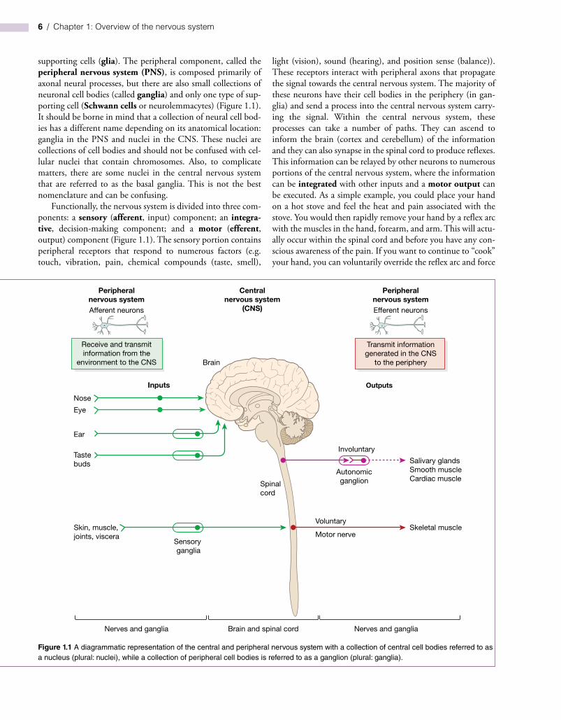
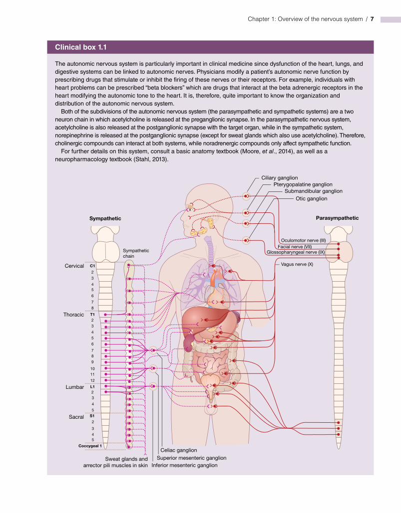
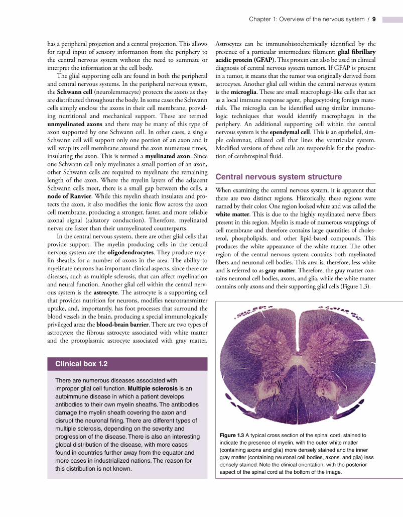
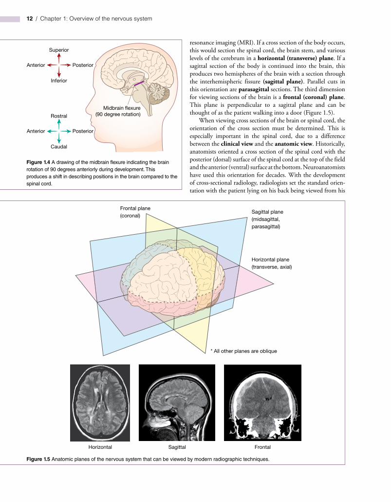
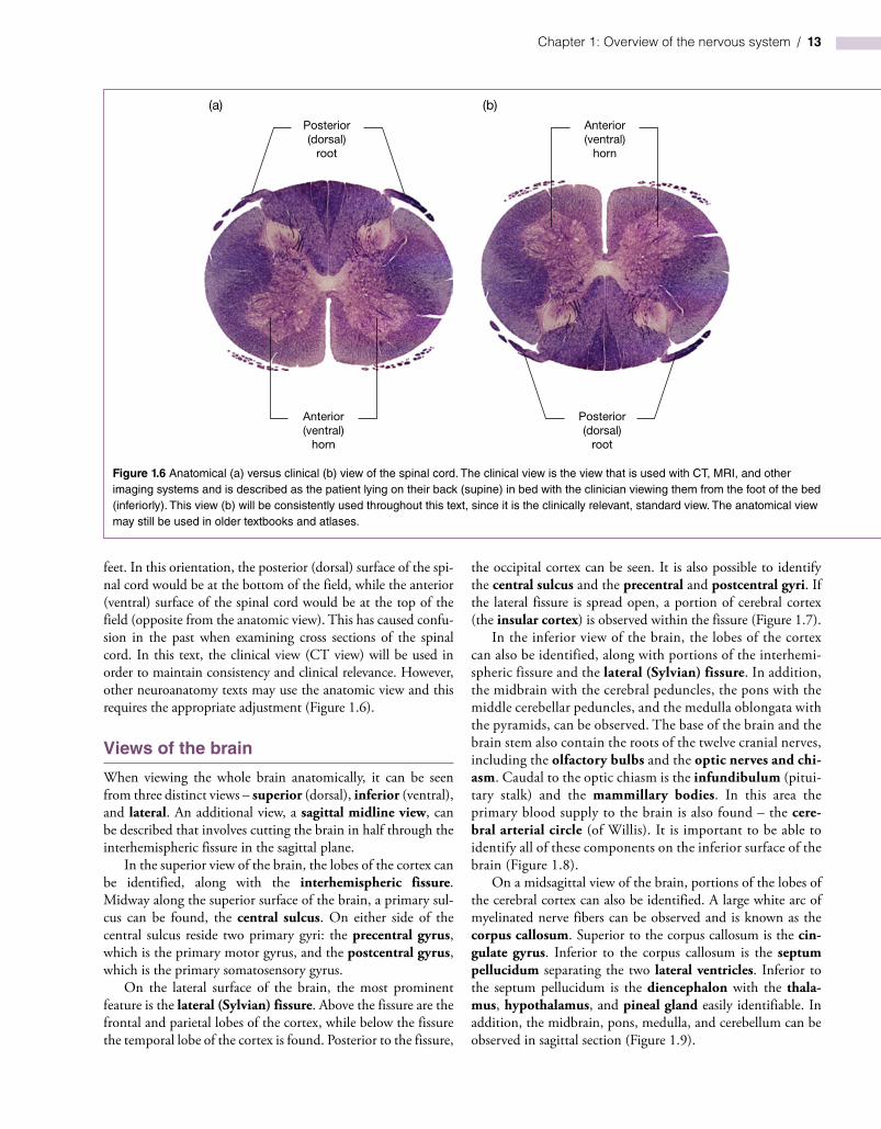
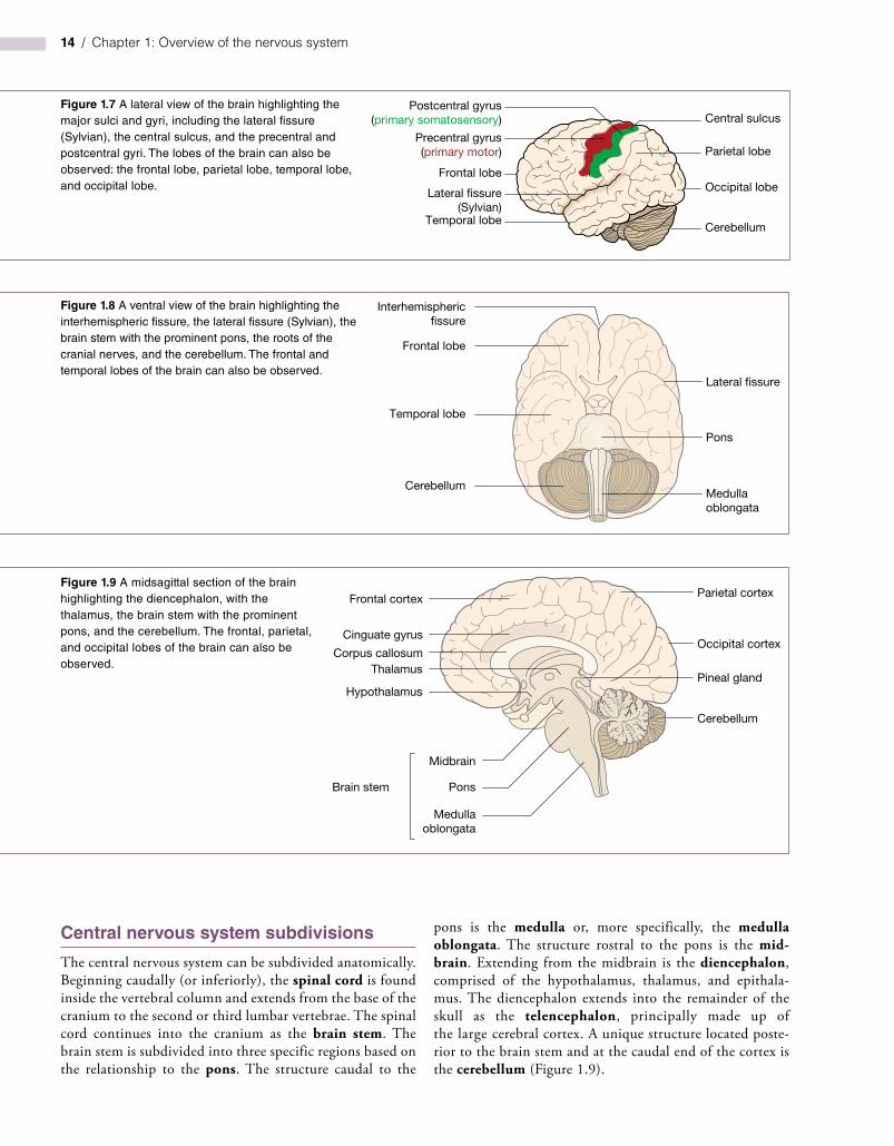
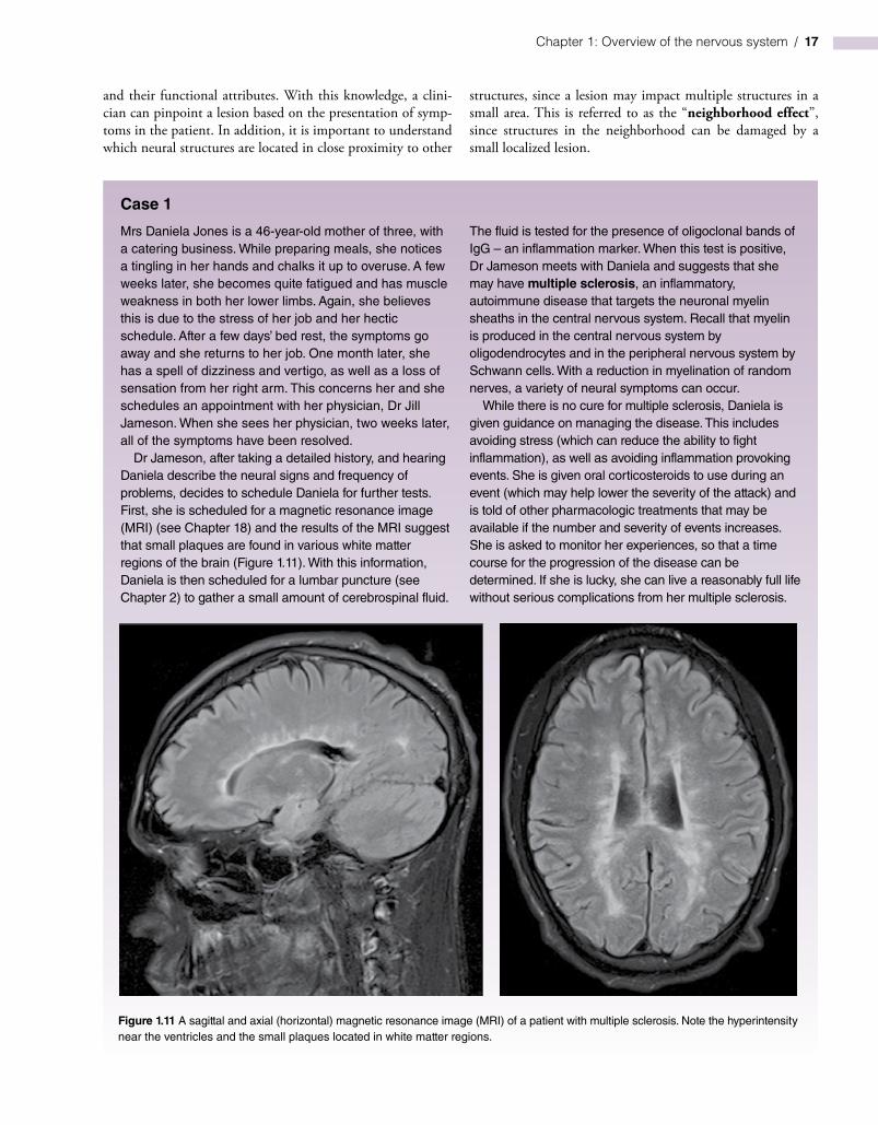