thymopentin treatment in atopic dermatitis-clinical and ... · thymopentin treatment in severe...
TRANSCRIPT

Archives ofDisease in Childhood 1992; 67: 1095-1102
Thymopentin treatment in severe atopicdermatitis-clinical and immunological evaluations
Kue-Hsiung Hsieh, Men-Fang Shaio, Tung-Nan Liao
AbstractAn open clinical trial of thymopentin wasconducted on 16 children with severe atopicdermatitis. The patients were treated withinjections three times a week of 50 mg thymo-pentin for six weeks. They were then dividedrandomly into two groups: group A continuedthymopentin for an additional six weeks, andgroup B were treated with normal saline.Clinical parameters and immunological func-tion were evaluated serially. The total severityscore started to decline from baseline signifi-cantly three weeks after treatment, and con-tinued throughout the study period in group Abut began to flare up in group B two weeksafter stopping thymopentin. AlU the eightpatients in group A completed the trial butthree out of eight in group B dropped outbecause of flaring up of skin lesion. In vitroproduction of interleukin-4 tended to decreaseand that of interferon gamma tended toincrease, but total serum IgE, in vitro IgEsynthesis, and abnormaliy low CD8+CD11b+ suppressor T cells remainedunchanged. Histamine releasing factor(HRF), plasma histamine, and respiratoryburst activities of polymorphonuclear leuco-cytes were appreciably decreased afterthymopentin treatment.
It is concluded that the clinical efficacy ofshort term thymopentin treatment verypossibly results from the decreased productionofHRF and decreased release of polymorpho-nuclear leucocyte derived inflammatorymediators and may have no relation withantigen-IgE immune reaction.
Department ofPaediatrics,National TaiwanUniversity Collegeof MedicineKue-Hsiung HsiehTung-Nan LiaoDepartment ofParasitology andTropical Medicine,National DefenseMedical Center,Taipei,Taiwan,Republic of ChinaMen-Fang ShaioCorrespondence to:Dr Kue-Hsiung Hsieh,Department of Paediatrics,National TaiwanUniversity Hospital,7 Chung-Shan S Road,Taipei, 10016,Taiwan,Republic of China.
Accepted 26 February 1992
(Arch Dis Child 1992;67:1095-102)
Atopic dermatitis is a chronic cutaneousinflammatory disease characterised by early ageof onset, severe pruritus, typical morphologyand distribution of skin rash, chronic relapsingcourse, and personal or family history ofatopy. ' 2
It is a common disease among infants andchildren with an incidence of 1-9% to 8-3% inwhite people"6 and 1 24% in Chinese school-children.7 Chronic atopic dermatitis may resultin significant morbidity, including hospital-isation for control of skin disease and infection,lost school days, psychological trauma fromphysical disfigurement, and occupationaldisability. The management of atopic dermatitishas been less than satisfactory. None of thecurrently available treatments are curative andtreatment is empirical.6Although the pathogenesis ofatopic dermatitis
remains unclear, a number of immunologicalabnormalities have been described. Theyinclude:
(i) Raised and sustained serum IgEconcentrations,8
(ii) Impaired T cell function,9(iii) Decreased natural killer cell activity,'0(iv) Defective capacity to generate alloreactive
cytotoxic T cells,"(v) Impaired autologous mixed lymphocyte
reaction due to defective CD4 responderT cells'2
(vi) Increased cell mediated cytotoxicityagainst skin fibroblasts,'3
(vii) Most importantly, immunological recon-stitution after bone marrow transplantationresults in the permanent resolution of eczema inWiskott-Aldrich syndrome, a primary T cellimmunodeficiency with raised serum IgE,providing in vivo evidence that immune defectspredispose to the development of eczema. 14Thymopoietin is a polypeptide hormone of
the thymus, originally isolated from bovinethymus extract by its effect on neuromusculartransmission. 5 It is secreted by thymic epithelialcells and has been shown to influence both thedifferentiation of thymocytes'6 and the functionof mature T cells. 17 18 Thymopentin (Timunox,Immunobiology Research Institute, Annandale,NJ) is the active pentapeptide (Arg-Lys-Asp-Val-Tyr) moiety corresponding to amino acids32 to 36 of the linear 49 amino acid sequence ofthymopoietin. The biological effects of thymo-pentin mimic those of thymopoietin in allsystems in which it has been evaluated.'8 19 Itwas marketed in Italy in 1985, in Belgium in1987, and in Germany in 1988, and has beenused in a number of diseases in which T celldeficiency may have a role.20
Recent studies suggest that thymopentininfluences IgE synthesis,2' and it might thereforeinfluence the course of atopic diseases in man.Recently, clinical improvement with thymo-pentin that produced reductions in overallseverity in patients with atopic dermatitis hasbeen reported,22 23 but the working mechanismshave not been studied extensively. We presentthe clinical efficacy of an open trial of thymo-pentin in children with severe atopic dermatitisand correlate the clinical efficacy with the invivo and in vitro immunological changes afterthymopentin treatment.
Subjects and methodsSUBJECTSSixteen children, nine boys and seven girls,aged 3 to 14 years, were enrolled into this study.
1095
on February 23, 2020 by guest. P
rotected by copyright.http://adc.bm
j.com/
Arch D
is Child: first published as 10.1136/adc.67.9.1095 on 1 S
eptember 1992. D
ownloaded from

Hsieh, Shaio, Liao
The clinical trial was approved by the humanresearch committee of this hospital and informedconsent was obtained for each patient. All of thepatients had severe pruritus, skin lesions ofcharacteristic morphology and distribution,chronic relapsing course, and a history of atopy,which met the criteria for the diagnosis of atopicdermatitis.' 2 The age of onset ranged from1 month to 1 year, the mean (SD) duration ofatopic dermatitis was 8-6 (3 0) years, the serumIgE concentrations ranged from 224 to 5241 IU/ml (mean (SD) 2318 (1525) IU/ml), and themean (SD) area of involved body surface was81-5 (25 0)%. The severity was graded on ascale of 0-3 for each of the five parameters:erythema, pruritus, oedema/papulation, ex-coriation, and scaling/dryness. To be enrolledthe patient had to have a severity score of 6 orhigher (including a score of 2 or more for botherythema and pruritus). The mean (SD) totalseverity score before treatment was 14-4 (0 9)(range 12-15). Twenty healthy schoolchildren,aged 7 to 15 years, were included as controls forimmunological study.
TREATMENT PROTOCOLAfter enrolment the patients received three sub-cutaneous injections each week of 50 mg ofthymopentin for a consecutive six weeks. Thepatients were then divided randomly into twogroups. One group (group A) continued thethymopentin treatment for an additional sixweeks, and the other group (group B) wasinjected with normal saline. Both groups werecomparable regarding age, severity score, andserum IgE concentration. During the treatmentpatients were allowed to continue antihista-mines and topical corticosteroids, but systemiccorticosteroids were discontinued for at leastfour weeks before the study. No recommenda-tion on food restriction and environmental con-trol was made.
EVALUATIONThe patients were seen every week by oneauthor (KHH). Dermatological assessment,routine laboratory tests (blood chemistry, hae-matology, and urinalysis), and immunologicaltests were performed before and at the end ofthe third, sixth, and 12th week after treatment.The clinical response was evaluated by a derma-tologist who did not know the treatment pro-tocol, and adverse reactions were observedcarefully. Parents' satisfaction was recorded.
IMMUNOLOGICAL TESTSEnumeration oflymphocyte subpopulationT cell subsets were enumerated by FACscan(Becton-Dickinson), using the monoclonal anti-bodies shown in table 1.
Preparations ofsera and culture supernatantsPeripheral blood mononuclear cells (MNCs)were obtained by Ficoll/Hypaque gradientdensity centrifugation. MNCs at a concentra-
Table I Monoclonal antibodies used to enumeratelymphocyte subpopulationsCD3 (total T cells)CD19 (total B cells)CD3-CD16+CD56+ (natural killer cells)CD4 (helper/inducer)CD4+CD45RA+ (suppressor/inducer)CD4+CD45R- (helper/inducer)CD8+ (suppressor/cytotoxic)CD8+CDl lb+ (suppressor)CD8+CD I Ib- (cytotoxic)CD3+CD25+ (activated CD3)CD4+CD25+ (activated CD4)CD8+CD25+ (activated CD8)CD3+HLA-DR+ (activated CD3)CD4+HLA-DR+ (activated CD4)CD8+HLA-DR+ (activated CD8)
tion of 2x 10⁶ cells/ml in complete culturemedium (Roswell Park Memorial Institute-1640supplemented with 10% heat inactivated fetalcalf serum, antibiotics and L glutamine) werestimulated with 2 [tg/ml phytohaemagglutinin(PHA) (Wellcome) for three days and thesupernatants were collected by centrifugationand stored at -20°C until testing.24 25 Theabove mentioned culture conditions, includingcell density, PHA concentration, and days ofcultivation, were found to be optimal forcytokine production in our previous studies.2F26
Measurements ofsoluble interleukin (IL)-2R, CD4molecule, and cytokines in supernatantsCell free CD4 and IL-2 receptor tests kits werepurchased from T Cell Sciences (Cambridge,MA), IL-2 and interferon gamma test kits fromGenzyme (Boston, MA), and IL-4 and IL-6 testkits from R and D System (Minneapolis, MN).The principle of those tests was sandwichenzyme immunoassay (ELISA), using twomonoclonal antibodies against different epitopesof the target molecule (CD4, IL-2, and IL-2R)or monoclonal antibody (first) followed by poly-clonal antibody (IL-4, IL-6, and interferongamma). The sensitivity of the ELISA was 50pg/ml for IL-2, 50 U/ml for IL-2R, 12 U/ml forCD4, 3 pg/ml for IL-4, 3 5 pg/ml for IL-6, and100 pg/ml for interferon gamma. The interassayvariation of all tests were around 5-10% and asimilar figure was found for intra-assay. All thesamples of a single individual were run induplicate simultaneously and at least onenormal control was included in each experiment.
Measurement of histamine releasing factor (HRF)activityHRF was measured as previously described.27MNCs (2 x 10⁶ cells/ml) were stimulated with2 [ig/ml PHA for four hours, washed, andincubated for additional 20 hours. The super-natant was collected, concentrated 50-fold, usinga YM-5 Amicon membrane. The HRF activityof the supernatant was measured by its capacityto release histamine from basophils obtainedfrom a healthy high HRF responder and wasexpressed as percent of total cellular hista-mine released. The histamine was determinedby using histamine radioimmunoassay kits(Immunotech, France). The sensitivity of thekit was 0 5 nmol/l.
1096
on February 23, 2020 by guest. P
rotected by copyright.http://adc.bm
j.com/
Arch D
is Child: first published as 10.1136/adc.67.9.1095 on 1 S
eptember 1992. D
ownloaded from

Thymopentin treatment in severe atopic dermatitis-clinical and immunological evaluations
Measurement ofserum IgG, IgA, IgM, and IgEThe IgG, IgA, and IgM were measured bynephelometry (Beckman). In vitro productionof IgE was done according to the method ofSaryan et al."8 The IgE concentrations in seraand culture supernatants were measured byPhadebas IgE PRIST kits (Pharmacia).
Lucigenin amplified chemiluminescence assayChemiluminescence was assayed by the methodofGyllenhammar,29 using a luminometer (model1251, LKB, Bromma) and recorded as mV.Reaction mixtures contained 1 x 10⁶ neutrophils,0 1 mg lucigenin, and 40 ng phorbol myristateacetate (PMA) or 1 mg opsonised zymosan; andHanks' balanced salt solution (HBSS) wasadded up to a final volume of 1 ml in eachreaction. The readings were started two minutesafter the addition ofreagents and were monitoredevery eight minutes up to an hour.
MEASUREMENTS OF SUPEROXIDE AND HYDROGENPEROXIDE PRODUCTIONSSuperoxide production was measured as super-oxide dismutase inhibitable reduction offerricytochrome c with PMA or opsonisedzymosan as stimulant.30 Quantitation of hydro-gen peroxide is based on the horseradish pero-xidase dependent oxidation of phenol red withPMA or opsonised zymosan as stimulant.3' Forthe superoxide assay the reaction mixture ineach well consisted of ferricytochrome c (2 mg/ml), PMA (40 ng/ml), or opsonised zymosan(1 mg/ml) with neutrophils in the presence orabsence of superoxide dismutase (300 U/ml).The microtitre plates were incubated and shakenat 37°C for one hour and absorbance was read at550 nm on a Microelisa reader (MR 700,Dynatech Laboratories). For the hydrogenperoxide assay the reaction mixture in each wellconsisted of HBSS supplemented with 0-56 mMphenol red, 19 U/ml horseradish peroxidase,PMA (40 ng/ml) or oponised zymosan (1 mg/ml), and neutrophils. The microtitre plateswere incubated and shaken at 37°C for one hourand terminated with 10 ,ul of IN sodiumhydroxide. The absorbance was read at 600 nm.Conversion of absorbance to nanomoles of bothsuperoxide and hydrogen peroxidewas calculatedby a standard method.32 The results wereexpressed as nmol of superoxide or hydrogenperoxide/60 minutes/mg of protein.
STATISTICSAll the data were expressed as mean (SEM)except T cell subsets which was expressed asmean (SD). Unpaired Student's t test was usedfor statistical analysis throughout the studyexcept that the Wilcoxon signed rank test wasused to analyse the immunoglobulin data. A pvalue of <0 05 was considered significant.
ResultsCLINICAL RESPONSEAll eight patients in group A who received 12weeks of continuous thymopentin injections
I.0)
0
Z 100.0)
I-
5
* Group AA Group B
*
0 1
**
**
3 6
Weeks after starting treatment12
Figure I Total severity score during treatment with 50 mgthymopentin. GroupA had 12 weeks continuous treatmentwith thymopentin and group B had six weeks ofthymopentinfollowed by six weeks ofnormal saline injections. Verticalbar represents the SEM; *p<003, compared with baseline;* *p<O0OOO1, comparison betveen groupsA andB at 12weeks.
completed the clinical trial. Three out of eightpatients in group B who received thymopentininjections for the first six weeks and thenswitched to normal saline injections during thelater half of clinical trial, however, dropped outbecause of flare up of skin lesions to a degreecomparable with the period before thymopentinfour weeks after stopping thymopentin. Nodifference in the total severity score was observedbetween two groups before and during thymo-pentin treatment, but the total severity score ofgroup B was much higher than that of group Aat 12 weeks (p<00001, fig 1). In both groups atherapeutic effect became evident after oneweek of treatment (p<003). The results ofevaluation by parents matched very well withthat of the physician (data not shown).
LABORATORY CHANGESFigure 2 demonstrates, as an example, that incase 6 the maculopapulovesicles and lichenifica-tion over the neck and antecubital areas (leftpanel) were much improved after three weeks ofthymopentin treatment (right panel). In general,erythema and pruritus were the parameters thatwere improved most quickly and noticeablyamong the five clinical parameters evaluated,whereas the improvement in scaling/drynesswas much less impressive.
Figure 3 shows the in vitro productions ofsoluble CD4, IL-2R, and cytokines during thecourse of treatment. the patients produced agreater amount of soluble CD4 (p<0.001),IL-2R (p<0 01), IL-2 (p<0 001), and IL-4(p<0001), but less interferon gamma (p<001)than normal controls. After treatment, in vitroproduction of soluble CD4, IL-2R, IL-2, andIL-4 tended to decrease, but only the decreasein IL-2 (p<0 01) and IL-4 (p<0.01) reached a
I
1097
on February 23, 2020 by guest. P
rotected by copyright.http://adc.bm
j.com/
Arch D
is Child: first published as 10.1136/adc.67.9.1095 on 1 S
eptember 1992. D
ownloaded from

Hsieh, Shaio, Liao1098
Figure 2 Thepapulovesicles andlichenification ofdiseased skin beforetreatnent (left panel)and after three weeksoftreatment (right panel).
300 I-
0 3 6 12Patients
240
E
0
-J
**
180 -
120 _-
60 F
01 l l l
Controls
***
**
120 3 6Patients
1500
1200
900
CJ
600
300
0
Controls Patients
E0)a
-
25 000 L
20 000
cm
15 000
10000 F
5000 F
I
I350
280
E210
z
U-
140
0 3 6 12 0 3 6 12 0 3 6 12Controls Patients Controls Patients Controls Patients
Weeks after starting treatment Weeks after starting treatment Weeks after starting treatment
Figure 3 In vitro productions ofsoluble CD4, IL-2R, and cytokines during thymopentin treatment. For CD4, 'p<OOO1; for IL-2, lp<0001,**p<0-01;forIL-2R, k'-p<0Ol;forIL4, '"[p<0001, ''p<0cOJ;forIFN-r, I'pOp<0J01, p<0-05. Data are expressed as mean (SEM).
D
C4a-
O I I I
Controls
--
.. I. au . .
ri
n
on February 23, 2020 by guest. P
rotected by copyright.http://adc.bm
j.com/
Arch D
is Child: first published as 10.1136/adc.67.9.1095 on 1 S
eptember 1992. D
ownloaded from

Thymopentin treatment in severe atopic derinatitis-clinical and immunological evaluations
significant degree. In vitro production of IFN-rwas increased (p<005). No change in IL-6production was found. Again, those changeswere reversed after stopping treatment (group B).The effect ofthymopentin on the distributions
of T cell subsets is shown in table 2. Whencompared with normal controls, the patients
Table 2 Changes ofdistributions ofT cell subsets during thymopentin treatment. Results aremean (SD)%
T cell subset Treatment period (n -10) Controls(n=20)
0 3 Weeks 6 Weeks
CD3+ 68-0(8-7) 66-8(8-4) 68-3(6-3) 67-1(8-2)CD19+ 14-1(4-8) 17 4 (4 9) 16-6 (7-0) 16 2 (4 9)CD3-CD16+CD56+ 17 3(90) 17 1(7-5) 16-3(5-3) 18 3(9 0)CD4+ 31L9(8 4) 32-0(8-1) 33-8(5-8) 34-8(6-1)CD4-CD45RA+ 10-7 (3-6) 108 (3 9) 11-8 (3-0) 11-9(3 9)CD4+CD45RA- 24-7(5-3) 25-4(6-8) 24-4(6 6) 26-7(7-3)CD8+ 28 5 (5 6) 26-3 (5-9) 29-2 (9-8) 26-7 (9-8)CD8+CD1 lb+ 5-0 (2 4):' 3-5 (2-1) 5-6 (2-7) 11-9(3-9)*CD8+CDllb- 24-2(5 2) 24-8(4 8) 26-1(5-7) 20-0(9-9)CD3+CD25+ 50(5-1) 45(09) 4-1(1-4) 5-1(2-8)CD4+CD25+ 5-2 (2-9) 5 6 (2-4) 5-0 (2-3) 6-6 (2-8)CD8+CD25+ 07 (13)* 0-2(04) 0-2(04) 30(19)''CD3+HLA-DR+ 8-2(58) 12-1(44) 15 1(87) 11-3(4-1)CD4+HLA-DR+ 3-8(0 9) 6-7(2 6) 7-4(2 6) 3-6(1 3)CD8+HLA-DR+ 4 5 (3-0):' 7-8 (3-5) 8 5 (4-8) 10-3 (4-5)
p=OOOO1, 'p<O005, '**p<0 002.
Table 3 Serum immunoglobulins and in vitro IgE synthesis during thymopentin treatment.Results are mean (SD)
Immunoglobulin Treatment period (n= 10)
0 3 Weeks 6 Weeks
IgG (g/l) 10-24 (3-43)-' 9-91 (332) 8-28 (1-37)IgA (g/l) 1-52 (0 60) 1-52 (0 56) 1-48 (0 38)IgM (g/1) 1-49(0 72) 1-49 (0-74) 1-19 (0-39)IgE (IU/ml) 3283-2 (1666-7) 2862-1 (1653-2) 3026-8 (1388-8)In vitro IgE
synthesis (pg/ml) 1714-5 (3004 2) 2117-6 (2578-3) 1335-8 (1447-6)
tp<0 05 (Wilcoxon signed rank test).
10
Z-0EC
-EC-Eco-Cl
Ea.
0 3 6 12Controls Patients
Weeks after starting treatment
with atopic dermatitis had a normal number (%)of total T cells (CD3+), B cells (CD19+),natural killer cells (CD3- CD16+CD56+),suppressor inducer T cells (CD4+CD45RA+),helper inducer T cells (CD4+CD45RA-),cytotoxic T cells (CD8+CDllb-), CD25+,and HLA-DR+ activated CD3 and CD4 cells.However, suppressor T cells (CD8+CDl lb+),and CD25+ and HLA-DR+activated sup-pressor/cytotoxic T cells (CD8+) wereappreciably decreased in patients. After thymo-pentin treatment the suppressor T cells (CD8+CDllb+) and CD25+ activated CD8+ cellsremained very low even at the end of 12 weeksof thymopentin treatment (table 2).The changes of serum immunoglobulin con-
centrations were followed up serially afterthymopentin treatment. IgG and IgM tended todecrease after six weeks of treatment, but not toa significant degree. No change was found forIgA, IgE, and in vitro IgE production through-out the course of trial (table 3).
Figure 4 demonstrates that both plasmahistamine and HRF activity were much higherin patients than in normal controls (p<O0OOl forthe former and p<O00l for the latter). Afterthymopentin treatment, both plasma histamineand HRF were decreased significantly. Thedecrease persisted in group A but reversed topretreatment value in group at the 12th week(A v B, p<0 Ol).The respiratory burst activities of poly-
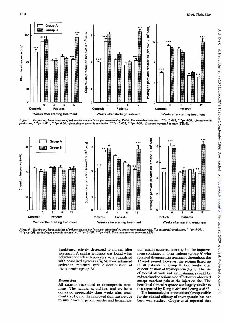
morphonuclear leucocytes after stimulation withPMA are shown in fig 5. The production ofsuperoxide (p<0001) and hydrogen peroxide(p<O-001) before thymopentin treatment wasmuch higher than normal controls and the
70 **
50
0-
CULLX
0 3 6 12Controls Patients
Weeks after starting treatment
Figure 4 Plasma histamine andHRF activity during thymopentin treatment. For plasma histamine, * '"p<0OO1,* Cp<0-0I;forHRF activity, p<0-01I, * 'p<0-01. Data are expressed as mean (SEM).
1099
on February 23, 2020 by guest. P
rotected by copyright.http://adc.bm
j.com/
Arch D
is Child: first published as 10.1136/adc.67.9.1095 on 1 S
eptember 1992. D
ownloaded from
Hsieh, Shaio, Liao
0 3 6 12Patients
-a57D 50uz
x
0EC
0 3._
Qx00.C,)
0'x
Controls0 3 6 12 0 3 6 12
Patients Controls PatientsWeeks after starting treatment Weeks after starting treatment Weeks after starting treatment
Figure 5 Respiratory burst activities ofpolymorphonuclear leucocytes stimulated by PMA. For chemiluminescence, ***p< 001, ***p<000l;for superoxideproduction, * * *p<0-001, * * *p<0.001;for hydrogen peroxide production, * * *p<0-001, * * -p<0001. Data are expressed as mean (SEM).
-a'E-iC.)U)0
xZ-0ECC0Cr.)-c0
'.20
Q0.00
Cn
5
3
(a
Un
0W-
x
Q
0ECC0I
*00L-
0
0100.
00
a
1
0
0 3 6 12Controls Patients
Weeks after starting treatment
0 3 6
Controls Patients12
6
4
2
0
ControlsWeeks after starting treatment
0 3 6 12Patients
Weeks after starting treatment
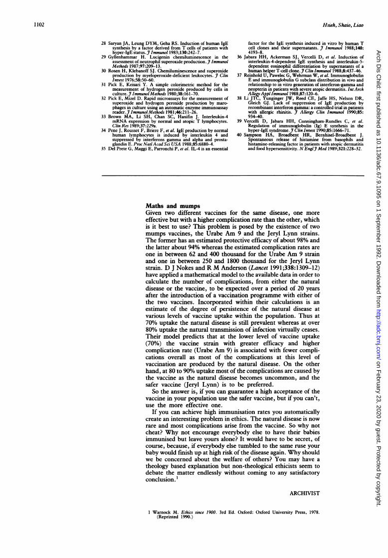
Figure 6 Respiratory burst activities ofpolymorphonuclear leucocytes stimulated by serum opsonised zymosan. For superoxide production, * * *p<O OOI,* * *p<0001;for hydrogen peroxide production, * **p<0001 , *p<0 OI. Data are expressed as mean (SEM).
heightened activity decreased to normal aftertreatment. A similar tendency was found whenpolymorphonuclear leucocytes were stimulatedwith opsonised zymosan (fig 6); their enhancedactivation returned after discontinuation ofthymopentin (group B).
DiscussionAll patients responded to thymopentin treat-ment. The itching, scratching, and erythemadecreased appreciably three weeks after treat-ment (fig 1), and the improved skin texture dueto subsidence of papulovesicles and lichenifica-
tion usually occurred later (fig 2). The improve-ment continued in those patients (group A) whoreceived thymopentin treatment throughout the12 week period, however, the eczema flared upin all patients of group B four weeks afterdiscontinuation of thymopentin (fig 1). The useof topical steroids and antihistamines could bereduced and no serious side effects were observedexcept transient pain at the injection site. Thebeneficial clinical response was largely similar tothat reported by Kang et a122 and Leung et al.23The immunological mechanism(s) responsible
for the clinical efficacy of thymopentin has notbeen well studied. Cooper et al reported that
E0UC0
0C
E
0
0
Controls
E0UC0U
U_
0
0-CE-
1100
on February 23, 2020 by guest. P
rotected by copyright.http://adc.bm
j.com/
Arch D
is Child: first published as 10.1136/adc.67.9.1095 on 1 S
eptember 1992. D
ownloaded from

Thymopentin treatment in severe atopic dermatitis-clinical and immunological evaluations 1101
thymopentin could influence the in vivo and invitro production by atopic dermatitis MNCs.2'The same group further described increasedCD8+ (suppressor/cytotoxic) cells after sixweeks of thymopentin administration.22 Theresults obtained in our study could not confirmtheir hypothesis: (i) total serum IgE and in vitroIgE production were not changed after treatment,and (ii) the CD8+CD1 lb+ suppressor T cells,but not the total number of CD8+ suppressor/cytotoxic T cells, were decreased in patientswith atopic dermatitis, and thymopentin treat-ment failed to increase the CD8+CD1 lb+suppressor and CD8+CD25+ activated T cells.Recent studies have demonstrated that peri-pheral blood MNCs from patients with atopicdermatitis produced increased concentrations ofIL-4,33a cytokine that induces IgE synthesis,3"36and decreased concentrations of interferongamma37 a cytokine that inhibits IL-4 dependentIgE synthesis.3'~36 Our results confirm thosereports,33 37 and furthermore this study showedthat thymopentin treatment tended to suppressthe production of IL-4 and enhanced theproduction of interferon gamma in vitro.However, Li et al recently reported that thetotal serum IgE and specific IgE antibodieswere not decreased in patients with allergicrhinitis after receiving interferon gammatreatment in both periods of off season andduring season.38 Vercelli et al also foundthat anti-IL-4 and interferon gamma wereunable to suppress the in vitro spontaneous IgEsynthesis in patients with hyper-IgE syndrome,39and no significant difference in mitogen inducedproductions of IL-4 and interferon gamma wasdetected between HIE patients and normalcontrols. Thus, mechanisms other than antigen-IgE immune reaction may account for the ratherquick appearance of clinical benefit of shortterm thymopentin administration in the treat-ment of atopic dermatitis.
It is important to note that HRF and plasmahistamine concentrations were decreased afterthymopentin treatment (fig 4). Furthermore,the augmented activation of polymorphonuclearleucocytes was also suppressed, as evidenced bythe lessened respiratory burst activities (fig 5and 6). Sampson et al recently reported thatpatients with atopic dermatitis caused by foodallergy produced significantly less HRF after theoffending food allergen was eliminated from thediet for an extended period.40 Our recent studyalso demonstrated decreased production of andresponsiveness to HRF in asthmatic patientsbenefited by successful immunotherapy.27 Thus,the decreased production ofHRF and decreasedrelease ofpolymorphonuclear leucocytes derivedinflammatory mediators may account partly forthe clinical efficacy of thymopentin treatment inpatients with atopic dermatitis.One of the shortcomings of this paper is lack
of immunohistological studies of diseased skinbefore and after thymopentin treatment. Suchstudies would provide the most direct informa-tion that could be used to explain the clinicalefficacy of thymopentin. Unfortunately, we areunable to obtain consents from parents to doskin biopsies.
Finally as no serious side effects are
encountered during the 12 week period of treat-ment and there is still no satisfactory manage-ment for severe atopic dermatitis, thymopentinmay be used in those patients with atopicdermatitis who are refractory to traditionaltreatment. Studies with longer duration andlarger number of patients are needed before therationale for such kind of treatment can bejustified.
The authors wish to thank Johnson and Johnson Company,Taiwan Branch, for supplying thymopentin preparations used inthis study.
I Hanifm JM, Raika G. Diagnostic features ofatopic dermatitis.Acta Derm Venerol (Stockh) 1980;92 (suppl):44-7.
2 Hanifm JM. Basic and clinical aspects of atopic dermatitis.Ann Allergy 1984;54:386-93.
3 Halpern SR, Sellars WA, Johnson RB, et al. Development ofchild allergy in infants fed breast, soy or cow's milk.JAllergy Clin Immunol 1973;66:465-71.
4 Kiellman N-JM. Atopic disease in seven-year-old children.Acta Paediatr Scand 1977;66:465-71.
5 Walker RB, Warin RP. Incidence of eczema in early child-hood. BrJ Dermatol 1956;68:182-3.
6 Hanifm JM. Atopic dermatitis. In: Middleton E Jr, Reed CE,Ellis EF, Adkinson NF Jr, Yunginger JW, eds. Allergyprinciples and practice. 3rd Ed. St Louis: CV Mosby, 1988:1403-28.
7 Hsieh KH, Shen JC. Prevalence of childhood asthma inTaipei, Taiwan, and other Asian Pacific countries. J Asthma1988;25:73-82.
8 Johnson E, Irons J, Patterson R, Roberts M. Serum IgEconcentration in atopic dermatitis. J Allergy Clin Immunol1974;54:94-9.
9 Leung DYM, Geha RS. Immunoregulatory abnormalities inatopic dermatitis. Clin Rev Allergy 1986;4:67-86.
10 Chiarelli F, Canfora G, Verrotti A, Amerio P, Morgese G.Humoral and cellular immunity in children with active andquiescent atopic dermatitis. Br J Dermatol 1987;116:651-60.
11 Leung DYM, Wood N, Dubey D, Rhodes AR, Geha RS.Cellular basis of defective cell-mediated lympholysis inatopic dermatitis. JImmunol 1983;130:1678-82.
12 Leung DYM, Saryan JA, Frankel R, Lareau M, Geha RS.Impairment of the autologous mixed lymphocyte reactionin atopic dermatitis. J Clin Invest 1983;72:1482-6.
13 Leung DYM, Parkman R, Feller J, Wood N, Geha RS. Cell-mediated cytotoxicity against skin fibroblasts in atopicdermatitis. J Immunol 1982;128:1736-41.
14 Saurat JH. Eczema in primary immune deficiencies: clues tothe pathogenesis of atopic dermatitis with special referenceto Wiskott-Aldrich syndrome. Acta Derm Venereol (Stockh)1985;114: 125-8.
15 Goldstein G, Audhya TK. Thymopoietin to thymopentin:experimental studies. Survey of Immunological Research1985;4 (suppl): 1-10.
16 Scheid MP, (Goldstein (G, Boyse EA. The generation andregulation of lymphocyte populations: evidence fromdifferentiative induction systems in vitro. J Exp Med 1978;147:1727-43.
17 Meroni PL, Barcellini W, Frasea D, et al. In vivo immuno-potentiating activity of thymopentin in aging human:increase of IL-2 production. Clin Immunol Immunopathol1987;42:151-9.
18 Diezel W, Waschke SR, Forner K. Induction and augmenta-tion of mitogen-induced immune interferon production inhuman lymphocytes by a synthetic thymopoietin pentapep-tide. BiomedBiochimActa 1984;43:9-12.
19 Goldstein G, Scheid MP, Boyse EA, et al. A synthetic penta-peptide with biologic activity characteristics of the thymichormone thymopoietin. Science 1979;204:1309-10.
20 Sundal E, Bolla K. Therapy with thymopentin: a clinicaloverview. In: Malaise MG, ed. Immune regulation by charac-terizedpolypeptides. New York: Alan R. Liss 1987:121-36.
21 Cooper KD, Kang K, Hanifin JM. Effects of thymopoietinpentapeptide (TP-5) on in vitro and in vivo IgE productionby atopic dermatitis cell subsets. Diagnostic Immunology1983;1:21 1-5.
22 Kang K, Cooper KD, Hanifin JM. Thymopentin pentapep-tide (TP-5) improves clinical parameters and lymphocytesubpopulations in atopic dermatitis. 7 Am Acad Dermatol1983;8:372-7.
23 Leung DYM, Hirsch RL, Schneider L, et al. Thymopentintherapy reduces the clinical severity of atopic dermatitis.J Allergy Clin Immunol 1990;85:927-33.
24 Hsieh KH. Altered interleukin-2 production and responsive-ness after hyposensitization to house dust. J Allergy ClinImmunol 1985;76: 188-94.
25 Hsieh KH. Decreased production of interleukin-2 receptorsafter immunotherapy to house dust. J Clin Immunol 1988;8:17 1-7.
26 Hsieh KH. Decreased production of CD8 (T8) antigen afterimmunotherapy. J Clin Immunol 1989,9: 111-8.
27 Liao TN, Hsieh KH. Decreased production of and respon-siveness to histamine releasing factor after immunotherapyin asthmatic children. J Allergy Clin Immunol 1990-86:894-901.
on February 23, 2020 by guest. P
rotected by copyright.http://adc.bm
j.com/
Arch D
is Child: first published as 10.1136/adc.67.9.1095 on 1 S
eptember 1992. D
ownloaded from
1102 Hsieh, Shaio, Liao
28 Saryan JA, Leung DYM, Geha RS. Induction of human IgEsynthesis by a factor derived from T cells of patients withhyper-IgE status. J Immunol 1983;130:242-7.
29 Gyllenhammar H. Lucigenin chemiluminescence in theassessment of neutrophil superoxide production. J ImmunolMethods 1987;97:209-13.
30 Rosen H, Klebanoff SJ. Chemiluminescence and superoxideproduction by myeloperoxide-deficient leukocytes. J ClinInvest 1976;58:50-60.
31 Pick E, Keisari Y. A simple colorimetric method for themeasurement of hydrogen peroxide produced by cells inculture. J ImmunolMethods 1980;38:161-70.
32 Pick E, Mizel D. Rapid microassays for the measurement ofsuperoxide and hydrogen peroxide production by maro-phages in culture using an automatic enzyme immunoassayreader. J ImmunolMethods 1981;46:211-26.
33 Brown MA, Li SH, Chan SC, Hanifm J. Interleukin-4mRNA expression by normal and atopic T lymphocytes.Clin Res 1989;37:229a.
34 Pene J, Rousset F, Briere F, et al. IgE production by normalhuman lymphocytes is induced by interleukin 4 andsuppressed by interferons gamma and alpha and prosta-glandin E. Proc NatlAcadSci USA 1988;85:6880-4.
35 Del Prete G, Maggi E, Parronchi P, et al. IL-4 is an essential
factor for the IgE synthesis induced in vitro by human Tcell clones and their supernatants. J Immunol 1988;140:4193-8.
36 Jabara HH, Ackerman SJ, Vercelli D, et al. Induction ofinterleukin-4-dependent IgE synthesis and interleukin-5-dependent eosinophil differentiation by supernatants of ahuman helper T-cell clone. J Clin Immunol 1988;8:437-46.
37 Reinhold U, Pawelec G, Wehrman W, et al. ImmunoglobulinE and immunoglobulin G subclass distribution in vivo andrelationship to in vitro generation of interferon-gamma andneopterin in patients with severe atopic dermatitis. Int ArchAllegy Appl Immunol 1988;87:120-6.
38 Li JTC, Yunginger JW, Reed CE, Jaffe HS, Nelson DR,Gleich Gj. Lack of suppression of IgE production byrecombinant interferon gamma: a controlled trial in patientswith allergic rhinitis. J Allergy Clin Immunol 1990;85:934-40.
39 Verceili D, Jabara HH, Cunningham-Rundles C, et al.Regulation of immunoglobulin (Ig) E synthesis in thehyper-IgE syndrome. J Clin Invest 1990;85: 1666-71.
40 Sampson HA, Broadbent HR, Bernhisel-Broadbent J.Spontaneous release of histamine from basophils andhistamine-releasing factor in patients with atopic dermatitisand food hypersensitivity. N EnglyMed 1989;321:228-32.
Maths and mumpsGiven two different vaccines for the same disease, one moreeffective but with a higher complication rate than the other, whichis it best to use? This problem is posed by the existence of twomumps vaccines, the Urabe Am 9 and the Jeryl Lynn strains.The former has an estimated protective efficacy of about 98% andthe latter about 94% whereas the estimated complication rates areone in between 62 and 400 thousand for the Urabe Am 9 strainand one in between 250 and 1800 thousand for the Jeryl Lynnstrain. D J Nokes and R M Anderson (Lancet 1991;338:1309-12)have applied a mathematical model to the available data in order tocalculate the number of complications, from either the naturaldisease or the vaccine, to be expected over a period of 20 yearsafter the introduction of a vaccination programme with either ofthe two vaccines. Incorporated within their calculations is anestimate of the degree of persistence of the natural disease atvarious levels of vaccine uptake within the population. Thus at70% uptake the natural disease is still prevalent whereas at over80% uptake the natural transmission of infection virtually ceases.Their model predicts that at the lower level of vaccine uptake(70%) the vaccine strain with greater efficacy and highercomplication rate (Urabe Am 9) is associated with fewer compli-cations overall as most of the complications at this level ofvaccination are produced by the natural disease. On the otherhand, at 80 to 90% uptake most of the complications are caused bythe vaccine as the natural disease becomes uncommon, and thesafer vaccine (Jeryl Lynn) is to be preferred.
So the answer is, if you can guarantee a high acceptance of thevaccine in your population use the safer vaccine, but if you can't,use the more effective one.
If you can achieve high immunisation rates you automaticallycreate an interesting problem in ethics. The natural disease is nowrare and most complications arise from the vaccine. So why notcheat? Why not encourage everybody else to have their babiesimmunised but leave yours alone? It would have to be secret, ofcourse, because, if everybody else tumbled to the same ruse yourbaby would finish up at high risk of the disease again. Why shouldwe be concerned about the welfare of others? You may have atheology based explanation but non-theological ethicists seem todebate the matter endlessly without coming to any satisfactoryconclusion.'
ARCHIVIST
1 Warnock M. Ethics since 1900. 3rd Ed. Oxford: Oxford University Press, 1978.(Reprinted 1990.)
on February 23, 2020 by guest. P
rotected by copyright.http://adc.bm
j.com/
Arch D
is Child: first published as 10.1136/adc.67.9.1095 on 1 S
eptember 1992. D
ownloaded from