timing of the critical period for plasticity of ocular
TRANSCRIPT
Timing of the Critical Period for Plasticity of Ocular DominanceColumns in Macaque Striate Cortex
Jonathan C. Horton and Davina R. Hocking
Beckman Vision Center, University of California, San Francisco, San Francisco, California 94143-0730
Visual deprivation induced by monocular eyelid suture, a lab-oratory model for congenital cataract, results in shrinkage ofocular dominance columns serving the closed eye. We per-formed monocular suture in macaques at ages 1, 3, 5, 7, and 12weeks to define the critical period for plasticity of ocular dom-inance columns. After a minimum survival of 8 months, com-plete montages of [3H]proline-labeled columns were recon-structed from flat-mounts of striate cortex in both hemispheres.In any given monkey, visual deprivation induced the columnsthroughout striate cortex (V1) to retract the same distance fromtheir original borders in layer IVcb. After deprivation, the widestcolumns remained in the foveal representation and along theV1/V2 border, where columns are widest in control animals. Thenarrowest deprived columns belonged to the ipsilateral eye,especially along the horizontal meridian and in the periphery,where columns are narrowest in control animals. At the earliestage that we tested (1 week), visual deprivation reduced thecolumns to fragments. These fragments always coincided witha cytochrome oxidase patch, or a short string of patches, in the
upper layers. More severe column shrinkage occurred in layerIVcb (parvo) than layer IVca (magno). The geniculate input tothe patches in layer III (konio) appeared normal after depriva-tion, despite loss of CO activity. Surprisingly, the blind spotrepresentation of the open eye was shrunken by monoculardeprivation, although binocular competition is absent in thisregion. Our principal finding was that eyelid suture at age 1week caused the most severe column shrinkage. With suture atlater ages, the degree of column shrinkage showed a progres-sive decline. Deprivation commencing at age 12 weeks causedno column shrinkage. These results imply that primate visualcortex is most vulnerable to deprivation during the first weeksof life. Our experiments should provide further impetus for thetreatment of children with congenital cataract at the earliestpossible age.
Key Words: ocular dominance column; critical period; ambly-opia; visual deprivation; cytochrome oxidase patch; flat-mount;striate cortex
The most severe form of amblyopia develops when a newbornchild is deprived of vision in one eye by a dense unilateral cataract(Boothe et al., 1985). In a series of landmark experiments, Wieseland Hubel (1963, 1965) raised kittens with the lids of one eyesutured to simulate congenital monocular cataract. Electrophysi-ological recordings from striate cortex (V1) revealed that few cellscould be driven via the deprived eye. Later, the anatomical cor-relate of this shift in ocular dominance was shown in cats andmonkeys by intraocular injection of [3H]proline (Hubel et al.,1977; Shatz and Stryker, 1978; Swindale et al., 1981). The oculardominance columns serving the deprived eye appeared severelyshrunken because of contraction of their geniculocortical terminalarbors. Wiesel and Hubel (see Wiesel, 1982) proposed that visualdeprivation produces amblyopia by causing the deprived eye tobecome disconnected from the cortical circuits required for thenormal processing of retinal input.
Ocular dominance columns are vulnerable to shrinkage for onlya short time after birth, called the “critical period” (Hubel andWiesel, 1970). Once neonates develop beyond the critical period,the width of their ocular dominance columns becomes immutable.To define the beginning and the end of the critical period, LeVayet al. (1980) performed monocular suture in a series of 10 ma-caques at successively later ages, ranging from 2 d to adult. Totheir surprise, the age at eyelid suture seemed to make no differ-ence for the first 6 weeks of life. The same degree of columnshrinkage was found in all animals sutured during this interval.The critical period did not appear to wane until ;10 weeks, wheneyelid closure caused only mild column shrinkage. The end of thecritical period was not defined exactly, but eyelid suture at 7months had no effect.
Results obtained from these animal experiments have alteredthe management of congenital cataract in children. Before 1980,the prognosis for monocular congenital cataract was considereddismal and successful visual rehabilitation was virtually unknown(Cordes, 1957; Costenbader and Albert, 1957; Ryan et al., 1965;Parkes and Hiles, 1967; Francois, 1979). After the discovery of thecritical period for plasticity of ocular dominance columns, a fewinnovative clinicians showed that good vision could be obtained inchildren if cataract surgery were performed at an early age,followed by immediate optical correction and vigorous patchingtherapy (Enoch and Rabinowicz, 1976; Beller et al., 1981; Pratt-Johnson and Tillson, 1981). It has now become accepted practicethat dense, monocular cataract should be removed before age 4months (Vaegan and Taylor, 1979; Rogers et al., 1981; Birch and
Received Dec. 19, 1996; revised Feb. 7, 1997; accepted Feb. 25, 1997.This work was supported by grants from the National Eye Institute, That Man May
See, and Research to Prevent Blindness. We thank the New England RegionalPrimate Research Center (especially Dr. Prabhat Sehgal) and the California Re-gional Primate Research Center (especially Dr. Celia Valverde, Jenny Short, andDavid Robb) for their help. The California Primate Center is supported by NationalInstitutes of Health Base Grant RR00169. Robin Troyer assisted with these exper-iments. The first experiment (Monkey 0) was performed in collaboration with Dr.Michael P. Stryker. We thank him for his critical review of this manuscript. Dr.Torsten N. Wiesel, Dr. Simon LeVay, and Dr. Michael C. Crair also provided manyuseful comments.
Correspondence should be addressed to Dr. Jonathan C. Horton, Beckman VisionCenter, 10 Kirkham Street, University of California, San Francisco, San Francisco,CA 94143-0730.Copyright © 1997 Society for Neuroscience 0270-6474/97/173684-26$05.00/0
The Journal of Neuroscience, May 15, 1997, 17(10):3684–3709
Stager, 1988; Drummond et al., 1989; Cheng et al., 1991; Wrightet al., 1992; Birch et al., 1993). However, there is still no consensusabout the urgency of cataract surgery within this time frame(Birch and Stager, 1996). Hoyt removed a dense, monocularcataract from a baby 7 hr after birth, and the child attained anacuity of 20/20 (Beller et al., 1981). Is such early action warranted,or does surgery anytime during a “grace period” of a few weeks ormonths after birth produce comparable results?
To furnish anatomical data pertinent to this issue, we repeated
the experiments of LeVay et al. (1980) by performing monoculareyelid suture in a series of baby macaques at successively laterages. After the monkeys were at least 8 months old, flat-mounts ofstriate cortex were prepared from each animal to measure theamount of column shrinkage. In most animals, we reconstructedthe entire pattern of ocular dominance columns to avoid samplingbias that might arise from examining only a small area. Preparingflat-mounts also allowed us to determine whether columns vary intheir susceptibility to shrinkage according to their location within
Table 1. Experimental animals
Monkey 1 Monkey 2 Monkey 3 Monkey 4 Monkey 5 Monkey 6 Monkey 7 Monkey 8
Sex male male male male female male male maleLength of gestation (d) E167 E166 E167 E164 E167 E164 E165 E165Macaque species fascicularis fascicularis mulatta mulatta mulatta mulatta mulatta mulattaAge at suture 7 d 7 d 7 d 21 d 35 d 35 d 49 d 84 dEye sutured right right right right right right right rightEye injected right right left left left left left leftEye enucleated right – right right – right right –Survival time
after enucleation10 d – 58 d 24 d – 42 d 20 d –
Age at perfusion 11 months 14 months 11 months 8 months 26 months 15 months 8 months 15 monthsWeight at Perfusion 1.47 kg 1.87 kg 1.53 kg 1.38 kg 3.14 kg 1.93 kg 1.64 kg 2.21 kg
Table 2. Measurements of striate cortex
Animal number Monkey 1 Monkey 2 Monkey 3 Monkey 4 Monkey 5 Monkey 6 Monkey 7 Monkey 8
Sutured 1 week 1 week 1 week 3 weeks 5 weeks 5 weeks 7 weeks 12 weeksHemisphere left right left right left right left right left right left right left right left rightV1 area (mm2) 461a 441a 404a 452a 1362 1414 1241 1226 895 1014 1075 1034 1575 1612 1379 1331Unreconstructable
area (mm2) 49 33 44 44 159 144 28 47 37 22 14 11 13 52 78 117Total # column
pairs intersect-ing V1 Border – – – – – – 118 124 127 123 145 145 122 115 136 136
Optic disc area(mm2) – – – – 12b 4 12 7 10 7 11 8 14 10 12 11
Monocular crescentarea (mm2) – – – – 78 84b 48 64 49 67 63 65 83 99 55 48
OperculumLeft (nondeprived)
eye column area 346 344 331 360 525 637 387 432 311 318 302 334 425 431 331 335(mm2) (83.8%) (84.1%)c (92.2%) (88.0%) (88.0%) (91.7%) (63.3%) (72.5%) (64.7%) (63.7%) (56.8%) (62.8%) (53.1%) (58.2%) (49.1%) (51.9%)
Right (deprived)eye column area 67 65 28 49 71 58 224 164 170 181 230 198 376 309 343 311(mm2) (16.2%) (15.9%) (7.8%) (12.0%) (12.0%) (8.3%) (36.7%) (27.5%) (35.3%) (36.3%) (43.2%) (37.2%) (46.9%) (41.8%) (50.9%) (48.1%)
Shrinkaged 36% 32% 44% 36% 40% 40% 15% 20% 17% 12% 9% 11% 5% 6% 1% 0%PeripheryLeft eye column 487 477 380 419 177 296 250 320 346 475 260 294
area (mm2) – – – – (94.4%) (98.1%) (70.1%) (82.0%) (55.7%) (70.8%) (54.7%) (76.6%) (52.0%) (66.9%) (46.5%) (57.8%)Right eye column 29 9 162 92 141 122 207 98 320 235 299 215
area (mm2) – – – – (5.6%) (1.9%) (29.9%) (18.0%) (44.3%) (29.2%) (45.3%) (23.4%) (48.0%) (33.1%) (53.5%) (42.2%)Shrinkaged – – – – 57% 35% 33% 19% 19% 8% 18% 14% 15% 4% 10% 25%
aData reflect area of the operculum only.bBoundary for this region was invisible in autoradiographs because the deprived eye’s ocular dominance columns were obliterated locally. Therefore, the boundary wasdetermined by reference to the appearance of CO patches in the upper layers.cUnderlined values denote contralateral eye.dThe shrinkage was calculated by subtracting the percentage of the right eye column area (line above) from the mean percentage in normal animals. In normal animals, themean percentage for the right eye is 52% in the contralateral operculum, 48% in the ipsilateral operculum; 63% in the contralateral periphery, 37% in the ipsilateral periphery(Horton and Hocking, 1996c).
Horton and Hocking • Timing of Eye Dominance Column Plasticity J. Neurosci., May 15, 1997, 17(10):3684–3709 3685
the overall mosaic (e.g., we measured shrinkage of foveal columnsvs peripheral columns, ipsilateral columns vs contralateral col-umns, etc.). We also compared how the three major projectionsfrom the lateral geniculate body (parvo 3 layer IVcb; magno 3layer IVca; konio 3 layers II and III) are affected by visualdeprivation. Finally, we examined the relationship between thecytochrome oxidase (CO) patches (“puffs” or “blobs”) and theshrunken islands of ocular dominance columns that survive inlayer IVc after early monocular suture. The main finding emerg-ing from this study is that the shrinkage of ocular dominancecolumns is greatest when visual deprivation begins right afterbirth, modifying the original conclusion reached by LeVay et al.(1980).
MATERIALS AND METHODSExperimental animals and surgical procedures. The first experiment wasperformed on a single male Macaca fascicularis (Monkey 0) born at theNew England Regional Primate Center. The right eyelids were suturedunder general anesthesia with ketamine HCl (10 mg/kg, i.m.) at age 1week. At age 11 months, the animal was transferred to the University ofCalifornia, San Francisco (UCSF) for injection of [3H]proline into thelateral geniculate body, following a protocol approved by the UCSFCommittee on Animal Research. Our procedures for making tracerinjections and for preparing histological sections have been describedelsewhere (Horton and Stryker, 1993a). In brief, 0.5 mCi of [3H]prolinein 2.5 ml was deposited by pressure injection at the border betweenlaminae 3 and 4 of the right lateral geniculate body. After a survival timeof 2 d, the monkey was given a lethal intravenous dose of thiopental andperfused with fixative. The right occipital lobe was sectioned parallel to
the opercular surface (without unfolding and flat-mounting), and alter-nate sections were prepared for CO and autoradiography.
The remaining experiments were performed in eight unrelated ma-caques (Monkeys 1–8) bred by timed matings at the California RegionalPrimate Research Center (Davis, CA). For the 1994 season, when most ofthese animals were born, mean gestation in the colony was 165 d (SD,68.9 d, n 5 61). This wide variability represented a potential confoundingfactor, because two animals sutured the same time after birth might easilydiffer by several weeks in their gestational age and, therefore, in theirsusceptibility to visual deprivation. To minimize this problem, the veter-inary staff allowed us to select babies from the colony born naturallywithin 2 d of full gestation [embryonic day 163 (E163) to E167].
All experimental procedures were approved by the Committees onAnimal Research at the University of California, Davis and UCSF. Toinduce amblyopia, the lids of the right eye were sutured at exactly 1, 3, 5,7, or 12 weeks after birth (Table 1). Tarsorrhaphy was performed underanesthesia with ketamine HCl (10 mg/kg, i.m.). After the eyelids wereinfiltrated with 0.5% lidocaine plus epinephrine (1:200,000), they weretrimmed to expose the tarsal plates and fused with 6-0 Vicryl interruptedhorizontal mattress sutures. The babies were examined daily for a week toverify that the eyelids were sealed. The eyelids healed completely in everyanimal within 1 week.
When the animals were at least 7 months old, they were transferred toUCSF to label their ocular dominance columns with [3H]proline. Thetracer was prepared by reconstituting 2 mCi of L-[2,3,4,5-3H]proline(specific activity 102–106 Ci/mmol; Amersham, Arlington Heights, I)L in20 ml of sterile balanced salt solution. It was injected into the mid-vitreousof either the amblyopic right eye or the normal left eye (Table 1) underanesthesia with ketamine HCl (10 mg/kg, i.m.) and topical proparacaineHCl. Immediately after, the fundus was examined with an indirect oph-thalmoscope to verify that no injury had occurred. For tracer injectioninto the amblyopic eye (Monkeys 1 and 2), the eyelids were opened
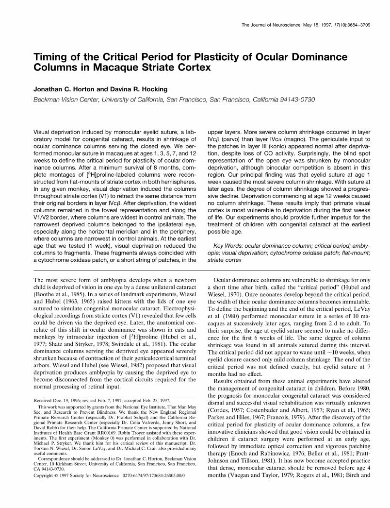
Figure 1. Monkey 0 (1 week suture). A, Autoradiograph of a single section passing through layer III in the medial half of the right operculum. Theradioactive tracer appears bright in dark-field illumination. The section just grazed the honeycomb in layer IVa at its deepest point (curved arrow). Parallelrows of patches were labeled by saturating the lateral geniculate body with a huge injection of [3H]proline. The arrows indicate five rows of patches servingthe normal left eye. Blood vessels used for section alignment are marked with arrowheads. B, Adjacent section showing the array of CO patches. Thestaining was poor, but one can detect alternating light and dark rows. The arrows mark five dark rows (left eye) that match the arrows in A. Comparisonof A and B indicated that every row of CO patches was well labeled by [3H]proline; deprivation of the right eye caused no obvious attenuation of thekoniocellular projection to its rows of CO patches. Scale bar, 2 mm.
3686 J. Neurosci., May 15, 1997, 17(10):3684–3709 Horton and Hocking • Timing of Eye Dominance Column Plasticity
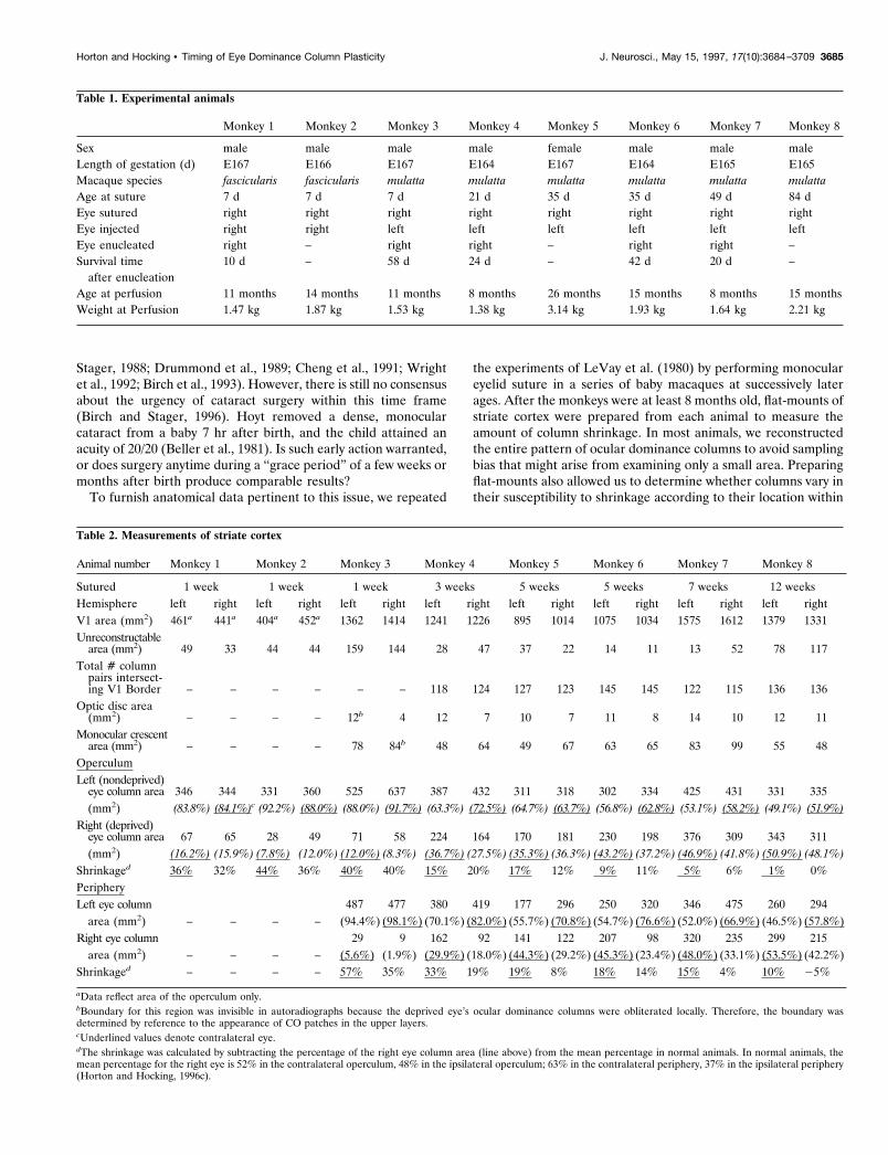
Figure 2. Monkey 1 (1 week suture). A, B, Montages of autoradiographs showing severe shrinkage of ocular dominance columns in layer IVcb. Thedeprived right eye was injected with [3H]proline at age 11 months and enucleated 1 week later. Tissues were processed after an additional 10 d survivalperiod. The region within the box is shown in Figure 3A at higher power. C, D, Montages of adjacent CO sections, showing a labeling pattern in layerIVcb matching the autoradiographs above. The boxed region in C is shown in Figure 3B. E, F, Drawing of the autoradiographs in A and B showing howthe deprived columns become gradually more fragmented moving from the fovea to the periphery (curved arrows) and from the vertical meridian to thehorizontal meridian (straight arrows). The latter effect is less striking. See Figure 7 for the retinotopic coordinates of V1. Areas shaded with gray couldnot be reconstructed.
Horton and Hocking • Timing of Eye Dominance Column Plasticity J. Neurosci., May 15, 1997, 17(10):3684–3709 3687
briefly and then resutured. Survival times after eye injection were 7–9 d,except for Monkey 1, whose survival time was 17 d.
In five monkeys, the ocular dominance columns were double-labeled byenucleating the amblyopic right eye and staining the cortex for CO (Table1). Enucleation was performed using sterile technique under anesthesiawith ketamine HCl (20 mg/kg, i.m.) and a retrobulbar injection of 2 ml of1% lidocaine plus epinephrine (1:100,000). After surgery, buprenorphineHCl (0.02 mg/kg, i.m.) was administered every 8 hr for 2 d to ensureanalgesia. Survival times ranged between 10 and 58 d (Table 1). Enucle-ation was done no earlier than age 7 months (well after the criticalperiod) so that it would have no confounding effect on the width of theocular dominance columns (LeVay et al., 1980).
We enucleated one eye to have CO available as a “back-up” label incase the autoradiography failed. In the end, the autoradiography suc-ceeded in each animal, so it was not necessary to reconstruct the oculardominance columns from the CO sections. However, for the sake ofcomparison we reconstructed the ocular dominance columns using bothmethods in Monkey 1.
Histological procedures. Before perfusion, the eye injected with[3H]proline was examined again under ketamine anesthesia (20 mg/kg,i.m.) with an indirect ophthalmoscope to verify that no injury had resultedfrom the tracer injection. In every case, the injected eye appeared
undamaged (in Monkey 1, this procedure was done before enucleation ofthe right eye, rather than before perfusion). After receiving a lethalinjection of sodium pentobarbital into the peritoneal cavity, each monkeywas perfused through the left ventricle with 1 l of normal saline followedby 1 l of 2% paraformaldehyde in 0.1 M phosphate buffer. Striate cortexfrom each occipital lobe was unfolded, flattened, and sectioned at 30 mm.Alternate sections were reacted for CO (Wong-Riley, 1979) or coatedwith emulsion for autoradiography (Wiesel et al., 1974). Our methods forpreparing flat-mounts and processing sections have been described indetail previously (Horton and Hocking, 1996b,c).
Data analysis. The autoradiographs were photographed in dark-fieldillumination using Technical Pan film (Eastman Kodak, Rochester, NY)and developed with Technidol developer (Eastman Kodak). CO sectionswere photographed in bright-field using Plus-X Pan film (EastmanKodak) and developed with Microdol-X (Eastman Kodak). Negativeswere scanned into the computer and montages were prepared usingPhotoshop 3.0 (Adobe Systems) (Horton and Hocking, 1996b,c). Toprepare black and white drawings, we created a transparent layer over-lying the completed montage, traced the borders of the dark columns athigh magnification on the monitor using the “pencil” function at a 1 pixelsetting, and then flood-filled them with black. For quantification ofcolumn areas, the drawings were analyzed with Mocha (Jandel Scientific,
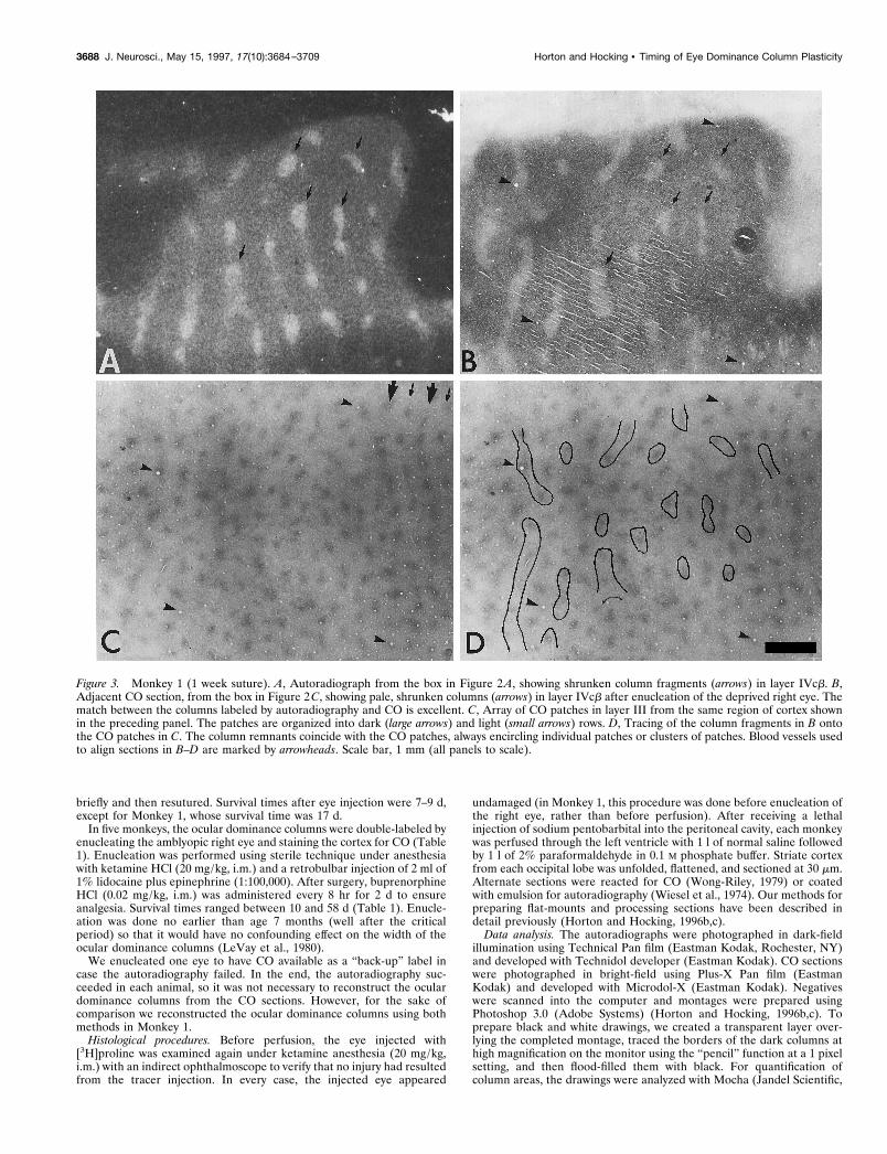
Figure 3. Monkey 1 (1 week suture). A, Autoradiograph from the box in Figure 2 A, showing shrunken column fragments (arrows) in layer IVcb. B,Adjacent CO section, from the box in Figure 2C, showing pale, shrunken columns (arrows) in layer IVcb after enucleation of the deprived right eye. Thematch between the columns labeled by autoradiography and CO is excellent. C, Array of CO patches in layer III from the same region of cortex shownin the preceding panel. The patches are organized into dark (large arrows) and light (small arrows) rows. D, Tracing of the column fragments in B ontothe CO patches in C. The column remnants coincide with the CO patches, always encircling individual patches or clusters of patches. Blood vessels usedto align sections in B–D are marked by arrowheads. Scale bar, 1 mm (all panels to scale).
3688 J. Neurosci., May 15, 1997, 17(10):3684–3709 Horton and Hocking • Timing of Eye Dominance Column Plasticity
San Rafael, CA). This program uses a thresholding function to assignpixels to black (level of gray 5 1) or white (level of gray 5 256). Pixelswere converted to mm2 by calibration with the scale bar. Intermediatelevels of gray were used to measure the unreconstructable areas, blindspots, and monocular crescents. To facilitate comparison between exper-iments, all montages in this paper were reproduced at identical scale.
RESULTSMonkey 0 (deprived at age 1 week)The goal of this experiment was to fill the thalamic projection tothe CO patches in the upper layers of striate cortex by depositinga massive quantity (0.5 mCi) of [3H]proline into the middle of theright lateral geniculate body. After injection, layer IVc wasdensely labeled throughout the entire flat, exposed lateral con-vexity of the right occipital lobe. This region, called the opercu-lum, represents the central 0°–8° of the contralateral visual hemi-field (see Fig. 7 for retinotopic coordinates of V1). The upperlayers were labeled only in the medial half of the operculum, from;2.5°–8° (Fig. 1A). Autoradiographs revealed rows of patches inlayer III, merging together like pearls on a string. An adjacent COsection showed alternating rows of light and dark patches (Fig.1B), as reported previously after visual deprivation at age 1 week(Horton and Stryker, 1993a). The adjacent sections matched:every row of CO patches coincided with a row of proline label.Despite shrinkage of the right eye’s ocular dominance columns inlayer IVc (which was not well seen, because the geniculocorticalafferents serving both eyes were flooded with [3H]proline), theprojection to the upper layers seemed normal. In fact, withoutreferring to the adjacent CO section, we could not tell which rowsof proline patches belonged to the deprived eye. This resultimplied that the koniocellular projection to the upper layers wasless susceptible to visual deprivation than the parvocellular pro-jection to layer IVcb.
Monkey 1 (deprived at age 1 week)This animal received an injection of [3H]proline into the deprivedright eye. Autoradiographs showed severe shrinkage of the oculardominance columns on the opercula (Fig. 2A,B). Only 16% oflayer IVcb in each hemisphere was occupied by geniculate affer-ents serving the amblyopic right eye (Table 2). The pattern ofcolumn shrinkage showed two gradients: the columns becamesmaller and more fragmented moving (1) from the fovea to theperiphery and (2) from the vertical meridian to the horizontalmeridian. Together, these two gradients reduced the deprivedeye’s columns to scattered islands along the horizontal meridian at8°. By contrast, the deprived eye’s columns remained relativelyintact within the foveal representation (Fig. 2E,F).
In this monkey, we double-labeled the ocular dominance col-umns with CO by enucleating the amblyopic right eye 1 week afterinjecting it with [3H]proline. After waiting an additional 10 d, toallow cortical CO levels to drop after enucleation, we preparedalternate sections for autoradiography and CO (compare Fig.2A,B with 2C,D). In Figure 3, A and B, a small region of layerIVcb from an autoradiograph and an adjacent CO section isshown at higher power. The two labels appeared nearly identical,but small differences were evident on close scrutiny. We attributedthese subtle discrepancies to the fact that the sections wereadjacent, not the same, and therefore passed through layer IVcbat slightly different depths. In addition, the columns were labeledmore intensely by [3H]proline than CO in this animal, causing theboundaries of the proline columns to appear slightly expanded.The match between the CO columns and the proline columnsconfirmed a previous study showing that CO accurately labels the
ocular dominance columns in deprived animals (Horton andStryker, 1993a).
In Monkey 1, CO staining showed alternating rows of light anddark patches in the upper layers. The shrunken fragments ofocular dominance columns in layer IVcb (Fig. 3B) were tracedonto the array of CO patches (Fig. 3C) to test their relationship(Fig. 3D). The remnants of ocular dominance columns werealways centered on CO patches. For example, where the oculardominance columns were reduced to small islands, they encircleda single CO patch. Where longer column fragments survived, theyencompassed a short string of CO patches. In these cases, theboundary of the shrunken column never ended on a CO patch but,instead, passed between two adjacent patches. These findingswere verified by examining three regions at high power from eachoperculum, containing a total of 78 column fragments in layerIVcb.
Monkey 2 (deprived at age 1 week)The opercula of this animal showed severe shrinkage of thedeprived right eye’s columns in layer IVcb (Fig. 4A,B,E,F). Theeffect was even more striking than in Monkey 1 (compare Figs.2E,F and 4E,F). This difference illustrates that two animalsdeprived in an identical manner can vary in their degree ofcolumn shrinkage. In Monkey 2, unlike Monkey 1, the deprivationeffect was asymmetric (compare Fig. 4A,B). The right eye’s col-umns occupied 12% of the ipsilateral operculum, but only 8% ofthe contralateral operculum (Table 2).
It is difficult to compare [3H]proline columns in different sub-layers of IVc, because they are more crisp and intense in layerIVcb than in layer IVca (see Fig. 30 in Horton, 1984). Nonethe-less, we observed an obvious difference in the shrinkage of layersIVca and IVcb (compare Fig. 4A,C). Rather than breaking intofragments, the columns in layer IVca appeared relatively intact,preserving their original stripe-like configuration. In all animalssutured at age 5 weeks or younger, there was less column shrink-age in layer IVca than in layer IVcb. This finding implies that themagnocellular projection (layer IVca) is less vulnerable than theparvocellular projection (layer IVcb) to the effects of visualdeprivation.
In layer IVc, the CO activity was homogeneous (Horton andStryker, 1993a). In layers II and III, there were alternating darkand pale rows of CO patches (Fig. 4D). We examined the rela-tionship between the shrunken ocular dominance columns in layerIVcb and the CO patches in layer III, seeking to corroborate ourfindings in Monkey 1. Figure 5A shows an autoradiograph fromthe right operculum of Monkey 2. Approximately 10 shrunkenislands were labeled in layer IVcb. The perimeter of each columnfragment encircled a single CO patch, or a cluster of patches, inthe upper layers (Fig. 5B,C). This finding was confirmed bytesting three regions from each operculum containing a total of 62column fragments.
In Monkey 2, the shrinkage of ocular dominance columns wasso severe that many CO patches in deprived rows came to besituated over regions of layer IVcb taken over by the normal eye.These patches were not spared the loss of CO activity suffered byfellow patches remaining in territory held by the deprived eye: COactivity was reduced in all patches located within a deprived row(Figs. 4D, 5B). This finding suggests that, although layer IVcb wastaken over by the open eye, functional connections serving theopen eye were not established to the deprived eye’s patches,leaving them with reduced metabolic activity.
In Monkeys 1 and 2, the ocular dominance columns of the
Horton and Hocking • Timing of Eye Dominance Column Plasticity J. Neurosci., May 15, 1997, 17(10):3684–3709 3689
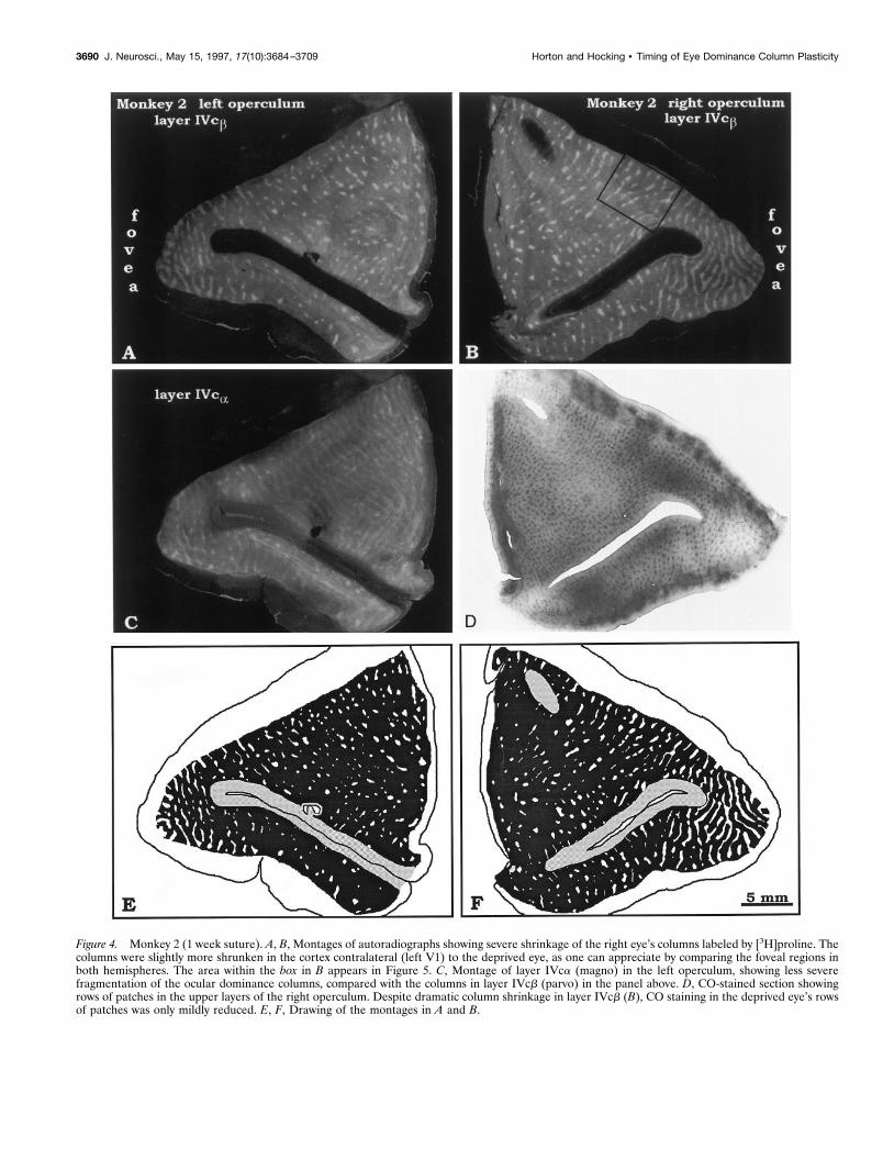
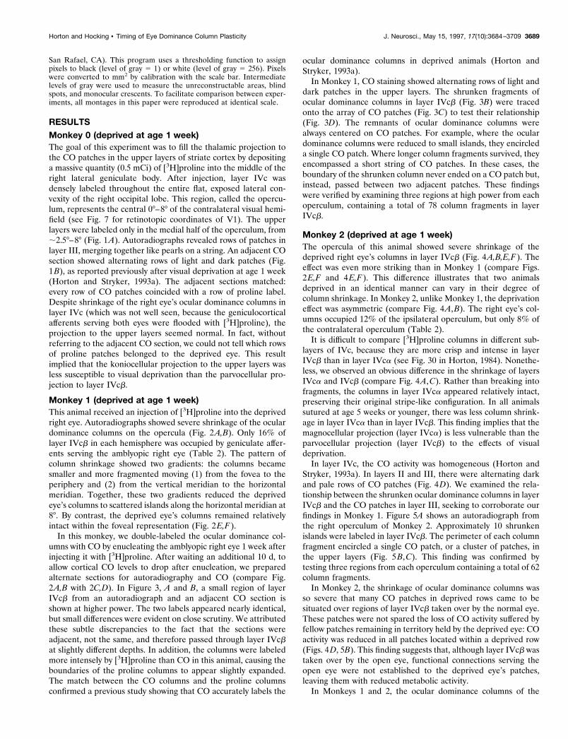
Figure 4. Monkey 2 (1 week suture). A, B, Montages of autoradiographs showing severe shrinkage of the right eye’s columns labeled by [3H]proline. Thecolumns were slightly more shrunken in the cortex contralateral (left V1) to the deprived eye, as one can appreciate by comparing the foveal regions inboth hemispheres. The area within the box in B appears in Figure 5. C, Montage of layer IVca (magno) in the left operculum, showing less severefragmentation of the ocular dominance columns, compared with the columns in layer IVcb (parvo) in the panel above. D, CO-stained section showingrows of patches in the upper layers of the right operculum. Despite dramatic column shrinkage in layer IVcb (B), CO staining in the deprived eye’s rowsof patches was only mildly reduced. E, F, Drawing of the montages in A and B.
3690 J. Neurosci., May 15, 1997, 17(10):3684–3709 Horton and Hocking • Timing of Eye Dominance Column Plasticity

deprived eye were best preserved in the foveal representation.This finding, also observed in a third monkey (Horton andStryker, 1993a), led us to suggest in a preliminary report thatocular dominance columns within the foveal representation areless susceptible to shrinkage induced by early monocular depriva-tion (Horton and Stryker, 1993b). However, our view has changedafter examining peripheral regions of striate cortex hidden withinthe calcarine fissure. Figure 6A shows layer IVcb from the pe-ripheral left cortex of Monkey 2, contralateral to the deprived eye.
Figure 5. Monkey 2 (1 week suture). A, Single autoradiograph from theregion enclosed by the box in Figure 4 B, showing fragments of deprivedright eye columns. B, CO section from the corresponding area in Figure4D, showing the patches in the upper layers. C, Tracing of the columnfragments in A onto the CO patches in B, demonstrating that columnremnants in layer IVcb always encircle a CO patch or a short string of COpatches. Note that the CO patches circled in this figure are no more palethan other CO patches in deprived rows. Blood vessels used for sectionalignment are marked by arrowheads. Scale bar, 1 mm for all three panels.
Figure 6. Monkey 2 (1 week suture). A, View of [3H]proline-labeledcolumns in the left peripheral cortex after injection of the deprived righteye. The columns are shrunken, but no more than on the operculum(compare with Fig. 4A). The labeled columns are more attenuated alongthe vertical meridian than the horizontal meridian. For retinotopic coor-dinates and orientation of this tissue segment within V1, see Figure 7. MC,Monocular crescent. B, Section from the upper layers of the same tissueblock, showing the CO patches. C, Montage of the koniocellular projec-tion to the CO patches in layer III, labeled by [3H]proline injection intothe deprived right eye. At higher power, we verified that every CO patchin rows corresponding to the deprived columns in layer IVcb received apuff of [3H]proline label. Curiously, no puffs of proline label were seen inthe monocular crescent representation, indicating that CO patches in thisarea receive no (or only a weak) thalamic projection.
Horton and Hocking • Timing of Eye Dominance Column Plasticity J. Neurosci., May 15, 1997, 17(10):3684–3709 3691
The location of this tissue segment within striate cortex is shownschematically in Figure 7 (left). The labeled columns of the de-prived right eye were shrunken, but they still occupied 19% oflayer IVcb. They were no more shrunken than the columnslocated within the foveal representation (compare Figs. 6A and4A). This finding invalidated our original notion, namely, thatcolumn shrinkage becomes increasingly severe across the cortexfrom the representation of the fovea to the periphery.
Figure 6B shows the CO patches in the upper layers from thesame block. In the monocular crescent, the CO patches werelarger and more widely spaced (Horton, 1984). A montage ofautoradiographs through layer III showed the projection to thepatches of the injected right eye (Fig. 6C). Despite visual depri-vation, all of the right eye’s patches were well labeled, except inthe monocular crescent. The absence of patch labeling in themonocular crescent was unexpected. We have since reviewedautoradiographs from previous experiments in four normal babymacaques that showed strong patch labeling after [3H]proline eyeinjection (Horton and Hocking, 1996a). In every animal, label inpatches within the contralateral monocular crescent and the ipsi-lateral blind spot was absent or was much weaker than in nearbybinocular cortex. We have no explanation for this observation.
For comparison, Figure 8 shows the matching piece of V1 fromthe right peripheral cortex, ipsilateral to the deprived eye (see Fig.7 for orientation). In layer IVcb, the ocular dominance columns ofthe right eye were obliterated, except for a few islands near thevertical meridian (Fig. 8A). The deprivation effect was morestriking than in the left peripheral cortex (compare Figs. 8A and6A). It was also more striking in layer IVcb than IVca (compareFig. 8A and 8B), confirming our observation from the operculumof the same animal (Fig. 4C). In the upper layers, the projectionto the right eye’s rows of patches (Fig. 8C) appeared intact. Thelabeling was indistinguishable from the pattern seen in normalmacaques (see Fig. 5 in Horton and Hocking, 1996a). This resultconfirmed our findings in Monkey 0 (Fig. 1). Because the righteye’s afferents to layer IVcb were decimated, preserved input toright eye patches was often situated over regions of layer IVcbsurrendered to the left eye. This contrast in the laminar pattern oflabeling within the same tissue block provided the most directevidence that visual deprivation had affected the parvocellularprojection more than the koniocellular projection.
Monkey 3 (deprived at age 1 week)V1 was flat-mounted as a single piece of tissue to compare columnshrinkage in the operculum and periphery in the same sections.The normal left eye was injected with [3H]proline, resulting inexpanded columns of label interrupted by dark gaps belonging tothe amblyopic right eye (Fig. 9). The labeling in the opercula wascomplementary to the pattern illustrated in Monkeys 1 and 2.Again, a gradient in apparent column shrinkage occurred fromthe fovea to the periphery and from the vertical meridian to thehorizontal meridian. The right eye’s columns were slightly moreshrunken in the ipsilateral cortex (8% of surface area) than thecontralateral cortex (12% of surface area; Table 2). Overall, thedegree of column shrinkage in Monkeys 1, 2, and 3 wascomparable.
Although the right and left opercula looked quite similar inMonkey 3, in the periphery the pattern of labeling was quitedifferent on the two sides. In the right hemisphere, the right eye’scolumns were nearly absent in the periphery, except near thevertical meridian (Fig. 9B,D). By contrast, in the left hemispherethe right eye’s columns experienced a renaissance as they ap-proached the monocular crescent (Figs. 9A,C). They were alsolarger near the horizontal meridian than the vertical meridian (theopposite of the pattern in the right hemisphere). These findingsconfirmed observations made in the peripheral fragments fromMonkey 2 (Figs. 6, 8). They also confirmed a report by Hubel etal. (1977) that in peripheral calcarine cortex the ocular dominancecolumns appear more shrunken ipsilateral to the sutured eye (seetheir Fig. 21A,B).
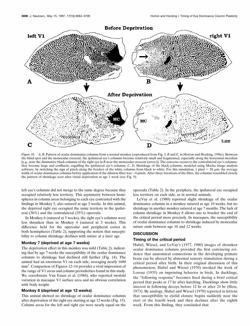
We believe the key to this ipsi /contra discrepancy in columnshrinkage lies with an observation by LeVay et al. (1985). Theydiscovered that the columns of the ipsilateral eye become frag-mented between the representation of the blind spot and themonocular crescent in peripheral cortex, especially along thehorizontal meridian. We have confirmed this finding (Fig. 10A,B).In a complementary manner, the columns of the contralateral eyebecome larger and more confluent in this region. To determinehow this asymmetry might be reflected in the appearance of thecolumns after visual deprivation, we simulated column shrinkageusing a binary filter provided with the Mocha image analysissoftware (Jandel Scientific). This filter dilates a white object byconverting a monolayer of pixels surrounding it from black to
Figure 7. Monkey 2. Schematic drawing showing the retinotopic coordinates in unfolded, flattened macaque V1, to aid in interpretation of our flat-mountmontages. The shaded area in the lateral portion of each cortex is the exposed, flat operculum of the occipital lobe, which represents the central ;0°–8°(Fig. 4A,B). The shaded areas in the upper, medial portion of the left and right V1 are illustrated in Figures 6 and 8, respectively. BS, Blind spot; MC,monocular crescent.
3692 J. Neurosci., May 15, 1997, 17(10):3684–3709 Horton and Hocking • Timing of Eye Dominance Column Plasticity
white. After applying three iterations of this filter to a normalmosaic of columns, the resultant pattern bore a close resemblanceto the columns in our monkeys deprived at age 1 week (compareFigs. 9C,D and 10C,D). In the peripheral right cortex, the righteye’s columns (which were small to begin with) were extinguished,except along the vertical meridian. In the peripheral left cortex,the right eye’s columns (which were big to begin with) fared muchbetter, especially along the horizontal meridian.
The correlation between the computer model of column shrink-age and our anatomical data suggests that visual deprivationinduces geniculocortical afferents to retract the same distancefrom the borders of the ocular dominance columns throughoutprimary visual cortex. The impression of greater shrinkage in theperipheral cortex ipsilateral to the deprived eye arises becausethere exists a normal, preexisting inequality in column widths inthis region. There is no need to invoke a greater sensitivity of theipsilateral eye to the effects of visual deprivation to explain why itscolumns appear smaller in the periphery after monocular suture.
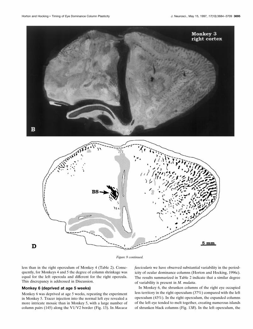
In monkey 3, the blind spot representation of the left eye in theright visual cortex was shrunken (Fig. 9B,D). It measured only 4mm2, compared with an average value of 10 mm2 in normalmacaques (Horton and Hocking, 1996c). In this special region,geniculate afferents serving the right eye face no direct competi-tion from the left eye because the left eye contributes no input.We expected this lack of competition to protect the blind spotrepresentation from shrinkage. Nevertheless, it appears that theleft eye’s afferents, presumably serving retina surrounding theoptic disk, invaded the disk’s cortical representation. This puzzlingresult will be considered further in Discussion.
Monkey 4 (deprived at age 3 weeks)Figure 11 shows flat-mounts prepared from Monkey 4, after[3H]proline injection into the the normal left eye. The intensity ofautoradiographic labeling was greater in peripheral calcarine cor-tex than on the operculum (Kaas et al., 1976; Hendrickson et al.,1978; LeVay et al., 1978; Spatz, 1979). This tendency was observedin all of the flat-mounts, although it was more striking in somethan in others. The reason is unclear, but it may reflect bettertracer uptake by ganglion cells in peripheral retina. In addition,the contrast of the autoradiographic label was greater in thecortex ipsilateral to the injected eye in all animals (Shatz et al.,1977). In cats, this effect has been attributed to greater tracerspillover in the lateral geniculate body on the contralateral side(LeVay et al., 1978). However, in macaques it may indicate morecomplete segregation of the geniculate afferents serving the ipsi-lateral eye (Horton and Hocking, 1996a).
Eyelid suture at 3 weeks in Monkey 4 caused much less shrink-age of ocular dominance columns than eyelid suture at age 1 weekin Monkeys 1, 2, and 3 (Table 2). On the opercula, the deprivedcolumns remained coherent stripes rather than breaking intoisolated fragments. The deprived right eye’s columns occupied 9%less surface area on the ipsilateral operculum compared with thecontralateral operculum. This imbalance was also marked in pe-ripheral cortex. On both opercula, the deprived columns wereslightly wider in the foveal region and along the V1/V2 border,echoing the tendency seen in Monkeys 1, 2, and 3. The blind spotwas also shrunken in the ipsilateral cortex (Fig. 11B), replicatingthe finding in the ipsilateral cortex of Monkey 3.
Monkey 5 (deprived at age 5 weeks)This monkey was the oldest and largest of our cohort but, ironi-cally, it had the smallest V1 (Table 2). On the left side, thecalcarine fissure did not unfold completely, leaving ;100 mm2
buried (Fig. 12). However, on the right side we obtained a com-plete reconstruction, and the surface area of striate cortex wasbarely 1000 mm2. The shrunken columns of the deprived right eyewere unlabeled, because [3H]proline was injected into the normalleft eye. In the left operculum, the column shrinkage was equal tothe degree seen in the left operculum of Monkey 4, sutured at 3weeks (compare Figs. 12A and 11A). However, in the left periph-
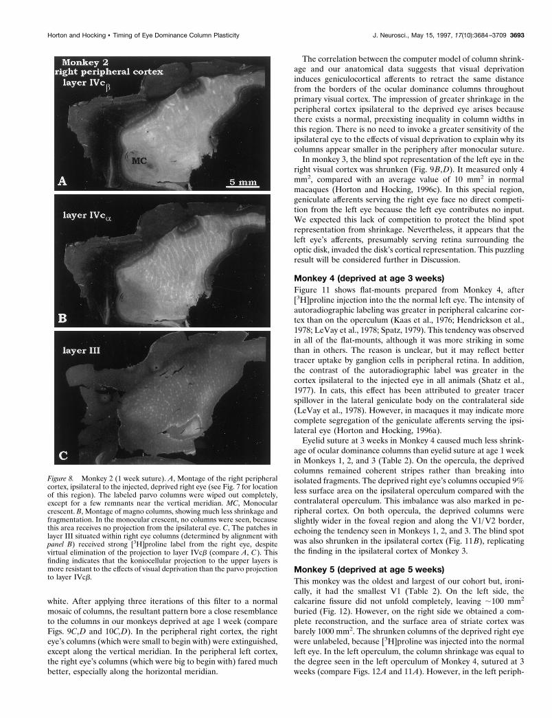
Figure 8. Monkey 2 (1 week suture). A, Montage of the right peripheralcortex, ipsilateral to the injected, deprived right eye (see Fig. 7 for locationof this region). The labeled parvo columns were wiped out completely,except for a few remnants near the vertical meridian. MC, Monocularcrescent. B, Montage of magno columns, showing much less shrinkage andfragmentation. In the monocular crescent, no columns were seen, becausethis area receives no projection from the ipsilateral eye. C, The patches inlayer III situated within right eye columns (determined by alignment withpanel B) received strong [3H]proline label from the right eye, despitevirtual elimination of the projection to layer IVcb (compare A, C). Thisfinding indicates that the koniocellular projection to the upper layers ismore resistant to the effects of visual deprivation than the parvo projectionto layer IVcb.
Horton and Hocking • Timing of Eye Dominance Column Plasticity J. Neurosci., May 15, 1997, 17(10):3684–3709 3693

eral cortex the column shrinkage in Monkey 5 was less severe thanin the corresponding region of Monkey 4. This point can beappreciated by observing that more black, unlabeled territory ispresent between the blind spot and monocular crescent in Figure
12A than in Figure 11A. Comparison of Monkeys 4 and 5 illus-trates that the relative column shrinkage in central versus periph-eral cortex can vary from animal to animal.
In the right operculum of Monkey 5, the column shrinkage was
Figure 9. Monkey 3 (1 week suture). A, B, Autoradiographic montage of layer IVcb after deprivation of the right eye. The normal left eye was injectedwith [3H]proline. The deprivation effect was severe and complemented the findings obtained in Monkeys 1 and 2 after injection the deprived eye. C, D,Drawing of the columns in A and B. This cortex was unusually large and did not flatten well. Despite the blemishes, one can appreciate that moreunlabeled (right eye) columns survived in the left periphery than in the right periphery. The border of the monocular crescent could not be discerned inthe right cortex, because the ocular dominance columns of the right eye were extinguished completely in the periphery. In the left cortex, the boundaryof the blind spot representation of the right eye was uncertain, for the same reason. In the right cortex, the blind spot (BS) representation of the left eyewas shrunken. MC, Monocular crescent. Figure continues.
3694 J. Neurosci., May 15, 1997, 17(10):3684–3709 Horton and Hocking • Timing of Eye Dominance Column Plasticity
less than in the right operculum of Monkey 4 (Table 2). Conse-quently, for Monkeys 4 and 5 the degree of column shrinkage wasequal for the left opercula and different for the right opercula.This discrepancy is addressed in Discussion.
Monkey 6 (deprived at age 5 weeks)Monkey 6 was deprived at age 5 weeks, repeating the experimentin Monkey 5. Tracer injection into the normal left eye revealed amore intricate mosaic than in Monkey 5, with a large number ofcolumn pairs (145) along the V1/V2 border (Fig. 13). In Macaca
fascicularis we have observed substantial variability in the period-icity of ocular dominance columns (Horton and Hocking, 1996c).The results summarized in Table 2 indicate that a similar degreeof variability is present in M. mulatta.
In Monkey 6, the shrunken columns of the right eye occupiedless territory in the right operculum (37%) compared with the leftoperculum (43%). In the right operculum, the expanded columnsof the left eye tended to melt together, creating numerous islandsof shrunken black columns (Fig. 13B). In the left operculum, the
Figure 9 continued.
Horton and Hocking • Timing of Eye Dominance Column Plasticity J. Neurosci., May 15, 1997, 17(10):3684–3709 3695
left eye’s columns did not merge to the same degree because theyoccupied relatively less territory. This asymmetry between hemi-spheres in column areas belonging to each eye contrasted with thefindings in Monkey 5, also sutured at age 5 weeks. In this animal,the deprived right eye occupied the same territory in the ipsilat-eral (36%) and the contralateral (35%) opercula.
In Monkey 6 (sutured at 5 weeks), the right eye’s columns wereless shrunken than in Monkey 4 (sutured at 3 weeks). Thisdifference held for the opercular and peripheral cortex inboth hemispheres (Table 2), supporting the notion that suscepti-bility to column shrinkage declines with suture at a later age.
Monkey 7 (deprived at age 7 weeks)The deprivation effect in this monkey was mild (Table 2), indicat-ing that by age 7 weeks the susceptibility of the ocular dominancecolumns to shrinkage had declined still further (Fig. 14). Theanimal had an enormous V1 on each side, averaging nearly 1600mm2. Comparison of Figures 12–14 provides a vivid impression ofthe range of V1 areas and column periodicities found in this study.We corroborate Van Essen et al. (1984), who reported twofoldvariation in macaque V1 surface area and no obvious correlationwith body weight.
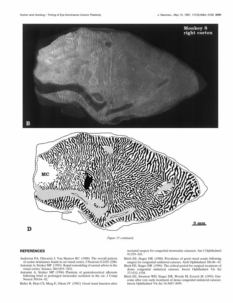
Monkey 8 (deprived at age 12 weeks)This animal showed no shrinkage of ocular dominance columnsafter deprivation of the right eye starting at age 12 weeks (Fig. 15).Column areas for the left and right eye were nearly equal on the
opercula (Table 2). In the periphery, the ipsilateral eye occupiedless territory on each side, as in normal animals.
LeVay et al. (1980) reported slight shrinkage of the oculardominance columns in a monkey sutured at age 10 weeks, but noshrinkage in another monkey sutured at age 7 months. The lack ofcolumn shrinkage in Monkey 8 allows one to bracket the end ofthe critical period more precisely. In macaques, the susceptibilityof ocular dominance columns to shrinkage induced by monocularsuture ends between age 10 and 12 weeks.
DISCUSSIONTiming of the critical periodHubel, Wiesel, and LeVay’s (1977, 1980) images of shrunkenocular dominance columns provided the first convincing evi-dence that anatomical connections in the developing primatebrain can be altered by abnormal sensory stimulation during acritical period after birth. In their original discussion of thisphenomenon, Hubel and Wiesel (1970) invoked the work ofLorenz (1935) on imprinting behavior in birds. In ducklings,the “following response” becomes fixed during a brief criticalperiod that peaks at 17 hr after hatching. Ducklings show littleinterest in following decoys before 12 hr or after 24 hr (Hess,1973). By analogy, Hubel and Wiesel (1970) reported in kittensthat susceptibility to eyelid closure begins suddenly near thestart of the fourth week and then declines after the eighthweek. From this finding, they concluded that
Figure 10. A, B, Pattern of ocular dominance columns from a normal monkey (reproduced from Fig. 3, B and E, in Horton and Hocking, 1996c). Betweenthe blind spot and the monocular crescent, the ipsilateral eye’s columns become relatively small and fragmented, especially along the horizontal meridian[e.g., note the diminutive black columns of the right eye in B near the monocular crescent (arrow)]. The converse occurs to the contralateral eye’s columns:they become large and confluent, engulfing the ipsilateral eye’s columns. C, D, Shrinkage of the black columns, modeled using Mocha image analysissoftware, by switching the sign of pixels along the borders of the white columns from black to white. For this simulation, 1 pixel 5 58 mm; the averagewidth of ocular dominance columns before application of the dilation filter was ;8 pixels. After three iterations of the filter, the columns resembled closelythe pattern of shrinkage seen after visual deprivation at age 1 week (see Fig. 9).
3696 J. Neurosci., May 15, 1997, 17(10):3684–3709 Horton and Hocking • Timing of Eye Dominance Column Plasticity
the kitten’s visual system is too immature for monocular de-privation to have much effect during the first weeks of life. Insupport of this notion, Olson and Freeman (1980) found nochange in the ocular dominance profiles of cortical neurons inkittens sutured from day 8 to day 19. Thereafter, just a few daysof monocular deprivation were sufficient to produce a majorshift in ocular preference.
In an initial report concerning macaques, Hubel et al. (1977)suggested that column shrinkage was more severe after lid sutureat age 2 weeks compared with 3 weeks. In their definitive study,LeVay et al. (1980) later retracted this observation by concluding:“deprivation begun at any age from birth to about 6 weeks hadapproximately the same effect.” This result might be explained ifmacaques, like cats, were relatively immune to the effects ofmonocular deprivation for the first few weeks after birth. How-ever, LeVay and coworkers found severe column shrinkage in amonkey sutured at 2 d and examined at 24 d, nullifying thisinterpretation.
Repeating the experiments by LeVay and coworkers, we havefound greater shrinkage and fragmentation of ocular dominancecolumns with lid suture starting at age 1 week compared with laterdates. For example, the column shrinkage after suture at age 1week was approximately twice as severe as the shrinkage aftersuture at age 5 weeks (Table 2). From these data, we concludethat macaques are highly sensitive to the effects of visual depri-vation within a week of birth (and probably even sooner). Unlikekittens, there is no significant delay after birth in the onset of thecritical period.
In making these new observations, we have benefited frominformation and methods not available to Hubel et al. when theyperformed their classic experiments. As LeVay et al. (1985) dis-covered only later, the ocular dominance columns in normalanimals show marked regional variation in width (e.g., the ipsi-lateral columns become broken and narrowed in the periphery).To make valid comparisons, it is crucial to reconstruct wideexpanses of cortex to obtain a coherent view of the shrunkencolumns or at least to survey the same region in every animal.LeVay et al. (1980) examined small fragments of tissue, averagingonly ;5% of the total V1 surface area, from different regions ineach animal. Improved methods for flat-mounting the cortex andfor preparing computer montages have made it easier to gaugethe effects of visual deprivation more accurately (Horton, 1984;Olavarria and Van Sluyters, 1985; Tootell and Silverman, 1985;Anderson et al., 1988; Rosa et al., 1988; Florence and Kaas,1992; Hata and Stryker, 1994; Horton and Hocking, 1996c).
Gradients in column shrinkagePrevious investigators have reported greater shrinkage of ocu-lar dominance columns ipsilateral to the sutured eye (Hubel etal., 1977; LeVay et al., 1980) or in peripheral cortex as opposedto opercular cortex (Swindale et al., 1981). We confirm thesetendencies but attribute them to preexisting inequalities incolumn widths present in normal mosaics before the onset ofvisual deprivation. For example, the smallest columns are lo-cated in the peripheral cortex, serving the ipsilateral eye. Aftermonocular deprivation, these columns become the most se-verely reduced in size. Visual deprivation seems to subtract thesame amount of territory from all of the columns throughoutany given mosaic, at least to a first approximation. This impres-sion was supported by simulation of column shrinkage using thedilation filter available with the Mocha image analysis program.Stripping pixels evenly from one set of columns and adding
them to the other set created a mosaic of artificially shrunkencolumns that resembled closely those from animals deprived bylid suture (compare Figs. 9 and 10).
The most direct way to prove that visual deprivation reducescolumns everywhere by the same amount would be to comparemosaics in the same animal, before and after visual deprivation.This is impossible, except with optical imaging. Unfortunately, thistechnique cannot image buried cortex and it renders less distinctcolumns than [3H]proline autoradiography. Until a better methodbecomes available, we shall be forced to rely on comparisonsbetween different animals—normal and deprived. This approachis dogged by marked intrinsic variability from one animal to thenext. A few examples suffice to illustrate how this variabilitycomplicated the interpretation of our data. In normal macaques,we reported recently that ocular dominance columns are wider inthe foveal representation and along the V1/V2 border (Hortonand Hocking, 1996c). In our mosaics, this tendency accounts forthe wider appearance of the columns in these regions after mo-nocular deprivation (e.g., see Figs. 2, 4, 9, 11–15). However,enlargement of the columns in the foveal region and along theV1/V2 border is quite variable in normal animals and in deprivedanimals. This variation makes it difficult to exclude the possibilitythat we suggested originally, namely, that columns in these regionsare less susceptible to shrinkage from monocular deprivation(Horton and Stryker, 1993b).
The second example concerns the degree of column shrinkageipsilateral versus contralateral to the deprived eye. In binocularV1 of normal macaques, the ipsilateral eye’s columns occupy lessterritory overall (43.2 6 SD, 2.4%, n 5 12) than the contralateraleye’s columns (56.8%; calculated from Table 2 in Horton andHocking, 1996c). Obviously, if the ipsilateral eye’s columns occupyless area in normal animals, they will appear more shrunken indeprived animals. The predominance of the contralateral eye ismore pronounced in peripheral cortex (mean ratio 63:37) than inopercular cortex (mean ratio 52:48) (Horton and Hocking, 1996c).In normal animals, there is a surprising range in the contralateraleye/ipsilateral eye ratio of opercular column areas, from 54:46 to48:52. In peripheral cortex, this variation is even greater. The ratiocan also differ between hemispheres in the same animal. Thisfluctuation is sufficient to account for the wide range that wemeasured in the ratio of contralateral eye/ipsilateral eye columnareas (Table 2) and for the fact that at least one animal had moreshrunken columns in the operculum contralateral to the deprivedeye (e.g., Monkey 2, Fig. 4).
Finally, there is real variability in column shrinkage from ani-mal to animal. Monkey 1 had less shrinkage than Monkey 2,despite suture at the same age. Monkey 4 (sutured at 3 weeks) hadmore column shrinkage in the right operculum than Monkey 5(sutured at 5 weeks), but the same shrinkage in the left opercu-lum. This variability can mislead, and it undoubtedly helpedpersuade LeVay et al. (1980) that suture at different dates resultsin the same degree of column shrinkage. In principle, variabilitycan be overcome by performing large numbers of experiments andthen subjecting the data to statistical analysis. However, thisapproach is not practical for primate studies, which are inevitablylimited to a handful of animals. We reduced variability by usingtime-mated animals and sutured enough animals at 1 week and 5weeks to convince ourselves that a genuine decline occurred in thesusceptibility to column shrinkage over this interval. The study byLeVay et al. (1980) was limited by their use of newborns ofunknown gestational age and by the fact that they tested only oneanimal at each suture date.
Horton and Hocking • Timing of Eye Dominance Column Plasticity J. Neurosci., May 15, 1997, 17(10):3684–3709 3697
Shrunken ocular dominance columns coalesce aroundCO patchesIn the foregoing discussion, we concluded that visual deprivationattenuates columns equally throughout striate cortex, at least to afirst approximation. We added the proviso because severely de-
prived animals exhibit a curious clumping of surviving columnremnants that cannot be explained by equal shrinkage alongcolumn frontiers. As columns become fragmented, they coalescein layer IVcb in register with CO patches (or short strings ofpatches) in the upper layers. We never observed a column rem-
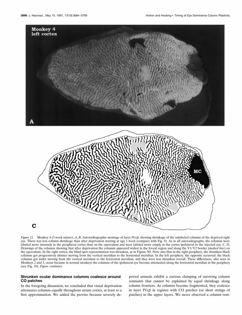
Figure 11. Monkey 4 (3 week suture). A, B, Autoradiographic montage of layer IVcb, showing shrinkage of the unlabeled columns of the deprived righteye. There was less column shrinkage than after deprivation starting at age 1 week (compare with Fig. 9). As in all autoradiographs, the columns werelabeled more intensely in the peripheral cortex than on the operculum and were labeled more crisply in the cortex ipsilateral to the injected eye. C, D,Drawings of the columns showing that after deprivation the columns appeared widest in the foveal region and along the V1/V2 border (dashed line) onthe operculum. In the right cortex, the blind spot representation was shrunken, as in Figure 9D. Note also that in the right periphery, the shrunken blackcolumns got progressively thinner moving from the vertical meridian to the horizontal meridian. In the left periphery, the opposite occurred: the blackcolumns got wider moving from the vertical meridian to the horizontal meridian, and they were less shrunken overall. These differences, also seen inMonkeys 2 and 3, occur because in normal monkeys the columns of the ipsilateral eye become attenuated along the horizontal meridian in the periphery(see Fig. 10). Figure continues.
3698 J. Neurosci., May 15, 1997, 17(10):3684–3709 Horton and Hocking • Timing of Eye Dominance Column Plasticity

nant in layer IVcb without an associated CO patch. In a sense, thisresult was not surprising, because columns shrink by erosion fromthe edge to the center, where CO patches are located (Horton andHubel, 1981; Horton, 1984). Patches also have a tendency to belocated at column bifurcations and excrescences, where oculardominance columns are wider to begin with and, therefore, morelikely to survive after shrinkage (Horton, 1984). In peripheralcortex of normal animals, where ocular dominance columns of theipsilateral eye become naturally fragmented, each fragment isaligned with a CO patch (J. Horton and D. Hocking, unpublishedobservations). Evidently, in macaques the association between
ocular dominance columns and CO patches remains strong, evenafter columns become fragmented by visual deprivation. By con-trast, in normal squirrel monkeys the ocular dominance columnsand CO patches are unrelated (Horton and Hocking, 1996b).
Differing susceptibility of parvo, magno, andkonio projectionsThe geniculate projection to layer IVcb was more shrunken thanthe projection to layer IVca (Figs. 4, 8), indicating that theparvocellular channel conveying fine spatial information to thecortex is affected more by visual deprivation than the magnocel-
Figure 11 continued.
Horton and Hocking • Timing of Eye Dominance Column Plasticity J. Neurosci., May 15, 1997, 17(10):3684–3709 3699

lular channel. This conclusion supports previous findings in ma-caques raised with blurred vision in one eye from atropine (Hen-drickson et al., 1987; Movshon et al., 1987). It is also consistentwith the observation in a reverse-sutured monkey that columnplasticity ends first for the magnocellular projection (LeVay et al.,1980).
The CO patches in the upper layers receive a direct thalamicprojection (Livingstone and Hubel, 1982; Fitzpatrick et al.,1983; Weber et al., 1983; Horton, 1984; Itaya et al., 1984).Recent evidence suggests that this projection arises from the
koniocellular laminae of the lateral geniculate body (Hendryand Yoshioka, 1994). In our first experiment (Fig. 1), thisprojection appeared normal, despite eyelid suture at age 1week. This result might be explained if the open eye’s konio-cellular afferents invaded the deprived eye’s patches. However,in another monkey we showed by ocular injection of [3H]pro-line that the projection indeed arises from the deprived eye(Figs. 6C, 8C). Recently, we have learned that konio cells in thelateral geniculate body are much less shrunken by visual depri-vation than parvo or magno cells (S. H. Hendry, personal
Figure 12. Monkey 5 (5 week suture). A, Montages of layer IVcb after injection of the normal left eye. In the left operculum, the column shrinkageequaled the shrinkage seen in the left operculum of Monkey 4 (Fig. 11A), despite starting visual deprivation 2 weeks later. However, in the left peripherythe deprivation effect was less marked. B, The right operculum showed a comparable deprivation effect, although the columns were less distinct. Thedeprivation effect in the right operculum was less pronounced than in the right operculum of Monkey 4, sutured at 3 weeks. C, D, Drawings of themontages in A, B. The calcarine fissure did not open completely in the left cortex. Figure continues.
3700 J. Neurosci., May 15, 1997, 17(10):3684–3709 Horton and Hocking • Timing of Eye Dominance Column Plasticity

communication). This finding is consistent with our observationof a spared koniocellular projection to the CO patches indeprived animals. It has been observed that the critical periodfor monocular deprivation persists for up to 1 year in the upperlayers of the cortex (LeVay et al., 1980; Daw et al., 1992). Weare unsure how to reconcile this fact with a shorter criticalperiod for the koniocellular projection than the parvocellularprojection. Perhaps cells in patches differ from cells betweenpatches in their susceptibility to monocular deprivation.
The significance of these differences among parvo, magno, andkonio projections in their susceptibility to visual deprivation isunclear. Macaques reared with early monocular suture have gross
light perception only (Von Noorden et al., 1970; Baker et al.,1974; Sparks et al., 1986), suggesting that all three perceptualchannels are cut off at some point between striate cortex and thelocus of visual consciousness.
Mechanism of binocular competitionThe geniculocortical afferents serving each eye are intermingledin layer IVc of fetal macaques (Rakic, 1977). Ocular dominancecolumns begin to emerge during the last few weeks of gestationand appear well formed at birth (Rakic, 1977; Horton and Hock-ing, 1996a). At the time of lid suture in our experiments, thegeniculocortical afferents were segregated almost completely.
Figure 12 continued.
Horton and Hocking • Timing of Eye Dominance Column Plasticity J. Neurosci., May 15, 1997, 17(10):3684–3709 3701
Therefore, column shrinkage and expansion occurred principallyby retraction and sprouting of fibers along the borders betweencolumns.
Guillery and Stelzner (1970) reported that cells within themonocular segment of the kitten’s lateral geniculate body arespared atrophy after eyelid suture, because their cortical arborsface no competition from the open eye. Although later studies
showed some shrinkage in the monocular segment (von Noordenand Middleditch, 1975; Von Noorden et al., 1976), presumablyfrom disuse, this area is less affected by deprivation (Casagrandeand Joseph, 1980). The blind spot representation is another re-gion where binocular competition is absent in striate cortex.Because the left eye’s blind spot representation receives inputfrom the right eye only, one would predict no shrinkage after
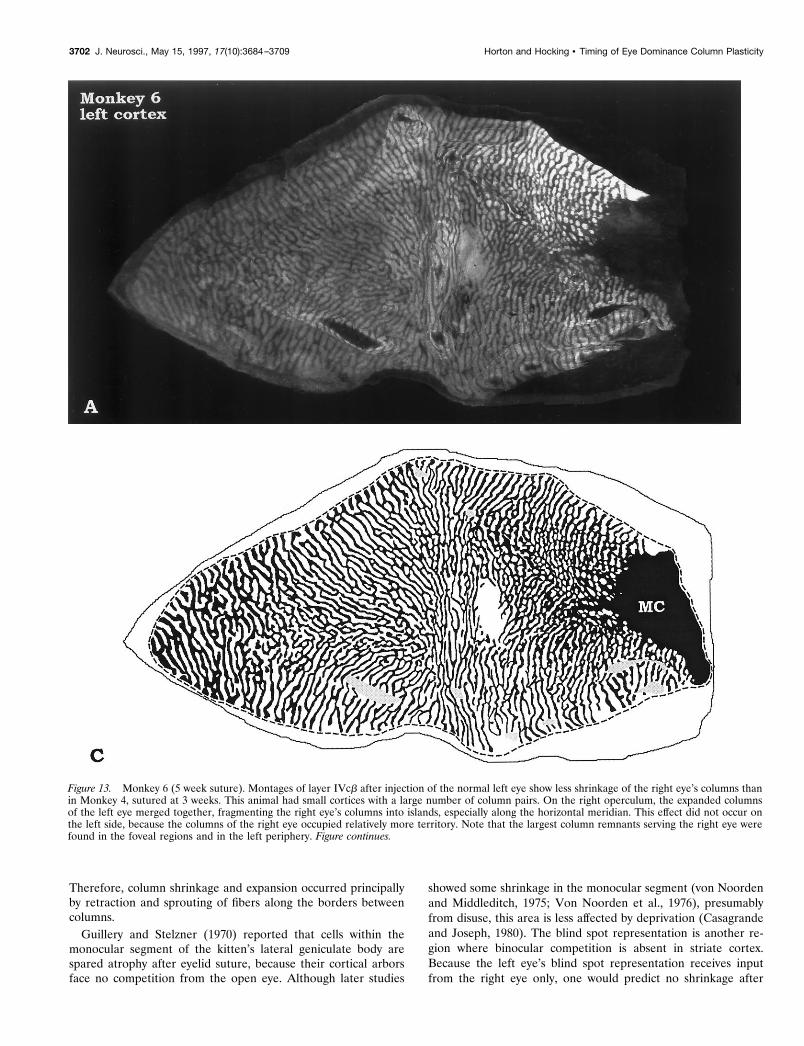
Figure 13. Monkey 6 (5 week suture). Montages of layer IVcb after injection of the normal left eye show less shrinkage of the right eye’s columns thanin Monkey 4, sutured at 3 weeks. This animal had small cortices with a large number of column pairs. On the right operculum, the expanded columnsof the left eye merged together, fragmenting the right eye’s columns into islands, especially along the horizontal meridian. This effect did not occur onthe left side, because the columns of the right eye occupied relatively more territory. Note that the largest column remnants serving the right eye werefound in the foveal regions and in the left periphery. Figure continues.
3702 J. Neurosci., May 15, 1997, 17(10):3684–3709 Horton and Hocking • Timing of Eye Dominance Column Plasticity
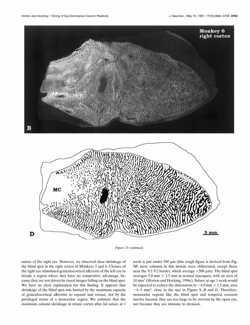
suture of the right eye. However, we observed clear shrinkage ofthe blind spot in the right cortex of Monkeys 3 and 4. Closure ofthe right eye stimulated geniculocortical afferents of the left eye toinvade a region where they have no competitive advantage, be-cause they are not driven by visual images falling on the blind spot.We have no clear explanation for this finding. It appears thatshrinkage of the blind spot was limited by the maximum capacityof geniculocortical afferents to expand and retract, not by theprivileged status of a monocular region. We estimate that themaximum column shrinkage in striate cortex after lid suture at 1
week is just under 500 mm (this rough figure is derived from Fig.9B: most columns in this mosaic were obliterated, except thosenear the V1/V2 border, which average .500 mm). The blind spotaverages 5.0 mm 3 2.5 mm in normal macaques, with an area of10 mm2 (Horton and Hocking, 1996c). Suture at age 1 week wouldbe expected to reduce the dimensions to ;4.0 mm 3 1.5 mm, area;4–5 mm2, close to the size in Figure 9, B and D. Therefore,monocular regions like the blind spot and temporal crescentsurvive because they are too large to be overrun by the open eye,not because they are immune to invasion.
Figure 13 continued.
Horton and Hocking • Timing of Eye Dominance Column Plasticity J. Neurosci., May 15, 1997, 17(10):3684–3709 3703

Clinical implicationsOur data offer further rationale for removal of dense, unilateralcataract at the earliest possible age. In macaque striate cortex, weshow that damage to the functional architecture from monocular
occlusion begins to accrue right from birth. Severe fragmentation(and obliteration in many areas) of columns was seen only in animalssutured at age 1 week. Less than 1 week of form deprivation duringthe critical period is sufficient to remodel axonal arbors in visual
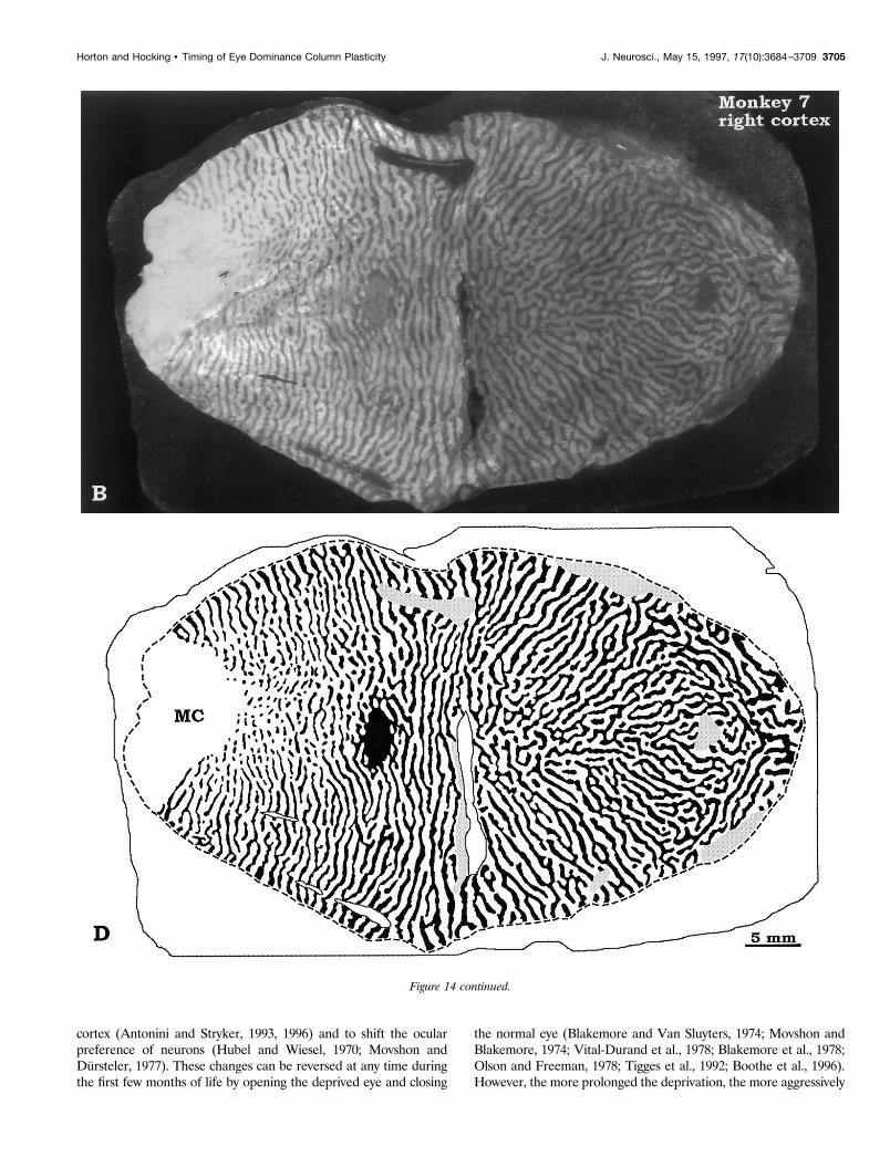
Figure 14. Monkey 7 (7 week suture). Montages of layer IVcb showed only mild shrinkage of the unlabeled columns of the right eye, signifying that thecortex was less vulnerable to the effects of visual deprivation by age 7 weeks. Note the gargantuan proportions of V1 and its columns in this animalcompared with Monkey 5 (Fig. 12) and Monkey 6 (Fig. 13). Figure continues.
3704 J. Neurosci., May 15, 1997, 17(10):3684–3709 Horton and Hocking • Timing of Eye Dominance Column Plasticity
cortex (Antonini and Stryker, 1993, 1996) and to shift the ocularpreference of neurons (Hubel and Wiesel, 1970; Movshon andDursteler, 1977). These changes can be reversed at any time duringthe first few months of life by opening the deprived eye and closing
the normal eye (Blakemore and Van Sluyters, 1974; Movshon andBlakemore, 1974; Vital-Durand et al., 1978; Blakemore et al., 1978;Olson and Freeman, 1978; Tigges et al., 1992; Boothe et al., 1996).However, the more prolonged the deprivation, the more aggressively
Figure 14 continued.
Horton and Hocking • Timing of Eye Dominance Column Plasticity J. Neurosci., May 15, 1997, 17(10):3684–3709 3705

the normal eye must be patched to compensate. The price of inten-sive patching therapy is loss of stereopsis. Gregg and Parks (1992)obtained normal stereopsis of 50 arc-sec in a child with monocularcataract by operating 24 hr after birth. Their experience demon-
strates that stereopsis can be preserved by optimal clinical manage-ment, and validates the trend toward surgery at the earliest feasibleage in infants with dense congenital cataract (Wright et al., 1992;Tytla et al., 1993).
Figure 15. Monkey 8 (12 week suture). Area measurements in layer IVcb showed normal column areas, indicating that the critical period forexpansion /contraction of columns was over by age 3 months. This animal had prominent widening of its ocular dominance columns within the fovealrepresentation and along the V1/V2 border (dashed line), an effect seen (to a variable extent) in all animals. Figure continues.
3706 J. Neurosci., May 15, 1997, 17(10):3684–3709 Horton and Hocking • Timing of Eye Dominance Column Plasticity
REFERENCES
Anderson PA, Olavarria J, Van Sluyters RC (1988) The overall patternof ocular dominance bands in cat visual cortex. J Neurosci 8:2183–2200.
Antonini A, Stryker MP (1993) Rapid remodeling of axonal arbors in thevisual cortex. Science 260:1819–1821.
Antonini A, Stryker MP (1996) Plasticity of geniculocortical afferentsfollowing brief or prolonged monocular occlusion in the cat. J CompNeurol 369:64–82.
Beller R, Hoyt CS, Marg E, Odom JV (1981) Good visual function after
neonatal surgery for congenital monocular cataracts. Am J Ophthalmol91:559–565.
Birch EE, Stager DR (1988) Prevalence of good visual acuity followingsurgery for congenital unilateral cataract. Arch Ophthalmol 106:40–43.
Birch EE, Stager DR (1996) The critical period for surgical treatment ofdense congenital unilateral cataract. Invest Ophthalmol Vis Sci37:1532–1538.
Birch EE, Swanson WH, Stager DR, Woody M, Everett M (1993) Out-come after very early treatment of dense congenital unilateral cataract.Invest Ophthalmol Vis Sci 34:3687–3699.
Figure 15 continued.
Horton and Hocking • Timing of Eye Dominance Column Plasticity J. Neurosci., May 15, 1997, 17(10):3684–3709 3707
Blakemore C, Van Sluyters RC (1974) Reversal of the physiologicaleffects of monocular deprivation in kittens: further evidence for asensitive period. J Physiol (Lond) 237:195–216.
Blakemore C, Garey LJ, Vital-Durand F (1978) The physiological effectsof monocular deprivation and their reversal in the monkey’s visualcortex. J Physiol (Lond) 283:223–262.
Boothe RG, Dobson V, Teller DY (1985) Postnatal development ofvision in human and nonhuman primates. Annu Rev Neurosci8:495–545.
Boothe RG, Louden TM, Lambert SR (1996) Acuity and contrast sen-sitivity in monkeys after neonatal intraocular lens implantation with andwithout parttime occlusion of the fellow eye. Invest Ophthalmol Vis Sci37:1520–1531.
Casagrande VA, Joseph R (1980) Morphological effects of monoculardeprivation and recovery on the dorsal lateral geniculate nucleus ingalago. J Comp Neurol 194:413–426.
Cheng KP, Hiles DA, Biglan AW, Pettapiece MC (1991) Visual resultsafter early surgical treatment of unilateral congenital cataracts. Oph-thalmology 98:903–910.
Cordes FC (1957) Failure in congenital cataract surgery. Am J Ophthal-mol 43:1–21.
Costenbader FD, Albert DG (1957) Conservatism in the management ofcongenital cataract. Arch Ophthalmol 58:426–430.
Daw NW, Fox K, Sato H, Czepita D (1992) Critical period for monoculardeprivation in the cat visual cortex. J Neurophysiol 67:197–202.
Drummond GT, Scott WE, Keech RV (1989) Management of monocu-lar congenital cataracts. Arch Ophthalmol 107:45–51.
Enoch JM, Rabinowicz IM (1976) Early surgery and visual correction ofan infant born with unilateral eye lens opacity. Documenta Ophthalmol41:371–382.
Fitzpatrick D, Itoh K, Diamond IT (1983) The laminar organization ofthe lateral geniculate body and the striate cortex in the squirrel monkey(Saimiri sciureus). J Neurosci 3:673–702.
Florence SL, Kaas JH (1992) Ocular dominance columns in area 17 ofold world macaque and talapoin monkeys: complete reconstructionsand quantitative analyses. Vis Neurosci 8:449–462.
Francois J (1979) Late results of congenital cataract surgery. Ophthal-mology 86:1586–1598.
Gregg FM, Parks MM (1992) Stereopsis after congenital monocular cat-aract extraction. Am J Ophthalmol 114:314–317.
Guillery RW, Stelzner DJ (1970) The differential effects of unilateral lidclosure upon the monocular and binocular segments of the dorsallateral geniculate nucleus in the cat. J Comp Neurol 139:413–422.
Hata Y, Stryker MP (1994) Control of thalamocortical afferent rear-rangement by postsynaptic activity in developing visual cortex. Science265:1732–1735.
Hendrickson AE, Wilson JR, Ogren MP (1978) The neuroanatomicalorganization of pathways between the dorsal lateral geniculate nucleusand visual cortex in old world and new world primates. J Comp Neurol182:123–136.
Hendrickson AE, Movshon JA, Eggers HM, Gizzi MS, Boothe RG,Kiorpes L (1987) Effects of early unilateral blur on the macaque’svisual system. II. Anatomical observations. J Neurosci 7:1327–1339.
Hendry SHC, Yoshioka T (1994) A neurochemically distinct third chan-nel in the macaque dorsal lateral geniculate nucleus. Science264:575–577.
Hess EH (1973) Imprinting: early experience and the developmentalpsychobiology of attachment. New York: Van Nostrand Reinhold.
Horton JC (1984) Cytochrome oxidase patches: a new cytoarchitectonicfeature of monkey visual cortex. Philos Trans R Soc Lond [Biol]304:199–253.
Horton JC, Hocking DR (1996a) An adult-like pattern of ocular domi-nance columns in striate cortex of newborn monkeys prior to visualexperience. J Neurosci 16:1791–1807.
Horton JC, Hocking DR (1996b) Anatomical demonstration of oculardominance columns in striate cortex of the squirrel monkey. J Neurosci16:5510–5522.
Horton JC, Hocking DR (1996c) Intrinsic variability of ocular domi-nance column periodicity in normal macaque monkeys. J Neurosci16:7228–7239.
Horton JC, Hubel DH (1981) Regular patchy distribution of cytochromeoxidase staining in primary visual cortex of macaque monkey. Nature292:762–764.
Horton JC, Stryker MP (1993a) Amblyopia induced by anisometropia
without shrinkage of ocular dominance columns in human striate cortex.Proc Natl Acad Sci USA 90:5494–5498.
Horton JC, Stryker MP (1993b) Ocular dominance columns within thefoveal representation of macaque striate cortex are less susceptible toshrinkage induced by early monocular deprivation. Soc Neurosci Abstr19:370.19.
Hubel DH, Wiesel TN (1970) The period of susceptibility to the physi-ological effects of unilateral eye closure in kittens. J Physiol (Lond)206:419–436.
Hubel DH, Wiesel TN, LeVay S (1977) Plasticity of ocular dominancecolumns in monkey striate cortex. Philos Trans R Soc Lond [Biol]278:377–409.
Itaya SK, Itaya PW, Van Hoesen GW (1984) Intracortical termination ofthe retino-geniculo-striate pathway studied with transsynaptic tracer(wheat germ agglutinin-horseradish peroxidase) and cytochrome oxi-dase staining in the macaque monkey. Brain Res 304:303–310.
Kaas JH, Lin CS, Casagrande VA (1976) The relay of ipsilateral andcontralateral retinal input from the lateral geniculate nucleus to striatecortex in the owl monkey: a transneuronal transport study. Brain Res106:371–378.
LeVay S, Stryker MP, Shatz CJ (1978) Ocular dominance columns andtheir development in layer IV of the cat’s visual cortex: a quantitativestudy. J Comp Neurol 179:223–244.
LeVay S, Wiesel TN, Hubel DH (1980) The development of oculardominance columns in normal and visually deprived monkeys. J CompNeurol 191:1–51.
LeVay S, Connolly M, Houde J, Van Essen DC (1985) The completepattern of ocular dominance stripes in the striate cortex and visual fieldof the macaque monkey. J Neurosci 5:486–501.
Livingstone MS, Hubel DH (1982) Thalamic inputs to cytochromeoxidase-rich regions in monkey visual cortex. Proc Natl Acad Sci USA79:6098–6101.
Lorenz KZ (1935) Der Kumpan in der Umwelt des Vogels: die Artgen-ossen als auslosendes Moment sozialer Verhaltensweisen. J Ornitholo-gie 83:137–213.
Movshon JA, Blakemore C (1974) Functional reinnervation in kittenvisual cortex. Nature 251:504–505.
Movshon JA, Dursteler MR (1977) Effects of brief periods of unilateraleye closure on the kitten’s visual system. J Neurophysiol 40:1255–1265.
Movshon JA, Egger HM, Gizzi MS, Hendrickson AE, Kiorpes L, BootheRG (1987) Effects of early unilateral blur on the macaque’s visualsystem. III. Physiological observations. J Neurosci 7:1340–1351.
Olavarria J, Van Sluyters RC (1985) Unfolding and flattening the cortexof gyrencephalic brains. J Neurosci Methods 15:191–202.
Olson CR, Freeman RD (1978) Monocular deprivation and recoveryduring sensitive period in kittens. J Neurophysiol 41:65–74.
Olson CR, Freeman RD (1980) Profile of the sensitive period for mo-nocular deprivation in kittens. Exp Brain Res 39:17–21.
Parks MM, Hiles DA (1967) Management of infantile cataracts. Am JOphthalmol 63:10–19.
Pratt-Johnson JA, Tillson G (1981) Visual results after removal of con-genital cataracts before the age of 1 year. Can J Ophthalmol 16:19–27.
Rakic P (1977) Prenatal development of the visual system in rhesusmonkey. Philos Trans R Soc Lond [Biol] 278:245–260.
Rogers GL, Tishler CL, Tsou BH, Hertle RW, Fellows RR (1981) Visualacuities in infants with congenital cataracts operated on prior to 6months of age. Arch Ophthalmol 99:999–1003.
Rosa MGP, Gatass R, Fiorani Jr M (1988) Complete pattern of oculardominance stripes in V1 of a new world monkey, Cebus apella. ExpBrain Res 72:645–648.
Ryan SJ, Blanton FM, Von Noorden GK (1965) Surgery of congenitalcataract. Am J Ophthalmol 60:583–587.
Shatz CJ, Lindstrom S, Wiesel TN (1977) The distribution of afferentsrepresenting the right and left eyes in the cat’s visual cortex. Brain Res131:103–116.
Shatz CJ, Stryker MP (1978) Ocular dominance in layer IV of the cat’svisual cortex and the effects of monocular deprivation. J Physiol (Lond)281:267–283.
Sparks DL, Mays LE, Gurski MR, Hickey TL (1986) Long- and short-term monocular deprivation in the rhesus monkey: effects on visualfields and optokinetic nystagmus. J Neurosci 6:1771–1780.
Spatz WB (1979) The retino-geniculo-cortical pathway in Callithrix. II.The geniculo-cortical projection. Exp Brain Res 36:401–410.
Swindale NV, Vital-Durand F, Blakemore C (1981) Recovery from mo-
3708 J. Neurosci., May 15, 1997, 17(10):3684–3709 Horton and Hocking • Timing of Eye Dominance Column Plasticity
nocular deprivation in the monkey. III. Reversal of anatomical effects inthe visual cortex. Proc R Soc Lond [Biol] 213:435–450.
Tigges M, Boothe RG, Tigges J, Wilson JR (1992) Competition betweenan aphakic and an occluded eye for territory in striate cortex of devel-oping rhesus monkeys: cytochrome oxidase histochemistry in layer 4C.J Comp Neurol 316:173–186.
Tootell RBH, Silverman MS (1985) Two methods for flat-mounting cor-tical tissue. J Neurosci Methods 15:177–190.
Tytla ME, Lewis TL, Maurer D, Brent HP (1993) Stereopsis after con-genital cataract. Invest Ophthalmol Vis Sci 34:1767–1773.
Vaegan, Taylor D (1979) Critical period for deprivation amblyopia inchildren. Trans Ophthalmol Soc [UK] 99:432–439.
Van Essen DC, Newsome WT, Maunsell JHR (1984) The visual fieldrepresentation in striate cortex of the macaque monkey: asymmetries,anisotropies, and individual variability. Vision Res 24:429–448.
Vital-Durand F, Garey LJ, Blakemore C (1978) Monocular and binocu-lar deprivation in the monkey: morphological effects and reversibility.Brain Res 158:45–64.
von Noorden GK, Middleditch PR (1975) Histology of the monkey’slateral geniculate nucleus after unilateral lid closure and experimentalstrabismus: further observations. Invest Ophthalmol Vis Sci14:674–683.
von Noorden GK, Crawford MLJ, Middleditch PR (1976) The effects ofmonocular visual deprivation: disuse or binocular interaction? BrainRes 111:277–285.
Weber JT, Huerta MF, Kaas JH, Harting JK (1983) The projections ofthe lateral geniculate nucleus of the squirrel monkey: studies of theinterlaminar zones and the S layers. J Comp Neurol 213:135–145.
Wiesel TN (1982) Postnatal development of the visual cortex and theinfluence of environment. Nature 299:583–591.
Wiesel TN, Hubel DH (1963) Single-cell responses in striate cortex ofkittens deprived of vision in one eye. J Neurophysiol 26:1003–1017.
Wiesel TN, Hubel DH (1965) Comparison of the effects of unilateral andbilateral eye closure on cortical unit responses in kittens. J Neurophysiol28:1029–1040.
Wiesel TN, Hubel DH, Lam DMK (1974) Autoradiographic demonstra-tion of ocular-dominance columns in the monkey striate cortex bymeans of transneuronal transport. Brain Res 79:273–279.
Wong-Riley M (1979) Changes in the visual system of monocularly su-tured or enucleated kittens demonstrable with cytochrome oxidasehistochemistry. Brain Res 171:11–28.
Wright KW, Matsumoto E, Edelman PM (1992) Binocular fusion andstereopsis associated with early surgery for monocular congenital cata-racts. Arch Ophthalmol 110:1607–1609.
Horton and Hocking • Timing of Eye Dominance Column Plasticity J. Neurosci., May 15, 1997, 17(10):3684–3709 3709