tissue engineering introduction for physicists - lecture two
TRANSCRIPT

Medical Imaging
● Develop a firm understanding of the fundamentals of
medical imaging, and principles underlying various
modalities and more importantly how use them in tissue
engineering
● Gain a basic understanding of the physical principles
underlying the major modalities, such as X-ray,
computed tomography and MRI.

Medical Imaging
What is the usage in Tissue Engineering?
Ultimate goal is to generate 3-D geometry of the Tissue/Organ

Medical Imaging
What does the human body look like on the inside?
It depends on how we look at it
● The most direct way → is to cut it open
○ A refinement of this procedure might be to use an endoscope
These are invasive techniques, which have the potential to cause damage or trauma to the body

Medical Imaging
Using Medical Imaging techniques means that we do not need to cut the body
or put a physical device into it in order to “see inside”
Various imaging techniques allow us to see inside the body in different ways -
the “signal” is different in each case and can reveal information that the other
methods cannot

Medical Imaging
Source
(e.g. light, x-ray)
Signal
System S Output g
Object
(e.g. tissue,
organ)
DetectionImage
reconstruct
● Excite the object with signal
● Acquire the image with detectors
● Reconstructing images
● Further image processing (i.e. generating 3D geometry)

Medical Imaging
For Example:
Functional Magnetic Resonance Imaging (fMRI) allows us to obtain images of
organ perfusion or blood flow
Positron Emission Tomography (PET) allows to obtain images of metabolism or
receptor binding

Medical Imaging
Again:
What does the human body look like on the inside?
It depends on the measured signal of interest

Medical Imaging
There are different methods of medical imaging measuring different signals
● Projection Radiography
● Computed Tomography
● Nuclear medicine
● Ultrasound Imaging
● Magnetic Resonance Imaging
● ...

Medical Imaging
● Projection Radiography
● Computed Tomography
● Nuclear medicine
● Ultrasound Imaging
● Magnetic Resonance Imaging
} Using ionizing radiation }}
Transmission imaging
Emission imaging
Reflection imaging}

Medical Imaging /Projection Radiography
● Routine diagnostic radiography
● Digital radiography
● Angiography
● Neuroradiology
● Mobile x-ray systems

Medical Imaging/X-Ray
x-ray imaging modality can be divided into two types
Projection radiography
Computed tomography
Whilhem C. Rontgen, the german physicist, father of x-
ray, named the rays coming out of the Crooke’s tube as
x-ray, because he didn’t know what are those rays
Today, we know that x-rays are electromagnetic waves
whose frequencies are much higher than visible light

Medical Imaging / X-Ray
The most common modality in projection radiography

Medical Imaging /Magnetic Resonance Imaging

Medical Imaging /Comparison
Characteristics X-Ray Imaging MRI
soft-tissue contrast Poor Excellent
Spatial resolution Excellent Good
Maximum imaging depth Excellent Excellent
Nonionizing radiation No Yes
Data acquisition Fast Slow
Cost Low High
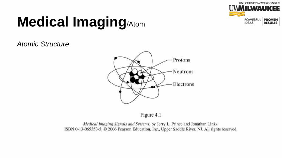
Medical Imaging/Atom
Atomic Structure

Medical Imaging/Atom
Electron Shells

Medical Imaging/Physics of Radiography
What is Binding energy?
Binding energy of electrons to its nucleus is the amount of energy is needed to
bind the energy to its shell
Binding energy for hydrogen atom is 13.6eV

Medical Imaging/Physics of Radiography
If radiation (particulate or electromagnetic) transfers energy to an orbiting
electron which is equal to or greater than the electron’s binding energy, then
the electron is ejected from the atom. This process called ionization.

Medical Imaging/Physics of Radiography
● Not all rays are ionizing
● High energy rays such as x-rays are ionizing which
results in free electron and an atom with positive charge
● Rays with energy higher than 13.6 eV are ionizing rays

Medical Imaging/Physics of Radiography
Excitation:
If an ionizing particle or ray transfers some energy to a bound electron but less
than the electron’s binding energy, then the electron is raised to a higher
energy state but is not ejected.

Medical Imaging/Physics of Radiography → Ionization
Ionizing can be divided into two broad categories:
● Particulate Radiation: Any subatomic particle (e.g, proton, neutron, or electron) can be
considered to be ionizing radiation if it possess enough kinetic energy to ionize an atom.
● Electromagnetic: Electromagnetic radiation consists of an electric wave and a magnetic
wave traveling together at perpendicular angle to each other.
Radio waves, microwaves, infrared light, x-ray, gamma rays and etc.

Medical Imaging/X-Ray

Medical Imaging / X-Ray
Object
X-ray
detector
X-ray
source

Medical Imaging/X-Ray
Medical Imaging/X-Ray
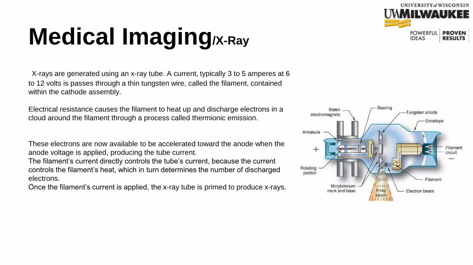
X-rays are generated using an x-ray tube. A current, typically 3 to 5 amperes at 6
to 12 volts is passes through a thin tungsten wire, called the filament, contained
within the cathode assembly.
Electrical resistance causes the filament to heat up and discharge electrons in a
cloud around the filament through a process called thermionic emission.
These electrons are now available to be accelerated toward the anode when the
anode voltage is applied, producing the tube current.
The filament’s current directly controls the tube’s current, because the current
controls the filament’s heat, which in turn determines the number of discharged
electrons.
Once the filament’s current is applied, the x-ray tube is primed to produce x-rays.
Medical Imaging/X-Ray
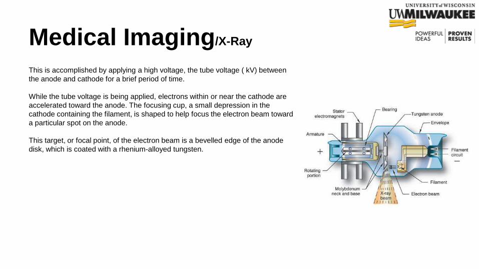
This is accomplished by applying a high voltage, the tube voltage ( kV) between
the anode and cathode for a brief period of time.
While the tube voltage is being applied, electrons within or near the cathode are
accelerated toward the anode. The focusing cup, a small depression in the
cathode containing the filament, is shaped to help focus the electron beam toward
a particular spot on the anode.
This target, or focal point, of the electron beam is a bevelled edge of the anode
disk, which is coated with a rhenium-alloyed tungsten.

Medical Imaging/X-Ray
X-Rays are generated by two different processes known as:
● Bremsstrahlung
● Characteristic X-Ray

Medical Imaging/X-Ray
➔ Characteristic radiation is produced when inner shell electrons of
the anode target are ejected by incident electrons
➔ The resultant vacancies are filled by other shell electrons, and the
energy difference emitted as characteristic radiation
➔ K-shell electrons are ejected only if incident electrons have
energies greater than K-shell binding energy
Medical Imaging/X-Ray
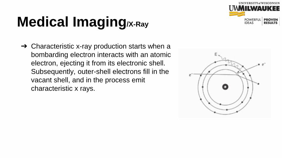
➔ Characteristic x-ray production starts when a
bombarding electron interacts with an atomic
electron, ejecting it from its electronic shell.
Subsequently, outer-shell electrons fill in the
vacant shell, and in the process emit
characteristic x rays.

Medical Imaging/X-Ray
➔ L-Shell characteristic x-rays have very low
energies and are absorbed by the glass of the
x-ray tube
➔ Most incident electrons interact with outer
shell electrons and produce heat but not x-ray
➔ (1) Characteristic radiation is produced when
an incoming electron (2) interacts with an
inner shell electron (3) and both are ejected
(4) when one of the electrons from any outer
shell moves to fill the inner shell vacancy, the
excess energy is emitted as characteristic
radiation

Medical Imaging/X-Ray
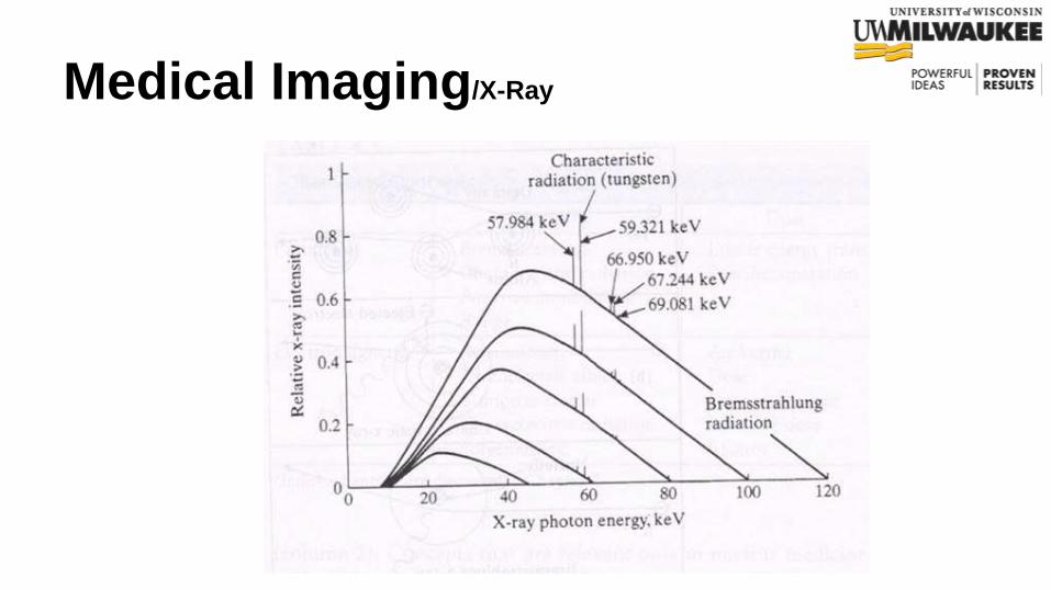
➔ Bremsstrahlung (braking) x-rays are produced
when incident electrons interact with electric
fields, which slow them down and change
their direction
➔ Bremsstrahlung x-ray production increases
with the accelerating voltage (kV) and the
atomic number (Z) of the anode
➔ Bremsstrahlung radiation is produced when
an energetic electron (with initial energy E1)
passes close to an atomic nucleus
➔ The attractive force of the positively charged
nucleus causes the electron to change
direction and lose energy

Medical Imaging/X-Ray
Medical Imaging/X-Ray

Medical Imaging/X-Ray
Main risk from ionizing radiation at high doses of x-ray is
cancer production
This is due to damage of Cell’s DNA due to radiation
The low dose of x-ray (like in dental x-ray or chest x-ray) is
not dangerous

Medical Imaging/X-Ray
X-ray radiation bioeffects
Low dose long term effects: genetic damage
Recommended maximal dose (NCRP, National Council
on Radiation Protection): 5 R per year
Rad: stands for a certain dose of energy absorbed by 1
gram of tissue
Rem: Multiply the dose in rads by a quality factor (Q)
for each type of radiation
Rem = Rad x Q

Medical Imaging/X-Ray
Filtration removes low-energy photons (long
wavelength or “soft” x-rays) from the beam by
absorbing them and permits higher energy
photons to pass through. This process reduces
the amount of radiation received by a patient
With Restriction, those rays that are not in the
certain area of interest are removed

Medical Imaging/X-Ray
X-ray interactions during passing through masster
Photons may:
1) Pass through
2) Absorbed (and transfer energy)
3) Scattered (change direction and lose energy)
which cause Compton scatter and
Photoelectric (PE) effects

Medical Imaging/X-Ray
Photoelectric (PE)
The PE effect occurs when an incident x-ray is
totally absorbed by an inner shell electron,
which is ejected as a photoelectron.
The vacancy is filled by an outer shell electron,
and the energy difference is emitted as
characteristic radiation

Medical Imaging/X-Ray
Compton scatter
Occurs when incoming x-ray photon interacts
with outer shell electron, x-ray photon loses
energy and changes direction and compton
electron carries away energy lost by scattered
photon
● This electron loses energy by ionizing
other atoms in the tissue, thereby
contributing to the patient dose
Medical Imaging/X-Ray
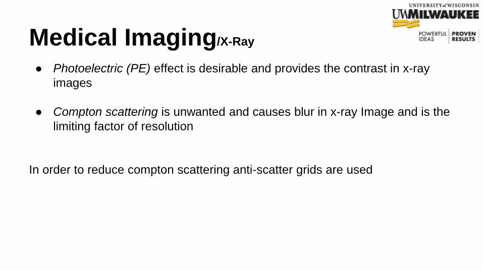
● Photoelectric (PE) effect is desirable and provides the contrast in x-ray
images
● Compton scattering is unwanted and causes blur in x-ray Image and is the
limiting factor of resolution
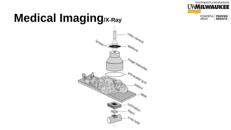
In order to reduce compton scattering anti-scatter grids are used

Medical Imaging/X-Ray
Medical Imaging/X-Ray

Medical Imaging / X-Ray
Object
X-ray
detector
X-ray
source
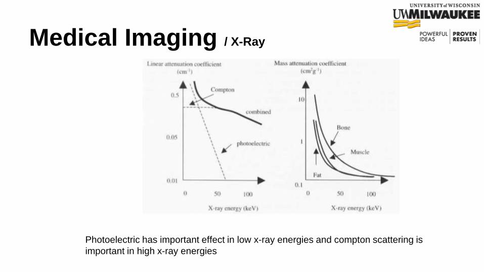
Linear attenuation coefficient
Linear attenuation coefficient depends on
density of the absorber, atomic number, and
incident photon energy
Mass attenuation coefficient
Medical Imaging / X-Ray
Photoelectric has important effect in low x-ray energies and compton scattering is
important in high x-ray energies

Medical Imaging /Computed Tomography

Medical Imaging /Computed Tomography

Medical Imaging/Computed Tomography (CT)
1st Generation CT: Parallel Projection

Medical Imaging/Computed Tomography (CT)
2nd Generation

Medical Imaging/Computed Tomography (CT)
3G

Medical Imaging/Computed Tomography (CT)
4G

Medical Imaging/Computed Tomography (CT)

Medical Imaging/Computed Tomography (CT)
● Horizontal movement of the bed as the x-ray is rotating
● Rapid volumetric data acquisition

Medical Imaging/Computed Tomography (CT)

Medical Imaging/Computed Tomography (CT)

Medical Imaging/Computed Tomography (CT)
CT Number
Consistency across CT scanners is desired. CT Number allow for the display of the image in terms of
the attenuation coefficient wrt water no matter what x-ray tube is used across different scanners
Where K is a constant (Usually = 1000), and are respectively the attenuation coefficients for
the pixel of interest and water
The CT number has a unit of Hounsfield or HU when K = 1000
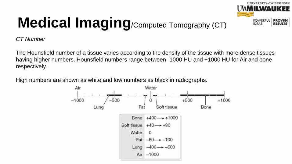
Medical Imaging/Computed Tomography (CT)
CT Number
The Hounsfield number of a tissue varies according to the density of the tissue with more dense tissues
having higher numbers. Hounsfield numbers range between -1000 HU and +1000 HU for Air and bone
respectively.
High numbers are shown as white and low numbers as black in radiographs.

Medical Imaging/Computed Tomography (CT)
Example:

Medical Imaging/Computed Tomography (CT)
Each projection contain M data point, and there are N rotations
For each projection, the signal intensity depends upon the composite attenuation coefficient of the
tissue corresponding to the particular beam path.

Medical Imaging /X-ray and CT
● Projection radiography → shadows of transmitted x-ray
intensities after absorption and scattering by body. 2D
projection
● CT is 3D x-ray imaging
● Tissue attenuates x-ray depending on their attenuation
coefficients and x-ray energies
● Both x-ray and CT uses transmission of ionizing radiation
through body
● Various organs change the intensity of beam differently
● The beams exiting the body contains shadows of tissue

Medical Imaging/Magnetic Resonance Imaging
MRI is making high contrast cross-sectional images like CT
taking advantage of magnetization of protons in the body
rather than using any ionizing radiation through the body

Medical Imaging/Magnetic Resonance Imaging
The five principal components:
1. The main magnet
2. A set of coils to provide a switchable spatial
gradient in the main magnetic field
3. coils for the transmission and reception of
radio-frequency pulses
4. Electronics for programming the timing of
transmission and reception of signals
5. Console

Medical Imaging/Magnetic Resonance Imaging
● Gradient coils provide the means to encode spatial information and to
choose slices of body to be images
● (a) axial by applying Gz
● (b) coronal by applying Gy
● (c) sagittal by applying Gx

Medical Imaging/Magnetic Resonance Imaging
Magnetic resonance imaging is an imaging modality
which is primarily used to construct pictures of the NMR
(nuclear magnetic resonance) signal from hydrogen
atoms in an object

Medical Imaging/Magnetic Resonance Imaging
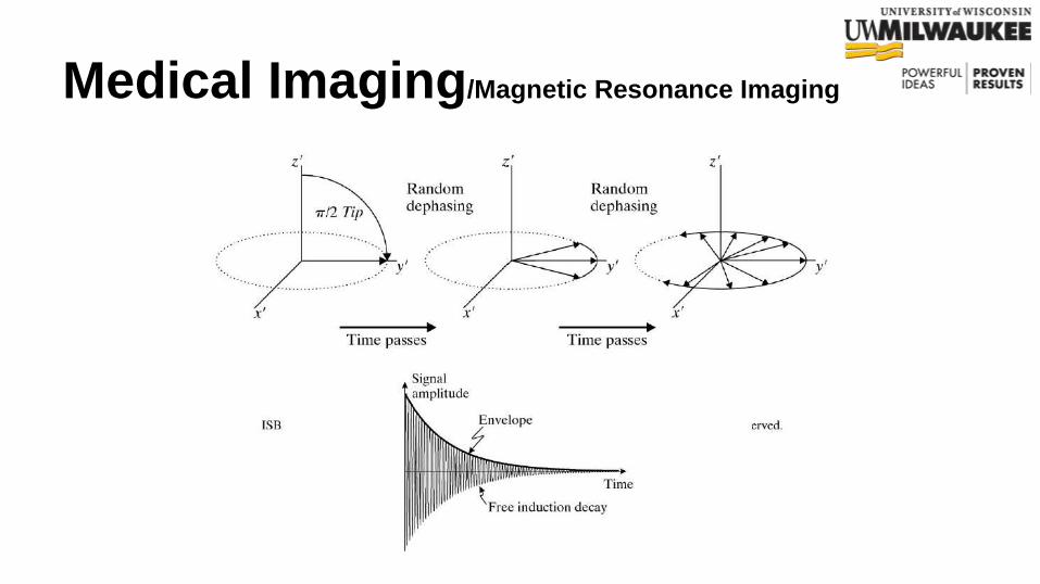
1. Magnetize your subject in strong magnetic field
2. Transmit radio waves into subject
3. Turn off radio waves transmitter
4. Receive radio waves echoed by subject
5. Manipulate echoes with additional magnetic fields
6. Store measured radio wave data vs. time
- Repeat steps 2 through 6
7. Process raw data to reconstruct images
What is the physical basis?

Medical Imaging/Magnetic Resonance Imaging
What is Gyroscope?
device for measuring or maintaining orientation, based
on the principles of angular momentum.

Medical Imaging/Magnetic Resonance Imaging
Why Gyroscope?
Atoms with electrons moving around them acting like a
Gyroscope
Each atom has it’s own small magnetic field

Medical Imaging/Magnetic Resonance Imaging

Medical Imaging/Magnetic Resonance Imaging

Medical Imaging/Magnetic Resonance Imaging
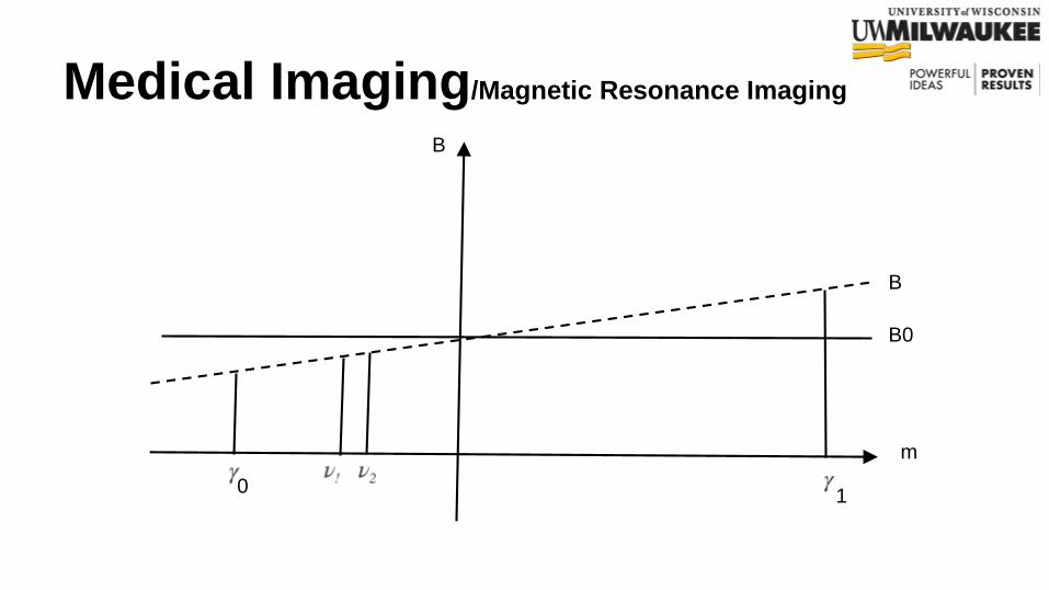
f is resonance frequency of a spin
is the gyromagnetic ratio and it’s equal to
265.5 M rad/s/T for hydrogen
is Larmor precession frequency

Medical Imaging/Magnetic Resonance Imaging
Medical Imaging/Magnetic Resonance Imaging
m
B0
B
B
01
Medical Imaging/Magnetic Resonance Imaging

Medical Imaging/Magnetic Resonance Imaging

Medical Imaging/Magnetic Resonance Imaging

Medical Imaging/Magnetic Resonance Imaging

Medical Imaging/Magnetic Resonance Imaging

Medical Imaging/Magnetic Resonance Imaging
m
B0
B
B
01

Medical Imaging/Magnetic Resonance Imaging

Medical Imaging/Magnetic Resonance Imaging

Medical Imaging /Magnetic Resonance Imaging
● Standard MRI
● Echo-planar imaging (EPI)
● Magnetic resonance spectroscopic imaging
● Functional MRI (fMRI)
Medical Imaging
References:
● EE437 Introduction to Biomedical Imaging, Dr. Mahsa Ranji
● Prince, J. L., & Links, J. (2006). Medical Imaging Signals and Systems.
Pearson (1st ed., p. 481).