tissue regeneration defective cholesterol …...tissue regeneration defective cholesterol clearance...
TRANSCRIPT
TISSUE REGENERATION
Defective cholesterol clearance limitsremyelination in the aged centralnervous systemLudovico Cantuti-Castelvetri,1,2,3,4* Dirk Fitzner,1,5* Mar Bosch-Queralt,1,2,3,4
Marie-Theres Weil,1,6 Minhui Su,1,2,3,4 Paromita Sen,1 Torben Ruhwedel,7
Miso Mitkovski,8 George Trendelenburg,5 Dieter Lütjohann,9
Wiebke Möbius,6,7 Mikael Simons1,2,3,4†
Age-associated decline in regeneration capacity limits the restoration of nervous systemfunctionality after injury. In a model for demyelination, we found that old mice fail toresolve the inflammatory response initiated after myelin damage. Aged phagocytesaccumulated excessive amounts of myelin debris, which triggered cholesterol crystalformation and phagolysosomal membrane rupture and stimulated inflammasomes.Myelin debris clearance required cholesterol transporters, including apolipoproteinE. Stimulation of reverse cholesterol transport was sufficient to restore the capacityof old mice to remyelinate lesioned tissue. Thus, cholesterol-rich myelin debris canoverwhelm the efflux capacity of phagocytes, resulting in a phase transition ofcholesterol into crystals and thereby inducing a maladaptive immune response thatimpedes tissue regeneration.
Remyelination restores rapid transmissionof nerve impulses and axonal function inthe central nervous system (CNS) of pa-tients with demyelinating diseases such asmultiple sclerosis (MS). Although remyeli-
nation can occur inMS, age-associated decline inmyelin repair contributes to chronic progressivedisease and disability (1). Thus, understandingthe cause of and preventing this decline are keygoals in regenerative medicine (2–4). So far, ep-igenetic changes within aging oligodendrocyteprogenitor cells and declines in phagocytic ca-pacity of aged blood-derived monocytes havebeen identified as possible mechanisms (5, 6).We implemented a toxin-inducedmodel, inwhicha single injection of lysolecithin (lysophosphati-dylcholine) induces a focal demyelinating lesionin the whitematter of the brain or spinal cord ofmice. In lesioned animals, demyelination is com-plete within 4 days, followed by a repair processthat is maximal between 14 and 21 days post-injection (dpi) and requires rapid clearance ofdamaged myelin for regeneration to occur (7).
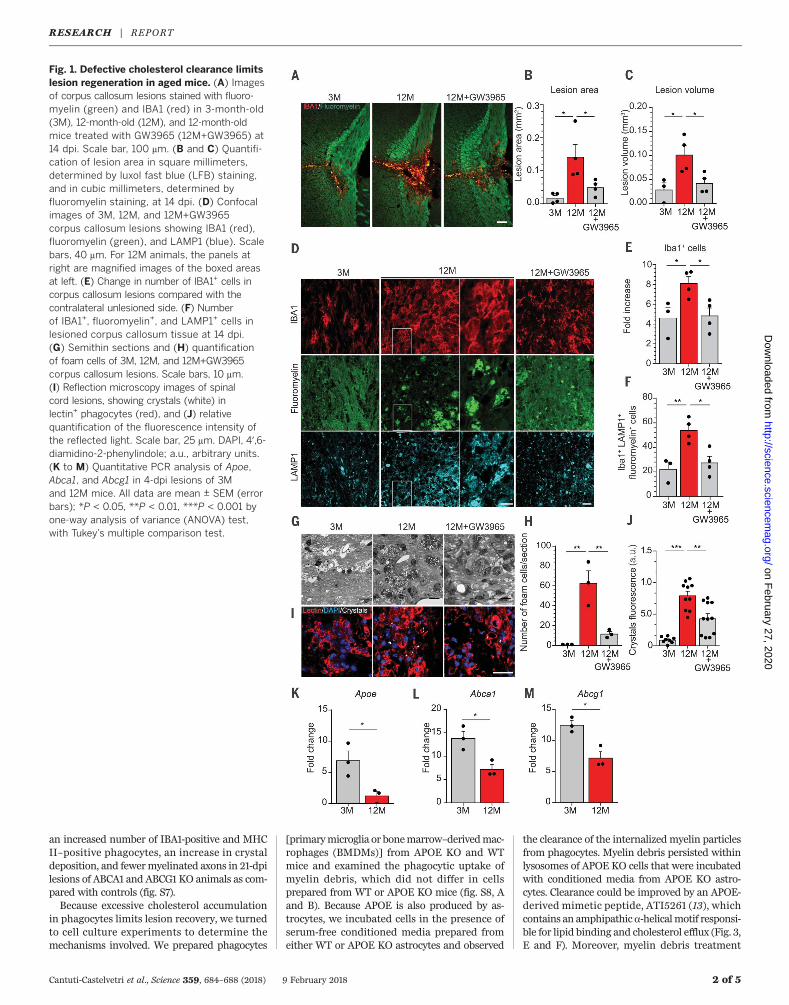
We induced focal demyelinating lesions in thecorpus callosum of young (3 months) and old(12months)miceby lysolecithin injections. Lesionswere of similar size at 4 dpi but not at 14 dpi,confirming the poor regenerative capacity ofold mice (Fig. 1, A to C, and fig. S1, A to E). Sus-tained immune infiltration, as determined byIBA1-positive and MAC2-positive cells, was de-tected in old mice at 14 dpi (Fig. 1, A and E, andfig. S1G). Myelin debris accumulation withinlysosomes of phagocytes (Fig. 1, D and F) andnumerous foam cells harboring lipid dropletsand needle-shaped cholesterol crystals—a typicalhallmark of cholesterol overloading—were foundin old mice (Fig. 1, G and H, and fig. S2G). More-over, by a combination of laser reflection andfluorescence confocal microscopy (reflection mi-croscopy) we confirmed the increase of crystaldeposition in spinal cord lesions of old mice(Fig. 1, I and J). Crystals were similarly observedin two other models of myelin injury (fig. S2).Toxic overload of cholesterol drives the forma-
tion of foamy macrophages and maladaptiveimmune responses in atherosclerosis. We hypoth-esized that the accumulation of cholesterol, themajor component of myelin, may overwhelm thecholesterol transport capacity of phagocytes,thereby forming a bottleneck for successful re-pair in the aged CNS. Because cholesterol cannotbe broken down, it must be transferred from thephagocytes back to the extracellular space viathe transporters ABCA1 and ABCG1 [adenosinetriphosphate–binding cassette (ABC) A1 and G1]in the plasma membrane where it binds high-density lipoprotein particles [e.g., apolipoproteinE(APOE)] (8). Real-time quantitative reverse tran-scription polymerase chain reaction (PCR) analy-ses revealed that the expression of ApoE, Abca1,andAbcg1was reduced in 4-dpi lesions of oldmice
compared with young mice (Fig. 1, K to M, andfig. S3). Oxysterols (hydroxylated cholesterol me-tabolites) such as 24S- and 27-hydroxycholesterolare endogenous ligands for the liver X receptor(LXR), thereby controlling the expression of genesinvolved in cholesterol efflux in cholesterol-loadedcells (9).Relative amountsof 24S-hydroxycholesteroldid not differ in lesions of young and old animals,but 27-hydroxycholesterol levels were reduced inlesions of oldmice (fig. S3). One possible explana-tion for lesion restitution failure in oldmice is theinability to clear excessive myelin-derived choles-terol fromphagocytes. Thus,weexaminedwhetherthe LXR agonist GW3965 (10) improve lesionrecovery in old mice by inducing the expressionof genes involved in lipid efflux, such as Abca1,Abcg1, and ApoE (fig. S3). GW3965 led to mark-edly improved lesion regeneration in old mice,with a reduction in the number of IBA1-positiveand MAC2-positive phagocytes (Fig. 1, A to C). Inaddition, the number of phagocytes containingmyelin debris, the number of foam cells, and theamount of cholesterol crystals were reduced bytreatment with GW3965 (Fig. 1, A to J).Because accumulation of cholesterol in phago-
cytes may pose a barrier for successful tissue re-generation,we analyzed the regenerative capacityof mouse mutants lacking central factors of thereverse cholesterol transport pathway. We in-duced demyelinating lesions in 3-month-oldNR1H3 (or LXRa) knockout (KO) mice and ob-served, as in aged wild-type (WT) mice, impairedlesion restitution and sustained phagocyte infil-tration at 21 dpi (fig. S4, A to C). In addition, wedetected accumulation of myelin debris in lyso-somes of phagocytes and crystal deposition inlesions of LXRa KO mice (fig. S4, D to G).Next, we analyzed the role of APOE, the major
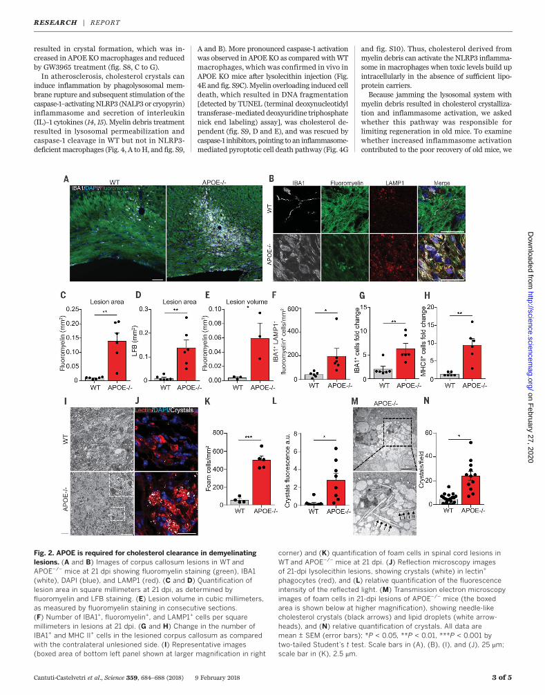
CNS cholesterol carrier that supports lipid effluxfrom cells (11). We induced focal demyelinationin the corpus callosum of 12-week-old APOE KOand WT control mice and quantified lesion sizeand recovery. Lesion size did not differ initially,at 4 dpi (fig. S4, H to J). However, when lesionswere analyzed at 21 dpi, we observed impairedlesion restitution.Wedetected increasednumbersof IBA1-positive, MAC2-positive, and major histo-compatibility complex class II (MHC II)–positivephagocytes, aswell as enhanced crystal depositionandmyelin debris accumulationwithin lysosomesof phagocytes in spinal cord and corpus callosumlesions of APOE KO animals as compared withcontrol mice (Fig. 2 and fig. S5). Because APOEhas functions beyond cholesterol transport (11),we tested the efficacy of cyclic oligosaccharide2-hydroxypropyl-b-cyclodextrin (HbCD), a com-pound that increases cholesterol efflux and sol-ubility (12), and found that HbCD treatmentattenuated the phenotype of APOE KO animals(Fig. 3 and fig. S6).Together with Abca1 and Abcg1, ApoE repre-
sents one of the major nuclear LXR–regulatedgenes involved inmediating cholesterol efflux fromphagocytes. We thus crossbred CX3CR1CreERanimals with Abca1fl/fl and Abcg1fl/fl mice to ob-tain microglia- and macrophage-specific double-KO mice (ABCA1 and ABCG1 KO). We observed
RESEARCH
Cantuti-Castelvetri et al., Science 359, 684–688 (2018) 9 February 2018 1 of 5
1Max Planck Institute of Experimental Medicine, 37075Göttingen, Germany. 2Munich Cluster for SystemsNeurology (SyNergy), 81377 Munich, Germany. 3Instituteof Neuronal Cell Biology, Technical University Munich,80805 Munich, Germany. 4German Center forNeurodegenerative Disease (DZNE), 81377 Munich,Germany. 5Department of Neurology, University ofGöttingen Medical Center, 37075 Göttingen, Germany.6Center for Nanoscale Microscopy and MolecularPhysiology of the Brain (CNMPB), 37075 Göttingen,Germany. 7Department of Neurogenetics, Max PlanckInstitute of Experimental Medicine, 37075 Göttingen,Germany. 8Light Microscopy Facility, Max Planck Instituteof Experimental Medicine, 37075 Göttingen Germany.9Institute for Clinical Chemistry and ClinicalPharmacology, University of Bonn, 53127 Bonn, Germany.*These authors contributed equally to this work.†Corresponding author. Email: [email protected]
on February 27, 2020
http://science.sciencem
ag.org/D
ownloaded from
an increased number of IBA1-positive and MHCII–positive phagocytes, an increase in crystaldeposition, and fewermyelinated axons in 21-dpilesions of ABCA1 and ABCG1 KO animals as com-pared with controls (fig. S7).Because excessive cholesterol accumulation
in phagocytes limits lesion recovery, we turnedto cell culture experiments to determine themechanisms involved. We prepared phagocytes
[primarymicroglia or bonemarrow–derivedmac-rophages (BMDMs)] from APOE KO and WTmice and examined the phagocytic uptake ofmyelin debris, which did not differ in cellsprepared from WT or APOE KO mice (fig. S8, Aand B). Because APOE is also produced by as-trocytes, we incubated cells in the presence ofserum-free conditioned media prepared fromeither WT or APOE KO astrocytes and observed
the clearance of the internalized myelin particlesfrom phagocytes. Myelin debris persisted withinlysosomes of APOE KO cells that were incubatedwith conditioned media from APOE KO astro-cytes. Clearance could be improved by an APOE-derived mimetic peptide, ATI5261 (13), whichcontains an amphipathica-helicalmotif responsi-ble for lipid binding and cholesterol efflux (Fig. 3,E and F). Moreover, myelin debris treatment
Cantuti-Castelvetri et al., Science 359, 684–688 (2018) 9 February 2018 2 of 5
Fig. 1. Defective cholesterol clearance limitslesion regeneration in aged mice. (A) Imagesof corpus callosum lesions stained with fluoro-myelin (green) and IBA1 (red) in 3-month-old(3M), 12-month-old (12M), and 12-month-oldmice treated with GW3965 (12M+GW3965) at14 dpi. Scale bar, 100 mm. (B and C) Quantifi-cation of lesion area in square millimeters,determined by luxol fast blue (LFB) staining,and in cubic millimeters, determined byfluoromyelin staining, at 14 dpi. (D) Confocalimages of 3M, 12M, and 12M+GW3965corpus callosum lesions showing IBA1 (red),fluoromyelin (green), and LAMP1 (blue). Scalebars, 40 mm. For 12M animals, the panels atright are magnified images of the boxed areasat left. (E) Change in number of IBA1+ cells incorpus callosum lesions compared with thecontralateral unlesioned side. (F) Numberof IBA1+, fluoromyelin+, and LAMP1+ cells inlesioned corpus callosum tissue at 14 dpi.(G) Semithin sections and (H) quantificationof foam cells of 3M, 12M, and 12M+GW3965corpus callosum lesions. Scale bars, 10 mm.(I) Reflection microscopy images of spinalcord lesions, showing crystals (white) inlectin+ phagocytes (red), and (J) relativequantification of the fluorescence intensity ofthe reflected light. Scale bar, 25 mm. DAPI, 4′,6-diamidino-2-phenylindole; a.u., arbitrary units.(K to M) Quantitative PCR analysis of Apoe,Abca1, and Abcg1 in 4-dpi lesions of 3Mand 12M mice. All data are mean ± SEM (errorbars); *P < 0.05, **P < 0.01, ***P < 0.001 byone-way analysis of variance (ANOVA) test,with Tukey’s multiple comparison test.
RESEARCH | REPORTon F
ebruary 27, 2020
http://science.sciencemag.org/
Dow
nloaded from
resulted in crystal formation, which was in-creased in APOEKOmacrophages and reducedby GW3965 treatment (fig. S8, C to G).In atherosclerosis, cholesterol crystals can
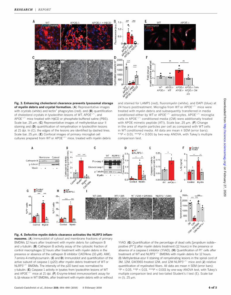
induce inflammation by phagolysosomal mem-brane rupture and subsequent stimulation of thecaspase-1–activatingNLRP3 (NALP3 or cryopyrin)inflammasome and secretion of interleukin(IL)–1 cytokines (14, 15). Myelin debris treatmentresulted in lysosomal permeabilization andcaspase-1 cleavage in WT but not in NLRP3-deficientmacrophages (Fig. 4, A toH, and fig. S9,
A and B). More pronounced caspase-1 activationwas observed in APOE KO as compared withWTmacrophages, which was confirmed in vivo inAPOE KO mice after lysolecithin injection (Fig.4E and fig. S9C). Myelin overloading induced celldeath, which resulted in DNA fragmentation[detected by TUNEL (terminal deoxynucleotidyltransferase–mediated deoxyuridine triphosphatenick end labeling) assay], was cholesterol de-pendent (fig. S9, D and E), and was rescued bycaspase-1 inhibitors, pointing to an inflammasome-mediated pyroptotic cell death pathway (Fig. 4G
and fig. S10). Thus, cholesterol derived frommyelin debris can activate the NLRP3 inflamma-some in macrophages when toxic levels build upintracellularly in the absence of sufficient lipo-protein carriers.Because jamming the lysosomal system with
myelin debris resulted in cholesterol crystalliza-tion and inflammasome activation, we askedwhether this pathway was responsible forlimiting regeneration in old mice. To examinewhether increased inflammasome activationcontributed to the poor recovery of old mice, we
Cantuti-Castelvetri et al., Science 359, 684–688 (2018) 9 February 2018 3 of 5
Fig. 2. APOE is required for cholesterol clearance in demyelinatinglesions. (A and B) Images of corpus callosum lesions in WT andAPOE−/− mice at 21 dpi showing fluoromyelin staining (green), IBA1(white), DAPI (blue), and LAMP1 (red). (C and D) Quantification oflesion area in square millimeters at 21 dpi, as determined byfluoromyelin and LFB staining. (E) Lesion volume in cubic millimeters,as measured by fluoromyelin staining in consecutive sections.(F) Number of IBA1+, fluoromyelin+, and LAMP1+ cells per squaremillimeters in lesions at 21 dpi. (G and H) Change in the number ofIBA1+ and MHC II+ cells in the lesioned corpus callosum as comparedwith the contralateral unlesioned side. (I) Representative images(boxed area of bottom left panel shown at larger magnification in right
corner) and (K) quantification of foam cells in spinal cord lesions inWT and APOE−/− mice at 21 dpi. (J) Reflection microscopy imagesof 21-dpi lysolecithin lesions, showing crystals (white) in lectin+
phagocytes (red), and (L) relative quantification of the fluorescenceintensity of the reflected light. (M) Transmission electron microscopyimages of foam cells in 21-dpi lesions of APOE−/− mice (the boxedarea is shown below at higher magnification), showing needle-likecholesterol crystals (black arrows) and lipid droplets (white arrow-heads), and (N) relative quantification of crystals. All data aremean ± SEM (error bars); *P < 0.05, **P < 0.01, ***P < 0.001 bytwo-tailed Student’s t test. Scale bars in (A), (B), (I), and (J), 25 mm;scale bar in (K), 2.5 mm.
RESEARCH | REPORTon F
ebruary 27, 2020
http://science.sciencemag.org/
Dow
nloaded from
Cantuti-Castelvetri et al., Science 359, 684–688 (2018) 9 February 2018 4 of 5
Fig. 3. Enhancing cholesterol clearance prevents lysosomal storageof myelin debris and crystal formation. (A) Representative imageswith crystals (white) and lectin+ phagocytes (red), and (B) quantificationof cholesterol crystals in lysolecithin lesions of WT, APOE−/−, andAPOE−/− mice treated with HbCD or phosphate-buffered saline (PBS).Scale bar, 25 mm. (C) Representative images of methylenblue-azur IIstaining and (D) quantification of remyelination in lysolecithin lesionsat 21 dpi. In (C), the edges of the lesions are identified by dashed lines.Scale bar, 25 mm. (E) Confocal images of primary microglial cellcultures prepared from WT or APOE−/− mice, treated with myelin debris
and stained for LAMP1 (red), fluoromyelin (white), and DAPI (blue) at24 hours posttreatment. Microglia from WT or APOE−/− mice weretreated with myelin debris and subsequently transferred in mediaconditioned either by WT or APOE−/− astrocytes. APOE−/− microgliacells in APOE−/− conditioned media (CM) were additionally treatedwith APOE mimetic peptide (ATI). Scale bar, 25 mm. (F) Changein the area of myelin particles per cell as compared with WT cellsin WT conditioned media. All data are mean ± SEM (error bars);**P < 0.01, ***P < 0.001 by two-way ANOVA, with Tukey’s multiplecomparison test.
Fig. 4. Defective myelin debris clearance activates the NLRP3 inflam-masome. (A) Immunoblot of cytosol and membrane fractions of primaryBMDMs 12 hours after treatment with myelin debris for cathepsin Band g-tubulin. (B) Cathepsin B activity assay of the cytosolic fraction ofcontrol macrophages 12 hours after treatment with myelin debris in thepresence or absence of the cathepsin B inhibitor CA074me (10 mM). AMC,7-amino-4-methylcoumarin. (C and D) Immunoblot and quantification of theactive subunit of caspase 1 (p20) after myelin debris treatment of WTorNLRP3−/− BMDMs. The intensity of the p20 band was normalized tog-tubulin. (E) Caspase 1 activity in lysates from lysolecithin lesions of WTand APOE−/− mice at 21 dpi. (F) Enzyme-linked immunosorbent assay forIL-1b release in WT BMDMs, after treatment with myelin debris with or without
YVAD. (G) Quantification of the percentage of dead cells [propidium iodide–positive (PI+)] after myelin debris treatment (12 hours) in the presence orabsence of a caspase-1 inhibitor (YVAD). (H) Quantification of PI+ cells aftertreatment of WT and NLRP3−/− BMDMs with myelin debris for 12 hours.(I) Methylenblue-azur II staining of remyelinating lesions in the spinal cord of3M, 12M, GW3965-treated 12M, and 12M NLRP3−/− mice and (J) relativequantification of myelinated fibers. All data are mean ± SEM (error bars);*P < 0.05, **P < 0.01, ***P < 0.001 by one-way ANOVA test, with Tukey’smultiple comparison test and two-tailed Student’s t test (E). Scale barin (I), 25 mm.
RESEARCH | REPORTon F
ebruary 27, 2020
http://science.sciencemag.org/
Dow
nloaded from
analyzed spinal cord lesions of aged WT andNLRP3-deficient mice. As in GW3965-treatedold animals (Fig. 4, I and J), we found signifi-cantly improved remyelination in aged NLRP3-deficient mice as compared with aged WT miceat 21 dpi. Thus, inflammasome activation, pos-sibly downstream of cholesterol accumulation,drives a maladaptive immune response thathampers inflammation resolution and repairin aged mice.Self-resolving inflammation is essential for a
proper restorative process after tissue damage,whereas uncontrolled inflammation can leavelasting marks that permanently alter tissuehomeostasis (16). Wemade the surprising discov-ery that the self-limiting inflammatory response,which is necessary to initiate a regenerative pro-cess, is maladaptive in the CNS of aged mice. Itappears that the inability of aged phagocytes toclear the enormous amounts of cholesterol thatare released frommyelin aftermyelin breakdownin demyelinating diseases results in a phase tran-sition of free cholesterol into crystals, inducinglysosomal rupture and inflammasome stimula-
tion, consistent with the beneficial effects ofnuclear receptor agonists in remyelination (17, 18).The unexpected link between lipid metabolismsand tissue regeneration provides opportunitiesfor the development of regenerative medicinesfor remyelination and for improving functionalrecovery after CNS injury (19).
REFERENCES AND NOTES
1. P. Patrikios et al., Brain 129, 3165–3172 (2006).2. F. J. Najm et al., Nature 522, 216–220 (2015).3. F. Mei et al., Nat. Med. 20, 954–960 (2014).4. V. A. Deshmukh et al., Nature 502, 327–332 (2013).5. J. M. Ruckh et al., Cell Stem Cell 10, 96–103 (2012).6. S. Shen et al., Nat. Neurosci. 11, 1024–1034 (2008).7. V. E. Miron et al., Nat. Neurosci. 16, 1211–1218 (2013).8. K. J. Moore, I. Tabas, Cell 145, 341–355 (2011).9. X. Fu et al., J. Biol. Chem. 276, 38378–38387 (2001).10. C. Hong, P. Tontonoz, Nat. Rev. Drug Discov. 13, 433–444
(2014).11. R. W. Mahley, Science 240, 622–630 (1988).12. S. Zimmer et al., Sci. Transl. Med. 8, 333ra50 (2016).13. A. Hafiane, J. K. Bielicki, J. O. Johansson, J. Genest, Biochim.
Biophys. Acta 1841, 1498–1512 (2014).14. P. Duewell et al., Nature 464, 1357–1361 (2010).15. K. Rajamäki et al., PLOS ONE 5, e11765 (2010).16. A. Aguzzi, B. A. Barres, M. L. Bennett, Science 339, 156–161
(2013).
17. J. K. Huang et al., Nat. Neurosci. 14, 45–53 (2011).18. D. Meffre et al., Proc. Natl. Acad. Sci. U.S.A. 112, 7587–7592
(2015).19. F. Bei et al., Cell 164, 219–232 (2016).
ACKNOWLEDGMENTS
We thank L. Vaculčiaková for help with the calculations ofthree-dimensional reconstructions and A. Kerksiek fortechnical assistance. This work was supported by a EuropeanResearch Council (ERC) Consolidator Grant (M.S.) andgrants from the German Research Foundation (DFG)(SI 746/9-1,10-1, SPP1757, TRR128, and TRR43), the KlausTschira Stiftung, the Adelson Foundation, Excellence Clusterfor Systems Neurology (SyNergy), and Excellence Clusterfor Nanoscale Microscopy and Molecular Physiology of theBrain (CNMPB). W.M. is supported by an ERC grant toK.-A. Nave. M.B.-Q. is supported by a Boehringer Ingelheimstipend. All data are presented in the main text andsupplementary materials.
SUPPLEMENTARY MATERIALS
www.sciencemag.org/content/359/6376/684/suppl/DC1Materials and MethodsFigs. S1 to S10References (20–29)
10 April 2017; resubmitted 5 November 2017Accepted 11 December 2017Published online 4 January 201810.1126/science.aan4183
Cantuti-Castelvetri et al., Science 359, 684–688 (2018) 9 February 2018 5 of 5
RESEARCH | REPORTon F
ebruary 27, 2020
http://science.sciencemag.org/
Dow
nloaded from

Defective cholesterol clearance limits remyelination in the aged central nervous system
Ruhwedel, Miso Mitkovski, George Trendelenburg, Dieter Lütjohann, Wiebke Möbius and Mikael SimonsLudovico Cantuti-Castelvetri, Dirk Fitzner, Mar Bosch-Queralt, Marie-Theres Weil, Minhui Su, Paromita Sen, Torben
originally published online January 4, 2018DOI: 10.1126/science.aan4183 (6376), 684-688.359Science
, this issue p. 684; see also p. 635Sciencegood candidates for regenerative medicine in the CNS.stimulation. Thus, drugs being developed to promote cholesterol clearance in human atherosclerosis lesions may also beresulting in a transition of free cholesterol into crystals, thereby inducing lysosomal rupture and inflammasome Perspective by Chen and Popko). Cholesterol-rich myelin debris overwhelmed the efflux capacity of phagocytes,which is necessary for remyelination to occur, is maladaptive in the central nervous system (CNS) of old mice (see the
report that the self-limiting inflammatory response,et al.failure is a key goal in regenerative medicine. Cantuti-Castelvetri sclerosis lesions contributes to chronic progressive disease and disability. Understanding the cause and preventing this
A decline in tissue repair is a universal hallmark of aging. The failure to regenerate myelin sheaths in multipleKeeping cholesterol at bay
ARTICLE TOOLS http://science.sciencemag.org/content/359/6376/684
MATERIALSSUPPLEMENTARY http://science.sciencemag.org/content/suppl/2018/01/03/science.aan4183.DC1
CONTENTRELATED
http://stke.sciencemag.org/content/sigtrans/11/524/eaao4180.fullhttp://science.sciencemag.org/content/sci/359/6376/635.full
REFERENCES
http://science.sciencemag.org/content/359/6376/684#BIBLThis article cites 29 articles, 9 of which you can access for free
PERMISSIONS http://www.sciencemag.org/help/reprints-and-permissions
Terms of ServiceUse of this article is subject to the
is a registered trademark of AAAS.ScienceScience, 1200 New York Avenue NW, Washington, DC 20005. The title (print ISSN 0036-8075; online ISSN 1095-9203) is published by the American Association for the Advancement ofScience
Science. No claim to original U.S. Government WorksCopyright © 2018 The Authors, some rights reserved; exclusive licensee American Association for the Advancement of
on February 27, 2020
http://science.sciencem
ag.org/D
ownloaded from