tissue response to a membrane of demineralized bovine ... · tissue response to a membrane of...
TRANSCRIPT
Braz Dent J 15(1) 2004
Demineralized bovine cortical bone membrane 3Braz Dent J (2004) 15(1): 3-8
Tissue Response to a Membrane of DemineralizedBovine Cortical Bone Implanted in the
Subcutaneous Tissue of Rats
Rodrigo Cardoso de OLIVEIRA1
Renato MENEZES1
Tânia Mary CESTARI2
Eulázio Mikio TAGA2
Rumio TAGA2
Marília Afonso Rabelo BUZALAF2
José Mauro GRANJEIRO2
1Department of Endodontics,and 2Department of Biological Sciences,Bauru Dental School, University of São Paulo, Bauru, SP, Brazil
The treatment of persistent bone defects has encouraged the search for proper techniques or bone substitutes. In Dentistry, a commonproblem in the treatment of periodontal bone defects is the growth of tissues within the lesion, such as the junctional epithelium, whichimpair regeneration of these tissues. Guided tissue regeneration (GTR), based on the separation of the tissues by means of membranesor barriers, was developed in an attempt to improve periodontal regeneration. The aim of this study was to histologically evaluate thetissue response to a membrane of demineralized bovine cortical bone implanted in the subcutaneous tissue of rats. The study periodswere 1, 3, 7, 15, 30 and 60 days after implantation. Analysis of the histological sections demonstrated a moderate to intenseinflammatory response at 1 and 3 days, moderate at 7 and 15 days, and almost absent at 30 and 60 days. Resorption of the membranebegan 15 days after implantation, and at 60 days only remnants could be detected in some animals. We concluded that the demineralizedbovine cortical bone membrane was well tolerated by the tissues and is completely resorbed after 30-60 days by mononuclear cells andmultinucleated giant cells, which disappear upon completion of the process.
Key Words: GTR, barrier, biomaterials, absorbable membrane.
Correspondence: Prof. Dr. José Mauro Granjeiro, Departamento de Ciências Biológicas/Bioquímica, Faculdade de Odontologia de Bauru,Universidade de São Paulo, Al. Octávio Pinheiro Brisolla 9-75, 17012-901 Aeroporto, Bauru, SP, Brasil. Tel: +55-14-235-8246. Fax: +55-14-226-2076. e-mail: [email protected]
ISSN 0103-6440
INTRODUCTION
Several aspects involved in bone regenerationhave been studied. It is known that, despite its remark-able ability of spontaneous regeneration, the body re-sponse does not regenerate tissue in extensive bonedefects, and therefore the application of a proper surgi-cal technique or biomaterials is required.
The concept of anatomical sealing with a physi-cal barrier to protect the clot and prevent the earlyinvasion by adjacent tissues in the defect has beenemployed in Periodontology to allow regeneration ofthe entire supporting apparatus of the tooth. This surgi-
cal technique is called guided tissue regeneration (GTR)(1). These principles were already employed in Medi-cine for the treatment of persistent extensive bonedefects through the guided bone regeneration (GBR)technique. These two surgical techniques employ mem-branes or biological barriers, either resorbable or non-resorbable, to separate the adjacent tissues from thesurgical site.
Despite the lack of clinical differences betweenthe two types of membrane, the resorbable membraneseliminate a second surgery for removal of the non-resorbable membranes, providing shorter surgical time,better acceptance by the patient and reduced risk of loss
Braz Dent J 15(1) 2004
4 R.C. de Oliveira et al.
of the new insertion (2). Moreover, the possibility ofassociating growth factors to the resorbable membranes(3) has encouraged their utilization instead of the non-resorbable membranes.
The association of membranes to materials forbone graft has improved the clinical outcome, espe-cially for the treatment of periodontal intraosseousdefects, furcation lesions and dehiscences (2,4).
Among the different types of resorbable mem-branes, collagen membranes have demonstrated excel-lent results, especially the membrane of demineralizedbovine bone matrix, which is produced in Brazil andhas been widely employed in dental clinics due to theaccessible cost and proven clinical quality. However,despite the important role played by this type of mem-brane in bone surgeries, the biological events involvedin the resorption of these membranes are not fullyunderstood (5). The aim of this study was to evaluatethe biocompatibility and resorption of a membrane ofdemineralized bovine cortical bone implanted in thesubcutaneous tissue of rats.
MATERIAL AND METHODS
Sixty male rats (Rattus norvegicus), weighingabout 250 g, from the Central Animal Laboratory ofBauru Dental School – University of Sao Paulo, wererandomly divided into groups of 10 animals accordingto the study periods. The study was carried out follow-ing the guidelines of the Ethics Committee for Teach-ing and Research in Animals of Bauru Dental School –USP.
Preparation of Animals and Procedures forImplantation
The same surgical sequence was followed for allanimals. The rats were anesthetized with an intramus-cular injection of a mixture of ketamine/xylazine(Agribrands do Brasil Ltda, Paulinia, SP, Brazil) with aratio of 1:1 (v/v), 0.5 mL per kg of weight. The dorsumof the animal, following the sagittal line, was submittedto trichotomy for exposure of the skin, followed byasepsis with gauze soaked in iodated alcohol. A straightincision was performed on the skin with a #10 blade(Becton-Dickson, Sao Paulo, SP, Brazil) in latero-lateral direction between the front legs of the animal,measuring approximately 1.5 cm, which exposed the
subcutaneous connective tissue. The margins of theincisions were then retracted and the connective tissuewas dissected for placement of the membranes (1 cm2),previously hydrated with 0.9% sodium chloride solu-tion. The membrane was produced with demineralizedbovine cortical bone (Gen-derm™, Baumer S.A., MogiMirim, SP, Brazil, Registry in the Ministry of Health#103.455.00007), sterilized with gamma radiation (25KGγ).
After membrane implantation, the margins ofthe wound were joined and closed with interruptedsuture (4-0 silk sutures) for a perfect coaptation, distantfrom the material. Asepsis was performed again aftersuture. All animals received normal diet and water adlibitum during the entire study period.
Biopsy and Histotechnical Preparation
The animals were anesthetized and a specimenof reaction tissue containing the material was removedat the study periods of 1, 3, 7, 15, 30 and 60 days afterimplantation. Thereafter, the animals were sacrificedby cervical displacement, according to the guidelines ofthe Brazilian College of Animal Experimentation(COBEA). The biopsies were fixed in 10% formalin inphosphate buffer for 24 hours. After histotechnicalprocessing, 6-µm thick alternate sections were takenand stained with hematoxylin-eosin.
Histological Analysis
The biological response was evaluated for in-flammatory alterations (presence of edema, vascularalterations and inflammatory infiltrate) and the repara-tive process (degree of fibrosis, angioblastic and fibro-blastic proliferation) of the tissues developed aroundthe material.
RESULTS
The rats were healthy and did not present signsof edema, suppuration or exposure of the membraneduring the entire postoperative period.
1 Day
The membrane was present and apparently in-tact in all animals of the sample. The inflammatory
Braz Dent J 15(1) 2004
Demineralized bovine cortical bone membrane 5
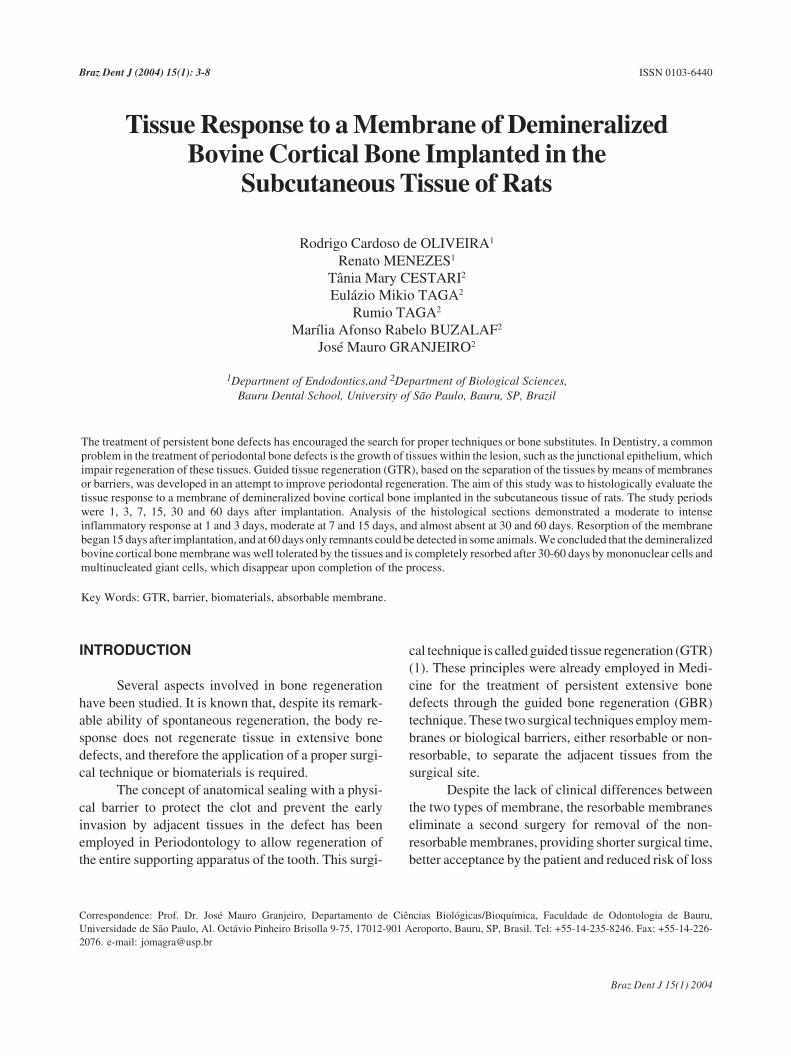
infiltrate was mild and comprised of mainly polymor-phonuclear leukocytes (PMNs) and lymphocytes (Fig-ure 1A). Some congested vessels were observed closeto the membrane.
3 Days
The membrane was apparently intact in all speci-mens (Figure 1B). The inflammatory infiltrate wasmoderate to intense and comprised of PMNs and lym-phocytes, revealing a larger number of blood vesselsand capillaries around the membrane.
7 Days
The membrane was still apparently intact, how-ever, with few sites of degradation (Figure 1C). Theinflammatory infiltrate comprising PMNs and lympho-cytes was intense, with a discrete degree of fibrosis andangioblastic proliferation. Angiogenesis in this periodwas remarkable in relation to the earlier periods. Giantcells were observed along the membrane, especially onthe borders.
15 Days
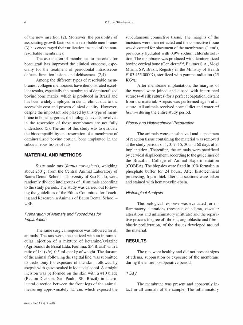
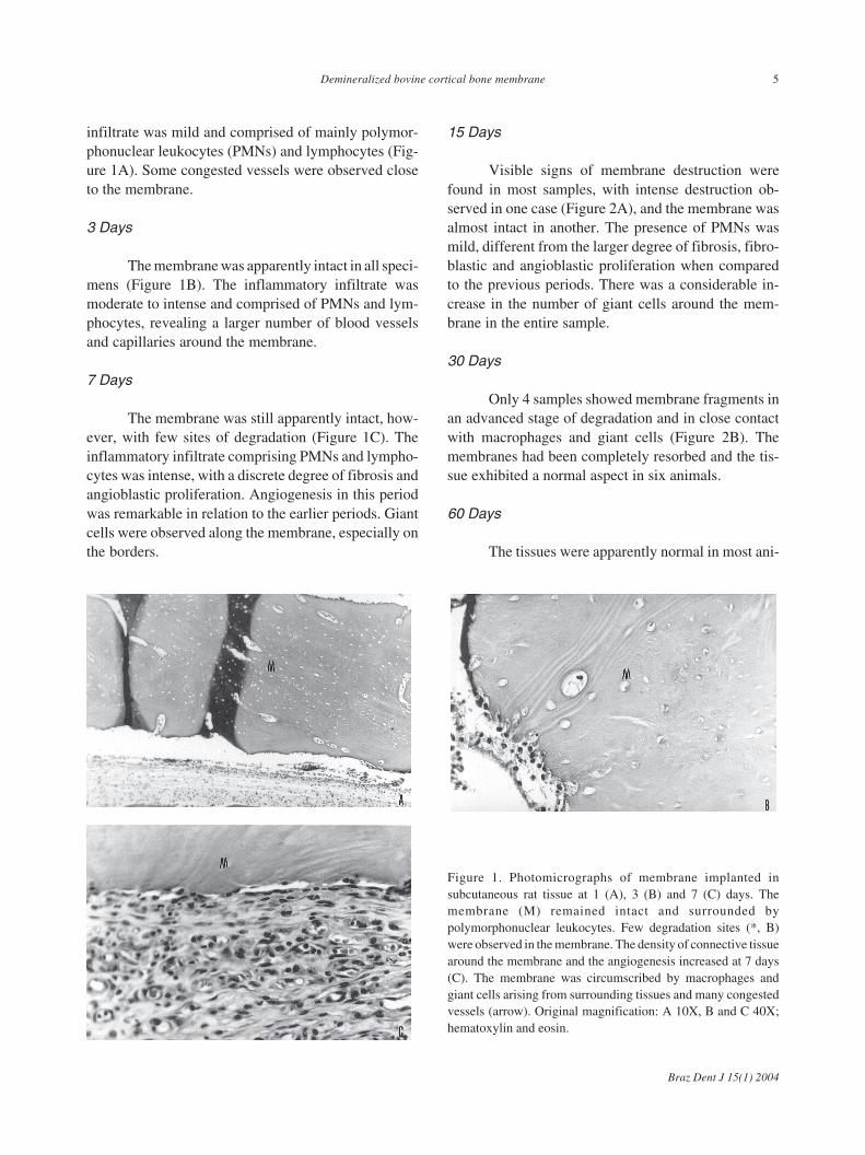
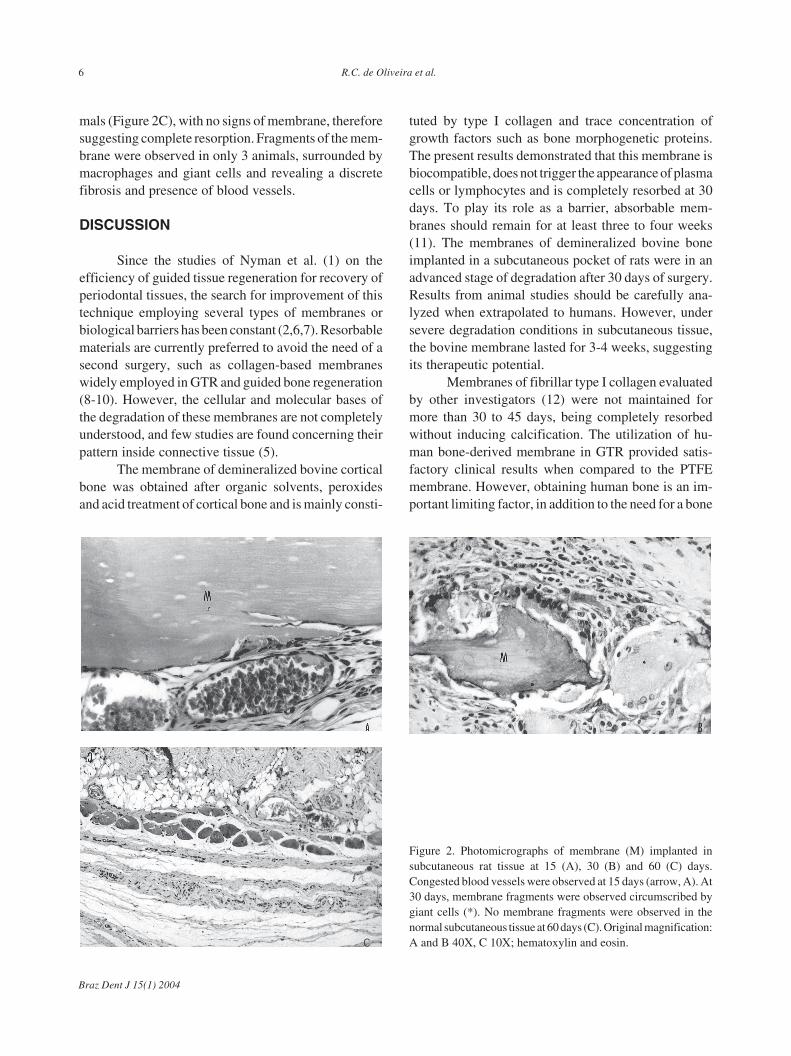
Visible signs of membrane destruction werefound in most samples, with intense destruction ob-served in one case (Figure 2A), and the membrane wasalmost intact in another. The presence of PMNs wasmild, different from the larger degree of fibrosis, fibro-blastic and angioblastic proliferation when comparedto the previous periods. There was a considerable in-crease in the number of giant cells around the mem-brane in the entire sample.
30 Days
Only 4 samples showed membrane fragments inan advanced stage of degradation and in close contactwith macrophages and giant cells (Figure 2B). Themembranes had been completely resorbed and the tis-sue exhibited a normal aspect in six animals.
60 Days
The tissues were apparently normal in most ani-
Figure 1. Photomicrographs of membrane implanted insubcutaneous rat tissue at 1 (A), 3 (B) and 7 (C) days. Themembrane (M) remained intact and surrounded bypolymorphonuclear leukocytes. Few degradation sites (*, B)were observed in the membrane. The density of connective tissuearound the membrane and the angiogenesis increased at 7 days(C). The membrane was circumscribed by macrophages andgiant cells arising from surrounding tissues and many congestedvessels (arrow). Original magnification: A 10X, B and C 40X;hematoxylin and eosin.
Braz Dent J 15(1) 2004
6 R.C. de Oliveira et al.
mals (Figure 2C), with no signs of membrane, thereforesuggesting complete resorption. Fragments of the mem-brane were observed in only 3 animals, surrounded bymacrophages and giant cells and revealing a discretefibrosis and presence of blood vessels.
DISCUSSION
Since the studies of Nyman et al. (1) on theefficiency of guided tissue regeneration for recovery ofperiodontal tissues, the search for improvement of thistechnique employing several types of membranes orbiological barriers has been constant (2,6,7). Resorbablematerials are currently preferred to avoid the need of asecond surgery, such as collagen-based membraneswidely employed in GTR and guided bone regeneration(8-10). However, the cellular and molecular bases ofthe degradation of these membranes are not completelyunderstood, and few studies are found concerning theirpattern inside connective tissue (5).
The membrane of demineralized bovine corticalbone was obtained after organic solvents, peroxidesand acid treatment of cortical bone and is mainly consti-
tuted by type I collagen and trace concentration ofgrowth factors such as bone morphogenetic proteins.The present results demonstrated that this membrane isbiocompatible, does not trigger the appearance of plasmacells or lymphocytes and is completely resorbed at 30days. To play its role as a barrier, absorbable mem-branes should remain for at least three to four weeks(11). The membranes of demineralized bovine boneimplanted in a subcutaneous pocket of rats were in anadvanced stage of degradation after 30 days of surgery.Results from animal studies should be carefully ana-lyzed when extrapolated to humans. However, undersevere degradation conditions in subcutaneous tissue,the bovine membrane lasted for 3-4 weeks, suggestingits therapeutic potential.
Membranes of fibrillar type I collagen evaluatedby other investigators (12) were not maintained formore than 30 to 45 days, being completely resorbedwithout inducing calcification. The utilization of hu-man bone-derived membrane in GTR provided satis-factory clinical results when compared to the PTFEmembrane. However, obtaining human bone is an im-portant limiting factor, in addition to the need for a bone
Figure 2. Photomicrographs of membrane (M) implanted insubcutaneous rat tissue at 15 (A), 30 (B) and 60 (C) days.Congested blood vessels were observed at 15 days (arrow, A). At30 days, membrane fragments were observed circumscribed bygiant cells (*). No membrane fragments were observed in thenormal subcutaneous tissue at 60 days (C). Original magnification:A and B 40X, C 10X; hematoxylin and eosin.C
Braz Dent J 15(1) 2004
Demineralized bovine cortical bone membrane 7
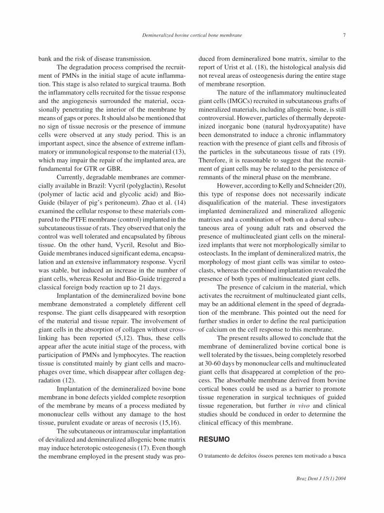
bank and the risk of disease transmission.The degradation process comprised the recruit-
ment of PMNs in the initial stage of acute inflamma-tion. This stage is also related to surgical trauma. Boththe inflammatory cells recruited for the tissue responseand the angiogenesis surrounded the material, occa-sionally penetrating the interior of the membrane bymeans of gaps or pores. It should also be mentioned thatno sign of tissue necrosis or the presence of immunecells were observed at any study period. This is animportant aspect, since the absence of extreme inflam-matory or immunological response to the material (13),which may impair the repair of the implanted area, arefundamental for GTR or GBR.
Currently, degradable membranes are commer-cially available in Brazil: Vycril (polyglactin), Resolut(polymer of lactic acid and glycolic acid) and Bio-Guide (bilayer of pig’s peritoneum). Zhao et al. (14)examined the cellular response to these materials com-pared to the PTFE membrane (control) implanted in thesubcutaneous tissue of rats. They observed that only thecontrol was well tolerated and encapsulated by fibroustissue. On the other hand, Vycril, Resolut and Bio-Guide membranes induced significant edema, encapsu-lation and an extensive inflammatory response. Vycrilwas stable, but induced an increase in the number ofgiant cells, whereas Resolut and Bio-Guide triggered aclassical foreign body reaction up to 21 days.
Implantation of the demineralized bovine bonemembrane demonstrated a completely different cellresponse. The giant cells disappeared with resorptionof the material and tissue repair. The involvement ofgiant cells in the absorption of collagen without cross-linking has been reported (5,12). Thus, these cellsappear after the acute initial stage of the process, withparticipation of PMNs and lymphocytes. The reactiontissue is constituted mainly by giant cells and macro-phages over time, which disappear after collagen deg-radation (12).
Implantation of the demineralized bovine bonemembrane in bone defects yielded complete resorptionof the membrane by means of a process mediated bymononuclear cells without any damage to the hosttissue, purulent exudate or areas of necrosis (15,16).
The subcutaneous or intramuscular implantationof devitalized and demineralized allogenic bone matrixmay induce heterotopic osteogenesis (17). Even thoughthe membrane employed in the present study was pro-
duced from demineralized bone matrix, similar to thereport of Urist et al. (18), the histological analysis didnot reveal areas of osteogenesis during the entire stageof membrane resorption.
The nature of the inflammatory multinucleatedgiant cells (IMGCs) recruited in subcutaneous grafts ofmineralized materials, including allogenic bone, is stillcontroversial. However, particles of thermally deprote-inized inorganic bone (natural hydroxyapatite) havebeen demonstrated to induce a chronic inflammatoryreaction with the presence of giant cells and fibrosis ofthe particles in the subcutaneous tissue of rats (19).Therefore, it is reasonable to suggest that the recruit-ment of giant cells may be related to the persistence ofremnants of the mineral phase on the membrane.
However, according to Kelly and Schneider (20),this type of response does not necessarily indicatedisqualification of the material. These investigatorsimplanted demineralized and mineralized allogenicmatrixes and a combination of both on a dorsal subcu-taneous area of young adult rats and observed thepresence of multinucleated giant cells on the mineral-ized implants that were not morphologically similar toosteoclasts. In the implant of demineralized matrix, themorphology of most giant cells was similar to osteo-clasts, whereas the combined implantation revealed thepresence of both types of multinucleated giant cells.
The presence of calcium in the material, whichactivates the recruitment of multinucleated giant cells,may be an additional element in the speed of degrada-tion of the membrane. This pointed out the need forfurther studies in order to define the real participationof calcium on the cell response to this membrane.
The present results allowed to conclude that themembrane of demineralized bovine cortical bone iswell tolerated by the tissues, being completely resorbedat 30-60 days by mononuclear cells and multinucleatedgiant cells that disappeared at completion of the pro-cess. The absorbable membrane derived from bovinecortical bones could be used as a barrier to promotetissue regeneration in surgical techniques of guidedtissue regeneration, but further in vivo and clinicalstudies should be conduced in order to determine theclinical efficacy of this membrane.
RESUMO
O tratamento de defeitos ósseos perenes tem motivado a busca
Braz Dent J 15(1) 2004
8 R.C. de Oliveira et al.
por técnicas ou substitutos ósseos adequados. Na odontologia,um problema comum no tratamento de defeitos ósseosperiodontais é o crescimento de tecidos competidores para ointerior da lesão, como o epitélio juncional da gengiva,prejudicando a regeneração desses tecidos. Buscando melhorar aregeneração periodontal foi desenvolvida a técnica da RegeneraçãoTecidual Guiada (RTG) baseada na separação dos tecidos atravésde membranas ou barreiras. Objetivo deste estudo foi avaliarhistologicamente a resposta tecidual à membrana obtida do ossocortical bovino desmineralizado, implantada em subcutâneo deratos. Os períodos analisados foram de 1, 3, 7, 15, 30 e 60 diasapós a implantação. A análise dos cortes histológicos mostrouresposta inflamatória de moderada a intensa nos períodos de 1 e3 dias, moderada aos 7 e 15 dias, e praticamente inexistente aos30 e 60 dias. A reabsorção da membrana se iniciou 15 dias pós-implantação e ao final de 60 dias apenas resquícios foramdetectados em alguns animais. Concluímos que a membranaderivada do osso cortical bovino desmineralizado é bem toleradapelos tecidos sendo completamente reabsorvida após 30-60 diaspor células mononucleadas e células gigantes multinucleadasque desaparecem ao final do processo.
ACKNOWLEDEGMENTS
The authors are very grateful to Thelma L. daSilva, Ovídio dos Santos Sobrinho, Gilmar V. da Silva,Pâmela R.A. Leoni, Luiz C. da Silva, Erasmo G. daSilva, and Daniele S. Ceolin for skillfull technicalassistance. This study was supported by Fundação deAmparo à Pesquisa do Estado de São Paulo, ConselhoNacional de Desenvolvimento Científico e Tecnológico(R.C.O. fellowship), Coordenação de Aperfeiçoamentode Pessoal de Nível Superior, and the University of SãoPaulo.
REFERENCES
1. Nyman S, Lindhe J, Karring T, Rylander H. New attachmentfollowing surgical treatment of human periodontal disease. J ClinPeriodontol 1982;9:290-296.
2. Wang HL, Macneil RL. Guided tissue regeneration. Absorbablebarriers. Dent Clin N Am 1998;42:505-522.
3. Chen CC, Wang HL, Lopatin DE, O’Neal RB, Macneil RL.Bacterial adherence to guided tissue regeneration barrier mem-branes exposed to the oral environment. J Periodontol1997;68:172-179.
4. Bunyaratavej P, Wang HL. Collagen membranes: A review. JPeriodontol 2001;72:215-229.
5. Galgut P, Pitrola R, Waite I, Doyle C, Smith R. Histologicalevaluation of biodegradable and non-degradable membranes
placed transcutaneously in rats. J Clin Periodontol 1991;18:581-586.
6. Blumenthal NM. A clinical comparison of collagen membraneswith ePTFE membranes in the treatment of human mandibularbuccal class II furcation defects. J Periodontol 1993;64:925-933.
7. Novaes Junior AB, Novaes AB. Guided tissue regeneration ver-sus hemisection in the treatment of furcation lesions. A clinicalanalysis. Braz Dent J 1993;3:99-102.
8. Nociti Junior FH, Caffesse RG, Sallum EA, Machado MA, StefaniCM, Sallum AW. Clinical study of guided bone regeneration and/or bone grafts in the treatment of ligature-induced peri-implantitisdefects in dogs. Braz Dent J 2001;12:127-131.
9. Oh TJ, Meraw SJ, Lee EJ, Giannobile WV, Wang HL. Compara-tive analysis of collagen membranes for the treatment of implantdehiscence defects. Clin Oral Impl Res 2003;14:80-90.
10. Dietrich T, Zunker P, Dietrich D, Bernimoulin JP. Periapical andperiodontal healing after osseous grafting and guided tissue re-generation treatment of apicomarginal defects in periradicularsurgery: Results after 12 months. Oral Surg Oral Med Oral PathOral Radiol Endod 2003;95:474-482.
11. Minabe M. Selective periodontal tissue reconstruction therapy. JPeriodontol 1991;62:171-179.
12. Boon ME, Ruijgrok JM, Vardaxis MJ. Collagen implants remainsupple not allowing fibroblast ingrowth. Biomaterials1995;16:1089-1093.
13. Holland R, de Souza V, Nery MJ, Faraco Junior IM, Bernabe PF,Otoboni Filho JA, Dezan Junior E. Reaction of rat connectivetissue to implanted dentin tube filled with mineral trioxide aggre-gate, Portland cement or calcium hydroxide. Braz Dent J2001;12:3-8.
14. Zhao S, Pinholt EM, Madsen JE, Donath K. Histological evalua-tion of different biodegradable and non-biodegradable mem-branes implanted subcutaneously in rats. J Cranio-MaxillofacialSurg 2000;2000:116-122.
15. Taga R, Hassunuma CY, Cestari TM, Ferreira PM. Destino demembrana de cortical óssea bovina colocada em posiçãosubperiostica na calvária de cobaia. Rev Bras Implant 1997;5:24-29.
16. Benoit Núñez JS. Análise histológica da reabsorção e dabiocompatibilidade de membrana de cortical óssea bovinacolocada sobre calvária de cobaia decorticada. [Master's Thesis].Bauru: Faculdade de Odontologia de Bauru, Universidade de SãoPaulo; 1999. 100p.
17. Reddi AH, Huggins C. Biochemical sequences in the transforma-tion of normal fibroblasts in adolescent rats. Proc Natl Acad SciUSA 1972;69:1601-1605.
18. Urist MR. Bone: formation by autoinduction. Science1965;150:893-899.
19. Oliveira RC, Sicca CM, Silva TL, Cestari TM, Oliveira DT,Buzalaf MAR, Taga R, Taga EM, Granjeiro JM. Efeito datemperatura de desproteinização no preparo de osso corticalbovino microgranular. Avaliação microscópica e bioquímica daresposta celular em subcutâneo de ratos. Rev FOB 1999;7:85-93.
20. Kelly JD, Schneider GB. Morphological and histochemical com-parison of cells elicited by ectopic bone implants and tibialosteoclasts. Am J Anat 1991;192:45-54.
Accepted September 29, 2003