tissues chapter 4 tissues chapter 4. tissues tissues are groups of cells and extracellular material...
TRANSCRIPT

TissuesChapter 4TissuesChapter 4

Tissues
Tissues are groups of cells and extracellular material that perform specific functions.
The four tissue types, in varying combinations, form all of the structures of the human body.
Histology - The study of tissues

Four Basic
Tissue Types Epithelial Connective Muscular Neural (nervous)
Copyright © 2007 Pearson Education, Inc., publishing as Benjamin Cummings

Epithelial Tissue (epithelium) Characteristics
Cells closely packed & attached to each other
Apical cells exposed to external environment or internal surface
Cells attached to connective tissue at basement membrane
Avascular - Lack blood vesselsRegenerates easily - cells continually
replaced
Epithelial Tissue
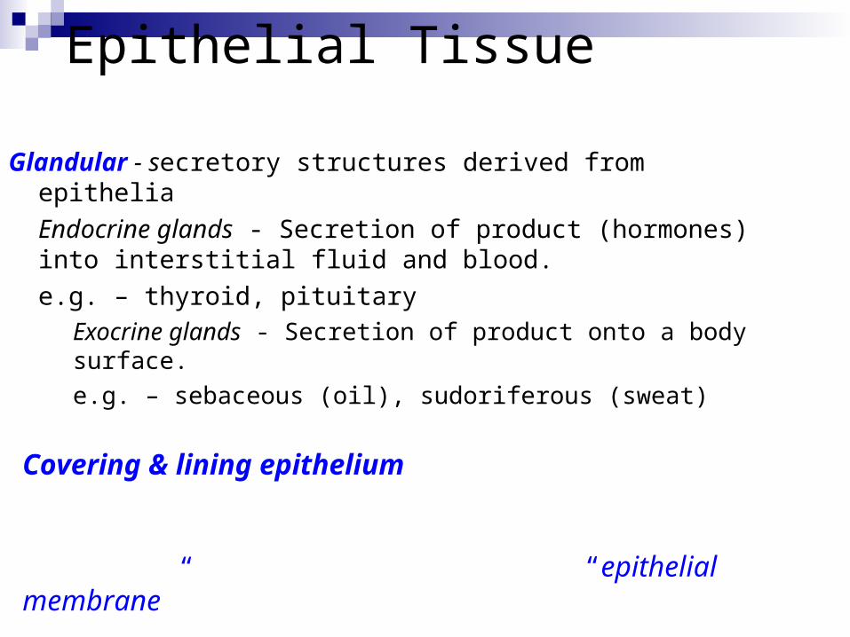
Glandular - secretory structures derived from epithelia
Endocrine glands - Secretion of product (hormones) into interstitial fluid and blood.
e.g. – thyroid, pituitaryExocrine glands - Secretion of product onto a body
surface.
e.g. – sebaceous (oil), sudoriferous (sweat)
Covering & lining epithelium - an avascular layer of cells that lines internal or external surfaces & covers organs within cavities; - always bound to CT creating an “epithelial membrane”
Subtypes of epithelial tissue -

Epithelial Membranes
Epithelial tissue bound to connective tissue
Connection of tissues occurs at the basement membrane - noncellular meshwork anchors basal cells
Basement membrane creates a semi-permeable junction for diffusion of O2, nutrients & wastes
Epithelial Membranes

Epithelial membranes Types of epithelial
membranes:Mucous membranes
(mucosa)Serous membranes
(serosa)Cutaneous membrane
(skin)

Mucous membranes (mucosa)
Line body cavities that open to exterior environment
Epithelial cells secrete mucus to moisten & protect surface
Examples: digestive tract, respiratory passageway, urinary tract, reproductive passageways
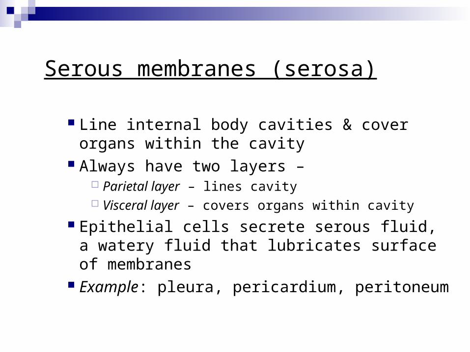
Serous membranes (serosa)
Line internal body cavities & cover organs within the cavity
Always have two layers – Parietal layer – lines cavity Visceral layer – covers organs within cavity
Epithelial cells secrete serous fluid, a watery fluid that lubricates surface of membranes
Example: pleura, pericardium, peritoneum

Cutaneous membrane (a.k.a. skin)
Covers & protects body surface from external environment
Epithelial layer is epidermis Connective layer is dermis

Simple - one layer of cells Stratified – more than one layer of cells Pseudostratified – looks like more than one layer but all
cells contact basement membrane
Cell shape
Classifying covering & lining Epithelia: Number of layers
Squamous - flat Cuboidal – cube-shaped Columnar - tall columns

Columnar cells may be modified with microvilli cilia goblet cells
Figure 4-3

Epithelial Tissue
Table 4-1

Epithelial Tissue Simple Squamous Epithelium
Figure 4-4(a)
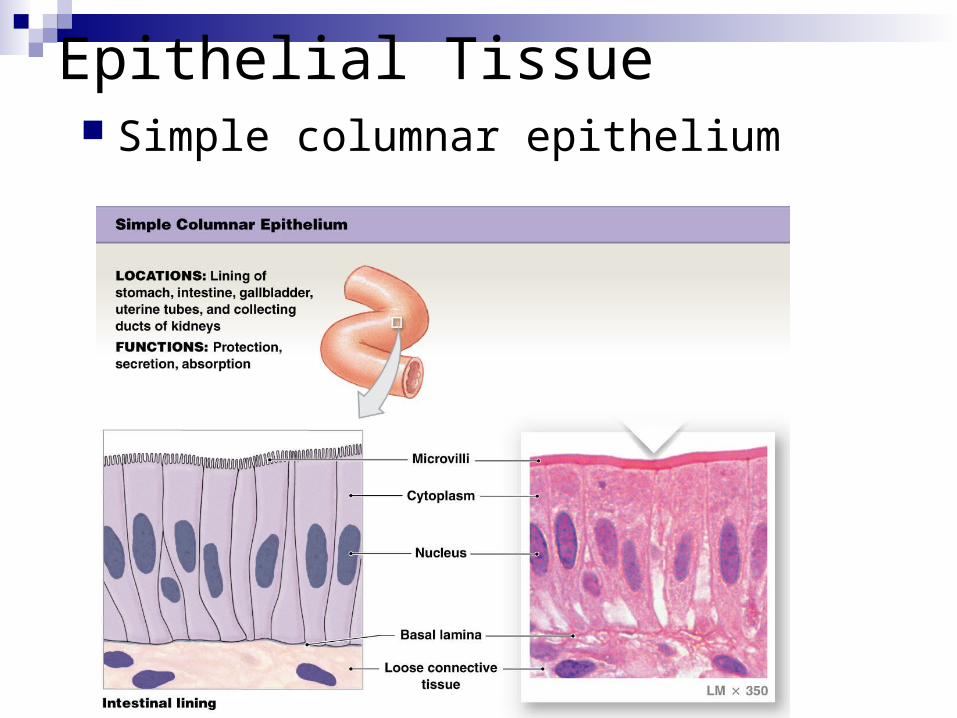
Epithelial Tissue Simple columnar epithelium

Epithelial Tissue Stratified Squamous Epithelium
Figure 4-5(c)
2 types: Keratinized
Non-keratinized
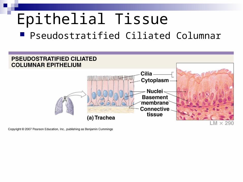
Epithelial Tissue Pseudostratified Ciliated Columnar
Figure 4-5(a)
Connective Tissue (CT)
Characteristics: Cells differ depending on specific type of CT (e.g.
mesenchymal, fibroblasts, chondrocytes, osteocytes) Cells usually widely spaced with intercellular matrix
between Usually well vascularized (exception: cartilage) Usually do not regenerate easily Repair ability varies depending on type of CT

Connective Tissues Components of Connective Tissues:
Specialized cellsExtracellular matrix
Ground substance Varies in consistency from liquid to gel-like to solid, depending on
specific CT
Protein fibers Collagen – provides strength Elastic – provides elasticity, resiliency Reticular – provides support

Connective Tissues
Whereas in epithelial tissue, the cell shape & layers were important to determine the function of the tissue, in connective tissues, the matrix of the tissue most directly determines the functional qualities of the tissue.
Classification of Connective Tissues
Embryonic CT - MesenchymeConnective tissue proper
Loose Dense
Supportive connective tissues Cartilage Bone (osseous) tissue
Fluid connective tissues Blood Lymph

Connective TissuesMajor Types of Connective Tissue
Figure 4-7
MESENCHYME

MesenchymeAn embryonic CT with mesenchymal cells in a thick
fluid ground substance with some collagen & reticular fibers.
Mesenchyme is the precursor to all other forms of CT
Connective Tissue “Proper”Most common type of cell present is the
fibroblast, but these CT’s may also contain adipocytes, macrophages, & other WBCs.
The cells are surrounded by a syrupy ground substance that contains hyaluronic acid.
Fibers vary & may be arranged loosely or densely packed together.

Connective Tissue ProperDepending on the arrangement of fibers, CT
proper can be classified into:
Loose CTsAreolarAdipose
Dense CTs
• Dense regular (collagenous)
• Dense irregular

Areolar Connective Tissue
Figure 4-9(a)

Adipose Connective Tissue
Figure 4-9(b)

Dense Regular Connective Tissue
Figure 4-9(c)

Dense Irregular Connective Tissue

Supportive Connective Tissues
Chondrocytes in lacunae of interstitial fluid within a firm gel-like ground substance of chondroitin sulfate
Avascular Covered by a fibrous perichondrium Three types of cartilage
Hyaline cartilageElastic cartilageFibrocartilage
Cartilage

Hyaline Cartilage
Figure 4-10(a)

Elastic Cartilage
Figure 4-10(b)

Fibrocartilage
Figure 4-10(c)

Supportive Connective Tissues
Osteocytes in lacunae of interstitial fluid within a calcified matrix
Matrix comprised of osteoid (mainly collagen) & calcium salts (mainly Ca3 (PO4)2 )
Because of the density of the matrix, osteocytes communicate & receive O2 / nutrients across canaliculi
Osseous tissue covered by fibrous periosteumWell vascularized tissue
Bone (Osseous Tissue)
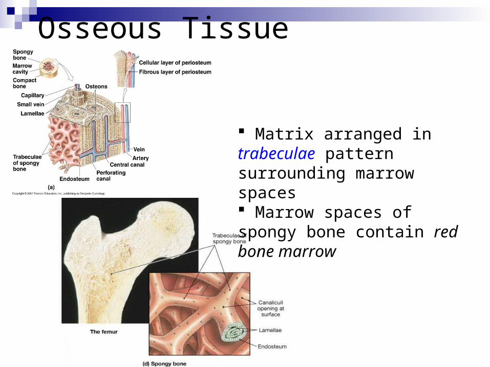
Osseous (Bone) Tissue
Bone tissue classified on the basis of the pattern (functional unit) of the matrix
Two types of bone tissue –
Spongy (cancellous)
Dense (compact)
Osseous Tissue
Fig. 6-3
Spongy (cancellous) bone tissue: Matrix arranged in trabeculae pattern surrounding marrow spaces Marrow spaces of spongy bone contain red bone marrow
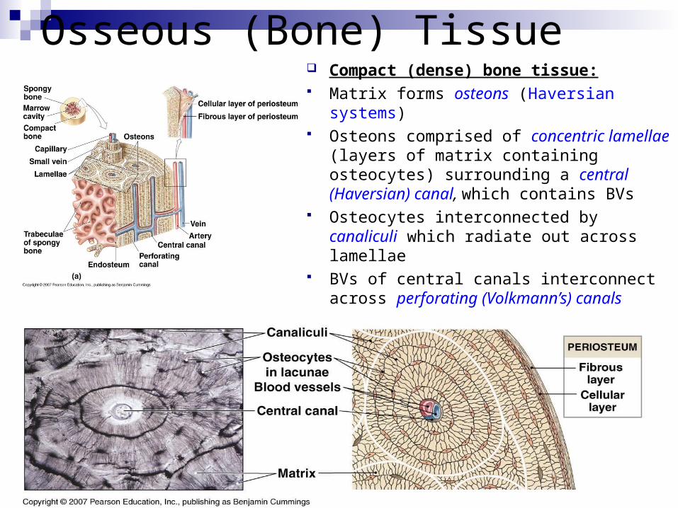
Osseous (Bone) Tissue Compact (dense) bone tissue: Matrix forms osteons (Haversian systems) Osteons comprised of concentric lamellae
(layers of matrix containing osteocytes) surrounding a central (Haversian) canal, which contains BVs
Osteocytes interconnected by canaliculi which radiate out across lamellae
BVs of central canals interconnect across perforating (Volkmann’s) canals
Fluid (liquid) Connective Tissues
Cells + a liquid ground substance Blood
RBCs, WBCs, platelets + plasma Lymph
Lymphocytes (WBCs) + lymph fluid

Tissue Injuries and Repair
Many different types of injuries can affect tissues – physical (e.g. cuts, bruises), thermal (e.g. burns), chemical, infections
An injury usually harms multiple tissues simultaneously
Tissues make coordinated response to restore homeostasis
Two response processes Inflammation Repair (Regeneration)
Tissue Injuries and Repair
Inflammation (a.k.a. the inflammatory response) Homeostatic response designed to isolate the injured
area & cleanup damaged tissue Cells within CTs known as mast cells release chemicals
(histamine, heparin) which cause vasodilation & increased capillary permeability

Tissue Injuries & Repair
Vasodilation (resulting in increased blood flow) & increased capillary permeability lead to the 4 classic signs of inflammation:
Warmth Redness Swelling Pain

Tissue Injuries and Repair
Repair (Regeneration) Response designed to repair/replace damaged
tissues & restore function Fibroblasts (CT cells) in damaged area & lay down
collagen fibers to create scar tissue (fibrous tissue)

Tissue Injuries and Repair
Degree of replacement to original tissue type depends on type of tissue
Epithelial cells regenerate to replace the damaged epithelial tissues
CT proper, bone tissues & smooth muscle heal fairly well
Cartilage, neural tissue, skeletal & cardiac muscle tissues do not heal well at all