title of the paper - applied ecology and … and technology houari boumediene- bp 32 el...
TRANSCRIPT
Bellout et al.: Impact of edaphic hydrocarbon pollution on pea roots
- 511 -
APPLIED ECOLOGY AND ENVIRONMENTAL RESEARCH 14(2): 511-525.
http://www.aloki.hu ● ISSN 1589 1623 (Print) ● ISSN 1785 0037 (Online)
DOI: http://dx.doi.org/10.15666/aeer/1402_511525
2016, ALÖKI Kft., Budapest, Hungary
IMPACT OF EDAPHIC HYDROCARBON POLLUTION ON THE
MORPHOLOGY AND PHYSIOLOGY OF PEA ROOTS
(PISUM SATIVUM L.)
BELLOUT, Y.1 – KHELIF, L.
2 – GUIVARCH, A.
3 – HAOUCHE, L.
4 – DJEBBAR, R.
2 –
CAROL, P.3 – ABROUS BELBACHIR, O.
2*
1Department of Biology, Faculty of Sciences, UniversityMhamad Bougara Boumerdes- Algeria
2Laboratory of biology and Physiology of organisms, Faculty of Sciences, University of
Sciences and Technology Houari Boumediene- BP 32 El Alia-16111-Algeria
3Sorbonne Universités, UPMC Univ. Paris 06, 4 Place Jussieu, 75005, Paris, France
4Scientific Institute of Public Service (ISSeP)-Rue de la platinerie, Colfontaine- Belgium
*Corresponding author
e-mail: [email protected]
(phone +213-21-24-72-17; fax +213-21-24-79-50)
(Received 6th Aug 2015; accepted 26th Feb 2016)
Abstract. Exposure to persistent organic hydrocarbon pollutants can have deleterious effects on the
growth, physiology and anatomy of plants. Sand collected at an oil-drilling quagmire in southern Algeria
was analyzed by GC-FID and found to contain 18mg.Kg-1
hydrocarbons. Pisum sativum L. (pea) plants
were grown in laboratory conditions in sandy soil from the site. Plants growing in hydrocarbon polluted
sandy soil had shorter primary roots and fewer lateral roots than control plants growing in non-polluted
sandy soil. However lateral root dry weight was 35% higher than control. Pollutant-induced oxidative
stress on pea roots resulted in lipid peroxidation and accumulation of MDA, H2O2 and O2- in root tips.
Enzymatic detox activities of superoxide dismutase and peroxidase were also over 40% higher in plants
growing on polluted soil than in controls. The anatomy of pea roots was also affected by hydrocarbon-
polluted soil, because xylem vessel differentiation was delayed and an unusual supplementary cell layer
was formed in the endoderm. These data suggest pea plants adapt morphologically and anatomically to
polluted soil.
Keywords: hydrocarbons, pea roots, growth, oxidative stress, anatomy
Introduction
Since the mid-1980s, hydrocarbon contamination has become a critical
environmental problem worldwide due to its adverse effects on the environment and
health (Li et al., 1993). In Algeria, petroleum is one of the main energy resources.
Petroleum exploration can cause soil pollution because drilling mud is stored in
quagmire spill sites so the soil surface becomes impregnated with total petroleum
hydrocarbons (TPHs). TPHs are complex mixtures of various hydrocarbons that can be
found at petrochemical sites and storage areas, waste disposal pits, refineries and oil
spill sites (McElroy et al., 1989).
Plants are a dominant biotic component of ecosystems and as sessile organisms; they
can be subjected to long-term pollution from hydrocarbons (McCarthy and Tschaplinski,
1991). Organic pollutants are able to penetrate plant organs through different mechanisms
(Gao and Zhu, 2004). In plant tissues, organic pollutants can migrate from roots to leaves,
and within the plant organs, they can be modified by conjugation, hydroxylation and by

Bellout et al.: Impact of edaphic hydrocarbon pollution on pea roots
- 512 -
APPLIED ECOLOGY AND ENVIRONMENTAL RESEARCH 14(2): 511-525.
http://www.aloki.hu ● ISSN 1589 1623 (Print) ● ISSN 1785 0037 (Online)
DOI: http://dx.doi.org/10.15666/aeer/1402_511525
2016, ALÖKI Kft., Budapest, Hungary
cytochrome containing monooxygenase enzymes (Korte et al., 2000). Plant growth is
affected by decrease of biomass in oiled areas (Culbertson et al., 2008). For maize
germination and growth in crude-oil polluted soil, the effect is proportional to the
concentration of the crude oil in the environment (Ogboghodo et al., 2004). The
considerable effect of polycyclic aromatic hydrocarbon fluoranthene (FLT) exposure was
to inhibit germination of seeds, retard growth and affect root morphology (Kummerova et
al, 2012). Anoliefo (1991) found evidence of cell disruption in roots and other organs and
the presence of oil films in the epidermal and cortical regions of the root, stem, and
leaves. Crude oil induced environmental stress in the seedlings causing inhibition of total
amylase and starch phosphorylase activities and mitotic activity of root meristems
(Achuba, 2006). The harmful effects of petroleum hydrocarbons in soils include
inhibition of seed germination, reduction of photosynthetic pigments, slowdown of
nutrient assimilation and shortening of roots and can disrupt the plant root architecture
(Smith et al., 2006). Some other workers have also used anatomical changes to monitor
environmental pollution (Gill et al., 1992). Petroleum hydrocarbons were reported to alter
the shape and size of parenchyma tissue and reduce the intercellular space in the cortex of
the stem and roots (Omosum et al., 2008).
In polluted soils, plants may experience a combined stress from nutritional
deficiency and chemical toxicity. Indeed, abiotic stress such as that caused by
polycyclic aromatic hydrocarbon exposure can also stress plants by generating reactive
oxygen species (ROS) (Sun et al., 2002). ROS such as superoxide radical (O2-),
hydrogen peroxide (H2O2) and hydroxyl radical (OH-), are generated as by-products of
normal metabolism in different subcellular compartments. Moreover, the imposition of
biotic or abiotic stress may give rise to an excessive concentration of ROS, resulting in
oxidative damage at cellular level that can be mitigated and repaired by a complex
antioxidant system (Romero-Puertas et al., 2007). Stress induced ROS accumulation is
counteracted by enzymatic antioxidant systems that include a variety of scavengers,
such as superoxide dismutase (SOD), ascorbate peroxidase (APX), peroxidase (POD),
glutathione S-transferase (GST), catalase (CAT) and non-enzymatic low molecular
metabolites (Mittler et al., 2004).
The impact of hydrocarbon contaminants on plant roots, which are in direct contact
with the pollutants, is not as well documented as the effect on leaves or
photosynthesis. For this reason, we aimed to evaluate the effect of hydrocarbons from
an edaphic pollution on the growth of pea roots (Pisum sativum L.) in controlled
laboratory conditions. We observed changes in ROS (O2-, H2O2,) and ROS detoxifying
enzymes, MDA levels were enhanced in pea roots growing in polluted soil.
Furthermore an unexpected morphological adaptation was observed at the level of the
endoderm cell layer.
Materials and methods
Soil
Soil samples were collected in the region of Hassi Messaoud-Ouargla province of
southern Algeria. Contaminated soil was sampled at the site of a disused oil quagmire.
Control soil was sampled at a site two kilometers from the quagmire. Three soil
samples were collected at 0-30 cm depth using a stainless steel sampler. The three
samples were mixed to form a single sample that was air-dried and then sieved (2 mm
sieve) before analysis.

Bellout et al.: Impact of edaphic hydrocarbon pollution on pea roots
- 513 -
APPLIED ECOLOGY AND ENVIRONMENTAL RESEARCH 14(2): 511-525.
http://www.aloki.hu ● ISSN 1589 1623 (Print) ● ISSN 1785 0037 (Online)
DOI: http://dx.doi.org/10.15666/aeer/1402_511525
2016, ALÖKI Kft., Budapest, Hungary
Hydrocarbon analysis
Before hydrocarbon measurement soil samples were chemically dried, by adding
Na2SO4. Analysis of hydrocarbons in soil was conducted in the ISSeP laboratory
(Scientific Institute of Public Service, Colfontaine, Belgium) using techniques
developed in the laboratory.
Polycyclic aromatic hydrocarbon (PAH) content in soil samples
The 16 PAH congeners listed as priority pollutants by the US Environmental
Protection Agency (US-EPA) were analyzed in the soil samples. The extraction of these
compounds was performed by the Accelerated Solvent Extractor (ASE) technology
(Dionex ASE 350), allowing a solid/liquid extraction with dichloromethane at 150 °C at
a pressure of 1500 psi. The dichloromethane extract was extracted with hexane and
cleaned up with aluminium oxide. The sample was extracted with acetonitrile and the
extract was concentrated at room temperature under a gentle stream of nitrogen. PAHs
were then separated by ultra-performance liquid chromatography (UPLC) and detected
using a fluorescence detector with appropriate excitation and emission wavelengths for
the 15 PAH and a diode array detector for acenaphthylene. Quantification was
performed by external standard calibration. Ultra performance liquid chromatography
(UPLC) was performed with a 1.8 µm, 2.1mm ID × 100 mm AZE-PAH column at a
flow rate of 0.4 ml/min. The mobile phase consisted of acetonitrile-water (50:50, v/v)
for 9 min and 100% acetonitrile for 3.5 min.
Hydrocarbon index
The extraction of total petroleum hydrocarbons (TPHs) was performed by a ASE
technology (Dionex ASE 350), allowing a solid/liquid extraction at 100°C under a
pressure of 1500 psi, with a solvent mixture of n-hexane-acetone (50:50, v/v). The
recovered extract was washed with an aqueous solution of hydrated magnesium sulfate
to remove acetone and then cleaned up on a Florisil column (6 mm diameter,6 cm long).
The eluate was then concentrated with a Syncore Analyst Evaporator to 0.5 ml to be
used for analysis by gas chromatography (GC-FID) using a Column VF-5ht 15 m × 0.25
mm × 0.10 μm with a “splitless” injection technique (30 sec) and a pulse injection (10
psi for 1 min). The analyses were conducted under the following conditions: injection
temperature, 300°C; injection volume, 1µl; carrier gas, helium; oven temperature
program, 40°C for 5 min to 300°C and 300 °C for 5 min; flame ionization detector
temperature, 330 °C. The following fractionation was performed: (C10-C12), (C12-
C16), (C16-C21), (C21-C35) and (C35-C40). The limits of integration were placed at
the corresponding retention times of n-alkanes (C12, C16, C21 and C35), areas of each
fraction were measured and the calculation based on the total area C10-C40.
Plant growth and root growth analysis
Pea seeds (Pisum sativum L.)“Kelvedon wonder” were surface-sterilized with 0.1%
sodium hypochlorite (NaClO) for 10 min, rinsed and soaked in distilled water at room
temperature for 12 h. The seeds were germinated on water-imbibed paper in sealed
plastic dishes. After three days, pea seedlings were transplanted into plastic containers
containing 250 g of a mixture of peat and sand, polluted or control, (80:20, w/w). Plants
were cultivated for 21 days under controlled conditions in a growth chamber with a 16 h

Bellout et al.: Impact of edaphic hydrocarbon pollution on pea roots
- 514 -
APPLIED ECOLOGY AND ENVIRONMENTAL RESEARCH 14(2): 511-525.
http://www.aloki.hu ● ISSN 1589 1623 (Print) ● ISSN 1785 0037 (Online)
DOI: http://dx.doi.org/10.15666/aeer/1402_511525
2016, ALÖKI Kft., Budapest, Hungary
light (90 E) and 8 h dark cycle at a constant temperature of 25 °C and relative air
humidity of 60%, watered by pure water according to usable water to field capacity
calculated in gram of water per gram of sandy soil and after wards, every other day till
the end of the experiment. The dry weight of roots, the number of lateral roots and the
length of the primary root of plants were measured after 21 days of culture.
Hydrogen peroxide detection
Hydrogen peroxide was detected by a colorimetric method using 3,3
diaminobenzidine (DAB). DAB is taken up by living plant tissue and can be used to
show H2O2 production when peroxidase activity is present (Thordal- Christensen et al.,
1997). The root apices (excised 1 cm from the tip) were immersed in the dark for 12 h
in a 1 mg.ml-1
DAB solution in water at room temperature with gentle stirring.
Hydrogen peroxide causes a redox polymerization with DAB molecule giving a stable
brown precipitate at the reaction site.
Superoxide anion detection
The superoxide anion O2- is detected by colorimetric method using
nitrobluetetrazolium (NBT) (Rao and Davis, 1999). Superoxide radicals reduce NBT to
form a stable formazan blue blue-indigo (Beyer and Fridovich, 1987). Root apices (5
cm from the tip) were immersed in a 0.5 mg/ml NBT solution in 0.1 M sodium
phosphate buffer pH (7.8) for 1 h at room temperature and in the dark. The root samples
were rinsed in boiling 96° ethanol for 10 minutes. The root samples were stored in a
glycerol-ethanol solution (1:4, v/v) until photographs were taken under a light
microscope.
Determination of lipid peroxidation
Lipid peroxidation was determined as the amount of malondialdehyde (MDA) in
roots. MDA is a thiobarbituric acid reactive substance (TBARS), which was measured
according to Achary et al (2008). Root fragments were homogenized in 1.5 ml of
reaction mixture containing 20% (w/w) trichloroacetic acid and 0.5% (W/V)
thiobarbituric acid, heated at 95 °C for 30 min, cooled on ice then centrifuged 10 min at
13000 g. The absorbance of the supernatant at 532 nm and 600 nm was measured. The
nonspecific absorbance at 600 nm was subtracted from that at 532 nm. The
concentration of MDA was calculated using an extinction coefficient of 155 mM cm−1
.
Extraction of enzymes
Plant roots (100 mg) were homogenized in 2 mL of 50 mM potassium phosphate
buffer (pH 7.0) containing 1 mM ethylene diamine tetraacetic acid (EDTA) and a small
amount of polyvinylpyrrolidone (PVP). The homogenate was centrifuged at 4°C for 20
min at 12000 × g. The resulting supernatant was used to measure peroxidase and
superoxide dismutase activities. An aliquot of 0.1 ml was used to determine the protein
content as per the method of Bradford (1976) using bovine serum albumin as standard.
Peroxidase activity
The peroxidase (POD) reaction solution (3 ml) contained 50 mM phosphate buffer
(pH 5), 20 mM guaiacol, 40 mM H2O2 and 0.1 ml of enzyme extract. Changes in

Bellout et al.: Impact of edaphic hydrocarbon pollution on pea roots
- 515 -
APPLIED ECOLOGY AND ENVIRONMENTAL RESEARCH 14(2): 511-525.
http://www.aloki.hu ● ISSN 1589 1623 (Print) ● ISSN 1785 0037 (Online)
DOI: http://dx.doi.org/10.15666/aeer/1402_511525
2016, ALÖKI Kft., Budapest, Hungary
absorbance of the reaction solution at 470 nm were determined every 20 s. One unit of
POD activity was defined as an absorbance change of 0.01 absorbance units per min.
The enzyme activities were expressed relative to the protein content (Chance and
Maehly, 1955).
Superoxide dismutase activity
Superoxide dismutase (SOD) activity was assayed by measuring the ability to inhibit
the photochemical reduction of nitro blue tetrazolium (NBT). The 3-ml reaction
solution contained 50 µM NBT, 1.3 µM riboflavin, 13 mM methionine, 75 nM EDTA,
50 mM phosphate buffer (pH 7.8) and 20-50 µl of enzyme extract. The test tubes
containing the reaction solution were irradiated by light. One unit of SOD activity was
defined as the amount of enzyme required to cause 50% inhibition of the reduction rate
of NBT as monitored by absorbance at 560 nm (Giannopolitis and Ries, 1977).
Anatomical studies
To investigate the internal structure of pea roots, we made cross sections at two
similar positions relative to the root tip (Figure. 1) to avoid the oscillation zone, a
region of the spatial and temporal definition of lateral roots pre-branching sites (Jung
and McCouch, 2013), and to have more differentiated tissues. Six plants were randomly
selected from both soils. Roots were fixed in FAA (95° ethanol - 35% formaldehyde -
acetic acid, 2:17:1, v/v) for 24 h, washed with distilled water several times then
progressively dehydrated through an ethanol series up to 70° ethanol. For
epifluorescence observations fresh samples were used. Fixed and fresh samples were
cut with a vibratome in order to obtain 60-micrometer transverse sections. Staining with
iodine green carmine was carried out as described in Locquin and Langeron (1978).
Briefly, sections were placed for 10 to 20 min in sodium hypochlorite, then washed
thoroughly with water, incubated in dilute acetic acid and stained with carmine-green
iodine. After staining sections were washed with water. Microphotographs were taken
by using a Zeiss Axioskop microscope equipped with an AxioCam camera MR (Zeiss)
using ×50 and ×100 magnification objective lenses and the images were processed and
archived with AxioVision software (Zeiss).
Figure 1. Diagram of root scheme with location of sections used for anatomical studies
To visualize lignified cells, sections were examined by using a digital imaging
station comprising a motorized Zeiss Axio Imager Z1 microscope equipped with a light

Bellout et al.: Impact of edaphic hydrocarbon pollution on pea roots
- 516 -
APPLIED ECOLOGY AND ENVIRONMENTAL RESEARCH 14(2): 511-525.
http://www.aloki.hu ● ISSN 1589 1623 (Print) ● ISSN 1785 0037 (Online)
DOI: http://dx.doi.org/10.15666/aeer/1402_511525
2016, ALÖKI Kft., Budapest, Hungary
sensing device with automated color correction (Apotome, Zeiss). This system allows
the observation of epifluorescence with a HBO mercury vapor light source type, which
provides excitation light in the 340-700nm range.
Lignin autofluorescence was detected using the Zeiss HE DAPI filter set 49
(excitation, 3656 nm; emission, 420-470 nm). Digital fluorescence images were
generated by an AxioCam MR Camera (Zeiss), using ×50 and ×100 magnification
objective lenses. Images were processed and archived with AxioVision software
(Zeiss).
Statistical analysis
All data presented are the mean values of five replicates ± standard deviation (SD).
Statistical analysis was carried out by ANOVA analysis at a 5%, 1% and 0.1%
significance level, using the statistical software package STATISTICA version 8.0.
Results
Soil analysis and hydrocarbon index
Samples of sandy soil were collected at a disused oil-drilling quagmire in southern
Algeria was analyzed by GC-FID and UPLC. Control soil samples, collected from a
non-industrial site 2 km away, had a similar sandy texture. Analysis showed presence of
molecules of low and high molar mass. Gas phase chromatography showed the presence
of 18 g of total petroleum hydrocarbon (TPH) per kg of polluted soil (Figure 2A).
Fractionation of these hydrocarbons (Fig. 2 A, C) showed that they are mainly a mixture
of C12-C21 molecules (Fig. 2A). No hydrocarbon was detected in control soil (Fig. 2B).
Polyaromatic hydrocarbon (PAH) content of polluted soil analysis by UPLC showed the
presence of fluorene (15.3µg kg-1
), phenanthrene (781.4 µg kg-1
), fluorenthene (30.9 µg
kg-1
) and pyrene (282.5 µg kg-1
). Concentrations of other PAHs were below the
detectable values.
Analysis showed an increase in soil moisture, total organic carbon, phosphorus and
nitrate in polluted soil, but no significant differences for nitrite, pH and conductivity
with control soil.
Figure 2. (A) Hydrocarbon index in control (CS) and polluted soil (PS) and fractionation of
hydrocarbons in (PS). Chromatogram of hydrocarbons from (B) control soil and (C) polluted
soil.
Bellout et al.: Impact of edaphic hydrocarbon pollution on pea roots
- 517 -
APPLIED ECOLOGY AND ENVIRONMENTAL RESEARCH 14(2): 511-525.
http://www.aloki.hu ● ISSN 1589 1623 (Print) ● ISSN 1785 0037 (Online)
DOI: http://dx.doi.org/10.15666/aeer/1402_511525
2016, ALÖKI Kft., Budapest, Hungary
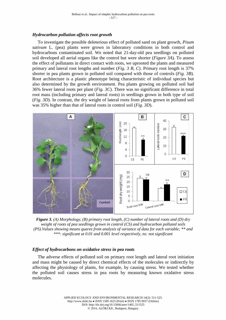
Hydrocarbon pollution affects root growth
To investigate the possible deleterious effect of polluted sand on plant growth, Pisum
sativum L. (pea) plants were grown in laboratory conditions in both control and
hydrocarbons contaminated soil. We noted that 21-day-old pea seedlings on polluted
soil developed all aerial organs like the control but were shorter (Figure 3A). To assess
the effect of pollutants in direct contact with roots, we uprooted the plants and measured
primary and lateral root lengths and number (Fig. 3 B, C). Primary root length is 37%
shorter in pea plants grown in polluted soil compared with those of controls (Fig. 3B).
Root architecture is a plastic phenotype being characteristic of individual species but
also determined by the growth environment. Pea plants growing on polluted soil had
36% fewer lateral roots per plant (Fig. 3C). There was no significant difference in total
root mass (including primary and lateral roots) in seedlings grown in both type of soil
(Fig. 3D). In contrast, the dry weight of lateral roots from plants grown in polluted soil
was 35% higher than that of lateral roots in control soil (Fig. 3D).
Figure 3. (A) Morphology, (B) primary root length, (C) number of lateral roots and (D) dry
weight of roots of pea seedlings grown in control (CS) and hydrocarbon polluted soils
(PS).Values showing means quares from analysis of variance of data for each variable; ** and
***: significant at 0.01 and 0.001 level respectively, ns: not significant
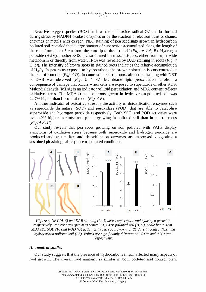
Effect of hydrocarbons on oxidative stress in pea roots
The adverse effects of polluted soil on primary root length and lateral root initiation
and mass might be caused by direct chemical effects of the molecules or indirectly by
affecting the physiology of plants, for example, by causing stress. We tested whether
the polluted soil causes stress in pea roots by measuring known oxidative stress
molecules.
Bellout et al.: Impact of edaphic hydrocarbon pollution on pea roots
- 518 -
APPLIED ECOLOGY AND ENVIRONMENTAL RESEARCH 14(2): 511-525.
http://www.aloki.hu ● ISSN 1589 1623 (Print) ● ISSN 1785 0037 (Online)
DOI: http://dx.doi.org/10.15666/aeer/1402_511525
2016, ALÖKI Kft., Budapest, Hungary
Reactive oxygen species (ROS) such as the superoxide radical O2- can be formed
during stress by NADPH-oxidase enzymes or by the reaction of electron transfer chains,
enzymes or metals with oxygen. NBT staining of pea seedlings grown in hydrocarbon
polluted soil revealed that a large amount of superoxide accumulated along the length of
the root from about 5 cm from the root tip to the tip itself (Figure 4 A, B). Hydrogen
peroxide (H2O2), another ROS, is also formed in stressed tissues, either from superoxide
metabolism or directly from water. H2O2 was revealed by DAB staining in roots (Fig. 4
C, D). The intensity of brown spots in stained roots indicates the relative accumulation
of H2O2. In pea roots exposed to hydrocarbons the brown coloration is concentrated at
the end of root tips (Fig. 4 D). In contrast in control roots, almost no staining with NBT
or DAB was observed (Fig. 4. A, C). Membrane lipid peroxidation is often a
consequence of damage that occurs when cells are exposed to superoxide or other ROS.
Malondialdehyde (MDA) is an indicator of lipid peroxidation and MDA content reflects
oxidative stress. The MDA content of roots grown in hydrocarbon-polluted soil was
22.7% higher than in control roots (Fig. 4 E).
Another indicator of oxidative stress is the activity of detoxification enzymes such
as superoxide dismutase (SOD) and peroxidase (POD) that are able to catabolise
superoxide and hydrogen peroxide respectively. Both SOD and POD activities were
over 40% higher in roots from plants growing in polluted soil than in control roots
(Fig. 4 F, G).
Our study reveals that pea roots growing on soil polluted with PAHs display
symptoms of oxidative stress because both superoxide and hydrogen peroxide are
produced and accumulate and detoxification enzymes are expressed suggesting a
sustained physiological response to polluted conditions.
Figure 4. NBT (A-B) and DAB staining (C-D) detect superoxide and hydrogen peroxide
respectively. Pea root tips grown in control (A, C) or polluted soil (B, D). Scale bar = 1cm.
MDA (E), SOD (F) and POD (G) activities in pea roots grown for 21 days in control (CS) and
hydrocarbon polluted soil (PS). Values are significantly different at 0.01** and 0.001***,
respectively.
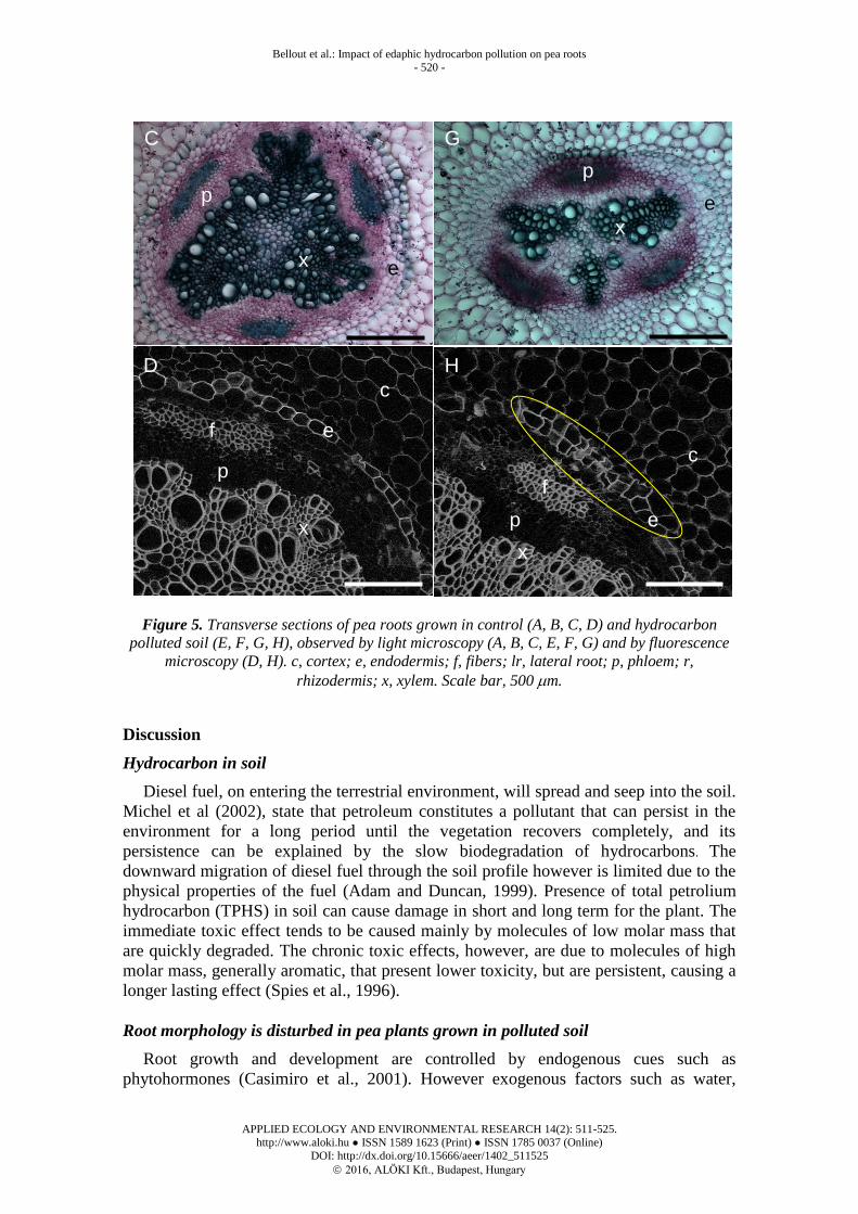
Anatomical studies
Our study suggests that the presence of hydrocarbons in soil affected many aspects of
root growth. The overall root anatomy is similar in both polluted and control plant

Bellout et al.: Impact of edaphic hydrocarbon pollution on pea roots
- 519 -
APPLIED ECOLOGY AND ENVIRONMENTAL RESEARCH 14(2): 511-525.
http://www.aloki.hu ● ISSN 1589 1623 (Print) ● ISSN 1785 0037 (Online)
DOI: http://dx.doi.org/10.15666/aeer/1402_511525
2016, ALÖKI Kft., Budapest, Hungary
samples with cortex cells similar in size both types of roots samples. However there was
a slight flattening of cortex cells in the primary roots that had been exposed to polluted
soil (Figure 5 A, E). Lateral root initiation was observed in both samples (Fig. 5 B, F).
When xylem vessels were viewed more closely (Fig. 5 C, G), evidence of the
centripetal differentiation of three primary xylem vessels was clearly observed in the
primary root in both samples. However secondary xylem differentiation appears
retarded in plants grown in the presence of hydrocarbons as much as less secondary
xylem is present compared to control (Fig. 5 C, G).
Assuming the presence of hydrocarbons in soil caused a delay in the differentiation
of secondary xylem, we looked for other signs that differentiation was affected. Lignin
fluorescence was observed in cell walls under UV-fluorescent microscopy. Lignified
xylem vessels are noticeably smaller in plants growing in polluted soil compared to
control plants (Fig. 5 D, H). Most surprisingly in plants grown in polluted soil we
observed a two-cell layered endodermis possibly adjacent to suberized cells (Figure 5
H, yellow circle). Roots grown in hydrocarbon-polluted soil therefore have unusual
xylem and endodermis differentiation.
F
lr
p x
r
c
e
B
lr p
x
r
c
e
E
c
r
p
x
e
A
c r
p x
e
Bellout et al.: Impact of edaphic hydrocarbon pollution on pea roots
- 520 -
APPLIED ECOLOGY AND ENVIRONMENTAL RESEARCH 14(2): 511-525.
http://www.aloki.hu ● ISSN 1589 1623 (Print) ● ISSN 1785 0037 (Online)
DOI: http://dx.doi.org/10.15666/aeer/1402_511525
2016, ALÖKI Kft., Budapest, Hungary
Figure 5. Transverse sections of pea roots grown in control (A, B, C, D) and hydrocarbon
polluted soil (E, F, G, H), observed by light microscopy (A, B, C, E, F, G) and by fluorescence
microscopy (D, H). c, cortex; e, endodermis; f, fibers; lr, lateral root; p, phloem; r,
rhizodermis; x, xylem. Scale bar, 500 m.
Discussion
Hydrocarbon in soil
Diesel fuel, on entering the terrestrial environment, will spread and seep into the soil.
Michel et al (2002), state that petroleum constitutes a pollutant that can persist in the
environment for a long period until the vegetation recovers completely, and its
persistence can be explained by the slow biodegradation of hydrocarbons. The
downward migration of diesel fuel through the soil profile however is limited due to the
physical properties of the fuel (Adam and Duncan, 1999). Presence of total petrolium
hydrocarbon (TPHS) in soil can cause damage in short and long term for the plant. The
immediate toxic effect tends to be caused mainly by molecules of low molar mass that
are quickly degraded. The chronic toxic effects, however, are due to molecules of high
molar mass, generally aromatic, that present lower toxicity, but are persistent, causing a
longer lasting effect (Spies et al., 1996).
Root morphology is disturbed in pea plants grown in polluted soil
Root growth and development are controlled by endogenous cues such as
phytohormones (Casimiro et al., 2001). However exogenous factors such as water,
C
D
G
H
e
e
e
e
x
x
c
c
x
p
p
x
p
p
f
f

Bellout et al.: Impact of edaphic hydrocarbon pollution on pea roots
- 521 -
APPLIED ECOLOGY AND ENVIRONMENTAL RESEARCH 14(2): 511-525.
http://www.aloki.hu ● ISSN 1589 1623 (Print) ● ISSN 1785 0037 (Online)
DOI: http://dx.doi.org/10.15666/aeer/1402_511525
2016, ALÖKI Kft., Budapest, Hungary
salinity, nutrients or the presence of toxic metals have a considerable impact on the final
root structure (Arduini et al., 1994). Many plant species are sensitive to petroleum
contaminants (Huang et al., 2004). Hydrocarbons in the soil may prevent uptake of
nutrients that are less mobile in contaminated soils (Atuanya, 1987). Water and nutrient
absorption can also be limited by hydrophobic molecules, which can form a layer over
the root when in excess in the soil (Quinones-Aquilar et al., 2003). Inhibition of plant
growth parameters (germination, plant length, and biomass) can be caused by toxic
compounds of petroleum hydrocarbons (Bossert and Bartha, 1985), such as low
molecular weight hydrocarbons.We observed both inhibition of primary root growth
and fewer lateral roots in pea plants growing on polluted soils. These results are
reminiscent of the known inhibition of lateral root formation and initiation of root
primordia by PAHs (Alkio et al., 2005; Baldyga et al., 2005).
There was no significant difference in the dry weight of total roots (primary and
lateral roots). Interestingly pea plants grown on oil-contaminated fields also had a
similar root dry weight as control plants after three weeks of growth (Xu and Johnson,
1995), although in older plants root weight was lower in polluted plants than in controls
(Xu and Johnson., 1995). Generally, lateral roots appear thicker in the polluted samples.
Hydrocarbon pollution is associated with oxidative stress in pea plants
Many environmental stresses induce ROS production (Apel and Hirt, 2004). Their
reaction with other molecules such as proteins or nucleic acids is often deleterious to the
cells. Lipids when peroxidised lead to MDA accumulation and altered cell integrity
(Apel and Hirt, 2004). ROS and ROS-detoxifying enzymes are more abundant in roots
from pea plants grown in polluted soil. The presence of PAH in polluted soil might be
directly responsible for ROS production, as it is generally observed that PAHs induce
ROS production in plants, as seen with phenanthrene (Alkio et al., 2005) and N-
heterocyclic PAHs (Paskova et al., 2006).
The observed increase in MDA in roots grown in polluted soil is suggestive of
oxidative damage as a consequence of ROS accumulation. It indicates that
hydrocarbon-induced stress alters biological membranes and affects cellular integrity.
Phenanthrene alone can induce ROS generation, MDA production, and oxidative stress
(Liu et al., 2009).
ROS abundance depends on rates of ROS generation and rate of ROS degradation
and scavenging/neutralizing by antioxidants whether through enzymatic and/or non-
enzymatic mechanisms (Amor et al., 2005). Plants have numerous detoxification
mechanisms, such as glutathione S-transferases, POD, catalases, and SOD and non-
enzymatic molecules like glutathione (Won et al., 2012). SOD activity and proteins
increase in response to stress in plants (Shalini and Dubey, 2003; Song et al., 2006).
Detoxifying enzyme activity or abundance is induced by hydrocarbons, such as
diethyl phthalate (Cheng and Cheng, 2012) and phenanthrene (Song et al., 2006) in
greater duckweed Spirodela polyrhiza. We found that ROS detoxifying activities SOD
and POD increased in roots of hydrocarbon-polluted pea plants suggesting that pea
plants respond to environmental stress by producing detoxifying enzymes. This finding
is broadly consistent with other abiotic stress responses, which quench excess ROS
through enzymatic reduction to water, and oxidize electron-rich buffers such as
ascorbate and glutathione (Apel and Hirt, 2004). However here not all of the stress-
induced ROS are eliminated, leading to MDA accumulation.

Bellout et al.: Impact of edaphic hydrocarbon pollution on pea roots
- 522 -
APPLIED ECOLOGY AND ENVIRONMENTAL RESEARCH 14(2): 511-525.
http://www.aloki.hu ● ISSN 1589 1623 (Print) ● ISSN 1785 0037 (Online)
DOI: http://dx.doi.org/10.15666/aeer/1402_511525
2016, ALÖKI Kft., Budapest, Hungary
Anatomy
The results of this study support the idea that the presence of hydrocarbons in soil
has affected not only the morphology and root development, but also their anatomical
structure. Indeed we show that roots grown in polluted soils are delayed in xylem
differentiation and have an additional cell layer in the endodermis.
Our results are in agreement with Kummerova et al. (2013), who showed that in pea
and maize roots, the proportion of xylem vessels in the stele decreased when exposed to
fluoranthene. Pea roots with less xylem in response to hydrocarbons in soil may be
interpreted as an adaptation to minimize absorption of polluted water, because vessel
number and diameter influence the amount of water flowing. Hernandez-Ortega et al.
(2014) reported that values of hydraulic parameters diminished, but the loss of hydraulic
conductivity was significantly enhanced as the diesel concentration increased. In
addition fluoranthene exposure triggers changes in the cell morphology of other organs
and tissues including the root tip, root cap, apical meristem and elongation zone
(Kummerova et al., 2013). Similar abnormal development patterns of xylem have been
also described in cotton grown in presence of high salinity (Reinhardt and Rost, 1995).
Thus the xylem tissue seems to be particularly sensitive to external abiotic pollutant.
A single layer of endodermis in plants is defined by an evolutionarily conserved
mechanism, where the SCARECROW (SCR) protein associated with the mobile
SHORT-ROOT (SHR) protein delimits endoderm and pericycle founder cells around
the quiescent center at the root tip (Cui et al., 2007). In our study, pea roots grown in
polluted soils showed an additional division in endodermis. Observing a supernumerary
cell division and cell differentiation in the developing root suggests that hydrocarbons
modify the existing differentiation pattern. The endodermis is the innermost layer of the
cortex and is characterized by the formation of Casparian bands in the anticlinal walls
of its cells (Enstone et al., 2003). An extra cell layer might contribute to limiting
exchanges between the cortex cells and the stele tissues reducing the import of
hydrocarbons in the xylem flux. This might be another anatomical adaptation to
pollutants like Casparian band and suberin lamellae thickening, increased suberization
and lignification of endodermis cells (Zelko and Lux, 2004; Vance et al., 1980; Kalaji
and Pietkiewicz, 1993; Shannon et al., 1994; Schreiber et al., 1999).
Overall hydrocarbon residues found in sand samples extracted from the quagmire site
profoundly modify plant growth and root architecture. ROS production and ROS
detoxifying enzymes are induced in pea, most likely a consequence of physiological
stress. We found that morphological and anatomical changes in pea roots exposed to
anthropogenic pollution might be an adaptation to abiotic stress limiting the impact of
the pollutant hydrocarbons on roots.
Acknowledgements. We thank members of the Laboratory of ISSeP in Colfontaine, Belgium and of the
UPMC-Paris Laboratory of Plant Adaptation to Environmental Constraints for their help and technical
assistance in soil hydrocarbon analysis and anatomical studies.
REFERENCES
[1] Achary, V. M. M., Jena, S., Panda, K. K., Panda, B. B. (2008): Aluminum induced
oxidative stress and DNA damage in root cells of Allium cepa L. ‒ Ecotoxicol and
Environmental Safety 70: 300-310.

Bellout et al.: Impact of edaphic hydrocarbon pollution on pea roots
- 523 -
APPLIED ECOLOGY AND ENVIRONMENTAL RESEARCH 14(2): 511-525.
http://www.aloki.hu ● ISSN 1589 1623 (Print) ● ISSN 1785 0037 (Online)
DOI: http://dx.doi.org/10.15666/aeer/1402_511525
2016, ALÖKI Kft., Budapest, Hungary
[2] Achuba, F.I. (2006): The effect of sublethal concentrations of crude oil on the growth and
metabolism of Cowpea (Vigna unguiculata) seedlings. ‒ The Environmentalist 26: 17-20.
[3] Adam, G., Duncan, H. (1999): Effect of diesel fuel on growth of selected plant species. ‒
Environmental Geochemistry and Health 21: 353- 357.
[4] Alkio, M., Tabuchi, T. M., Wang, X., Colon-Carmona, A. (2005): Stress responses to
polycyclic aromatic hydrocarbons in Arabidopsis include growth inhibition and
hypersensitive responses-like symptoms. ‒ Journal of Experimental Botany 56 (421):
2983-2994.
[5] Amor, N. B., Hamed, K. B., Debez, A., Grignon, C., Abdelly, C. (2005): Physiological
and antioxidant responses of the perennial halophyte Crithmum maritimum to salinity. ‒
Plant Science 168: 889-899.
[6] Anoliefo, G. O. (1991): Forcados blend crude oil effect in respiration, metabolism,
elemental composition and growth of Citrullus vulgaris (Schrad). Ph.D. Thesis, Benin.
[7] Apel, K., Hirt, H. (2004): Reactive oxygen species: metabolism, oxidative stress, and
signal transduction. ‒ Annual Review of Plant Biology 55 (1): 373-399.
[8] Arduini, S., Godbold, D. L., Onnis, A. (1994): Cadmium and coper change root growth
and morphology of Pinus pinea and Pinus pinaster seedlings. ‒ Plant Physiology 92: 675-
680.
[9] Atuanya, E.J. (1987): Effect of oil pollution on physical and chemical properties of soil, a
case study of waste oil contaminated delta soil in Bendel State. Nigeria. ‒ Journal of
Applied Science 55: 155-176.
[10] Baldygà, B., Weiczorek, J., Smoczynski, S., Weiczorek, Z., Smoczinska, K. (2005): Pea
plant response to Anthracene present in soil. ‒ Polish Journal of Environmental Studies
14: 397-401.
[11] Beyer, W. F., Fridovich, I. Jr. (1987): Assaying for superoxide dismutase activity: some
large consequences of minor changes in conditions. ‒ Analytical Biochemistry 161: 559-
566.
[12] Bossert, I., Bartha, R. (1985): Plant growth in soils with a history of oily sludge disposal.
‒ Soil Science 140: 75-77.
[13] Bradford, M. M. 1976. A rapid and sensitive method for the quantification of microgram
quantities of protein utilizing the principle of protein-dye binding. ‒ Analytical
Biochemistry 72: 248-254.
[14] Casimiro, L., Marchant, A., Bhalerao, R.P., Beeckman, T., Dhooge, S., Swarup, R.,
Graham, N., Inzé, D., Sandberg, G., Casero, P.J., Bennett, M. (2001): Auxin transport
promoters Arabidopsis lateral root initiation. ‒ Plant Cell 13: 843-852.
[15] Chance, M., Maehly, A.C. (1955): Assay of catalases and peroxidases. ‒ Methods
Enzymology 2: 764-817.
[16] Cheng, L-J., Cheng, T-S. (2012): Oxidative effects and metabolic changes following
exposure of greater duckweed (Spirodela polyrhiza) to diethyl phthalate. ‒ Aquatic
Toxicology 109: 166-175.
[17] Cui, H., Levesque, M. P., Vernoux, T. (2007): An evolutionarily conserved mechanism
delimiting SHR movement defines a single layer of endodermis in plants. ‒ Science 316:
421-425.
[18] Culbertson, J.B., Valiela, I., Pickart, M., Peacock, E. E., Reddy, C. M. (2008): Long-
term consequences of residual petroleum on salt marsh grass. ‒ Journal of Applied
Ecology 45: 1284-1292.
[19] De Jong, E. (1980). Effect of a crude oil spill on cereals. ‒ Environmental Pollution 22:
187-307.
[20] Enstone, D. E., Peterson, C. A., Ma, F. (2003): Root Endodermis and Exodermis:
Structure, Function, and Responses to the Environment. ‒ Journal of Plant Growth
Regulation 21: 335-351.
[21] Gao, Y. Z., Zhu, L. Z. (2004): Plant uptake, accumulation and translocation of
phenanthrene and pyrene in soils. ‒ Chemosphere 55: 1169-1178.

Bellout et al.: Impact of edaphic hydrocarbon pollution on pea roots
- 524 -
APPLIED ECOLOGY AND ENVIRONMENTAL RESEARCH 14(2): 511-525.
http://www.aloki.hu ● ISSN 1589 1623 (Print) ● ISSN 1785 0037 (Online)
DOI: http://dx.doi.org/10.15666/aeer/1402_511525
2016, ALÖKI Kft., Budapest, Hungary
[22] Giannopolitis, C. N., Ries, S. K. (1977): Superoxide dismutases: Occurrence in higher
plants. ‒ Plant Physiology 59: 309-314.
[23] Gill, L. S., Nyawuame, H. G. K., Ehikhametalor, A. O. (1992). Effect of crude oil on the
growth and anatomical features of Chromolaena odorata L. ‒ Chromolaena odorata
Newsletter 6:1-6.
[24] Hernandez-Ortega, H. A., Quintanar-Isaias, P.A., Jaramillo-Pérez, A. T., Alancon, A.,
Ferrera-Cerato, R., Lazzarini Lechuga, R. (2014): Diesel effects on root hydraulic
conductivity and morphological changes of the vascular cylinder in Medicago sativa. ‒
Environmental and Experimental Botany 105:1-9.
[25] Huang, X. D., El-Alawi, Y., Penrose, D. M., Glick, B. R., Greenberg, B. M. (2004): A
multi-process phytoremediation system for removal of polycyclic aromatic hydrocarbons
from contaminated soils. ‒ Environmental Pollution 130: 465-476.
[26] Jung, J. K. H., McCouch, S. (2013): Getting to the root of it: genetic and hormonal
control of root architecture. ‒ Frontiers in Plant Science 4: 186.
[27] Kalaji, M.H.,Pietkiewicz, S. (1993): Salinity effects on plant growth and other
physiological processes. ‒ Acta Physiologiae Plantarum 15: 89-124.
[28] Korte, F., Kvesitadze, G., Ugrekhelidze, D., Gordeziani, M., Khatisashvili, G., Buadze,
O., Zaalishvili, G., Coulston, F. (2000): Organic toxicants and plants. ‒ Ecotoxicology
and Environmental Safety 47: 1-26.
[29] Kummerová, M., Zezulka, S., Váňová, L., Fišerová, H. (2012): Effect of organic
pollutant treatment on the growth of pea and maize seedlings. ‒ Central European Journal
of Biology 7(1) : 159-166.
[30] Kummerova, M., Zezulka, S., Babula, P., Vànovà, L. (2013): Root response in Pisum
sativum and Zea mays under fluoranthene stress: Morphological and anatomical traits. ‒
Chemosphere 90: 665-673.
[31] Li, K. Y., Kane, A. J., Wang, J. J., Cawley, W. A. (1993): Measurement of
biodegradation rate constants of a water extract from petroleum contaminated soil. ‒
Waste Manage 13: 245-251.
[32] Liu, H., Weisman, D., Ye, Y.B., Cui, B., Huang, Y. H., Colon-Carmona, A., Wang, Z. H.
(2009): An oxidative stress response to polycyclic aromatic hydrocarbon exposure is
rapid and complex in Arabidopsis thaliana. ‒ Plant Science 176: 375-382.
[33] Locquin, M., Langeron, M. (1978): Manuel de microscopie. Ed. Masson. France.
[34] McCarthy, J. F., Tschaplinski, T. J. (1991): Biological markers in environmental sentinels
to establish exposure to and effects of atmospheric toxicants: an overview. ‒ In: Moser,
T.J., Baker, J. R., Tingey, D. T. (Eds). Ecological exposure and effects of airborne toxic
chemicals: USA.
[35] McElroy, E., Farrington, J. W., Teal, J. M. (1989): Bioavailability of aromatic
hydrocarbons in the aquatic Environment. ‒ In: Varanasi, U. (Ed.) Metabolism of
Polycyclic Aromatic Hydrocarbons in the Aquatic Environment. CRC Press Inc. Boca
Raton, Florida, USA. 1-39.
[36] Michel, J., Henry, JR., C.B. Thumm, S. (2002): Shoreline assessment and environmental
impacts from the M/T Westchester oil spill in the Mississippi River. ‒ Spill Science &
Technology Bulletin 7 (3-4): 155-161.
[37] Mittler, R., Vanderauwera, S., Gollery, M., Van Breusegem, F. (2004): Reactive oxygen
gene network of plants. ‒ Trends in Plant Science 9: 490-498.
[38] Ogboghodo, I. A., Iruaga, E. A., Osemwota, I. O., Chokor, J. U. (2004): An assessment
of the effects of crude pollution on soil properties, germination and growth of maize (Zea
mays) using two crude types - Forcados light and Escravos light. ‒ Environmental
Monitoring and Assessment 96:142-152.
[39] Omosun, G., Markson, A. A., Mbanasor, O. (2008): Growth and anatomy of Amaranthus
hybridus as affected by different crude oil concentrations. Am-Euras. ‒ Jornal of
Scientific Research 3: 70-74.

Bellout et al.: Impact of edaphic hydrocarbon pollution on pea roots
- 525 -
APPLIED ECOLOGY AND ENVIRONMENTAL RESEARCH 14(2): 511-525.
http://www.aloki.hu ● ISSN 1589 1623 (Print) ● ISSN 1785 0037 (Online)
DOI: http://dx.doi.org/10.15666/aeer/1402_511525
2016, ALÖKI Kft., Budapest, Hungary
[40] Paskova, V., Hilscherovà, K., Feldmannovà, M., Blaha, L. (2006): Toxic effects and
oxidative stress in higher plants exposed to polycyclic aromatic hydrocarbons and their n
heterocyclic derivatives. ‒ Environmental Toxicology and Chemistry 25 (12): 3238-3245.
[41] Quinones-Aquilar, E. E., Ferra-Cerrato, R., Gavi, R. F., Fernandez, L., Rodriguez, V. R.,
Alarcom, A. (2003) : Emergence and growth of maize in a crude oil polluted soil. ‒
Agrociencia 37: 585-594.
[42] Rao, M. V., Davis, K. R. (1999): Ozone-induced cell death occurs via two distinct
mechanisms in Arabidopsis: the role of salicylic acid. ‒ The Plant Journal 17: 603-614.
[43] Reinhardt, D. H., Rost, T. L. (1995): On the correlation of the primary root growth and
treachery element size and distance from the tip in cotton seedlings grown under salinity.
‒ Environmental and Experimental Botany 35: 575-588.
[44] Romero- Puertas, M. C., Corpas, F. J., Rodríguez-Serrano, M., Gómez, M., Del Río, L.
A., Sandalio, L. M. (2007) : Differential expression and regulation of antioxidative
enzymes by cadmium in pea plants. ‒ Journal of Plant Physiology 164: 1346-1357.
[45] Schreiber, L., Hartmann, K., Skrabs, M., Zeier, J. (1999): Apoplastic barriers in roots:
chemical composition of endodermal and hypodermal cell walls. ‒ Journal of
Experimental Botany 50: 1267-1280.
[46] Shalini, C., Dubey, R. S. (2003): Lead toxicity induces lipid peroxidation and alters the
activities of antioxidant enzymes in growing rice plant. ‒ Plant Science 164: 645- 655.
[47] Shannon, M. C., Grieve, C. M., Francois, L. E. (1994): Whole-plant response to salinity.
‒ In: Wilkinsow, R.E. (ed) Plant-environment interactions. New York.
[48] Sharma, G. K., Chandler, C., Salemi, L. (1980): Environmental pollution and leaf
cuticular variation in Kudzu (Pueraria lobata Willd). ‒ Annals of Botany 45: 77-80.
[49] Smith, M. J., Flowers, T.H., Duncan, H.J., Alder, J. (2006): Effects of polycyclic
aromatic hydrocarbons on germination and subsequent growth of grasses and legumes in
freshly contaminated soil and soil with aged PAHs residues. ‒ Environmental Pollution
141: 519-525.
[50] Song, G. L., Hou, W. H., Wang, Q. H., Wang, J. L., Jin, X. G. (2006): Effect of low
temperature on eutrophicated waterbody restoration by Spirodela polyrhiza. ‒
Bioresource Technology 97: 1865-1869.
[51] Spies, R. B., Rice, S. D., Wolfe, D. A., Wright, B. A. (1996): The effects of the Exxon
Valdez oil spill on the Alaskan coastal environment. ‒ In: Rice, S. D., Spies, R. B.,
Wolfe, D. A. Wright, B. A. (Eds.). Proceedings of the Exxon Valdez Oil Spill
Symposium. Alaska. ‒ American Fish Society Symposium 18: 1-16.
[52] Sun, W., Montagu, M. V., Verbruggen, N. (2002): Small heat shock proteins and stress
tolerance in plants. ‒ Biochimica and Biophysica Acta 1577: 1-9.
[53] Thordal-Christensen, H., Zhang, Z., Wei, Y., Collinge, D. B. (1997): Subcellular
localization of H2O2 in plants, H2O2 accumulation in papillae and hypersensitive response
during barley-powdery mildew interaction. ‒ Plant Journal 11:1187-1194.
[54] Vance, C. P., Kirk, T. K., Sherwood, R. T. (1980): Lignification as a mechanism of
disease resistance. ‒ Annual Review of Phytopathology 18: 259-288.
[55] Won, E. J., Rhee, J. S., Kim, R. O., Ra, K., Kim, K.T., Shin, K. H., Lee, J. S. (2012):
Susceptibility to oxidative stress and modulated expression of antioxidant genes in the
copper-exposed polychaete Perinereis nuntia. ‒ Comparative Biochemistry and
Physiology 155: 344-351.
[56] Xu, J.G., Johnson, R.L. (1995): Root growth, microbial activity and phosphatase activity
in oil-contaminated, remediated and uncontaminated soils planted to barley and field pea.
‒ Plant and Soil 173: 3-10.
[57] Zelko, I., Lux, A. (2004): Effect of cadmium on Karwinskia humboldtiana roots. ‒
Biologia 59 (13): 205-209.