title page serum human epididymis protein 4 he4) and...
TRANSCRIPT
1
Title page
Serum human epididymis protein 4 (HE4) and Risk for Ovarian Malignancy Algorithm
(ROMA) as new diagnostic and prognostic tools for epithelial ovarian cancer management
Elisabetta Bandiera1, Chiara Romani1, Claudia Specchia2, Laura Zanotti1, Claudio Galli3,
Giuseppina Ruggeri4, Germana Tognon5, Eliana Bignotti1, Renata A. Tassi1, Franco Odicino5, Luigi
Caimi4, Enrico Sartori5, Alessandro D. Santin6, Sergio Pecorelli1 and Antonella Ravaggi1
1”Angelo Nocivelli” Institute of Molecular Medicine, Division of Gynecologic Oncology,
University of Brescia, Italy
2Department of Biomedical Sciences and Biotechnology, University of Brescia, Italy
3 Scientific Affairs, Abbott Diagnostics, Roma, Italy
4Department of Laboratory Medicine, A.O. Spedali Civili, Brescia, Italy
5Department of Obstetrics and Gynecology, University of Brescia, Italy
6Department of Obstetrics and Gynecology and Reproductive Sciences, Yale University School of
Medicine, New Haven, USA
Running title
Serum HE4 and ROMA algorithm in epithelial ovarian cancer
Keywords
Epithelial ovarian cancer, HE4, ROMA, diagnosis, prognosis
Financial support
on July 6, 2018. © 2011 American Association for Cancer Research. cebp.aacrjournals.org Downloaded from
Author manuscripts have been peer reviewed and accepted for publication but have not yet been edited. Author Manuscript Published OnlineFirst on October 25, 2011; DOI: 10.1158/1055-9965.EPI-11-0635
2
Supported in part by grants from the Nocivelli, Foundation, Brescia, Italy, by grants from the
Istituto Superiore di Sanità (Progetto Oncoproteomica, Programma Italia-USA “Farmacogenomica
Oncologica”, convenzione 527/B4/4), Rome, Italy and by grants from the Ministero dell’Istruzione,
dell’Università e della Ricerca (PRIN project, prot2008AZJM9E) Rome, Italy. This investigation
was also supported by grants from NIH R01 CA122728-01A2 and R01 CA154460-01A1 to ADS,
grants 501/A3/3 and 0027557 from the Italian Institute of Health (ISS) to ADS and by NIH
Research Grant CA-16359 from the National Cancer Institute.
Corresponding author
Dr. Elisabetta Bandiera
Mailing address: Istituto di Medicina Molecolare “A. Nocivelli”, Spedali Civili 1, 25123 Brescia,
Italy
E-mail address: [email protected]
Phone number: 00390303996286
Fax number: 00390303996059
Conflict of interest statement
Dr. Claudio Galli is currently employed by Abbott Diagnostics as the Scientific Affairs Manager,
Italy
Word count: 4091
Total number of figures and tables: 6
on July 6, 2018. © 2011 American Association for Cancer Research. cebp.aacrjournals.org Downloaded from
Author manuscripts have been peer reviewed and accepted for publication but have not yet been edited. Author Manuscript Published OnlineFirst on October 25, 2011; DOI: 10.1158/1055-9965.EPI-11-0635

3
BACKGROUND:
The aim of this work was to analyze the diagnostic and prognostic value of serum human
epididymis protein 4 (HE4) and Risk for Ovarian Malignancy Algorithm (ROMA) in epithelial
ovarian cancer (EOC).
METHODS:
Preoperative serum samples of 419 women (140 healthy controls, 131 ovarian benign cysts, 34
endometriosis, 114 EOC) were tested for CA125 and HE4 using fully automated methods (Abbott
ARCHITECT) and validated cut-off values.
RESULTS:
For the discrimination of benign masses from EOC, in pre-menopausal women the sensitivity and
specificity were 92.3% and 59.4% for CA125, 84.6% and 94.2% for HE4, and 84.6% and 81.2% for
ROMA while in post-menopausal women the sensitivity and specificity were 94.3% and 82.3% for
CA125, 78.2% and 99.0% for HE4, 93.1% and 84.4% for ROMA.
In patients with EOC, elevated CA125, HE4 and ROMA levels were associated with advanced
FIGO stage, sub-optimally debulking, ascites, positive cytology, lymph node involvement and
advanced age (all p≤0.05). Elevated HE4 and ROMA (both p≤0.01), but not CA125 (p=0.0579),
were associated with undifferentiated tumours. In multivariable analysis, elevated HE4 and ROMA
(all p≤0.05) were independent prognostic factors for shorter overall survival, disease free survival
and progression free survival.
CONCLUSIONS and IMPACT:
This study underlines the high specificity of HE4 in discriminating endometriosis and ovarian
benign cysts from EOC and the high sensitivity of CA125 in detecting EOC. We demonstrated HE4
and ROMA as independent prognostic factors. Multicenter studies are needed to draw firm
conclusions about the applicability of HE4 and ROMA in clinical practice.
on July 6, 2018. © 2011 American Association for Cancer Research. cebp.aacrjournals.org Downloaded from
Author manuscripts have been peer reviewed and accepted for publication but have not yet been edited. Author Manuscript Published OnlineFirst on October 25, 2011; DOI: 10.1158/1055-9965.EPI-11-0635

4
Introduction
Epithelial ovarian cancer (EOC) is the most frequent cause of death from gynaecological cancer. It
has the highest fatality-to-case ratio of all gynaecological malignancies, being characterized by
early widespread metastasis and high-grade malignancy at diagnosis. The five-year survival rate is
about 80-90% for patients with stage I disease and only 30% for patients with stage III or IV.
Although survival has improved with the use of maximal cytoreductive surgery along with
platinum- and taxol-based chemotherapy, nearly 80% of ovarian cancers relapse and patients
inevitably succumb to the development of chemotherapy-resistant disease (1).
At the moment, serum CA125 is the commonly used biomarker for EOC diagnosis. Jacobs and
colleagues (2) developed the widely used Risk of Malignancy Index (RMI), an algorithm that uses
ultrasound findings, architectural features of pelvic mass, CA125 levels and menopausal status to
stratify patients into high- and low-risk groups. However, since CA125 is associated with a high
false-positive rate among benign gynaecologic conditions, such as endometriosis that affects mainly
women in pre-menopause, its use for EOC detection is almost exclusively reserved for post-
menopausal cases (3-6). Furthermore, CA125 has low sensitivity in identifying patients with early
EOC disease, being increased in only 50% of patients with stage I (7). CA125 is also used to
monitor response to therapy and in early detection of ovarian cancer recurrence after treatment (8-
11), but the value of preoperative CA125 is not associated with prognosis of EOC patients (12-13).
Clinicopathological features known to be prognostic variables for EOC are age, surgical stage
(FIGO stage), histological subtype, differentiation grade, ascites, lymph node involvement and
residual tumour after cytoreductive surgery. According to the three-yearly analysis of the FIGO
Annual Report on the Results of Treatment in Gynaecological Cancer, stage, grade and residual
tumour have the greatest prognostic value (14). However, these factors provide an insufficient
picture of EOC biology and they are frequently interrelated.
Therefore, there is a pressing need to develop new methods for EOC diagnosis and prognosis. First,
the preoperative diagnosis of EOC would refer patients to centers specialized in optimal tumor
on July 6, 2018. © 2011 American Association for Cancer Research. cebp.aacrjournals.org Downloaded from
Author manuscripts have been peer reviewed and accepted for publication but have not yet been edited. Author Manuscript Published OnlineFirst on October 25, 2011; DOI: 10.1158/1055-9965.EPI-11-0635

5
debulking and complete surgical staging, since it has been shown that optimal surgery treatment
improves overall survival in EOC patients (15-20). Moreover, the prediction of disease outcome in
EOC patients could be useful for developing individually tailored and possibly more effective post-
surgical treatments.
Serum analysis is a low-cost, non-invasive technique and it is not subjected to operator variability,
such as imaging analysis. Therefore, considerable efforts are underway to identify new serum
biomarkers that alone or in combination with CA125 could improve EOC diagnosis (7, 21-36).
In the majority of studies, HE4 has emerged as one of the most promising new serum biomarker in
EOC diagnosis. Previous reports evaluated the clinical utility of the HE4 and CA125 combination
(ROMA algorithm) in order to assess the risk of EOC pathology in patients presenting with a pelvic
mass (32, 37-42). However, some recent papers (39, 41-42) showed that diagnostic accuracy of
ROMA compared to CA125 and HE4 alone is still controversial. At the present the prognostic value
of HE4 has been investigated by only one study (43) in patients with advanced EOC. This report
showed HE4 as an independent prognostic factor for progression free survival. However, in such
study, the prognostic analysis was conducted in a small sample size, the comparison with
prognostic value of CA125 was not evaluated and EOC patients were not dichotomized by median
value of biomarker, as usually performed in prognostic studies.
The aim of this work was to analyze diagnostic and prognostic value of serum HE4, CA125 and
ROMA in a large number of patients, using fully automated methods for biomarkers determination
that guarantee a higher reproducibility and robustness of assay results (41).
Initially, we analyzed the diagnostic performance of HE4 compared to CA125 in discriminating
among subjects with EOC, ovarian benign cysts, endometriosis and healthy controls. Then, we
analyzed ROMA algorithm, compared to CA125 and HE4 alone, for the differential diagnosis
between benign pelvic mass and EOC.
on July 6, 2018. © 2011 American Association for Cancer Research. cebp.aacrjournals.org Downloaded from
Author manuscripts have been peer reviewed and accepted for publication but have not yet been edited. Author Manuscript Published OnlineFirst on October 25, 2011; DOI: 10.1158/1055-9965.EPI-11-0635

6
Finally, we investigated the role of HE4 and ROMA, in comparison with CA125 and established
prognostic factors, in predicting overall survival (OS), disease-free survival (DFS), and progression
free survival (PFS) in EOC patients.
Material and Methods
Patients’ characteristics
A total of 419 patients referred to the Gynaecologic Oncology Department of the University of
Brescia from 2003 to 2010 were included in the study. All patients signed an informed consent
approved by the Institutional Review Board. Patients with a past or concomitant history of
malignancy were excluded from the study. Cohorts of patients in pre- and post-menopause were
balanced as regards the number and age. Pre-menopausal women included 39 healthy controls (age:
mean 39 years; range 21-53), 34 patients with endometriosis (age: mean 36.5 years; range 25-51),
35 patients with ovarian benign cysts (age mean: 41.5 years; range 18-59) and 26 patients with EOC
(age mean: 44.7 years; range 33-54). Post-menopausal women included 101 healthy controls (age:
mean 63.3 years; range 40-76), 96 patients with ovarian benign cysts (age mean: 64.0 years; range
46-89) and 87 patients with EOC (age mean: 66.3 years; range 46-87). One EOC patient had an
unknown menopausal status and thus was included only in prognostic analysis. Women were
considered in post-menopause if they reported no menstrual periods within the 12 months before
blood collection.
EOC patients’ charts were reviewed to obtain all clinical and pathological features at the moment of
the diagnosis treatment and during follow-up. Low malignant potential tumors were excluded from
this study. Standard treatment for EOC consisted in complete pelvic surgery with cytoreductive
surgery in advanved stages and platinum-based chemotherapy. Cytoreductive surgery included total
abdominal hysterectomy, bilateral salpingo-oophorectomy, omentectomy and pelvic and periaortic
lymph node sampling, with cytological evaluation of ascites or peritoneal washing. The staging
procedure was performed according to the International Federation of Gynaecologists and
on July 6, 2018. © 2011 American Association for Cancer Research. cebp.aacrjournals.org Downloaded from
Author manuscripts have been peer reviewed and accepted for publication but have not yet been edited. Author Manuscript Published OnlineFirst on October 25, 2011; DOI: 10.1158/1055-9965.EPI-11-0635

7
Obstetricians (FIGO) system standards. Histological subtype and differentiation grade were
assigned according to World Health Organization criteria.
A group of 98 EOC patients was evaluated for survival analysis. The remaining 16 EOC patients
were excluded from survival analysis because 2 refused chemotherapy, 4 were not eligible for
primary surgery because of their poor medical conditions and 10 had incomplete follow-up.
Patients were followed up from the date of surgery until death or November 30, 2010 (median
follow-up: 19.5 months, range 1-85 months).
HE4 and CA125 immunoassays
Blood was drawn before any surgical or chemotherapeutic treatment and centrifuged within half an
hour for serum collection. Serum samples were stored at -80°C until analysis. Levels of CA125 and
HE4 were measured by chemiluminescent microparticle immunoassays (CMIA) on the fully
automated ARCHITECT instrument (Abbott Diagnostics Division, Wiesbaden, Germany) at the III
Laboratory Service, Spedali Civili di Brescia, Italy. According to the indications of the HE4
manufacturer, the normal ranges were ≤70 pmol/L for pre-menopausal state and ≤140 pmol/L in
menopausal state.
Roma Algorithm
ROMA utilizes the results for HE4 and CA125 to generate a predictive index (PI) for EOC (32),
calculated by these formulas:
For pre-menopausal women: PI = -12.0 + 2.38*LN[HE4] + 0.0626*LN[CA125]
For post-menopausal women: PI = -8.09 + 1.04*LN[HE4] + 0.732*LN[CA125]
Then, ROMA value is calculated as follow: ROMA value (%) = exp(PI) / [1+exp(PI)] *100.
According to the indications of the HE4 manufacturer, indexes of at least 7.4% and 25.3% indicate
a high risk for the presence of EOC in pre- or post-menopause, respectively.
on July 6, 2018. © 2011 American Association for Cancer Research. cebp.aacrjournals.org Downloaded from
Author manuscripts have been peer reviewed and accepted for publication but have not yet been edited. Author Manuscript Published OnlineFirst on October 25, 2011; DOI: 10.1158/1055-9965.EPI-11-0635

8
Statistical analysis
The Wilcoxon-Mann-Whitney test and the Kruskal-Wallis test were used to compare
biomarkers distributions across two and more than two subgroups of patients respectively.
Differences between the proportions of patients with level of biomarkers above thresholds have
been compared within the same subgroup of patients with Mc Nemar test.
Area under the receiver operating characteristic (ROC) curves were used to quantify each
biomarker’s ability to discriminate between diagnostic groups. Areas under the ROC curves
(AUCs) were compared with the method described by DeLong (44).
For survival analysis, three end-points (cancer relapse, cancer progression and cancer related death)
were used to calculate DFS, PFS, OS, respectively. DFS was defined as the time interval between
the date of surgery and the date of identification of disease recurrence, PFS was defined as the time
interval between the date of surgery and the date of identification of progressive disease (disease
not treatable with curative intent) and OS was defined as the time interval between the date of
surgery and the date of death. For all three end-points the last date of follow-up was used for
censored subjects.
Survival curves were calculated using Kaplan–Meier method, and differences in survival between
subgroups of patients were tested using the log-rank test.
The effect of HE4, CA125 and ROMA serum levels on prognosis was evaluated categorizing them
on the basis of the median values, computed on the whole cohort (Low; High).
Univariate Cox proportional hazard models were fitted to evaluate the role of CA125, HE4, ROMA
and established prognostic factors on the considered outcomes. Multivariable Cox regression
models were used to estimate the effect of biomarkers adjusted for FIGO stage, residual tumour and
histological subtype, the most important established prognostic factors.
All P-values were two-sided. A P-value less than 0.05 was considered to indicate statistical
significance. All the analyses were performed using STATA 11.0 software (Stata Corporation,
College Station, Texas)
on July 6, 2018. © 2011 American Association for Cancer Research. cebp.aacrjournals.org Downloaded from
Author manuscripts have been peer reviewed and accepted for publication but have not yet been edited. Author Manuscript Published OnlineFirst on October 25, 2011; DOI: 10.1158/1055-9965.EPI-11-0635

9
Results
CA125, HE4 and ROMA diagnostic performances
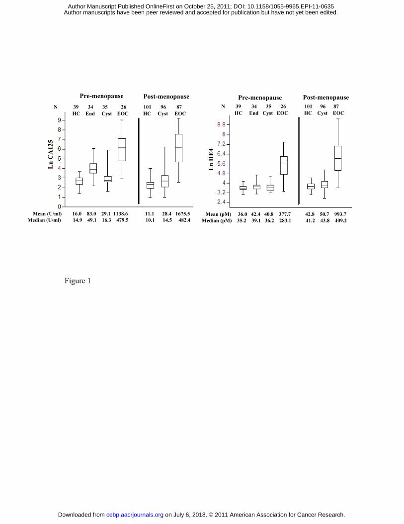
Comparison of CA125 and HE4 levels between pre- and post-menopausal healthy controls showed
that CA125 is significantly higher in pre-menopausal than in post-menopausal status (14.9 U/ml vs
10.1 U/ml, p=0.0001), while HE4 is inversely significantly higher in post-menopausal than in pre-
menopausal status (41.2 pM vs 35.2 pM, p=0.001). For this reason CA125 and HE4 diagnostic
performances were analyzed separately in pre- and post-menopausal women. CA125 and HE4
values detected in healthy controls and in patients with endometriosis, ovarian benign cysts and
EOCs are represented in Figure 1.
The levels of HE4 and CA125 were significantly higher in EOC patients compared with healthy
controls, endometriosis and ovarian benign cysts, independently from menopausal status (all
p<0.0001). Both CA125 and HE4 levels were slightly higher in patients with ovarian cysts when
compared with healthy controls, but these differences reached the statistical significance only in
post-menopausal women (14.5 U/ml vs 10.1 U/ml, p<0.0001 for CA125; 43.8 pM vs 41.2 pM,
p=0.0381 for HE4) and not in pre-menopausal ones (p=0.1561 for CA125; p=0.2718 for HE4). In
pre-menopausal women, HE4 and CA125 showed different ability in discriminating endometriosis
from healthy controls and ovarian benign cysts. CA125 was significantly higher in patients with
endometriosis (49.1 U/ml) than in healthy controls (14.9 U/ml) and ovarian benign cysts (16.3
U/ml) (both p<0.0001). On the contrary, HE4 showed a marginally significant increase in
endometriosis (39.1 pM) towards healthy controls (35.2 pM) (p=0.0447) and a not statistically
significant increase in endometriosis towards ovarian benign cysts (36.2 pM) (p=0.5015).
In order to evaluate the differences in diagnostic abilities between CA125 and HE4, we used the
reference value indicated by standard clinical use for CA125 (35 U/ml) or proposed by the
manufacturer for HE4. In patients with EOC, CA125 and HE4 levels were above cut-off in 82/87
(94.3%) and in 68/87 (78.1%) post-menopausal patients, respectively, and in 24/26 (92.3%) and in
on July 6, 2018. © 2011 American Association for Cancer Research. cebp.aacrjournals.org Downloaded from
Author manuscripts have been peer reviewed and accepted for publication but have not yet been edited. Author Manuscript Published OnlineFirst on October 25, 2011; DOI: 10.1158/1055-9965.EPI-11-0635

10
22/24 (84.6%) pre-menopausal patients. The difference between CA125 and HE4 was statistically
significant only in post-menopausal women (p=0.0002). In patients with ovarian benign cysts,
CA125 and HE4 levels were above the cut-off values in 17/96 (17.7%) and in 1/96 (1.0%) post-
menopausal women, respectively (p=0.002), and in 3/35 (8.5%) and 2/35 (5.7%) pre-menopausal
women, respectively (p=0.6547). In patients with endometriosis, all in pre-menopausal status,
CA125 and HE4 were above cut-off in significantly (p=0.0001) different percentages: 25/34
(73.5%) and 2/34 (5.8%), respectively. Moreover, at these cut-offs, 2 out of 140 (1.4%) healthy
controls were identified as positive by CA125, while no healthy control (0%) was positive for HE4.
The overall abilities of CA125 and HE4 to discriminate among subjects belonging to the four
cohorts were also evaluated by ROC curves (Table 1). In post-menopausal status, CA125 ROC-
AUC was significantly higher than HE4 ROC-AUC when comparing EOCs vs healthy controls; in
pre-menopausal status, CA125-AUCs were significantly higher than HE4-AUCs when comparing
endometriosis vs ovarian benign cysts or healthy controls. Other differences between CA125-AUCs
and HE4-AUCs did not reach statistical significance.
ROMA algorithm was calculated in 278 patients presenting with pelvic mass (endometriosis,
ovarian benign cysts and EOC). Distribution of patients with EOC, endometriosis and ovarian
benign cysts according to their positivity and negativity for CA125, HE4, ROMA and diagnostic
performances of the three serum markers are reported in Table 2.
Of note, CA125, HE4 and ROMA detected 15 (6 in pre- and 9 in post-menopause), 11 (4 in pre-
and 7 post-menopause) and 14 (4 in pre- and 10 post-menopause) of 21 EOC patients with stage I of
disease.
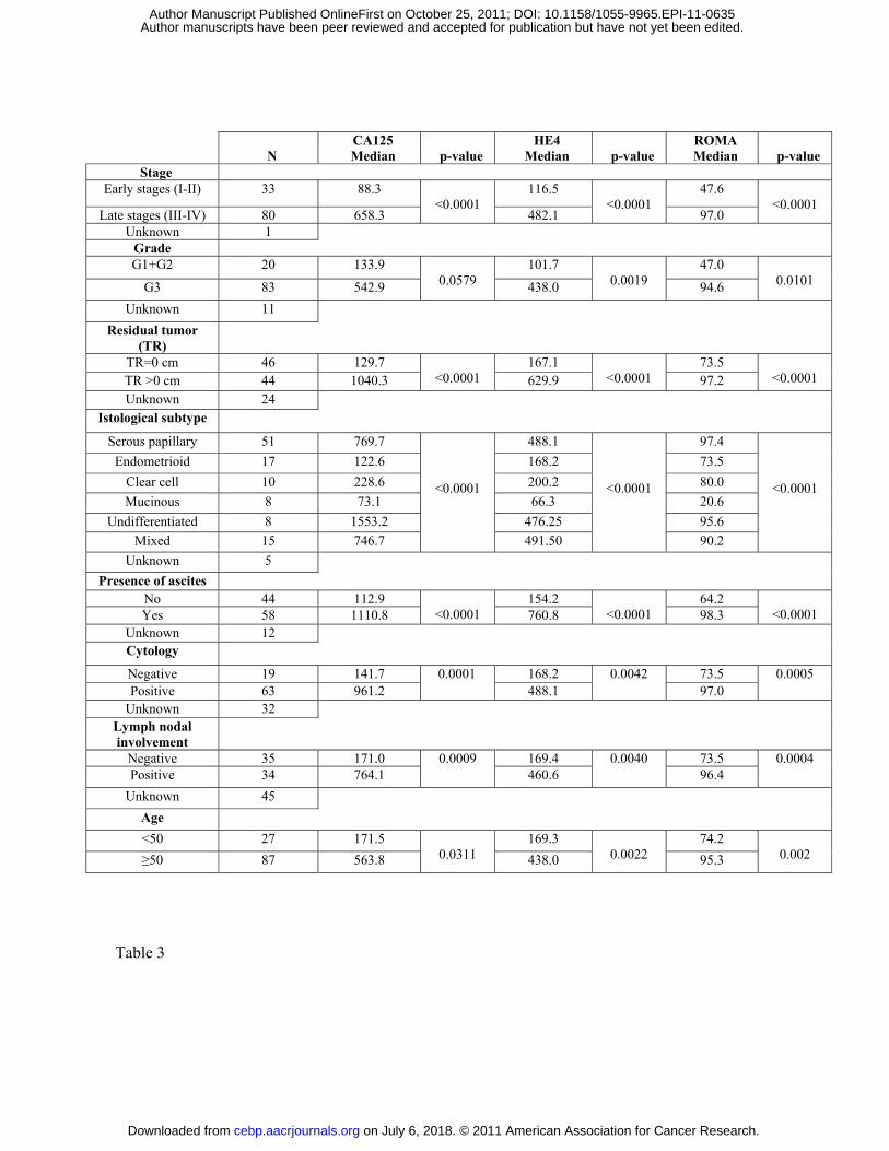
CA125, HE4, ROMA serum levels and clinicopathological features of EOC patients
Relationships between CA125, HE4 and ROMA levels and clinicopathological characteristics were
illustrated in Table 3. Elevated CA125, HE4 and ROMA levels were associated with advanced
FIGO stage, suboptimally debulked tumor, ascites, positive cytology, lymph node involvement and
on July 6, 2018. © 2011 American Association for Cancer Research. cebp.aacrjournals.org Downloaded from
Author manuscripts have been peer reviewed and accepted for publication but have not yet been edited. Author Manuscript Published OnlineFirst on October 25, 2011; DOI: 10.1158/1055-9965.EPI-11-0635

11
advanced age (all p≤0.05). Elevated HE4 and ROMA levels (p≤0.01), but not CA125 levels
(p=0.0579), were associated with undifferentiated tumours. Finally, CA125, HE4 and ROMA levels
were associated with histological subtypes (all p≤0.0001). Indeed serous papillary, undifferentiated
and mixed type showed higher levels of CA125, HE4, and ROMA, than endometrioid, clear cell
and mucinous subtypes.
CA125, HE4 and ROMA prognostic performances
At the time of the last follow-up, 42 (42.8%) patients were alive without evidence of disease, 11
(11.2%) patients were alive with disease, 33 (33.7%) patients died of disease (median OS, 46
months, 95% CI=32-n.e.) and 12 patients were alive with unknown status. The number of events for
OS, DFS and PFS were 33, 47 and 38, respectively.
Survival analyses of OS, DFS and PFS on the basis of high vs low CA125, HE4, ROMA levels
were significantly different (all p≤0.0001). The 2-yrs OS, DFS and PFS for EOC patients with low
CA125 levels were 76.3% (95%CI: 60.3%-86.6%), 65.0% (95%CI: 47.9%-77.7%) and 71.1%
(95%CI: 54.6%-82.5%) respectively, and decreased to 63.2% (95%CI: 44.4%-77.2%), 13.9%
(95%CI: 4.0%- 29.6%) and 44.9% (95%CI: 27.3%- 61.1%) for patients with high levels. The 2-yrs
OS, DFS and PFS for EOC patients with low HE4 levels were 90.1% (95%CI: 75.8%-96.2%),
71.3% (95%CI: 54.0%-83.1%) and 82.4% (95%CI: 66.5%-91.3%) respectively, and decreased to
47.3% (95%CI: 29.6%-63.2%), 8.9% (95%CI: 1.8%- 23.5%) and 32.6% (95%CI: 1.7%- 49.1%) for
patients with high levels. The 2-yrs OS, DFS and PFS for EOC patients with low ROMA levels
were 85.9% (95%CI: 0.71.3%-93.5%), 69.2% (95%CI: 51.9%-81.3%) and 77.9% (95%CI: 61.6%-
87.9%) respectively, and decreased to 50.4% (95%CI: 32.0%-66.2%), 8.4% (95%CI: 1.6%-22.6%)
and 35.7% (95%CI: 19.4%-52.4%) for patients with high levels. The Kaplan-Meier curves for OS
on the basis of high vs low CA125, HE4 and ROMA levels were shown in Figure 2.
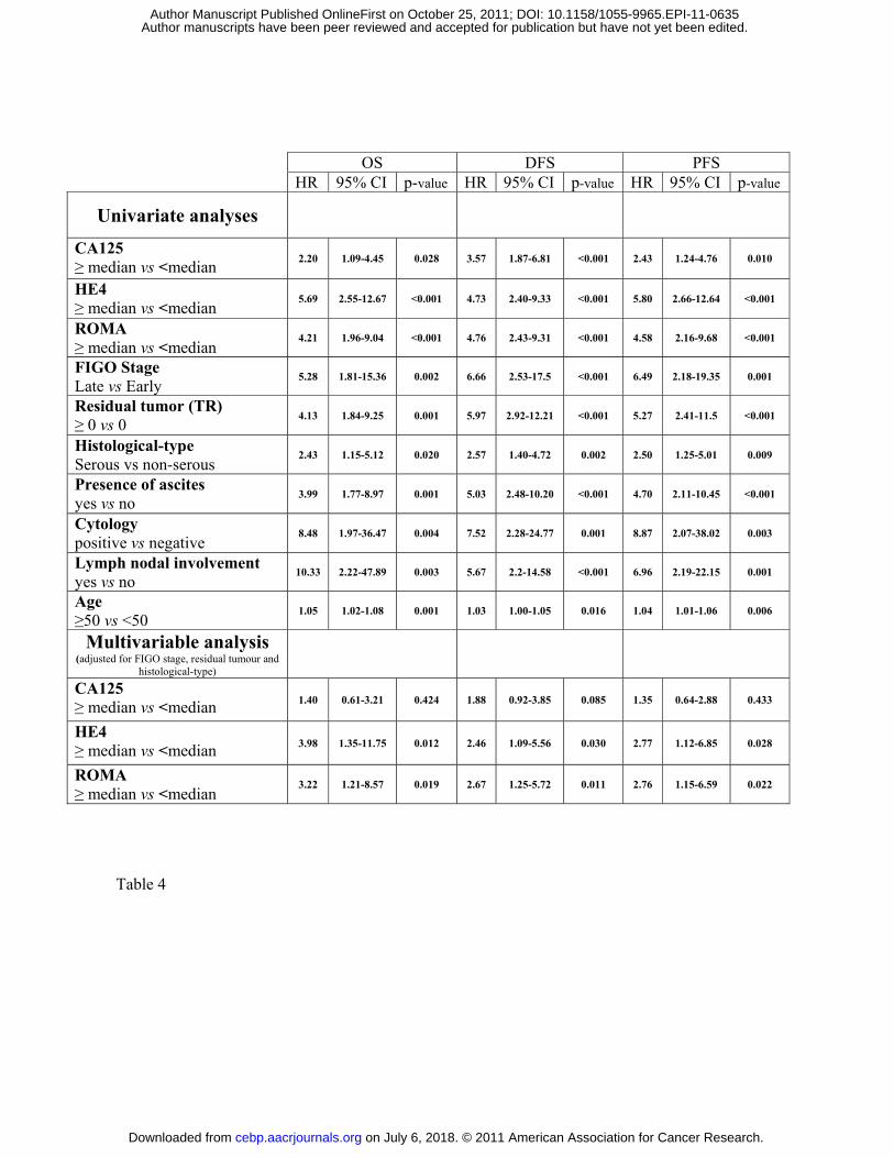
Univariate and multivariable analyses for survival were reported in Table 4. We couldn’t analyze
the prognostic impact of tumor grade, due to extremely unbalanced number of events available for
on July 6, 2018. © 2011 American Association for Cancer Research. cebp.aacrjournals.org Downloaded from
Author manuscripts have been peer reviewed and accepted for publication but have not yet been edited. Author Manuscript Published OnlineFirst on October 25, 2011; DOI: 10.1158/1055-9965.EPI-11-0635

12
the analyses in the three subgroups (G1, G2, G3). As expected, clinicopathological features known
to be prognostic variables for EOC such as FIGO stage, residual tumour after cytoreductive surgery,
histological subtype, presence of ascites, positive cytology, lymph node involvement and age
showed a statistically significant association with OS, DFS and PFS in univariate analyses, proving
the validity of the patients cohort recruited in this study (all p≤0.02). In addition, univariate analyses
demonstrated that elevated levels of CA125, HE4 and ROMA were significantly associated with
shorter OS, DFS and PFS (all p≤0.028).
Multivariable analysis was performed to estimate the effect of biomarkers adjusted for FIGO stage,
residual tumour and histological subtype, the most important established prognostic factors.
Interestingly, among the three biomarkers analyzed, we found that HE4 and ROMA (all p≤0.039),
but not CA125 (all p≥0.082), are independent prognostic factors for OS, DFS and PFS. About the
established prognostic factors only residual tumour has been found as independent prognostic factor
for DFS (HR=3.05, 95%CI=1.29-7.21, p=0.011) and for PFS (HR=3.00, 95%CI=1.15-7.83,
p=0.025).
Discussion
Epithelial ovarian cancer is often detected at an advanced stage and is characterized by poor
survival. As a result it is the most frequent cause of death from gynaecological cancer. At the
moment, transvaginal ultrasonography and CA125 assay are mainly used for EOC diagnosis. Two
longitudinal randomized screening trials, PLCO and UKCTOCS, are ongoing to determine their
potential role in early detection of EOC. However, transvaginal ultrasonography requires
specialized knowledge to reduce inter-operator interpretation variability and, like CA125, it is only
partially effective in discriminating benign and malignant lesions. Therefore, there is a pressing
need for clinical practice to develop new, easy, low cost and standardized serum analysis methods
for an accurate differential diagnosis between benign and malignant lesions. In fact, it has been
demonstrated that discrimination between malignant and benign pelvic masses improves EOC
patients care and outcome (15-20). Moore (32) proposed ROMA algorithm for the differential
on July 6, 2018. © 2011 American Association for Cancer Research. cebp.aacrjournals.org Downloaded from
Author manuscripts have been peer reviewed and accepted for publication but have not yet been edited. Author Manuscript Published OnlineFirst on October 25, 2011; DOI: 10.1158/1055-9965.EPI-11-0635

13
diagnosis between benign and malignant lesions and Nolen and colleagues (33), among 65
biomarkers tested, reaffirmed the superiority of the CA125/HE4 combination. Moreover, Moore
(37) showed that ROMA algorithm has better diagnostic performance than the widely used RMI.
However, diagnostic accuracy of ROMA compared to CA125 and HE4 alone is still controversial.
This is mainly due to the fact that different assays for CA125 and HE4, different thresholds for HE4
and ROMA and different patient selection criteria were used in each study to compare diagnostic
performances of HE4, CA125 and ROMA (32, 37-42). In the present study, in order to report
consistent results about HE4, CA125 and ROMA diagnostic performances, we enrolled a large
number (419) of women, selecting only invasive epithelial ovarian cancer as malignant ovarian
pathology. Moreover, we analyzed HE4 and CA125 employing fully automated methods and using
biomarkers’ thresholds largely validated by manufactures or by clinical practice. With these
thresholds, CA125 classified correctly 98.6% of healthy controls, while HE4 classified correctly
100% of healthy controls; consequently, these thresholds seemed adequate for our study.
Our analysis performed on healthy controls, revealed that HE4 and CA125 are differentially
expressed in pre- and post-menopause, in agreement with the literature (3, 45). Being the expression
of the markers and the risk of developing benign and malignant pathologies correlated to the
menopausal status, we analyzed separately pre- and post-menopausal women.
The biological function of HE4 has not been identified yet. HE4 is a member of the whey acidic
protein (WAP) four-disulfide core gene cluster that harbors 15 small serine protease inhibitor genes.
Comparative genomic hybridization studies showed that WAP gene cluster is among the most
frequent amplified chromosomal regions in EOC (46-49), suggesting the potential presence of
oncogenes in this region.
This study, according to previous reports (5-6, 39, 50), proved the high specificity of HE4 in
differential diagnosis between lesions of malignant and benign nature, as it is less frequently
increased in patients with benign cysts or endometriosis. In our experience, 17.7 % of post-
menopausal patients with ovarian benign cysts and 73.5% of patients with endometriosis exceeded
on July 6, 2018. © 2011 American Association for Cancer Research. cebp.aacrjournals.org Downloaded from
Author manuscripts have been peer reviewed and accepted for publication but have not yet been edited. Author Manuscript Published OnlineFirst on October 25, 2011; DOI: 10.1158/1055-9965.EPI-11-0635
14
the threshold value for CA125, while only 1.0% of the same post-menopausal patients with ovarian
benign cysts and 5.8% of patients with endometriosis exceeded the threshold value for HE4 (CA125
vs HE4: p≤0.002). According to the fact that CA125 is frequently increased in patients with
endometriosis, when we compare endometriosis vs healthy controls we observed that CA125 ROC-
AUC was significantly higher than HE4-AUC (0.8982 vs 0.6369: p= 0.0011).
There are conflicting results about CA125 sensitivity. Prior studies (23, 50-51) showed that HE4 is
more sensitive than CA125 in detecting EOC, being elevated in patients with EOC that do not
express CA125. However, the current study, in agreement with other observations (38, 52, 53)
showed that CA125 has greater sensitivity than HE4 in detecting EOC. Indeed, in postmenopausal
EOC, CA125 exceeded the threshold value in 94.2% of patients while HE4 exceeded the threshold
value in only 78.1% of patients (94.2% vs 78.1% p=0.0002). Notably, we didn’t find any EOC
patient positive for HE4 and negative for CA125, while 16 (including 5 stages I) EOC patients were
positive only for CA125. Being CA125 more sensitive than HE4 in detecting patients with EOC,
when we compare EOCs vs healthy controls by ROC curves, CA125-AUC was significantly higher
than HE4-AUC (0.9940 vs 0.9576, p=0.0147).
Among pre-menopausal women, we observed that CA125 is more sensitive in detecting EOCs,
while HE4 is more specific in discriminating ovarian cysts from EOCs. However, this analysis
didn’t reach statistical significance, possibly because of the small sample size.
Concerning ROMA, some previous reports showed that CA125/HE4 combination yielded a higher
diagnostic accuracy compared to single markers (23, 32), while other papers didn’t reach the same
conclusions (38-39). In our experience, ROMA inherits the strengths and the weakness of CA125
and HE4 alone. Indeed, ROMA is more sensitive than HE4 but less sensitive than CA125 and it is
more specific than CA125 but less specific than HE4. Unfortunately, because of the heterogeneity
of previous studies, it is not possible to pool data and to perform a meta-analysis in order to obtain
univocal indications about the clinical application of this algorithm in EOC diagnosis.
on July 6, 2018. © 2011 American Association for Cancer Research. cebp.aacrjournals.org Downloaded from
Author manuscripts have been peer reviewed and accepted for publication but have not yet been edited. Author Manuscript Published OnlineFirst on October 25, 2011; DOI: 10.1158/1055-9965.EPI-11-0635

15
Ovarian cancer management could also be improved through the development of new methods for
EOC prognosis. Therefore, the need for additional prognostic data to calibrate therapeutic tools on
an individual basis in women with EOC seems obvious. In order to find out how CA125, HE4 and
ROMA influence EOC biology, we analyzed the association between the biomarkers and the
clinicopathological characteristics of the patients. Remarkably, elevated HE4 and ROMA levels
(both p≤0.01), but not CA125 levels (p=0.0579), were associated with undifferentiated tumors,
suggesting that they are correlated with cancer aggressiveness. Moreover, since elevated levels of
CA125, HE4 and ROMA were found in patients suboptimally debulked, we could suppose that
these biomarkers, associated with others parameters (i.e. like imaging analysis), would be useful in
preoperative assessment of residual disease and, eventually, in the evaluation of a neoadiuvant
treatment. Univariate analyses showed that CA125, HE4 and ROMA, as well as established
clinicopathological prognostic variables for EOC, were significantly associated with shorter OS,
DFS, and PFS (all p≤0.05). However, when multivariable analysis was performed, we found that
only HE4 and ROMA were independent prognostic factors for OS, DFS and PFS. Moreover,
multivariable analysis proved that HE4 and ROMA appear to be better prognostic factors than
FIGO stage, residual tumor and histological subtype for OS, DFS, and PFS. These data suggest that
HE4 and ROMA could reflect intrinsic tumor aggressiveness that established prognostic factors
were not able to identify in our cohort of EOCs. Thus, elevated HE4 and ROMA levels, in
association with the traditional prognostic factors, could be clinically useful in identifying high-risk
EOC patients for a more aggressive tailored therapy, consisting in consolidation of treatment with
chemotherapeutic drugs or novel biologic agents. To date, we are the first group that demonstrated
HE4 and ROMA as independent prognostic factors for OS, DFS and PFS in EOC. Differently, the
Peak’ study showed that HE4 is an independent prognostic factor only for PFS and only in patients
with advanced (IIIc-IV) stage EOC. This could be explained by the fact that this latter analysis has
been carried out in a small group of women and the effect of HE4 serum levels on prognosis was
on July 6, 2018. © 2011 American Association for Cancer Research. cebp.aacrjournals.org Downloaded from
Author manuscripts have been peer reviewed and accepted for publication but have not yet been edited. Author Manuscript Published OnlineFirst on October 25, 2011; DOI: 10.1158/1055-9965.EPI-11-0635
16
evaluated non categorizing HE4 on the basis of the median values, but on the basis of cut-off value
proposed by Moore (23) for diagnostic purpose.
Recently, our group has reported the prognostic value of serum HE4 levels in poorly differentiated
endometrial carcinoma patients. Higher serum HE4 levels were correlated with a more aggressive
tumor phenotype and worse outcome (54) and similar results have been shown for breast and lung
cancer (55-56). Moreover, it has already been demonstrated that Leukocyte Protease 1 (SLPI), one
of the best-studied member of the WAP protein family, contrary to what expected for a protease
inhibitor, promotes malignant potential of cancer cells (57). On the basis of the similarity of HE4
and SLPI, it is tempting to speculate that HE4, like SLP1, could directly enhance malignant
potential of tumor.
In conclusion, this study confirms the diagnostic role of HE4 and ROMA already suggested by
other reports, but it adds a clinically relevant information, as for the first time we showed that high
levels of HE4 and ROMA are promising prognostic factors in EOC, identifying a subgroup of
patients with poor survival and at higher risk of death and, subsequently, directing to a more
aggressive adjuvant therapy. Further multicenter studies with homogeneous laboratory procedures
for HE4 and CA125 assays, as well as patients’ selection criteria, are needed to draw firm
conclusions about the applicability of HE4 and ROMA in clinical practice.
Acknoledgements
We wish to thank all the nurses working in the Department of Obstetrics and Gynaecology of the
University of Brescia, and especially Ms. Margherita Franzoni, for the assistance in collecting blood
samples. We are also grateful to Ms. Michela Faustini for her essential technical contribution in
performing the ARCHITECT assays.
Financial support
Supported in part by grants from the Nocivelli, Foundation, Brescia, Italy, by grants from the
Istituto Superiore di Sanità (Progetto Oncoproteomica, Programma Italia-USA “Farmacogenomica
on July 6, 2018. © 2011 American Association for Cancer Research. cebp.aacrjournals.org Downloaded from
Author manuscripts have been peer reviewed and accepted for publication but have not yet been edited. Author Manuscript Published OnlineFirst on October 25, 2011; DOI: 10.1158/1055-9965.EPI-11-0635

17
Oncologica”, convenzione 527/B4/4), Rome, Italy and by grants from the Ministero dell’Istruzione,
dell’Università e della Ricerca (PRIN project, prot2008AZJM9E) Rome, Italy. This investigation
was also supported by grants from NIH R01 CA122728-01A2 and R01 CA154460-01A1 to ADS,
grants 501/A3/3 and 0027557 from the Italian Institute of Health (ISS) to ADS and by NIH
Research Grant CA-16359 from the National Cancer Institute.
Conflict of interest statement
Dr. Claudio Galli is currently employed by Abbott Diagnostics as the Scientific Affairs Manager,
Italy
References
1. Holschneider CH, Berek JS. Ovarian cancer: epidemiology, biology, and prognostic factors.
Semin Surg Oncol 2000;19(1):3-10
2. Jacobs I, Oram D, Fairbanks J, Turner J, Frost C, Grudzinskas JG. A risk of malignancy index
incorporating CA 125, ultrasound and menopausal status for the accurate preoperative diagnosis of
ovarian cancer. Br J Obstet Gynaecol 1990;97(10):922-9
3. Malkasian GD Jr, Knapp RC, Lavin PT, Zurawski VR Jr, Podratz KC, Stanhope CR, et al.
Preoperative evaluation of serum CA 125 levels in premenopausal and postmenopausal patients
with pelvic masses: discrimination of benign from malignant disease. Am J Obstet Gynecol
1988;159(2):341-6.
4. Rustin GJ, Bast RC Jr, Kelloff GJ, Barrett JC, Carter SK, Nisen PD, et al. Use of CA-125 in
clinical trial evaluation of new therapeutic drugs for ovarian cancer. Clin Cancer Res
2004;10(11):3919-26.
on July 6, 2018. © 2011 American Association for Cancer Research. cebp.aacrjournals.org Downloaded from
Author manuscripts have been peer reviewed and accepted for publication but have not yet been edited. Author Manuscript Published OnlineFirst on October 25, 2011; DOI: 10.1158/1055-9965.EPI-11-0635
18
5. Huhtinen K, Suvitie P, Hiissa J, Junnila J, Huvila J, Kujari H et al. Serum HE4 concentration
differentiates malignant ovarian tumours from ovarian endometriotic cysts. Br J Cancer
2009;100(8):1315-9.
6. Bordin L, Fiore C, Donà G, Andrisani A, Ambrosini G, Faggian D et al. Evaluation of
erythrocyte band 3 phosphotyrosine level, glutathione content, CA-125, and human epididymal
secretory protein E4 as combined parameters in endometriosis. Fertil Steril 2010;94(5):1616-21.
7. Bast RC Jr, Badgwell D, Lu Z, Marquez R, Rosen D, Liu J et al. New tumor markers: CA125 and
beyond. Int J Gynecol Cancer 2005;15 Suppl 3:274-81.
8. Gadducci A, Landoni F, Maggino T, Sartori E, Zola P, Ferdeghini M, et al. Serum CA125 assay
at the time of relapse has no prognostic relevance in patients undergoing chemotherapy for recurrent
ovarian cancer: a multicenter Italian study. Int J Gynecol Cancer 1997;7(1):78-83.
9. Gadducci A, Cosio S, Zola P, Landoni F, Maggino T, Sartori E. Surveillance procedures for
patients treated for epithelial ovarian cancer: a review of the literature. Int J Gynecol Cancer
2007;17(1):21-31.
10. Bhoola S, Hoskins WJ. Diagnosis and management of epithelial ovarian cancer. Obstet Gynecol
2006;107(6):1399-410.
11. Aebi S, Castiglione M; ESMO Guidelines Working Group. Newly and relapsed epithelial
ovarian carcinoma: ESMO clinical recommendations for diagnosis, treatment and follow-up. Ann
Oncol 2009;20 Suppl 4:21-3.
on July 6, 2018. © 2011 American Association for Cancer Research. cebp.aacrjournals.org Downloaded from
Author manuscripts have been peer reviewed and accepted for publication but have not yet been edited. Author Manuscript Published OnlineFirst on October 25, 2011; DOI: 10.1158/1055-9965.EPI-11-0635

19
12. Høgdall EV, Christensen L, Kjaer SK, Blaakaer J, Kjaerbye-Thygesen A, Gayther S, et al.
CA125 expression pattern, prognosis and correlation with serum CA125 in ovarian tumor patients.
From The Danish "MALOVA" Ovarian Cancer Study. Gynecol Oncol 2007;104(3):508-15.
13. Markmann S, Gerber B, Briese V. Prognostic value of Ca 125 levels during primary therapy.
Anticancer Res 2007;27(4A):1837-9.
14. FIGO (International Federation of Gynecology and Obstetrics) 26th Annual Report on the
Results of Treatment in Gynecological Cancer. Int J Gynaecol Obstet 2006, 95 Suppl 1: S1-257
15. Hunter RW, Alexander ND, Soutter WP. Meta-analysis of surgery in advanced ovarian
carcinoma: is maximum cytoreductive surgery an independent determinant of prognosis? Am J
Obstet Gynecol 1992;166(2):504-11
16. Bristow RE, Gossett DR, Shook DR, Zahurak ML, Tomacruz RS, Armstrong DK, et al.
Micropapillary serous ovarian carcinoma: surgical management and clinical outcome. Gynecol
Oncol 2002;86(2):163-70.
17. Giede KC, Kieser K, Dodge J, Rosen B. Who should operate on patients with ovarian cancer?
An evidence-based review. Gynecol Oncol 2005;99(2):447-61.
18. Earle CC, Schrag D, Neville BA, Yabroff KR, Topor M, Fahey A, et al. Effect of surgeon
specialty on processes of care and outcomes for ovarian cancer patients. J Natl Cancer Inst
2006;98(3):172-80.
on July 6, 2018. © 2011 American Association for Cancer Research. cebp.aacrjournals.org Downloaded from
Author manuscripts have been peer reviewed and accepted for publication but have not yet been edited. Author Manuscript Published OnlineFirst on October 25, 2011; DOI: 10.1158/1055-9965.EPI-11-0635

20
19. Paulsen T, Kjaerheim K, Kaern J, Tretli S, Tropé C. Improved short-term survival for advanced
ovarian, tubal, and peritoneal cancer patients operated at teaching hospitals. Int J Gynecol Cancer
2006;16 Suppl 1:11-7.
20. Engelen MJ, Kos HE, Willemse PH, Aalders JG, de Vries EG, Schaapveld M, et al. Surgery by
consultant gynecologic oncologists improves survival in patients with ovarian carcinoma. Cancer
2006;106(3):589-98
21. Zhang Z, Yu Y, Xu F, Berchuck A, van Haaften-Day C, Havrilesky LJn, et al. Combining
multiple serum tumor markers improves detection of stage I epithelial ovarian cancer. Gynecol
Oncol 2007;107(3):526-31.
22. McIntosh MW, Liu Y, Drescher C, Urban N, Diamandis EP. Validation and characterization of
human kallikrein 11 as a serum marker for diagnosis of ovarian carcinoma. Clin Cancer Res
2007;13(15 Pt 1):4422-8.
23. Moore RG, Brown AK, Miller MC, Skates S, Allard WJ, Verch T, et al The use of multiple
novel tumor biomarkers for the detection of ovarian carcinoma in patients with a pelvic mass.
Gynecol Oncol 2008;108(2):402-8.
24. Havrilesky LJ, Whitehead CM, Rubatt JM, Cheek RL, Groelke J, He Q, et al. Evaluation of
biomarker panels for early stage ovarian cancer detection and monitoring for disease recurrence.
Gynecol Oncol 2008;110(3):374-82.
on July 6, 2018. © 2011 American Association for Cancer Research. cebp.aacrjournals.org Downloaded from
Author manuscripts have been peer reviewed and accepted for publication but have not yet been edited. Author Manuscript Published OnlineFirst on October 25, 2011; DOI: 10.1158/1055-9965.EPI-11-0635
21
25. Bertenshaw GP, Yip P, Seshaiah P, Zhao J, Chen TH, Wiggins WS, et al. Multianalyte profiling
of serum antigens and autoimmune and infectious disease molecules to identify biomarkers
dysregulated in epithelial ovarian cancer. Cancer Epidemiol Biomarkers Prev 2008;17(10):2872-81.
26. Visintin I, Feng Z, Longton G, Ward DC, Alvero AB, Lai Y, et al. Diagnostic markers for early
detection of ovarian cancer. Clin Cancer Res 2008;14(4):1065-72.
27. Palmer C, Duan X, Hawley S, Scholler N, Thorpe JD, Sahota RA, et al. Systematic evaluation
of candidate blood markers for detecting ovarian cancer. PLoS One 2008;3(7):e2633.
28. Henic E, Borgfeldt C, Christensen IJ, Casslén B, Høyer-Hansen G. Cleaved forms of the
urokinase plasminogen activator receptor in plasma have diagnostic potential and predict
postoperative survival in patients with ovarian cancer. Clin Cancer Res 2008;14(18):5785-93.
29. Nolen B, Marrangoni A, Velikokhatnaya L, Prosser D, Winans M, Gorelik E, et al. A serum
based analysis of ovarian epithelial tumorigenesis. Gynecol Oncol 2009;112(1):47-54.
30. Nosov V, Su F, Amneus M, Birrer M, Robins T, Kotlerman J, et al. Validation of serum
biomarkers for detection of early-stage ovarian cancer. Am J Obstet Gynecol 2009;200(6):639.e1-5.
31. Amonkar SD, Bertenshaw GP, Chen TH, Bergstrom KJ, Zhao J, Seshaiah P, et al. Development
and preliminary evaluation of a multivariate index assay for ovarian cancer. PLoS One
2009;4(2):e4599.
on July 6, 2018. © 2011 American Association for Cancer Research. cebp.aacrjournals.org Downloaded from
Author manuscripts have been peer reviewed and accepted for publication but have not yet been edited. Author Manuscript Published OnlineFirst on October 25, 2011; DOI: 10.1158/1055-9965.EPI-11-0635
22
32. Moore RG, McMeekin DS, Brown AK, DiSilvestro P, Miller MC, Allard WJ, et al. A novel
multiple marker bioassay utilizing HE4 and CA125 for the prediction of ovarian cancer in patients
with a pelvic mass. Gynecol Oncol 2009;112(1):40-6.
33. Nolen B, Velikokhatnaya L, Marrangoni A, De Geest K, Lomakin A, Bast RC Jr, et al. Serum
biomarker panels for the discrimination of benign from malignant cases in patients with an adnexal
mass. Gynecol Oncol 2010;117(3):440-5.
34. Yurkovetsky Z, Skates S, Lomakin A, Nolen B, Pulsipher T, Modugno F, et al. Development of
a multimarker assay for early detection of ovarian cancer. J Clin Oncol 2010;28(13):2159-66.
35. Abdel-Azeez HA, Labib HA, Sharaf SM, Refai AN. HE4 and mesothelin: novel biomarkers of
ovarian carcinoma in patients with pelvic masses. Asian Pac J Cancer Prev 2010;11(1):111-6.
36. Cramer DW, Bast RC Jr, Berg CD, Diamandis EP, Godwin AK, Hartge P, et al. Ovarian cancer
biomarker performance in prostate, lung, colorectal, and ovarian cancer screening trial specimens.
Cancer Prev Res (Phila) 2011;4(3):365-74
37. Moore RG, Jabre-Raughley M, Brown AK, Robison KM, Miller MC, Allard WJ, et al.
Comparison of a novel multiple marker assay vs the Risk of Malignancy Index for the prediction of
epithelial ovarian cancer in patients with a pelvic mass. Am J Obstet Gynecol 2010;203(3):228.e1-
6.
38. Van Gorp T, Cadron I, Despierre E, Daemen A, Leunen K, Amant F, et al. HE4 and CA125 as
a diagnostic test in ovarian cancer: prospective validation of the Risk of Ovarian Malignancy
Algorithm. Br J Cancer 2011;104(5):863-70.
on July 6, 2018. © 2011 American Association for Cancer Research. cebp.aacrjournals.org Downloaded from
Author manuscripts have been peer reviewed and accepted for publication but have not yet been edited. Author Manuscript Published OnlineFirst on October 25, 2011; DOI: 10.1158/1055-9965.EPI-11-0635
23
39. Montagnana M, Danese E, Ruzzenente O, Bresciani V, Nuzzo T, Gelati M. The ROMA (Risk
of Ovarian Malignancy Algorithm) for estimating the risk of epithelial ovarian cancer in women
presenting with pelvic mass: is it really useful? Clin Chem Lab Med 2011;49(3):521-5.
40. Kim YM, Whang DH, Park J, Kim SH, Lee SW, Park HA, et al. Evaluation of the accuracy of
serum human epididymis protein 4 in combination with CA125 for detecting ovarian cancer: a
prospective case-control study in a Korean population. Clin Chem Lab Med 2011;49(3):527-34.
41. Ruggeri G, Bandiera E, Zanotti L, Belloli S, Ravaggi A, Romani C, et al. HE4 and epithelial
ovarian cancer: Comparison and clinical evaluation of two immunoassays and a combination
algorithm. Clin Chim Acta 2011;412(15-16):1447-53.
42. Jacob F, Meier M, Caduff R, Goldstein D, Pochechueva T, Hacker N, et al. No benefit from
combining HE4 and CA125 as ovarian tumor markers in a clinical setting. Gynecol Oncol
2011;121(3):487-91.
43. Paek J, Lee SH, Yim GW, Lee M, Kim YJ, Nam EJ, et al. Prognostic significance of human
epididymis protein 4 in epithelial ovarian cancer. Eur J Obstet Gynecol Reprod Biol 2011. [Epub
ahead of print].
44. DeLong ER, DeLong DM, Clarke-Pearson DL. Comparing the areas under two or more
correlated receiver operating characteristic curves: a nonparametric approach. Biometrics
1988;44(3):837-45.
on July 6, 2018. © 2011 American Association for Cancer Research. cebp.aacrjournals.org Downloaded from
Author manuscripts have been peer reviewed and accepted for publication but have not yet been edited. Author Manuscript Published OnlineFirst on October 25, 2011; DOI: 10.1158/1055-9965.EPI-11-0635

24
45. Bon GG, Kenemans P, Verstraeten R, van Kamp GJ, Hilgers J. Serum tumor marker
immunoassays in gynecologic oncology: establishment of reference values. Am J Obstet Gynecol
1996;174(1 Pt 1):107-14.
46. Iwabuchi H, Sakamoto M, Sakunaga H, Ma YY, Carcangiu ML, Pinkel D, et al. Genetic
analysis of benign, low-grade, and high-grade ovarian tumors. Cancer Res 1995;55(24):6172-80.
47. Sonoda G, Palazzo J, du Manoir S, Godwin AK, Feder M, Yakushiji M, et al. Comparative
genomic hybridization detects frequent overrepresentation of chromosomal material from 3q26,
8q24, and 20q13 in human ovarian carcinomas. Genes Chromosomes Cancer 1997;20(4):320-8.
48. Kiechle M, Jacobsen A, Schwarz-Boeger U, Hedderich J, Pfisterer J, Arnold N. Comparative
genomic hybridization detects genetic imbalances in primary ovarian carcinomas as correlated with
grade of differentiation. Cancer 2001;91(3):534-40.
49. Ferreira Z, Hurle B, Rocha J, Seixas S. Differing evolutionary histories of WFDC8 (short-term
balancing) in Europeans and SPINT4 (incomplete selective sweep) in Africans. Mol Biol Evol
2011. [Epub ahead of print]
50. Holcomb K, Vucetic Z, Miller MC, Knapp RC. Human epididymis protein 4 offers superior
specificity in the differentiation of benign and malignant adnexal masses in premenopausal women.
Am J Obstet Gynecol 2011. [Epub ahead of print].
51. Anastasi E, Marchei GG, Viggiani V, Gennarini G, Frati L, Reale MG. HE4: a new potential
early biomarker for the recurrence of ovarian cancer. Tumour Biol 2010;31(2):113-9.
on July 6, 2018. © 2011 American Association for Cancer Research. cebp.aacrjournals.org Downloaded from
Author manuscripts have been peer reviewed and accepted for publication but have not yet been edited. Author Manuscript Published OnlineFirst on October 25, 2011; DOI: 10.1158/1055-9965.EPI-11-0635

25
52. Andersen MR, Goff BA, Lowe KA, Scholler N, Bergan L, Drescher CW, et al. Use of a
Symptom Index, CA125, and HE4 to predict ovarian cancer. Gynecol Oncol 2010;116(3):378-83.
53. Park Y, Lee JH, Hong DJ, Lee EY, Kim HS. Diagnostic performances of HE4 and CA125 for
the detection of ovarian cancer from patients with various gynecologic and non-gynecologic
diseases. Clin Biochem 2011. [Epub ahead of print]
54. Bignotti E, Ragnoli M, Zanotti L, Calza S, Falchetti M, Lonardi S, et al. Diagnostic and
prognostic impact of serum HE4 detection in endometrial carcinoma patients. Br J Cancer
2011;104(9):1418-25.
55. Yamashita S, Tokuishi K, Hashimoto T, Moroga T, Kamei M, Ono K, et al. Prognostic
significance of HE4 expression in pulmonary adenocarcinoma. Tumour Biol 2011;32(2):265-71.
56. Kamei M, Yamashita S, Tokuishi K, Hashioto T, Moroga T, Suehiro S, et al. HE4 expression
can be associated with lymph node metastases and disease-free survival in breast cancer. Anticancer
Res 2010;30(11):4779-83.
57. Devoogdt N, Hassanzadeh Ghassabeh G, Zhang J, Brys L, De Baetselier P, Revets H. Secretory
leukocyte protease inhibitor promotes the tumorigenic and metastatic potential of cancer cells. Proc
Natl Acad Sci U S A 2003;100(10):5778-82.
on July 6, 2018. © 2011 American Association for Cancer Research. cebp.aacrjournals.org Downloaded from
Author manuscripts have been peer reviewed and accepted for publication but have not yet been edited. Author Manuscript Published OnlineFirst on October 25, 2011; DOI: 10.1158/1055-9965.EPI-11-0635

26
Figure 1: Serum CA125 and HE4 levels detected in healthy controls and in patients with
endometriosis, ovarian cysts and epithelial ovarian cancer
HC, healthy controls; End, patients with endometriosis; Cysts, patients with ovarian benign cysts;
EOC, patients with epithelial ovarian cancer; HE4, human epididymis protein 4
Figure 2: Kaplan-Meier survival curves in relation to serum CA125, HE4 and ROMA levels
for epithelial ovarian cancer patients
Serum human epididymis protein 4 (HE4) levels and overall survival (OS) (A), serum CA125 levels
and overall survival (OS) (B), Risk for Ovarian Malignancy Algorithm (ROMA) levels and OS (C),
Table 1: Comparisons of the ROC-AUCs for CA125 and HE4 across the groups enrolled in
this study
on July 6, 2018. © 2011 American Association for Cancer Research. cebp.aacrjournals.org Downloaded from
Author manuscripts have been peer reviewed and accepted for publication but have not yet been edited. Author Manuscript Published OnlineFirst on October 25, 2011; DOI: 10.1158/1055-9965.EPI-11-0635

27
ROC-AUC, area under the receiver operating characteristic curve; HE4, human epididymis protein
4; CI, confidence interval; HC, healthy controls; Cysts, patients with ovarian benign cysts; End,
patients with endometriosis; EOC, patients with epithelial ovarian cancer
Table 2: Distribution of patients with epithelial ovarian cancer, endometriosis and ovarian
benign cysts according to their positivity for CA125, HE4 and ROMA and diagnostic
performances
HE4, human epididymis protein 4; ROMA, Risk for Ovarian Malignancy Algorithm; EOC, patients
with epithelial ovarian; positive, above cut-off; negative, under cut-off; CA125 cut-off value: 35
U/ml; HE4 cut-off values: ≤70 pmol/L for pre-menopausal state and ≤140 pmol/L in menopausal
state; ROMA cut-off values: ≤7.4% for pre-menopausal state and ≤25.3% menopausal state
Table 3: Association between serum CA125, HE4 and ROMA levels and clinicopathological
characteristics of epithelial ovarian cancer patients
HE4, human epididymis protein 4; ROMA, Risk for Ovarian Malignancy Algorithm; SD, standad
deviation
Table 4: Univariate and multivariable survival analyses in relation to serum CA125, HE4,
ROMA levels and clinicopathological prognostic variables for epithelial ovarian cancer
patients
OS, overall survival; DFS, disease-free survival; PFS, progression free survival; HE4, human
epididymis protein 4; ROMA, Risk for Ovarian Malignancy Algorithm
on July 6, 2018. © 2011 American Association for Cancer Research. cebp.aacrjournals.org Downloaded from
Author manuscripts have been peer reviewed and accepted for publication but have not yet been edited. Author Manuscript Published OnlineFirst on October 25, 2011; DOI: 10.1158/1055-9965.EPI-11-0635
5
HC End Cyst EOC HC Cyst EOC HC End Cyst EOC HC Cyst EOC
Pre-menopause Pre-menopausePost-menopause Post-menopauseN 39 34 35 26 101 96 87 N 39 34 35 26 101 96 87
Ln
CA
125
Ln H
E4Mean (pM) 36.0 42.4 40.8 377.7 42.8 50.7 993.7
Median (pM) 35.2 39.1 36.2 283.1 41.2 43.8 409.2Mean (U/ml) 16.0 83.0 29.1 1138.6 11.1 28.4 1675.5
Median (U/ml) 14.9 49.1 16.3 479.5 10.1 14.5 482.4
Figure 1
on July 6, 2018. © 2011 American Association for Cancer Research. cebp.aacrjournals.org Downloaded from
Author manuscripts have been peer reviewed and accepted for publication but have not yet been edited. Author Manuscript Published OnlineFirst on October 25, 2011; DOI: 10.1158/1055-9965.EPI-11-0635
CA125 ROC-AUC (95% CI)
HE4 ROC-AUC (95% CI)
p-value Po
st-
men
opau
se EOC vs HC 0.9940
(0.9866-1.0000) 0.9576
(0.9279-0.9873) 0.0147
EOC vs Cysts 0.9602 (0.9353-0.9852)
0.9400 (0.9039-0.97599)
0.2439
Cysts vs HC 0.6671 (0.5898-0.7444)
0.5856 (0.5055-0.6656)
0.1420
Pre-
m
enop
ause
EOC vs HC 0.9773 (0.9457-1.0000)
0.9177 (0.8279-1.0000)
0.1233
EOC vs Cysts 0.9549 (0.9053-1.0000)
0.9016 (0.8068-0.9965)
0.1867
EOC vs End 0.8473 (0.7397-0.9549)
0.8925 (0.7907-0.9944)
0.3211
Cysts vs HC 0.5960 (0.4644-0.7275)
0.5744 (0.4390-0.7097)
0.8280
End vs HC 0.8982 (0.8183-0.9781)
0.6369 (0.5055-0.7682)
0.0011
End vs Cysts 0.8361 (0.7307-0.9416)
0.5471 (0.4075-0.6867)
0.0019
Table 1
on July 6, 2018. © 2011 American Association for Cancer Research. cebp.aacrjournals.org Downloaded from
Author manuscripts have been peer reviewed and accepted for publication but have not yet been edited. Author Manuscript Published OnlineFirst on October 25, 2011; DOI: 10.1158/1055-9965.EPI-11-0635

Pre-menopause Post-menopause
EOC Cysts and Endometriosis EOC Cysts
(N=26) (N=69) (N=87) (N=96) CA125 positive 24 28 82 17 negative 2 41 5 79 Accuracy (95% CI) 68.4 (58.1-77.6) 88.0 (82.4-92.3) Sensitivity (95% CI) 92.3 (74.9-99.1) 94.3 (87.1-98.1) Specificity (95% CI) 59.4 (46.9-71.1) 82.3 (73.2-89.3) HE4 positive 22 4 68 1 negative 4 65 19 95 Accuracy (95% CI) 91.6 (84.1-96.3) 89.1 (83.6-93.2) Sensitivity (95% CI) 84.6 (65.1-95.6) 78.2 (68.0-86.3) Specificity (95% CI) 94.2 (85.8-98.4) 99.0 (94.3-100.0) ROMA positive 22 4 81 15 negative 13 56 6 81 Accuracy (95% CI) 82.1 (72.9-89.2) 88.5 (83.0-92.8) Sensitivity (95% CI) 84.6 (65.1-95.6) 93.1 (85.6-97.4) Specificity (95% CI) 81.2 (69.9-89.6) 84.4 (75.5-91.0)
Table 2
on July 6, 2018. © 2011 American Association for Cancer Research. cebp.aacrjournals.org Downloaded from
Author manuscripts have been peer reviewed and accepted for publication but have not yet been edited. Author Manuscript Published OnlineFirst on October 25, 2011; DOI: 10.1158/1055-9965.EPI-11-0635
N
CA125 Median
p-value
HE4 Median
p-value
ROMA Median
p-value
Stage Early stages (I-II) 33 88.3
<0.0001 116.5
<0.0001 47.6
<0.0001 Late stages (III-IV) 80 658.3 482.1 97.0
Unknown 1 Grade G1+G2 20 133.9
0.0579 101.7
0.0019 47.0
0.0101 G3 83 542.9 438.0 94.6
Unknown 11
Residual tumor (TR)
TR=0 cm 46 129.7 <0.0001
167.1 <0.0001
73.5 <0.0001 TR >0 cm 44 1040.3 629.9 97.2
Unknown 24 Istological subtype
Serous papillary 51 769.7
<0.0001
488.1
<0.0001
97.4
<0.0001
Endometrioid 17 122.6 168.2 73.5
Clear cell 10 228.6 200.2 80.0
Mucinous 8 73.1 66.3 20.6
Undifferentiated 8 1553.2 476.25 95.6
Mixed 15 746.7 491.50 90.2
Unknown 5
Presence of ascites No 44 112.9
<0.0001 154.2
<0.0001 64.2
<0.0001 Yes 58 1110.8 760.8 98.3 Unknown 12 Cytology
Negative 19 141.7 0.0001 168.2 0.0042 73.5 0.0005 Positive 63 961.2 488.1 97.0
Unknown 32 Lymph nodal involvement
Negative 35 171.0 0.0009 169.4 0.0040 73.5 0.0004 Positive 34 764.1 460.6 96.4
Unknown 45
Age
<50 27 171.5 0.0311
169.3 0.0022
74.2 0.002 ≥50 87 563.8 438.0 95.3
Table 3
on July 6, 2018. © 2011 American Association for Cancer Research. cebp.aacrjournals.org Downloaded from
Author manuscripts have been peer reviewed and accepted for publication but have not yet been edited. Author Manuscript Published OnlineFirst on October 25, 2011; DOI: 10.1158/1055-9965.EPI-11-0635

babi
lity
abili
ty
Kaplan-Meier curves: ROMA and OS
ROMA≥Median
Kaplan-Meier curves: CA125 and OS
CA125≥Median
A C
Surv
ival
pro
b
Surv
ival
pro
ba
ROMA<Median
P<0.0001
0 20 40 60 80 Months0 20 40 60 80 Months0 20 40 60 80 Months
CA125<Median
CA125≥Median
P<0.0001
Kaplan-Meier curves: HE4 and OS
ty
0 20 40 60 80 Months
49 17 5 0 0 Low48 31 23 12 4 High
0 20 40 60 80 Months48 31 23 12 4 High49 17 5 0 0 Low
0 20 40 60 80 Months50 31 22 11 3 High48 18 6 1 1 Low
B
Surv
ival
pro
babi
li HE4≥Median
HE4<Median
P<0.0001
0 20 40 60 80 Months48 33 24 12 4 High50 16 4 0 0 Low
Figure 2Figure 2
on July 6, 2018. © 2011 American Association for Cancer Research. cebp.aacrjournals.org Downloaded from
Author manuscripts have been peer reviewed and accepted for publication but have not yet been edited. Author Manuscript Published OnlineFirst on October 25, 2011; DOI: 10.1158/1055-9965.EPI-11-0635
OS DFS PFS HR 95% CI p-value HR 95% CI p-value HR 95% CI p-value
Univariate analyses
CA125 ≥ median vs <median
2.20 1.09-4.45 0.028 3.57 1.87-6.81 <0.001 2.43 1.24-4.76 0.010
HE4 ≥ median vs <median
5.69 2.55-12.67 <0.001 4.73 2.40-9.33 <0.001 5.80 2.66-12.64 <0.001
ROMA ≥ median vs <median
4.21 1.96-9.04 <0.001 4.76 2.43-9.31 <0.001 4.58 2.16-9.68 <0.001
FIGO Stage Late vs Early
5.28 1.81-15.36 0.002 6.66 2.53-17.5 <0.001 6.49 2.18-19.35 0.001
Residual tumor (TR) ≥ 0 vs 0
4.13 1.84-9.25 0.001 5.97 2.92-12.21 <0.001 5.27 2.41-11.5 <0.001
Histological-type Serous vs non-serous
2.43 1.15-5.12 0.020 2.57 1.40-4.72 0.002 2.50 1.25-5.01 0.009
Presence of ascites yes vs no
3.99 1.77-8.97 0.001 5.03 2.48-10.20 <0.001 4.70 2.11-10.45 <0.001
Cytology positive vs negative
8.48 1.97-36.47 0.004 7.52 2.28-24.77 0.001 8.87 2.07-38.02 0.003
Lymph nodal involvement yes vs no
10.33 2.22-47.89 0.003 5.67 2.2-14.58 <0.001 6.96 2.19-22.15 0.001
Age ≥50 vs <50
1.05 1.02-1.08 0.001 1.03 1.00-1.05 0.016 1.04 1.01-1.06 0.006
Multivariable analysis (adjusted for FIGO stage, residual tumour and
histological-type)
CA125 ≥ median vs <median 1.40 0.61-3.21 0.424 1.88 0.92-3.85 0.085 1.35 0.64-2.88 0.433
HE4 ≥ median vs <median 3.98 1.35-11.75 0.012 2.46 1.09-5.56 0.030 2.77 1.12-6.85 0.028
ROMA ≥ median vs <median
3.22 1.21-8.57 0.019 2.67 1.25-5.72 0.011 2.76 1.15-6.59 0.022
Table 4
on July 6, 2018. © 2011 American Association for Cancer Research. cebp.aacrjournals.org Downloaded from
Author manuscripts have been peer reviewed and accepted for publication but have not yet been edited. Author Manuscript Published OnlineFirst on October 25, 2011; DOI: 10.1158/1055-9965.EPI-11-0635

Published OnlineFirst October 25, 2011.Cancer Epidemiol Biomarkers Prev Elisabetta Bandiera, Chiara Romani, Claudia Specchia, et al. prognostic tools for epithelial ovarian cancer managementMalignancy Algorithm (ROMA) as new diagnostic and Serum human epididymis protein 4 (HE4) and Risk for Ovarian
Updated version
10.1158/1055-9965.EPI-11-0635doi:
Access the most recent version of this article at:
Manuscript
Authoredited. Author manuscripts have been peer reviewed and accepted for publication but have not yet been
E-mail alerts related to this article or journal.Sign up to receive free email-alerts
Subscriptions
Reprints and
To order reprints of this article or to subscribe to the journal, contact the AACR Publications
Permissions
Rightslink site. Click on "Request Permissions" which will take you to the Copyright Clearance Center's (CCC)
.http://cebp.aacrjournals.org/content/early/2011/10/24/1055-9965.EPI-11-0635To request permission to re-use all or part of this article, use this link
on July 6, 2018. © 2011 American Association for Cancer Research. cebp.aacrjournals.org Downloaded from
Author manuscripts have been peer reviewed and accepted for publication but have not yet been edited. Author Manuscript Published OnlineFirst on October 25, 2011; DOI: 10.1158/1055-9965.EPI-11-0635