title: the uim protein, s5a, is rapidly ...the uim protein, s5a, is ubiquitinated by all types of...
TRANSCRIPT

THE UIM PROTEIN, S5A, IS UBIQUITINATED BY ALL TYPES OF UBIQUITIN LIGASES BY A MECHANISM DIFFERENT FROM TYPICAL SUBSTRATE RECOGNITION
Tomoaki Uchiki1*, Hyoung Tae Kim1*, Bo Zhai1,2, Steven P. Gygi1,2, Jennifer A. Johnston3, John P. O’Bryan4, Alfred L. Goldberg1
From the 1Department of Cell Biology, the 2Taplin Biological Mass Spectrometry Facility, Harvard Medical School, Boston, MA 02115, the 3Elan Pharmaceuticals, Inc, South San Francisco, CA 94080, the
4Department of Pharmacology, University of Illinois College of Medicine, Chicago, IL 60612 *Both authors contributed equally to this work
Address correspondence to: Alfred L. Goldberg, Department of Cell Biology, Harvard Medical School, 240 Longwood Avenue, Boston, MA 02115. Tel: 617-432-1855; Fax: 617-432-0173; Email: [email protected]
S5a/Rpn10 is a ubiquitin (Ub)-binding protein that is a subunit of the 26S proteasome, but also exists free in the cytosol. It binds polyUb chains through its two Ub-interacting motifs (UIMs). We discovered that, unlike typical substrates of Ub ligases (E3s), S5a can be ubiquitinated by all E3s tested including multimeric and monomeric Ring-finger E3s (MuRF1, Siah2, Parkin, APC and SCF TRCP1),the U-box E3, CHIP, and HECT domain E3s (E6AP and Nedd4), when assayed with UbcH5 or related Ub-conjugating enzymes. However, the E2s, UbcH1 and UbcH13/Uev1a, which function by distinct mechanisms, do not support S5a ubiquitination. Thus, S5a can be used for assay of probably all E3s with UbcH5. Ubiquitination of S5a results from its binding to Ub chains on the E3 (after self-ubiquitination) or on the substrate, since a mutant lacking the UIM domain was not ubiquitinated. Furthermore, if S5a’s UIM domains were fused to GST, the protein was rapidly ubiquitinated by MuRF1 and CHIP. In addition, polyubiquitination (but not monoubiquitination) of MuRF1 allowed S5a to bind to MuRF1 and accelerated S5a ubiquitination. This tendency of S5a to associate with the growing Ub chain can explain how S5a, unlike typical substrates, which are recognized by certain E3s through specific motifs, is ubiquitinated by all E3s tested and is rapidly degraded in vivo.
INTRODUCTIONIn eukaryotes, ubiquitination of proteins plays a key role in the regulation of many cellular processes ranging from the cell cycle and antigen presentation to gene transcription (1,2). A primary biochemical function of ubiquitination is to serve
as a substrate recognition mechanism that targets specific proteins for degradation by the 26S proteasome. This large complex degrades most cellular proteins in an ATP-hydrolysis dependent manner (1). Ubiquitination of protein requires the sequential action of three types of enzymes. First, the Ub-activating enzyme (E1) activates the ubiquitin (Ub) in an ATP-dependent reaction and forms a thioester bond between the C-terminal carboxyl group of the Ub and a cysteine on the E1. The activated Ub is then transferred to a cysteine residue on a Ub-conjugating enzyme (E2), and finally a Ub-protein ligase (E3) facilitates formation of isopeptide linkage between the C-terminus of the Ub and a lysine residue on the substrate or on the preceding Ub (3). In the final step, the Ub ligase (E3) binds both the substrate and the E2-Ub thioester, and thus determines which substrate is ubiquitinated. E3s can be classified into two types based on their mechanism of transferring Ub to the substrate. One type, which comprises most E3s, contains a RING motif (or the related U-box motif) and facilitates transfer of the activated Ub directly from the E2 to the substrate (3). The second type contains a HECT domain, which forms a thioester intermediate with Ub and then transfers the Ub to the substrate (3). Eukaryotic cells contain a large number of different E3s (probably hundreds), and each E3 is believed to recognize only a limited number of proteins as substrates (1). In most cases, the E3 recognizes a specific structure or amino-acid sequence in the substrate (often termed the “degron”) (3). For example, the “N-end rule” E3, Ubr-1, recognizes particular types of amino-acids at the N-terminus of the substrates (4), while the E3 SCFCdc4 complex recognizes a specific phosphorylated amino acid sequence in its substrate, Sic1 (5).
1
http://www.jbc.org/cgi/doi/10.1074/jbc.M900556200The latest version is at JBC Papers in Press. Published on February 24, 2009 as Manuscript M900556200
Copyright 2009 by The American Society for Biochemistry and Molecular Biology, Inc.
by guest on March 9, 2020
http://ww
w.jbc.org/
Dow
nloaded from

Cells contain a variety of proteins that non-covalently bind Ub or Ub chains through several distinct Ub-binding domains (UBD) (6). Many of these proteins have been reported to become ubiquitinated in cells or in vitro, and deletion or mutation of UBDs abolishes their ubiquitination (6-10). Interestingly, ubiquitination of some of these proteins reportedly blocks their binding to other ubiquitinated proteins and seems to reduce the activities of these Ub-binding proteins (9,11). However, despite an increasing number of reports about ubiquitination of Ub-binding proteins, only a few studies have identified E2s and E3s responsible for this process (8,12), and the mechanisms for their ubiqutination remain uncertain. S5a/Rpn10 is a major Ub-binding protein that binds preferentially to poly-Ub chains (13). It is found as a subunit of the 26S proteasome, but unlike other proteasome subunits, S5a exists predominantly as a free protein in the cytosol (i.e. not incorporated into the proteasome) (14,15). S5a contains two stretches of about 15 amino acids called the ubiquitin interacting motif (UIM) which is responsible for its affinity for the Ub chains (16,17). In yeast, the homolog of S5a, Rpn10, is required for degradation of a subset of cellular proteins by the proteasome (14,18-20). While investigating how the presence of free S5a affects ubiquitination of typical substrates of E3s, we observed that S5a can be ubiquitinated by two very different E3s (MuRF1 and CHIP) with the E2, UbcH5. Since a substrate is usually ubiquitinated by only a particular E3 and since these two enzymes recognize substrates by distinct mechanisms, this observation suggests that S5a interacts with E3s in an atypical manner. The present study demonstrates systematically that a large variety of E3s, which differ widely in enzymatic mechanism, size, structure and specificity, can ubiquitinate S5a provided they function with UbcH5 and related E2s. S5a thus can be considered as a new type of substrate that is ubiquitinated by a novel mechanism involving the association of S5a with growing poly-Ub chains.
EXPERIMENTAL PROCEDURES PlasmidsThe bacterial expression plasmids for His(10X)S5a and its mutants (16) were kindly provided by Dr. Patrick Young (Stockholm
University), GST fusion Siah2 (21) by Dr. Ze’ev Ronai (Burnham Institute), GST fusion MuRF1 (22) by Dr. David J. Glass (Novartis Institute), GST fusion E6AP and Nedd4 (23) by Dr. Allan M. Weissman (NIH) and His(6X)CHIP (24) by Dr. Cam Patterson (University of North Carolina Chapel Hill) were obtained as kind gifts. GST fusion UIM domain of S5a, MJD1, KIAA1594, KIAA1386, Eps15 and Epsin were subcloned from YFP-UIMs (7). Expression plasmids for His(6X)Parkin and GST-Mdm2 were generated by using cDNAs inserted into pGEX-6X expression plasmids.
Expression and purification of proteins Most proteins were expressed in E. coli (BL21(DE3)STAR) by induction with 0.2mM IPTG at 16 or 25°C for 14h. Cells were lysed in PBS with a French Press, and the crude lysate was cleared by centrifugation at 100,000g for 1h. All GST fusion proteins were purified using glutathione 4B sepharose (GE Healthcare), and all His-tagged proteins were purified using Ni-NTA agarose (Qiagen). His(6X)E1 was purified with Ub-agarose (Sigma) in lysis buffer (50mM Tris-Cl pH7.5, 1mM dithiothreitol (DTT), 5 mM MgCl2,20 mM KCl and 2 mM ATP). UbcH5 was purified with Ub-agarose in lysis buffer containing 10nM E1. The SCF TRCP1 complex was purified from SF9 insect cells expressing His-Cul1, myc-Rbx1, myc-Skp1, and Flag- TRCP1 isolated as previously described (25). APCCdc20 was immunoprecipitated from Xenopus oocytes, as described by Kirkpatrick et al (26).
Radiolabeling of S5a To radiolabel S5a by 125I, 150 l of 70 M of purified His(10X)S5a in PBS was first incubated with IODO-BEADS (Pierce) for 5min at room temperature. Then, 250 l of 0.5mCi of 125I in PBS were added and the mixture was incubated for an additional 10min at room temperature. Free iodine was removed with a PD10 desalting column (GE Healthcare). To metabolically label S5a, His(10x)S5a was expressed in E.coli (BL21STAR(DE3)) in a media containing 35S-methionine, and the 35S-labeled S5a was purified with an Ni/NTA column, as described above.
Ubiquitination assays
2
by guest on March 9, 2020
http://ww
w.jbc.org/
Dow
nloaded from

To assay the ubiquitination of S5a, 400 nM His(10X)S5a was incubated at 37°C for 30 to 60min with 20 nM E1, 350-700 nM E2, 150-700 nM E3, and 58 M Ub in conjugation buffer A (20 mM Tris-Cl pH 7.5, 20 mM KCl, 5 mM MgCl2, 2 mM ATP, and 1 mM DTT). The same procedure was performed for the ubiquitination of GST-UIMs. To ubiquitinate S5a by SCF TRCP1, 200 nM of His(10X)S5a was incubated at 30°C for 60min with 230 nM E1, 1.5 M UbcH5c, 58 M Ub, 1
M Ub-aldehyde, 6 nM SCF TRCP1, 12 nM Nedd8, 6 nM UbcH12 and 6 nM Nedd8-activating enzyme in conjugation buffer A supplemented with 5 mM NaF, 20 mM creatine phosphate, and 5 Mcreatine kinase. To measure ubiquitination of S5a by Parkin, 200 nM of His(10X)S5a were incubated at 37°C for 30 to 60min with conjugation buffer B (400 nM E1, 5 M UbcH7, 1.4 M Parkin and 200 M Ub in 50 mM HEPES-KCl pH8.8, 50 mM NaCl, 10 mM MgCl2, 1 mM ATP). To study ubiquitination of S5a by Mdm2, 1 M of His(10x)-S5a was incubated at 37°C for 30-60min with 400 nM E1, 5 M UbcH5c, 900 nM GST-Mdm2, 200 M Ub in the conjugation buffer B. To measure ubiquitination of troponin I, 400 nM of troponin I were incubated A at 37°C for 30 to 120min with 20 nM E1, 350 nM E2, 150 nM GST-MuRF1, and 58 M Ub in conjugation buffer. Ubiquitination of S5a by APC was assayed as described by Kirkpatrick et al. (26). Ubiquitinated proteins were analyzed by SDS-PAGE followed by auto-radiography or immunoblotting using specific antibodies.
Binding of S5a to immobilized MuRF1 GST-MuRF1 from 1 L of E.coli culture was immobilized on 400 l (bed volume) of the glutathione resin and washed with PBS. The resin was equilibrated with the conjugation buffer. Then, E1 was added to reach final concentrations of 20 nM, UbcH5c 350 nM, Ub 58 M, and ATP , , 2 mM. The mixture was incubated at 37°C for 1h with gentle rotation. A control sample was incubated in the same manner without E1, UbcH5c or Ubs. After incubation, the resin was washed three times with 40 bed volumes of the Ub-binding buffer [25 mM HEPES-KCl pH7.0, 125 mM potassium acetate, 5 mM EGTA, 0.5% (v/v) Triton-X100, and 1 mM DTT]. The resin was then equilibrated with the Ub-binding buffer containing
1mg/ml BSA. 5 l of the resin was mixed with 125I-S5a at a final concentration of 400nM and rotated at 4°C for 2h. The resin was then transferred to a spin column (BioRad) and was washed twice with 500 l of the Ub binding buffer. The bound proteins were eluted with the SDS-PAGE sample loading buffer, and the radioactive 125I-S5a in the eluate was measured using a gamma counter. A similar experiment was performed under conditions where GST-MuRF1 and S5a were ubiquitinated in the same test tube. GST-MuRF1 immobilized on the glutathione resin was prepared as above, and 125I-S5a (400 nM) were added to reach final concentrations of E1 20 nM, UbcH5c 350 nM, Ub 58 M, and ATP 2 mM respectively. In the control reaction, Ub was not included in the mixture. The mixtures were rotated at 37°C, and aliquots were taken every 15min. The resin was washed and the bound S5a was measured as described above.
Mass spectrometry analysis His(10X)S5a was ubiquitinated by GST-MuRF1 or GST-E6AP as described above. The sample was resolved on SDS-PAGE and visualized with Coomassie Blue. The upper portion of the gel, corresponding to ubiquitinated S5a was excised and subjected to in-gel trypsin digestion. The tryptic peptides were extracted from the gel and analyzed by liquid chromatography MS/MS (LC-MS/MS). Peptides were separated across a 50-min gradient ranging from 7% to 30% (v/v) acetonitrile in 0.1% (v/v) trifluoroacetic acid in a microcapillary (125 µm X 18 cm) column packed with C18 reverse-phase material (Magic C18AQ, 5 µm particles, 200 Å pore size, Michrom Bioresources) and analyzed on-line on a hybrid linear ion trap–Orbitrap mass spectrometer (LTQ-Orbitrap, ThermoElectron). For each cycle, one full MS scan acquired on the Orbitrap at high mass resolution was followed by ten MS/MS spectra on the linear ion trap from the ten most abundant ions. MS/MS spectra were searched using the Sequest algorithm against the human IPI protein database. Dynamic modifications of 114.0429275 Da on Lysine was allowed for ubiquitination. All peptidematches were initially filtered based on enzyme specificity, mass measurement error, Xcorr and dCorr scores and further manually validated for peptide identification and site localization.
3
by guest on March 9, 2020
http://ww
w.jbc.org/
Dow
nloaded from

RESULTSAll E3s tested can ubiquitinate S5a During recent studies, we observed that S5a was rapidly ubiquitinated by the E2, UbcH5, functioning with two different ubiquitin ligases, the muscle specific Ring-finger E3, MuRF1, which is induced in atrophying muscles (27), and the U-box E3, CHIP, which ubiquitinates unfolded proteins bound to Hsp70 (28,29). We therefore speculated that S5a, unlike typical substrates, can be ubiquitinated by many different types of E3s. To test this possibility, we initially examined whether S5a could be a substrate of the E2, UbcH5, with several types of monomeric RING E3s, including MuRF1, Siah2 (which is involved in apoptosis induced by TNF (21)), and Mdm2 (which catalyzes the degradation of p53 (30)) (Figure 1). We also examined its ubiquitination by an oligomeric RING E3, SCF TRCP1 (whichcatalyzes the degradation of various phosphoproteins including I- B and -catenin (31)), a U-box E3, CHIP, and the HECT domain E3s, E6AP (which is responsible for degradation of p53 upon infection by human papillomaviruses (32)) and Nedd4 (which is involved in endocytosis of membrane proteins (33)) (Figure 1). Ubiquitination of S5a and its detection were performed as described in the Experimental Procedures. In a typical reaction with MuRF1, S5a (400 nM) was incubated with MuRF1 (700nM), E1 (20 nM), UbcH5 (700nM), Ub (58 M) and ATP (2 mM). All these E3s formed long Ub-chains on S5a with UbcH5, and no ubiquitination was observed without an E3 added. Furthermore, to demonstrate that S5a is also ubiquitinated when it is in molar excess over E3, we performed the ubiquitination reaction with 2000 nM S5a and 400, 200 or 100 nM GST-MuRF1. Even at 100 nM of GST-MuRF1, the majority of S5a in the reaction mixture was ubiquitinated within 1h (Figure S2). This finding indicates that during these reactions, MuRF1 (and presumably other E3s) ubiquitinates S5a in a catalytic (not a stoichiometric) manner. It is noteworthy that ubiquitination of S5a by CHIP does not require the presence of a molecular chaperone for substrate recognition (Figure 2). Typical substrates of CHIP are proteins recognized and bound to the molecular chaperones, Hsp70 or Hsp90 (28). Accordingly CHIP
ubiquitinates the model substrate, luciferase, only when it is denatured and bound to Hsp70 (Figure 2). By contrast, in the absence of chaperones, CHIP ubiquitinates itself, which seems to be important in the ubiquitination of S5a without Hsp70 or Hsp90 present (see below).
Ubiquitination of S5a results from binding to the growing Ub chain
These findings raised the possibility that S5a is promiscuously ubiquitinated by many E3s due to a mechanism which does not require a substrate recognition site of E3. Several studies have reported the ubiquitination of Ub-binding proteins in vivo, which has been proposed to occur due to their high affinity for Ub, because deletion or mutation in their UBDs diminishes their ubiquitination in vivo (7,11,34,35) and in vitro(10,36). Therefore, we tested systematically if the ubiquitination of S5a by various E3s requires the UIMs using mutant S5a in which the critical hydrophobic residues within the UIM1 and/or UIM2 region are replaced with alanines (16). These mutations abolish the affinity of the UIMs for Ub (Figure S1 and (16)). Mutations in both the UIM1 and UIM2 domains of S5a abolished ubiquitination of S5a by MuRF1, CHIP and E6AP, with UbcH5 as the E2 (Figure 5, S1). However, S5a lacking just one functional UIM (UIM1 or UIM2) domain was still rapidly ubiquitinated. Thus, UIMs are required for the ubiquitination of S5a, but either of these UIMs is sufficient to allow S5a ubiquitination (Figure 5, S1). To test whether the UIM regions of S5a are sufficient to promote ubiquitination of a protein, we assayed ubiquitination of GST fused at its C-terminus to residues 203-329 of S5a that contains both UIM1 and UIM2. This fusion protein, GST-UIMS5a, was rapidly ubiquitinated by MuRF1 or CHIP with UbcH5, while GST without the UIM was not ubiquitinated (Figure 6A&B). To determine if the UIM of S5a is unique in this regard, we also assayed the ubiquitination of GST-fusion proteins containing UIMs of various other proteins (EPSIN, Eps15, MJD1, KIAA1594 or KIAA1386). These GST fusions were all ubiquitinated by GST-MuRF1 with UbcH5 (Figure 6A). Also, His(6x)CHIP with UbcH5 ubiquitinated all these GST-fusions, but not GST, itself and did so even in the absence of a molecular chaperone (Figure 6B). Since these two E3s have
4
by guest on March 9, 2020
http://ww
w.jbc.org/
Dow
nloaded from
very different specificities for substrates, and CHIP does not directly bind substrates, these UIM proteins must be functioning as substrates due to their tendency to bind to Ub chains. Accordingly, all these GST fusions bound to free poly-Ub chains (not attached to a protein substrate) containing 2 to 7 Ub molecules (Figure S1). These observations make it very likely that binding to the growing Ub chains on the E3s or on the substrate proteins leads to the ubiquitination of the UIM proteins. To determine whether this self-ubiquitination of a E3 promotes the ubiquitination of S5a, MuRF1 was first incubated with E1, UbcH5, Ub and ATP for 25min to allow self-ubiquitination of MuRF1, and then S5a was added to the reaction. In a control reaction, MuRF1 was first incubated with E1, UbcH5 and ATP but without Ub, and then the capacity to ubiquitinate S5a was assayed by adding S5a and Ub. The ubiquitination of S5a was significantly faster when MuRF1 had been pre-ubiquitinated before the addition of S5a (Figure 7A). For example, ubiquitination of S5a by ubiquitinated MuRF1 was clearly evident 4 minutes after S5a addition, while in the reaction with control MuRF1, ubiquitination of S5a was not observed until 8 minutes.
Self-Ubiquitination of MuRF1 specifically accelerated the ubiquitination of S5a
In contrast to S5a, troponin I was ubiquitinated more slowly by self-ubiquitinated MuRF1 than by unmodified MuRF1 (Figure 7B). Ubiquitination of a typical substrate thus does not require and seems to be rather reduced by the presence of Ub chains on the E3. Furthermore, the self-ubiquitination of MuRF1 appeared to precede the ubiquitination of S5a in the control reaction, while S5a ubiquitination was slow in onset but accelerated later when MuRF1 had been self-ubiquitinated (Figure 7A). By contrast, ubiquitination of troponin I and of MuRF1 appeared to start simultaneously (Figure 7B). These observations further support our hypothesis that self-ubiquitination of MuRF1 is a pre-requisite for the ubiquitination of S5a, but not for ubiquitination of a typical substrate. If ubiquitination of S5a is caused by binding of S5a to the growing Ub chains on the E3, S5a should bind to ubiquitinated MuRF1, but not to unmodified MuRF1. To test this prediction, we
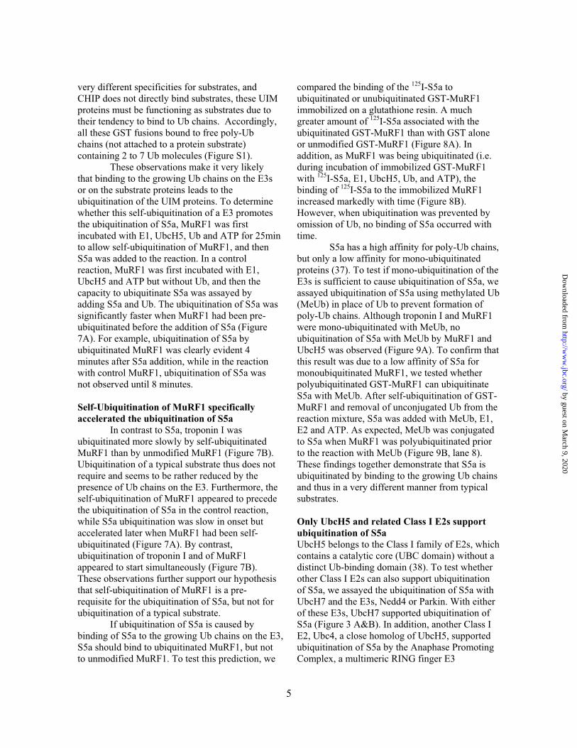
compared the binding of the 125I-S5a to ubiquitinated or unubiquitinated GST-MuRF1 immobilized on a glutathione resin. A much greater amount of 125I-S5a associated with the ubiquitinated GST-MuRF1 than with GST alone or unmodified GST-MuRF1 (Figure 8A). In addition, as MuRF1 was being ubiquitinated (i.e. during incubation of immobilized GST-MuRF1 with 125I-S5a, E1, UbcH5, Ub, and ATP), the binding of 125I-S5a to the immobilized MuRF1 increased markedly with time (Figure 8B). However, when ubiquitination was prevented by omission of Ub, no binding of S5a occurred with time. S5a has a high affinity for poly-Ub chains, but only a low affinity for mono-ubiquitinated proteins (37). To test if mono-ubiquitination of the E3s is sufficient to cause ubiquitination of S5a, we assayed ubiquitination of S5a using methylated Ub (MeUb) in place of Ub to prevent formation of poly-Ub chains. Although troponin I and MuRF1 were mono-ubiquitinated with MeUb, no ubiquitination of S5a with MeUb by MuRF1 and UbcH5 was observed (Figure 9A). To confirm that this result was due to a low affinity of S5a for monoubiquitinated MuRF1, we tested whether polyubiquitinated GST-MuRF1 can ubiquitinate S5a with MeUb. After self-ubiquitination of GST-MuRF1 and removal of unconjugated Ub from the reaction mixture, S5a was added with MeUb, E1, E2 and ATP. As expected, MeUb was conjugated to S5a when MuRF1 was polyubiquitinated prior to the reaction with MeUb (Figure 9B, lane 8). These findings together demonstrate that S5a is ubiquitinated by binding to the growing Ub chains and thus in a very different manner from typical substrates.
Only UbcH5 and related Class I E2s support ubiquitination of S5a UbcH5 belongs to the Class I family of E2s, which contains a catalytic core (UBC domain) without a distinct Ub-binding domain (38). To test whether other Class I E2s can also support ubiquitination of S5a, we assayed the ubiquitination of S5a with UbcH7 and the E3s, Nedd4 or Parkin. With either of these E3s, UbcH7 supported ubiquitination of S5a (Figure 3 A&B). In addition, another Class I E2, Ubc4, a close homolog of UbcH5, supported ubiquitination of S5a by the Anaphase Promoting Complex, a multimeric RING finger E3
5
by guest on March 9, 2020
http://ww
w.jbc.org/
Dow
nloaded from
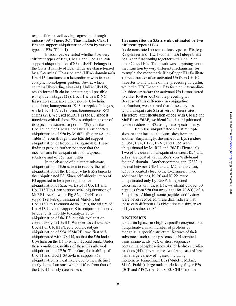
responsible for cell cycle progression through mitosis (39) (Figure 3C). Thus multiple Class I E2s can support ubiquitination of S5a by various types of E3s (Table 1). In addition, we tested whether two very different types of E2s, UbcH1 and UbcH13, can support ubiquitination of S5a. UbcH1 belongs to the Class II family of E2s, which are characterized by a C-terminal Ub-associated (UBA) domain (40). UbcH13 functions as a heterodimer with its non-catalytic homologous protein, Uev1a, which contains Ub-binding sites (41). Unlike UbcH5, which forms Ub chains containing all possible isopeptide linkages (29), UbcH1 with a RING finger E3 synthesizes processively Ub-chains containing homogeneous K48 isopeptide linkages, while UbcH13/Uev1a forms homogeneous K63 chains (29). We used MuRF1 as the E3 since it functions with all these E2s to ubiquitinate one of its typical substrates, troponin I (29). Unlike UbcH5, neither UbcH1 nor UbcH13 supported ubiquitination of S5a by MuRF1 (Figure 4A and Table 1), even though these E2s did support ubiquitination of troponin I (Figure 4B). These findings provide further evidence that the mechanisms for ubiquitination of a typical substrate and of S5a must differ. In the absence of a distinct substrate, ubiquitination of S5a seems to require the self-ubiquitination of the E3 after which S5a binds to the ubiquitinated E3. Since self-ubiquitination of E3 appeared to be a pre-requisite for ubiquitination of S5a, we tested if UbcH1 and UbcH13/Uev1 can support self-ubiquitination of MuRF1. As shown in Fig S5a, UbcH1 can support self-ubiquitination of MuRF1, but UbcH13/Uev1a cannot do so. Thus, the failure of UbcH13/Uevla to support S5a ubiquitination may be due to its inability to catalyze auto-ubiquitination of the E3, but this explanation cannot apply to UbcH1. We then tested whether UbcH1 or UbcH13/Uevla could catalyze ubiquitination of S5a if MuRF1 was first self-ubiquitinated with UbcH5, so that the S5a had a Ub-chain on the E3 to which it could bind,. Under these conditions, neither of these E2s allowed ubiquitination of S5a. Therefore, the inability of UbcH1 and UbcH13/Uevla to support S5a ubiquitination is most likely due to their distinct catalytic mechanisms, which differs from that of the UbcH5 family (see below).
The same sites on S5a are ubiquitinated by two different types of E3s As demonstrated above, various types of E3s (e.g. Ring-finger and HECT-domain E3s) ubiquitinate S5a when functioning together with UbcH5 or other Class I E2s. This result was surprising since they function by very different mechanisms; for example, the monomeric Ring-finger E3s facilitate a direct transfer of an activated Ub from Ub~E2 thioester to any lysine on the preceding ubiquitin, while the HECT-domain E3s form an intermediate Ub-thioester before the activated Ub is transferred to either K48 or K63 on the preceding Ub. Because of this difference in conjugation mechanism, we expected that these enzymes would ubiquitinate S5a at very different sites. Therefore, after incubation of S5a with UbcH5 and MuRF1 or E6AP, we identified the ubiquitinated lysine residues on S5a using mass spectrometry. Both E3s ubiquitinated S5a at multiple sites that are located at distant sites from one another. Surprisingly, the same four Lys residues on S5a, K74, K122, K262, and K365 were ubiquitinated by MuRF1 and E6AP (Figure 10). Two of the common ubiquitination sites, K74 and K122, are located within S5a’s von Willebrand factor A domain. Another common site, K262, is located between UIM1 and UIM2, and the last, K365 is located close to the C-terminus. Two additional lysines, K126 and K122, were ubiquitinated only by E6AP. In repeated experiments with these E3s, we identified over 30 peptides from S5a that accounted for 70-80% of its 24 lysines. Although some peptides and lysines were never recovered, these data indicate that these very different E3s ubiquitinate a similar set of Lys residues on S5a.
DISCUSSION Ubiquitin ligases are highly specific enzymes that ubiquitinate a small number of proteins by recognizing specific structural features of their substrates, such as the presence of N-terminal basic amino acids (42), or short sequences containing phosphoserines (43) or hydroxylproline residues (44). Nevertheless, we demonstrated here that a large variety of ligases, including monomeric Ring-finger E3s (MuRF1, Mdm2, Siah2, Parkin), large multimeric Ring-finger E3s (SCF and APC), the U-box E3, CHIP, and the
6
by guest on March 9, 2020
http://ww
w.jbc.org/
Dow
nloaded from
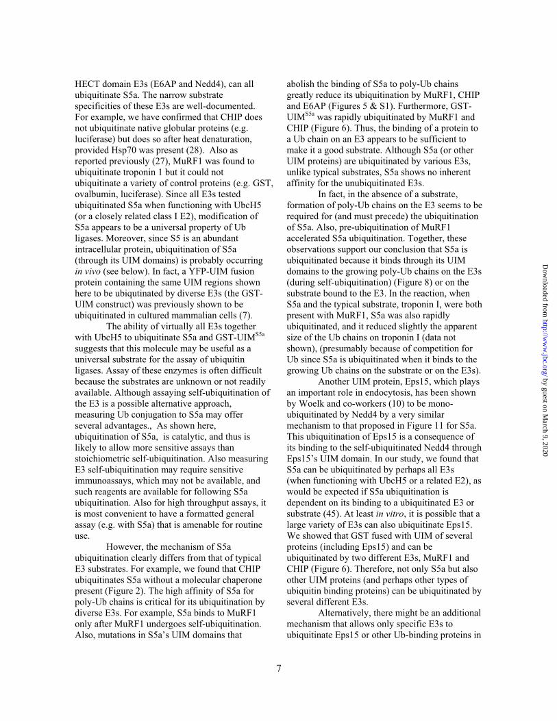
HECT domain E3s (E6AP and Nedd4), can all ubiquitinate S5a. The narrow substrate specificities of these E3s are well-documented. For example, we have confirmed that CHIP does not ubiquitinate native globular proteins (e.g. luciferase) but does so after heat denaturation, provided Hsp70 was present (28). Also as reported previously (27), MuRF1 was found to ubiquitinate troponin 1 but it could not ubiquitinate a variety of control proteins (e.g. GST, ovalbumin, luciferase). Since all E3s tested ubiquitinated S5a when functioning with UbcH5 (or a closely related class I E2), modification of S5a appears to be a universal property of Ub ligases. Moreover, since S5 is an abundant intracellular protein, ubiquitination of S5a (through its UIM domains) is probably occurring in vivo (see below). In fact, a YFP-UIM fusion protein containing the same UIM regions shown here to be ubiqutinated by diverse E3s (the GST-UIM construct) was previously shown to be ubiquitinated in cultured mammalian cells (7).
The ability of virtually all E3s together with UbcH5 to ubiquitinate S5a and GST-UIMS5a
suggests that this molecule may be useful as a universal substrate for the assay of ubiquitin ligases. Assay of these enzymes is often difficult because the substrates are unknown or not readily available. Although assaying self-ubiquitination of the E3 is a possible alternative approach, measuring Ub conjugation to S5a may offer several advantages., As shown here, ubiquitination of S5a, is catalytic, and thus is likely to allow more sensitive assays than stoichiometric self-ubiquitination. Also measuring E3 self-ubiquitination may require sensitive immunoassays, which may not be available, and such reagents are available for following S5a ubiquitination. Also for high throughput assays, it is most convenient to have a formatted general assay (e.g. with S5a) that is amenable for routine use. However, the mechanism of S5a ubiquitination clearly differs from that of typical E3 substrates. For example, we found that CHIP ubiquitinates S5a without a molecular chaperone present (Figure 2). The high affinity of S5a for poly-Ub chains is critical for its ubiquitination by diverse E3s. For example, S5a binds to MuRF1 only after MuRF1 undergoes self-ubiquitination. Also, mutations in S5a’s UIM domains that
abolish the binding of S5a to poly-Ub chains greatly reduce its ubiquitination by MuRF1, CHIP and E6AP (Figures 5 & S1). Furthermore, GST-UIMS5a was rapidly ubiquitinated by MuRF1 and CHIP (Figure 6). Thus, the binding of a protein to a Ub chain on an E3 appears to be sufficient to make it a good substrate. Although S5a (or other UIM proteins) are ubiquitinated by various E3s, unlike typical substrates, S5a shows no inherent affinity for the unubiquitinated E3s.
In fact, in the absence of a substrate, formation of poly-Ub chains on the E3 seems to be required for (and must precede) the ubiquitination of S5a. Also, pre-ubiquitination of MuRF1 accelerated S5a ubiquitination. Together, these observations support our conclusion that S5a is ubiquitinated because it binds through its UIM domains to the growing poly-Ub chains on the E3s (during self-ubiquitination) (Figure 8) or on the substrate bound to the E3. In the reaction, when S5a and the typical substrate, troponin I, were both present with MuRF1, S5a was also rapidly ubiquitinated, and it reduced slightly the apparent size of the Ub chains on troponin I (data not shown), (presumably because of competition for Ub since S5a is ubiquitinated when it binds to the growing Ub chains on the substrate or on the E3s). Another UIM protein, Eps15, which plays an important role in endocytosis, has been shown by Woelk and co-workers (10) to be mono-ubiquitinated by Nedd4 by a very similar mechanism to that proposed in Figure 11 for S5a. This ubiquitination of Eps15 is a consequence of its binding to the self-ubiquitinated Nedd4 through Eps15’s UIM domain. In our study, we found that S5a can be ubiquitinated by perhaps all E3s(when functioning with UbcH5 or a related E2), as would be expected if S5a ubiquitination is dependent on its binding to a ubiquitinated E3 or substrate (45). At least in vitro, it is possible that a large variety of E3s can also ubiquitinate Eps15. We showed that GST fused with UIM of several proteins (including Eps15) and can be ubiquitinated by two different E3s, MuRF1 and CHIP (Figure 6). Therefore, not only S5a but also other UIM proteins (and perhaps other types of ubiquitin binding proteins) can be ubiquitinated by several different E3s. Alternatively, there might be an additional mechanism that allows only specific E3s to ubiquitinate Eps15 or other Ub-binding proteins in
7
by guest on March 9, 2020
http://ww
w.jbc.org/
Dow
nloaded from
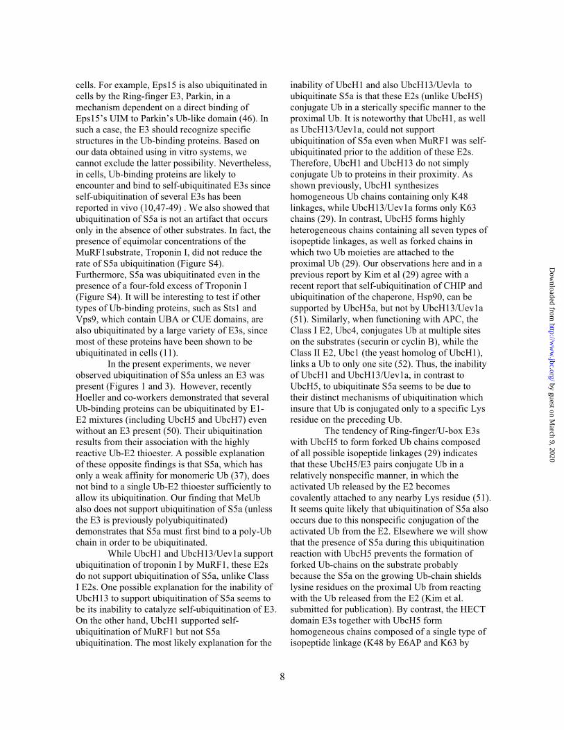
cells. For example, Eps15 is also ubiquitinated in cells by the Ring-finger E3, Parkin, in a mechanism dependent on a direct binding of Eps15’s UIM to Parkin’s Ub-like domain (46). In such a case, the E3 should recognize specific structures in the Ub-binding proteins. Based on our data obtained using in vitro systems, we cannot exclude the latter possibility. Nevertheless, in cells, Ub-binding proteins are likely to encounter and bind to self-ubiquitinated E3s since self-ubiquitination of several E3s has been reported in vivo (10,47-49) . We also showed that ubiquitination of S5a is not an artifact that occurs only in the absence of other substrates. In fact, the presence of equimolar concentrations of the MuRF1substrate, Troponin I, did not reduce the rate of S5a ubiquitination (Figure S4). Furthermore, S5a was ubiquitinated even in the presence of a four-fold excess of Troponin I (Figure S4). It will be interesting to test if other types of Ub-binding proteins, such as Sts1 and Vps9, which contain UBA or CUE domains, are also ubiquitinated by a large variety of E3s, since most of these proteins have been shown to be ubiquitinated in cells (11). In the present experiments, we never observed ubiquitination of S5a unless an E3 was present (Figures 1 and 3). However, recently Hoeller and co-workers demonstrated that several Ub-binding proteins can be ubiquitinated by E1-E2 mixtures (including UbcH5 and UbcH7) even without an E3 present (50). Their ubiquitination results from their association with the highly reactive Ub-E2 thioester. A possible explanation of these opposite findings is that S5a, which has only a weak affinity for monomeric Ub (37), does not bind to a single Ub-E2 thioester sufficiently to allow its ubiquitination. Our finding that MeUb also does not support ubiquitination of S5a (unless the E3 is previously polyubiquitinated) demonstrates that S5a must first bind to a poly-Ub chain in order to be ubiquitinated. While UbcH1 and UbcH13/Uev1a support ubiquitination of troponin I by MuRF1, these E2s do not support ubiquitination of S5a, unlike Class I E2s. One possible explanation for the inability of UbcH13 to support ubiquitination of S5a seems to be its inability to catalyze self-ubiquitination of E3. On the other hand, UbcH1 supported self-ubiquitination of MuRF1 but not S5a ubiquitination. The most likely explanation for the
inability of UbcH1 and also UbcH13/Uevla to ubiquitinate S5a is that these E2s (unlike UbcH5) conjugate Ub in a sterically specific manner to the proximal Ub. It is noteworthy that UbcH1, as well as UbcH13/Uev1a, could not support ubiquitination of S5a even when MuRF1 was self-ubiquitinated prior to the addition of these E2s. Therefore, UbcH1 and UbcH13 do not simply conjugate Ub to proteins in their proximity. As shown previously, UbcH1 synthesizes homogeneous Ub chains containing only K48 linkages, while UbcH13/Uev1a forms only K63 chains (29). In contrast, UbcH5 forms highly heterogeneous chains containing all seven types of isopeptide linkages, as well as forked chains in which two Ub moieties are attached to the proximal Ub (29). Our observations here and in a previous report by Kim et al (29) agree with a recent report that self-ubiquitination of CHIP and ubiquitination of the chaperone, Hsp90, can be supported by UbcH5a, but not by UbcH13/Uev1a (51). Similarly, when functioning with APC, the Class I E2, Ubc4, conjugates Ub at multiple sites on the substrates (securin or cyclin B), while the Class II E2, Ubc1 (the yeast homolog of UbcH1), links a Ub to only one site (52). Thus, the inability of UbcH1 and UbcH13/Uev1a, in contrast to UbcH5, to ubiquitinate S5a seems to be due to their distinct mechanisms of ubiquitination which insure that Ub is conjugated only to a specific Lys residue on the preceding Ub. The tendency of Ring-finger/U-box E3s with UbcH5 to form forked Ub chains composed of all possible isopeptide linkages (29) indicates that these UbcH5/E3 pairs conjugate Ub in a relatively nonspecific manner, in which the activated Ub released by the E2 becomes covalently attached to any nearby Lys residue (51). It seems quite likely that ubiquitination of S5a also occurs due to this nonspecific conjugation of the activated Ub from the E2. Elsewhere we will show that the presence of S5a during this ubiquitination reaction with UbcH5 prevents the formation of forked Ub-chains on the substrate probably because the S5a on the growing Ub-chain shields lysine residues on the proximal Ub from reacting with the Ub released from the E2 (Kim et al. submitted for publication). By contrast, the HECT domain E3s together with UbcH5 form homogeneous chains composed of a single type of isopeptide linkage (K48 by E6AP and K63 by
8
by guest on March 9, 2020
http://ww
w.jbc.org/
Dow
nloaded from
9
Nedd4). Thus, these enzymes must transfer the activated Ub in a rather precise, stereo-specific manner (29). Nevertheless, both E6AP and Nedd4 extensively ubiquitinated S5a, and E6AP ubiquitinates S5a at multiple Lys residues. This finding suggests that HECT domain E3s require a less specific orientation of the Ub-acceptor Lys residues in the substrate than in the proximal Ub during Ub chain synthesis. The mechanism of Ub conjugation to the substrate and that of Ub-chain elongation by the HECT domain E3s differ markedly. For example, the HECT domain E3s, E6AP and KIAA10, appear to pre-synthesize Ub chains before transferring them to their substrates (53). Thus, chain synthesis and Ub conjugation to the substrate might be two distinct processes. These observations suggest that Ub-chain formation by E3s involves two kinds of specificity that are dependent on types of the E2/E3 pairs: The first specificity defines the Lys residue of the substrate to which the first Ub is attached, and the second concerns the types of isopeptide linkages formed within the Ub chain. These two types of specificity may not necessarily correlate with each other. Accordingly, MuRF1 and E6AP, despite
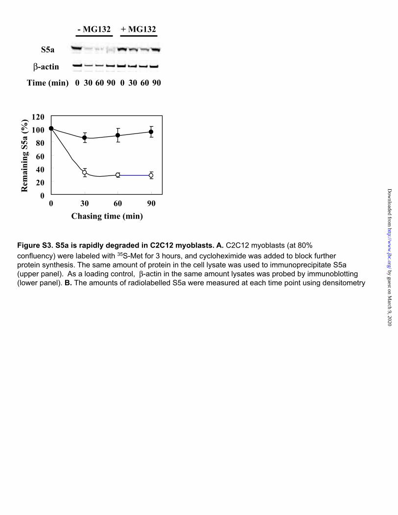
their contrasting mechanisms, conjugated multiple Ub residues to the same four sites on S5a. These findings further argue that the conjugation of the initial Ub to a substrate protein is quite a different process from the elongation of Ub-chains. The rapid ubiquitination of S5a by various E3s in vitro would predict that this protein, at least its free form present in the cytosol (not in the proteasome) would be rather short-lived. In fact, in yeast, the S5a homolog, Rpn10 (54), is extensively ubiquitinated and rapidly degraded by the proteasome in vivo (54), and in C2C12 (mouse) myoblasts, we observed that S5a has a half-life of about 30min (Figure S3). The short half-life of S5a presumably is due to the presence of the UIM domain and reflects the ubiquitination of free S5a by many E3s . At first glance this rapid turnover would appear to be quite wasteful for the cell.Although ubiquitination of S5a may perhaps serve some important regulatory purpose, it seems more likely from the present findings to represent an untoward side effect of substrate ubiquitination that would require cells to continually synthesize and destroy (or deubiquitinate) S5a molecules.
REFERENCES
1. Glickman, M. H., and Ciechanover, A. (2002) Physiol Rev 82(2), 373-428 2. Goldberg, A. L. (2003) Nature 426(6968), 895-899 3. Pickart, C. M. (2001) Annu Rev Biochem 70, 503-533 4. Varshavsky, A. (1997) Genes Cells 2(1), 13-28 5. Nash, P., Tang, X., Orlicky, S., Chen, Q., Gertler, F. B., Mendenhall, M. D., Sicheri, F., Pawson,
T., and Tyers, M. (2001) Nature 414(6863), 514-521 6. Hicke, L., Schubert, H. L., and Hill, C. P. (2005) Nat Rev Mol Cell Biol 6(8), 610-621 7. Miller, S. L., Malotky, E., and O'Bryan, J. P. (2004) J Biol Chem 279(32), 33528-33537 8. Polo, S., Sigismund, S., Faretta, M., Guidi, M., Capua, M. R., Bossi, G., Chen, H., De Camilli, P.,
and Di Fiore, P. P. (2002) Nature 416(6879), 451-455 9. Meray, R. K., and Lansbury, P. T., Jr. (2007) J Biol Chem 282(14), 10567-10575 10. Woelk, T., Oldrini, B., Maspero, E., Confalonieri, S., Cavallaro, E., Di Fiore, P. P., and Polo, S.
(2006) Nat Cell Biol 8(11), 1246-1254 11. Hoeller, D., Crosetto, N., Blagoev, B., Raiborg, C., Tikkanen, R., Wagner, S., Kowanetz, K.,
Breitling, R., Mann, M., Stenmark, H., and Dikic, I. (2006) Nat Cell Biol 8(2), 163-169 12. Timsit, Y. E., Miller, S. L., Mohney, R. P., and O'Bryan, J. P. (2005) Biochem Biophys Res
Commun 328(2), 550-559 13. Deveraux, Q., Ustrell, V., Pickart, C., and Rechsteiner, M. (1994) J Biol Chem 269(10), 7059-
706114. van Nocker, S., Sadis, S., Rubin, D. M., Glickman, M., Fu, H., Coux, O., Wefes, I., Finley, D.,
and Vierstra, R. D. (1996) Mol Cell Biol 16(11), 6020-6028 15. Rubin, D. M., van Nocker, S., Glickman, M., Coux, O., Wefes, I., Sadis, S., Fu, H., Goldberg, A.,
Vierstra, R., and Finley, D. (1997) Mol Biol Rep 24(1-2), 17-26
by guest on March 9, 2020
http://ww
w.jbc.org/
Dow
nloaded from
16. Young, P., Deveraux, Q., Beal, R. E., Pickart, C. M., and Rechsteiner, M. (1998) J Biol Chem273(10), 5461-5467
17. Fu, H., Sadis, S., Rubin, D. M., Glickman, M., van Nocker, S., Finley, D., and Vierstra, R. D. (1998) J Biol Chem 273(4), 1970-1981
18. Elsasser, S., Chandler-Militello, D., Muller, B., Hanna, J., and Finley, D. (2004) J Biol Chem279(26), 26817-26822
19. Mayor, T., Lipford, J. R., Graumann, J., Smith, G. T., and Deshaies, R. J. (2005) Mol Cell Proteomics 4(6), 741-751
20. Verma, R., Oania, R., Graumann, J., and Deshaies, R. J. (2004) Cell 118(1), 99-110 21. Habelhah, H., Frew, I. J., Laine, A., Janes, P. W., Relaix, F., Sassoon, D., Bowtell, D. D., and
Ronai, Z. (2002) Embo J 21(21), 5756-5765 22. Bodine, S. C., Latres, E., Baumhueter, S., Lai, V. K., Nunez, L., Clarke, B. A., Poueymirou, W.
T., Panaro, F. J., Na, E., Dharmarajan, K., Pan, Z. Q., Valenzuela, D. M., DeChiara, T. M., Stitt, T. N., Yancopoulos, G. D., and Glass, D. J. (2001) Science 294(5547), 1704-1708
23. Hatakeyama, S., Jensen, J. P., and Weissman, A. M. (1997) J Biol Chem 272(24), 15085-15092 24. Ballinger, C. A., Connell, P., Wu, Y., Hu, Z., Thompson, L. J., Yin, L. Y., and Patterson, C.
(1999) Mol Cell Biol 19(6), 4535-4545 25. Skowyra, D., Koepp, D. M., Kamura, T., Conrad, M. N., Conaway, R. C., Conaway, J. W.,
Elledge, S. J., and Harper, J. W. (1999) Science 284(5414), 662-665 26. Kirkpatrick, D. S., Hathaway, N. A., Hanna, J., Elsasser, S., Rush, J., Finley, D., King, R. W., and
Gygi, S. P. (2006) Nat Cell Biol 8(7), 700-710 27. Kedar, V., McDonough, H., Arya, R., Li, H. H., Rockman, H. A., and Patterson, C. (2004) Proc
Natl Acad Sci U S A 101(52), 18135-18140 28. Murata, S., Minami, Y., Minami, M., Chiba, T., and Tanaka, K. (2001) EMBO Rep 2(12), 1133-
113829. Kim, H. T., Kim, K. P., Lledias, F., Kisselev, A. F., Scaglione, K. M., Skowyra, D., Gygi, S. P.,
and Goldberg, A. L. (2007) J Biol Chem 282(24), 17375-17386 30. Oliner, J. D., Kinzler, K. W., Meltzer, P. S., George, D. L., and Vogelstein, B. (1992) Nature
358(6381), 80-83 31. Nakayama, K. I., and Nakayama, K. (2005) Semin Cell Dev Biol 16(3), 323-333 32. Huibregtse, J. M., Scheffner, M., and Howley, P. M. (1991) Embo J 10(13), 4129-4135 33. Rotin, D., Staub, O., and Haguenauer-Tsapis, R. (2000) J Membr Biol 176(1), 1-17 34. Shih, S. C., Prag, G., Francis, S. A., Sutanto, M. A., Hurley, J. H., and Hicke, L. (2003) Embo J
22(6), 1273-1281 35. van Delft, S., Govers, R., Strous, G. J., Verkleij, A. J., and van Bergen en Henegouwen, P. M.
(1997) J Biol Chem 272(22), 14013-14016 36. Oldham, C. E., Mohney, R. P., Miller, S. L., Hanes, R. N., and O'Bryan, J. P. (2002) Curr Biol
12(13), 1112-1116 37. Wang, Q., Young, P., and Walters, K. J. (2005) J Mol Biol 348(3), 727-739 38. Jentsch, S. (1992) Annu Rev Genet 26, 179-207 39. Baker, D. J., Dawlaty, M. M., Galardy, P., and van Deursen, J. M. (2007) Cell Mol Life Sci 64(5),
589-600 40. Haldeman, M. T., Xia, G., Kasperek, E. M., and Pickart, C. M. (1997) Biochemistry 36(34),
10526-10537 41. McKenna, S., Spyracopoulos, L., Moraes, T., Pastushok, L., Ptak, C., Xiao, W., and Ellison, M. J.
(2001) J Biol Chem 276(43), 40120-40126 42. Bartel, B., Wunning, I., and Varshavsky, A. (1990) Embo J 9(10), 3179-3189 43. Hao, B., Oehlmann, S., Sowa, M. E., Harper, J. W., and Pavletich, N. P. (2007) Mol Cell 26(1),
131-143 44. Kaelin, W. G. (2005) Cold Spring Harb Symp Quant Biol 70, 159-166 45. Haglund, K., and Stenmark, H. (2006) Nat Cell Biol 8(11), 1218-1219
10
by guest on March 9, 2020
http://ww
w.jbc.org/
Dow
nloaded from

46. Fallon, L., Belanger, C. M., Corera, A. T., Kontogiannea, M., Regan-Klapisz, E., Moreau, F., Voortman, J., Haber, M., Rouleau, G., Thorarinsdottir, T., Brice, A., van Bergen En Henegouwen, P. M., and Fon, E. A. (2006) Nat Cell Biol 8(8), 834-842
47. Sato, S., Aoyama, H., Miyachi, H., Naito, M., and Hashimoto, Y. (2008) Bioorg Med Chem Lett18(11), 3354-3358
48. Sasiela, C. A., Stewart, D. H., Kitagaki, J., Safiran, Y. J., Yang, Y., Weissman, A. M., Oberoi, P., Davydov, I. V., Goncharova, E., Beutler, J. A., McMahon, J. B., and O'Keefe, B. R. (2008) JBiomol Screen 13(3), 229-237
49. Nuber, U., Schwarz, S. E., and Scheffner, M. (1998) Eur J Biochem 254(3), 643-649 50. Hoeller, D., Hecker, C. M., Wagner, S., Rogov, V., Dotsch, V., and Dikic, I. (2007) Mol Cell
26(6), 891-898 51. Windheim, M., Peggie, M., and Cohen, P. (2008) Biochem J 409(3), 723-729 52. Rodrigo-Brenni, M. C., and Morgan, D. O. (2007) Cell 130(1), 127-139 53. Wang, M., and Pickart, C. M. (2005) Embo J 24(24), 4324-4333 54. Crosas, B., Hanna, J., Kirkpatrick, D. S., Zhang, D. P., Tone, Y., Hathaway, N. A., Buecker, C.,
Leggett, D. S., Schmidt, M., King, R. W., Gygi, S. P., and Finley, D. (2006) Cell 127(7), 1401-1413
FOOTNOTES *We thank Lulu Ang for providing materials for the reaction SCF TRCP1 and Dr. NathanielHathaway for performing the ubiquitination reaction with APC.
Abbreviations used are: Ub, ubiquitin; E1, Ubiquitin-activating enzyme; E2, Ubiquitin-conjugating enzymes; E3, Ubiquitin-protein ligase; UIM, Ubiquitin Interacting Motif; UBD, Ubiquitin Binding Domain; MeUb, methylated Ub.
FIGURE LEGENDS
Figure 1. S5a can be ubiquitinated by various types of E3s with UbcH5.His(10X)S5a was incubated with various types of E3s for 60min as described in the Experimental Procedures. A. Ubiquitination by MuRF1 requires E1, E2, Ub and ATP. S5a was probed by immunoblotting using anti-6His antibody. B. Siah2, MuRF1, CHIP, E6AP and Nedd4 can all polyubiuquitinate S5a. 35S-labeled S5a (700cpm/pmol) was detected by auto-radiography. C.Ubiquitination of S5a by SCF TRCP1 was assayed by immunoblotting using the anti-S5a antibody. D.Ubiquitination of S5a of GST-MDM2 was assayed by immunoblotting with an anti S5a.
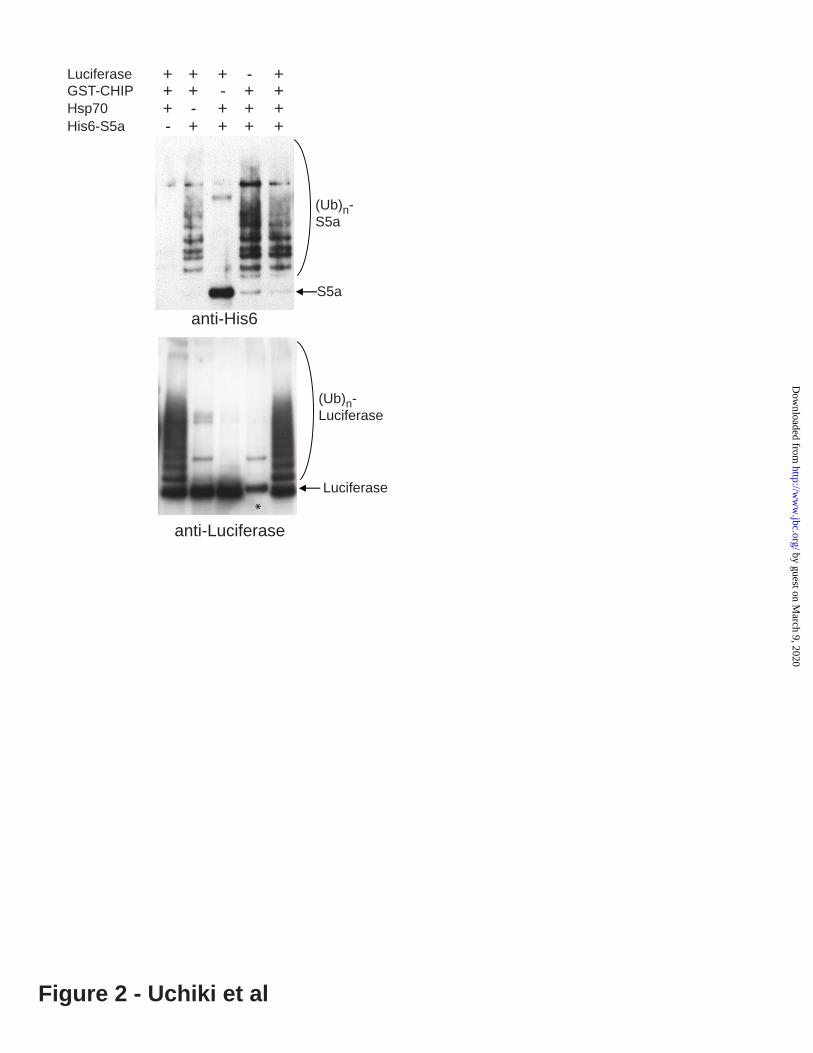
Figure 2. S5a ubiquitination by CHIP does not require Hsp70 unlike ubiquitination of a typical substrate, denatured luciferase. 200nM of His(10x)-S5a or luciferase were ubiquitinated in the presence or absence of 200nM Hsp70 for 60min. (Before the ubiquitination reaction, the luciferase, 2 M was denatured by pre-incubation with 2 M Hsp70 at 43°C for 10 min.) S5a was ubiquitinated by CHIP even in the absence of Hsp70 or without heat-denaturation, while luciferase was ubiquitinated only in the presence of Hsp70. Ubiquitination of S5a and luciferace was assayed by immunoblotting with an anti-6His or anti-luciferase antibody respectively. * The anti-luciferase antibody cross-reacted with GST-MuRF1.
Figure 3. Other Class I E2s, UbcH7 and Ubc4, can support ubiquitination of S5a.S5a was ubiquitinated as described in Figure 1 using UbcH7 or Ubc4 as the E2 for 60min. A.Ubiquitination by Nedd4 with UbcH7. 125I-labeled S5a (7500cpm/pmol) was detected by autoradiography. B. Ubiquitination by Parkin with UbcH7. S5a was probed by immunoblotting using an anti-S5a antibody.. C. Uquitination by Xenopus APC with Ubc4. S5a was probed by immunoblotting using anti-S5a antibody.
11
by guest on March 9, 2020
http://ww
w.jbc.org/
Dow
nloaded from
Figure 4. Unlike UbcH5a, UbcH1 and UbcH13 cannot support ubiquitination of S5a by MuRF1, although they support ubiquitination of a typical MuRF1 substrate, troponin I. A. 125I-labeled S5a (7500cpm/pmol) was incubated with MuRF1 and UbcH1, UbcH5a, or UbcH13/Uev1a for 120min. S5a was detected by autoradiography. B. Troponin I was also ubiquitinated under the same conditions and probed by immunoblotting using an anti-troponin I antibody.
Figure 5. The rapid ubiquitination of S5a by MuRF1 or E6AP requires its UIM domains.His(10x)-S5a and the mutants, which lack hydrophobic residues in either or both UIM domains (16), were incubated with MuRF1(A) or E6AP(B) and UbcH5 for 60 min. S5a was probed by immunoblotting using an anti-6His antibody. The asterisk shows His(6X)-E1 present in the reaction.
Figure 6. The attachments of a UIM domain from various UIM proteins to GST is sufficient to cause its ubiquitination by multiple E3s.GST or GST fused with UIM domains fromS5a, Eps15, EPSIN, MJD1, KIAA1594, or KIAA1386 were subjected to ubiquitination by MuRF1 (A) or CHIP in the absence of molecular chaperones (B) with UbcH5, for 60min. Ubiquitination of GST-UIMs was assayed by immunoblotting using anti-GST antibody.
Figure 7. Self-ubiquitination of MuRF1 with UbcH5 accelerates ubiquitination of S5a, but inhibits ubiquitination of troponin I. A. GST-MuRF1 was first self-ubiquitinated by incubation with UbcH5, ATP, E1, and E2 at 37°C for 25min, and then S5a was added. For comparison, MuRF1 with E1 and UbcH5 was incubated first without Ub, and then S5a and Ub were added to the reaction mixture. GST-MuRF1 (i) (ii) in the reaction was assayed using an anti-GST antibody and S5a with an anti 6His antibody. The asterisk shows His(6X)-E1 present in the reaction. iii. Unmodified S5a was quantified by densitometric analysis of the immunoblot image shown in Figure 7Aii. B. A similar experiment was conducted with troponin I in place of S5a, and GST-MuRF1 (i) or troponin I (ii) was probed by immunoblotting using anti-GST or anti-troponin I antibody respectively. iii. Unmodified troponin I was quantified by densitometric analysis of the immunoblot image shown in Figure 7Bii.
Figure 8. S5a binds to ubiquitinated MuRF1 but not unmodified MuRF1. A. GST-MuRF1 was immobilized on a glutathione resin and allowed to self-ubiquitinate for 30min by incubation with UbcH5. The resulting Ubn-GST-MuRF1, GST-MuRF1 (unmodified) and GST on the glutathione resin were then incubated with 125I-labeled S5a (7500cpm/pmol). i. S5a bound to the resin was measured using a gamma counter. (The data represent averages of 3 replicates, and the error bar represents S.E.M.) ii.Autoradiograph of the samples corresponding to Figure 8Ai. iii. Self-ubiquitination of the immobilized GST-MuRF1 corresponding to Figure 8Ai is shown by immunoblotting with anti-GST antibody. B. 125I-labeled S5a was incubated with E1, UbcH5, ATP, Ub and GST-MuRF1 immobilized on glutathione resin. Aliquots were taken at different times, and radioactivity bound to the resin was analyzed by gamma counting (i) or autoradiography (ii). iii. Self-ubiquitination of GST-MuRF1 in the same aliquots is shown by immunoblotting using an anti-GST antibody. As a control, the same procedure was performed with the reaction mixture lacking Ub.
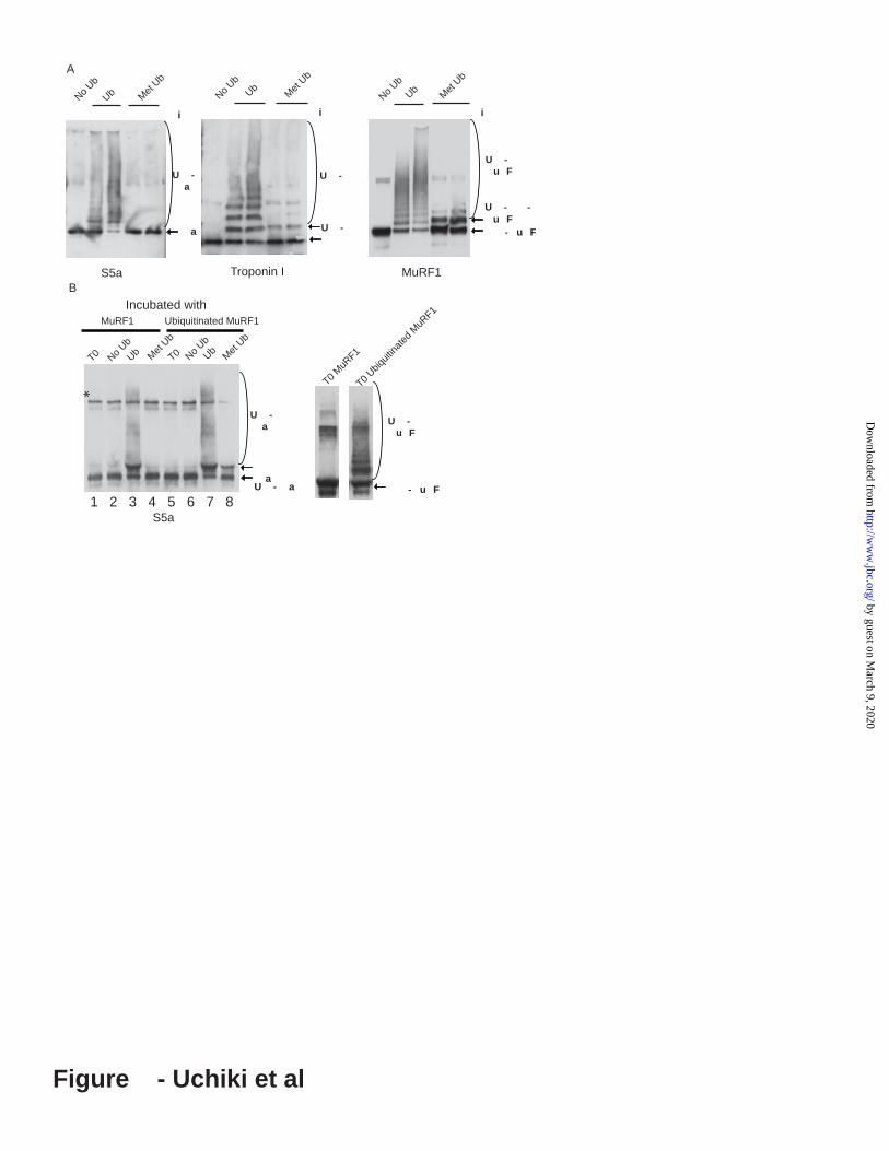
Figure 9. Methylated Ub, though supporting mono-ubiquitination of MuRF1 and troponin I, does not support ubiquitination of S5a, unless the E3, MuRF1, is first polyubiquitinated (i.e. to be ubiquitinated, S5a requires a Ub-chain rather than mono-Ub on the E3 or substrate). A. His(10x)-S5a and troponin I were ubiquitinated by GST-MuRF1 with Methylated Ub (MeUb) or Ub for 60min. Ubiquitination of His(10x)-S5a was assayed by immunoblotting with anti-6His Troponin1, and MuRF1 with anti-GST antibody. B. To test if S5a can be conjugated to MetUb if MuRF1 is polyubiquitinated, GST-MuRF1 immobilized on the glutathione resin was first polyubiquitinated by incubation with E1, UbcH5, Ub and ATP at 37°C for 30min. As a control, the immobilized GST-MuRF1 was pre-incubated
12
by guest on March 9, 2020
http://ww
w.jbc.org/
Dow
nloaded from
similarly but without E1 and E2. The resin containing GST-MuRF1 was washed extensively and then incubated with S5a, E1, UbcH5, ATP and MeUb at 37°C for 60min (Lanes 4 and 8). No Ub was added to a set of reaction mixtures (Lanes 2 and 6) to show that the washing after the first incubation was complete. In parallel, Ub was added to another set of reaction mixtures to confirm that the GST-MuRF1 retained its activity after the first incubation. The asterisk shows His(6X)-E1 present in the reaction (Lanes 3 and 7).
Figure 10. MuRF1 and E6AP attach ubiquitins to the same four lysines in S5a S5a was ubiquitinated by MuRF1 or E6AP, and the proteins in the sample were resolved on SDS-PAGE and digested by trypsin. The tryptic peptides were analyzed by LC-MS/MS using a LTQ-Orbitrap hybrid mass spectrometer, and the ubiquitination sites were identified by searching a database using SEQUEAST. An increase in mass of Lys residues by 114.0429275 Da was considered as the ubiquitination signature. Twenty (with MuRF1) or 17 (by E6AP) Lys residues out of total 24 Lys in S5a were identified. Four Lys residues (K74, K122, K262 and K365) were ubiqutinated by both of E3s. Two additional Lys residues (K126 and K135) were ubiquitinated by E6AP. The data represent two independent experiments for each E3 which yielded similar results.
Figure 11. The proposed mechanism of ubiquitination of S5a. S5a binds to growing Ub chain on the E3 and is ubiquitinated due to its proximity to the highly reactive Ub thioester. After multiple round of ubiquitination, S5a is supposedly released as our data showed that ubiquitination of S5a is a catalytic process.
Figure S1. (A& B) Ubiquitination of S5a by CHIP depends on binding of S5a to poly-ubiquitin via UIM domains.A. His(10x)-S5a and mutants lacking either UIM1 or UIM2 or both were ubiquitinated by CHIP with UbcH5 for 60min as in Figure 5A. B. His(10X)-S5a and the mutants were immobilized on Ni/NTA resin and incubated with unanchored poly-Ub chains (Ub1-7). The Ub chains bound to S5a on the Ni/NTA resin were analyzed by immunoblotting using an anti-polyubiquitin antibody. His(10X)S5a bound to the resin is shown after immunoblotting using anti-6His antibody.(C) All GST-UIM fusions bind to poly-Ub chain. C. GST or GST-UIMs presented in Figure 6B was immobilized on a glutathione resin and incubated with unanchored poly-Ub chains (Ub1-7). The GST fusion proteins that bound to the resin are shown after immunoblotting with an anti-GST antibody.
Figure S2. S5a was extensively ubiquitinated even at molar excesses of MuRF1. 2000nM His(10X)S5a was incubated with 0, 100, 200 and 400nM GST-MuRF1 in the presence of 750nM UbcH5b, 100nM E1, 25mMUb and 2mM ATP at 37°C for 1h. Ubiquitination of S5a (A) and GST-MuRF1 (B) was assayed by immunoblotting using anti-S5a and anti-GST antibodies. The same concentrations of GST-MuRF1 did not ubiquitinate 2000nM GST (as negative control) showing that ubiquitination of S5a was specific in these conditions (C).
Figure S3. S5a is rapidly degraded in C2C12 myoblasts. A. C2C12 myoblasts (at 80% confluency) were labeled with 35S-Met for 3 hours, and cycloheximide was added to block further protein synthesis. The same amount of protein in the cell lysate was used to immunoprecipitate S5a (upper panel). As a loading control, -actin in the same amount lysates was probed by immunoblotting (lower panel). B. The amounts of radiolabelled S5a were measured at each time point using densitometry.
Figure S4. S5a can be ubiquitinated by MuRF1 with UbcH5 in the presence of a typical substrate, troponin I . A. In the presence or absence of 1000nM of TnI, 250nM of His(10x)S5a was incubated with MuRF1 and UbcH5, UbcH1 and UbcH13/Uev1. Ubiquitination of S5a was assayed by immunoblotting
13
by guest on March 9, 2020
http://ww
w.jbc.org/
Dow
nloaded from
14
using an anti-S5a antibody. B. 400nM of His(10x)S5a and 400nM of troponin I were ubiquitinated by MuRF1 either in separate test tubes or in the same test tube. Ubiquitination of S5a and troponin I was assayed by immunoblotting with anti-6His and anti-troponin I antibodies. The asterisk shows His(6X)-E1 present in the reaction.
Figure S5. UbcH1 and UbcH13/Uev1 cannot support ubiquitination of S5a by MuRF1 even when MuRf1 is self-ubiquitinated. A. GST-MuRF1 and TnI were incubated with E1, Ub and indicated E2 at 37°C for 1h. Self-ubiquitination of MuRF1 was assayed by immunoblotting using an anti-GST antibody. As a internal control, ubiquitination of TnI in the same reaction mixture was also shown by immunoblotting using anti-TnI antibody. B. GST-MuRF1 on glutathione resin was self-ubiquitinated during incubation with UbcH5, E1 and ubiquitin. After the resin was washed, UbcH1, UbcH13/Uev1 or UbcH5 were added together with S5a, E1 and ubiquitin and incubated at 37°C for 1h. Ubiquitination of S5a was assayed by immunoblotting using an anti-S5a antibody.
by guest on March 9, 2020
http://ww
w.jbc.org/
Dow
nloaded from
E2 type Class I Class II Hetero-dimeric E2
E3 type E2 E3
UbcH5 Ubc4 UbcH7 UbcH1 UbcH13/Uev1
U-box CHIP Yes NoMuRF1 Yes No NoSiah2 YesMdm2 Yes
Monomeric RING
Parkin YesSCF TRCP1 YesOrigomeric
RING APC YesE6AP YesHECTNedd4 Yes Yes
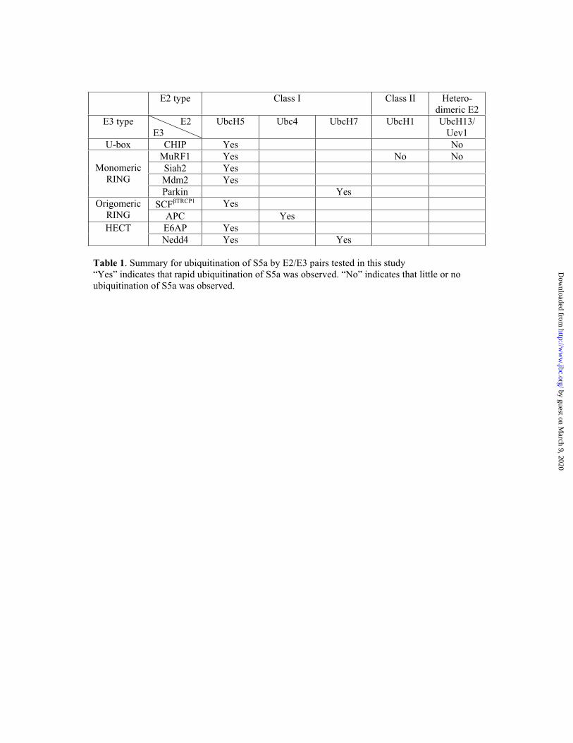
Table 1. Summary for ubiquitination of S5a by E2/E3 pairs tested in this study “Yes” indicates that rapid ubiquitination of S5a was observed. “No” indicates that little or no ubiquitination of S5a was observed.
by guest on March 9, 2020
http://ww
w.jbc.org/
Dow
nloaded from

Figure 1 - Uchiki et al
S5a
191kD97
64
51
39 No S
5aC
omplete reaction
ATP
Ubiquitin
E3
E2
E1
Ubiquitination by MuRF1
Without
A
(Ub)n-S5a
C
250kD
148
98
64
Com
plete
No ubiquitin
No E
3
S5a
Ubiquitination by SCF TRCP1
(Ub)n-S5a
reaction
S5a
0 30 60 30 60 MinUbcH: 5a 5b
(Ub)n-S5a
D
Ubiquitination by Mdm2
188kD
98
62
49
Nedd4
E6A
PC
HIP
MuR
F1S
iah2N
o E3
T0
S5a
HE
CT
RIN
G
B
(Ub)n-S5a
Ubiquitination by
U-box
by guest on March 9, 2020
http://ww
w.jbc.org/
Dow
nloaded from
anti-His6
S5a
(Ub)n-S5a
anti-Luciferase
Luciferase
(Ub)n-Luciferase
Luciferase + - +++
Hsp70 ++++ -GST-CHIP +++ + -
His6-S5a +- +++
Figure 2 - Uchiki et al
*
by guest on March 9, 2020
http://ww
w.jbc.org/
Dow
nloaded from

S5a
T UI UI UI UI utant utant utant
S5a
(Ub)n-
S5a
A
Ubi uitinate b u
Ub - + - + - + - +
B S5a
T UI UI UI UIutant utant utant
Ub - + - + - + - +
S5a
(Ub)n-
S5a
Ubi uitinate b 6 P
*
Figure - Uchiki et al
by guest on March 9, 2020
http://ww
w.jbc.org/
Dow
nloaded from

GST S5a MJD KIAA KIAA Eps15 EPSIN 1386 1594
Ub - + - + - + - + - + - + - +
GST fused with UIM of
GST
A
Ubiquitinated by MuRF1
GST
S5a GST KIAA Eps15 EPSIN MJD1 KIAA 1594 1386
Min 0 60 0 60 0 60 0 60 0 60 0 60 0 60
GST fused with UIM ofB
Figure 4 - Uchiki et al
Ubiquitinated by CHIP
by guest on March 9, 2020
http://ww
w.jbc.org/
Dow
nloaded from

Figure - Uchiki et al
i ter rei cu ati ith
U U 2 2 i
U -u F
u F
u F
iii
0 0
0 0
0 0
60 0
0 0
00 0
0 0
0 5 0 5 0
ui
ie
a
reai
ig
U i uiti ateu F
u F
i
iii
0 0
0 0
0 0
60 0
0 0
00 0
0 0
0 5 0 5 0
ui
ie
re
ai
ig
u F
U i uiti ateu F
i
U -u F
u F
u F
iter rei cu ati ith
U U 2 2 i
U -
r i
iiter rei cu ati ith
U U 2 2 i
U -a
a
ii
*
ter rei cu ati ith
U U 2 2 i
a
by guest on March 9, 2020
http://ww
w.jbc.org/
Dow
nloaded from

0.0
2.0
4.0
6.0
8.0
10.0
12.0
0 20 40 60 80
Min
% c
pm
bo
un
d
0.0
10.0
20.0
30.0
40.0
50.0
60.0
70.0
80.0
GST No E1 No Ub Ub-GST-
MuRF1
% c
pm
bo
un
d
Aiii
ii
iii
GST-MuRF1
(Ub)n-GST-
MuRF1
S5a
(Ub)n-S5a
GST-MuRF1
(Ub)n-GST-MuRF1
i
i
B
Complete reaction
No Ub
Completereaction No Ub0 8 15 30 60 0 8 15 30 60min
Completereaction No Ub0 8 15 30 60 0 8 15 30 60min
GST No E1 No Ub Ub-GST- MuRF1
GS
TN
o E
1N
o U
bU
b-G
ST-M
uR
F1
S5a
ii
Figure 6 - Uchiki et al
by guest on March 9, 2020
http://ww
w.jbc.org/
Dow
nloaded from
Met
Ub
UbN
o Ub
i
Met
Ub
Ub
No
Ub
i
a U - - u F
U - -u F
Met
Ub
Ub
No
Ub
i
A
S5a Troponin I MuRF1
Met
Ub
Ub
No
Ub
T0 Met
Ub
Ub
No
Ub
T0
aU - a
MuRF1 Ubiquitinated MuRF1
B
S5a
*
1 2 3 4 5 6 7 8
Figure - Uchiki et al
U -a
U -a
U -
U -u F
T0 M
uRF1
T0 Ubiqu
itina
ted
MuR
F1Incubated with
- u F
U -u F
by guest on March 9, 2020
http://ww
w.jbc.org/
Dow
nloaded from

Figure 8 - Uchiki et al
(Ub)n-S5a
S5a
(Ub)n-S5a
S5a
(Ub)n-S5a
S5a
*
by guest on March 9, 2020
http://ww
w.jbc.org/
Dow
nloaded from

188kD
62
Substrate: S5a Troponin I
No UbcHE2 1 5 13
49
28
S5a
(Ub)n-S5a
188kD
62
No UbcHE2 1 5 13
49
28 TnI
(Ub)n-TnI
96 96
Figure - Uchiki et al
A B
by guest on March 9, 2020
http://ww
w.jbc.org/
Dow
nloaded from

S5a ainsC
L cati ns f i entifie pepti es
Ubi uitinate L sines 22 2 2
L cati ns f i entifie pepti es
Ubi uitinate L sines22 2 2
UI UI
Ubi uitinate b u
Ubi utinate b 6 P
Figure - Uchiki et al
6 5 Ubi uitinate n b 6 P
by guest on March 9, 2020
http://ww
w.jbc.org/
Dow
nloaded from
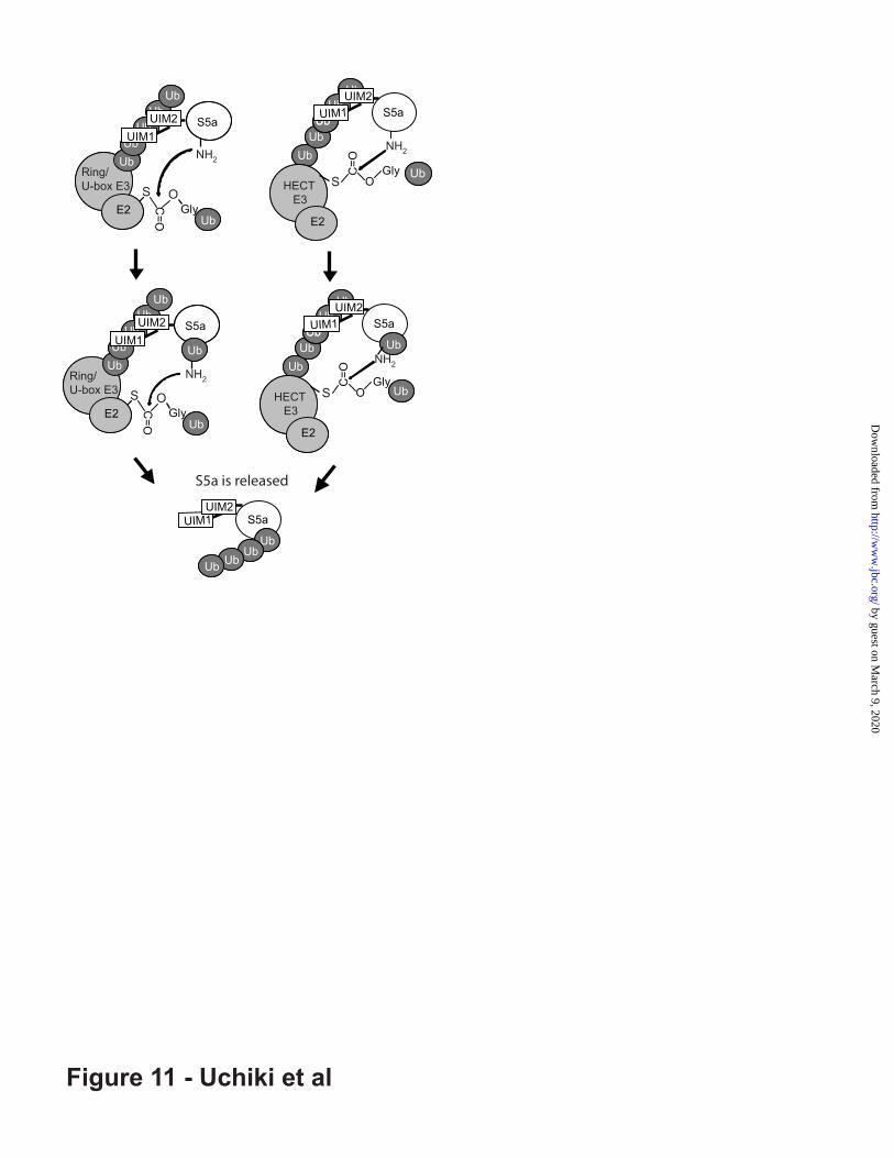
Figure 11 - Uchiki et al
UbRING/U-boxE3
E2S C=O
S
C=O
OGlyUb Ub
UbUb
UbUb
Ub
S5a
E2 Gly
UbUbUb
UbUb
S5a
UbRing/U-box E3
NH2
UIM2UIM1 Ub
UbUb
UbUb
UIM1
NH2
RING/U-boxE3
E2E2
HECT E3
Ub
Ub
S OS
UbUIM2
UbRING/U-box
E3
E2S C=O
S
C=O
OGlyUb Ub
UbUb
UbUb
Ub
S5a
E2 Gly
UbUbUb
UbUb
S5a
UbRing/U-box E3
NH2
UIM2UIM1 Ub
UbUb
UbUb
UIM1
NH2
RING/U-boxE3
E2E2
HECT E3
Ub
Ub
S O S
UbUIM2
Ub Ub
S5aUIM1
UbUIM2
UbUb
UbUb
S5a is released
by guest on March 9, 2020
http://ww
w.jbc.org/
Dow
nloaded from

Supplemental Experimental Procedure
Binding of Ub chains to immobilized Ub binding proteins
Glutathione resin was equilibrated with the Ub binding buffer, and 12 l of the resin was mixed with 5 M GST-UIMs. The resin was rotated at 4°C overnight. Then, 230 l of the Ub binding buffer and 3 g of Ub1-7 chains (Boston Biochem) were added and the sample was further incubated at 4°C for 2h. The resin was washed twice with 500 l of the Ub binding buffer. The bound proteins were eluted by SDS-PAGE sample loading buffer and analyzed by SDS-PAGE followed by immunoblotting with anti-Ub antibody (BIOMOL). To assay binding of Ub chains to S5a, the same procedure was performed with His(10X) S5a immobilized on Ni-NTA resin in the Ub binding buffer not containing EGTA.
by guest on March 9, 2020
http://ww
w.jbc.org/
Dow
nloaded from

S5a
(Ub)n-S5a
S5a WT UIM1 UIM2 UIM1/UIM2
mutant mutant mutantUb - + - + - + - +
148 kD
98
64
50
A
Ubiquitinated by CHIP
98
6249
38
28
17
Ubiquitin
Pol
y -U
bN
i/NTA
resi
nW
ild ty
peU
IM1
mut
ant
UIM
2 m
utan
tU
IM1/
UIM
2 m
utan
t
S5a
6249
Pol
y-U
bG
ST
S5a
MJD
1
KIA
A15
94
KIA
A13
86
Eps
15
EP
SIN
28
64
51
39
14
GST fusion with UIM of
28
51
39
Ubiquitin
GST fusion proteins
B C
Figure S1.(A& B) Ubiquitination of S5a by CHIP depneds on binding of S5a to poly-ubiquitin via UIM domains.A. His(10x)-S5a and mutants lacking either UIM1 or UIM2 or both were ubiquitinated by CHIP with UbcH5 for 60min as in Figure 3A. B. His(10X)-S5a and the mutants were immobilized on Ni/NTA resin and incubated with unanchored poly-Ub chains (Ub1-7). The Ub chains bound to S5a on the Ni/NTA resin were analyzed by immunoblotting using an anti-polyubiquitin antibody. His(10X)S5a bound to the resin is shown after immunoblotting using an anti-6His antibody. (C) All GST-UIM fusions bind to poly-Ub chain. C. GST or GST-UIMs presented in Figure 4B was immobilized on a glutathione resin and incubated with unanchored poly-Ub chains (Ub1-7). The GST fusion proteins that bound to the resin are shown after immunoblotting with an anti-GST antibody.
by guest on March 9, 2020
http://ww
w.jbc.org/
Dow
nloaded from

Figure S2. S5a was extensively ubiquitinated molar excess of MuRF1. 2000nM His(10X)S5a was incubated with 0, 100, 200 and 400nM GST-MuRF1 in the presence of 750nM UbcH5b, 100nM E1, 25 MUb and 2mM ATP at 37°C for 1h. Ubiquitination of S5a (A) and GST-MuRF1 (B) was assayed by immunoblotting using anti-S5a and anti-GST antibodies. The same concentrations of GST-MuRF1 did not ubiquitinate 2000nM GST (as negative control) showing that ubiquitination of S5a was specific in these conditions
S5a
S5a (2000 nM)
A
B
C
Ubn-S5a
Incubated with 100 200 400 0 nM GST-MuRF1
GST
GST (2000nM)
Incubated with 100 200 400 0 nM GST-MuRF1
GST-MuRF1
Ubn-GSTMuRF1
100 200 400nM GST-MuRF1
by guest on March 9, 2020
http://ww
w.jbc.org/
Dow
nloaded from
- MG132 + MG132
0 30 60 90 0 30 60 90Time (min)
-actin
S5a
020406080
100120
0 30 60 90
Rem
aini
ng S
5a (%
)
Chasing time (min)
Figure S3. S5a is rapidly degraded in C2C12 myoblasts. A. C2C12 myoblasts (at 80% confluency) were labeled with 35S-Met for 3 hours, and cycloheximide was added to block further protein synthesis. The same amount of protein in the cell lysate was used to immunoprecipitate S5a (upper panel). As a loading control, -actin in the same amount lysates was probed by immunoblotting (lower panel). B. The amounts of radiolabelled S5a were measured at each time point using densitometry
by guest on March 9, 2020
http://ww
w.jbc.org/
Dow
nloaded from

Figure S4. S5a is ubiquitinated by MuRF1 with UbcH5 in the presence of a typical substrate, troponin I . A. In the presence or absence of 1000nM of tropoinin I (TnI), 250nM of S5a was incubated with MuRF1 and UbcH5, UbcH1 and UbcH13/Uev1. Ubiquitination of S5a was assayed by immunoblotting using an anti-S5a antibody. B. 400nM of His(10x)S5a and 400nM of troponin I were ubiquitinated by MuRF1 either in separate test tubes or in the same test tube. Ubiquitination of S5a and troponin I was assayed by immunoblotting with anti-6His and anti-troponin I antibodies. The asterisk shows His(6X)-E1 present in the reaction.
S5a
E2 - UbcTroponin I - - + - + - +
H1 H5 H13
A+ Troponin I
0 2 4 8 16 0 2 4 8 16min
S5a
S5a
0 2 4 8 16min
+S5a
Ubn-S5a
Ubn-TnI
TnI
Troponin I
*
B
by guest on March 9, 2020
http://ww
w.jbc.org/
Dow
nloaded from
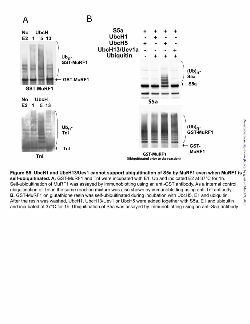
Figure S5. UbcH1 and UbcH13/Uev1 cannot support ubiquitination of S5a by MuRF1 even when MuRF1 is self-ubiquitinated. A. GST-MuRF1 and TnI were incubated with E1, Ub and indicated E2 at 37°C for 1h. Self-ubiquitination of MuRF1 was assayed by immunoblotting using an anti-GST antibody. As a internal control, ubiquitination of TnI in the same reaction mixture was also shown by immunoblotting using anti-TnI antibody. B. GST-MuRF1 on glutathione resin was self-ubiquitinated during incubation with UbcH5, E1 and ubiquitin. After the resin was washed, UbcH1, UbcH13/Uev1 or UbcH5 were added together with S5a, E1 and ubiquitin and incubated at 37°C for 1h. Ubiquitination of S5a was assayed by immunoblotting using an anti-S5a antibody
GST-MuRF1
TnI
No UbcH E2 1 5 13
A B
GST-MuRF1
Ubn-TnI
Ubn-GST-MuRF1
TnI
No UbcH E2 1 5 13
S5a
GST-MuRF1 (Ubiquitinated prior to the reaction)
S5a
GST-MuRF1
(Ub)n-S5a
(Ub)n-GST-MuRF1
S5a ++++UbcH1 -- -+UbcH5 -- ++
UbcH13/Uev1a --- +Ubiquitin - +++
by guest on March 9, 2020
http://ww
w.jbc.org/
Dow
nloaded from

O'Bryan and Alfred L. GoldbergTomoaki Uchiki, Hyoung Tae Kim, Bo Zhai, Steven P. Gygi, Jennifer A. Johnston, John P.
mechanism different from typical substrate recognitionThe UIM protein, S5A, is ubiquitinated by all types of ubiquitin ligases by a
published online February 24, 2009J. Biol. Chem.
10.1074/jbc.M900556200Access the most updated version of this article at doi:
Alerts:
When a correction for this article is posted•
When this article is cited•
to choose from all of JBC's e-mail alertsClick here
Supplemental material:
http://www.jbc.org/content/suppl/2009/02/25/M900556200.DC1
by guest on March 9, 2020
http://ww
w.jbc.org/
Dow
nloaded from