tive pulmonary disease doi: 10.1185…
TRANSCRIPT

ORIGINAL ARTICLE
PLATELET ACTIVATION AND CARDIOVASCULAR CO-MORBIDITIES IN PATIENTS WITH CHRONIC OBSTRUC-TIVE PULMONARY DISEASEMario Malerba, Alessia Olivini, Alessandro Radaeli, Fabio Luigi Massimo Ricciardolo,Enrico Clini
Doi: 10.1185/03007995.2016.1149054
Abstract
Objective: Platelet activation in COPD patients is associated with an increased riskof cardiovascular events. We aimed at assessing the mean platelet volume (MPV),as an index of platelet activation, in COPD patients both when stable or during acuteexacerbation (AE).Research design and methods: 478 patients (75 with AE) and 72 age-matchedhealthy controls were enrolled. Medical history, co-morbidities, medications, pulmo‐nary function tests, MPV and blood cell count, erythrocyte sedimentation rate (ERS)
and C reactive protein (CRP) were recorded.Results: MPV was higher in COPD than in controls (8.7 ± 1.1 fL and 8.4 ± 0.8 fL respectively, p = 0.025) and increasedacross the severity of the diseases as assessed by post bronchodilator FEV1 categorized I to IV (p > 0.05). MPV was higherin COPD patients during AE as compared with stable condition (8.7 ± 1.0 fL and 8.9 ± 1.0 fL, p = 0.021).MPV ≥ 10.5 fL correlated with the presence of at least one co-existing cardiovascular disease (p = 0.008) . No correlationwas observed between MPV and CRP or ERS in patients or in controls. A negative correlation was found between plateletcount and MPV in COPD patients taken together.Limitations: The retrospective design did not allow to assess a clear cause-effect relationship between MPV and all thepathophysiological factors consideredConclusions: Elevated MPV is associated with lower platelet count and with cardiovascular co-morbidity in COPD patients.MPV value is higher in the more severe COPD and during AE. Present findings warrant future studies to confirm a possibleclinically relevant role for platelet activation on cardiovascular risk in the population of COPD.
Just Accepted by Current Medical Research and Opinion
© 2016 Taylor & Francis. This provisional PDF corresponds to the article as it appeared upon acceptance. Fully formattedPDF and full text (HTML) versions will be made available soon.DISCLAIMER: The ideas and opinions expressed in the journal's Just Accepted articles do not necessarily reflect those of Taylor & Francis (the Publisher),the Editors or the journal. The Publisher does not assume any responsibility for any injury and/or damage to persons or property arising from or related toany use of the material contained in these articles. The reader is advised to check the appropriate medical literature and the product information currentlyprovided by the manufacturer of each drug to be administered to verify the dosages, the method and duration of administration, and contraindications. It isthe responsibility of the treating physician or other health care professional, relying on his or her independent experience and knowledge of the patient, todetermine drug dosages and the best treatment for the patient. Just Accepted articles have undergone full scientific review but none of the additional editorialpreparation, such as copyediting, typesetting, and proofreading, as have articles published in the traditional manner. There may, therefore, be errors inJust Accepted articles that will be corrected in the final print and final online version of the article. Any use of the Just Accepted articles is subject to theexpress understanding that the papers have not yet gone through the full quality control process prior to publication.

ORIGINAL ARTICLE
PLATELET ACTIVATION AND CARDIOVASCULAR CO-MORBIDITIES IN PATIENTS
WITH CHRONIC OBSTRUCTIVE PULMONARY DISEASE
Malerba Mario1, Olivini Alessia
1, Radaeli Alessandro
2, Ricciardolo Fabio Luigi Massimo
3, Clini
Enrico4
1 Department of Internal Medicine, University of Brescia and AOU Spedali Civili, Brescia, Italy
2 Department of Emergency AOU Spedali Civili, Brescia, Italy
3 Department of Clinical and Biological Sciences, University of Torino, San Luigi Hospital,
Orbassano, Italy.
4 Department of Medical and Surgical Sciences, Ospedale Villa Pineta, University of Modena-
Reggio Emilia, Modena, Italy.
Address for correspondence: Mario Malerba, Department of Internal Medicine, University of
Brescia and AOU Spedali Civili, Brescia, Italy. Tel: +39 030 3995250; [email protected]
Key words: mean platelet volume, COPD, cardiovascular co-morbidities, platelet, inflammation.
[Short title: MPV and cardiovascular co-morbidity in COPD]
JUST A
CCEPTED

ABSTRACT
Objective: Platelet activation in COPD patients is associated with an increased risk of
cardiovascular events. We aimed at assessing the mean platelet volume (MPV), as an index of
platelet activation, in COPD patients both when stable or during acute exacerbation (AE).
Research design and methods: 478 patients (75 with AE) and 72 age-matched healthy controls were
enrolled. Medical history, co-morbidities, medications, pulmonary function tests, MPV and blood
cell count, erythrocyte sedimentation rate (ERS) and C reactive protein (CRP) were recorded.
Results: MPV was higher in COPD than in controls (8.7 ± 1.1 fL and 8.4 ± 0.8 fL respectively, p =
0.025) and increased across the severity of the diseases as assessed by post bronchodilator FEV1
categorized I to IV (p>0.05). MPV was higher in COPD patients during AE as compared with
stable condition (8.7 ± 1.0 fL and 8.9 ± 1.0 fL, p = 0.021).
MPV ≥ 10.5 fL correlated with the presence of at least one co-existing cardiovascular disease (p =
0.008) . No correlation was observed between MPV and CRP or ERS in patients or in controls. A
negative correlation was found between platelet count and MPV in COPD patients taken together.
Limitations: The retrospective design did not allow to assess a clear cause-effect relationship
between MPV and all the pathophysiological factors considered
Conclusions: Elevated MPV is associated with lower platelet count and with cardiovascular co-
morbidity in COPD patients. MPV value is higher in the more severe COPD and during AE.
Present findings warrant future studies to confirm a possible clinically relevant role for platelet
activation on cardiovascular risk in the population of COPD.
JUST A
CCEPTED

INTRODUCTION
An important association was observed between chronic obstructive pulmonary disease (COPD)
and cardiovascular diseases [1,2], being events such as myocardial infarction [3] and stroke [4]
more frequent in these patients than in the healthy population. Indeed, both COPD and
cardiovascular diseases share common risk factors, smoking habit as most important [5].
A large body of research aimed at finding the explanation of this enhanced risk in the COPD
population, focusing in particular on systemic inflammation as a driving mechanism for
atherothrombosis [6, 7]. Recent studies have shown the presence of increased platelet activation in
patients with COPD [8-10], which may be correlated with the presence of circulating inflammatory
mediators and with the hypoxemia which is typical in the advanced stages of the disease [11]. The
increased mean platelet volume (MPV) is an index of accelerated platelet activation, derives from
the change of the platelets’ shape, and leads to enhanced thrombosis [12-13]. Very recently, it has
been demonstrated that high MPV is correlated with higher risk of developing atherosclerotic
thrombotic cardiovascular events, mainly in chronic patients suffering from diabetes, hypertension
and obesity, where platelet activation is enhanced [14]. Moreover, in elderly male patients with
COPD, the MVP was inversely correlated with the level of FEV1, and the impairment of
cardiopulmonary function [15].
In the present study we aimed at assessing the MPV value in a large population of COPD patients
both when stable or during an episode of acute exacerbation (AE) and to determine whether
correlation exists between MPV and the stage of disease severity, the systemic inflammation and
the increased prevalence of atherothrombotic cardiovascular events in the studied population. JUST A
CCEPTED
METHODS
Study protocol
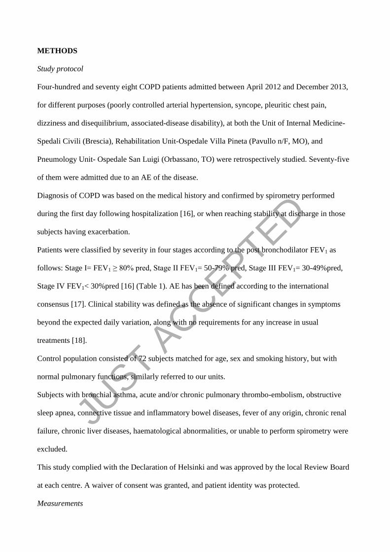
Four-hundred and seventy eight COPD patients admitted between April 2012 and December 2013,
for different purposes (poorly controlled arterial hypertension, syncope, pleuritic chest pain,
dizziness and disequilibrium, associated-disease disability), at both the Unit of Internal Medicine-
Spedali Civili (Brescia), Rehabilitation Unit-Ospedale Villa Pineta (Pavullo n/F, MO), and
Pneumology Unit- Ospedale San Luigi (Orbassano, TO) were retrospectively studied. Seventy-five
of them were admitted due to an AE of the disease.
Diagnosis of COPD was based on the medical history and confirmed by spirometry performed
during the first day following hospitalization [16], or when reaching stability at discharge in those
subjects having exacerbation.
Patients were classified by severity in four stages according to the post bronchodilator FEV1 as
follows: Stage I= FEV1 ≥ 80% pred, Stage II FEV1= 50-79% pred, Stage III FEV1= 30-49%pred,
Stage IV FEV1< 30%pred [16] (Table 1). AE has been defined according to the international
consensus [17]. Clinical stability was defined as the absence of significant changes in symptoms
beyond the expected daily variation, along with no requirements for any increase in usual
treatments [18].
Control population consisted of 72 subjects matched for age, sex and smoking history, but with
normal pulmonary functions, similarly referred to our units.
Subjects with bronchial asthma, acute and/or chronic pulmonary thrombo-embolism, obstructive
sleep apnea, connective tissue and inflammatory bowel diseases, fever of any origin, chronic renal
failure, chronic liver diseases, haematological abnormalities, or unable to perform spirometry were
excluded.
This study complied with the Declaration of Helsinki and was approved by the local Review Board
at each centre. A waiver of consent was granted, and patient identity was protected.
Measurements
JUST A
CCEPTED

Medical history, respiratory function tests, and blood analysis was recorded in each participant.
Active smoking was defined if a current use of >2 cigarettes/day was reported; the number of
pack/year was also recorded.
Respiratory Function Tests- Lung spirometry was performed in accordance with the standard
procedure [19]. Forced Vital Capacity (FVC) and Forced Expiratory Volume in the first second
(FEV1) were recorded.
Peripheral Blood analysis- Blood sample was taken at admission. Ten millilitres of fasting bloods
were drawn from each subject enrolled, tubes containing the dipotassium salt of thylenediamine
tetraacetic acid (EDTA) were used for measurement of MPV [20]. Samples were immediately taken
to the Hospital central laboratory to be analysed.
Total count on platelets, red and white cell, the MPV, and the level of both erythrocyte
sedimentation rate (ESR) and C-reactive protein (CRP) were recorded for study purposes. The
inter- and intra-assays coefficients of variation of these assays were below 5%.
Co-morbidities- Co-existing chronic diseases were identified by examining the clinical
documentation provided by each subject at admission and/or by the results from diagnostic tests
performed during hospitalization.
Diabetes mellitus was diagnosed if plasma glucose > 126 mg/dL, and/or treatment with anti-
diabetic drugs, and/or presence of haemoglobin A1c > 6.5% [21, 22].
Systemic arterial hypertension was defined if assuming antihypertensive treatment, in the presence
of a medical history with documented high blood pressure (BP), if systolic BP >140 mm Hg and/or
diastolic BP > 90 mm Hg were recorded on 3 separate occasions [23].
Dyslipidemia was defined in the presence of hypolipidemic treatment, and if total cholesterol >200
mg/dL, and/or triglycerides >200 mg/dL, high-density lipoprotein cholesterol >40 mg/dL, low-
density lipoprotein cholesterol >130 mg/dL [24].
Coronary artery disease was based on the history of previously documented myocardial infarction
and/or suggestive changes at the electrocardiogram (ECG) [25].
JUST A
CCEPTED

Heart failure was defined according to the medical history and the presence of documented left
ventricular ejection fraction <55% (Simpson method) together with some degree of exertional
dyspnea [26].
Obesity was defined when BMI was greater than or equal to 30.
The presence of cerebrovascular disease was based on the past medical history of cerebral
infarction, cerebral thrombosis, transient ischemic attack, artery occlusion at the carotid or vertebral
vessels.
Statistical Analysis
Data were presented as mean ± standard deviation (SD). Subjects were analysed according to
different groups (stable or exacerbated COPD and Controls) or categories (presence of at least one
cardiovascular co-morbidity, MPV value ≥10.5 fL as the median cut-off of distribution).
After verifying that data were normally distributed, group comparisons were performed using t-test
for independent variables. Analysis of correlation among variables was performed by linear
regression with a straight trend and through the calculation of Pearson coefficient. Categorical
variables were compared by the chi-square test method.
A p value <0.05 was considered to be significant. All analysed were performed with a dedicated
software (SPSS 21.0 for Windows; SPSS, Chicago, IL).
JUST A
CCEPTED

RESULTS
Characteristics of subjects in study are displayed in Table 1. Most of the COPD patients were in
stage II or III according to the post bronchodilator FEV1. Seventy-five patients were under AE,
being twenty-five (33%) of them in stage IV of the disease.
Chronic co-morbidities were similar between COPD and controls, as well as the use of
antiaggregants and/or anticoagulants was similar.
Two-hundred twenty seven (47.4%) out of the 478 COPD patients (192/403 stable and 35/75 during
AE) had at least one cardiovascular disease (hypertension, acute myocardial infarction, exertional
angina, coronary artery disease, cerebral vascular disease, arterial disease of the lower limbs,
thromboembolic disease). The proportion of patients with at least one cardiovascular co-morbidity
was also similar (48.6%) in the control group.
Two-hundred fifty eight COPD were taking acetylsalicylic acid (ASA) as an antiplatelet therapy (11
subjects were under dual antiplatelet therapy with ASA and clopidogrel); 89 patients were receiving
anticoagulation therapy with warfarin (n=86) or enoxaparin (n=3). Forty-one out of 72 controls
were receiving ASA (one subject was under dual therapy with ASA and clopidogrel), while 11 were
assuming warfarin. No difference was noted in the medication taken in the group of COPD patients
under AE compared to the group of COPD patients in stable condition.
Table 2 resumes data of both lung function and peripheral blood in the study groups. In particular,
the mean MPV differed significantly between COPD patients taken together and controls
(respectively 8.7 ± 1.1 fL and 8.4 ± 0.8 fL in COPD patients and in controls p = 0.025), despite
platelet count was similar. The mean values of ESR and PCR were similar in patients and in
controls (see Table 2).
Noteworthy, we observed an inverse correlation between the platelets count and the MPV in
patients with COPD (Pearson = -0.115, p <0.05, Figure 1) but not in controls (Pearson = -0.009, ns).
Mean MPV did not correlate with ESR, PCR, the total number of co-morbidities and the use of
JUST A
CCEPTED

antiplatelet/anticoagulant therapies in the whole population in study, nor in the COPD group either
when stable or during AE.
Sub-group analysis: COPD patients without or with AE.
Table 3 and Figure 2 show comparison of mean MPV value between study groups including COPD
with or without AE. In COPD patients, MPV level was lower when stable as compared during AE
(8.7 ± 1.0 fL, and 8.9 ± 1.0 fL respectively, p = 0.021). Moreover, MPV was higher in COPD stage
II to IV when compared with stage I (see Table 3 and Figure3). Mean MPV was also compared
within each post bronchodilator category in stable patients and in those under AE, but no significant
differences were reported. Among the COPD patients, neither ESR nor PCR differed according to
the different clinical condition (stable state or AE).
Platelet count was lower during AE than in stable state (211.2 ± 96.5 and 218.7 ± 64.2 x 10³ / mm3
respectively, p<0.05).
Finally, the presence of a MPV ≥10.5 fL significantly correlated with the presence of at least one
cardiovascular co-morbidity in COPD patients taken together (but not in controls) (see Table 4).
JUST A
CCEPTED

DISCUSSION
Our study shows that MPV values are higher in COPD patients than in healthy controls and
increases with the degree of severity and during AE. Moreover, high level of MPV is associated
with at least one cardiovascular co-morbidity in the population of COPD but not in comparable
controls.
Data of mean MPV in COPD are still inconsistent and contradictory. Onder et al. [27] observed for
the first time higher MPV values in hypoxic COPD patients when compared with controls.
Additionally, Bansal et al. [28] as well as Ulasli et al [29] reported higher MPV in patients with
COPD. The latter study also measured a reduced MPV during AE, however the smoking status of
patients was not reported and COPD patients had a significantly lower BMI, at difference with our
study.
A recent paper [30] has evaluated 269 COPD patients and finding have shown that, in disagreement
with our data, lower MPV during AE negatively correlated with PCR values. This discrepancy is
probably due to different characteristics between the two populations studied, furthermore neither
BMI nor smoking history were reported and lung function (FEV1 and FEV1/FVC) was surprisingly
similar both in stable and exacerbated COPD patients.
Cui et al [15] was the first to observe that increased MPV in very old male patients with COPD was
associated with impaired cardiopulmonary function, linking the concept of platelet activation with
the prevalence of cardiovascular co-morbidities in this population. Recent awareness on the
enhanced risk of cardiovascular co-morbidities in COPD patients has stimulated research to study
the possible pathophysiological mechanisms that stand behind this phenomenon [31]. Several
mechanisms including hypoxia, systemic inflammation and imbalance of the proteases to
antiproteases ratio (with an increased expression of neutrophil elastase) have been considered able
to promote platelet activation and hyperaggregability and to stimulate atheromasic plaque
progression [8-11]. The mechanism of platelet activation involves different phenomena such as
change in the shape of platelets, increase in the MPV, expression of P-selectin in the cell’s
JUST A
CCEPTED

membrane [32], formation of aggregates with platelets and monocytes [33], thus favouring the
endothelial activation and the atherosclerotic plaque formation.
The increased values of MPV could follow the hypoxemia that is typical in patients with COPD, in
particular at peripheral tissue level, which may trigger bone marrow stimulation or increase the
sequestration of smaller platelets [28]. COPD is associated with systemic inflammation, oxidative
stress, activation of circulating inflammatory cells and increased levels of inflammatory mediators,
mechanisms that may be partly responsible of platelet activation as observed in COPD [29,31],
where the presence of a pro-thrombotic state with high production of thrombin and pro-coagulative
mediators was reported [34].
By confirming previous data on increased MPV in the COPD population, present study is the first
to show that it is present in the more severe stages of the disease and/or during AE. Therefore, the
severity of COPD is possibly associated with a progressive increase of MPV, despite the
pathophysiological mechanism(s) involved, among all those mentioned above, has not yet been
completely clarified. Indeed, our data do not suggest a direct relationship between hypoxemia and
elevation of MPV, neither demonstrated a significant difference of inflammatory serum markers
(ESR and CRP) between COPD and controls. Notwithstanding, values of both CRP and ESR are
higher in the two groups compared to the normal range.
Interestingly, we have found a significant negative correlation between platelet count and MPV
value only among the COPD population. This could be explained by the increased proportion of
activated platelets sequestered at the peripheral level and by the precipitation of the latter in the
form of aggregates of platelets and monocytes, which are typically seen during the process of
platelet activation [35,36]. One of the most interesting and novel aspect of our study is the
observation of a direct association between high MPV level (above the pre-defined limit of ≥10.5
fL) and the at least one cardiovascular co-morbidity in patients with COPD but not in the controls.
This finding could support the above mentioned hypothesis about the presence of a tight
relationship between platelet activation, systemic atherosclerosis, plaque rupture and cardiovascular
JUST A
CCEPTED

events in COPD patients. In a large population cohort (the “Rotterdam Study”) it has been recently
demonstrated a higher prevalence of carotid atherosclerosis in elderly COPD compared with
healthy controls, which is independent on the stage of the disease [37]. This study also advanced the
concrete hypothesis that COPD may be an independent predictor for the presence of carotid
atherosclerosis.
Despite our findings in a consistent population of COPD confirm the role of platelet activation in
the disease and also clarify the concept that this happens according to the degree of severity and
during AE, present study has some limitations that deserves to be outlined. A first potential limit is
the retrospective design, thus a selection bias (e.g. the similar systemic inflammation level in both
groups) may have occurred. Second, we were not able to report a patent cause-effect relationship
between variable of interest (MPV) and all the pathophysiological factors behind (such as arterial
oxygen pressure level). Notwithstanding, the strong point of our study is primarily the large sample
size of COPD patients and the objective recording of co-morbidities [38].
JUST A
CCEPTED

CONCLUSION
Our observational study underlines the presence of platelet activation (i.e. elevated MPV and reduce
platelet count) in COPD patients, being patients with higher degree of severity an/or those under
AE at higher risk of developing cardiovascular co-morbidity. This indirectly suggests a clear
relationship between platelet activation and the excess of cardiovascular events commonly observed
in the COPD population. On the basis of the present data it would be possible to hypothesize the
prophylactic use of antiplatelets or anticoagulants in order to reduce the future risk of
cardiovascular events [39] in patients with COPD, especially when severe or frequent exacerbators.
To test this hypothesis, however, further prospective studies on larger populations are needed.
Transparency
Declaration of funding:
There was no funding for this study.
Declaration of financial/other relationship:
MM, AO, AR, FLMRA, ECR have disclosed that they have no significant relationships with or
financial interests in any commercial companies related to this study or article. CMRO Peer
Reviewers on this manuscript have no relevant financial relationships to disclose.
JUST A
CCEPTED
REFERENCES
1- Curkendall SM, DeLuise C, Jones JK, Lanes S, Stang MR, Goehring E Jr, She D. Cardiovascular
disease in patients with chronic obstructive pulmonary disease, Saskatchewan Canada
cardiovascular disease in COPD patients. Ann Epidemiol 2006; 16(1):63-70.
2- Fimognari FL, Scarlata S, Conte ME, Incalzi RA. Mechanisms of atherothrombosis in chronic
obstructive pulmonary disease. Int J Chron Obstruct Pulmon Dis 2008;3(1):89-96.
3- Hansell AL, Walk JA, Soriano JB. What do chronic obstructive pulmonary disease patients die
from? A multiple cause coding analysis. Eur Respir J 2003; 22 :809–14.
4- Truelsen T, Prescott E, Lange P, Schnohr P, Boysen G.. Lung function and risk of fatal and non-
fatal stroke. The Copenhagen City Heart Study. Int J Epidemiol 2001; 30:145–51.
5- Sin DD, Wu L, Man SF. The relationship between reduced lung function and cardiovascular
mortality: a population-based study and a systematic review of the literature. Chest 2005; 127:
1952–9.
6- Davì G, Basili S, Vieri M, Cipollone F, Santarone S, Alessandri C, Gazzaniga P, Cordova C,
Violi F. Enhanced thromboxane biosynthesis in patients with chronic obstructive pulmonary
disease. The Chronic Obstructive Bronchitis and Haemostasis Study Group. Am J Respir Crit Care
Med 1997;156:1794–9.
7- Ghoorah K, De Soyza A, Kunadian V. Increased cardiovascular risk in patients with chronic
obstructive pulmonary disease and the potential mechanisms linking the two conditions: a review.
Cardiol Rev 2013;;21(4):196-202.
8- Guzmán-Grenfell A, Nieto-Velázquez N, Torres-Ramos Y, Montoya-Estrada A, Ramírez-
Venegas A, Ochoa-Cautiño L, Flores-Trujillo F, Hicks JJ. Increased platelet and erythrocyte
arginase activity in chronic obstructive pulmonary disease associated with tobacco or wood smoke
exposure. J. Investig.Med 2011; 59: 587–92.
JUST A
CCEPTED

9- Biljak VR, Pancirov D, Cepelak I, Popovic´-Grle S, Stjepanovic´ G, Grubisˇic´ TZˇ . Platelet
count, mean platelet volume and smoking status in stable chronic obstructive pulmonary disease.
Platelets. 2011;22(6):466-470.
10- Steiropoulos P, Papanas N, Nena E, Xanthoudaki M, Goula T, Froudarakis M, Pita E, Maltezos
E, Bouros D. Mean platelet volume and platelet distribution width in patients with chronic
obstructive pulmonary disease: the role of co-morbidities. Angiology 2013;64(7):535-9.
11- Cordova C, Musca A, Violi F, Alessandri C, Perrone A, Balsano F. Platelet hyperfunction in
patients with chronic airways obstruction. Eur J Respir Dis 1985;66:9–12.
12- Gasparyan AY, Ayvazyan L, Mikhailidis DP, Kitas GD. Mean platelet volume: a link between
thrombosis and inflammation? Curr Pharm Des 2011;17(1):47-58.
13- Vizioli L, Muscari S, Muscari A. The relationship of mean platelet volume with the risk and
prognosis of cardiovascular diseases. Int J Clin Pract 2009;63(10):1509-1515.
14- Sansanayudh N, Anothaisintawee T, Muntham D, McEvoy M, Attia J, Thakkinstian A. Mean
platelet volume and coronary artery disease: a systematic review and meta-analysis. Int J Cardiol.
2014;175(3):433-40.
15- Cui H, Liu L, Wei Z, Wang D, Hu Y, Hu G, Fan L. Clinical value of mean platelet volume for
impaired cardiopulmonary function in very old male patients with chronic obstructive pulmonary
disease. Arch Gerontol Geriatr 201;54(2):e109-12.
16- Global Strategy for the Diagnosis, Management and Prevention of COPD, Global Initiative for
Chronic Obstructive Lung Disease (GOLD) 2015. Available from: http://www.goldcopd.org/.
17- Rodriguez-Roisin R. Toward a consensus definition for COPD exacerbations. Chest 2000; 117:
398S–401S.
18- Celli BR, MacNee W. Standards for the diagnosis and treatment of patients with COPD: a
summary of the ATS/ERS position paper. Eur. Respir. J 2004; 23: 932–46.
19- American Thoracic Society: Standardisation of Spirometry. Am. J. Respir. Crit. Care Med
1995; 152:1107-1136.
JUST A
CCEPTED

20- O’Malley T, Ludlam CA, Fox KA, Elton RA. Measurement of platelet volume using a variety
of different anticoagulant and antiplatelet mixtures. Blood Coagul Fibrinolysis 1996;7(4):431-436.
21- American Diabetes Association. Diagnosis and classification of diabetes mellitus. Diabetes
Care. 2011;34(suppl 1):S62-S69.
22- Katsiki N, Papanas N, Mikhailidis DP, Fonseca VA. Glycated hemoglobin A(1c) (HbA1c) and
diabetes: a new era? Curr Med Res Opin 2011;27(suppl 3):7-11.
23- Chobanian AV, Bakris GL, Black HR, Cushman WC, Green LA, Izzo JL Jr, Jones DW,
Materson BJ, Oparil S, Wright JT Jr, Roccella EJ. Seventh report of the joint national committee on
prevention, detection, evaluation, and treatment of high blood pressure. Hypertension 2003;
42(6):1206-1252.
24- National Cholesterol Education Program (NCEP) Expert Panel on Detection, Evaluation, and
Treatment of High Blood Cholesterol in Adults (Adult Treatment Panel III). Third Report of the
National Cholesterol Education Program (NCEP) Expert Panel on Detection, Evaluation, and
Treatment of High Blood Cholesterol in Adults (Adult Treatment Panel III) final report.
Circulation. 2002;106(25):3143-3421.
25- Blackburn H, Keys A, Simonson E, Rautaharju P, Punsar S. The electrocardiogram in
population studies. A classification system. Circulation 1960;21:1160-1175.
26- Dickstein K, Cohen-Solal A, Filippatos G, McMurray JJ, Ponikowski P, Poole-Wilson PA,
Strömberg A, van Veldhuisen DJ, Atar D, Hoes AW, Keren A, Mebazaa A, Nieminen M, Priori
SG, Swedberg K. ESC guidelines for the diagnosis and treatment of acute and chronic heart failure
2008: the Task Force for the diagnosis and treatment of acute and chronic heart failure 2008 of the
European Society of Cardiology. Developed in collaboration with the Heart Failure Association of
the ESC (HFA) and endorsed by the European Society of Intensive Care Medicine (ESICM). Eur J
Heart Fail 2008;10(10):933-989.
27- Onder I, Topcu S, Dökmetas HS, Türkay C, Seyfikli Z. Platelet aggregation size and volume in
chronic obstructive pulmonary disease. Mater Med Pol. 1997 ; 29(1-4):11-3.
JUST A
CCEPTED
28- Bansal R, Gupta HL, Goel A, Yadav M. Association of increased platelet volume in patients of
chronic obstructive pulmonary disease: clinical implications. J Indian Acad Clin Med. 2002;3(2):
169-172.
29- Ulasli SS, Ozyurek BA, Yilmaz EB, Ulubay G. Mean platelet volume as an inflammatory
marker in acute exacerbation of chronic obstructive pulmonary disease. Pol Arch Med Wewn. 2012;
122(6):284-290.
30- Günay E, Sarınç Ulaşlı S, Akar O, Ahsen A, Günay S, Koyuncu T, Unlü M. Neutrophil-to-
lymphocyte ratio in chronic obstructive pulmonary disease: a retrospective study. Inflammation.
2014 Apr;37(2):374-80.
31- Maclay JD, MacNee W. Cardiovascular disease in COPD: mechanisms. Chest.
2013;143(3):798-807.
32- Ferroni P, Basili S, Martini F, Vieri M, Labbadia G, Cordova C, Alessandri C, Gazzaniga PP.
Soluble P-selectin as a marker of platelet hyperactivity in patients with chronic obstructive
pulmonary disease. J. Investig.Med. 2000; 48: 21–7.
33- Maclay JD, McAllister DA, Johnston S, Raftis J, McGuinnes C, Deans A, Newby DE, Mills
NL, MacNee W. Increased platelet activation in patients with stable and acute exacerbation of
COPD. Thorax. 2011;66(9):769-74.
34- Malerba M, Clini E, Malagola M, Avanzi GC. Platelet activation as a novel mechanism of
atherothrombotic risk in chronic obstructive pulmonary disease. Expert Rev Hematol. 2013;
6(4):475-83
35- Sarma J, Laan CA, Alam S, Jha A, Fox KA, Dransfield I. Increased platelet binding to
circulating monocytes in acute coronary syndromes. Circulation 2002;105:2166e71.
36- Michelson AD, Barnard MR, Krueger LA, Valeri CR, Furman MI. Circulating monocyte-
platelet aggregates are a more sensitive marker of in vivo platelet activation than platelet surface P-
selectin: studies in baboons, human coronary intervention, and human acute myocardial infarction.
Circulation 2001;104:1533e7.
JUST A
CCEPTED

37- Lahousse L, Niemeijer MN, van den Berg ME, Rijnbeek PR, Joos GF, Hofman A, Franco OH,
Deckers JW, Eijgelsheim M, Stricker BH, Brusselle GG. Chronic obstructive pulmonary disease
and sudden cardiac death: the Rotterdam study. Eur Heart J. 2015;36(27):1754-61.
38- Vanfleteren LE, Spruit MA, Groenen M, Gaffron S, van Empel VP, Bruijnzeel PL, Rutten EP,
Op 't Roodt J, Wouters EF, Franssen FM. Clusters of comorbidities based on validated objective
measurements and systemic inflammation in patients with chronic obstructive pulmonary disease.
Am J Respir Crit Care Med. 2013 1;187(7):728-35
39- Harrison MT, Short P, Williamson PA, Singanayagam A, Chalmers JD, Schembri S.
Thrombocytosis is associated with increased short and long term mortality after exacerbation of
chronic obstructive pulmonary disease: a role for antiplatelet therapy? Thorax. 2014 ;69(7):609-15.
JUST A
CCEPTED

FIGURE LEGENDS
Figure 1: Relationship between MPV and Platelet count in COPD patients.
Figure 2: Comparison between MPV in controls vs stable COPD vs COPD with exacerbation.
(horizontal black lines show the means, boxes show values between 25 and 75%, vertical black
lines show maximum and minimum values).
Figure 3: Comparison between MPV in COPD Stage I vs COPD Stage II, III and IV. (horizontal
black lines show the means, boxes show values between 25 and 75%, vertical black lines show
maximum and minimum values).
JUST A
CCEPTED

Table 1: Demographic and clinical characteristics of the subjects studied.
COPD COPD
stable COPD
exacerbation Controls p
Total number 478 403 75 72
Age years, mean ± SD 74.26 ± 7.8 74.26 ± 7.8 74.26 ± 7.8 75.83 ± 9.19 N.S. SEX, % of Male 62% 64% 60% 60% N. S. BMI, kg/m2 27.17 ± 4.51 28.47 ± 3.61 26.87 ± 5.51 27.75 ± 5.2 N. S. Smoking status, % Yes No Former Packs/year, mean ± SD
32% 18% 50%
40.82 ± 25.23
30% 20% 51%
38.43 ± 33.21
32% 18% 50%
40.23 ± 43.21
31% 20% 49%
39.72 ± 18.58
N.S. N.S. N.S. N.S.
Patients with acute exacerbation 75 (18.6%) 0 75 -
Disease Severity, n/% Stage I (FEV1≥80%pred) Stage II (FEV1=50-79% pred) Stage III (FEV1=30-49% pred) Stage IV (FEV1<30% pred)
50 (10.5%) 175 (36.6%) 165 (34.5%) 88 (18.4%)
36 (8,9%)
156 (38,7%) 148 (36,7%) 63 (15,6%)
14 (18,7)
19 (25,3%) 17 (22,7%) 25 (33,3%)
- - - -
Co-morbidities, n/% Diabetes Dyslipidemia Arterial Hypertension Obesity Myocardial infarction, angina, coronaroscl. Cerebrovascular disease Heart failure Other cardiovascular diseases
143 (30%) 86 (18%)
282 (59%) 52 (11%)
162 (34%) 158 (33%) 258 (54%) 124 (26%)
118 (29%) 72 (18%)
231 (57%) 44 (11%)
135 (33%) 132 (33%) 219 (54%) 105 (26%)
25(33%) 14 (19%) 51 (68%) 8 (10%)
27 (36%) 26 (34%) 39 (52%) 19 (25%)
23 (32%) 15 (21%) 45 (63%)
5 (7%) 20 (29%) 21 (30%) 34 (47%) 17 (24%)
N.S. N.S. N.S. N.S. N.S. N.S. N.S. N.S.
Medication, n/% Antiaggregation ASA Clopidogrel
258 (54%)
11 (2%)
219(54%)
9(2%)
39(52%)
2(2%)
41 (57%)
1 (1%)
N.S. N.S.
Anticoagulation Warfarin sodico Enoxaparina
86 (18%)
3 (1%)
73(18) 3(1%)
13(17)
0
11 (15%)
0 (0%)
N.S. N.S.
Inhaled drugs Anticholinergics β2-agonists Corticosteroids Oxygen
224 (47%) 200 (42%) 230 (48%)
33 (7%)
190(47%) 170(42%) 195(48%)
28(7%)
34(45%) 30(40%) 35(46%)
5(6%)
- - - -
COPD: chronic obstructive pulmonary disease. BMI: body mass index. FEV1: Forced expiratory volume at first second. ASA: acid acetylsalicylic.
JUST A
CCEPTED
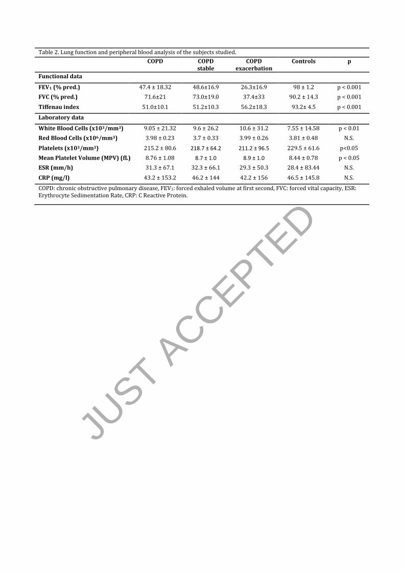
Table 2. Lung function and peripheral blood analysis of the subjects studied.
COPD COPD stable
COPD exacerbation
Controls p
Functional data
FEV1 (% pred.) 47.4 ± 18.32 48.6±16.9 26.3±16.9 98 ± 1.2 p < 0.001
FVC (% pred.) 71.6±21 73.0±19.0 37.4±33 90.2 ± 14.3 p < 0.001
Tiffenau index 51.0±10.1 51.2±10.3 56.2±18.3 93.2± 4.5 p < 0.001
Laboratory data
White Blood Cells (x103/mm3) 9.05 ± 21.32 9.6 ± 26.2 10.6 ± 31.2 7.55 ± 14.58 p < 0.01
Red Blood Cells (x106/mm3) 3.98 ± 0.23 3.7 ± 0.33 3.99 ± 0.26 3.81 ± 0.48 N.S.
Platelets (x103/mm3) 215.2 ± 80.6 218.7 ± 64.2 211.2 ± 96.5 229.5 ± 61.6 p<0.05
Mean Platelet Volume (MPV) (fL) 8.76 ± 1.08 8.7 ± 1.0 8.9 ± 1.0 8.44 ± 0.78 p < 0.05
ESR (mm/h) 31.3 ± 67.1 32.3 ± 66.1 29.3 ± 50.3 28.4 ± 83.44 N.S.
CRP (mg/l) 43.2 ± 153.2 46.2 ± 144 42.2 ± 156 46.5 ± 145.8 N.S.
COPD: chronic obstructive pulmonary disease, FEV1: forced exhaled volume at first second, FVC: forced vital capacity, ESR: Erythrocyte Sedimentation Rate, CRP: C Reactive Protein.
JUST A
CCEPTED

Table 3. Statistical comparison of MPV in the study groups and within the COPD population.
MPV (fL) p
COPD 8.7 ± 1.1 0.025
Controls 8.4 ± 0.8
COPD stable 8.7 ± 1.0 0.021
COPD with exacerbation 8.9 ± 1.0
Stage I(FEV1≥80% pred) 8.3 ± 0.9
0.011* Stage II (FEV1=50-79% pred) 8.7 ± 0.8 Stage III(FEV1=30-49% pred) 9.1 ± 1.5 Stage IV(FEV1<30% pred) 8.8 ± 1.0 Data expressed as mean ± standard deviation COPD: chronic obstructive pulmonary disease, MPV: mean platelet volume *ANOVA
JUST A
CCEPTED

JUST A
CCEPTED

JUST A
CCEPTED

JUST A
CCEPTED