to seizure disorder - nancy mullan,...
TRANSCRIPT
86 • THE AUTISM FILE GLOBAL ISSUE 38 REPRINTED WITH PERMISSION © THE AUTISM FILE www.autismfile.com
biomedical
How Bacterial Imbalances May Predispose
to Seizure DisorderBy Amy Yasko, PhD, AMD, FAAIM and Nancy Mullan, MD
ather than discussing the well-recognized role of glutamate and GABA balance in the production of seizures,22,5 the focus here is on
a less well appreciated area: the ability of gastrointestinal bacteria to harbor aluminum and heavy metals in the body and aluminum’s role in producing neurologic dysfunction of many kinds, including seizure disorders.
Bacteria have been shown to retain heavy metals and aluminum. A number of different bacteria can accumulate various types of metals and may retain those metals in the body by several mechanisms.50,44,42,35,13,3 These metals interfere with the body’s biochemical pathways in well-documented ways. The large intestine harbors a large, complex ecosystem of microbes. For example, a healthy adult carries between 1.5 and 2.0 kg of bacteria in his gastrointestinal lumen.16,53 This space retains the organisms as well as the toxic and heavy metals that they contain. In some cases, up to 50 percent of the dry weight of the bacteria can be heavy metals.13 While bacteria, in particular staphylococci, retain aluminum,3 it is likely that other bacteria can do so also. Aluminum may increase the propensity for bacteria to form a biofilm in part because of its pro-oxidant activity,8 and it has been characterized as having direct
effects on biofilm activity in other systems.41 The elimination of abnormal gastrointestinal flora—along with the excretion of the metals they contain, and the maintenance of proper balance among the organisms that should be present in the gastrointestinal tract—may be essential for proper function of the biochemical pathways in the body.51
Aluminum is a documented neurotoxin that is associated with cognitive, psychological, and motor abnormalities. There is no argument about this in the literature. Both clinical observation and animal experiments have documented neurotoxicity from excess brain exposure to aluminum.47 It has been found in elevated levels in the brains of patients with Parkinsonism,14,37,32 amyotrophic lateral sclerosis,37,32 and Alzheimer-type dementia.7,10,30,45,4 It produces aluminum-induced encephalopathy 17,15,32 and causes neuroanatomical and neurochemical changes in the brain, including neurofilament disturbances 40,36,52,4,14 followed by nerve cell loss.2,15 Direct evidence of aluminum inducing seizures has been obtained from nonhuman primate studies in which aluminum provoked well-documented and characterized seizures.9,20,24,29 There is a quantitative threshold in monkeys for seizure activity that is generated simply by adding aluminum.9 (See
Amy Yasko, PhD, holds a doctorate in microbiology, immunology, and infectious diseases. She completed two research fellowships at Strong Memorial Hospital in Rochester NY; one as a member of the Dept. of Pediatrics and Infectious Diseases, the other as a member of the Wilmont Cancer Center. Dr. Yasko was a fellow in the Department of Hematology at Yale Medical Center prior to joining a biotechnology company. She later cofounded a successful biotechnology company, where she was recognized as an expert in the field of DNA/RNA based diagnostics and therapeutics. She was a consultant to the medical, pharmaceutical and research community for almost 20 years with an expertise in biochemistry, molecular biology and biotechnology. Dr. Yasko continued her education in the area of alternative healthcare to graduate with high honors from the Clayton College of Natural Health, receiving Doctor of Naturopathy and Doctor of Natural Health degrees.
Nancy Mullan, MD, received her MD from Tufts University. She completed an internship and residency in psychiatry and a fellowship in child psychiatry at the University of Chicago Hospitals and Clinics. While there she studied at the Chicago Institute for Psychoanalysis and taught at the Psychosomatic and Psychiatric Institute for Research and Training at Michael Reese Hospital. Dr. Mullan joined the medical staff at Cedars-Sinai Medical Center and taught at both UCLA and USC Schools of Medicine. She earned psychoanalytic certification from the Psychoanalytic Center of California. Currently, Dr. Mullan is practicing nutritional medicine and psychiatry in Burbank, CA, treating children on the autism spectrum and adults with neurologic, immunologic, metabolic and/or gastroenterologic dysfunction that might otherwise be thought of as psychiatric illness. Bacteria have been shown to retain heavy metals and aluminum.
www.autismfile.com REPRINTED WITH PERMISSION © THE AUTISM FILE THE AUTISM FILE GLOBAL ISSUE 38 • 87
biomedical Table 1.) Clinical observation has linked aluminum with seizure disorders,11 and aluminum has also been documented to affect myelin formation.34 Myelin, the protective sheath on the axonal fiber that promotes proper conduction of the nervous signal, is critical to neuron signal transduction. Deficient myelination of nerves may be another contributing factor in seizure activity as the absence of myelin has been demonstrated to cause seizures.18,39
Aluminum has a suppressing influence on the cholinergic system.2,17 It inhibits the activity of acetylcholinesterase in the brain, cerebrospinal fluid, erythrocytes, plasma, and lymphocytes of rodents.6,19,26 This results in increased acetylcholine levels. Acetylcholine is a key component, the significant neurotransmitter, in the cholinergic system. Increased stimulation of muscarinic receptors may produce pinpoint pupils, blurred vision, hypersecretion, and bladder incontinence. Increased stimulation of nicotinic receptors may be responsible for muscle twitching, muscle weakness, and dilated pupils.
The impacts of aluminum on the cholinergic system of rodents are reported by Dave, et. al.:
There is some evidence of neurochemical alterations induced in the cholinergic system of several rodent species by Al in vivo. We have reported the long-term effect of long-term Al feeding on oxidative energy metabolism in rat liver, brain and heart mitochondria. The toxic effects of Al on different membrane systems have been evaluated. Our own studies have shown that the phosholipid compositions of rat brain synaptic plasma membranes, microsomes and myelin, as well as Na+, K+ ATPase kinetics, were significantly altered after long-term Al feeding (6, p.225).
While aluminum is a toxic, it is not a heavy metal. It is a non-redox reactive metal and causes oxidative damage at increased levels. Both the mitochondria and the methionine cycle are negatively impacted by high levels of aluminum. The function of the mitochondria is to produce energy to drive biochemical reactions, transport substrates into and out of the cells, and perform other functions in the body that require energy.
The energy is produced in a biochemical pathway called the Krebs cycle. In generating this energy, the mitochondria make reactive oxygen species, molecules which attach to other molecules or structures and deplete them of electrons. If the body cannot neutralize these radicals, then important molecules are at risk of becoming oxidized. One of these important molecules is glutathione, which is responsible for detoxifying metals and other toxins. Glutathione is functional only in its reduced state.
B12 is another critical molecule which is sensitive to oxidation. It is also functional only in its reduced state. Lack of functional B12 has direct negative impacts on the Krebs energy cycle in the mitochondria12 and induces the cycle to flow in a retrograde direction.27 Additionally, B12 deficiency has been linked with seizures.21,28 B12 also has impacts on the methionine cycle. The methionine cycle is that metabolic pathway that uses methionine synthase along with methyltetrahydrofolate and reduced B12 to reattach a methyl group to homocysteine and continue the vital
process of methyl group production. Methionine synthase, a critical enzyme in the methionine cycle, is sensitive to oxidation. In the pro-oxidant environment generated by aluminum, the activity of B12 is inhibited as is methionine synthase. Because the Krebs cycle and the methionine cycle are central biological processes, body function would be expected to be compromised from the non-optimal function of either one of these two systems.
Taurine is an end product of the methionine cycle by way of a sulfur metabolizing pathway called the transsulfuration pathway. Because taurine is well known to help reduce seizure activity, lack of taurine due to impaired methionine cycle function may be another contributing factor in the etiology of seizure disorder. This is an indirect impact of aluminum resulting from its pro-oxidant character. Whether it is the indirect impact of aluminum on methionine synthase and B12 by oxidation that may decrease taurine levels or the direct effect of aluminum on generating seizure activity as observed
Glutamate Pathway alumina cream-induced epilepsy in the monkey offers a vast opportunity for further study of the basic mechanisms of temporal lobe epilepsy.
Table 1 Summary of Clinical Manifestation in Psychomotor Epilepsy in Man (Gastaut, H., 1954)
1. Sensory and Sensorial SymptomsA. In relation to external stimuli
gustatory, olfactory, auditory, vertigious, visual, somastaesthetic
B. Autonomic oropharyngeal, oesophageal, epigastric,
abdominal, genital, retrosternal, precordial nausea, asphyxia, palpitation, heat or cold, hunger or thirst, urinate or defaecate
2. Mental SymptomsA. Changes in the state of consciousnessB. Perceptive illusions or hallucinations (macropsia,
micropsia, macrosacusia, distortion of image, metamorphopsia, deja vu, jamais vu, incoherence, strangeness, depersonalization, complex hallucination)
C. Ideational blocking of thought, interfering idea, etc.
D. Affective fear, sadness, anger, joy, etc.3. Motor SymptomsA. In relation to external stimuli 1. Simple motor manifestations:
clonic, tonic, rotatory 2. Complex motor manifestation orientating or investigating reaction,
straightening reaction, gesticulatory response, gestures accompanying confusional state
B. Automatic respiratory, circulatory, digestive
(mastication, salivation), visceral (urination, defaecation), pupillary
C. Speech aphasic, expressing the sensation, a flow
of words
Symptoms which could be reproduced in the monkeys are in italicsSource: Faeth WH, Walker AE, Kaplan AD, et.al. Threshold studies on production of experimental epilepsy with alumina cream. Proc Soc Exp Biol Med. 1955;88:329-31.
Addressing aluminum toxicity by decreasing bacteria in the system that harbor aluminum is a critical intervention.
88 • THE AUTISM FILE GLOBAL ISSUE 38 REPRINTED WITH PERMISSION © THE AUTISM FILE www.autismfile.com
in non-human primate studies, it is imperative to decrease excess aluminum in the body.*
Addressing aluminum toxicity by decreasing bacteria in the system that harbor aluminum is a critical intervention. Aluminum tremendously increases the neurotoxicity of glutamate. Worse, glutamate has aluminum binding capacities that can act to hold aluminum in the system.32 Aluminum has pleotrophic effects in the body that culminate in a number of consequences, many of which may contribute to the development of seizures. The intention here is not to discount the significant role of glutamate in producing seizure disorder; rather, it is to assert that balancing glutamate alone is not likely to be sufficient to mitigate seizure activity in the presence of aluminum.
Aluminum has also been shown to directly inhibit regeneration of tetrahydrobiopterin (BH4), an important intermediate in the pathways of two critical neurotransmitters, serotonin and dopamine.1 In this way, aluminum may not only directly impact neurological parameters but also indirectly affect them by virtue of its inhibition of neurotransmitter generation.
The presence of pro-oxidant aluminum reduces the levels of methionine in the
biomedical
cells. The same amino acid methionine that functions in the methionine cycle also acts as an endogenous antioxidant in cells, and it combines readily with a variety of pro-oxidative molecules and becomes methionine oxidase. Optimally, methionine oxidase can be reduced to become methionine again. The cell has methionine oxidase reductases, enzymes that catalyze this process when thioredoxin and NADH are present. Virtually all organisms – from bacteria to mammals – have several methionine oxidase reductases in their cytoplasm to provide reversibility between methionine oxide and methionine. This reversibility is the chemical basis for efficient scavenging of reactive species by methionine.25 Reduction of methionine levels reduces this protection.
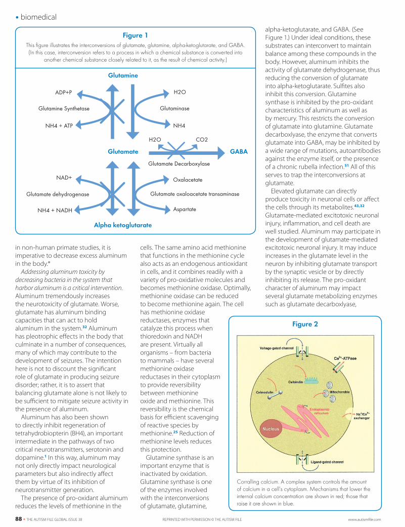
Glutamine synthase is an important enzyme that is inactivated by oxidation. Glutamine synthase is one of the enzymes involved with the interconversions of glutamate, glutamine,
alpha-ketoglutarate, and GABA. (See Figure 1.) Under ideal conditions, these substrates can interconvert to maintain balance among these compounds in the body. However, aluminum inhibits the activity of glutamate dehydrogenase, thus reducing the conversion of glutamate into alpha-ketoglutarate. Sulfites also inhibit this conversion. Glutamine synthase is inhibited by the pro-oxidant characteristics of aluminum as well as by mercury. This restricts the conversion of glutamate into glutamine. Glutamate decarboxlyase, the enzyme that converts glutamate into GABA, may be inhibited by a wide range of mutations, autoantibodies against the enzyme itself, or the presence of a chronic rubella infection.51 All of this serves to trap the interconversions at glutamate.
Elevated glutamate can directly produce toxicity in neuronal cells or affect the cells through its metabolites.43,32 Glutamate-mediated excitotoxic neuronal injury, inflammation, and cell death are well studied. Aluminum may participate in the development of glutamate-mediated excitotoxic neuronal injury. It may induce increases in the glutamate level in the neuron by inhibiting glutamate transport by the synaptic vesicle or by directly inhibiting its release. The pro-oxidant character of aluminum may impact several glutamate metabolizing enzymes such as glutamate decarboxlyase,
Figure 1This figure illustrates the interconversions of glutamate, glutamine, alpha-ketoglutarate, and GABA.(In this case, interconversion refers to a process in which a chemical substance is converted into
another chemical substance closely related to it, as the result of chemical activity.)
ADP+P
Glutamine
Glutamate
Alpha ketoglutarate
GABA
Glutamine Synthetase
NH4 + ATP NH4
H2O
Glutaminase
Glutamate Decarboxylase
H2O CO2
NAD+ Oxalacetate
Glutamate dehydrogenase Glutamate oxaloacetate transaminase
AspartateNH4 + NADH
www.autismfile.com REPRINTED WITH PERMISSION © THE AUTISM FILE THE AUTISM FILE GLOBAL ISSUE 38 • 89
biomedical glutamate aminotransferases, glutamate dehydrogenase, gamma-glutamyl transferase, and glutamine synthase, among others.32,43 These modulations can lead to the increase of glutamate concentration. Glutamate decarboxlyase is the sole enzyme responsible for the decarboxylation of glutamate into GABA. In addition to the GABA-producing enzyme being impacted by aluminum, the GABA-degrading enzyme is also.
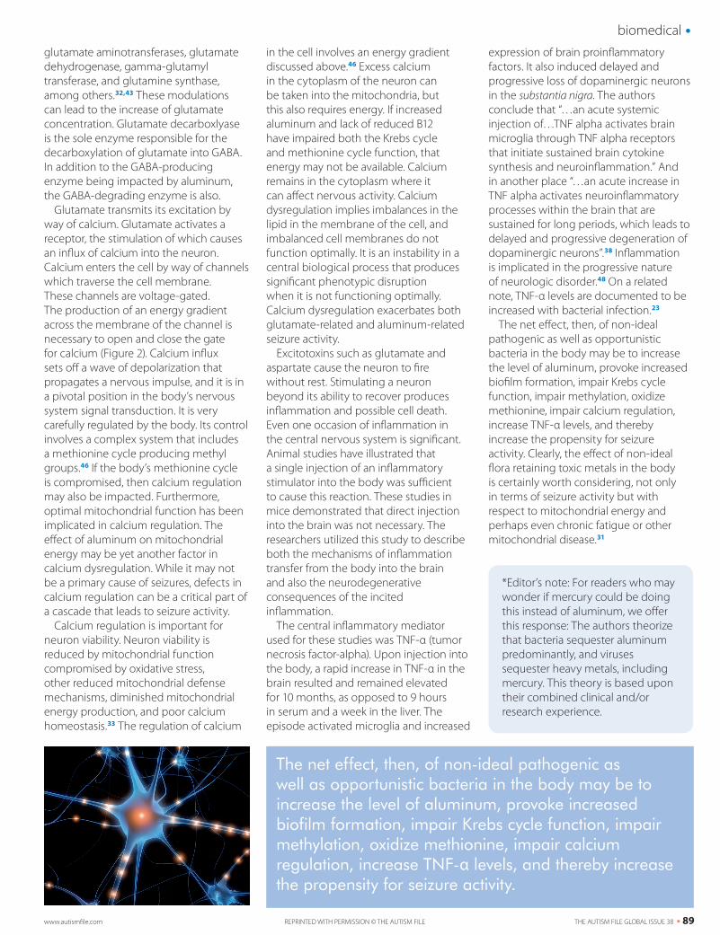
Glutamate transmits its excitation by way of calcium. Glutamate activates a receptor, the stimulation of which causes an influx of calcium into the neuron. Calcium enters the cell by way of channels which traverse the cell membrane. These channels are voltage-gated. The production of an energy gradient across the membrane of the channel is necessary to open and close the gate for calcium (Figure 2). Calcium influx sets off a wave of depolarization that propagates a nervous impulse, and it is in a pivotal position in the body’s nervous system signal transduction. It is very carefully regulated by the body. Its control involves a complex system that includes a methionine cycle producing methyl groups.46 If the body’s methionine cycle is compromised, then calcium regulation may also be impacted. Furthermore, optimal mitochondrial function has been implicated in calcium regulation. The effect of aluminum on mitochondrial energy may be yet another factor in calcium dysregulation. While it may not be a primary cause of seizures, defects in calcium regulation can be a critical part of a cascade that leads to seizure activity.
Calcium regulation is important for neuron viability. Neuron viability is reduced by mitochondrial function compromised by oxidative stress, other reduced mitochondrial defense mechanisms, diminished mitochondrial energy production, and poor calcium homeostasis.33 The regulation of calcium
in the cell involves an energy gradient discussed above.46 Excess calcium in the cytoplasm of the neuron can be taken into the mitochondria, but this also requires energy. If increased aluminum and lack of reduced B12 have impaired both the Krebs cycle and methionine cycle function, that energy may not be available. Calcium remains in the cytoplasm where it can affect nervous activity. Calcium dysregulation implies imbalances in the lipid in the membrane of the cell, and imbalanced cell membranes do not function optimally. It is an instability in a central biological process that produces significant phenotypic disruption when it is not functioning optimally. Calcium dysregulation exacerbates both glutamate-related and aluminum-related seizure activity.
Excitotoxins such as glutamate and aspartate cause the neuron to fire without rest. Stimulating a neuron beyond its ability to recover produces inflammation and possible cell death. Even one occasion of inflammation in the central nervous system is significant. Animal studies have illustrated that a single injection of an inflammatory stimulator into the body was sufficient to cause this reaction. These studies in mice demonstrated that direct injection into the brain was not necessary. The researchers utilized this study to describe both the mechanisms of inflammation transfer from the body into the brain and also the neurodegenerative consequences of the incited inflammation.
The central inflammatory mediator used for these studies was TNF-α (tumor necrosis factor-alpha). Upon injection into the body, a rapid increase in TNF-α in the brain resulted and remained elevated for 10 months, as opposed to 9 hours in serum and a week in the liver. The episode activated microglia and increased
expression of brain proinflammatory factors. It also induced delayed and progressive loss of dopaminergic neurons in the substantia nigra. The authors conclude that “…an acute systemic injection of…TNF alpha activates brain microglia through TNF alpha receptors that initiate sustained brain cytokine synthesis and neuroinflammation.” And in another place “…an acute increase in TNF alpha activates neuroinflammatory processes within the brain that are sustained for long periods, which leads to delayed and progressive degeneration of dopaminergic neurons”.38 Inflammation is implicated in the progressive nature of neurologic disorder.48 On a related note, TNF-α levels are documented to be increased with bacterial infection.23
The net effect, then, of non-ideal pathogenic as well as opportunistic bacteria in the body may be to increase the level of aluminum, provoke increased biofilm formation, impair Krebs cycle function, impair methylation, oxidize methionine, impair calcium regulation, increase TNF-α levels, and thereby increase the propensity for seizure activity. Clearly, the effect of non-ideal flora retaining toxic metals in the body is certainly worth considering, not only in terms of seizure activity but with respect to mitochondrial energy and perhaps even chronic fatigue or other mitochondrial disease.31
*Editor’s note: For readers who may wonder if mercury could be doing this instead of aluminum, we offer this response: The authors theorize that bacteria sequester aluminum predominantly, and viruses sequester heavy metals, including mercury. This theory is based upon their combined clinical and/or research experience.
The net effect, then, of non-ideal pathogenic as well as opportunistic bacteria in the body may be to increase the level of aluminum, provoke increased biofilm formation, impair Krebs cycle function, impair methylation, oxidize methionine, impair calcium regulation, increase TNF-α levels, and thereby increase the propensity for seizure activity.
90 • THE AUTISM FILE GLOBAL ISSUE 38 REPRINTED WITH PERMISSION © THE AUTISM FILE www.autismfile.com
biomedical
References1 Altindag ZZ, Baydar T, Elgin AB, Sahin G. Effects of the metals on dihydropteridine reductase activity. Toxicol In Vitro. 2003;17:533-7.2 Bilkei-Gorzo A. Neurotoxic effect of enteral aluminum. Food Chem Toxicol. 1993;31:357-61.3 Bradley TJ, Parker MS. Binding of aluminum ions by Staphylococcus aureus 893. Experientia.1968;24:1175-6.4 Crapper DR, Krishnan SS, Quirrkat S. Aluminum neurofibrillary degeneration and Alzheimer’s disease. Brain. 1976;99:67-80.5 Dalby NO, Thompsen C. Modulation of seizure activity in mice by metabotrophic glutamate receptor ligands. J Pharmacol Exp Ther. 1996;276:516-22.6 Dave KR, Syal Ar, Katyare SS. Effect of long-term aluminum feeding on kinetics attributes of tissue cholinesterases. Brain Research Bulletin. 2002;58:225-33.7 Deloncle R, Guillard O. Mechanism of Alzheimer’s disease arguments for a neurotransmitter-aluminium complex implication. Neurochem Res.1990; 7:195-206.8 Exley C. The pro-oxidant activity of aluminum. Free Radical Biol Med. 2004;36:380-7.9 Faeth WH, Walker AE, Kaplan AD, et.al. Threshold studies on production of experimental epilepsy with alumina cream. Proc Soc Exp Biol Med. 1955;88:329-31.10 Flaten TP. Aluminum as a risk factor in Alzheimer’s disease with emphasis on drinking water. Brain Res. Bull2001;55:187-96.11 Freiman SB, Holler J, Wittler M, Raymond L. Seizure and elevated blood aluminum in a remelt furnace operator: connection or coincidence? Am J Emerg Med. 2005; 23:419-20.12 Frenkel EP, Mukherjee H, Hackenbrock CR, Shere PA. Biochemical and ultrastructual hepatic changes during vitamin b deficiency in animals and man. J Biol Chem.1976;251:2147-54.13 Gadd GM. Heavy metal accumulation by bacteria and other microorganisms. Experientia. 1990:46:834-40.14 Garruto RM, Fukatsu R, Yanagihera R, Gajdusek DC. Hook G, Fiori CE. Imaging of calcium and aluminum neurofibrillary tangle-bearing neurons in Parkinsonism-dementia of Guam. Pro Natl Acad Sci USA. 1984;81:1875-79.15 Ghetti B, Musicco M, Norton J, Bugiani O. Nerve cell loss in the progressive encephalopathy induced by aluminum powder. A morphologic and semi-quantitative study of the Purkinje cells. Neuropathol Appl Neurobiol.1985;11:31-53.16 Guarner F, Malagelada JR. Gut flora in health and disease. The Lancet. 2003;361:512-19.17 Gulya K, Rakonczay Z, Kasa P. Cholinotoxic effects of aluminum in rat brain. J Neurochem. 1990;54:1020-26.18 Hoffman K, Lindner M, Groticke I, Stangel M, Loscher W. Epileptic seizures and hippocampal damage after cuprizone-induces demyelination in C57BL/6 mice. Exp Neurol. 2008;210(2):308-21.19 Inestrosa NC, Alaercon R, Arriagada J, Domoso A, Alvarez J, Campos EO, Blood markers in alzheimer disease: subnormal acetylcholinesterase and butyrylcholinesterase in lymphocytes and erythyrocytes. J Neurol Sci. 1994;122:1-5.20 Kopeloff LM, Chusid JG, Kopeloff N. Chronic experimental epilepsy in Macaca mulatta. Neurology. 1954;4:218-27.21 Kumar S. Recurrent seizures: An unusual manifestation of vitamin B12 deficiency. Neurol India [serial online] 2004 [cited 2010 Oct 7];52:122-3. Available from: http://www.neurologyindia.com/text.asp?2004/52/1/122/672122 Lee AC, Wong RKS, Chuang SC, Shin HS, Bianchi R. Role of synaptic metabotropic glutamate receptors in epileptiform discharges in hippocampal slices. J Neurophysiol. 2002;88:1625-33.23 Leslie M. This is your brain…and this is your brain on calcium. Sci. Aging Knowl. Environ. 2002;15:4. 24 Lockard JS, Wyler AR. The influence of attending on seizure activity in epileptic monkeys. Epilepsia 1979;20:157-68.25 Luo S, Levine R, Methionine in protein defends against oxidative stress. FASEB J. 2009;23:464-72.26 Hofstetter JR, Vincent I, Bugiani O, Ghetti B, Richter JA. Aluminum-induced decrease in choline acetyl transferase, tyrosine hydroxylase and glutamate decarboxylate in selected regions of rabbit brain. Neurochemical Pathology. 1987;6:177-93.27 Mailloux RJ, Hamel R, Appanna VD. Aluminum toxicity elicits a dysfunctional TCA cycle and succinate accumulation in hepatocytes. Journal of Biochemical and Molecular Toxicology. 2006;20:198-208.
28 Matsumoto A, Shiga Y, Shimizu H, Kimura I, Hisanaga K. Encephalomyelopathy due to vitamin b12 deficiency with seizures as a predominant symptom [article in Japanese]. Rinsho Shinkeigaku. 2009;243-54. 29 Mayanagi Y. Alumina cream-induced temporal lobe epilepsy in the monkey as an experimental model. Folia Psychiatrica et Neurologica Japonica. 1979;33:457-62.30 McDermott JR, Smith AI, Iqubal K, Wisiniewiski HM. Brain aluminum in ageing and Alzheimer’s disease. Neurology. 1979;29:809-14.31 Myhill S, Booth NE, McLaren-Howard J. Chronic fatigue syndrome and mitochondrial dysfunction. Int J Clin Exp Med. 2009;2:1-16.32 Nayak P, Chatterjee. Effects of aluminum exposure on brain glutamate and GABA systems: an experimental study in rats. Food and Chemical Toxiocolgy. 2001;39:1285-128933 Nilsen J, Brinton RD. Mitochondria as therapeutic targets of estrogen action in the central nervous system. CNS & Neurological Disorders. 2004;3:297-313.34 Pandya JD, Dave KR, Katyare SS. Effect of long-term aluminum feeding on lipid/phospholipid profiles of rat brain synaptic plasma membranes and microsomes. J Alzheimer’s Dis. 2001;3:531-39.35 Perdrial N, Liewig N, Delphin JE, Elsass J. TEM evidence for intracellular accumulation of lead by bacteria in subsurface environments. Chemical Geology. 2008;15:196-204.36 Perl DP, Brody AR. Alzheimer’s disease: X-ray spectrometric evidence of aluminum accumulation in neurofibrillary tangle bearing neurons. Science. 1980;208:297-9.37 Perl DP, Gajdusek DC, Garruta RM, Yanagihata RT, Gibba CJ. Intraneuronal aluminum accumulation in amyotrophic lateral sclerosis and Parkinsonism-dementia of Guam. Science 1982;217:1052-55.38 Qin et.al. Systemic LPS causes chronic neuroinflammation and progressive neurodegeneration. Glia. 2007;55:453-62.39 Rosenbluth J, Guo D, Liu Z, Liang WL, Schiff R. Effects of cerebellar lesions on tonic seizures, tremor and lifespan in myelin deficient rats. Brain Res. 1994;650(1):85-92.40 Simpson J, Yates CM, WHyler DK, Wilson H, Dewar AJ, Gordon A. Biochemical studies on rabbits with aluminum induced neurofilament accumulations. Neurochem Res. 1985;10:229-38.41 Sonak S, Bhosle N. (1995). Observations on biofilm bacteria isolated from aluminium panels immersed in estuarine waters. Biofouling. 1995;8:243-54.42 Strandberg GW, Shumate SK, Parrott JR. Microbial cells as biosorbents for heavy metals: accumulation of uranium by Saccharomyces cervisiae and Pseudomonas aeruginosa. Applied and Environmental Microbiology. 1961;41:237-45.43 Struys-Ponsar C, Guillard O, de Aguilar P. Effects of aluminum exposure on glutamate metabolism: A possible explanation for its toxicity. Experimental Neurology. 2000;163:157-164.44 Summers AO, Silver S. Microbial transformations of metals. Ann Rev Microbiol. 1978;32:637-72.45 Swegert CV, Dave KR, Katyare SS. Effect of aluminum-induced Alzheimer like condition on oxidative energy metabolism in rat liver, brain and heart mitochondria. Mech Ageing Dev.1999;112:27-42.46 Toyota M, et.al., Inactivation of CACNA1G, a T-Type calcium channel gene, by abberant methylation of its 5’ CpG island in human tumors. Cancer Research. 1989;4535-31.47 US Dept of Health and Human Services, Agency for Toxic Substances and Disease Registry, Altlanta, GA 30333. ATSDR’s Toxicol Profiles. 2004[Aluminum]:11-30.48 Wang Q, Tang XN, Yenari MA. The inflammatory response in stroke. J Neuroimmunol. 2003;184:53-68.49 Wang SD, Huang KJ, Lin YS, Lei HY. Sepsis induced apoptosis of the thymocytes in mice. Journal of Immunology. 1994;152:5014-21.50 Wood JM, Wang HK. Microbial resistance to heavy metals. Environ Sci Technol.1983;17:730-50.51 Yasko A. Autism: Pathways to Recovery. Neurological Research Institute, Bethel, Maine. 2009;61-70.52 Yates CM, Simpson J, Russell D, Gordon A. Cholinergic enzymes in neurofibrillary degeneration produced by aluminum. Brain Res. 1980;197:269-274.53 Zoetendal EG, von Wright A, Vilpponen-Salmela T, Ben-Amor K, Akkermans ADL, de Vos VM. Mucosa-associated bacteria in the human gastrointestinal tract are uniformly distributed along the colon and differ from the community recovered from feces. Appl Environ Microbiol. 2002;68:3401-7.