today is tuesday, october 28 th, 2014 pre-class: what is/are the female hormone(s)? what is/are the...
TRANSCRIPT
Today is Tuesday,October 28th, 2014
Pre-Class:What is/are the female hormone(s)?What is/are the male hormone(s)?
Guided readings should be on your desks.
In This Lesson:Cell
Communication and the
Endocrine System(Lesson 5 of 5)

Today’s Agenda
• Biostatistics: Standard Error and Standard Deviation
• Cell Communication– AKA membrane function in like…super detail.
• Hormones and the Endocrine System
• Where is this in my book?– Chapters 11 and 45

By the end of this lesson…
• You should be able to calculate standard deviation and standard error for a set of data.
• You should be able to describe the G protein-coupled receptor, receptor tyrosine-kinase, and intracellular receptor mechanisms of cell communication.
• You should know how hormones work.• You should be able to explain feedback loops
and how they function to maintain homeostasis.

Biostatistics
• Part of AP Biology is learning how to do the statistical analyses necessary to validate the results of an experiment.
• The Chi-Squared Analysis was part of our Animal Behavior lesson, although it doesn’t really have anything to do with animal behavior directly.
• Today we learn Standard Error and Standard Deviation, even though they don’t really have anything to do with cell communication.– Or the endocrine system.
• Fact Sheet – Unit 3 – Standard Deviation and Standard Error Tutorial
Cell Communication
• So far nearly everything we’ve done has been about the cell in isolation.– Except for maybe the junctions between cells.
• In reality, unless you’re unicellular, your cells need to communicate with one another.– Actually, even being unicellular doesn’t excuse you
from the need to communicate.– Even yeast cells actually have two sexes (a and α) that
mate – a process which requires its fair share of communication.
– For more: TED: Bonnie Bassler – How Bacteria Talk
Communication Modes
• Cells actually can respond to light and touch, in addition to the usual chemical signals.– We’re talking chemical signals right now.
• The general chemical signaling process leads to what’s known as a signal transduction pathway.– This sounds fancy, but it really just means that a stimulus (a
signal) is received by the cell and changed into a response (is transduced) through a series of molecules (a pathway).
• Signal transduction pathways are remarkably similar across a wide range of organisms, suggesting they’ve been around awhile.

Communication Ranges
• Think for a second how you might communicate with someone else.– How would you communicate if you were in the same room?– How would you communicate if you were in different states?
• Consider also how you would communicate in light of the speed necessary to do so.– Needing to deliver something urgently might change your
decision over how to deliver it.• In the same way:– Electrical signaling (nerves) = fast, urgent communication.– Chemical signaling (molecules) = gradual communication.

Cell Communication
• Local Signaling:– Paracrine Signaling: when
a signal affects a neighboring cell.
– Synaptic Signaling: paracrine signaling in neurons – the end of one nerve cell releases neurotransmitter into the synapse (space between neurons) to signal the next.
Cell Communication
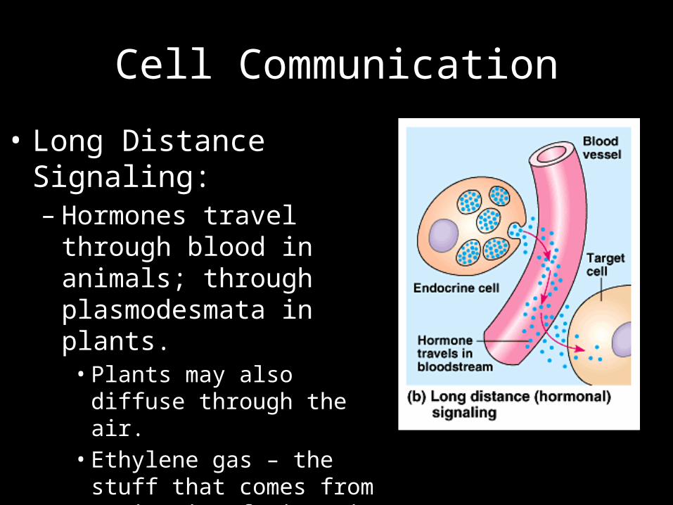
• Long Distance Signaling:– Hormones travel through
blood in animals; through plasmodesmata in plants.• Plants may also diffuse through
the air.• Ethylene gas – the stuff that
comes from a ripening fruit – is an example of an air hormone.

The Signal Transduction PathwayThree Steps
• Step 1: Reception:– The cell needs to receive a chemical signal (a ligand).• A ligand is any small molecule that binds to a larger one.• The larger molecule is usually a receptor protein.

The Signal Transduction PathwayThree Steps
• Step 2: Transduction– The membrane receptor protein then activates one or more
other molecules to carry the signal deeper into the cell.– These other molecules are called relay molecules and may be
involved in a phosphorylation cascade (more later).

The Signal Transduction PathwayThree Steps
• Step 3: Response– The cell does something.– This could be the activation of a gene, change in the
cytoskeleton, activity of an enzyme, or just about anything else.
Signal Transduction Pathways
• If it helps, think of signal transduction pathways like what happens when you get a text message:– Reception = Your phone vibrates or dings.– Transduction = You unlock the phone and read the
message.– Response = You write back, smile, cry, or throw the
phone against the wall.

Key Enzymes in Signal Transduction
• Before we look at the individual signal transduction forms, keep in mind the following two types of enzymes and their jobs:– Protein Kinase – a kinase removes phosphate groups from
(dephosphorylates) ATP and adds them to (phosphorylates) a protein.
– Protein Phosphatases – a phosphatase takes the phosphate back from the protein.
• These may act in a phosphorylation cascade to make a response.– A phosphorylation cascade is just a lot of
phosphorylation/dephosphorylation and is associated with the “transduction” step of cell communication.
– It also helps in amplification – more later.
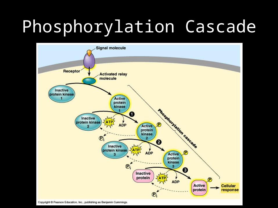
Phosphorylation Cascade
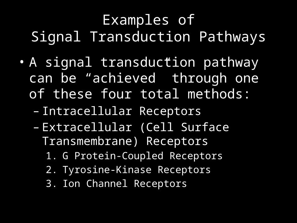
Examples ofSignal Transduction Pathways
• A signal transduction pathway can be “achieved” through one of these four total methods:– Intracellular Receptors– Extracellular (Cell Surface Transmembrane)
Receptors1. G Protein-Coupled Receptors2. Tyrosine-Kinase Receptors3. Ion Channel Receptors

Intracellular Receptors
• This is when a signal molecule, still called a ligand, enters a cell to elicit a response.
• Inside the cell, it binds to a receptor protein in the cytoplasm and then can affect transcription or other cell activities.– In this case, we could call the
ligand/receptor complex a transcription factor.
• Onto the extracellular receptors!

1. G Protein-Coupled Receptor
• G proteins are guanine nucleotide-binding proteins.
• So, a G protein-coupled receptor (GPCR) is a membrane receptor that is linked in some way to a G protein.– There’s the G protein and there’s the
receptor (they’re different).• G protein-linked receptors have
seven α helices spanning the membrane.
• These receptors are responsible for relaying a signal from a ligand to the interior of the cell (NOT relaying the ligand itself).
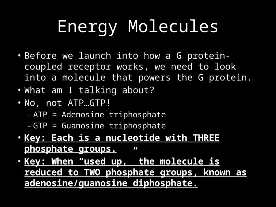
Energy Molecules
• Before we launch into how a G protein-coupled receptor works, we need to look into a molecule that powers the G protein.
• What am I talking about?• No, not ATP…GTP!– ATP = Adenosine triphosphate– GTP = Guanosine triphosphate
• Key: Each is a nucleotide with THREE phosphate groups.• Key: When “used up,” the molecule is reduced to TWO
phosphate groups, known as adenosine/guanosine diphosphate.

ATP vs. GTP
• They’re similar, but different in the same way that adenine and guanine are different.– Adenosine = adenine (a nitrogenous base) + ribose– Guanosine = guanine (a nitrogenous base) + ribose
• ATP is the more familiar energy “currency” of the cell, but GTP plays a role too.– The key is not so much the “adenosine” or
“guanosine” part as is the “triphosphate” part.– The bonds between the phosphate groups contain
the energy.

Back to G Protein-Coupled Receptors• Inactive:
– The receptor is spanning the membrane.– The G protein is bound to GDP and stuck to the inner membrane.– An enzyme also exists on the inner surface of the cell membrane.
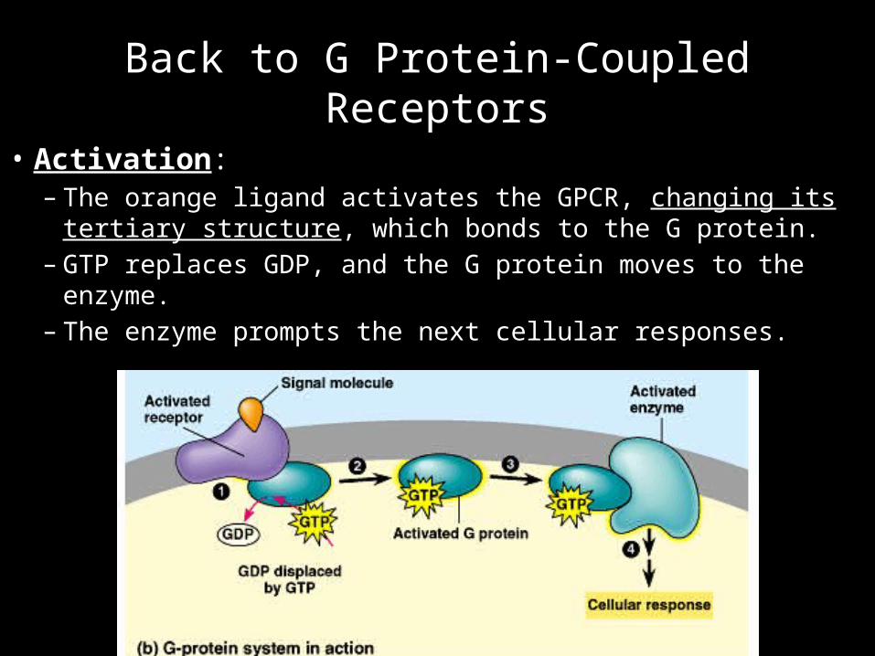
Back to G Protein-Coupled Receptors
• Activation:– The orange ligand activates the GPCR, changing its tertiary
structure, which bonds to the G protein.– GTP replaces GDP, and the G protein moves to the enzyme.– The enzyme prompts the next cellular responses.
G Protein Activation In Depth
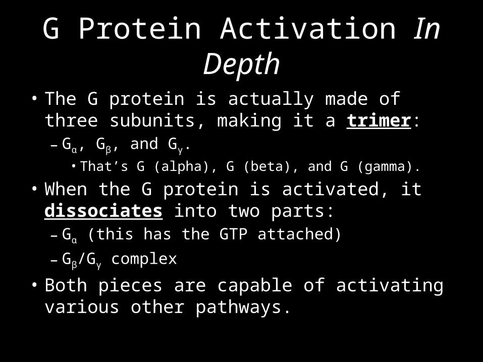
• The G protein is actually made of three subunits, making it a trimer:– Gα, Gβ, and Gγ.
• That’s G (alpha), G (beta), and G (gamma).
• When the G protein is activated, it dissociates into two parts:– Gα (this has the GTP attached)
– Gβ/Gγ complex
• Both pieces are capable of activating various other pathways.
G Proteins
• For a mental image…– Bat G-Protein video– Think of the Bat-Pod like a Gα subunit (and it’s got the
GTP, like Batman, attached to it).– Think of the rest of the Batmobile like the Gβ/Gγ
complex.• Except Gβ/Gγ complexes don’t normally explode.
• Overall Metaphor:– The G protein is like a pull-back toy car. Pull it back to
“wind it up” (have it interact with the receptor), then “let it go” (have it interact with the enzyme).

Back to G Protein-Coupled Receptors
• Deactivation:– The enzyme hydrolyzes GTP and removes a phosphate.– The G protein is released. The process can start again.
And I care…because?
• So why are G protein-coupled receptors important?– Your vision and smell senses use G protein-
coupled receptors.– Diseases like botulism, pertussis (whooping
cough), and cholera produce toxins that interfere with GPCRs.
– Around 60% of medicine works by affecting GPCRs, and a whole lot of drugs (including heroin) work the same way.

G Protein-Coupled ReceptorCase in Point
• Cholera is caused by the bacterium Vibrio cholerae.
• The bacterium releases a toxin that prevents GTP from being dephosphorylated, leading to a ton of salt secretion from intestinal cells, followed by water loss through osmosis.– This leads to fatal diarrhea if
not treated.http://sameens.dia.uned.es/Trabajos10/Trab_Publicos/Trab_2/Navarro_De_La_Cruz_2/Imagenes/vibrio_cholerae%5B1%5D.jpg

2. Tyrosine-Kinase Receptors
• Tyrosine-kinase is an enzyme stuck in the cell membrane.
• Its job is to dephosphorylate ATP and move that phosphate group to the attached tyrosine.
• It has a binding site in the ECM for signal molecules and single α helix in the membrane.

Tyrosine-Kinase Receptors• Inactive– The tyrosine-kinase receptor proteins are two separate
monomers, and relay proteins are not active.• Take a guess where this is going…

Tyrosine-Kinase Receptors• Activated– A ligand activates the monomers and they make a dimer.– Once joined, the kinase dephosphorylates ATP and adds that
phosphate group to its tyrosine (amino acid), which activates relay proteins.
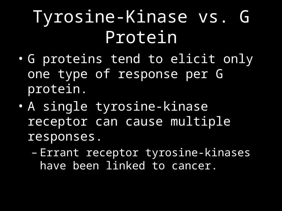
Tyrosine-Kinase vs. G Protein
• G proteins tend to elicit only one type of response per G protein.
• A single tyrosine-kinase receptor can cause multiple responses.– Errant receptor tyrosine-kinases have been linked
to cancer.

3. Ion Channel Receptors
• These are protein channels that open only when activated by a ligand.
• Nerve cells use these frequently.– Uh…that’s it here.
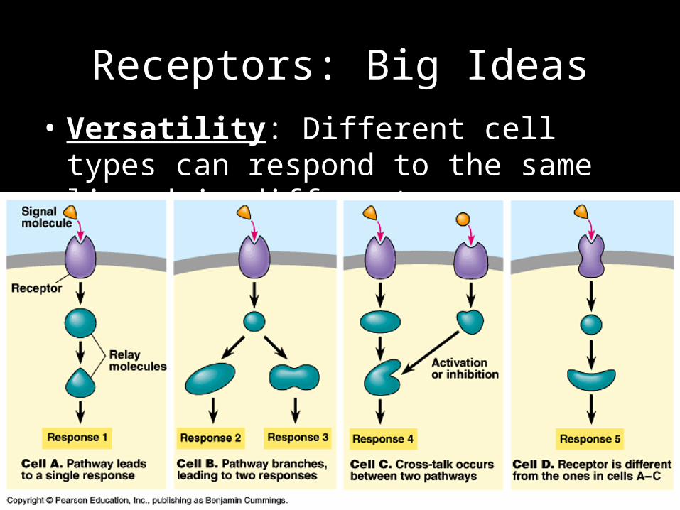
Receptors: Big Ideas• Versatility: Different cell types can respond to
the same ligand in different ways:
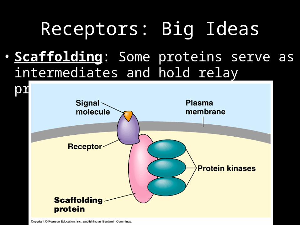
Receptors: Big Ideas• Scaffolding: Some proteins serve as intermediates
and hold relay proteins together.

Receptors: Big Ideas• Amplification: A single
signal molecule can lead to a massive response.– This is the point of a
phosphorylation cascade.

Second Messengers
• Signal transduction pathways often activate second messengers.– These are molecules within cells that act as signals just
like the original extracellular signal.• The three major classes of second messengers:– Cyclic nucleotides– DAG and IP3
– Calcium ions (Ca2+)

Second Messenger:Cyclic Nucleotides
• The enzyme adenylyl cyclase is activated by a G protein.– Adenylyl cyclase uses ATP to make cAMP, or cyclic
AMP.– AMP = adenosine monophosphate
• Similarly, guanylyl cyclase uses GTP to make cGMP (cyclic GMP).
• These second messengers then serve to turn on other responses within cells.

Second Messenger:IP3 and DAG
• DAG is diacylglycerol which stays in the cell membrane and activates other enzymes, which often use…
• …IP3, which is inositol triphosphate.– This helps release Ca2+ ions (which are themselves
considered a second messenger) from the ER.• Calcium ions, by the way, are used widely
throughout the body.– Including making your muscles contract.
Time to Practice
• Signal Transduction Pathways POGIL

The Endocrine System
• Those signaling methods we just saw operate on relatively “local” distances.
• When operating on long distances, we’re talking about hormones.– In Greek, harmon means “to excite.”
• Key: Hormones reach every cell in the body but only affect those with specific receptors.– Everyone hears it, but only some can respond.• Local signaling = whispering.• Hormones = YELLING but in a different language.
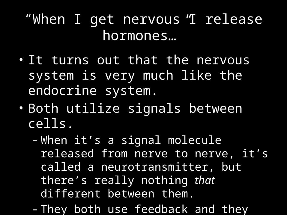
“When I get nervous I release hormones…”
• It turns out that the nervous system is very much like the endocrine system.
• Both utilize signals between cells.– When it’s a signal molecule released from nerve to
nerve, it’s called a neurotransmitter, but there’s really nothing that different between them.
– They both use feedback and they also can sometimes be released by the same structures.

The Endocrine System
• Endocrine glands secrete chemical signals within the body.– Hence the name “endocrine.”
• Exocrine glands secrete chemical signals onto the outside of the body or into a cavity.– Examples include salivary glands and sweat glands.
• Women’s menstrual cycles can be influenced by other women’s sweat.
– Other examples include anal glands in dogs and cats (and other animals).• Don’t worry, no photo.

The Endocrine System
• Hormones tend to be either water-soluble (polar) or lipid-soluble (non-polar).– Polar molecules use the same mechanisms as
extracellular responses (GPCRs, receptor tyrosine kinases).
– Non-polar molecules act like intracellular signals.• They go all the way into the cell and bind with
receptors within the cytoplasm, remember?

Hormone Mechanisms

Another View

Important Components of the Endocrine System
• Pineal Gland– Produces melatonin, which regulates circadian (day/seasonal) rhythms.
• Pituitary Gland/Hypothalamus– Makes growth hormone (GH), regulates menstrual cycle, and pigmentation.
• Pancreas• Ovaries/Testes (gonads)
– Male hormones = androgens (including testosterone)– Female hormones = estrogens (including estradiol) and progestogens
(including progesterone)• Thyroid/Parathyroid Gland
– Regulate energy usage and nervous system function.• Gastrointestinal Tract• Adrenal Glands
– Respond to stress, release epinephrine (adrenaline).

The Endocrine System

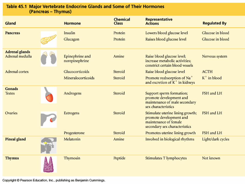

Some Important Hormone Information• The illegal anabolic steroids in sports are actually analogs of
androgens.• Insulin (blood sugar reducer) and glucagon (blood sugar
increaser) are hormones.• Dwarfism is caused by a lack of growth hormone from the
pituitary.• Acromegaly is a sort of dwarfism opposite caused by
increased growth hormone.• The thyroid gland makes thyroxine (an iodine-based
hormone) used in regulating basal metabolic rates.• Epinephrine (adrenaline) and norepinephrine are made by the
adrenal glands that sit atop the kidneys (the renal organs).
It’s Peanut Butter POGIL Time!
• Cell Communication POGIL

The Catch: Homeostasis
• All this signaling is great, but there’s one major catch: organisms still need to maintain homeostasis.
• They can achieve this through feedback loops.• Positive feedback amplifies the original signal.• Negative feedback inhibits the original signal.– As you might guess, negative feedback is far more
useful to homeostasis.

Negative FeedbackExamples
• Predator-Prey Relationships– Increase in prey leads to an
increase in predators…which decreases prey.
• Body Temperature– A rise in body temperature is
sensed by neurons which signal the brain, which sends signals to dilate the blood vessels (vasodilation), decreasing temperature.• And making you red in the face.

Negative FeedbackAnother Example
• If the pH in the duodenum (part of the intestine) drops too low…
• …the cells in the intestine release secretin, a chemical signal, into the blood.
• Secretin travels to the pancreas, which releases bicarbonate…
• …which raises the pH.

Positive FeedbackExamples
• Stampedes– A few animals start to
stampede, causing more to run, leading to a mass movement.
• Uterine contractions– Oxytocin causes uterine
contractions, which moves the fetus further down the birth canal, which stimulates more oxytocin release.
• Students packing up at the end of class?
Awkward stock photo from old PowerPoint…at what exactly are they
all looking?
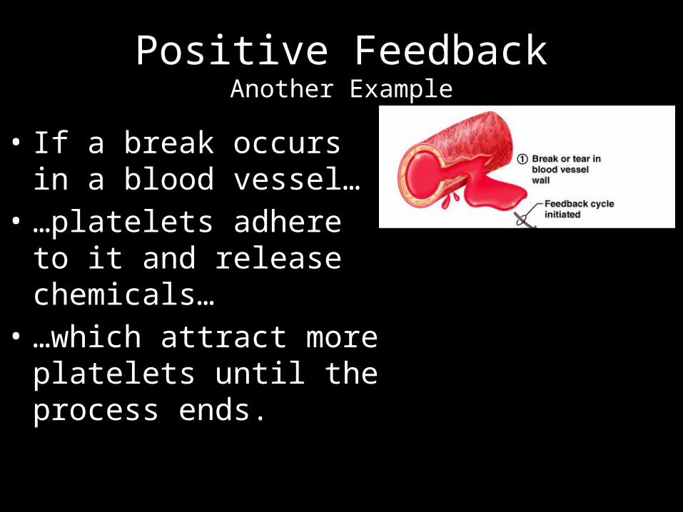
Positive FeedbackAnother Example
• If a break occurs in a blood vessel…
• …platelets adhere to it and release chemicals…
• …which attract more platelets until the process ends.

Positive FeedbackOne last one…
• The ocean is a major carbon sink.• Carbon dioxide dissolves best in cold water.• As CO2 levels cause temperatures to rise, more
CO2 precipitates from the ocean.
• More CO2 coming out of the ocean raises temperatures…
• …which releases more CO2.

Coupling Feedback Loops
• Remember that negative feedback loops are best for homeostasis.
• To prevent any level or rate from getting too high, you need a feedback loop.
• To prevent any level or rate from getting too low, you need another feedback loop.
• Key: A coupled (or double) feedback loop is needed to keep homeostasis.– Let’s look at some examples. Know the key
components.

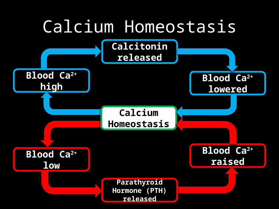
Calcium Homeostasis
Blood Ca2+ high
Calcitonin released
Blood Ca2+ lowered
Blood Ca2+ lowBlood Ca2+
raised
Calcium Homeostasis
Parathyroid Hormone (PTH) released


Glucose Homeostasis
Blood glucose high
Insulinreleased
Blood glucose lowered
Blood glucose low
Blood glucoseraised
GlucoseHomeostasis
Glucagon released

Closure: Feedback Mechanisms POGIL
• It’s time to put our knowledge of feedback mechanisms to the test using a POGIL.
• Feedback Mechanisms POGIL