topic 5: the folding of biopolymers rna and...
TRANSCRIPT

Topic 5: the folding of biopolymers – RNA and Protein
Overview:
The main functional biomolecules in cells are polymers – DNA, RNA
and proteins
For RNA and Proteins, the specific sequence of the polymer dictates
its final structure
Can we predict the final structure of RNA or protein given just the
sequence information?
Can we design any biomolecular structure that we want?

The world of RNA
RNA is a linear polymer built from
4 possible monomers: A, G, U, C
These monomers can form complimentary
interactions to form base-pairings:
A with U and C with G
Most often RNA is found as a single-stranded
polymer that via base-pairing with complementary
regions within its own sequence, is able to fold in
on itself
RNA has a wide variety of functions that we will
now explore

RNA function in cell
mRNA = messenger RNA
RNA’s main function in the cell is to act as a messenger molecule
in the process of making a protein
DNA (gene) mRNA Protein
tRNA = transfer RNA
these are used in translation to recognize the 64 codons. There
is one tRNA for each codon, each representing one of the 20
amino acids

rRNA = ribosomal RNA
these are RNAs that get incorporated into the ribosome to give it
part of its function.
microRNA or siRNA
these are relatively recent discovered form of RNA that is used to regulate
gene expression – so called RNA interference (won Nobel prize)
RNA function in cell
destroys
target
mRNA
many developmental genes are regulated by miRNAs in your genome

RNA function in cell
Riboswitches – many mRNA molecules can detect metabolites by binding them
and changing the structure of the mRNA = regulation

RNA function in cell
Ribozymes – like enzymes (catalytic proteins) except made from RNA. Able to
catalyze reactions.
in-vitro selection experiments – can select RNA molecules out of a random library
of sequences to catalyze a specific chemical reaction

RNA structure:
A polymer of RNA first folds by forming complimentary base pairings: G = C and A = U
The simplest form of RNA structure is a hairpin loop that in 3D looks like a double helix
2ndary structure double helix

RNA structure representation
The 2ndary structure of RNA is a particular pairing of its complimentary bases
RNA tertiary structure is the final 3D fold of the polymer
2ndary structure possesses 3 hairpin loops Tertiary structure

RNA structure representation
We will be interested in studying the formation of RNA secondary structure – tertiary
is too hard of a problem
We need ways to represent the structure in diagrams or strings for use in calculations
Rainbow diagram – shows pairings as
loops
no loops are allowed to cross = pseudo-knot
these occur but are computationally hard to
deal with
String representation:
(((((..))((..)))))…((…))…
1,1,1,1,0,0,-1,-1,1,1,0,0,-1,-1, …
nice property: sum of the string = 0

RNA folding
A biomolecules function depends on it’s structure. Can we predict the
most probable 2ndary structure of an RNA molecule by just knowing it’s sequence?
Ans: yes! enumerate all the possible structures (states), each has an energy, then
use Boltzmann distribution to determine the probabilities
Consecutive base-pairs is a stack – a lone base-pair is not

Simple model for RNA folding
Since stacking energy dominates, ignore the contribution of lone base-pairs and
only consider the energy that comes from forming stacks
Each stack lowers the energy of the structure by, − 𝜀𝑆
A structure that has n stacks has an energy of 𝐸 = −𝑛 |𝜀𝑆|
2 stacks 1 stack 0 stacks
For small RNA sequences, we can enumerate all possible structures that possess
stacks (do not draw structures that have lone base-pairs that are not part of a stack)
Calculate their energy (and possible entropy for the unpaired bases).
Ground state is the lowest (free) energy structure
For probabilities use Boltzman: P(structure) = exp(-Estructure/kT)/Z

http://rna.urmc.rochester.edu/RNAstructureWeb/Servers/Predict1/Predict1.html
RNA structure prediction in the real world
In reality, we can not draw all these structures by hand. Use a computer to
enumerate possible structure sequences and calculate the energy of the
sequence on each structure
Real-world RNA secondary structure prediction uses energies for base-pairing,
stacking, looping and forming pseudo-knots
Wide range of applications from predicting mRNA secondary structure, the
locations of miRNAs in a genome, designing PCR templates

Proteins:
● Proteins are biopolymers that form most of the cellular machinery
● The function of a protein depends on its 'fold' – its 3D structure
Chaperone
Motor
Walker

PRIMARY
F T P A
V
L F A H
D
K F L A
S
V T S V
Levels of Folding:
SECONDARY
TERTIARY
QUATERNARY

The Backbone
N
C
C'
N
C
C'
N
a
a
H
H
R
1
R
2
H
O
O
Peptide Bond
f
1
y
1
f
2
y
2
● Amino acids linked together
by peptide bonds
Steric constraints lead
only to a subset of possible angles
--> Ramachandran plot
Glycine residues
can adopt many
angles

a Helices
3.6 residues/turn

b Sheets
parallel sheet
anti-parallel sheet
other topologies possible
but much more rare
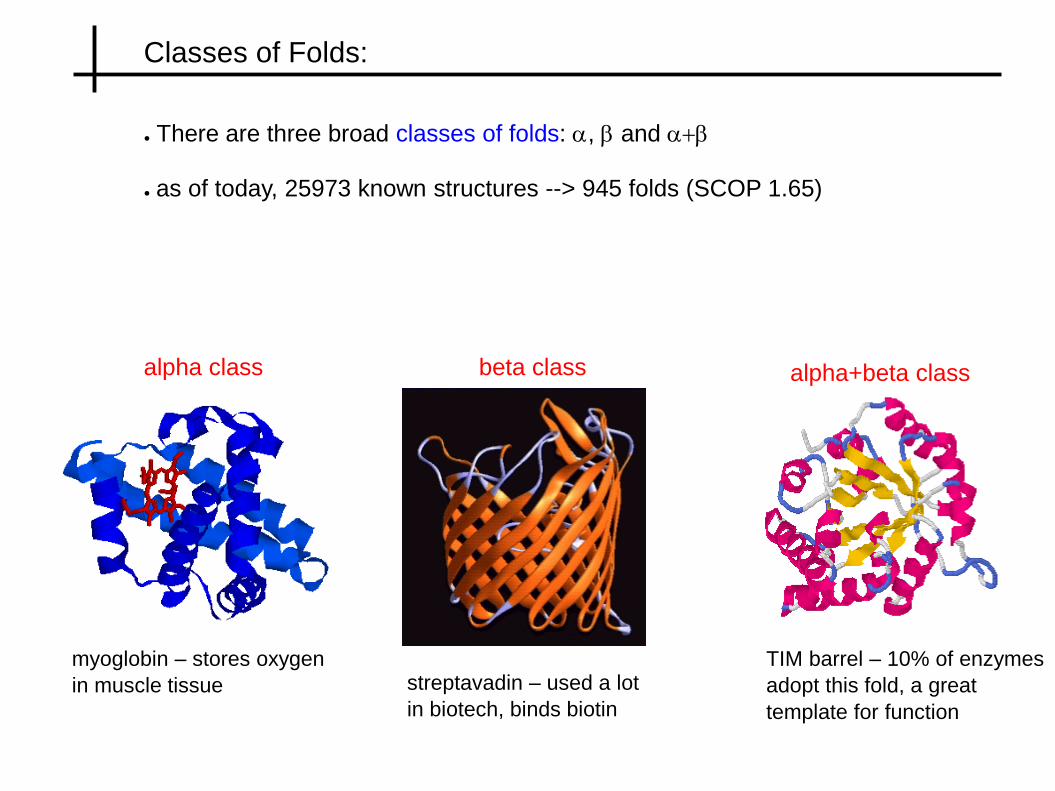
Classes of Folds:
● There are three broad classes of folds: a, b and a+b
● as of today, 25973 known structures --> 945 folds (SCOP 1.65)
alpha class beta class alpha+beta class
myoglobin – stores oxygen
in muscle tissue streptavadin – used a lot
in biotech, binds biotin
TIM barrel – 10% of enzymes
adopt this fold, a great
template for function

Databases:
SWISSPROT:
contains sequence data of proteins – 100,000s of sequences
Protein Data Bank (PDB):
contains 3D structural data for proteins – 20,000 structures, x-ray & NMR
SCOP:
classifies all known structures into fold classes ~ 800 folds

Protein Folding:
amino acid sequence structure
FOLDING
DESIGN
● naturally occurring sequences seem to have a unique 3D structure
Levinthal paradox: if the polymer doesn't search all of conformation space, how on
earth does it find its ground state, and in a reasonable time?
if 2 conformation/residue & dt ~ 10-12 -> t=1025 years for a protein of L = 150!!!
Reality: t = .1 to 1000 s
How do we resolve the paradox?

Paradox Resolved: Funnels
Slow path
Fast path
● there are multiple folding pathways on the energy landscape – slow & fast
● If a protein gets stuck (misfolded) there are chaperones to help finish the fold

Factors Influencing folding:
Hydrogen bonding:
doesn't drive folding since unfolded structure can form H-bonds with H20
drives 2ndary structure formation after compaction
Hydrophobicity:
main driving force
significant energy gain from burying hydrophobic side-chains
leads to much smaller space to search
Other interactions:
give specificity and ultimately favour final unique state
disulfide bridges = formed between contacting Cystine residues
salt-bridges = formed between contacting -ve and +ve charged residues
secondary structure preferences = from entropy
Open Molten (compacted) Native state
hydrophobic
force
H-bonds &
specific interactions

More on Hydrophobicity:
• Hydrophobicity is an entropic force – water loses entropy due to the presence
of non-polar solvent
H20 molecules form a tetrahedral structure, and there are 6 hydrogen-bonding
Orientations/H20
When a non-polar molecule occupies a vertex reduces to only 3 orientations
dS = k ln 3 – k ln 6 = - k ln 2 dG = + kT ln 2 costs energy to dissolve

Hydrophobicity and Packing:
For an O2 molecule in H20, A = 0.2 nm2 so G ~ 1 kT. So O2 easily dissolves in H20
For an octane molecule, G ~ 15 kT, so octane will aggregate so as to minimize the
combined exposed area
Non-polar
molecule
Area = A
A non-polar object with area A will disrupt
The local H20 environment
For 1 nm2 of area ~ 10 H20 molecules are affected
So hydrophobic cost per unit area
g = 10 k T ln 2/nm2 = 7 k T / nm2
Hydrophobic energy cost = G = g A

Simple Models of Folding: Getting at the big picture
● folding proteins in 3D with full atomic detail is HARD!!! essentially unsolved
--> study tractable models that contain the essential elements
SIMPLE STRUCTURE MODEL = LATTICE MODELS:
● enumerate all compact structures that completely fill a 2D or 3D grid
● can also study non-compact structures by making larger grid

Simple Energy functions:
H-P Models:
●amino acids come in only two types, H = hydrophobic, P = polar
●interactions: H-H, H-P & P-P with EPP
> EHP
> EHH
Energy = S Eij D(r
i – r
j)
●could use full blown 20 x 20 Eij matrix = Miyazawa-Jernigan matrix
Solvation Models:
●energy is gained for burying hyrdophobic residues
●if residue is buried, surface exposure, s = 1
●if residue is exposed, surface exposure, s = 0
●hydrophobicity scale: H: h = -1, P: h= 1
●Energy = S hi s
i
favourable contact
core site, s = 1 with H
surface site, s=0 with P
Ground state structure has the lowest energy
for given sequence

Model Results: Designability Principle
● Fold random HP sequences, and determine the ground state for each
● Designability = # of sequences which fold into a given structure
HPHPHPHHHHPHHP
PHPHPPHPHPHPPH
HPHPHHHPHPHPHH
PHPHPHHHPHPHHH
HPHHHPHPPPPHHP
PPHHPHHHPHHPPP
HPHPHPHHHHPPHP
HPHPHHHPPHHPHP
PHPHPHHHPPHHPH
PHPHPHHPHPPHHP
Designability, D
D = 3
D = 6
D = 1
D = 0
Designability Principle: there are only a few highly designable structure,
most structures have very few sequences that fold into them
Energy
Function

Thermodynamic Stability
● high designabilty implies mutational stability, does it imply thermodynamic stability?
YES
E0 E0
Eavg
D Eavg
D
Excited state spectra
HIGH DESIGN STRUCTURE LOW DESIGN STRUCTURE
● Highly designable structures are characterized by a large energy gap, D

Fast Folding
● High designability structures are fast folders, since there are few low lying
energy structures to compete with – no kinetic traps
● Low designability structures are slow – have many competing low energy
alternatives which act as kinetic traps
● Determine kinetics using Metropolis Monte-carlo
t ~ # of monte-carlo steps needed to first achieve near native state (90%)

Neutral Networks in Protein Folding:
well
separated
Sequence Space
Structure Space
● Just like RNA, designable proteins have well connected neutral networks
● Unlike RNA, these neutral networks are well separated, so they are not
space covering
● Prototype sequence tends to have best thermodynamic properties (cluster center)
prototype
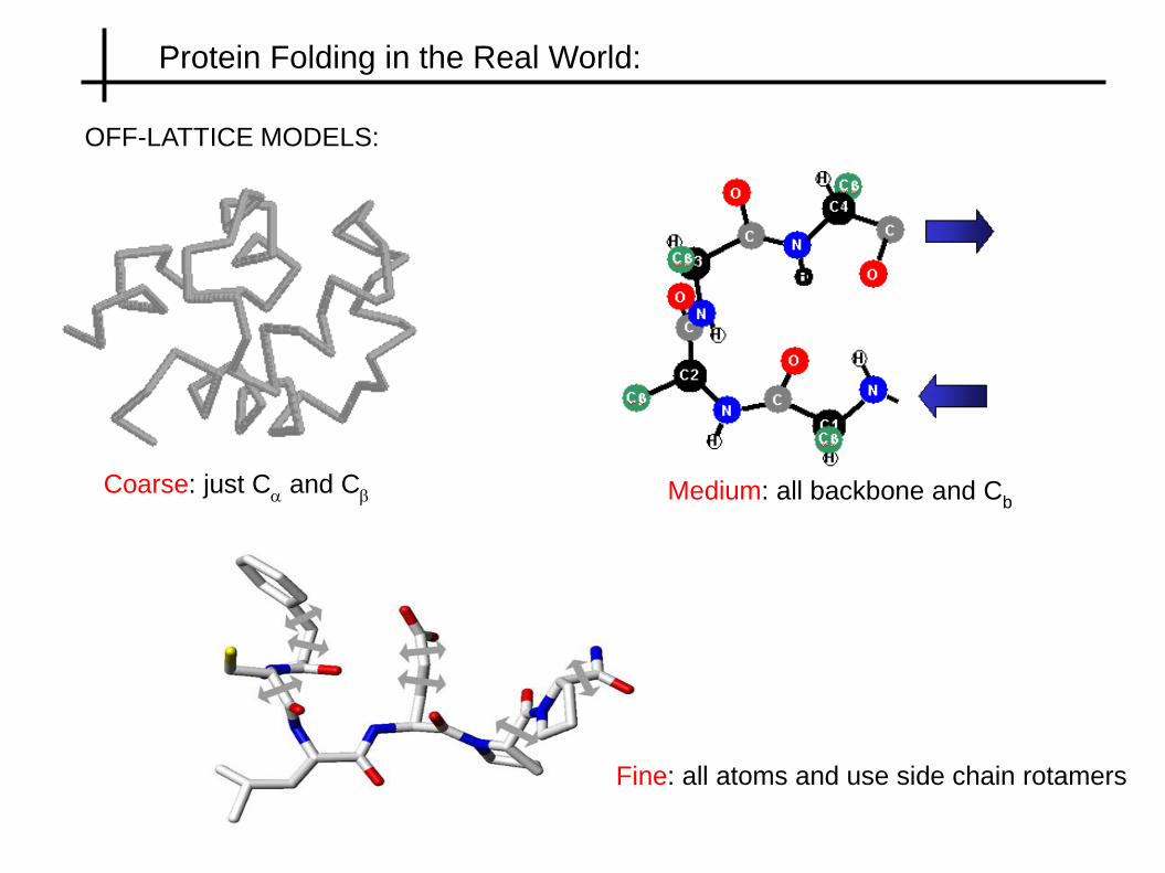
Protein Folding in the Real World:
OFF-LATTICE MODELS:
Coarse: just Ca and C
b Medium: all backbone and Cb
Fine: all atoms and use side chain rotamers

Structure Construction:
Enumerate structures:
●enumerate all structures that are possible using a finite # of (f,y) angles
●e.g. 4 pairs, L = 20 --> 420 = 1 x 10¹² structures!!!
Packing of secondary elements:
●pack together in 3D a fixed set of secondary structural elements
●can go to much larger structures
●must sample the space
Packing function

Protein Design:
Inverse folding problem: given a structure find a compatible sequence for which
the structure is the ground state fold
1) Improve natural folds:
give natural proteins new function, stability, kinetics
2) The search for novel folds: for L = 100 --> 10020 sequences !!!
There may be sequences that fold into structures not seen in nature
VLMQEGGFVLMS
MQEFTDGVMAA
AAVKRGTWWSR
EFVKLILAAIRST
Can we design any structure we want? NO, designability principle.
E(s)

Successful Designs
Redesigned Zinc Finger (Steve Mayo Lab)
Design of right-handed coiled coil (Harbury & Kim)
Binary patterning of helical bundle (Michael Hecht Lab)
Design of novel fold (David Baker Lab)