topological patterns in two-dimensional gel ... · topological patterns in two-dimensional gel...
TRANSCRIPT
Topological patterns in two-dimensional gelelectrophoresis of DNA knotsDavide Michielettoa,1, Davide Marenduzzob, and Enzo Orlandinic
aDepartment of Physics and Complexity Science, University of Warwick, Coventry CV4 7AL, United Kingdom; bSchool of Physics and Astronomy, University ofEdinburgh, Edinburgh EH9 3FD, United Kingdom; and cDipartimento di Fisica e Astronomia and Sezione, Istituto Nazionale di Fisica Nucleare, Universitá diPadova, 35131 Padova, Italy
Edited by Tom C. Lubensky, University of Pennsylvania, Philadelphia, PA, and approved August 14, 2015 (received for review April 9, 2015)
Gel electrophoresis is a powerful experimental method to probethe topology of DNA and other biopolymers. Although there is alarge body of experimental work that allows us to accuratelyseparate different topoisomers of a molecule, a full theoretical un-derstanding of these experiments has not yet been achieved. Herewe show that the mobility of DNA knots depends crucially andsubtly on the physical properties of the gel and, in particular, on thepresence of dangling ends. The topological interactions betweenthese and DNA molecules can be described in terms of an “entan-glement number” and yield a nonmonotonic mobility at moderatefields. Consequently, in 2D electrophoresis, gel bands display a char-acteristic arc pattern; this turns into a straight line when the densityof dangling ends vanishes. We also provide a novel framework toaccurately predict the shape of such arcs as a function of moleculelength and topological complexity, which may be used to informfuture experiments.
DNA knots | topology | gel electrophoresis
Topology plays a key role in the biophysics of DNA and isintimately related to its functioning. For instance, transcrip-
tion of a gene redistributes twist locally to create what is knownas supercoiling, whereas catenanes or knots can prevent cell di-vision; hence they need to be quickly and accurately removed byspecialized enzymes known as topoisomerases. How can oneestablish experimentally the topological state of a given DNAmolecule? By far the most successful and widely used techniqueto do so is gel electrophoresis (1, 2). This method exploits theempirical observation that the mobility of a charged DNA mol-ecule under an electric field depends on its size, shape, and to-pology (2). Gel electrophoresis is so reliable that it can be used,for instance, to map replication origins and stalled replicationforks (3), to separate plasmids with different amount of super-coiling (3, 4), and to identify DNA knots (5, 6). The most widelyused variant of this technique nowadays is 2D gel electropho-resis, where a DNA molecule is subjected to a sequence of twofields, applied along orthogonal directions (2). The two runs arecharacterized by different field strengths and sometimes also gelconcentrations (4); with suitable choices, the joint responses leadto increased sensitivity.Although gel electrophoresis is used very often, and is ex-
tremely well characterized empirically, there is still no compre-hensive theory to quantitatively understand, or predict, whatresults will be observed in a particular experiment. Some aspectsare reasonably well established. For instance, it is now widelyaccepted that the physics of the size-dependent migration oflinear polymers can be explained by the theory of biased polymerreptation (7–12). Likewise, the behavior of, for example, nicked,torsionally relaxed, DNA knots in a sparse gel and under a weakfield is analogous to that of molecules sedimenting under gravity(13–15). The terminal velocity can be estimated via a balancebetween the applied force and the frictional opposing force, whichis proportional to the average size of the molecule; as a result,more-complex knots, which are smaller, move faster under thefield. However, the mechanisms regulating the electrophoretic
mobility of DNA knots at intermediate fields, and in more-con-centrated agar gels, are much less understood (4, 13, 16). Here,experiments suggest that the mobility of knots is usually a non-monotonic function of the knot complexity, or, more precisely, oftheir average crossing number (5, 17) (ACN): Initially, knots movemore slowly as their ACN increases, whereas, past a critical ACN,more-complex knots move faster. The combination of the re-sponses to external fields directed along two perpendicular di-rections leads to a characteristic electrophoretic arc, which allowsseparation of the first simple knots more clearly in a 2D slab (4, 6,18, 19). To our knowledge, there is currently no theoreticalframework that quantitatively explains the nonmonotonic behav-ior at intermediate or large fields and the consequent formation ofarc patterns.To address this issue, here we present large-scale Brownian
dynamics simulations of knotted DNA chains migrating througha gel, and subjected to a sequence of fields of different strengthand direction, as in 2D gel electrophoresis experiments (seecartoon in Fig. 1A). We model the gel as an imperfect cubicmesh (20), where some of the bonds have been cut (seeMaterialsand Methods) to simulate the presence of open strands, ordangling ends, which have been observed in physical agarose gels(21–27). Our results confirm the linear relation of the electro-phoretic mobility with ACN for the first simple knots (we studyACN up to 12) in a sparse gel and under a weak field. However,our simulations also suggest that, due to a nonnegligible proba-bility of forming “impalements” where a dangling end of the gelpierces a knot, the response of the chain to stronger fields isdifferent. We suggest that, in this regime, the sole radius of gy-ration is not enough to explain the observed dynamics, and weintroduce an average “entanglement number” that increases withthe ACN and provides a measure of the likelihood of forming an
Significance
Gel electrophoresis is a ubiquitous biophysical technique. Itconsists of dragging charged biopolymers through a porousgel, by applying an electric field. Because the migration speeddepends on topology, this method can be used to classify DNAknots. Currently, electrophoresis relies on empirical observa-tions, and its theoretical understanding is limited. No theorycan explain why knot mobility under strong fields dependsnonmonotonically on complexity. Our study reveals a possiblereason: Although complex knots have a smaller size, and hencemove faster through the gel, they can become severely entangledwith the gel, causing longer pauses. Our results can improve thedesign of future electrophoresis experiments.
Author contributions: D. Michieletto, D. Marenduzzo, and E.O. designed research;D. Michieletto performed research; D. Michieletto analyzed data; and D. Michieletto,D. Marenduzzo, and E.O. wrote the paper.
The authors declare no conflict of interest.
This article is a PNAS Direct Submission.1To whom correspondence should be addressed. Email: [email protected].
This article contains supporting information online at www.pnas.org/lookup/suppl/doi:10.1073/pnas.1506907112/-/DCSupplemental.
www.pnas.org/cgi/doi/10.1073/pnas.1506907112 PNAS | Published online September 8, 2015 | E5471–E5477
BIOPH
YSICSAND
COMPU
TATIONALBIOLO
GY
APP
LIED
PHYS
ICAL
SCIENCE
SPN
ASPL
US

impalement. The time needed by a knot to disentangle from thegel increases with its average entanglement number (AEN), orknot complexity, and this slows down the motion, thus competingwith the Stokes friction, which leads to an increase of mobilitywith ACN. As a result of this contest, one typically gets a non-monotonic behavior of the terminal speed with ACN, and anelectrophoretic arc in two dimensions.We also provide a simple model, based on a mapping between the
DNA knot dynamics and a biased continuous-time random walk,which faithfully reproduces our Brownian dynamics simulationsstarting from a minimal number of assumptions. This approach canthen be used to predict how the shape of the electrophoretic arcshould depend on system parameters such as the average latticespacing (pore size) of the gel and the contour length of DNA knots;as we shall see, the predicted trends are in agreement with existing2D electrophoresis data. This constitutes, to our knowledge, the firstexample of a quantitative prediction of 2D electrophoretic diagrams;hence we suggest that the approach we present could potentiallylead to even more accurate and targeted experiments to separatetopoisomers in DNA or other polymers.
DNA Knots Form an Electrophoretic Arc only in IrregularGelsThe system we studied, sketched in Fig. 1 (see also Materials andMethods and Supporting Information), consists of 10 nicked, i.e.,torsionally relaxed, DNA loops of ∼ 3.7 kilobase pairs (kbp)within a model agarose gel with dangling ends. These loops areeither unknotted or form one of the first few simple knots [withup to nine crossing in their minimal projection (28)]. The loopswere first equilibrated within the gel (see Supporting Information)and then subjected to an in silico gel electrophoresis processwhere a weak electric field is first applied (K 50 V/cm) along thevertical (z) direction, followed by a stronger field (J 150 V/cm)
along a transversal, say y, direction. We refer to these two fields as“weak” and “stronger,” or “moderate,” in what follows. The com-plete equations of motions and force fields used in our Browniandynamics simulations are detailed in Supporting Information.By monitoring the trajectories of the knots through the gel, we
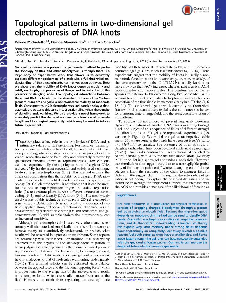
computed the average speed of their center of mass along eachof the field directions (see Supporting Information and Fig. 2 Aand B). As expected, the mobility along the direction of the weakfield increases with the topological complexity of the configu-rations. Along the direction of the moderate field, however, themobility of the knots displays a nonmonotonic behavior. Inparticular, the unknot now moves faster than either the trefoil orthe 41 twist knot, and has an average speed similar to the 51 knot.This nonmonotonic behavior of the knot mobility, as a functionof the ACN, was previously observed in typical experiments withtorsionally relaxed DNA knots (4, 6, 18, 19, 29). It is worth no-ticing that, within the five-crossings family, the 51 torus knotmoves more slowly than the 52 twist knots. This is similar to whatwas observed in the weak field case (although much less en-hanced) but different from what is observed in experiments ofsedimentation (15).To better compare our findings with experiments, we report,
in Fig. 2C, the spatial distribution of the knots as Gaussianscentered in [vzðKÞtz,vyðKÞty] where vzðKÞ and vyðKÞ are the ve-locities along z (weak field direction) and y (stronger field di-rection) of knot K, and ty and tz are the electrophoretic run times.The width of the Gaussians is set to be proportional to the SD ofthe velocities. The resulting spots can be seen as the in silicoanalog of the ones observed in gel electrophoretic experiments.Note that the combination of a monotonic behavior along theweak field direction with a nonmonotonic one along the strongerfield gives rise to the arc-shaped distribution of the spots char-acteristic of 2D electrophoresis experiments run either on knottedconfigurations or on supercoiled plasmids (4) (see Fig. 2D).It is interesting to ask whether one can observe the electro-
phoretic arc also in simulations where the gel is a regular cubicmesh, i.e., a mesh with no dangling ends, as this has been, so far, thetypical way to model an agarose gel (16). Remarkably, unlike thecase of gel with dangling ends, also called “irregular” hereafter, noexample of nonmonotonic behavior of the knot mobility is found forregular gels (for comparison, see Fig. 2C and Fig. S1). This result isin line with previous simulations based on lattice knots in regulargels (16) and persists for different field strengths (1.25− 600 V/cm)and gel pore sizes (200− 500 nm) (see Supporting Information). [Wenote that physical gels also have an inhomogeneous pore size; al-though considering this aspect will affect our results quantitatively,the common understanding is that the knot speed should dependmonotonically on size (1). This is qualitatively different from thecase of dangling ends, where the gel–polymer interactions stronglydepend on topological complexity as well.] Hence our simulationsstrongly suggest that the causes for the nonmonotonic behavior,observed in the case of irregular gels, are to be sought in the in-teraction between the knots and the gel dangling ends.This conjecture is also supported by the fact that linear (open)
DNA samples are frequently observed to migrate faster thancovalently closed (unknotted) ones in gel electrophoresis ex-periments performed in both strong and weak fields (4, 6, 18,29). This is in line with the outcome of a recent computer ex-periment probing the dynamics of linear and unknotted circularmolecules through an irregular gel (20).
DNA Samples Become Severely Entangled with the DanglingEndsHaving established that the presence of dangling ends in gelsseverely affect the transport properties of the knotted DNAloops under moderate electric fields, it is natural to look at thepossible mechanisms ruling this phenomenon.
B
A
Fig. 1. (A) Snapshot (to scale) of the model gel with some examples ofknotted configurations. Note that, to model a physical gel, a simple cubicstructure is randomly cut to create dangling ends. (B) Equilibrium configu-rations of some of the knots considered; it can be readily seen that the sizetends to be smaller as the knot becomes more complex. The knots picturedin A and B are trefoil (31), red; figure of eight (41), orange; pentafoil (51),yellow; Stevedore’s (61), green; and nonafoil (91), gray.
E5472 | www.pnas.org/cgi/doi/10.1073/pnas.1506907112 Michieletto et al.
The typical trajectories and average extension of some knottedloops as they move through a regular model gel and a gel withdangling ends are markedly different at moderate fields (see Fig.S1). In a regular gel, knots respond to the field, by shrinking theirsize so as to channel through the pores of the gel more effi-ciently. This mechanism, also known as “channeling,” in whichpolymers squeeze through the gel pores, has already been ob-served in previous works (30, 31), and it was previously con-jectured to play a role in the nonmonotonic separation of DNAknots in gels, as more-complex knots could have a differentability in deforming their overall shape when squeezing throughthe pores (4). On the other hand, as discussed in DNA KnotsForm an Electrophoretic Arc, we find that this behavior is notsufficient to explain the electrophoretic arc, as, for regular gels, wealways observe a monotonic separation of the knots as a function ofthe ACN (see Fig. S1). Conversely, in the case of irregular gels,knotted loops are much more prone to entangle with one (or more)dangling ends (see Insets of Fig. 2C for some examples). Theseentangled states (or impalements) require some time to be unrav-eled, and this is the reason for the anomalously long pauses ob-served in the knot trajectories (see, in particular, Fig. S1). Clearly, asthe DNA gets longer, impalements, which can either be parallel orperpendicular with respect the direction of the field, become pro-gressively more likely. As a matter of fact, this could be one of thereasons why it is, in practice, unfeasible to perform efficient gelelectrophoresis experiments with circular DNA longer than 10 kbp(32): At these sizes, impalements are so frequent that they maycause DNA breakage.In analogy with the phenomenon of threading, which slows
down the dynamics of unknotted loops either in a melt or in a gel(20, 33, 34), and that of “crawling” of knots around obstacles(35), it is reasonable to expect that more-complicated knots willtake longer to disentangle themselves from an impalement. We
argue that this mechanism, when competing with the reducedStokes drag of more-complex knots in gels, is ultimately responsiblefor producing a nonmonotonic dependence as a function of theircomplexity, i.e., their ACN.
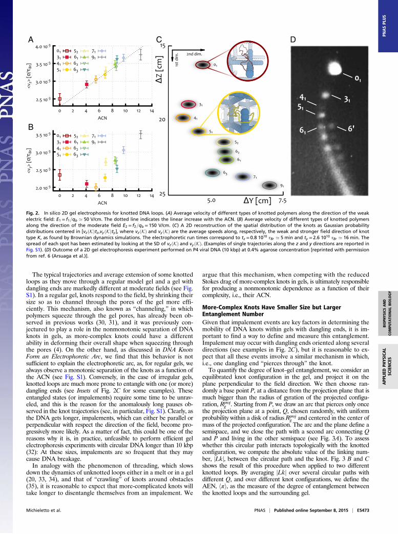
More-Complex Knots Have Smaller Size but LargerEntanglement NumberGiven that impalement events are key factors in determining themobility of DNA knots within gels with dangling ends, it is im-portant to find a way to define and measure this entanglement.Impalement may occur with dangling ends oriented along severaldirections (see examples in Fig. 2C), but it is reasonable to ex-pect that all these events involve a similar mechanism in which,i.e., one dangling end “pierces through” the knot.To quantify the degree of knot–gel entanglement, we consider an
equilibrated knot configuration in the gel, and project it on theplane perpendicular to the field direction. We then choose ran-domly a base point P, at a distance from the projection plane that ismuch bigger than the radius of gyration of the projected configu-ration, Rproj
g . Starting from P, we draw an arc that pierces only oncethe projection plane at a point, Q, chosen randomly, with uniformprobability within a disk of radius Rproj
g and centered in the center ofmass of the projected configuration. The arc and the plane define asemispace, and we close the path with a second arc connecting Qand P and living in the other semispace (see Fig. 3A). To assesswhether this circular path interacts topologically with the knottedconfiguration, we compute the absolute value of the linking num-ber, jLkj, between the circular path and the knot. Fig. 3 B and Cshows the result of this procedure when applied to two differentknotted loops. By averaging jLkj over several circular paths withdifferent Q, and over different knot configurations, we define theAEN, hπi, as the measure of the degree of entanglement betweenthe knotted loops and the surrounding gel.
A
B
C D
Fig. 2. In silico 2D gel electrophoresis for knotted DNA loops. (A) Average velocity of different types of knotted polymers along the direction of the weakelectric field: E1 = f1=qb ’ 50 V/cm. The dotted line indicates the linear increase with the ACN. (B) Average velocity of different types of knotted polymersalong the direction of the moderate field E2 = f2=qb = 150 V/cm. (C) A 2D reconstruction of the spatial distribution of the knots as Gaussian probabilitydistributions centered in [vzðKÞtz,vyðKÞty], where vzðKÞ and vy ðKÞ are the average speeds along, respectively, the weak and stronger field direction of knottype K, as found by Brownian dynamics simulations. The electrophoretic run times correspond to tz = 0.8 1010 τBr ’ 5 min and ty = 2.6 1010 τBr ’ 16 min. Thespread of each spot has been estimated by looking at the SD of vzðKÞ and vyðKÞ. (Examples of single trajectories along the z and y directions are reported inFig. S1). (D) Outcome of a 2D gel electrophoresis experiment performed on P4 viral DNA (10 kbp) at 0.4% agarose concentration [reprinted with permissionfrom ref. 6 (Arsuaga et al.)].
Michieletto et al. PNAS | Published online September 8, 2015 | E5473
BIOPH
YSICSAND
COMPU
TATIONALBIOLO
GY
APP
LIED
PHYS
ICAL
SCIENCE
SPN
ASPL
US
From Fig. 3D, we see that hπi grows approximately linearly withcan; as one would expect, more-complex knots can, on average,become more entangled with the surrounding irregular gel. It isinteresting to compare this behavior with that of the mean squaredradius of gyration normalized with respect to half the gel pore size(see Fig. 3D): Unlike the AEN, the average extension of the loop is,to a good approximation, inversely proportional to the ACN, i.e., tothe knot complexity. This corresponds to the well-known fact that,for a given loop contour length, more-complex knots are on averageless extended (5, 15) (see also the equilibrium configurationsin Fig. 1).The plots in Fig. 3D suggest a possible interpretation of the
nonmonotonic mobility of the knots in irregular gels based on theinterplay between the average size and the degree of entangle-ment with the gel. On one hand, more-complex knots, beingsmaller in size, experience less frequent collision with the gel andhence should travel more easily through it; this is just anothervariant of the Stokes friction argument discussed previously. Onthe other hand, once knot–gel collisions occur, more-complexknots experience a more intricate entanglement with the gel(higher values of AEN are more probable) that will take longerto unravel (5, 14, 16, 35).The above argument suggests the existence of two time scales in
the process: one is the time τf between two successive knot–gelcollisions yielding a local entanglement; the other, τdis, is the time
needed by the knotted loop to fully disentangle from the impale-ment. The time scale τf increases as the knot average size decreases,and hence increases with knot complexity (ACN). In other words,more-complex knots experience, on average, less collisions with thegel than their simpler counterpart. The second time scale, τdis, isinstead an increasing function of hπi (see Fig. 3D) and hence of theknot complexity (measured in terms of ACN). According to thispicture, the slowest topoisomer in an irregular gel with a givenlattice spacing will be the one with the best compromise between ahigh rate of collisions, and a sufficiently high value hπi.To investigate more quantitatively the dependence of τf and τdis
on the knot type (ACN), we analyze the trajectories of the knottedloops in the gel by computing (i) the average number of times aknot arrests its motion in the gel (entanglement event), hnei, and (ii)the distribution of the duration of these entanglement events.As specified in Supporting Information, the duration of the en-
tanglement events can be identified as the time intervals where thespatial position of the center of mass of the configuration deviatessignificantly from the expected collision-free field-driven linearmotion with speed vfree =Fz,y=Mζ= fz,y=ζ.As reported in Supporting Information (Fig. S2), the average
fraction of time in which the knot is trapped, τw=τtot (τtot is the timeof the full trajectory), is a nonmonotonic function of the ACN, inline with the result on the mobility under moderate field (Fig. 2B).In Fig. 3A, we show that the average number of entanglement
A
B
C
D
Fig. 3. (A) Sketch of the procedure used to define the piercing, or entanglement number: For a given projection of the configuration, the crossings define a setof regions whose intersections with the disk of radius Rproj
g are highlighted in blue. Starting from different points P far away from the projections, closed paths(two in the example) that pierce the disk once at different locationsQ are built at random. The absolute value of the linking number is then computed betweenthe knot configuration and each closed path. The average over the set of closed paths is finally taken: This is the AEN. B and C show the results of the proceduredescribed in A for a configuration with knot type 91 (C) and for an unknotted configuration (B). The regions are colored according to the computed absolutevalue of the linking number (see color map at left). Note that for the 91 case, there are regions of high jLkj(3), which are more prone to become entangled withthe dangling ends of the gel. (D) The AEN hπi and the mean squared radius of gyration divided by ðl=2Þ2 (l is the gel pore size) for different knot types classifiedin terms of ACN.
E5474 | www.pnas.org/cgi/doi/10.1073/pnas.1506907112 Michieletto et al.

events hnei decreases with the knot complexity (i.e., ACN). On theother hand, the distribution of the duration of these events displaysan intriguing bimodal shape, with two peaks occurring respectivelyat short and very long times (see Fig. S3). The peak at long timescan be interpreted as the signature of head-on collisions with thegel, where the dangling ends involved are opposite to the directionof the knot motion. The resulting entanglement is, in this case, verydifficult to unravel, especially in the presence of a strong electricfield (see, for instance, Inset of Fig. 2 for the unknot). Nonetheless,whether or not we exclude from the statistic the entanglementevents corresponding to the peak at long times, the characteristicdisentanglement time τdis turns out to increase (linearly) with theknot complexity, i.e., with ACN (see Fig. 4B).We assume that this bimodal shape is due to a shift in the energy
barrier that the knots have to overcome to disentangle from thedangling ends. In particular, one can think of this process as anArrhenius process, where the energy barrier is a function of thelength (projected along the field direction) of the dangling end, theknot complexity, and, more importantly, the magnitude of the ex-ternal field. When the external bias is too strong, disentanglement
events are very rare, and all knots will end up being permanentlyentangled with the gel structure; on the other hand, when it is tooweak, the typical disentanglement time is very short, and the de-pendence of τf as a function of the ACN dominates the motion ofthe polymers, reestablishing the usual linear relationship.
Random Walk Model with Topology-Dependent RatesCaptures the Observed Nonmonotonic BehaviorAs shown in DNA Knots Form an Electrophoretic Arc only in Ir-regular Gels, 2D electrophoresis experiments and Brownian dy-namics simulations of knotted loops in irregular gels are inqualitative agreement in many aspects. In this section, we pro-pose a simple model that reproduces the main findings of thesimulations and furnishes a simple but accurate way to predictthe arc shapes of the experimental patterns as a function both ofthe knot complexity and of the loop contour length.In this model, we describe the knotted loop moving within the
irregular gel as a biased random walk on a 1D lattice, i.e., a ran-dom walk that moves to the right (direction of the external field)unless it is trapped into an entangled state (due to impale-ment), with probability λeðKÞ= τ−1f ðKÞ (see Supporting Informationfor more details). Once in the entangled state, the walker has towait a given amount of time that is picked randomly from a bimodaldistribution consisting of an exponential decay, modeling the shorttime disentanglement, and a smaller probability peak at large times,describing the long disentanglement time from a head-on collision(see Supporting Information for the details). In this simple de-scription, the only relevant parameters are the hitting rate and theparameters characterizing the bimodal distribution of waiting (i.e.,disentanglement) times.Once the values of these parameters are set to reproduce the
data reported in Fig. 3 (see also Supporting Information), the modelcan be used to predict the mobility of the electrophoretic arc as afunction of ACN. As shown in Fig. 5, this procedure reproduceswith remarkably good agreement the simulation data, and, in par-ticular, it captures the physical mechanism leading to the non-monotonic mobility at moderate field. Note that, as the randomwalker solely moves to the right, the field strength enters into themodel only through the waiting times and the hitting rates.More importantly, once the parameter values of the biased ran-
dom walk model are set for a given pore size of the gel, l1, theirvalues for a different pore size, l2, can be estimated from generalarguments (see Supporting Information). We can therefore use thissimplified model to predict the moderate field mobility and theshape of the electrophoretic arcs of DNA knots in gels of variablepore size, e.g., tuned via agarose concentration (36) or nanowiregrowth cycle (37).
A
B
Fig. 4. (A) Average number of events in which the knot is entangledwith the surrounding gel (entanglement events) as a function of ACN.(B) Average disentanglement time as a function of ACN. In these esti-mates, only entanglement events with duration shorter than 200 τBr areconsidered.
A B C D
Fig. 5. (A) Average speed along the direction of the moderate field from Fig. 2B. The dashed line is obtained from the biased continuous random walkmodel, and corresponds to the (shifted and rescaled) red curve in B. (B) Average relative separation (in units of lattice spacing over time) of the knots as afunction of the ACN for different parameters, as predicted by the continuous random walk model. The gray dashed line in A is obtained by shifting the redcurve in B by the value of hvyið01Þ and rescaling it by the free velocity vfree. (C) Reconstruction of a 2D gel electrophoresis experiment from the data in B andzoom over the relative position of the family of six-crossings knots for two cases in which the minimum of the arc is at their left and their right. (D) Outcomesof a 2D gel electrophoresis experiments performed on P4 viral DNA with different lengths, respectively 4.7 (black) and 10 (white) kbp at equal agaroseconcentration (0.4%) [reprinted with permission from ref. 29 (Trigueros and Roca)].
Michieletto et al. PNAS | Published online September 8, 2015 | E5475
BIOPH
YSICSAND
COMPU
TATIONALBIOLO
GY
APP
LIED
PHYS
ICAL
SCIENCE
SPN
ASPL
US

The plots presented in Fig. 5B and Fig. S4 suggest that tightergels give rise to more curved (or deeper) arcs where the slowestknot has a higher ACN with respect to sparser gels. Moreover,because the entanglement rate λe and the disentanglement timeτdis (both of these relative to the same quantities for the unknot)should depend only on the ratio between the knot extension andthe gel pore size l, a similar trend should also be observed byincreasing the DNA loop contour length by keeping fixed l (seeSupporting Information). This is in qualitative agreement withexperiments, as electrophoretic arcs are straighter for shorterDNA molecules (Fig. 5D). A further quantitative prediction wecan draw from our arguments is that the relative position of thethree six-crossing knots can be controlled by tuning the pore size(Fig. 5) of the gel. Indeed, the size of the pores determineswhether the 61 Stevedore’s knot is to the left or to the right of theminimum of the mobility curve: In the former case, 61 will movesfaster in the gel than the 62 and 63 knots (which, having highercan, also have higher AEN), whereas, in the latter case, it willmove more slowly. This detailed prediction could be tested infuture electrophoresis experiments with knotted DNA loopsmoving within different gels.
ConclusionsWe have studied the role of topology in the gel electrophoreticmobility of DNA knots by means of Brownian dynamics simu-lations and a minimal model of biased random walk. We showedthat, when the knots are driven through a physical gel, i.e., onepossessing dangling ends, the knots’ mobility, as a function oftheir ACN, depends on the strength of the external field.At weak fields, we recover the well-known linear relationship
between migrating speed and knot type; at stronger fields, weobserve instead a nonmonotonic behavior. We argue that thispuzzling feature, routinely observed in experiments but not yetfully explained, can be better understood by taking into accountthe topological interactions, or entanglements, of the knots withthe irregularities of the surrounding gel. Although more-complexknots assume more-compact configurations, and hence smallerStokes friction than simpler knots, they also experience more-complex entanglements with the gel and, hence, longer disen-tanglement times. These two competing effects give rise to thenonmonotonic speed of the knots observed in the experiments, afeature that, remarkably, is absent for knotted loops moving in aregular gel (i.e., no dangling ends). Although most of our sim-ulations were performed with a rigid gel, we tested that the re-sults are qualitatively unchanged for gels with flexible danglingends (see Materials and Methods and Supporting Information andFig. S5).We also propose a model that describes the motion of knotted
DNA loops as a biased continuous-time random walk. This model,although minimal, by focusing on the competition between Stokesfriction and topological entanglements highlighted by the simu-lations, is able to predict the shape of electrophoretic patterns ofDNA knots of different contour length observed in gels withtunable physical properties. In particular, we predict that, bychanging the ratio between the radius of gyration of the unknotand the gel pore size, 2D gel electrophoresis experiments shouldlead to deep electrophoretic arcs for tight gels (or long knots),and shallow ones for sparse gels (or short knots).We hope that our results will prompt further experimental and
numerical verification of the role of topology in the anomalouselectrophoretic mobility of knotted polymers and, consequently,suggest new and more accurate setups to separate biopolymers ofdifferent topology. Lastly, we highlight that it may be possible tounderstand the patterns of DNA molecules with different den-sities of supercoiling within the presented framework as a similarcompetition between loop size and loop–gel interactions may beresponsible for their characteristic behavior.
Materials and MethodsDouble-stranded (ds) and nicked, i.e., torsionally relaxed, DNA (dsDNA) knotsare modeled as closed and knotted semiflexible bead-spring chains (38), withbeads of diameter σ = 2.5 nm, which reflects the thickness of hydrated B-DNAnear physiological conditions (39). The persistence length is set to lp = 20σ=50 nm, and the chosen contour length Lc = 512σ corresponds to DNA loops oflength ∼ 4 kbp ’ 1.3 μm.
The gel is modeled as an imperfect and rigid cubic mesh, with latticespacing l= 80σ ’ 200 nm compatible with the average pore size of agarosegels at 5% and artificial gels made of solid nanowires (36, 37) (for moredetails on the model and comparison with the case of flexible dangling ends,see Supporting Information). The irregularities, or dangling ends, of the gelare created by starting from a regular cubic mesh and then halving some ofthe edges randomly, with probability p= 0.4. Although this probability ischosen arbitrarily, it is possible to map it to a real value of disorder found inan agarose gel at a given concentration by comparing the mobility of linearand ring polymers running through it, similarly to what was done in ref. 20.The edges of the mesh are discretized with beads of size σg = 10σ ’ 25 nm,which is compatible with the observed diameter of agarose bundles (36, 40,41). For simplicity and computational efficiency, we model the gel as a staticmesh, meaning that the mesh structure is not deforming under eitherthermal of mechanical strains. This is an approximation for an agarose gel,whose bundles are generally, at the concentrations used in gel electropho-resis, found to be made of tens of fibers whose persistence length has beenobserved to be around 2− 10 nm (40). In light of this, a conservative esti-mation of the persistence length of an agarose bundle is comparable withthat of DNA, i.e., lp ’ 50 nm (this assumes weak attraction between the fi-bers; see Supporting Information). In this case, whose analysis is detailed inSupporting Information, we do not observe significant deviations from theresults presented here in DNA Knots Form an Electrophoretic Arc only inIrregular Gels. It is also worth noticing that this perfectly rigid environmentclosely resembles artificial gels made of solid nanowires (37), which possess amuch higher Young modulus and have been found to be optimal media forgel electrophoresis experiments.
The external field is modeled as a force f acting on each bead forming thepolymers. Assuming that, in physiological conditions, half of the chargesfrom the phosphate groups are screened by counter ions (42), one can thinkthat each bead (σ = 2.5 nm ’ 8 bp) contains a total charge of qb = 16qe=2,where qe is the electron charge. Within this assumption, we can map theexternal force applied onto each bead to an effective electric field E = f=qb.Although this mapping is a crude approximation of the Coulomb interactionbetween the charged DNA, the ions in solution, and the applied electricfield, we find that we can recover a weak field behavior of the knottedsamples, i.e., linear increase of the speed as a function of their ACN, up to∼ 50 V/cm, which is roughly comparable with the field intensity used in ex-periments. In this work, we used field intensities in the range from E = 1.25V/cm to E= 625 V/cm.
The ACNs used in this work were obtained from ref. 43, where the authorscomputed the ACN corresponding to Möbius energy minimizing knotted con-figurations. The thermally averaged ACN of the samples used in this work hasbeen computed from equilibrated configurations and has been found to be in aone-to-one correspondence to the values in ref. 43, confirming the linear re-lationship between the ACN of ideal and thermally agitated configurations (17).
The hydrodynamics is here considered only implicitly, as is customary forBrownian dynamics simulations. This means that the polymers do not feelone another via hydrodynamical interactions but are subject to thermalfluctuations due to a surrounding bath at fixed temperature T (see Sup-porting Information for more details).
The simulation time scale is given in terms of the Brownian time, whichcorresponds to the time taken by a bead of size σ to diffuse its own size, i.e.,τBr = σ2=Dσ, where Dσ = kBT=ξ= kBTð3πηsolσÞ−1 is the diffusion coefficient ofone bead and ηsol = 10 cP (centipoise) is the solution (water) viscosity. Fromthis, we obtain τBr = 3πηsolσ
3=kBT ’ 40 ns.
ACKNOWLEDGMENTS. D. Michieletto acknowledges support from the Com-plexity Science Doctoral Training Centre at the University of Warwick withfunding provided by the Engineering and Physical Sciences Research Council(EPSRC) (EP/E501311). The computing facilities were provided by the Centre forScientific Computing of the University of Warwick with support from theScience Research Investment Fund. D. Marenduzzo thanks EPSRC GrantEP/I034661/1 for support. E.O. acknowledges support from the Italian Ministryof Education Grant PRIN 2010HXAW77.
E5476 | www.pnas.org/cgi/doi/10.1073/pnas.1506907112 Michieletto et al.

1. Calladine CR, Drew H, Luisi FB, Travers AA (1997) Understanding DNA: The Moleculeand How It Works (Elsevier, New York).
2. Bates A, Maxwell A (2005) DNA Topology (Oxford Univ Press, New York).3. Olavarrieta L, et al. (2002) Supercoiling, knotting and replication fork reversal in
partially replicated plasmids. Nucleic Acids Res 30(3):656–666.4. Cebrián J, et al. (2014) Electrophoretic mobility of supercoiled, catenated and knotted
DNA molecules. Nucleic Acids Res 43(4):e24.5. Stasiak A, Katritch V, Bednar J, Michoud D, Dubochet J (1996) Electrophoretic mobility
of DNA knots. Nature 384(6605):122.6. Arsuaga J, et al. (2005) DNA knots reveal a chiral organization of DNA in phage
capsids. Proc Natl Acad Sci USA 102(26):9165–9169.7. de Gennes PG (1979) Scaling Concepts in Polymer Physics (Cornell Univ Press,
Ithaca, NY).8. Rubinstein M (1987) Discretized model of entangled-polymer dynamics. Phys Rev Lett
59(17):1946–1949.9. Duke TA (1989) Tube model of field-inversion electrophoresis. Phys Rev Lett 62(24):
2877–2880.10. Viovy JL, Duke T (1993) DNA electrophoresis in polymer solutions: Ogston sieving,
reptation and constraint release. Electrophoresis 14(4):322–329.11. Barkema GT, Marko JF, Widom B (1994) Electrophoresis of charged polymers: Simu-
lation and scaling in a lattice model of reptation. Phys Rev E Stat Phys Plasmas FluidsRelat Interdiscip Topics 49(6):5303–5309.
12. Viovy J (2000) Electrophoresis of DNA and other polyelectrolytes: Physical mecha-nisms. Rev Mod Phys 72(3):813–872.
13. Vologodskii AV, et al. (1998) Sedimentation and electrophoretic migration of DNAknots and catenanes. J Mol Biol 278(1):1–3.
14. Weber C, Carlen M, Dietler G, Rawdon EJ, Stasiak A (2013) Sedimentation of mac-roscopic rigid knots and its relation to gel electrophoretic mobility of DNA knots. SciRep 3:1091.
15. Piili J, Marenduzzo D, Kaski K, Linna RP (2013) Sedimentation of knotted polymers.Phys Rev E Stat Nonlin Soft Matter Phys 87(1):012728.
16. Weber C, Stasiak A, De Los Rios P, Dietler G (2006) Numerical simulation of gelelectrophoresis of DNA knots in weak and strong electric fields. Biophys J 90(9):3100–3105.
17. Katritch V, et al. (1996) Geometry and physics of knots. Nature 384:142–145.18. Trigueros S, Arsuaga J, Vazquez ME, Sumners DW, Roca J (2001) Novel display of
knotted DNA molecules by two-dimensional gel electrophoresis. Nucleic Acids Res29(13):E67.
19. Arsuaga J, Vázquez M, Trigueros S, Sumners D, Roca J (2002) Knotting probability ofDNA molecules confined in restricted volumes: DNA knotting in phage capsids. ProcNatl Acad Sci USA 99(8):5373–5377.
20. Michieletto D, Baiesi M, Orlandini E, Turner MS (2015) Rings in random environments:Sensing disorder through topology. Soft Matter 11(6):1100–1106.
21. Mickel S, Arena V, Jr, Bauer W (1977) Physical properties and gel electrophoresisbehavior of R12-derived plasmid DNAs. Nucleic Acids Res 4(5):1465–1482.
22. Levene SD, Zimm BH (1987) Separations of open-circular DNA using pulsed-fieldelectrophoresis. Proc Natl Acad Sci USA 84(12):4054–4057.
23. Turmel C, Brassard E, Slater GW, Noolandi J (1990) Molecular detrapping and bandnarrowing with high frequency modulation of pulsed field electrophoresis. NucleicAcids Res 18(3):569–575.
24. Åkerman B, Cole KD (2002) Electrophoretic capture of circular DNA in gels.Electrophoresis 23(16):2549–2561.
25. Cole KD, Åkerman B (2003) The influence of agarose concentration in gels on theelectrophoretic trapping of circular DNA. Sep Sci Technol 38(10):2121–2136.
26. Robertson RM, Smith DE (2007) Strong effects of molecular topology on diffusion ofentangled DNA molecules. Proc Natl Acad Sci USA 104(12):4824–4827.
27. Stellwagen NC, Stellwagen E (2009) Effect of the matrix on DNA electrophoreticmobility. J Chromatogr A 1216(10):1917–1929.
28. Adams CC (1994) The Knot Book: An Elementary Introduction to the MathematicalTheory of Knots (Freeman, New York).
29. Trigueros S, Roca J (2007) Production of highly knotted DNA by means of cosmidcircularization inside phage capsids. BMC Biotechnol 7:94.
30. Mohan A, Doyle PS (2007) Stochastic modeling and simulation of DNA electrophoreticseparation in a microfluidic obstacle array. Macromolecules 40(24):8794–8806.
31. Mohan A, Doyle PS (2007) Effect of disorder on DNA electrophoresis in a microfluidicarray of obstacles. Phys Rev E Stat Nonlin Soft Matter Phys 76(4 Pt 1):040903.
32. Dorfman KD (2010) DNA electrophoresis in microfabricated devices. Rev Mod Phys82(4):2903–2947.
33. Michieletto D, Marenduzzo D, Orlandini E, Alexander G, Turner M (2014) Threadingdynamics of ring polymers in a gel. ACS Macro Lett 3:255–259.
34. Michieletto D, Marenduzzo D, Orlandini E, Alexander GP, Turner MS (2014) Dynamicsof self-threading ring polymers in a gel. Soft Matter 10(32):5936–5944.
35. Weber C, De Los Rios P, Dietler G, Stasiak A (2006) Simulations of electrophoreticcollisions of DNA knots with gel obstacles. J Phys Condens Matter 18(14):S161–S171.
36. Pernodet N, Maaloum M, Tinland B (1997) Pore size of agarose gels by atomic forcemicroscopy. Electrophoresis 18(1):55–58.
37. Rahong S, et al. (2014) Ultrafast and wide range analysis of DNA molecules using rigidnetwork structure of solid nanowires. Sci Rep 4:5252.
38. Kremer K, Grest GS (1990) Dynamics of entangled linear polymer melts: A molecular-dynamics simulation. J Chem Phys 92(8):5057.
39. Rybenkov VV, Cozzarelli NR, Vologodskii AV (1993) Probability of DNA knotting and theeffective diameter of the DNA double helix. Proc Natl Acad Sci USA 90(11):5307–5311.
40. Guenet JM, Rochas C (2006) Agarose sols and gels revisited. Macromol Symp 242(1):65–70.
41. Sugiyama J, Rochas C, Turquois T, Taravel F, Chanzy H (1994) Direct imaging ofpolysaccharide aggregates in frozen aqueous dilute systems. Carbohydr Polym 23(4):261–264.
42. Maffeo C, et al. (2010) DNA–DNA interactions in tight supercoils are described by asmall effective charge density. Phys Rev Lett 105(15):158101.
43. Kusner R, Sullivan J (1994) Möbius energies for knots and links, surfaces and sub-manifolds. Geometric Topology: Proceedings of the 1993 Georgia InternationalTopology Conference, ed Kazez WH (Am Math Soc, Cambridge, MA), pp 570–604.
44. Mogilner A, Rubinstein B (2005) The physics of filopodial protrusion. Biophys J 89(2):782–795.
Michieletto et al. PNAS | Published online September 8, 2015 | E5477
BIOPH
YSICSAND
COMPU
TATIONALBIOLO
GY
APP
LIED
PHYS
ICAL
SCIENCE
SPN
ASPL
US