torpor and timing: impact of endogenously controlled ......contents chapter 1 introduction 1 chapter...
TRANSCRIPT
Institute of Zoology University of Veterinary Medicine Hannover Centre for Systems Neuroscience Hannover
in cooperation with the Laboratory for Neurobiology of Rhythms
University Louis Pasteur Strasbourg
Torpor and timing:
Impact of endogenously controlled hypothermia on the circadian system of two hamster species.
Thesis Submitted in Partial Fulfilment of the requirements for the degree
Dr. rer. nat.
at the University of Veterinary Medicine Hannover
by
Annika Herwig
Kiel, Germany
Hannover, Germany 2007

1. Supervisor: Prof. Stephan Steinlechner, University of Veterinary Medicine Hannover
2. Supervisor: Dr. Michel Saboureau, University Louis Pasteur, Strasbourg
Advisory Committee:
Prof. Wolfgang Löscher, University of Veterinary Medicine Hannover
Prof. Alexandru Stan, Hannover Medical School
Dr. Paul Pévet, University Louis Pasteur, Strasbourg
Prof. François Lasbennes, University Louis Pasteur, Strasbourg
First Evaluation :
Prof. Stephan Steinlechner, University of Veterinary Medicine Hannover
Dr. Michel Saboureau, University Louis Pasteur, Strasbourg
Prof. Wolfgang Löscher, University of Veterinary Medicine Hannover
Prof. Alexandru Stan, Hannover Medical School
Dr. Paul Pévet, University Louis Pasteur, Strasbourg
Prof. François Lasbennes, University Louis Pasteur, Strasbourg
Second Evaluation:
Prof. Franziska Wollnik, University of Stuttgart
Date of oral examination: 26.04.2007
Ms Annika Herwig was a member of the European Doctoral College of the Universities of
Strasbourg during the preparation of his/her PhD, from 2004 to 2007, class name Périclès.
She has benefited from specific financial supports offered by the College and, along with
his/her mainstream research, has followed a special course on topics of general European
interests presented by international experts. This PhD research project has been led with the
collaboration of two universities: the University of Veterinary Medicine Hannover and the
University Louis Pasteur in Strasbourg.

�Van Burd dött ik ober doch ne gohn, ne?�
�Och du Dösbattel, kannst du ok van Burd gohn?
Büst doch up See, is doch all Woter üm di rüm.�
Gorch Fock „Seefahrt ist not!“
Contents
Chapter 1 Introduction 1
Chapter 2 Daily torpor alters multiple gene expression in 13
suprachiasmatic nuclei and pineal gland of the
Djungarian hamster (Phodopus sungorus)
Chapter 3 Daily torpor affects the molecular machinery of the 21
circadian clock in Djungarian hamsters (Phodopus sungorus)
Chapter 4 Trans pineal microdialysis in the Djungarian hamster 39
(Phodopus sungorus): a tool to study seasonal changes of
circadian clock activities
Chapter 5 In vivo melatonin measurement during torpor in the 53
Djungarian hamster (Phodopus sungorus)
Chapter 6 The circadian clock stops ticking during hibernation 65
Chapter 7 Histamine H3 receptor and Orexin A expression during 77
daily torpor in the Djungarian hamster (Phodopus sungorus)
Chapter 8 Discussion 91
References 101
Summary 119
Zusammenfassung 121
Résumé 123
Acknowledgements 125
Publications 128

Abbreviations
AA-NAT Arylalkylamine-N-acetyltransferase
ARC Arcuate nucleus
AVP Arginine-vasopressin
CLOCK Circadian locomotor output cycles kaput
CRY Cryptochrome
CNS Central nervous system
HP Hibernation protein
DD Constant darkness
DLG Dorsal lateral geniculate
DMH Dorso-medial hypothalamus
H1-4R Histamine 1-4 receptor
LD Long day
L:D Light:dark
LP Long photoperiod
MR Metabolic rate
NA Noradrenaline
OX1R Orexin receptor 1
OX2R Orexin receptor 2
PER Period
REV-ERBα Orphan nuclear receptor
RORα Retinoid-orphan receptor
SCN Suprachiasmatic nuclei
Ta Ambient temperature
Tb Body temperature
Τ Endogenous period
TM Tuberomammilary nucleus
VLPO ventrolateral preoptic area
ZT Zeitgeber-time

1
1 Introduction

Chapter 1
2
Introduction
Our earth rotates around itself once a day and around the sun once a year, which is why most
organisms face periodic changes of their environment. The alteration of day and night is the
most important Zeitgeber in our lives and build the framework for all doings. Activity and
resting, food and water intake are only the most obvious behaviours that are timed. However,
also most physiological processes like body temperature, hormone release, moods and
emotions underlie rhythmicity triggered by the light dark cycle (FOSTER and KREITZMAN
2004). The ratio of day and night length changes throughout the year and with it the
conditions of living. Humans mostly elude those seasonal variations by electric light, central
heating and always available food stocks, but animals have evolved different strategies to live
with and adapt to varying conditions of light, temperature, food supply or predator abundance.
Seasonal influences are extremely marked at high latitudes, where changes in external
conditions are most drastic. Some species, especially birds, migrate over long distances to
exploit local resources as best as possible (GWINNER 1996). Mammals often anticipate
profound environmental changes throughout the year facilitating survival in winter when
energy supply is low, but metabolic costs are high. Generally reproduction and growth are
shut down in less inviting seasons. Reproduction is strictly bound to times of the year when
resources cover the demanding energy costs of pregnancy and lactation and warrant survival
of the offspring. Some species increase their body weight during summer in order to live on
fat reserves in winter, until food availability improves again in spring. Others, especially
small mammals that can only store a low amount of fat relative to their body mass, even
decrease their body weight towards winter � lower body mass costs less energy. Many
mammals like sheep, deer, rabbits or bears change their fur to a better insulating coat to
decrease heat loss. For endotherms, thermoregulation is the highest expenditure in cold
seasons. The rate of heat production is proportionally enhanced to the rate of heat loss to
maintain body temperature (Tb) at the desired level allowing maximal physical performance.
Energy costs for endotherms are eight times higher than for ectotherms of comparable body
size and ambient temperature (Ta) (ELSE and HULBERT 1981) and cannot always be
afforded.

Chapter 1
3
Torpor To lower metabolic costs, many species evolved endogenously controlled hypothermia which
is probably the most powerful tool for saving energy in harsh seasons. Those heterotherms
appear to use mainly two different forms of torpor to adapt to their environment and their
physiological needs as best as possible: hibernation or prolonged torpor in �true� hibernators
and daily torpor in the daily heterotherms (Fig. 1). Deep hibernators like e.g. European
hamsters (Cricetus cricetus), European ground squirrels (Spermophilus citellus) or Syrian
hamsters (Mesocricetus auratus) show a controlled decrease in Tb to values approaching
ambient temperature (Ta) during the hibernation season which can last 6-7 months. Periods of
deep torpor are interrupted by regular arousals and brief 1-2 day periods of euthermia every 5
to 20 days (KÖRTNER and GEISER 2000). Mostly, the preferred Tb during deep hibernation
ranges between 1 and 6 °C, but some species like the Arctic ground squirrel can even tolerate
supercooling at -3 °C (BARNES 1989). Hibernating mammals reduce their metabolic costs by
around 90% (HELDMAIER et al. 2004) which allows survival entirely on body fat that has
been stored prior to winter, making this economizing strategy most effective. Thus, energy
intake and expenditure in hibernators are not balanced on a daily, but rather a yearly basis.
Smaller animals like the Djungarian hamster (Phodopus sungorus), various bat species,
hummingbirds and also lemurs predominantly undergo bouts of daily torpor (GEISER 2004,
DAUSMANN 2004). During this relatively shallow form of torpor they decrease Tb
depending on Ta to barely lower than 15 °C. Generally, daily torpor is restricted to the
circadian resting phase of the animal, hence not exceeding a duration of some hours (GEISER
2004, HELDMAIER et al. 2004). During a bout of daily torpor the extent of hypothermia and
hypometabolism, i.e. energy saving, is usually less pronounced than in deep hibernation.
Nevertheless, a 60-70% reduction in energy requirements is reached, also because feeding-
related activities are strongly reduced in torpid animals (RUF et al. 1991, 1993, SCHMID et
al. 2000). The advantage of daily torpor compared to deep hibernation is that territorial and
social activities can be maintained during the torpor season. Generally, daily torpor is less
strictly seasonal in most species and therefore can also be used as a response to acute energy
shortage.
It has been questioned whether different forms of hypothermia underlie different mechanisms,
but so far physiological properties have turned out to be very similar (HELDMAIER et al.
2004).

Chapter 1
4
Figure 1: Examples of daily torpor (upper panel) and hibernation (lower panel) over 20 days in
midwinter. The minimum Tb achieved during a daily torpor bout of this Djungarian hamster, which was
kept at 15 °C Ta, does not drop below 20 °C. Note that daily torpor is not expressed every day. During
deep hibernation preferred Tb of golden-mantled ground squirrels generally lies at around 6 °C at 5 °C
Ta. Regular arousals occur every 7-8 days in this example (modified from RUBY et al. 2003).
First, metabolic rate is reduced, followed by a drop in Tb which can be precisely regulated and
maintained at low degrees, before rapidly increasing again to the normometabolic and
normothermic state (GEISER and HELDMAIER 1995, LOVEGROVE et al. 1999,

Chapter 1
5
ORTMANN and HELDMAIER 2000). At present, the different classifications have been
considered to simply represent gradual differences in timing, duration and amplitude of an
analogue physiological state (HELDMAIER et al. 2004).
Cooling down poses several biochemical problems. Humans cannot even tolerate a decrease
in Tb of more than 10 °C (VAN BREUKELEN and MARTIN 2002 a,b) before cardiac arrest
and already at a Tb of 34 °C a state of profound exhaustion is reached in which consciousness
is severely reduced. Hibernators are capable of attaining, withstanding and reversing low Tbs,
so obviously something in their physiology must be different. It has been discussed whether
those abilities to reduce rates of biochemical processes are based on specific adaptations and
activation of important metabolic pathways, or whether they are undirected simple
temperature effects the animal passively needs to tolerate (VAN BREUKELEN and MARTIN
2002b). Regular arousals between torpor bouts would then serve to recover from the �freeze�.
Certain is that the animal takes advantage of a low Tb in torpor for a profound reduction in
biochemical reactions that serve to maintain balance in extremely hypoxic and nutrient
deficient periods. Translational processes slow down dramatically during torpor, protein
synthesis is affected at the levels of initiation and elongation (KNIGHT et al. 2000, VAN
BREUKELEN and MARTIN 2001). Also initiation as well as elongation of transcription is
temperature sensitive as expected from Q10 effects and ceases during deep hibernation in
golden-mantled ground squirrels (BOCHAROVA et al. 1992, VAN BREUKELEN and
MARTIN 2002a). Thus, generally central nervous system (CNS) activity is reduced.
Nevertheless, it is crucial that functional integrity is maintained. The hibernating brain needs
to accurately control the complex metabolic processes and to remain sensitive to external and
internal stimuli. Continued CNS function could be demonstrated in Arctic ground squirrels,
where metabolic rate was increased when Ta fell below 0 °C indicating that the CNS actively
triggers heat production to maintain a constant Tb (BUCK and BARNES 2000).
It seems that specific adaptations keep the cellular and molecular machinery functional and
resistant to damage and ensure correct and fast reactivation during interbout arousal.
Although transcription is inhibited during torpor (BOCHAROVA et al. 1992), there is no
evidence for mRNA loss during hypothermia (FRERICHS et al. 1998, O´HARA et al. 1999).
Therefore, it follows that there must be an extension of mRNA half life. Indeed, Poly (A) tail
lengths of liver transcripts are not reduced and there is an association of mRNA with a
Poly (A) binding protein in torpor. This might prevent transcripts from being degraded and

Chapter 1
6
facilitate translation after rewarming (KNIGHT et al. 2000). Special nuclear bodies (RNPs)
have been described in torpid, but not euthermic hibernators (MALATESTA et al. 1994,
1999, 2001, TAMBURINI et al. 1996). They may store transcripts and various splicing
factors that then could be rapidly processed for expression during arousal.
Finally, there exists evidence that special brain systems abide full activity during torpor and
do not follow expected Q10 effects. The subject of this thesis is to describe systems that are
supposed to remain functional in the physiological state of endogenously controlled
hypothermia better.
The circadian system
One of the systems believed to be active during torpor is the circadian system. On the one
hand, this system translates seasonal information to the organism, enabling the animal to
anticipate environmental changes well in advance. On the other hand, it times daily events,
thus controlling and interlocking various internal physiological processes on a circadian basis,
which might be of benefit for animals with rare external time information during the torpor
season. The circadian clock is situated in the suprachiasmatic nuclei (SCN) of the
hypothalamus and oscillates endogenously in a ~24 hours rhythm (MOORE and EICHLER
1972, STEPHAN and ZUCKER 1972). This endogenous rhythm (τ) that is expressed in
constant conditions without any external Zeitgebers (light-dark or temperature cycles) differs
between individuals, but is under normal circumstances precisely entrained to the light-dark
cycle.
According to current knowledge, the molecular basis of SCN´s oscillation are feedback loops
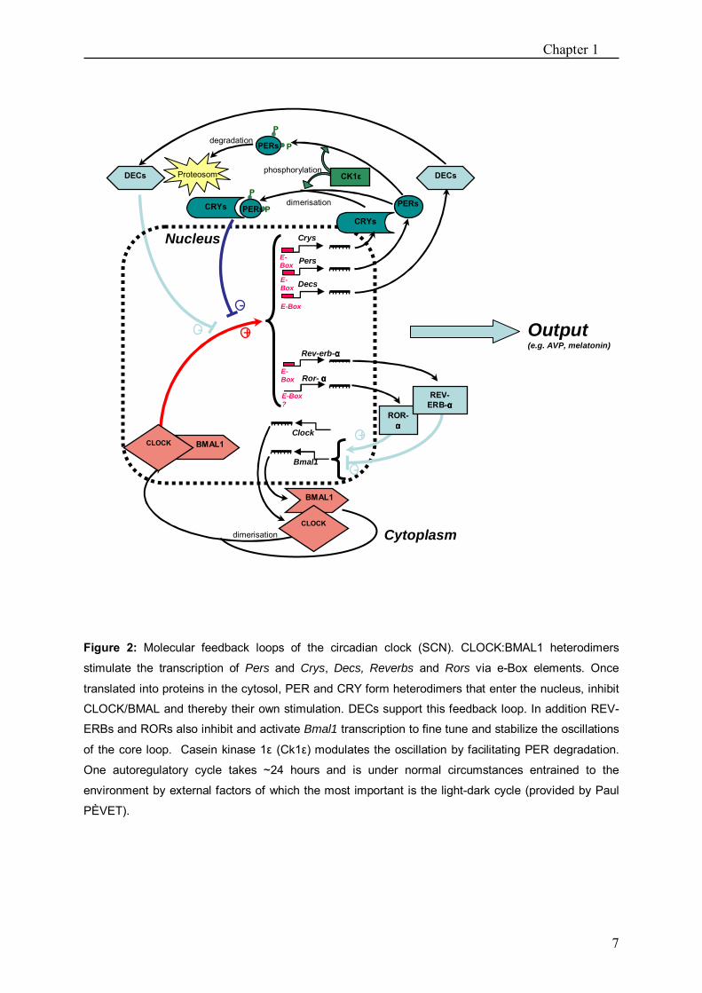
of so-called clock genes which are expressed rhythmically (Fig. 2). Circadian rhythm
generation is based on the transcriptional regulation of two sets of genes, Pers (Per1, Per2,
Per3) and Crys (Cry1, Cry2), by an autoregulatory feedback loop. Transcription of these
genes is driven by CLOCK:BMAL1 protein heterodimers acting on E-box sequences in their
promoters. Per and Cry mRNA are built up and followed by an increase of cytoplasmatic
concentrations of PER and CRY proteins.
Chapter 1
7
Figure 2: Molecular feedback loops of the circadian clock (SCN). CLOCK:BMAL1 heterodimers
stimulate the transcription of Pers and Crys, Decs, Reverbs and Rors via e-Box elements. Once
translated into proteins in the cytosol, PER and CRY form heterodimers that enter the nucleus, inhibit
CLOCK/BMAL and thereby their own stimulation. DECs support this feedback loop. In addition REV-
ERBs and RORs also inhibit and activate Bmal1 transcription to fine tune and stabilize the oscillations
of the core loop. Casein kinase 1ε (Ck1ε) modulates the oscillation by facilitating PER degradation.
One autoregulatory cycle takes ~24 hours and is under normal circumstances entrained to the
environment by external factors of which the most important is the light-dark cycle (provided by Paul
PÈVET).
Pers
Crys
Decs
PERs
CRYs
DECs
Rev-erb-αααα
Bmal1
Clock
E-Box
Nucleus
Cytoplasm
BMAL1 CLOCK
-
E-Box
Ror- ααααE-Box
-
+
dimerisation
+
E-Box ?
E-Box
dimerisation
P
PPERs
degradation
DECs
-
phosphorylation
P PERs
P
Proteosom
ROR-αααα
REV-ERB-αααα
Output(e.g. AVP, melatonin)
CRYs
BMAL1
CLOCK
CK1ε

Chapter 1
8
When their concentration in the cytoplasm is high enough, these proteins again form
heterodimers which then are likely to enter the nucleus. Here the PER:CRY dimers bind to
CLOCK:BMAL1 and thereby prevent its stimulatory action on their own transcription. Other
factors like Casein Kinase 1ε, REV-ERB α and ROR α are thought to fine tune the oscillation
by either facilitating PER degradation or acting on Bmal1 expression. Entrainment to the
light/dark cycle is arranged through receptors within the retina and their action on Per1 and
Per2 transcription which is rapidly induced by light (FOSTER and KREITZMANN 2004).
Finally, the molecular circadian machinery forwards the �time� to the organism via different
outputs of which the rhythmic melatonin secretion from the pineal gland is the best
characterised. Melatonin release is exclusively under SCN control which restricts production
of melatonin to night-time by controlling the activity of its rhythm-generating enzyme,
arylalkylamine-N-acetyltransferase (Aa-nat). Hence, melatonin secretion precisely reflects the
day-night ratio. Its most important function is to provide the organism with photoperiodic
information and it has been demonstrated to be crucial for seasonal adaptations
(STEINLECHNER et al. 1987, PÉVET 1988, BARTNESS 1989, STEINLECHNER 1992,
STEINLECHNER and NIKLOWITZ 2002). Another clock output is considered to be
arginine-vasopressin (AVP). This neuropeptide is expressed rhythmically in the SCN, peaking
during the light phase of the circadian cycle (VAN ESSEVELDT et al. 2000). Its precise role
in rhythm generation is not entirely clarified, but it appears that the amplitude in AVP
rhythms accounts for the amplitude of behavioural rhythms (VAN ESSEVELDT et al. 2000).
One of the particularities of circadian clocks is that they are strongly temperature
compensated. Free running circadian rhythms that are the endogenous rhythms of individual
organisms without external Zeitgebers, show Q10 values that lie close to 1 in all investigated
species (SWEENEY and HASTINGS 1960, PITTENDRIGH 1954, MENAKER 1954,
JOHNSON et al. 2004, BELL-PEDERSEN et al. 2005). This makes sense especially for
ectotherms. A temperature-sensitive process would be useless as a clock because it would run
faster on a warm day than on a cold day. Also endotherms may display large Tb changes and
for those hetreotherms as well time-keeping is crucial.
Photoperiodically induced daily torpor is an event that is precisely timed relative to the resting
phase of the animal. This timing makes it possible that torpor does not interfere with foraging
so that a positive energetic balance can be maintained. Hence, it seems likely that the

Chapter 1
9
circadian system is not disturbed by Tbs of ~15 °C as reached during this hypothermia in the
Djungarian hamster and is still capable of keeping time.
Also in deep hibernation a functional circadian clock appears useful for synchronising
endogenous physiological functions and for timing the regularly occurring interbout arousals.
This would even mean that the circadian clock tolerates temperatures below 10 °C.
Studies that addressed the question of a functional circadian clock during daily torpor or
hibernation, though, have been controversial (FRENCH 1977, FLORANT et al. 1984,
VANECEK et al. 1984, 1985, STANTON et al. 1986, KILDUFF et al. 1989, GRAHN et al.
1994, WOLLNIK and SCHMIDT 1995, KÖRTNER and GEISER 2000, RUBY et al. 2002,
LARKIN et al. 2002, HUT et al. 2002, RUBY 2003, HELLER and RUBY 2004).
In chapter 2, 3 and 6 we investigated for the first time the effect of the two different forms of
endogenously controlled hypothermia, daily torpor in Djungarian hamsters (Chapter 2, 3) and
deep hibernation in European hamsters (Chapter 6), on the very core of the clock, the
molecular feedback loops generating rhythms in the SCN. We set up experiments to
investigate rhythmic clock and clock related gene expression in normo- and hypothermic
animals and tried to reveal whether alterations in rhythm amplitude might be directly
temperature dependent. Moreover, to better understand how daily torpor affects the phase of
the circadian clock in Djungarian hamsters, we set up trans pineal microdialysis to directly
measure melatonin release as a phase marker in vivo (Chapter 4, 5).
The histaminergic system Another system that has already been shown to be upregulated and which plays an important
role in deep hibernation is the histaminergic system. The neurotransmitter histamine plays a
prominent role in various neurophysiological processes such as in the regulation of
wakefulness and thermoregulation in nonhibernators (SCHWARTZ et al. 1991). Already in
early studies, it was suggested that histamine acts as a �waking substance� (MONNIER et al.
1970) and hitherto many studies indicate that histamine is required for arousal. Lesioning or
inactivation of the posterior hypothalamus has been shown to cause hypersomnia (LIN et al.
1989). Mice lacking histamine show a deficit in waking, attention and interest in a new
environment (PARMENTIER et al. 2002). Histaminergic neurons fire at a rate dependent on
the behavioural state. Their activity is high during waking and attention and low or absent

Chapter 1
10
during sleep, inhibited by GABAergic neurons from the ventrolateral preoptic area (VLPO)
(STEVENS et al. 2001).
In mammals, histamine synthesising neurons are exclusively found in the tuberomammillary
nucleus (TM) and project to almost all parts of the brain (INAGAKI et al. 1988, PANULA et
al. 1989) where 3 of the 4 cloned histamine receptors (H1-H3) mediate its action. The H1 and
H2 receptors mostly excite neurons or potentiate excitatory inputs. By contrast, H3 receptor
activation causes autoinhibition of TM neurons and inhibition of neurotransmitter release. H4
receptors are predominantly detected in the periphery (NGUYEN et al. 2001). In addition to
histamine, the TM neurons co-express several other neurotransmitters and modulators of
which γ-aminobutyric acid (GABA) is probably the most prominent (ERICSON et al. 1991).
This is likely to be of functional relevance because TM neurons constitute an extensive far-
reaching system that could mediate general inhibition throughout the CNS. General inhibition
of CNS activity has also been reported during hibernation (HELLER 1979) and these findings
have drawn interest to the histaminergic system in hibernation research. In golden-mantled
ground squirrels (Citellus lateralis) the histaminergic projections have been demonstrated to
be more extensive than in non-hibernators, such as the rat (SALLMEN et al. 1999) which
could be interpreted as indication of an enhanced importance of histamine in the CNS of
hibernators. Indeed, the rate of histamine turnover is high during hibernation; moreover,
histamine infused into the hippocampus prolongs the hibernation bout implying that histamine
might be involved in the maintenance of the hibernating state and inhibits the arousal
(SALLMEN et al. 1999, 2003b). It could be demonstrated that the histaminergic system
blocks the arousal from hibernation via inhibitory H3 receptors which are up-regulated in
cortex and basal ganglia in hibernating ground squirrels (SALLMEN et al. 1999, 2003a).
In Chapter 7 we investigated the expression of the H3 receptor during daily torpor in
Djungarian hamsters. If torpor indeed underlies mechanisms comparable to deep hibernation
we may expect a resembling, but less pronounced up-regulation in Djungarian hamsters
compared to deep hibernating golden-mantled ground squirrels.

Chapter 1
11
Orexins Moreover, we explored in chapter 7 whether reactivation of the other systems could be
mediated by orexins. Orexins are a group of neuropeptides in the lateral hypothalamus that
have been more and more linked to the arousal state of the animal and probably directly
activate the histaminergic system (RODGERS et al. 2001, ISHII et al. 2005). Orexin positive
neurons are found in the dorsal, perifornical, posterior and lateral hypothalamic areas and
project throughout the hypothalamus including paraventricular, arcuate, lateral, perifornical
and ventromedial areas as well as to several extrahypothalamic sites like the septal nuclei, the
bed nucleus of the stria terminalis, the paraventricular and reunions nuclei of the thalamus, the
zona incerta, the subthalamic nucleus, the central grey, the substantia nigra, the dorsal raphe
nuclei, the parabranchial nucleus, the medullary reticular formation, the area postrema and the
nucleus of the solitary tract (PEYRON et al. 1998). Two G-protein-coupled receptors (OX1R,
OX2R) have been identified and are distributed widely throughout the brain (TRIVEDI et al.
1998). Binding studies indicate that OX1R is selective to orexin A, whereas OX2R binds with
similar activity to both orexins (SAKURAI et al. 1998). OX2R appears to be more strongly
associated with arousal, as it is the lack of this receptor that causes narcolepsy in dogs. The
arousal effect depends on activation of the histaminergic neurons in the TM where solely
OX2 receptors are expressed (ERIKSSON et al. 2001, HUANG et al. 2001, ERIKSSON et al.
2004). Moreover, orexin A injection into several brain areas has been shown to provoke
enhanced thermogenesis and energy expenditure due to activation of sympathetic nervous
system (GEERLING et al. 2003, KOTZ 2006). Therefore, we hypothesised that orexins might
be a good candidate for provoking arousal from hypothermia and tested orexin A expression
in the lateral hypothalamus in the course of a torpor bout in chapter 7.
All in all, this thesis aims at providing insight into brain systems that are likely to be
important for timing, maintaining and reversing endogenously controlled hypothermia.

12

13
2
Daily torpor alters multiple gene expression in
suprachiasmatic nuclei and pineal gland of the
Djungarian hamster (Phodopus sungorus).
Annika Herwig1,2, Florent Revel2, Michel Saboureau2, Paul Pévet2,
Stephan Steinlechner1
1 Institute of Zoology, University of Veterinary Medicine, Bünteweg 17, D-30559 Hannover, Germany 2 Département de Neurobiologie des Rythmes, Institut des Neurosciences Cellulaires et Intégratives,
UMR-7168/LC2, CNRS - Université Louis Pasteur, 5 rue Blaise Pascal, 67084 Strasbourg Cedex,
France
Published in Chronobiology International (2006) 23:269-76.

Chapter 2
14
Abstract
Circadian rhythms are still expressed in animals that display daily torpor, implying a
temperature compensation of the pacemaker. Nevertheless, it remains unclear how the clock
works in hypothermic states and whether torpor itself as a temperature pulse affects the
circadian system. In order to reveal changes in the clockwork during torpor, we compared
clock gene and neuropeptide expression by in situ hybridisation in the SCN and pineal gland
of normothermic and torpid Djungarian hamsters (Phodopus sungorus). Animals from LD
8:16 were sacrificed at 8 time points throughout 24 hours. To investigate the effect of a
previous torpor episode on the clock, we killed a group of normothermic hamsters one day
after torpor. In normothermic animals Per1 peaked at ZT4, whereas Bmal1 reached maximal
expression between ZT16 and ZT19. Avp mRNA in the SCN showed highest levels at ZT7.
On the day of torpor only, Avp mRNA contents did not differ from the control group, showing
increased mRNA levels during the torpor bout. Moreover, the Bmal1 rhythm was advanced.
On the day after the hypothermia Bmal1 and Avp rhythms showed a severely depressed
amplitude. Those distinct amplitude changes in Bmal1 and Avp on the day after a torpor
episode expression might give a hint that torpor affects the circadian system, probably by
altered translational processes that might lead to a modified protein feedback on gene
expression.
In the pineal gland, an important clock output, Aa-nat expression peaked between ZT16 and
ZT22 in normothermic animals. Aa-nat levels were significantly advanced on the day of
hypothermia, an effect which was still visible one day afterwards.
In this study we showed that daily torpor affects the phase and amplitude of rhythmic clock
gene and clock controlled gene expression in the SCN. Furthermore, the rhythmic gene
expression in a peripheral oscillator, the pineal gland, is also affected.
Introduction
One of the particularities of circadian clocks is the strong temperature compensation that has
been found in all investigated species from flagellates to mammals. Circadian rhythms show a
Chapter 2
15
Q10 close to 1 which has been shown for both homeotherms and heterotherms (PITTENDRIGH
1954, SWEENEY and HASTINGS 1960). However, temperature compensation does not mean
that temperature has no impact on circadian organisation at all. It has been shown that changes
in temperature may even significantly affect the endogenous period length (τ), both, in vivo and
in vitro (THOMAS et al. 1993, RUBY et al. 1999). This suggests at least a sensitivity of the
pacemaker to temperature. It appears, however that changes in body temperature (Tb) rather than
ambient temperature (Ta) are necessary to bias circadian organisation in homeothermic animals.
Thus, animal models like hibernators or species that display daily torpor can provide insight into
the clock´s temperature compensation. Such an animal is the Djungarian hamster (Phodopus
sungorus). This small rodent from Siberia shows diverse traits that vary seasonally, such as
body weight, thermoregulation, reproduction, pelage, pituitary and gonadal hormone
concentrations. The photoperiod is the cue for a proper timing of annual rhythms and it is well
established that the pineal gland by way of its hormone melatonin conveys the photoperiodic
message (PÉVET 1988, BARTNESS and GOLDMAN 1989). The pineal gland transforms the
nervous signal coming from the suprachiasmatic nuclei (SCN) of the hypothalamus, the
master circadian clock. The molecular basis of the clock are believed to be the clock genes
and their expression has been shown to be photoperiod-dependent in Syrian hamsters, rats,
mice and also Djungarian hamsters (STEINLECHNER et al. 2002a, CARR et al. 2003,
TOURNIER et al. 2003, SUMOVA et al. 2003, JOHNSTON et al. 2005).
Another well-known seasonal adaptation of this species is the daily torpor which is expressed
in short photoperiod. During this form of hypothermia body temperature is reduced from 37 °C
to 15-25 °C for several hours, whereby around 20 % of daily energy consumption is saved
(KIRSCH et al. 1991, RUF and HELDMAIER 1992). In the laboratory torpor occurs
spontaneously after ~3 months in short photoperiod and the frequency of torpor bouts can be
influenced by food availability and ambient temperature. On average, torpor bouts occur 2-4
times a week.
Daily hypothermia shows a very precise circadian organisation. Entry into torpor generally
precedes the time of lights-on in nocturnal species and torpor bouts readily reentrain to phase
shifts of the photocycle and they free-run in DD (KIRSCH et al. 1991, RUBY and ZUCKER
1992, RUF and HELDMAIER 1992). After ablation of the SCN, torpor bouts occur at random
during the day (RUBY and ZUCKER 1992). These studies demonstrate that it is the circadian
system that times daily torpor, and that the clock does not seem to be severely disturbed by low

Chapter 2
16
temperatures during hypothermia. Nevertheless, a study by THOMAS et al. (1993) revealed that
torpor shortens the τ of Djungarian hamsters, indicating at least a modulatory effect of
temperature on the clock.
In this study we wanted to have a closer look on how the circadian clock works at low
temperature at the molecular level and if a torpor bout itself, as a temperature pulse, has even an
after-effect on the circadian system.
Material and Methods
Hamsters and experimental set-up
Male and female Djungarian hamsters (Phodopus sungorus) from our own breeding colony were
raised under long photoperiod (LP; LD 16:8) until 3-6 months of age. Food and water were
available ad libitum at any time of the experiment. Three months before the experiments started,
the LD cycle was stepwise changed to short photoperiod (SP) by reducing the light phase for two
hours every two weeks until the final photoperiod of. LD 8:16 was reached. Ta amounted to
18±2 °C. The light intensity during the light phase was between 150 and 300 lux at cage level. A
dim red light provided less than 10 lux light intensity during the dark phase.
After three months, torpor bouts occurred spontaneously in most animals. To study the circadian
clockwork during torpor, 3 groups of animals were sacrificed at eight time points throughout 24
hours starting with ZT1 (ZT0 = lights on). We first compared animals of a normothermic control
group with animals that were killed on the day of torpor. For the normothermic control group
(ZT1, 4, 7 n=8; ZT10, 13, 16, 19, 22, n=3) we chose animals that had never been observed
torpid, but clearly showed a SP phenotype with decreased body weight and changed pelage.
Animals that were killed during torpor had been implanted with radiofrequency transmitters at
least one week before sacrifice to continuously measure body temperature (Tb) and to ensure that
hamsters were killed during entry into daily torpor (ZT1, n=8), deep torpor (ZT4, n=8) and
arousal (ZT7, n=8; all other time points n=4). To investigate the after-effect of the torpor bout on
the circadian clock, a third group of animals was sacrificed in a normothermic state on the day
following a torpor bout at the same time points (n=4) in LD conditions. Brain tissue was rapidly
removed and frozen on dry ice. Samples were stored at -80 °C until analysis.

Chapter 2
17
In situ hybridizations Serial coronal sections (16 µm) were made through the brain regions containing SCN and pineal
gland and were mounted onto Superfrost glass slides and stored at -80 °C.
We used riboprobes of rPer1 and mBmal1b plasmids as described by MENDOZA et al. (2005)
that were made available by Dr. H. Okamura, University of Kobe, Japan. AVP oligoprobes were
kindly provided by Jens Mikkelsen, NeuroSearch, Ballerup, Denmark (MAYWOOD et al.
1995). Fixation and hybridisation were performed as previously described (TOURNIER et al.,
2003). For each gene one hybridisation was made including all samples. Hybridized sections
were then exposed to autoradiographic films (BioMax, Kodak, Amersham, France) for 5 days.
Quantitative analysis of the autoradiograms was performed using Image J software. For each
time point, three sections per SCN were analysed. Relative mRNA abundance values were
calculated by defining the highest value of each experiment as 100%.
Satistical analysis All data were analysed using SigmaStat1.0 software (Jandel Corp, San Rafael, CA, USA). To
see whether a significant rhythm occurred within one group a one way ANOVA was carried out.
Comparison between the 3 animal groups was made using a Kruskal Wallis test followed by a
Dunn´s test post hoc. Statistical significance was defined as P < 0.05.
Results
In normothermic animals all genes analysed showed temporal changes in mRNA expression
in the SCN. In these conditions Per1 peaked during the day (P<0.05), whereas Bmal1 reached
high levels between ZT10 and ZT19 (P<0.001). Avp mRNA rhythm in the SCN showed
maximal expression at ZT7 (P<0.001), Aa-nat expression in the pineal gland between ZT16
and ZT22 in non-torpid animals (P<0.001, Fig. 1).
On the day of torpor Per1 (P<0.001) expression was significantly increased at ZT7 (P<0.05)
and at ZT22 (P<0.05). Bmal1 (P<0.001) showed significantly higher levels at ZT4 (P<0.05)
during hypothermia, so the onset of expression was advanced. Avp rhythm (P<0.001) was not
different compared to normothermic animals. Aa-nat expression (P<0.001) showed a

Chapter 2
18
significantly advanced onset at ZT10 (P<0.05) on the day of torpor and reached its maximum
at ZT13 (P<0.05, Fig. 1).
One day after torpor Per1 rhythm (P<0.001) was comparable to the control group again.
Bmal1 showed low mRNA levels with no rhythm on the day following torpor and expression
differed significantly from group n at ZT19 (p<0.05) and ZT22 (p<0.05), whereas Avp
expression (P<0.05) was reduced at ZT4, ZT7 and ZT10 (P<0.05) compared to normothermic
animals. Aa-nat rhythm (P<0.05) was still advanced and showed significant difference to the
control group at ZT13 (P<0.05, Fig. 1).
Per1 SCN
ZT22 ZT1 ZT4 ZT7 ZT10 ZT13 ZT16 ZT 19 ZT 22
% s
peci
fic b
indi
ng
0
20
40
60
80
100
*
*
Avp SCN
Zeitgeber time
ZT22 ZT1 ZT4 ZT7 ZT10 ZT13 ZT16 ZT19 ZT 22
% s
peci
fic b
indi
ng
0
20
40
60
80
100
°°
°
Bmal1 SCN
ZT22 ZT1 ZT4 ZT7 ZT10 ZT13 ZT16 ZT19 ZT 2220
40
60
80
100
*°
°
*
*
Aanat pineal gland
Zeitgeber time
ZT22 ZT1 ZT4 ZT7 ZT10 ZT13 ZT16 ZT19 ZT 220
20
40
60
80
100
**°
Figure 1: Expression profiles of clock genes and neuropeptides in SCN and pineal gland of
Djungarian hamsters. Animals were sacrificed either in a normothermic state (ZT1, 4, 7 n=8; ZT 10,
13, 16, 19, 22 n=3; filled circles, solid line) on a day of torpor (ZT1, 4, 7 n=8; ZT 10, 13, 16, 19, 22
n=4; open squares, dotted line) or one day after a torpor bout in a normothermic state (n=4, filled
triangles, dash-dot line). Data are presented as mean ± SEM. Solid and open bars represent the dark
and light period. Significant differences are indicated by * for differences between normothermic vs
day of torpor and ° for differences between normothermic and day after torpor.

Chapter 2
19
Discusssion
In normothermic animals, expression of the investigated genes in the SCN showed clear
rhythmic changes throughout 24 hours. Profiles are comparable to a previous study by Johnston
et al. (2005), who showed similar expression patterns for Per1, Bmal1 and Avp in the SCN in SP.
On the day of torpor, expression of the investigated genes did show significant changes. mRNA
levels of Per1 as well as Bmal1 were significantly higher during the hypothermia. This could
either mean that low temperatures stimulate those genes ́ expression or that a delay in protein
synthesis results in an accumulation of mRNA. Moreover, the onset of Bmal1 expression was
significantly advanced on the day of torpor. These results are congruent with the prediction of
ASCHOFF (1979) that τ becomes shorter in nocturnal species as temperature decreases and the
findings of THOMAS et al. 1993) that torpor shortens Djungarian hamsters` circadian rhythms
in constant conditions. This makes sense in order to maintain entrainment close to 24 hours on
days where no light is seen, as it occurs in nature after a torpor bout which is longer than the light
phase. Hereby Tb seems to be the important stimulus rather than Ta (LEE et al. 1990, THOMAS
et al. 1993). Of course Tb is to a certain extent dependent on Ta, given that the torpid animal can
not drop its temperature below Ta. In our study, animals did not drop their temperature below
22 °C although it is known from other studies that Tb can fall to 15 °C during a torpor bout at
lower ambient temperatures (HELDMAIER and STEINLECHNER 1981). It would be worth
investigating whether the feedback of hypothermia on the clock might be directly temperature
dependent and increases with decreasing Tb. This seems likely as τ changes show a much
stronger magnitude in other species like squirrels or pocket mice that show longer periods of
hypothermia with a Tb decreased to 3-5 °C above Ta (FRENCH 1977, LEE et al. 1990).
One day after torpor we found severe changes in the amplitude of Avp and Bmal1 expression
profiles. Bmal1 showed no rhythm at all and also Avp rhythm was significantly depressed.
This is striking given that during hypothermia those rhythms were clearly expressed. A
possible explanation for these results might be a modified protein feedback after the
hypothermia. Although the transcriptional rhythms` amplitude of the investigated clock genes
does not seem to be attenuated by temperature, this does not give information about
translational, transport, or other post transcriptional processes. Indeed, protein synthesis
seems to be more dramatically affected during hypothermia than gene transcription
Chapter 2
20
(FRERICHS et al. 1998, KNIGHT et al. 2000) and this could lead to a lack of gene induction
on the day after torpor. These results, however, need to be confirmed and extended by
measuring protein synthesis. The Per1 rhythm on the day after torpor did not differ from that
of the control group. This might be due to the fact that Per1 is directly induced by light and
that our experiment was done in an LD cycle.
The best known clock output, the Aa-nat expression within the pineal, showed a late onset in
normothermic control animals compared to other literature where Aa-nat activity was already
elevated 2 hours after dark onset (HOFFMANN et al. 1981). We might have observed a different
profile due to strain differences because it is known that melatonin profiles can differ
considerably between different strains of the same species and it is likely that those differences
may occur as well in Aa-nat as in the rate limiting enzyme in melatonin production (BARASSIN
et al. 1999). Moreover, it is not clear if the animals from other studies had been torpid during the
experiments, and we demonstrate here that torpor seems to change the Aa-nat expression profile.
On the day of torpor we observed a strong Aa-nat induction in the pineal gland at the very
beginning of the night which is likely to be caused by the enhanced sympathetic activation
associated with intense thermogenesis during arousal of the animal. Similar results have been
obtained by LARKIN et al (2003) demonstrating an increased melatonin production during
arousal. This could also potentially contribute to a phase advance (and thereby entrainment)
of the circadian clock after a day of torpor. Aa-nat mRNA levels in the pineal gland were still
advanced on the day after hypothermia which might indicate that there is nevertheless a
longer lasting effect of the temperature pulse.
In this study we showed for the first time a direct impact of daily torpor on the molecular
clockwork. Further studies have to confirm these results and to reveal whether protein
synthesis is altered in the SCN during hypothermia and whether the impact of hypothermia is
directly temperature dependent.
21
3
Daily torpor affects the molecular machinery of the
circadian clock in Djungarian hamsters
(Phodopus sungorus).
Annika Herwig 1,2, Michel Saboureau2, Paul Pévet2 and
Stephan Steinlechner1 1 Institute of Zoology, University of Veterinary Medicine, Bünteweg 17, D-30559 Hannover, Germany 2 Département de Neurobiologie des Rythmes, Institut des Neurosciences Cellulaires et Intégratives,
UMR-7168/LC2, CNRS - Université Louis Pasteur, 5 rue Blaise Pascal, 67084 Strasbourg Cedex,
France
In preparation

Chapter 3
22
Abstract
Daily torpor in the Djungarian hamster (Phodopus sungorus) is a precisely timed event gated
by the circadian clock situated in the suprachiasmatic nuclei (SCN) of the hypothalamus.
Timing controlled hypothermia during which body temperature (Tb) decreases to ~15 °C
implies temperature compensation of the circadian system. Nevertheless, it remains
controversial as to how the molecular clockwork functions at those low Tbs and whether the
torpor bout affects the circadian system. In this study we investigated rhythmic clock and
clock related gene as well as protein expression in the SCN and pineal gland of torpid and
normothermic Djungarian hamsters over a 48 hours cycle. We demonstrate clearly rhythmic
gene expression of Per1, Bmal1 and Avp in the SCN as well as Aa-nat in the pineal gland on a
day of torpor. Alterations in phase and amplitude of these rhythms, however, may be due to
decreased protein synthesis during hypothermia. This decreased protein feedback resulting
from the hypothermia, might also be responsible for changes in gene expression observed one
day after a torpor bout. Although we could not show direct temperature dependency of altered
gene expression, we conclude that temperature has at least a modulatory effect on the
circadian system.
Introduction
Temperature compensation is one of the fundamental properties of circadian clocks which
prescinds their abilities from most other biological processes that are usually very sensitive to
temperature in a predictable way. Biochemical reaction rates generally double or triple with
every 10 °C increase in temperature, but the Q10 of circadian rhythms, at least in the range of
temperature tested, lies close to 1 in all species investigated from flagellates to mammals
(PITTENDRIGH 1954, SWEENEY and HASTINGS 1960). For ectotherms the need for a
temperature compensated clock seems obvious, as their rhythms would otherwise depend on
changes of ambient temperature (Ta) and consequently body temperature (Tb), so that
synchrony with the most important Zeitgeber, the light-dark (LD) cycle, could not be
warranted. But, also endothermic species undergo Tb changes of more than 10 °C. Such

Chapter 3
23
seasonally occurring periods of hypothermia, like hibernation or daily torpor provide good
models to study the clock´s temperature compensation in mammals in a precise and controlled
range of temperature.
Daily torpor occurs in the Djungarian hamster (Phodopus sungorus) as one of various
adaptations (body weight reduction, winter coat, gonadal regression) to reduce metabolic
costs during harsh Siberian winters (FIGALA 1973). Those distinct changes in morphology
and physiology are induced by decreasing photoperiod. Photoperiod is perceived and
integrated by the circadian clock located in the hypothalamic suprachiasmatic nuclei (SCN).
In LD conditions the endogenous pacemaker oscillates rhythmically over 24 hours, precisely
reflecting a day length. This oscillation is based on positive (e.g. BMAL1) and negative (e.g.
PER1) feedback loops of so-called clock genes that are expressed rhythmically and generate
output signals like vasopressin (AVP) release from the SCN or melatonin secretion from the
pineal gland. Their proportional expression has been shown to be photoperiod dependent in
various species including the Djungarian hamster (STEINLECHNER et al. 2002a,
TOURNIER et al. 2003, SUMOVA et al. 2003, JOHNSTON et al. 2005) and it is well
established that seasonal information is conveyed to the organism via melatonin release from
the pineal gland (STEINLECHNER et al. 1987, PÉVET 1988, BARTNESS and GOLDMAN
1989, STEINLECHNER 1992, STEINLECHNER and NIKLOWITZ 1992).
In short photoperiod, Djungarian hamsters spontaneously undergo bouts of daily torpor.
During this hypothermia Tb is reduced from 37 to 15-25 °C for several hours a day, whereas
~20% of daily energy expenditure is saved (RUF and HELDMAIER 1992, KIRSCH et al.
1992). Torpor occurs spontaneously on average 2-4 times a week, but frequency, amplitude
and duration of the bouts can be influenced by food availability and Ta. Lower Tas result in
lower Tbs during torpor which never drop far below 15 °C, though (RUF et al. 1993).
The temporal organisation of daily torpor is accurately timed by the circadian system. Entry
into hypothermia generally precedes the light phase, torpor bouts quickly reentrain to shifts of
the photocycle, free run in constant darkness and occur at random times of the day after SCN
lesions (KIRSCH et al. 1991, RUBY and ZUCKER 1992, RUF and HELDMAIER 1992).
These behavioural findings suggest that the circadian clock compensates for the temperature
effects. However, THOMAS et al. (1993) have shown that daily torpor shortens the free-
running period (τ) of Djungarian hamsters probably in a directly temperature-dependent
manner, indicating at least a modulatory effect of temperature on the circadian system.
Chapter 3
24
Nonetheless, it is still not clear whether and how temperature affects the core clock on the
molecular level or if modulations occur further downstream so that only certain behavioural
outputs are altered.
In this study we wanted to clarify if the molecular machinery of the circadian clock is affected
by the low Tbs of daily torpor and if there might be a sustained after-effect of the torpor bout
(= low temperature pulse) on the circadian system. Therefore, we investigated clock gene
(Bmal1, Per1) and clock related gene (Avp) as well as protein expression in the SCN in
normothermic and hypothermic Djungarian hamsters. Furthermore, we studied the expression
of Aa-nat, the rate limiting enzyme for melatonin in the pineal gland, another very well
defined clock output. Moreover, by setting up different experiments in different Tas resulting
at different Tbs, we addressed the question whether changes in clock gene expression might
be directly temperature dependent.
Materials and Methods
Animals
Male and female Djungarian hamsters (Phodopus sungorus) from the breeding colony of the
Institute for Zoology of the University of Veterinary Medicine Hannover were raised under
long photoperiod (LP) of light-dark (LD) 16:8 (8:00 lights on; 16:00 lights off) and Ta of
22±2 °C until 3 to 6 months of age. Light intensity during the light phase was between 150
and 300 lux at cage level. Constant dim red light gave ~1lux light intensity during the dark
phase.
Three months before the experiments, the light period was decreased stepwise for 2 hours
every two weeks until a short photoperiod (SP) of LD 8:16 was reached. Then Ta was reduced
to 18±2 °C. Bouts of daily torpor occurred spontaneously in most animals 12 weeks after the
beginning of decreasing photoperiod. Each sampling was carried out within 2 weeks.
Experiment 1: mRNA expression at 18 ± 2 °C Ta To assess acute changes in the circadian clockwork during daily torpor and after-effects of the
hypothermia, mRNA expression of clock and clock-related genes were investigated in 3
Chapter 3
25
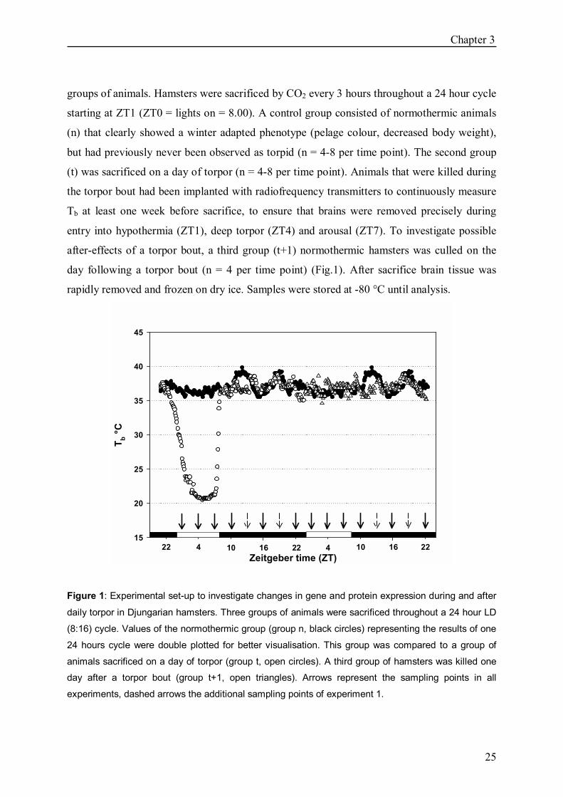
groups of animals. Hamsters were sacrificed by CO2 every 3 hours throughout a 24 hour cycle
starting at ZT1 (ZT0 = lights on = 8.00). A control group consisted of normothermic animals
(n) that clearly showed a winter adapted phenotype (pelage colour, decreased body weight),
but had previously never been observed as torpid (n = 4-8 per time point). The second group
(t) was sacrificed on a day of torpor (n = 4-8 per time point). Animals that were killed during
the torpor bout had been implanted with radiofrequency transmitters to continuously measure
Tb at least one week before sacrifice, to ensure that brains were removed precisely during
entry into hypothermia (ZT1), deep torpor (ZT4) and arousal (ZT7). To investigate possible
after-effects of a torpor bout, a third group (t+1) normothermic hamsters was culled on the
day following a torpor bout (n = 4 per time point) (Fig.1). After sacrifice brain tissue was
rapidly removed and frozen on dry ice. Samples were stored at -80 °C until analysis.
Zeitgeber time (ZT)
T b °C
15
20
25
30
35
40
45
22 4 10 16 22 4 10 16 22
Figure 1: Experimental set-up to investigate changes in gene and protein expression during and after
daily torpor in Djungarian hamsters. Three groups of animals were sacrificed throughout a 24 hour LD
(8:16) cycle. Values of the normothermic group (group n, black circles) representing the results of one
24 hours cycle were double plotted for better visualisation. This group was compared to a group of
animals sacrificed on a day of torpor (group t, open circles). A third group of hamsters was killed one
day after a torpor bout (group t+1, open triangles). Arrows represent the sampling points in all
experiments, dashed arrows the additional sampling points of experiment 1.

Chapter 3
26
Experiment 2: Protein expression at 18 ± 2 °C Ta In a second experiment we determined changes in protein expression during and after daily
torpor. The experiment was carried out as described for experiment 1. Animals were perfused
with 4% paraformaldehyde in 0.1m phosphatebuffer (PBS) (pH 7,4) before brains were
removed. To reduce the animal number we decreased the number of control animals to n = 3
for each time point and n = 4 in the two other groups and sampled at ZT1, ZT4, ZT7, ZT10,
ZT16 and ZT22, hence leaving out the sampling points ZT13 and ZT19.
Experiment 3: mRNA expression at 4°+2 °C Ta As Tb during torpor also depends on Ta another experiment was set up at Ta of 4±2 °C to
investigate whether changes in clock gene expression during torpor might be directly
temperature dependent. One week before sampling Ta was decreased from 18±2 °C to 4±2 °C.
Samples were taken at ZT1, ZT4, ZT7, ZT10, ZT16 and ZT22 with n = 3 in the control group
and n = 4 in the others and brain tissue was rapidly removed and frozen on dry ice. Samples
were stored at -80 °C until analysis.
In situ hybridizations
Serial coronal kryostat sections (16µ) of SCN and pineal gland were mounted on Superfrost
Plus® glass slides and stored at -80 °C until further treatment.
Antisense and sense RNA probes were generated with an in vitro transcription kit (Maxiscript;
Ambion, Austin, TX). We used riboprobes of rPer1 (from plasmids provided by Dr. H.
Okamura, University of Kobe, Kobe, Japan), rVasopressin (Avp) (DARDENTE et al., 2002),
and mBmal1b (MENDOZA et al. 2005). Fixation and hybridization was carried out as
previously described (TOURNIER et al. 2003). For the Aanat in situ hybridization we used a
mixture of antisense oligonucleotide synthetic probes labelled with 35S ATP, proceeding as
reported by SINITSKAYA et al. 2006. Slides were exposed to autoradiographic films
(Biomax, Kodak) together with 14C standards for 5 days. Densiometric analysis of the
autoradiograms was performed using Image J software. For each animal 3 SCN sections were
analysed bilaterally and averaged. Relative mRNA abundance values were calculated by
defining the highest value of an individual animal as 100%.

Chapter 3
27
Immunohistochemistry Brains were post-fixed for 24 hours in 4% paraformaldehyde in 0.1m phosphate buffer (pH
7,4) after removal and kryoprotected in 30% glucose for 3 days. The tissue including the
hypothalamus was serially sectioned 25µ on a kryostat and 2 series were mounted on gelatine
covered slides for PER1 and AVP staining respectively. After rinsing in 0.2 m PBS for 10
min, slides were cooked for 15 min in 0.01m citrate buffer to permeabilise the tissue.
Endogenous peroxidase was blocked for 30 min in 1% H2O2 before the sections were rinsed
2x10 min in 0.2m PBS + 0.1% Triton. After blocking non specific binding sites for 1 h in 2%
albumin bovine + 2% goat normal serum in 0.2m PBS + 0,1% Triton, sections were incubated
with the primary antibody for 24 hours (PER1 1:8000, provided by David Weaver, AVP
1:8000 Fitzgerald), then washed again and incubated 1h with 5% anti rabbit IgG (Vector
labs) in 0.2m PBS + 0.1% Triton. The signal was amplified with a 2% avidin/biotin complex
(Vectastain Elite ABC Kit, Vector labs) and immunolabelling was developed using 1%nickel-
3,3´-diaminobenzidine (Ni-DAB, Sigma).
BMAL1 immunohistochemistry was performed on free-floating sections following the same
protocol, leaving out the first step (citrate buffer) and incubating with the first antibody
(1:500, Santa Cruz) for 48 hours. Cells were counted in 4 SCN sections per animal using
image J software.
Statistical analysis
All data were analysed using Graph Pad Prism 4.0 software. Rhythms in any group were
detected using one-way analysis of variance (ANOVA). Comparisons between the 3 animal
groups were made by a Kruskal Wallis test followed by a Dunn´s test post hoc. Statistical
significance was defined as p<0.05.
Results
Tbs To asses Tb at the nadir of torpor (ZT4) in the different experimental conditions (Ta 18±2 °C,
Ta 4±2 °C), Tb was measured immediately after decapitation in the particular animals. In
experiment 1, Tb dropped to 22.6 ± 0.65 °C, in experiment 2 to 18.5 ± 1.04 °C.

Chapter 3
28
Per1
Per1 gene expression at 18 ± 2 °C Ta In all groups, Per1 expression showed a clear circadian rhythm (p<0.05) with a peak at ZT4
in the control group (n) as well as group t+1. During torpor (t) the peak was shifted to ZT7
and increased (p<0.05) compared to group n (Fig. 2a).
PER1 protein expression at 18 ± 2 °C Ta The PER1 protein was rhythmic in all groups (p<0.05) and reached highest values at ZT10 in
groups n and t+1. Group t differed from group n at ZT4 (p<0.05) and showed a decreased
number of immunopositive cells (Fig. 2b).
Per1 gene expression at 4 ± 2 °C Ta Per1 expression showed a similar pattern as in the 18±2 °C Ta experiment with rhythms in all
groups (p<0.05) peaking at ZT4 in group n and t+1 and increased expression at ZT7 in group t
(p<0.05) compared to group n (Fig. 2c).
Bmal1
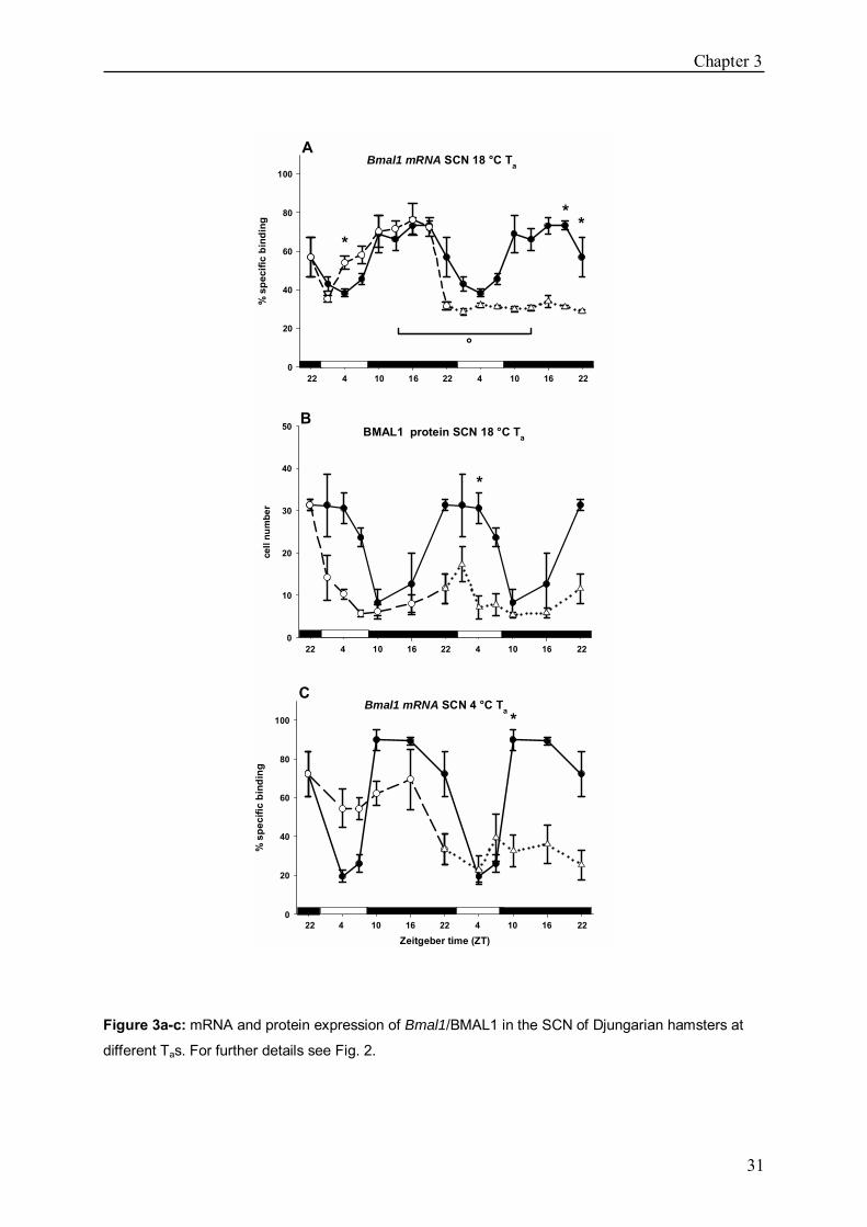
Bmal1 gene expression at 18 ± 2 °C Ta Bmal1 was rhythmically expressed in group n and group t (p<0.05). Highest amounts of
mRNA were detected between ZT10 and ZT16. In group t expression was significantly
increased at ZT4 (p<0.05). Group t+1 did not show a detectable rhythm and expression
differed significantly from group n at ZT19 (p<0.05) and ZT22 (p<0.05) from group t at ZT13
(p<0.05) (Fig. 3a).
BMAL1 protein expression at 18 ± 2 °C Ta
BMAL protein levels were rhythmic in group n (p<0.05) with highest values between ZT22
and ZT4 (p<0.05). No rhythm could be revealed in group t or t+1. In group t+1 the cell
number differed significantly at ZT4 from that of group n (Fig. 3b).
Bmal1 gene expression at 4 ± 2 °C Ta
Bmal1 showed a rhythmic expression in group n and group t (p<0.05) and peaked between
ZT10 and ZT16. No rhythm was detected in group t+1, values differing from the control
group n at ZT10 (p<0.05) (Fig. 3c).

Chapter 3
29
Avp
Avp gene expression at 18 ± 2 °C Ta
Avp rhythm was significant in all 3 groups (p<0.05). In group n and t the clock related gene
was expressed highest between ZT4 and ZT10. Group t+1 showed decreased values at ZT4
(p<0.05), ZT7 (p<0.05) and ZT10 (p<0.05) compared to group n (Fig. 4a).
AVP protein expression at 18 ± 2 °C Ta AVP protein was rhythmic in group n and peaked at ZT16. No significant rhythm was
detected in group t or group t+1. In group t we found decreased cell numbers at ZT10
(p<0.05) relative to group n. Group t+1 showed a decreased number of immunopositive cells
at ZT10 (p<0.05) (Fig. 4b).
Avp gene expression at 4 ± 2 °C Ta Avp expression showed comparable rhythms (p<0.05) to those in the 18 °C Ta experiment
with rhythms peaking ZT4 and ZT10 in group n and t and decreased expression at ZT4
(p<0.05), ZT7 (p<0.05) and ZT10 (p<0.05) compared to group n (Fig. 4c).
Aa-nat
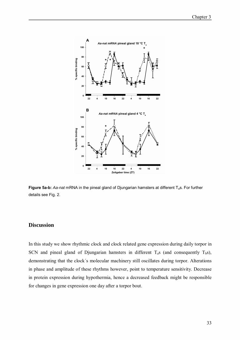
Aa-nat gene expression at 18 ± 2 °C Ta Aa-nat was rhythmic in the pineal gland in all 3 groups (p<0.05). In group n the expression
peaked between ZT16 and ZT22, in group t and t+1 between ZT13 and ZT22. Increased
mRNA amounts were found in group t at ZT10 (p<0.05) and ZT13 (p<0.05), in group t+1 at
ZT13 (p<0.05) compared to group n (Fig. 5a).
Aa-nat gene expression at 4 ± 2 °C Ta
Aa-nat rhythms were comparable to those in the 18 °C Ta experiment (p<0.05) with gene
expression peaking at ZT16 in all groups. In group t expression was increased at ZT10
compared to group n (p<0.05) (Fig. 5b).

Chapter 3
30
Per1 mRNA SCN 18 °C Ta
% s
peci
fic b
indi
ng
0
20
40
60
80
100
PER1 protein SCN 18 °C Ta
cell
num
ber
0
100
200
300
Per1 mRNA SCN 4 °C Ta
Zeitgeber time (ZT)
% s
peci
fic b
indi
ng
0
20
40
60
80
100
*
*
*
A
B
C
22 4 10 16 22 4 10 16 22
22 4 10 16 22 4 10 16 22
22 4 10 16 22 4 10 16 22
Figure 2a-c: mRNA and protein expression of Per1/PER1 in the SCN of Djungarian hamsters at
different Tas. In each experiment, 3 animal groups were sacrificed throughout a 24 hour cycle. Values
of the normothermic control group (n, black circles, solid line) represent the results of one 24 hour
cycle and were double plotted. The second group (t, open circles, dashed line) was killed on a day of
daily torpor. Finally samples of a third group (t+1, open triangles, dotted line) were taken one day after
a torpor bout. Data are presented as mean ± SEM. Solid and open bars represent dark and light
periods, respectively. Significant differences are indicated by * for differences between the control
group n and group t or t+1, by ° for differences between group t and group t+1.
Chapter 3
31
Bmal1 mRNA SCN 18 °C Ta
% s
peci
fic b
indi
ng
0
20
40
60
80
100
B
22 4 10 16 22 4 10 16 22
A
C
BMAL1 protein SCN 18 °C Ta
cell
num
ber
0
10
20
30
40
50
*
*
*
Bmal1 mRNA SCN 4 °C Ta
Zeitgeber time (ZT)
% s
peci
fic b
indi
ng
0
20
40
60
80
100 *
22 4 10 16 22 4 10 16 22
22 4 10 16 22 4 10 16 22
*
°
Figure 3a-c: mRNA and protein expression of Bmal1/BMAL1 in the SCN of Djungarian hamsters at
different Tas. For further details see Fig. 2.

Chapter 3
32
Avp mRNA SCN 18 °C Ta
% s
peci
fic b
indi
ng
0
20
40
60
80
100
AVP protein SCN 18 °C Ta
cell
num
ber
0
20
40
60
Avp mRNA SCN 4 °C Ta
Zeitgeber time (ZT)
% s
peci
fic b
indi
ng
0
20
40
60
80
100
***
* *
*
*
A
B
C
22 4 10 16 22 4 10 16 22
22 4 10 16 22 4 10 16 22
22 4 10 16 22 4 10 16 22
Figure 4a-c: mRNA and protein expression of Avp/AVP in the SCN of Djungarian hamsters at
different Tas. For further details see Fig. 2.
Chapter 3
33
Aa-nat mRNA pineal gland 4 °C Ta
Zeitgeber time (ZT)
% s
peci
fic b
indi
ng
0
20
40
60
80
100
Aa-nat mRNA pineal gland 18 °C Ta
% s
peci
fic b
indi
ng
0
20
40
60
80
100
* *
*
*
A
22 4 10 16 22 4 10 16 22
B
22 4 10 16 22 4 10 16 22
Figure 5a-b: Aa-nat mRNA in the pineal gland of Djungarian hamsters at different Tas. For further
details see Fig. 2.
Discussion
In this study we show rhythmic clock and clock related gene expression during daily torpor in
SCN and pineal gland of Djungarian hamsters in different Tas (and consequently Tbs),
demonstrating that the clock´s molecular machinery still oscillates during torpor. Alterations
in phase and amplitude of these rhythms however, point to temperature sensitivity. Decrease
in protein expression during hypothermia, hence a decreased feedback might be responsible
for changes in gene expression one day after a torpor bout.
Chapter 3
34
All investigated genes showed a rhythmic mRNA expression in normothermic animals (group
n) at both Tas. In this normothermic state Ta does of course not influence Tb which generally
lies between 36 °C and 39 °C, and hence identical rhythms were found in both groups. These
rhythms resemble those described by JOHNSTON et al. (2005) who showed similar
expression patterns of Per1, Bmal1 and Avp in the SCN of Djungarian hamsters in short
photoperiod (SP), reflecting the typical short day feedbackloops with Per1 as example for the
negative and Bmal1 as the marker for the positive limb of the circadian molecular machinery.
Also protein expression of PER1, BMAL1 and AVP showed clear circadian rhythms at Ta of
18± 2°C peaking with a delay to mRNA expression of 6-9 hours for PER1 and AVP and a
delay of 9-12 hours for BMAL1. This is in accordance with previously shown results
(REPPERT and WEAVER 2001) and seems to be the normal delay for clock protein
synthesis.
On the day of torpor (group t) all genes were rhythmically expressed at both Tas, suggesting
temperature compensation of the clock at the transcriptional level in the investigated Tb range
between ~18 and 39 °C. With regard to behavioural data these results are not surprising, as
the circadian system is of utmost importance for the precise timing of daily torpor which is
maintained over the whole winter season and can easily be adapted to shifts in the photocycle
(KIRSCH et al. 1991, RUBY and ZUCKER 1992, RUF and HELDMAIER 1992). However,
studies investigating transcriptional processes in hibernating ground squirrels, demonstrate a
directly temperature-dependent transcript elongation which is arrested in deep hibernation (Tb
~3 °C) to reduce transcriptional expenses (BOCHAROVA et al. 1992, VAN BREUKELEN
and MARTIN 2002a). Only few exceptions in systems that are of vital importance for
controlling the hypothermia, as for example the histaminergic system, are made (SALMEN et
al. 1999, 2003a) and here transcription appears to be even upregulated during hypothermia.
Admittedly, daily torpor is only a relatively shallow form of hypothermia. Therefore, it is
likely that the attenuating temperature effect on transcription is less pronounced at those still
relatively high Tbs. In addition, it could be shown that despite reduced transcription total
mRNA amounts in both brain and liver were surprisingly constant during deep hibernation,
implicating an extension of mRNA half life (FRERICHS et al. 1998, O´HARA et al. 1999).
This could e.g. happen through specific poly(A) binding proteins protecting poly(A)tail length
(KNIGHT et al. 2000) or heterogenous ectopic RNP-derived structures (HERDS) that have

Chapter 3
35
been described in torpid but not euthermic hibernators (MALATESTA et al. 1999,
TAMBURINI et al. 1996) and might serve to store transcripts.
We did not observe differences in gene expression between the groups at 18±2 °C and 4±2 °C
Ta. Tb during torpor depends to a certain extent on Ta , given that torpid animals cannot drop
their Tb below Ta. Despite a 14 °C difference in Ta, Tbs only differed for 4±2,5 °C at the nadir
of torpor between both groups. Probably this difference is not great enough to reveal changes
in gene expression by in situ hybridisation and requires a more sensitive quantitative method
like qPCR to determine differences in amount of mRNA. It is also possible that temperature
compensation is not gradual but limited to a certain temperature range. In vitro studies have
shown that electrical SCN activity is arrested below 16.6 °C (MILLER et al. 1994). In
hibernating European hamsters (Cricetus cricetus) with Tbs ~8 °C during the torpor bout we
did not observe any rhythmic clock gene expression (REVEL et al. submitted) so it may be
that oscillation does not disappear gradually, but once a certain low temperature (probably
16.6 °C) is reached, rhythmic clock gene expression is no longer possible.
mRNA amounts of Per1 and Bmal1 were increased during the torpor bout. This might either
result from a directed upregulation of gene expression or a passive accumulation of mRNA
due to extension of mRNA half life in hypothermia (KNIGHT et al. 2000) and attenuated
protein synthesis. Decline in protein synthesis during hypothermia has been demonstrated
before and is likely to result from active suppression through regulatory phosphorylation of
translational initiation factors rather than from simple Q10 effects (FRERICHS et al. 1998,
VAN BREUKELEN and MARTIN 2001). Indeed we found a decreased number of
immunopositive cells in all investigated genes during hypothermia supporting this hypothesis.
Yet, Avp mRNA was not increased during torpor compared to the control group n (that had
never shown torpor before the experiment), although protein expression was reduced as a
consequence of torpor. This might result from a decreased BMAL1 feedback after
hypothermia. The precise role of AVP in the SCN is still unclear. Lower amplitude in AVP
rhythms causes lower amplitude in SCN electrical activity and may therefore account for less
pronounced behavioural rhythms (VAN ESSEVELDT et al. 2000). Indeed behavioural
rhythms are reduced or even completely absent during torpor season in the Djungarian
hamster (unpublished observation), activity levels are generally low and amplify energy
saving (RUF and HELDMAIER 1992). Lower AVP amplitude, as also seen on the day after
torpor (t+1), could at least support this behaviour. We discussed previously (HERWIG et al.,
Chapter 3
36
2006b) that the onset of Bmal1 expression seems to be advanced on the day of torpor and that
this advance may probably explain the shortened τ accompanying hypothermia in DD that has
been described by Thomas et al. 1993. With the additional immunohistochemistry results that
reveal a depressed amplitude in BMAL1 protein during torpor, it seems more likely that the
increase in Bmal1 mRNA amount is rather a passive accumulation of mRNA. Moreover, we
did our experiments in LD conditions and therefore cannot predict the endogenous period.
On the day after torpor, we found a depression of Bmal1/BMAL1 as well as Avp/AVP
rhythms in all experiments. It is striking that no Bmal1/BMAL1 rhythm was detected one day
after torpor. However, PER1 protein rhythms were significantly dampened during
hypothermia, probably resulting in a diminished feedback on Bmal1 transcription. Diminished
rhythms in one of the feedback loops of the circadian molecular machinery though, would
support diminished output, namely Avp/AVP rhythms and consequently decreased
behavioural rhythms. Moreover, also hypocaloric fed mice displayed decreased Bmal1
amplitude (FEILLET et al. 2006). As torpor serves as an energy saving process, animals
displaying torpor decrease their food intake which might have the same effect as food
restriction. It is striking though, that we did not observe changes in Per1 expression despite
the lack of Bmal1/BMAL1 feedback, as this feedback seems necessary for restarting the next
circadian cycle (REPPERT and WEAVER 2001). However, Per1 expression is directly
induced by light and as our experiments were carried out in an LD cycle the light input might
be the more crucial factor here.
Besides the molecular machinery within the SCN, we also investigated Aa-nat expression in
the pineal gland, a well studied output of the circadian clock. Aa-nat as the rate limiting
enzyme for melatonin, has been shown to be directly under SCN control and is a good marker
for the actual output of the circadian clock. Its expression was rhythmic in all groups. On the
day of torpor (group t) we observed a strong Aa-nat induction at the very beginning of the
night, most likely caused by the strong sympathetic activity during the arousal from
hypothermia. Indeed already LARKIN et al. (2003) found an increased melatonin production
accompanying the arousal from torpor. The advanced increase in Aa-nat expression was still
visible on the day after the torpor bout. It has been discussed whether such �melatonin pulses�
might contribute to a phase advance of the circadian system and consequently shorten τ in
constant conditions (THOMAS et al. 2003). Our results would support such a hypothesis, but
as the animals were always kept in LD conditions we do not know the endogenous phase.

Chapter 3
37
However, we set up long term microdialysis experiments to continuously measure melatonin
in the pineal gland (HERWIG et al. 2006a) to further investigate phase changes caused by
daily torpor in vivo and in LD as well as constant conditions.
Overall, our results demonstrate transcriptional temperature compensation during daily
hypothermia. No difference in gene expression could be observed in varying Tas and
consequently Tbs. However, a decreased protein feedback results in altered clock and clock
related gene expression after hypothermia.

38
39
4
Trans-pineal microdialysis in the Djungarian
hamster (Phodopus sungorus): a tool to study
seasonal changes of circadian clock activities.
Annika Herwig1,2, Paul Pévet2, Béatrice Bothorel2,
Stephan Steinlechner1, Michel Saboureau2 1 Institute of Zoology, University of Veterinary Medicine, Bünteweg 17, D-30559 Hannover, Germany 2 Département de Neurobiologie des Rythmes, Institut des Neurosciences Cellulaires et Intégratives,
UMR-7168/LC2, CNRS - Université Louis Pasteur, 5 rue Blaise Pascal, 67084 Strasbourg Cedex,
France
Published in Journal of Pineal Research (2006) 40:177-183

Chapter 4
40
Abstract
The Djungarian hamster is a highly seasonal small mammal. The rhythmic secretion of
melatonin by the pineal gland is controlled by the circadian clock, conveying the
photoperiodic message to the organism. Trans-pineal microdialysis permits the in vivo study
of this well-defined and precise clock output by measuring melatonin release directly in the
pineal gland. The aim of this study was to adapt this method to the Djungarian hamster in
order to monitor clock properties during photoperiodic changes. Male adult Djungarian
hamsters were kept in a long photoperiod (LD 16:8) and melatonin release was measured
hourly during the dark period for several weeks. Melatonin showed a regular secretion
between ZT 17 and ZT 23.5 whereas the amplitude became stable only after the 3rd day of
perfusion. To test how quickly changes in melatonin profile can be measured, 15 min light
pulses were given at different time points throughout the scotophase. Light-pulses
immediately interrupted the melatonin secretion at any time point during the scotophase and
the temporal resolution for measurement could be reduced to 30 minutes. In accordance with
studies in the rat, long term effects of light on the clock could only be observed when a light
pulse was administered in the 2nd half of the night. For the first time we established a method
to precisely measure a direct and reliable clock-output in a highly seasonal species which
allows us now to study the circadian and seasonal properties of the clock in detail.
Introduction
The central biological clock, located in the suprachiasmatic nuclei (SCN) of the
hypothalamus, is known to control the daily rhythm of many behavioural and physiological
functions via endocrine and/or neuronal pathways (PERREAU-LENZ et al. 2004). In
mammals, diurnal or nocturnal, the hormone melatonin, mainly synthesised in the pineal
gland, is released during the night in a circadian manner and in proportion to the night length.
Numerous studies on mammals have shown that the daily rhythm of melatonin synthesis is
exclusively under SCN control, which is connected to the pineal gland through a
multisynaptic pathway (MOORE 1996). Depending on inhibitory and stimulatory signals

Chapter 4
41
from the clock, the release of noradrenaline (NA) into the pineal gland by the sympathetic
fibres stimulates the melatonin synthesis pathway by activation of adrenergic receptors
(PERREAU-LENZ et al. 2004). Melatonin is thus a stable and reliable output of the
biological clock and detailed analysis of this output allows us to study clock properties
(REITER 1993, PÉVET 2003).
Several approaches have been used to follow melatonin rhythm generation continuously over
a long period of time, as for example repeated blood sampling with a jugular catheter.
However, except for some large mammals such as sheep the amount and the volume of blood
samples that can be taken from the same animal limit the data points per 24h cycle and the
duration of the experiment (RAVAULT and CHESNEAU 1996). The indirect evaluation by
analysis of the urinary melatonin metabolite 6-sulphatoxy-melatonin is also not very precise
as data may be dependent on liver activity and show a phase-lag of two to four hours
(RAYNAUD et al. 1993, STIEGLITZ et al. 1995, KENNAWAY et al. 2002).
It is the development of the trans-pineal microdialysis by AZEKAWA et al. (1990) and
DRIJFHOUT et al. (1993), which allows the precise long term measurement of the melatonin
rhythm with a high temporal resolution in its production site, in intact and freely moving
animals. This technique permits longitudinal studies, in which each animal is its own control
and has been useful especially in the rat to identify the nervous pathways and the
neurotransmitters involved in distributing the circadian message (PERREAU-LENZ et al.
2004). Melatonin, however, through the change in duration of its nocturnal peak is known to
be involved in the distribution of seasonal information (STEINLECHNER et al. 1987,
BARTNESS et al. 1993, PÉVET 2003). In the last few years, it has been observed that a
number of clock genes show day-length dependent changes in expression in the SCN
supporting the idea that the clock directly generates the seasonal messages (VUILLEZ 1996,
NUESSLEIN-HILDESHEIM et al. 2000, TOURNIER et al. 2003).
Trans-pineal microdialysis appears to be a good technical tool for studying clock properties
and pathways involved in photoperiodic responses. Since rats only marginally respond to such
seasonal changes, the development of trans-pineal microdialysis in a photoperiodic species is
needed. We have set up such trans-pineal microdialysis approaches for the Djungarian
hamster (Phodopus sungorus), a well known model in seasonal studies in which the role of
photoperiod and melatonin is well defined (HOFFMANN 1979, HELDMAIER and

Chapter 4
42
STEINLECHNER 1981, BARTNESS and GOLDMAN 1989, STEINLECHNER and
NIKLOWITZ 1992).
Materials and methods
Animals
For the experiments male three to five-month-old Djungarian hamsters (Phodopus sungorus)
from our own breeding colony were used. The animals weighed between 30 and 40 g and
were housed in a temperature-controlled room (21 ± 2 °C) under a reversed 16:8 h light-dark
(L:D) cycle (lights off from 10.00 a.m. to 6.00 p.m.) for at least three weeks before surgery.
Light onset corresponds to Zeitgeber time ZT 0, thus in this photoperiodic condition night
starts at ZT16. Water and food were available ad libitum.
All experiments were performed in accordance with �Principles of Laboratory Animal Care´
(NIH pub.no., 86-23, revised 1985) and in accordance with French laws.
Surgery and dialysis
The animals were operated at least one week before the microdialysis experiment. They were
anesthetised with intraperitoneal injections of 0.2 ml Zolétil and 0.1 ml Rompun (1:8). After
the operation they were allowed to recover for one week in individual cages before starting
the first perfusion.
Surgery was performed as previously described (BARASSIN et al. 1999) but with a modified
dialysis probe. The active dialysis area measured 1 mm and the probe was implanted 0.8 mm
ventral to the skull surface and 0.4 mm posterior to Lambda. Perfusion was performed at a
flow rate of 2 µl/min
Experimental procedure In all experiments, pineal melatonin release was measured during five subsequent days.
Animals were always housed in long photoperiod (LD 16:8). Samples were collected hourly
from ZT 14 (8.00 a.m., two hours before dark onset) to ZT 3 (9.00 p.m., three hours after light
onset), except for the first two hours after the photic stimulus (exp 2) when samples were

Chapter 4
43
collected every 30 min. The dialysates were stored at �20 °C until being assayed for
melatonin by radioimmunoassay (RIA). In order to determine the location of the probe,
cryostat sections of the brain/pineal (25µm) were stained with cresyl violet at the end of the
experiments.
Experiment 1: To validate the transpineal microdialysis approach in the Djungarian hamster
and to characterise the daily profile of the melatonin excretion under these conditions, in a
first experiment daily melatonin rhythms were measured hourly for five subsequent days
without any manipulations (n = 11).
Experiment 2: To assess how reliable changes in melatonin release can be measured, the
influence of single 15 min light pulses on the melatonin secretion at different time points
throughout the dark phase was tested in a second experiment. After two control days without
any manipulation and hourly sampling, a 15 min light pulse (200 lux) was given on the third
day at ZT 18 (n = 4), ZT 19 (n = 3), ZT 20 (n = 3), ZT 21 (n = 3), and ZT 22 (n = 4). In the
first two hours after the light pulses, samples were collected every 30 min. The following two
days the animals were kept in undisturbed LD 16:8 cycles.
Melatonin assay Melatonin in the dialysates was measured by radioimmunoassay in 25 µl duplicate samples
using specific rabbit antiserum (R19540, INRA, Nouzilly, France) and labelled (125 I) -2-
iodomelatonin. The sensitivity limit of the assay was 0.5 pg/tube. For details see BARASSIN
et al. (2003).
Data analysis and statistics
All melatonin data are expressed in absolute values (nM). For each animal the pattern of
melatonin secretion was characterised and fitted by a non-linear regression analysis with
Sigma Plot software (Jandel Scientific). All pineal Mel profiles were fitted with the following
equation: y = Y0 + (Ymax / ((1 + exp(A*(IT50-x)))*(1 + exp(x � DT50)))) where y was the
nth data point, x the time point of the nth point, Y0 the basal level measured during daytime,
Ymax the maximum of the nocturnal peak, IT50 was defined as the time point of 50 %
increase in melatonin levels and DT50 as the time point of 50 % decrease. The multiplicative
factors in the exponentials were chosen arbitrarily to fit the observed slopes and in this work

Chapter 4
44
fixed to 1.62 and 3.58 respectively. IT50 and DT50 were used to characterise the timings of
the onset and offset of melatonin peak for each day of the experiments. The duration and the
amplitude of the melatonin peak were determined as the difference between DT50, IT50 and
Ymax, respectively.
First, an ANOVA for repeated measurements was carried out for each group of animals on
IT50, DT50 and Ymax with animals and control days (Ctrl 1 and Ctrl 2) as factors; a Scheffé’s
test was used as post hoc test. The differences were considered significant for a P<0.05(*).
Results
Under our experimental photoperiodic conditions (LD 16:8), extracellular pineal melatonin
concentrations were low during the light period and peaked during darkness between ZT 20
and ZT 0. Figure 1 shows averaged nocturnal melatonin profiles of eleven animals measured
for five successive days without any manipulation. The melatonin release showed a regular
onset at ZT 18.49 ± 0.08 h and offset at ZT 0.13 ± 0.05 h corresponding to the time of lights-
on in all animals. The amplitude showed a significant increase between day one and day four
(Scheffé’s: P = 0,054) as well as day one and day five (Scheffé’s: P = 0.03). Moreover the
amplitude varied between individuals correlating to the position of the probe within the pineal
gland.
Figure 2 presents representative individual melatonin profiles of animals given light pulses at
different time points throughout the dark phase. Ctrl 1 shows the normal melatonin pattern in
the night before the photic stimulus. LP corresponds to the day where the 15 min light pulse
was given, whereas Ctrl 2 presents the melatonin profiles two days after the light pulse. The
15 min photic stimulus given during darkness caused an immediate decrease in melatonin
concentrations down to basal levels within one hour in every case.
After a light pulse at ZT 18 and ZT 19, melatonin concentrations increased again two hours
after the light pulse and reached normal levels by the end of the night. Two days after the
manipulation (Ctrl 2) no significant effect could be seen at the onset (Anova: ZT 18 P = 0.08;
ZT 19 P = 0.24), offset (Anova: ZT 18 P = 0.59; ZT 19 P = 0.41) or amplitude (Anova: ZT 18
P = 0.68; ZT 19 P = 0.19).

Chapter 4
45
The light pulse at ZT 20 caused an instant reduction in the hormone in the pineal gland which
did not recover to normal (Ctrl 1) levels during the same night. Moreover the profile showed a
significantly earlier offset (Scheffé’s: 0.0015) of 1.45 ± 0.13 h. On Ctrl 2 only slight
alterations could be seen at the onset (ANOVA: P = 0.90) but the offset took place 1.03 ± 0.08
hours earlier (Scheffé’s: P = 0.0056) and the amplitude was significantly reduced (Scheffé’s:
P = 0.016).
At ZT 21 and ZT 22 the photic stimulus had a similarly acute effect. The melatonin
concentrations did not or only hardly rose again after the light induced decrease. While the
manipulation had no long term effect on the onset at ZT 21 (ANOVA: P = 0.43) the melatonin
release set off significantly 0.95 ± 0.08 hours earlier (Scheffé’s: P = 0.025) on Ctrl 2. At ZT
22 also the onset advanced significantly for 0.80 ± 0.13 hours (Scheffé’s: P = 0.009), the
offset shifting for 0.74 ± 0.28 hours (Scheffé’s: P = 0.001).
Zeitgeber time (hours)14 15 16 17 18 19 20 21 22 23 0 1 2 3
MEL
ATO
NIN
(nM
)
0
1
2
3
4
D1 D2 D3D4D5
Figure 1: Averaged nocturnal profiles of pineal melatonin concentrations measured by in vivo
microdialysis in Phodopus sungorus during 5 successive days (D1-D5) without any manipulations.
Data are plotted as the mean ± SEM (n = 11). The grey bar represents the dark period (from ZT16 to
ZT0) of the LD 16:8 cycle.

Chapter 4
46
Figure 2: Examples of individual melatonin profiles in the pineal gland during the first control day (Ctrl
1), during the experimental day (LP) where a 15 min light pulse was given at different Zeitgeber times
and during the second day after the photic stimulus (Ctrl 2). The grey bar represents the dark period of
the LD 16:8 cycle the black arrow points to the 15 min light pulse.
The individual phase shifts of melatonin on- and offsets are presented in Figure 3a/b besides
the means for each group. The individual values for the amplitude are illustrated in Figure 3c.

Chapter 4
47
Figure 3: Effect of a 15 min light pulse given at different Zeitgeber times on the onset IT50 (A) and the
offset DT 50 (B) of the nocturnal melatonin profiles revealed by in vivo microdialysis. Light pulses were
given between ZT 18 and ZT 22 under LD 16:8 conditions and pineal dialysis samples were collected
until 2 days afterwards (Ctrl 2). The variations in on- and offsets are presented as changes observed
on the Ctrl 2 vs. a control day. Variations in the amplitude of the melatonin peak are presented in
Figure 3 C. Open symbols: individual values. Closed symbols: means ± SEM.

Chapter 4
48
Discussion
The melatonin rhythm in the pineal gland is an excellent marker of the circadian pacemaker
because it accurately reflects and amplifies SCN electrical and metabolic activity in both
diurnal and nocturnal mammals. Trans-pineal microdialysis, developed by AZEKAWA et al.
(1990) and DRIJFHOUT et al. (1993) allows precise and continuous measurement of this
marker directly in the pineal gland with a high temporal resolution and in the same animal for
a period of up to five weeks (unpublished data). The adaptation of the trans-pineal
microdialysis technique to a photoperiodic species as demonstrated in this study is important
as melatonin is not only an SCN generated circadian signal but also conveys seasonal
photoperiodic changes.
Undisturbed melatonin release showed profiles with regular on- and offsets during 5
consecutive days. In all animals tested and kept under LD 16:8, onsets occurred regularly two
to three hours after the beginning of the dark period. Our results tally with pineal melatonin
profiles or NAT activities described in this species (ILLNEROVA et al. 1984, HOFFMANN
et al. 1985, STEINLECHNER et al. 1987). The offset differs from what has been described in
the rat. Instead of declining about one hour before light onset, therefore being necessarily
driven by an endogenous SCN-induced cessation of NA release (DRIJFHOUT et al. 1996) the
melatonin peak in the Djungarian hamster did not decrease before ZT 0. This suggests that not
an endogenous stimulus but the light itself terminates the melatonin release in LD 16:8.
Interestingly STEINLECHNER et al. (1987) described a similar phenomenon for AA-NAT
activities in long photoperiod, while in short photoperiod the AA-NAT activity already
decreased before dawn. This observation of a light inhibitory signal in melatonin/AA-NAT
production under LP but not under SP, also described in the Syrian hamster (PÉVET 1988)
and Arvicanthi ansorgei (GARIDOU-BOOF et al. 2005) seems to be characteristic of
photoperiodic species. Due to the short half life of circulating melatonin (20 min)
(STANKOV et al. 1993), secretion dropped to day time levels within less than an hour.
The increase in amplitude within the first 3 days of melatonin measurement observed in this
study has not been reported in other intra-pineal microdialysis studies in rats or birds, where
the measured melatonin peak appeared to be stable from the first day of dialysis (BARASSIN
et al. 1999). Very probably, this instability in amplitude during the first days of measurement

Chapter 4
49
in the Djungarian hamster results from the technique itself. Implantation of the probe causes a
more severe tissue trauma in the very small pineal gland of the Djungarian hamster which
needs a longer time to recover. After the beginning of perfusion it takes some days until the
dialysis system is equilibrated. The fact that after few days we obtained a reproducible
nocturnal melatonin release similar to that described in the literature, demonstrates that this
trauma is only temporary. Nevertheless, to use intra-pineal microdialysis in good conditions,
the physiological experiments should not be started before 3 days of perfusion without
sampling in this species.
As expected, all 15 min light pulses caused an immediate decrease in melatonin release down
to basal levels within one hour. These results tally with all the other data in literature,
describing immediate suppression of AA-NAT activity and melatonin synthesis by even short
white light pulses (KLEIN and WELLER 1972, ILLNEROVA and VANECEK 1972).
Depending on the time they were given, the photic stimuli showed different impacts on the
melatonin rhythm. When the light pulse was given before midnight (ZT 20), only a transient
decrease was seen, which recovered thereafter to normal levels and set off at the usual time.
Light pulses after ZT 20 resulted in a generally sustained decrease in melatonin for the rest of
the night. These results are in accordance with those of VANECEK and ILLNEROVA
(1982), ILLNEROVA et al. (1986), showing an initial drop in AA-NAT activity and a
subsequent rise when the light pulse was applied before midnight, and a sustained decrease
when given after midnight in rats. Similar findings have been reported by Kanematsu et al.
(1994) for the melatonin release. These different effects of light on melatonin release may
result from the circadian change in sensitivity in the light pathway (PANGERL et al. 1990,
HONMA et al. 1992). Moreover all components involved in melatonin regulation such as
AA-NAT and HIOMT show complex and changing kinetics within a circadian phase
(RIBELAYGA et al. 1999). Contrary to findings of Kanematsu et al. (1994) we did not find
any long term alteration of the melatonin rhythm when the light pulse was given before
midnight [ZT 20]. Very likely we had no such effect because our experiment was done in
continued LD cycles, while Kanematsu et al. chose constant conditions for their experiments.
Our results tally with studies, showing only a weak c-Fos induction in the SCN after a light
stimulus in early on at night compared to a stronger induction late at night in hamsters
(SUTIN and KILDUFF 1992). The protein c-Fos serves as marker for efferent SCN signals
and is localised in VIP expressing cells. This efferent signal might modify the NA release in

Chapter 4
50
the pineal gland and consequentially the melatonin rhythm (KALSBEEK et al. 1999). When
the photic stimulus was applied in the middle of the night, at ZT 20, the melatonin rhythm
was strongly reduced after two days. The duration was markedly shorter with an advanced
offset, and the amplitude was significantly decreased. SUMOVA and ILLNEROVA (1998)
showed a decreased amplitude of c-Fos immunoreactivity as well as an advance in the
morning decline after a light stimulus around midnight in rats, suggesting a comparable effect
of the light on the clock itself. In accordance with our study, LERCHL (1995) reported
significant effects on melatonin amplitude and phase one night after a light pulse at midnight
in the same species. However our results show that those alterations are still visible 2 days
after the photic stimulus, tallying with results from STEINLECHNER et al. (2002b) who
showed that already short light pulses can have a long-lasting or even destructive effect on
circadian rhythms. It is known as well that during resetting of the mammalian circadian clock,
not only the clock�s phase is shifted, but amplitude of overt rhythms driven by the clock may
be temporarily reduced or even abolished (ILLNEROVA 1992). Our results are not
astonishing, knowing that hamsters in short photoperiod exposed to light pulses during every
second or fourth night is virtually indistinguishable from a complete transfer to long-day
photoperiod (HOFFMANN et al. 1981). It would make sense that nocturnal seasonally
breeding animals can react very sensitively to even very little photoperiodic information to
time the re-onset of reproduction correctly. A light pulse in the second half of the night
resulted in an offset advance (ZT 21) or a complete phase advance in onset and offset (ZT 22)
up to one hour. KANEMATSU et al. (1994) observed similar phase advances in rats, although
they used a different protocol and released the animals in constant darkness after the light
pulse. This means that even despite a present entrainment stimulus, namely the LD cycle,
light at night has an enormous impact on the clock. In this study on- and offsets are not
equally affected. In most cases the melatonin offset is more altered than the onset. This
"uncoupling" of onset and offset supports the model of a complex two-component pacemaker
system controlling the pineal melatonin production. The model by PITTENDRIGH and
DAAN (1976) describes a clock with a morning (M) oscillator controlling the onset of the
resting period and an evening (E) oscillator controlling the onset of locomotor activity. This
theory was extended to pineal AA-NAT activity which appeared to be differentially affected
by light pulses given at different circadian times in constant darkness (ILLNEROVA and
VANECEK 1982). In this study we validated an important method in the Djungarian hamster,

Chapter 4
51
a highly seasonal animal. We are now able to measure very precisely a direct and reliable
clock output which is not only involved in circadian physiology but also responsible for
seasonal changes, thus allowing us to study in detail the seasonal properties of the clock.
Acknowledgments
This study was funded by a Marie-Curie grant to AH.

52

53
5
In vivo melatonin measurement during torpor in the
Djungarian hamster (Phodopus sungorus).
Annika Herwig1,2, Paul Pévet2, Stephan Steinlechner1,
Michel Saboureau2 1 Institute of Zoology, University of Veterinary Medicine, Bünteweg 17, D-30559 Hannover, Germany 2 Département de Neurobiologie des Rythmes, Institut des Neurosciences Cellulaires et Intégratives,
UMR-7168/LC2, CNRS - Université Louis Pasteur, 5 rue Blaise Pascal, 67084 Strasbourg Cedex,
France
Preliminary data

Chapter 5
54
Introduction
As described in chapter 4, the rhythmic melatonin secretion from the pineal gland is a precise
and very well-defined output of the circadian clock that reflects both seasonal and daily
changes in light-dark (LD) ratio. It is exclusively regulated by the suprachisamatic nuclei
(SCN) and therefore a very reliable phase marker for the clock´s activity. In chapter 2 and 3
we described alterations of the clock´s oscillation during and after daily torpor in the
Djungarian hamster (Phodopus sungorus). Daily torpor mainly affected the amplitude of
oscillation: decreased clock protein expression during the torpor bout probably led to
decreased clock gene expression after the hypothermia. Yet, after arousal from hypothermia,
which usually takes place at the end of the light phase, we observed a phase advanced
increase in Aa-nat mRNA (the rate limiting enzyme for melatonin) relative to the
normothermic control group. As melatonin feeds back on the clock and is capable of inducing
phase shifts (UNDERWOOD 1982, REDMAN et al. 1983, LEWI et al. 1992, REDMAN
1997, AGEZ et al. 2007), it is possible that an advanced melatonin induction after arousal
from daily torpor might also have an effect on the phase or, in constant conditions, the
endogenous period (τ) of the circadian clock. Temperature-provoked phase and τ changes of
circadian rhythms have been described before. Already ASCHOFF (1979) predicted that τ
becomes shorter in nocturnal species as temperature decreases. Low temperature pulses of
25 °C caused a 2 hours shortening in the SCN´s τ of golden mantled ground squirrels
(Spermophilus citellus) in vitro and consequently a nearly 5 hour phase advance after only 48
hours at low temperatures (RUBY and HELLER 1996). Also THOMAS et al. (1993)
observed a shortening of τ in Djungarian hamsters that displayed daily torpor. To assess
changes in phase and τ, it is necessary to continuously measure a clock output in the same
individual because τ varies between animals. Measuring melatonin by trans-pineal
microdialysis allows a long-term investigation of the most precise clock output known today
and allows us to study in detail the effect of daily torpor on the phase and τ of the circadian
clock.
Chapter 5
55
Materials and methods
Animals
For the experiments we used male three to five-month-old Djungarian hamsters (Phodopus
sungorus) from the breeding colony of the Laboratory for Neurobiology of Rhythms at the
Louis Pasteur University, Strasbourg. The animals were housed in a temperature-controlled
room (21 ± 2 °C) under a 16:8 h light-dark (LD) cycle (lights on from 4.00 a.m. to 8.00 p.m.).
To facilitate working we inversed the photocycle to an LD 16:8 with lights on from 6.00 p.m.
to 10.00 a.m. Three months before the experiments the light period was decreased step-wise
for 2 hours every two weeks until a short photoperiod (SP) of LD 8:16 was reached (lights on
from 10.00 p.m. to 6.00 a.m.). Then Ta was reduced to 18 ± 2 °C. Bouts of daily torpor
occurred spontaneously in most animals 12 weeks after the start of decreasing the
photoperiod. Light onset corresponds to Zeitgeber time zero (ZT0). Water and food were
available ad libitum.
Body temperature (Tb) After torpor bouts had been observed, the animals were implanted with thermo-sensitive
radio-transmitters (Model VM-FH-LT, Mini-Mitter Co., Sunriver, OR, USA) in the
abdominal cavity under isoflurane anesthesia. Radiofrequency signals from the implanted
transmitters were averaged every 5 min by receivers placed under each cage and collected by
an automated computer software (Dataquest, St. Paul, MN). After Mini-Mitter implantation
the animals were allowed to recover for at least four weeks before microdialysis surgery.
Microdialysis surgery and dialysis
Microdialysis surgery and dialysis were performed as described in chapter 4. After
implantation of the dialysis probe the animals were allowed to recover for at least two weeks.
Experimental procedure Pineal melatonin release was measured during 8-16 days depending on the animals. To
primarily complete the results in chapter 3, the animals were first kept under an LD 8:16
cycle. Samples were collected hourly throughout 24 hours by automatic fraction collectors
Chapter 5
56
(Microsampler Univentor, Phymep, France). The dialysates were stored at �20 °C until being
assayed for melatonin by radioimmunoassay (RIA, see chapter 4).
To gain insight into how torpor affects τ the animals were released in constant darkness for 3-
4 days at the end of the experiments.
Data analysis and statistics
Due to the small number (n=3) of successfully monitored animals that displayed torpor after
surgery we desisted from statistical analysis and provide here only a description of the
observed results.
Results
As expected under our experimental photoperiodic conditions (LD 8:16), extracellular pineal
melatonin concentrations were low during the light period and peaked during darkness. A
double plot of a normothermic control day (n) is plotted together with a day of torpor (t) and
the day following a torpor bout (t+1). To better illustrate the changes in melatonin after the
arousal from a torpor bout, an extra figure shows the melatonin release on one normothermic
day and on a day of torpor together with the Tb data.
Animal Hs 2 displayed two torpor bouts during the microdialysis experiment (Figs. 1 and 2).
Relative to the normothermic control day the onset of melatonin release was delayed for
~20 min on the day of torpor in the first torpor episode (torpor 1, Fig. 1A,B), and for
approximately one hour in the second (torpor 2, Fig. 2A,B). The animal aroused between ZT8
and 9 in both cases. The same pattern, a delay of melatonin secretion onset of approximately
one hour, occurred in an additional animal which aroused from torpor between ZT7 and 8 (Hs
14, data not shown). No changes in the melatonin profile could be observed one day after a
torpor bout (Fig. 1 A, 2 A).
Also animal Hs 15 showed two torpor bouts (Figs. 3 and 4). On the first day of torpor the
melatonin increased ~30 min earlier on a day of torpor compared to the normothermic control
day (Fig. 3A,B). During the second torpor episode the onset was approximately one hour
advanced (Fig. 4A,B). The animal aroused between ZT 4 and 6. Again we did not observe
changes in the melatonin profile on the day after a torpor bout (Fig. 3 A, Fig 4 A).
In DD none of the monitored animals displayed torpor.

Chapter 5
57
A Hs 2 torpor 1
Zeitgeber time (ZT)
Mel
aton
in (n
M)
0,0
0,5
1,0
1,5
2,0
2,5
00 020 2214 16 188 10 122 4 6 20 2214 16 188 10 122 4 6
B Hs 2 torpor 1
Zeitgeber time (ZT)
Mel
aton
in (n
M)
0,0
0,5
1,0
1,5
2,0
2,5
T b (°
C)
20
25
30
35
40
0 2 4 6 8 10 12 14 16 18 20 022
Figure 1: Profiles of pineal melatonin concentrations of animal Hs 2 (torpor 1) measured by in vivo
microdialysis in Phodopus sungorus. A. Values of the normothermic control day (black solid line) were
double plotted for better visualisation. This day was compared to a day of torpor (grey dashed line)
and a day following the torpor bout. B. Pineal melatonin profile on the day of topor (grey dashed line)
versus the normothermic control day (black solid line). The dotted line represents the body
temperature (Tb) on the day of torpor. The white and black bar on the x-axis represents the LD cycle.
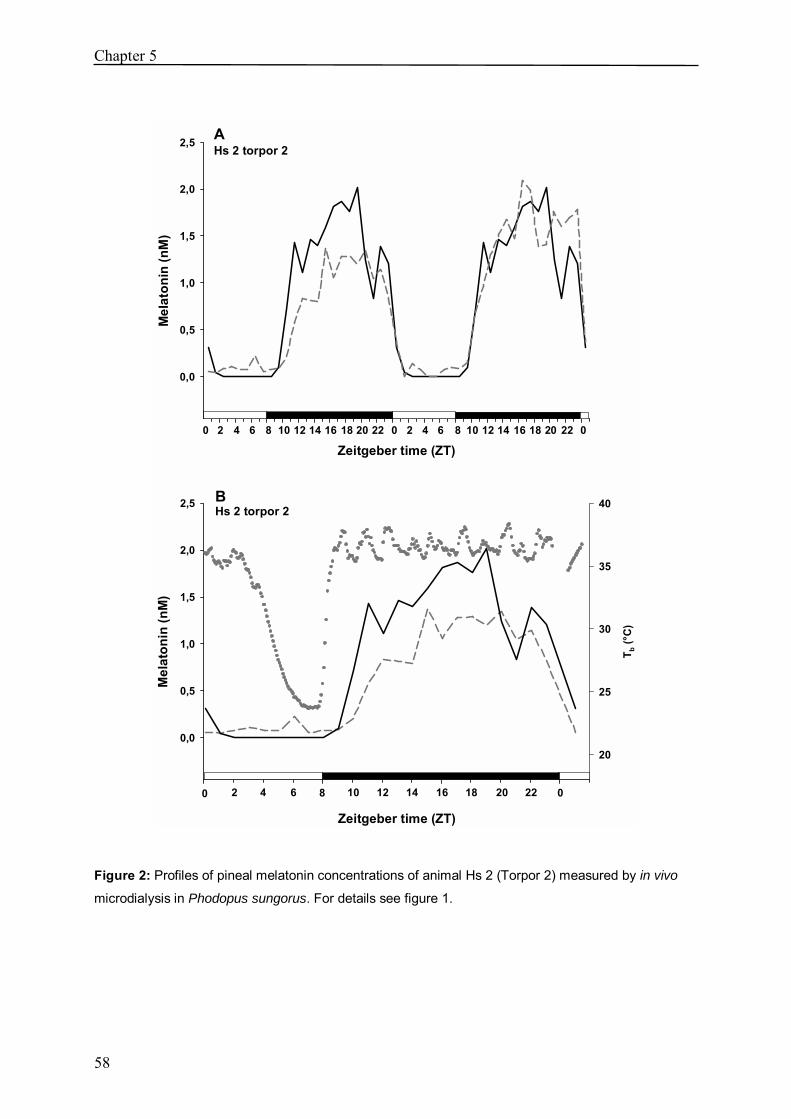
Chapter 5
58
B Hs 2 torpor 2
Zeitgeber time (ZT)
Mel
aton
in (n
M)
0,0
0,5
1,0
1,5
2,0
2,5
T b (°
C)
20
25
30
35
40
A Hs 2 torpor 2
Zeitgeber time (ZT)
Mel
aton
in (n
M)
0,0
0,5
1,0
1,5
2,0
2,5
00 020 2214 16 188 10 122 4 6 20 2214 16 188 10 122 4 6
0 2 4 6 8 10 12 14 16 18 20 022
Figure 2: Profiles of pineal melatonin concentrations of animal Hs 2 (Torpor 2) measured by in vivo
microdialysis in Phodopus sungorus. For details see figure 1.
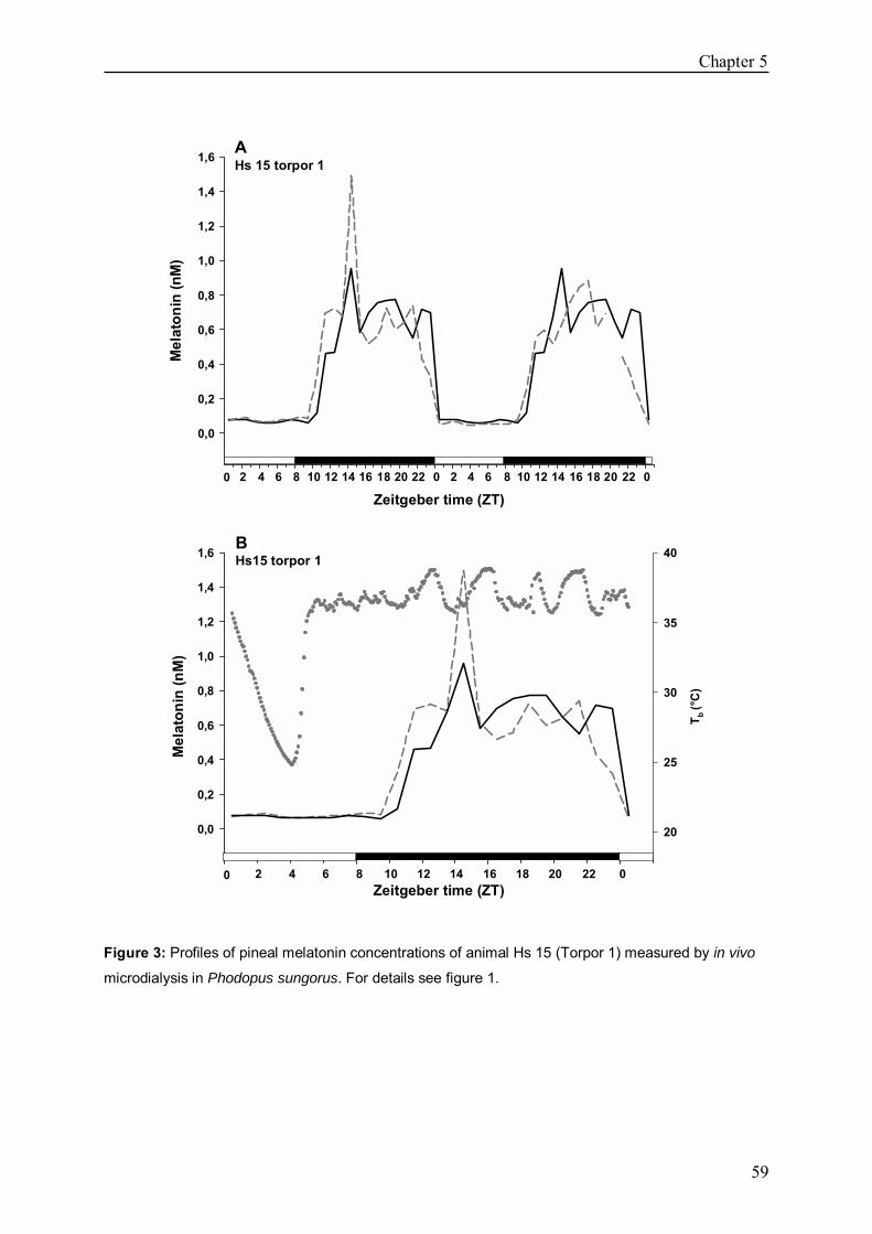
Chapter 5
59
0 2 4 6 8 10 12 14 16 18 20 022
A Hs 15 torpor 1
Zeitgeber time (ZT)
Mel
aton
in (n
M)
0,0
0,2
0,4
0,6
0,8
1,0
1,2
1,4
1,6
00 020 2214 16 188 10 122 4 6 20 2214 16 188 10 122 4 6
B Hs15 torpor 1
Zeitgeber time (ZT)
T b (°
C)
20
25
30
35
40
Mel
aton
in (n
M)
0,0
0,2
0,4
0,6
0,8
1,0
1,2
1,4
1,6
Figure 3: Profiles of pineal melatonin concentrations of animal Hs 15 (Torpor 1) measured by in vivo
microdialysis in Phodopus sungorus. For details see figure 1.
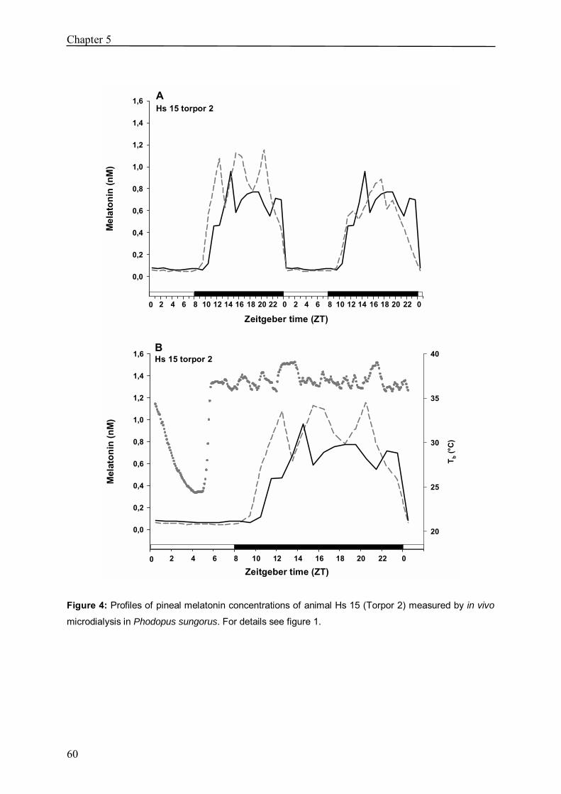
Chapter 5
60
A Hs 15 torpor 2
Zeitgeber time (ZT)
Mel
aton
in (n
M)
0,0
0,2
0,4
0,6
0,8
1,0
1,2
1,4
1,6
00 020 2214 16 188 10 122 4 6 20 2214 16 188 10 122 4 6
0 2 4 6 8 10 12 14 16 18 20 022
B Hs 15 torpor 2
Zeitgeber time (ZT)
T b (°C
)
20
25
30
35
40
Mel
aton
in (n
M)
0,0
0,2
0,4
0,6
0,8
1,0
1,2
1,4
1,6
Figure 4: Profiles of pineal melatonin concentrations of animal Hs 15 (Torpor 2) measured by in vivo
microdialysis in Phodopus sungorus. For details see figure 1.
Chapter 5
61
Discussion
During the course of the experiment, 5 torpor episodes displayed by three animals could be
observed. In all cases the torpor bout affected the onset of melatonin secretion in the night
directly following arousal. It has previously been hypothesised that �melatonin pulses� after a
torpor bout contribute to entrain and reset the circadian clock in the torpor season where
animals retreat to an enclosed nest or burrow to enter torpor and light exposure is rare (RUBY
2003).
From our results in chapter 2 and 3 where we showed an advanced onset of Aa-nat as well as
from the results of THOMAS et al. (1993), who showed a shortened τ in torpid Djungarian
hamsters, we expected that the melatonin secretion would be advanced after arousal from
torpor. This hypothesis is appealing because ecologically it would make sense. In DD, short
day adapted Djungarian hamsters usually free run with a τ longer than 24 hours. Thus, in
times with rare or no light exposure a regular phase advance after a torpor bout would ensure
that the animal´s endogenous rhythm remains closer to the external LD cycle. Moreover, the
sensitivity of the clock for melatonin is highest at the end of the day and very early night
(REDMAN et al. 1983, LEWI et al. 1992, REDMAN 1997). In this experiment we saw both
phase advance as well as phase delay of melatonin secretion after arousal from torpor.
Hs 2 showed two torpor episodes in which at both times the melatonin secretion was delayed.
Regarding the Tb data it is noticeable that the torpor bouts of this animal occur relatively late
on in the light phase and also the arousals are atypically late, i.e. already in the dark phase of
the LD cycle. A comparable pattern with a late arousal between ZT7 and ZT8 and delayed
melatonin secretion onset was observed in another animal (Hs 14, data not shown) where we
only have data during one torpor bout. At first sight this appears to point against a role for
melatonin resetting the clock, as an advance would be necessary to maintain entrainment.
However, the clock´s sensitivity for melatonin decreases soon after dark onset (REDMAN et
al. 1983, REDMAN 1997), so a delayed onset of melatonin secretion would probably have no
effect at all.
In contrast, Hs 15 showed an advanced melatonin release as expected. The Tb data indicate
that also the arousal from torpor occurred much earlier, namely in the early afternoon. This
might point to a direct relation between time of arousal and time of melatonin release onset.
Chapter 5
62
Such a relation has already been assumed by LARKIN et al. (2003) who demonstrated that
arousal from torpor is accompanied by melatonin production due to the strong sympathetic
activation during rewarming. Our observation would support this hypothesis. In Hs 15 the
melatonin secretion starts indeed only after ~5 hours after arousal (whereas in Hs 2 it is
elevated already 2 hours after rewarming), but this may well result from the presence of light
which strongly inhibits melatonin release (chapter 4). As soon as the lights go off, melatonin
concentrations rapidly increase.
Contrary to our results in chapter 2 and 3, where we observed a still advanced Aa-nat
expression on the day following a torpor bout, we did not see an aftereffect on the melatonin
release. It must be taken into account, though that in the experiments in chapter 2 and 3 we
chose animals for the control group that have never been observed torpidly before. In the
experiment in this chapter all investigated animals had displayed torpor before the experiment
to increase the possibility that they still would do so after surgery. It is likely that displaying
torpor generally alters the circadian organisation compared to animals that do not at all show
hypothermia.
However, with only three investigated animals we cannot draw firm conclusions and need to
study the issue of torpor affecting the circadian phase further.
Unfortunately none of the monitored animals showed torpor in DD. DD conditions started
only at the end of the experiments and either the microdialysis probes broke or the animals
did just not enter torpor.
Nevertheless, these results are a huge success, as the experimental setup is extremely difficult.
Already the trans-pineal microdialysis approach in such a small animal which weighs only
20-30 g in short day conditions is tricky and the rate of successfully implanted animals lies by
only 50%. In our setup it was not possible to monitor Tb and melatonin release more than 10
animals per experiment and the hourly melatonin sampling (which is done by collectors that
have to be emptied every 10 hours) is very demanding. Moreover, this experiment was critical
because we could not predict that the animals would display torpor at all. Torpor is a very
sensitive process as animals are very vulnerable in this condition. Therefore, if the animals are
disturbed or feel �unsafe� they do not enter hypothermia; so, we were very pleased to see that
even despite the implanted Mini-Mitters and the microdialysis probe the animals showed
hypothermia.

Chapter 5
63
This study is proof of the fact that the microdialysis approach is valuable in order to
investigate the effect of daily torpor on the melatonin release. Further, it can now confirm and
broaden the observations of these preliminary data.

64

65
6
The circadian clock stops ticking during hibernation.
Florent G. Revel*1, Annika Herwig*1,2, Marie-Laure Garidou1, Hugues
Dardente1, Jérôme S. Menet1, Mireille Masson-Pévet1, Valérie
Simonneaux1, Michel Saboureau1 and Paul Pévet1
* These authors contributed equally to this work
1 Département de Neurobiologie des Rythmes, Institut des Neurosciences Cellulaires et Intégratives,
UMR-7168/LC2, CNRS - Université Louis Pasteur, 5 rue Blaise Pascal, 67084 Strasbourg Cedex,
France 2 Institute of Zoology, University of Veterinary Medicine, Bünteweg 17, Building 218, D-30559
Hannover, Germany
Submitted

Chapter 6
66
Abstract
Hibernation represents one of the most fascinating, yet enigmatic physiological phenomena
(CAREY et al. 2003). During the harsh season this strategy allows substantial energy saving
by reducing body temperature and metabolism (CAREY et al. 2003, GEISER 2004).
Accordingly, biological processes are considerably slowed down and reduced to a minimum.
However, the persistence of a temperature-compensated, functional biological clock in
hibernating mammals has long been debated (KÖRTNER et al. 2000, RUBY 2003, HELLER
and RUBY 2004). Here we show that the master circadian pacemaker no longer displays 24-h
molecular oscillations under hypothermia, suggesting an arrest of the clock. We found that in
the suprachiasmatic nucleus (SCN) of the European hamster, the expression of the clock
genes Per1, Per2 and Bmal1 remains constant over 24-h in hypothermic animals. Similarly,
the expression of the clock-controlled gene arginine-vasopressin is constantly suppressed.
Finally, the melatonin rhythm generating enzyme, arylalkylamine-n-acetyl transferase,
rhythmic expression in the pineal gland being controlled by the SCN, no longer displays day-
night changes of expression, but constantly elevated mRNA levels over 24-h. Our results
strongly suggest that during hibernation the circadian clock is stalled at the molecular level,
and may no longer deliver rhythmic output signals.
Text
In mammals, the hibernation season consists of recurring bouts of hypothermia (or torpor)
alternated with short periods of euthermia, during which body temperature (Tb) returns to
~37 °C (KILDUFF et al. 1993, KÖRTNER and GEISER 2000, CAREY et al. 2003, RUBY
2003, GEISER 2004). During deep torpor, energetically expensive cellular processes like
transcription and translation are severely depressed, and physiological functions like heart
rate, respiration, immune and renal functions, and neural activity run at greatly reduced rates
(CAREY et al. 2003, GEISER 2004). The circadian system temporarily coordinates internal
biological processes to ensure health and survival (KALSBEEK and BUIJS 2002,
HASTINGS and HERZOG 2004, BELL-PEDERSEN et al. 2005). Circadian rhythms are
Chapter 6
67
temperature compensated and run at a relatively constant pace under various temperatures
(BELL-PEDERSEN et al. 2005). However, it has long been debated whether the circadian
system remains functional during deep hibernation (KÖRTNER and GEISER 2000, RUBY
2003, HELLER and RUBY 2004). Although several studies have already addressed this
question, uncertainty prevails since clock outputs rather than core clockwork mechanisms
were examined (FRENCH 1977, FLORANT et al. 1984, VANECEK et al. 1984, 1985,
STANTON et al. 1986, KILDUFF et al. 1989, GRAHN et al. 1994, CANGUILHEM et al.
1994, WOLLNIK and SCHMIDT 1995, HUT et al. 2002, RUBY et al. 2002, LARKIN et al.
2002). In mammals, the master circadian clock is located in the suprachiasmatic nuclei of the
hypothalamus (SCN; KALSBEEK and BUIJS 2002, HASTINGS and HERZOG 2004, BELL-
PEDERSEN et al. 2005). Circadian oscillations within SCN neurons result from recurrent
expression of so-called clock genes that interact in complex, interlocked transcription /
translation feedback loops (HASTINGS and HERZOG 2004, BELL-PEDERSEN et al. 2005).
Assessing such molecular oscillations in the SCN of hibernating animals is a valuable
approach for reconsidering the issue of a functional circadian clockwork during deep torpor.
For this we used the European hamster (Cricetus cricetus), a well-defined hibernator
(HERMES et al. 1989, CANGUILHEM et al. 1994, WOLLNIK and SCHMIDT 1995,
MAGARINOS et al. 2006). Hamsters raised outdoor were transferred in September to a
climatic room kept at 6±2 °C under short photoperiodic regimen. Individual Tbs were
recorded via a telemetric system. Under these conditions, comparable to outdoor conditions in
December, the bouts of hypothermia characteristic of the hibernation cycle were regularly
expressed (Fig. 1a; CANGUILHEM et al. 1994, MAGARINOS et al. 2006). During deep
torpor, basal metabolic rate is typically reduced to 2-4% of euthermic rates, and Tb drops
close to ambient temperature (Ta; CAREY et al. 2003, GEISER 2004). In this experiment, the
Tb of hypothermic hamsters was 8-10 °C (Fig. 1b). The European hamster enters hibernation
preferentially around mid-night (CANGUILHEM et al. 1994, WOLLNIK and SCHMIDT
1995). We took advantage of this characteristic to design our experiment, as illustrated in
Figure 1c. Radioactive in-situ hybridisation was used to assess gene expression in the SCN of
euthermic versus hypothermic hamsters, killed during daytime (D), nighttime (N) or during
the subjective day (SD).

Chapter 6
68
Figure 1: European hamster�s hibernation pattern and experimental paradigm. a, b, Typical body
temperature (Tb) recording (animal #B17) illustrating the major phases of the hibernation cycle of the
European hamster. a, Three weeks of recording (25th Nov. to 14th Dec., 2005) showing 5 hibernation
cycles. The horizontal black bar represents the period detailed in b. The ambient temperature of the
climatic room was set at 6±2 °C (dotted line). b, Focus on a single hibernation cycle (6th Dec. to 10th
Dec., 2005). Entrance into hibernation (Tb < 35 °C) preferentially occurred during night-time. c,
Experimental paradigm used for the experiment. For the euthermic group, the hamsters had a Tb close
to 37 °C, and were aroused from hibernation for more than 48-h. These animals were sacrificed at
mid-day (13:00; D, day; n = 6), mid-night (01:00; N, night; n = 6) or mid-subjective day (13:00;
darkness from the previous day; SD, subjective day; n = 5). For the hypothermic group, only the
animals entering hibernation (Tb < 35 °C) between 00:00 and 02:00 (mid-night) were considered. Their
sacrifice occurred 36-h (D, day; n = 4), 48-h (N, night; n = 5) or 60-h (SD, subjective day; no lights on
for the last day; n = 7) later. Light and dark bars: day and night; Dark-grey bar: subjective day.

Chapter 6
69
In the molecular core of the clock, the transcription factors CLOCK and BMAL1 drive the
expression of the Period (Per) and Cryptochrome (Cry) genes, and in turn PER and CRY
proteins heterodimerize and repress CLOCK/BMAL1-driven transcription (HASTINGS and
HERZOG 2004, BELL-PEDERSEN et al. 2005). To compare the state of these molecular
loops during hypo- versus euthermia, we examined the expression of 3 major clock genes:
Per1, Per2 and Bmal1 (Fig. 2a,b,c). As expected in euthermic animals significant day-night
changes of expression were observed, with high expression of Per1 and Per2 during the day
and on the subjective day, and high expression of Bmal1 during night-time. In contrast to
euthermic animals, no day-night change of expression occurred in the SCN of hypothermic
hamsters (Fig. 2a,b,c). In this condition, Per1 mRNA levels were persistently elevated (Fig.
2a), whereas those of Per2 were constantly depressed (Fig. 2b). For Bmal1, the nocturnal
peak of expression was lost (Fig. 2c), and mRNA levels were intermediate between daytime
and night-time values in euthermic hamsters. All in all, these results show that the robust day-
night oscillations of Per1, Per2 and Bmal1 expression disappear during hibernation, strongly
suggesting that the circadian clockwork ceases to function. A potential slow-down of the
circadian oscillations and extended period of rhythm seem unlikely, because gene expression
was not systematically restricted to intermediate levels. Importantly, we observed that the
circadian molecular oscillations were clearly re-expressed during the interbout intervals. This
�recovery� and re-synchronisation of the clock was presumably facilitated by the presence of
the light-dark cycle.
If the molecular circadian clock is stopped during hypothermia, then the 24-h rhythm of its
output signals should be lost Thus, we examined the expression of arginine-vasopressin
(Avp), a clock-controlled gene whose rhythmic expression in the SCN directly depends upon
the molecular circadian machinery (JIN et al. 1999, KALSBEEK and BUIJS 2002,
HASTINGS and HERZOG 2004). As expected, Avp expression displayed a clear day-night
change of expression in euthermic hamsters (Fig. 2d), with high mRNA levels during the day
and on the subjective day. This was in marked contrast to hypothermic animals, in which Avp
expression was persistently depressed (Fig. 2d). This result confirms and extends our previous
conclusion, suggesting that the SCN molecular clockwork and at least one of its molecular
output cease during hypothermia.
Interestingly, the SCN maintain a relatively high metabolic activity during hibernation, as
assessed by [14C] 2-deoxyglucose uptake and c-Fos expression {KILDUFF et al. 1982, 1989

Chapter 6
70
BITTING et al. 1994). In the present study, examination of c-Fos mRNA levels in the SCN
also revealed a dramatic increase of expression in hibernating hamsters relative to euthermic
animals (Fig. 2e), without significant day-night differences. This suggests that the loss of
circadian organisation in the SCN of hibernating hamsters does not simply result from
reduced metabolic activity or transcription, and favours the hypothesis that the SCN may
serve a non-circadian role during hibernation (RUBY 2003).
Finally, we questioned whether hibernation equally affects the rhythmic activity of SCN
targets. Synthesis of melatonin by the pineal gland represents one of the best characterised
outputs of the SCN (KALSBEEK and BUIJS 2002, PERREAU-LENZ 2003, GARIDOU et
al. 2003, BELL-PEDERSEN et al. 2005). The SCN restricts production of melatonin to night-
time by controlling the activity of its rhythm-generating enzyme, the arylalkylamine-N-
acetyltransferase (AA-NAT). In the European hamster, the daily variations of AA-NAT
activity result from changes in transcription of the Aa-nat gene (GARIDOU et al. 2003). The
expression of this gene was therefore used as an index of the pineal gland activation by the
SCN. As expected in euthermic animals, Aa-nat expression was increased more than 17 fold
during night-time (Fig. 3a, GARIDOU et al. 2003). This was faithfully translated into changes
of plasma melatonin, as measured by radioimmunoassay (Fig. 3b). In contrast, Aa-nat
expression was persistently high during hibernation (Fig. 3a), comparable to that in euthermic
animals during night-time, suggesting that melatonin synthesis is no longer rhythmic. This
was confirmed at the melatonin level, although plasma melatonin concentrations were
continuously low during hypothermia, as already described in other species. Interestingly,
plasma melatonin level has been reported to rise rapidly after arousal from hibernation,
whatever the time of arousal (FLORANT et al. 1984, VANECEK et al. 1984, 1985,
STANTON et al. 1986). The fact that Aa-nat mRNA levels remain persistently elevated
during hibernation may well explain these observations, and suggests a possible decoupling
between transcriptional activity, and protein synthesis / enzymatic function.

Chapter 6
71
Figure 2: The circadian expression of clock genes in the suprachiasmatic nucleus is eradicated under
deep hibernation. a-e, In-situ hybridisation was used to examine gene expression in euthermic and
hypothermic hamsters during daytime (D), night-time (N) or during the subjective day (SD). For all
genes, the upper panel displays representative autoradiograms for each condition; the lower panel
shows the quantified mRNA levels. Expression of the clock genes Per1 (a), Per2 (b) and Bmal1 (c)
exhibits circadian fluctuations in euthermic hamsters (Per1, Per2: P < 0.001, N versus D and SD;
Bmal1: P = 0.008, N versus D and SD), whereas their mRNA level is constant in hypothermic animals

Chapter 6
72
(Per1, Per2, Bmal1: P > 0.05). During hibernation, Per1 expression is elevated (a; no significant
difference with daytime levels in euthermic animals; P > 0.05), Per2 expression remains low (b; no
statistical difference with night-time levels in euthermic animals; P > 0.05), and Bmal1 expression is
intermediate between daytime and night-time levels in euthermic hamsters (c; P > 0.05 versus D and
SD; P > 0.05 versus N). Similarly, the expression of the clock controlled gene Avp (d) is rhythmic in
euthermic (P < 0.001, N versus D and SD), but not in hibernating hamsters (P > 0.05), in which its
expression remains low (for all hypothermic conditions, P > 0.05 versus euthermic N, and P < 0.001
versus euthermic D and SD). The expression of the immediate early gene c-Fos (e) is dramatically
increased in hypothermic relative to euthermic hamsters (P < 0.001), without significant day-night
fluctuations (P > 0.05). Data are shown as percentage of the maximum and represent the means ±
s.e.m. (n = 4-7). Error bars denote s.e.m.. Different characters (a, b, c) indicate significant differences
(P < 0.05).
Figure 3: Aa-nat is constitutively expressed in the pineal gland of hibernating hamsters. a,
Representative autoradiograms and quantification of Aa-nat mRNA demonstrate marked day-night
changes of expression in euthermic animals (P < 0.001, N versus D and SD), while in hypothermic
hamsters it is persistently expressed (P > 0.05) and displays high mRNA levels (for all hypothermic
conditions, P > 0.05 versus euthermic N, and P < 0.001 versus euthermic D and SD). Data are shown
as percentage of the maximum and represent the means ± s.e.m. (n = 4-7). Error bars denote s.e.m..
***, p < 0.001. b, A rhythmic concentrations of plasma melatonin is observed in euthermic animals (P
< 0.001, N versus D and SD), but not in hibernating hamsters which exhibit constantly low plasma
melatonin levels (for all hypothermic conditions, P > 0.05 versus euthermic D and SD). Data are
means ± s.e.m. (n = 4-7). Error bars denotes s.e.m. . ***, p < 0.001.

Chapter 6
73
Overall, our results show that the circadian clock of the SCN stops generating 24-h rhythms
during hibernation. This is the first physiological condition in which an arrest of the master
circadian clock over several days is described. The issue has been debated for many years
with various evidence either denying or supporting the existence of a functional pacemaker
during deep torpor (FRENCH 1977, FLORANT et al. 1984, VANECEK et al. 1984, 1985,
STANTON et al. 1986, KILDUFF et al. 1989, GRAHN et al. 1994, WOLLNIK and
SCHMIDT 1995, KÖRTNER and GEISER 2000, RUBY et al. 2002, LARKIN et al. 2002,
HUT et al. 2002, RUBY 2003, HELLER and RUBY 2004). These contradictions may stem
from species difference as well as from the variety of markers used to assess the functional
state of the clock. Based on the recording of small circadian fluctuations in Tb, some reports
have suggested the maintenance of circadian rhythms in hibernating ground squirrels
(GRAHN et al. 1994, RUBY et al. 2002, LARKIN et al. 2002). However, consistent with
other reports we did not detect such variations in the European hamster (CANGUILHEM et
al. 1994, WOLLNIK and SCHMIDT 1995, HUT et al. 2002,). In this study we assessed gene
transcription. Thus, we cannot rule out the possibility that the circadian clock might continue
to function based on separate mechanisms like post-transcriptional regulations. Furthermore,
the technique we used may not be suitable for detecting either small subsets of cells that retain
temperature-compensated time measurement, or desynchronization amongst individual SCN
oscillators (GRAHN et al. 1994, HELLER and RUBY 2004). However, we did not
systematically observe intermediate mRNA levels, and the rhythmicity of both Aa-nat
expression and plasma melatonin levels were lost during hibernation. The possibility that an
extra-SCN oscillator could take over the circadian clock function is also unlikely, since the
SCN is one of the most active brain structure during hibernation (KILDUFF et al. 1982,
1989). Furthermore, we and others previously demonstrated that arousals are randomly
distributed over 24-h, supporting the absence of a circadian timer during hibernation
(CANGUILHEM et al. 1994, WOLLNIK and SCHMIDT 1995). Electrophysiological studies
indicate that the circadian clock of hibernators still functions at temperatures lower than 37
°C, but is likely to stop at temperatures below 16.6 °C (MILLER et al. 1994, RUBY 2003,
HELLER and RUBY 2004). The absence of action potentials, together with the vanishment of
rhythmic oscillation in clock gene expression, make temperature compensation unlikely at
low Tb (5-10 °C). This property of the clock may be limited to a range of temperatures, as
rhythmic clock gene expression still occurs during daily torpor, a shallower form of

Chapter 6
74
hypothermia during which Tb decreases to 15-20 °C for only several hours (HERWIG et al.
2006b).
In conclusion, our data demonstrate that in the European hamster the molecular clock of the
SCN is arrested during deep hibernation and no longer delivers circadian signals.
METHODS
Animals All experiments were performed in accordance with the rules of the French Department of
Agriculture (license no 67-38) and the European Committee Council Directive of November
24, 1986 (86/609/EEC). The adult male European hamsters used in this study were bred in-
house. The colony was established from animals caught in the fields near Strasbourg, France
between 1994 and 1996 Hamsters were raised under semi-natural conditions (natural
photoperiod) with ad libitum access to water and food. In September, animals born in May of
the previous year (16-17 months old) were transferred to a climatic room kept at 6±2 °C,
under a 10-h light (150 lux; lights off at 18:00) / 14-h dark (2 lux dim red light) cycle. The
core body temperature (Tb) was continually registered via thermo-sensitive radio-transmitters
(Model VM-FH-LT, Mini-Mitter Co., Sunriver, OR, USA) implanted in the abdominal cavity
under halothane anesthesia, before placing the hamsters in the climatic room. Radiofrequency
signals from the implanted transmitters were averaged every 5 min by receivers placed under
each cage and collected by an automated computer software (Dataquest, St. Paul, MN). Straw
was given as nest-building material. All the animals considered in this study had completed at
least 3 bouts of hypothermia. Sacrifice occurred as depicted in Figure 1b. Animals were
anesthetised with N2O and killed by decapitation. Brains were rapidly removed, snap-frozen
at -30 °C, and stored at -80 °C until in-situ hybridization. Trunk blood was collected,
centrifuged at 1,500 x g for 15 min and plasma was stored at -20 °C until melatonin assay.
In-situ hybridisation Radioactive in-situ hybridisation was performed as previously described (GARIDOU et al.
2003), using riboprobes for rat Period1 (bases 638-1618 from GenBank accession number

Chapter 6
75
NM_001034125), Period2 (bases 1170-1930 from NM_031678; donated by Dr. H. Okamura,
Department of Anatomy and Brain Science, Kobe University School of Medicine, Japan),
Bmal1 (bases 75-1809 from AB012600) and arginine vasopressin (bases 68-539 from
M25646), or for Syrian hamster c-fos (bases 625-1025 from AF061881) and Aa-nat (bases 1-
1045 from AF092100). After they were fixed, acetylated, and dehydrated, 20-µm thick
coronal brains sections were hybridised overnight at 54 °C with 35S-UTP labelled riboprobes
The sections were then treated with Rnase (10 µg/ml), washed (SSC 0.1x, 60 °C), dehydrated
and exposed to a BioMax MR Film (Kodak) for 3-5 days along with 14C-radioactive standards
to allow standardisation of densitometric measurements across films. X-ray films were
scanned on an Epson 4990 transmittance scanner, and background subtracted, calibrated
optical density (OD) measurements of mRNA levels were performed using ImageJ (NIH).
Melatonin radioimmunoassay Plasma melatonin was analysed by a radioimmunoassay previously validated in European
hamsters and described in the reference (GARIDOU et al. 2003).
Data and Statistical Analyses Experiments were performed twice with similar results, and the data reported here are those
from the second experiment. Results are shown as a percentage of the maximum and
represent the mean ± SEM. Data were analysed by two-way analysis of variance (ANOVA),
followed by HSD Tukey�s analysis. Statistical significance was set at P < 0.05.
Acknowledgements We wish to thank D. Bonn and A. Senser for taking care of the animals; Prof. André Malan,
Dr. Etienne Challet and Dr. Jorge Mendoza for helpful discussions; Dr. David Hicks for
revision of the English language. This work was supported by the Women�s support of the
University of Veterinary Medicine Hannover (TiHo; A.H.), the ACI �Plates-formes
d�explorations fonctionnelles thématiques� from the Ministère Délégué à la Recherche (P.P.)
and the Centre National de la Recherche Scientifique.

Chapter 6
76
Authors` contributions
FGR and AH contributed equally to this work, and performed all molecular and statistical
analysis. PP conceived the original experiment and coordinated the study. MS performed the
physiological studies and monitored the animals on a daily basis for several years. MLG, HD,
JSM, MMP and VS performed the preliminary experiments. All authors discussed the results
and commented on the manuscript.
77
7
Histamine H3 receptor and Orexin A expression
during daily torpor in the Djungarian hamster
(Phodopus sungorus).
Annika Herwig1, Elena A. Ivanova2, Helen Lydon2, Perry Barrett3,
Stephan Steinlechner1 and Andrew S. Loudon2
1 Institute of Zoology, Universtiy of Veterinary Medicine, Hannover, Germany 2 Faculties of Life Sciences, University of Manchester, M13 9PT, UK 3 Rowett Research Institute, Aberdeen Centre for Energy Regulation and Obesity (ACERO),
Bucksburn Aberdeen, AB21 9SB UK
Submitted

Chapter 7
78
Abstract
Controlled hypometabolism and hypothermia lead to a reduction in CNS activity.
Nevertheless, brain systems of importance for controlling and maintaining the hypothermic
state have been shown to increase activity. During hibernation the histaminergic system
seems to actively inhibit other neurotransmitter pathways through the inhibitory auto � as well
as hetero-histamine H3 receptor. In deep hypothermia H3 receptors have been shown to be
up-regulated in several brain areas. In this study we demonstrate an up-regulation of H3
receptors in several brain nuclei including arcuate nucleus, dorsomedial hypothalamus,
suprachiasmatic nucleus dorsal lateral geniculate nucleus and tuberomammilary nucleus
during daily torpor, a shallow form of hypothermia in the Djungarian hamster (Phodopus
sungorus). This suggests that inhibition through histaminergic pathways may play a more
general role in maintaining low body temperature and torpor state in mammals. Moreover, we
showed that mRNA for orexins, a group of neuropeptides that have been shown to increase
wakefulness, remain unchanged during the arousal from daily torpor. Therefore, it is likely
that this �arousal� pathway is not involved in the arousal from hypothermia.
Introduction
Controlled hypothermia is a powerful strategy for reducing the high metabolic costs of a
constant body temperature (Tb) during periods of prolonged food shortage. Different forms of
hypothermia have evolved. In contrast to long enduring hibernation bouts lasting several days,
some small mammals undergo bouts of daily torpor where the circadian timing system is used
to precisely time hypo-metabolism and hypothermia (HELDMAIER et al. 2004). Decreasing
day-length provides an anticipatory mechanism of acclimatisation to winter for the
Djungarian hamster, also known as Siberian hamster (Phodopus sungorus), evoking
physiological adaptations to winter conditions, including daily torpor. During a torpor bout
the extent of hypothermia and hypo-metabolism is less pronounced than in deep hibernation
and consequently less energy is saved (HELDMAIER and RUF 1992, GEISER and
HELDMAIER 1995). However, the physiological properties are very similar, and simply

Chapter 7
79
represent gradual differences in timing, duration and amplitude of an analogous physiological
state.
Invariably is associated with a depression of CNS activity but it is crucial that functional
integrity is maintained. The hibernating brain needs to accurately control the complex
metabolic processes and to remain sensitive to external and internal stimuli (HELLER 1979).
It has been shown that the histaminergic system, involved in the regulation of sleep-wake
cycle, Tb and energy metabolism (SCHWARTZ et al. 1991), is important in initiating and
maintaining the hibernating state in ground squirrels (Citellus lateralis) (SALLMEN et al.
1999, 2003a,b). Contrary to many studies that have shown a general depression of neuronal
transcription and translation activity in this state (FRERICHS et al. 1998, VAN
BREUKELEN et al. 2002a), perhaps serving to maintain homeostatic balance in extremely
hypoxic and nutrient deficient periods, histamine turnover in the hypothermic brain is known
to increase. The increased histamine activity seems to be linked to the up-regulation and
elevated binding of the inhibitory histamine H3 receptors in several brain areas including
cortex, caudate nucleus and putamen as well as globus pallidus and substantia nigra
(SALLMEN et al. 2003a). The inhibitory Gi/o protein coupled receptor has auto- as well as
heteroreceptor functions and it is likely that histamine thereby influences and regulates other
neurotransmitter systems. In the Siberian hamster, H3 receptors are expressed in various
nuclei of the hypothalamus, the principal brain region controlling autonomic functions and for
which a role for seasonal energy regulation has been assumed (BARRETT et al. 2005). In this
study we investigated whether H3 receptors are also involved in controlling daily torpor. If
torpor indeed underlies mechanisms comparable to deep hibernation, the histaminergic
system might also actively inhibit CNS activity and H3 receptors should be up-regulated in
the brain of torpid Djungarian hamsters.
Molecular mechanisms underlying and preparing the arousal from daily torpor, accompanied
by strong activation of the sympathetic nervous system, are still unknown. Orexins are a
group of peptides that have been linked to the arousal state of animals (RODGERS et al.
2001, ISHII et al. 2005). Orexin neurons are mainly located in the lateral hypothalamus and
project to other hypothalamic regions such as the paraventricular, arcuate, lateral, perifornical
and ventromedial nuclei, areas that have all been associated with feeding and wakefulness
(KHOROOSHI et al. 2005, KOTZ 2006). The absence of a functional orexin OX2R receptor
causes narcolepsy in dogs and the absence of the peptide orexin in humans also results in
Chapter 7
80
narcolepsy whilst orexin administration enhances arousal (KOTZ 2006). As orexins have also
been related to non-shivering thermogenesis in brown adipose tissue (YASUDA et al. 2005,
KOTZ 2006), i.e. the principal mechanism for regaining normal Tb after hypothermia, we also
investigated whether orexin may be important for the arousal from daily torpor.
Material and Methods Animals Male Djungarian hamsters (Phodopus sungorus) were raised in the breeding colony of the
University of Veterinary Medicine, Hanover, Germany. Animals were born between April
and July and raised under natural photoperiod. Body weight and pelage score were monitored
to ensure hamsters responded appropriately to changing photoperiod (FIGALA et al. 1973).
Animals were implanted with radiofrequency transmitters to monitor body temperature and
torpor state. They were acclimatised to short photoperiod of 8:16h light:dark for 2 weeks prior
to transfer to climatic chambers with an ambient temperature (Ta) of 18 °C on the same
photoperiod. Brains were removed during entry into torpor (n=6; Tb:30,94±1,61 °C) at ZT1
with ZT0 corresponding to �lights-on�, deep torpor (n=7; Tb:23,87±1,34 °C) at ZT4, arousal
(n=6; Tb:33,63±1,23 °C) at ZT8 and in a normothermic state following the torpor bout (n=6;
ZT12). Control brains of normothermic animals were sampled at the same time points (n=6
each). Brain tissue was rapidly removed and frozen on dry ice. Cryostat sections of 14 µm
thickness were thaw mounted on SuperfrostPlus® sildes and stored at -80 °C until further use.
In situ hybridisation A probe for the histamine H3 receptor has been described previously (10). 33P-UTP
(3000Ci/mmol, ICN) labelled sense and antisense riboprobes were generated with SP6
polymerase (Sph1 digestion) and T7 polymerase (Spe1 digestion), respectively. The probes
were purified with Chromaspin 100 columns (BD Bioscience) Sections were fixed for 15 min
in 4% paraformaldehyde, rinsed twice in 1× PBS 5 min each and then acetylated for 10 min in
0.1 µM triethanolamine, 0.375% acetic anhydride. After rinsing in 1× PBS for 5 min, sections
were dehydrated in graded ethanol series (70%, 80%, 90%, 95%, 100%, and Chloroform 5
min each) and finally dried at room temperature (RT). The cover-slipped slides were
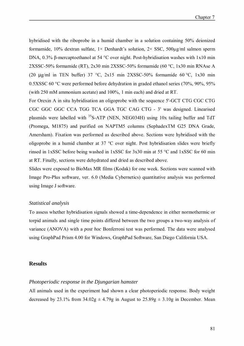
Chapter 7
81
hybridised with the riboprobe in a humid chamber in a solution containing 50% deionized
formamide, 10% dextran sulfate, 1× Denhardt�s solution, 2× SSC, 500µg/ml salmon sperm
DNA, 0.3% β-mercaptoethanol at 54 °C over night. Post-hybridisation washes with 1x10 min
2XSSC-50% formamide (RT), 2x30 min 2XSSC-50% formamide (60 °C, 1x30 min RNAse A
(20 µg/ml in TEN buffer) 37 °C, 2x15 min 2XSSC-50% formamide 60 °C, 1x30 min
0.5XSSC 60 °C were performed before dehydration in graded ethanol series (70%, 90%, 95%
(with 250 mM ammonium acetate) and 100%, 1 min each) and dried at RT.
For Orexin A in situ hybridisation an oligoprobe with the sequence 5'-GCT CTG CGC CTG
CGC GGC GGC CCA TGG TCA GGA TGC CAG CTG - 3' was designed. Linearised
plasmids were labelled with 35S-ATP (NEN, NEG034H) using 10x tailing buffer and TdT
(Promega, M1875) and purified on NAPTM5 columns (SephadexTM G25 DNA Grade,
Amersham). Fixation was performed as described above. Sections were hybridised with the
oligoprobe in a humid chamber at 37 °C over night. Post hybridisation slides were briefly
rinsed in 1xSSC before being washed in 1xSSC for 3x30 min at 55 °C and 1xSSC for 60 min
at RT. Finally, sections were dehydrated and dried as described above.
Slides were exposed to BioMax MR films (Kodak) for one week. Sections were scanned with
Image Pro-Plus software, ver. 6.0 (Media Cybernetics) quantitative analysis was performed
using Image J software.
Statistical analysis
To assess whether hybridisation signals showed a time-dependence in either normothermic or
torpid animals and single time points differed between the two groups a two-way analysis of
variance (ANOVA) with a post hoc Bonferroni test was performed. The data were analysed
using GraphPad Prism 4.00 for Windows, GraphPad Software, San Diego California USA.
Results
Photoperiodic response in the Djungarian hamster All animals used in the experiment had shown a clear photoperiodic response. Body weight
decreased by 23.1% from 34.02g ± 4.79g in August to 25.89g ± 3.10g in December. Mean

Chapter 7
82
pelage score changed during this time from 1 to 4 (according to 16), that is from summer
agouti colour to nearly white winter coat.
Body temperature Radio-telemetry successfully revealed the daily body temperature and torpor response in our
Djungarian hamsters. Hamsters entered torpor approximately 1h before lights on and
terminated torpor approximately 8h later at lights off and showed a 17 °C drop in body
temperature compared to normothermic hamsters (Fig. 1a,b)
Histamine H3 receptor (H3R) mRNA expression
H3R expression is shown in Figure 2. Expression was significantly up-regulated during torpor
in the arcuate nucleus (ARC) (p<0.001; Fig. 2a), dorso medial hypothalamus (DMH) (p<0.01;
Fig. 2b), the suprachiasmatic nucleus (SCN) (p<0.050; Fig. 2c), dorsal lateral geniculate
nucleus (DLG) (p<0.010; Fig. 2d) and tuberomammilary nucleus (TM) (p<0.001; Fig. 2g).
Within the suprachiasmatic nucleus, H3R expression showed a diurnal change, with lowest
expression at ZT8 and peak expression between ZT1 and ZT4 (p=0,005; Fig. 2c) in
normothermic animals. This rhythm was eradicated in torpid animals where expression was
upregulated at ZT8 (p<0.050; Fig. 2c). Also in the tuberomammilary nucleus H3R expression
appeared to be time dependent (p=0.048) in the normothermic animals with high night-time
expression compared to low daytime levels. No significant changes were observed in the
cortex (Fig. 2e), caudate/putamen (Fig. 2f), ventromedial hypothalamic nucleus (VMH, data
not shown), and ventral posterior medial nucleus of the thalamus (VPM, data not shown). No
time-dependent expression was seen in other structures in torpid or normothermic animals.
Orexin A mRNA expression
Orexin A mRNA expression was confined to the lateral hypothalamus (LHA; Fig. 3).
Expression was rhythmical in normothermic animals (ANOVA p=0.04) with highest
expression at ZT4 and lowest at ZT12. There was no rhythmical change in expression
observed in torpid hamsters. In torpid animals, orexin A mRNA was significantly down-
regulated relative to normothermia animals at ZT4 (t-test p=0.03), and also during arousal at
ZT8 (t-test p=0.04).

Chapter 7
83
Fig 1
Tb °C
A Tb #5
12.00 20.00 4.00 12.00 20.00 4.00
20
25
30
35
40
time(hr)
Tb #7
12.00 20.00 4.00 12.00 20.00 4.00
Tb °C
20
25
30
35
40
B
Figure 1: Representative body temperature rhythms (Tb) in short photoperiod adapted Djungarian
hamsters (LD8:16). Panel A illustrates Tb of a normothermic animal, panel B of an individual that
displayed daily torpor at an ambient temperature (Ta) of 18±2 °C. Tb was recorded at 6 min intervals
with i.p. implanted radiofrequency transmitters. Black arrows indicate the sampling points at ZT1, ZT4,
ZT8 and ZT12, with ZT0 corresponding to lights on.

Chapter 7
84
Figure 2: H3 receptor expression in various brain regions of normothermic (black bars, n=6) and
hypothermic (grey bars, n=6) Djungarian hamsters. Animals were kept in short photoperiod (LD8:16).
Samples were taken at ZT1, ZT4, ZT8 and ZT12 (ZT0=lights on) along the torpor cycle and hybridised
with a 33P labelled H3 receptor riboporobe. Significantly increased H3 receptor mRNA levels were
found in the ARC (panel A, p<0.001), DMH (panel B, p<0.050), SCN (panel C, p<0.010 ), DLG (panel
D, p<0.010) and TM (panel G, p<0.001) during torpor, but not in cortex (panel E) or caudate putamen
(panel F). Statistical significances are indicated through * for p<0.05. The upper and lower images
represent sections from torpid and normothermic animals respectively at ZT4 in the ARC, DMH, DLG,
TM, Cortex, Caudate Putamen, at ZT8 in the SCN.
Chapter 7
85
Figure 3: Orexin A expression in the LHA of normothermic (black bars, n=6) and hypothermic (grey
bars, n=6) Djungarian hamsters. Animals were kept in short photoperiod (LD8:16). Samples were
taken at ZT1, ZT4, ZT8 and ZT12 (ZT0=lights on) along the torpor cycle and hybridised with a 35S
labelled orexin A oligoprobe. Orexin A mRNA was significantly reduced during torpor (ZT4, p=0.03) as
well as arousal (ZT8, p=0.04). The upper and lower panels show representative sections from
normothermic and torpid animals, respectively.
Discussion
H3 receptors We show significant increases of histamine H3 receptor mRNA in the arcuate nucleus,
dorsomedial hypothalamus, suprachiasmatic nucleus, dorsal lateral geniculate nucleus and
tuberomammilary nucleus during daily torpor. Increased histamine H3 receptor mRNA
expression during hypothermia has already been described in the brain of hibernating golden-

Chapter 7
86
mantled ground squirrels (SALLMEN 2003a). Our data in a torpid model suggest that the
histaminergic system may play an important role in the hibernation process � a state where
transcription as well as translation are generally depressed (CHEN et al. 2001, VAN
BREUKELEN and MARTIN 2001, 2002a,b). During torpor, the animal takes advantage of
low Tb to induce a profound reduction of metabolic rate, but specific adaptations exist to keep
the cellular and molecular machinery functional in order to ensure a rapid reactivation during
inter-bout arousal (KNIGHT et al. 2000, VAN BREUKELEN and MARTIN 2002a,b).
SALLMEN et al. 2003 suggested that the specific up-regulation of inhibitory H3 receptors
could mediate the active down-regulation of various neurotransmitter systems at cold
temperatures and may therefore be important in the control of hibernation state. Our data,
showing significant changes in expression over the course of a shallow torpor bout, suggest
that deep hibernation and daily torpor may share common histamine-dependent features over
the course of a low-temperature bout.
In our study H3 receptors were up-regulated in various hypothalamic nuclei implying
involvement in metabolic processes and autonomic regulation. As previously described
(BARRETT et al. 2005, ROSS et al. 2005) H3 receptor mRNA is expressed in the arcuate
nucleus, a structure which is subject to photoperiod-dependent seasonal variation in
expression in this species. Since histamine-synthesizing neurons are confined to the
tuberomamillary nucleus (SCHWARTZ et al. 1991) and are not present in the arcuate
nucleus, H3 receptors might inhibit hypothalamic neurons of the arcuate nucleus which are
involved in acute responses to energy demand (EBLING et al. 1998). These neurons contain
neuropeptide Y, agouti-related protein, and the inhibitory neurotransmitter GABA, both of
which are key regulators of feeding behaviour, and local regulation by H3 receptor up-
regulation may serve to suppress this behaviour during torpor (RUF et al. 1991). In our study
we did not observe changes in H3 receptor expression in the dmpARC, a subdivision of the
arcuate nucleus which has been shown to be an area of the brain that is highly responsive to
photoperiod (BARRETT et al. 2005). Here we found low H3 receptor expression as would be
expected for hamsters housed in short photoperiod (BARRETT et al. 2005). The lack of H3
receptor induction during torpor suggests that the dmpARC is not directly involved in acute
physiological responses to energy demand over the course of a torpor bout.
In the tuberomammilary nucleus H3 recptors were significantly up-regulated during torpor.
Tuberomammilary neurons innervate the whole brain and are the sole source neuronal

Chapter 7
87
histamine. The activity of tuberomammilary neurons is strongly associated with behavioural
state. They fire tonically during waking, less so during slow wave sleep and not at all during
rapid eye movement sleep. Further, histamine has been shown to influence the ability to
sustain wakefulness likely through activation of the H1 receptor (VANNI-MERCIER et al.
1984, MONTI et al. 1994). During torpor, this system might be inhibited through H3
receptors and hence serve to maintain a low state of arousal.
At the lowest point of the body temperature cycle, up-regulation of H3 receptors were also
detected in the DMH, an important thermoregulatory site within the hypothalamus.
Disinhibition of DMH neurons by microinjections of the GABAA receptor antagonist
bicuculline methiodide has been shown to markedly elevate core Tb in freely moving rats at
room temperature, while similar microinjections evoked rapid and dramatic increases in
temperature within brown adipose tissue (BAT) (DIMICCO and ZARETSKY 2002,
ZARETSKAIA et al. 2002). As non-shivering thermogenesis via BAT is the main re-warming
mechanism employed to establish normothermy, inhibition of DMH neuronal activity may be
mediated via the H3 receptor during hypothermia in order to reduce thermogenesis. The
decreased expression of H3 receptor at the end of the torpor phase would consequently permit
activation of DMH neurons, an increase in BAT activity, renal and cardiac pathways, which
have all been shown to be activated following electrical stimulation of the dorsomedial
nucleus in rats, as well as dorsomedial-dependent altered cutaneous vasoconstriction, all
adaptive processes during torpor when thermogenesis is suppressed (DIMICCO and
ZARETSKY 2002, CAO et al. 2004).
H3 receptor expression was up-regulated in two structures involved in circadian timing. In the
SCN, expression was up-regulated at the end of the torpor bout, and elevated expression was
also detected during hypothermia in the thalamic dorsal lateral geniculate nucleus. The dorsal
lateral geniculate nucleus is a subdivision of the lateral geniculate nucleus which presents the
main thalamic relay of retinal information to the striatal cortex and is part of the non-image-
forming visual network originating in the melanopsin-expressing retinal ganglion cells
(BERSON et al. 2002, HATTAR et al. 2002, 2006). Entry into daily torpor is a precisely
timed event for which an entrained circadian system is a key feature in this species, always
precedes the onset of the photo-phase, and is endogenously timed by the circadian clock
(RUBY and ZUCKER 1992, RUBY 2003). In contrast, arousal times exhibit considerable
individual variation and can occur during both the light or dark phase. Compared to torpor
Chapter 7
88
entry, precise endogenous timing of arousal may have less selective advantage, as the animal
needs to be able react to external stimuli and if needed arouse early. Since daily torpor is
timed by the circadian clock, possible H3 receptor mediated inhibition during torpor through
H3 receptors may appear paradoxical. However, inhibition of neurotransmitters in the
pacemaker during the variably-timed arousal period might prevent re-setting of endogenous
rhythms evoked by sympathetic activation.
In contrast to the studies of SALLMEN et al. 2003 on hibernating ground squirrels, we did
not detect increased H3 receptor mRNA expression in the caudate putamen or cortex. It is
possible that H3 receptor up-regulation in the basal ganglia may serve to inhibit movement
during deep hypothermia, but in contrast, mobility is only partially reduced during a daily
torpor bout. A similar reason may explain why we found no changes in cortical H3 receptors
in torpid animals. In hibernating ground squirrels mRNA expression was significantly
increased and the authors suggest that this increase plays an important role in controlling the
release of newly activated neurotransmitters during arousal (SALLMEN et al. 2003a).
Orexin A Orexin fibers projecting through the whole hypothalamus to hypothalamic structures such as
paraventricular nucleus, arcuate nucleus, lateral, perifornical and ventromedial nucleus of the
hypothalamus, have been associated with enhanced feeding behaviour and arousal (IDA et al.
1999, KOTZ et al. 2002, KOTZ 2006). The arousal effect of orexin A has been shown to
involve the histaminergic neurons in the tuberomammilary nucleus (HUANG et al. 2001,
YAMANAKA et al. 2002). The stimulation of pronounced spontaneous physical activity after
orexin A injection to several brain areas has been shown to be accompanied by enhanced
thermogenesis and energy expenditure due to activation of sympathetic nerve activity (IDA et
al. 1999, KOTZ et al. 2002, GEERLING et al. 2003). We may therefore predict that orexin A
could play a role in arousal from torpor and be up-regulated during the re-warming.
In situ hybridisation revealed orexin expression in the LHA which was elevated within the
initial period of the light phase of normothermic hamsters but did not change across a
circadian cycle in torpid hamsters. Activation of histaminergic neurons by orexin may
constitute part of the mechanism of arousal in a sleep-wake cycle. It was therefore surprising
that we did not observe any tendency for increased expression in both torpid and
normothermic hamsters at the time of arousal for nocturnal activity (YAMANAKA et al.

Chapter 7
89
2002, ISHIZUKA et al. 2002). However, it is possible that local peptide release may be
enhanced during arousal despite reduced gene expression. It is also plausible that orexin-
stimulated arousal to wakefulness in normothermic experimental animals and arousal from
low body temperature represent different physiological processes which employ different
neurochemical substrates. Although at present this temporal pattern of orexin expression
seems counter-intuitive in torpid hamsters, the absence of an increase at ZT4 may have a
causal link to state of torpor as enhanced release of histamine from the tuberamammillary
neurons via the OXR2 receptor may be part of a mechanism to prevent entry into the torpid
state (YAMANAKA et al. 2002).
In conclusion, we show that the inhibitory H3 receptor exhibits significant up-regulation in
several hypothalamic structures during the process of torpor in Siberian hamsters. These
observations, together with the earlier studies showing a role for this receptor in deep
hibernation in ground squirrels (SALLMEN et al. 2003a), suggest that inhibition through
histaminergic pathways may play a vital role in maintaining the low body temperature and
torpor in mammals more generally. In contrast, we did not show expected changes in orexin
expression during torpor, suggesting that the orexin-mediated �arousal� pathway may not
operate during natural bouts of hypothermia.
Acknowledgements: We wish to thank the Biotechnology and Biological Sciences Research
Council (UK) (AL) and the Scottish Executive Environment Rural Affairs Department (PB)
for supporting this work.

Chapter 7
90

91
8 Discussion

Chapter 8
92
Discussion
The circadian system Hibernating animals have been successful models for studying fundamental properties of
many physiological systems. The extremely low metabolic rate (MR) and body temperature
(Tb) reached during hypothermia pose great challenges for all kinds of biological processes.
Furthermore, the manner in which hibernators deal with these problems are of great interest
for various biological fields. Over the past 40 years interest in the role of the circadian system
in control and expression of torpor has been aroused. Convergent lines of evidence have
supported a role for the biological clock in timing daily torpor and controlling several aspects
of deep hibernation (KÖRTNER and GEISER 2000, RUBY 2003). However, the two distinct
patterns of hypothermia differ in their interaction with the circadian system. When
photoperiodically induced, daily torpor is integrated into the usual daily activity and rest cycle
of the animal which pursues foraging, territorial and social activities throughout winter
(KIRSCH et al. 1991, RUF et al. 1991, DEBOER and TOBLER 1994, KÖRTNER and
GEISER 2000, RUBY 2003). Torpor should not interfere with foraging, so as to maintain a
positive energetic balance. Thus, the need for a fully functional circadian clock may be
obvious.
In Chapters 2 and 3 we provide for the first time a detailed analysis of the molecular basis of
the circadian system in the suprachiasmatic nuclei (SCN) and pineal gland of torpid and
normothermic Djungarian hamsters (Phodopus sungorus), indeed indicating an oscillating
pacemaker during torpor. These results could be expected from previous studies that clearly
demonstrated maintenance of circadian Tb and activity rhythms despite daily hypothermia,
though disappearing after SCN lesions (GEISER 1985, RUF and HELDMAIER 1987,
KIRSCH et al. 1991, RUBY and ZUCKER 1992, RUBY et al. 2002, THOMAS et al. 1993,
RUBY 2003). Interestingly enough, however, remains the question as to how the circadian
system of daily heterotherms maintains entrainment, despite rare light availability and a daily
low temperature pulse. For nocturnal animals that retreat into their burrows to enter torpor
before dawn, light exposure is limited to the end of the day. Additionally, although the
circadian system has been shown to be temperature compensated (PITTENDRIGH 1954,

Chapter 8
93
MENAKER 1959, SWEENEY and HASTINGS 1960, RUBY et al. 1999), temperature pulses
and transitions are nevertheless capable of phase-shifting the clock and affecting its
endogenous period length (τ) (PALVIDIS and ZIMMERMANN 1968, RUBY and HELLER
1996, RUBY et al. 1999).
Thus, the question arises as to whether daily torpor feeds back on the organisation of
circadian rhythms. Behavioural observations have supported the idea of a feedback of daily
torpor on the circadian system (THOMAS et al. 1993), but what kind of changes that
feedback might induce in the clock or its outputs remained unclear.
In chapter 2 and 3 we could demonstrate that the daily low temperature pulse indeed affected
the molecular circadian clockwork. Although transcription appeared to be temperature
compensated, the amount of immunopositive cells was decreased during the torpor bout in all
investigated genes, supporting former findings of decreased translational processes in the cold
(FRERICHS et al. 1998, VAN BREUKELEN and MARTIN 2001). Hence, it seems that no
specific biochemical adaptations compensate for protein synthesis during hypothermia. It is
likely that the decreased protein feedback after hypothermia changes the amplitude of the
circadian oscillation as observed in our study for Bmal1/BMAL1 and Avp/AVP expression on
the day after a torpor bout. This raises the question as to how the circadian system would then
be able to keep the time throughout the whole torpor season, because according to our results
oscillation would simply fade away. It must be taken into account though that daily torpor is
not an �everyday� event. In Djungarian hamsters it occurs on average 2-4 times a week (RUF
et al. 1991). On the other days the animals remain normothermic, hence normal
transcription/translation cycles in the SCN can take place to reset the clock and assure
consistent entrainment to the photocycle.
In chapter 3 we could not show a direct temperature dependent alteration of the molecular
clockwork. We decreased Ta from 18 °C to 4 °C and consequently the animals´ Tb from
~22 °C to ~18 °C during hypothermia and could not reveal differences in gene or protein
expression; so, either this decrease of Tb of only 4 °C was not great enough to provoke
essential changes that could be revealed by in situ hybridization in the investigated molecular
mechanisms, or the clock does not react to temperature in a gradual way and is somehow
�buffered� until a certain lower threshold.
It is likely that daily torpor may not only affect the amplitude, but also the phase of the
pacemaker in order to maintain entrainment. The τ of Djungarian hamster is longer than 24

Chapter 8
94
hours and on a day without light perception (e.g. a day of torpor on which the animal does not
leave its burrow) the system would need a phase advance. A shortened τ in torpid Djungarian
hamsters supported this hypothesis (THOMAS et al. 1993). It was assumed that a phase
advance might happen through non-photic cues, such as an advance in melatonin secretion
which resets the clock via melatonin receptors in the SCN (RUBY 2003).
In chapter 4 and 5 we made the effort to test this assumption and set up in vivo microdialysis
experiments to be able to measure melatonin continuously. Well established in rats, it was
challenging to adapt this method to the very small (~30 g) Djungarian hamster and we
validated this method as best as possible (Chapter 4). Moreover, we successfully set up an
experiment with torpid Djungarian hamsters which is very difficult, because daily torpor is a
very sensitive event and any kind of disturbance prevents it. Due to the low number of
animals investigated that did not show an equal advance of melatonin secretion after a torpor
bout, we cannot draw any firm conclusions from our preliminary data. Yet, it seems that the
onset of melatonin release is linked to the time of arousal from torpor. Thus, it is possible that
melatonin contributes to entrainment, but this stills remains to be clarified by further studies.
All in all, in chapter 2-5 we could clearly demonstrate an effect of daily torpor on the
circadian oscillation of the core clock, supporting the idea that the pacemaker is not entirely
insensitive to temperature.
This has been assumed before and it was discussed for a long time as to whether a functional
circadian clock in deep hibernation, during which the animal drops its Tb even below 10 °C,
could be realistic and would be necessary (MENAKER 1959, POHL 1961, FLORANT et al.
1984, VANECEK et al. 1984, 1985, STANTON et al. 1986, JANSKY et al. 1989, GRAHN et
al. 1994, CANGUILHEM et al. 1994, WOLLNIK and SCHMIDT 1995, KÖRTNER and
GEISER 2000, HUT et al. 2002, RUBY et al. 2002, LARKIN et al. 2002, RUBY 2003). In
deep hypothermia that lasts for several days or even weeks, clearly a day is no longer divided
into phases of activity and rest and the need for a timing system is not apparent. Moreover,
many animals remain underground throughout the entire hibernating season. However, such
animals might benefit from a clock that maintains synchrony of internal rhythms and times
regular events like arousals (KÖRTNER and GEISER 2000, RUBY 2003). Regular arousals
appear to be counterproductive, because the animals waste a lot of energy through
endogenous heat production. In spite of this, all hibernators regularly reheat throughout the
hibernation season and several hypotheses have been put forward. One is that accumulation of

Chapter 8
95
certain metabolites during torpor requires normothermic periods for re-establishing
homeostatic balance (GALSTER and MORRISON 1970, FRENCH 1985), but this could
never be clearly demonstrated. Another theory is based on the observation that hibernators
predominantly sleep during normothermic periods. It has been proposed that an accumulation
of a sleep deficit induces arousals (DAAN et al. 1991, TRACHSEL et al. 1991). Indeed, it
could be shown by EEG studies that the function of sleep may be impaired by hypothermia as
slow wave activity (SWA) patterns after arousals resemble those of sleep deprived animals
(DEBOER and TOBLER 1994, 2003). Another explanation for periodic arousals deals with
the lengthening of the normal circadian rhythm due to low Tb during hibernation. This theory
is favoured as hypothermic bouts become longer with decreasing Ta, but this relationship only
holds true until the Tb threshold for thermoregulation during torpor is reached (KÖRTNER
and GEISER 2000). Below this point the animal defends its Tb by actively increasing its
metabolic rate and hypothermic bouts shorten (GEISER and BROOME 1993). Also the
persistence of daily Tb fluctuations during deep hibernation in some species would not
support a slowing down of the endogenous period, but imply a fully functional circadian
clock (GRAHN et al. 1994). The persistence of circadian rhythms and especially of daily Tb
fluctuations in deep torpor has been discussed controversially as they can interfere with
fluctuation in Ta and hence, lead to false positive conclusions regarding rhythm persistence
(WOLLNIK and SCHMIDT 1995, RUBY et al. 2002). In addition, Tb rhythms are often
absent when Tb falls below 10 °C and are restored when Ta and consequently Tb is increased
to 12 °C pointing to a probable direct effect of temperature on rhythm generation (RUBY et
al. 2002). Hence, the question of a functional circadian clock during hibernation has never
really been resolved. This divergence might result from the fact that so far mainly peripheral
rhythms (Tb, activity) have been measured that do not always reflect the endogenous clock´s
time, but can be masked by external factors like light or Ta. Chapter 6 provides for the first time insight into the molecular SCN mechanisms and lack of
any rhythmic gene expression strongly supports the hypothesis of a non-functional circadian
system during deep torpor. It is the first physiological state in which an arrest of the master
circadian clock over several days is described. However, we did not find overall depressed
mRNA levels suggesting that the clock somehow remains in an �alert state� to facilitate fast
rhythm generation after arousal. This assumption is underlined by the fact that after 48 hours
of normothermia, gene oscillation had fully recovered. Moreover, the question arises as to

Chapter 8
96
whether the SCN might serve a non-circadian role in hibernation related to energy balance
(RUBY et al. 1998, RUBY 2003). This was assumed from findings that SCN-lesioned
golden-manteled ground squirrels (Spermophilus lateralis) were not able to defend their high
winter body mass after cold exposure despite rich food supply (RUBY et al.1998). This
excessive weight loss in the cold was accredited to increased energy costs associated with
arousals that occurred more frequently, but also ground squirrels with partial lesions that
maintained robust Tb rhythms lost substantial amounts of weight. So far a non-circadian role
of the SCN is not entirely clear, but some authors have suggested an influence on regulation
of blood glucose and glucose tolerance that is unrelated to its role as circadian pacemaker
(NAGAI et al. 1994, LA FLEUR et al. 2001).
If the SCN does not continue to oscillate during hibernation the question obtrudes as to how
the animal then remains synchronised with the environment, can arouse regularly and how it
�knows� when to finally rewarm in spring and thereby end the hibernation season. Interbout
arousals might not be an actively timed process, but simply occur after a certain accumulation
of negative factors like e.g., metabolites or sleep deficits as discussed above. Alternatively
arousal could be the result of a depletion of metabolites as has been suggested for melatonin
(STANTON et al. 1986). A solution for maintaining entrainment to yearly cycles appears to
be a circannual clock that might work independently from the circadian system. Such a
system was proposed after the observation that many long-lived hibernators display a stable
circannual cycle of reproduction, activity, food consumption, body mass and torpor under
constant Ta and photoperiod in the laboratory (PENGELLEY and KELLY 1966, KENAGY
1980, MROSOVSKY 1980, WARD and ARMITAGE 1981, PÉVET et al. 1989).
Furthermore, this cycle can persist after ablation of the SCN, whereas the circadian rhythm is
abolished (RUBY et al. 1996, 1998). The long-term timing and structure of the circannual
cycle is altered after SCN ablation, though. Therefore, it appears that the circadian clock has
only a modulating function on circannual rhythms (ZUCKER et al. 1983). Feedback of the
circadian on the circannual clock might be provided during euthermic periods throughout the
hibernation season. We show in chapter 6 that the circadian oscillation fully recovers within
48 hours after rewarming. What the common circannual pacemaker or signal exactly might be
remains unknown. Yet, a recent study from KONDO et al. (2006) described a circannually
controlled hibernation-specific protein (HP) in chipmunks (Tamias sibiricus) which is
synthesised in the liver and transported to the brain prior to and during the hibernation season.

Chapter 8
97
Such HP regulation proceeds independently of photoperiod and Tb changes, but animals only
display torpor when HP levels in the brain are increased. Blocking HP activity decreased
duration of hibernation in a dose-dependent manner, leading to the suggestion that this
hormonal signal is essential for developing tolerance to hibernation in the brain. The
identification of a signalling pathway and target sites of HP in future studies may be useful in
order to develop a molecular approach to mechanisms governing circannual rhythms.
The histaminergic system Since the discovery of sedative actions of classical antihistamines, it has become clear that
histamine has important functions in the central nervous system (CNS). Today the role of
histamine as a neurotransmitter is clearly established and has been shown to be involved in
many vital brain functions such as body temperature regulation, feeding and sleeping
(SCHWARTZ et al. 1991). Moreover, it could be demonstrated to be implicated in the
hibernation process by actively inhibiting other neurotransmitter systems through the
inhibitory histamine 3 (H3) receptor (SALLMEN et al. 2003a).
In chapter 7 we could demonstrate an up-regulation of H3 receptors during daily torpor in
various brain nuclei including arcuate nucleus, dorso medial hypothalamus, suprachiasmatic
nucleus and dorsal lateral geniculate, important relay stations for energy balance,
thermoregulation, timing and vision. This suggests that inhibition through histaminergic
pathways is crucial for maintaining low body temperature and low arousal state in mammals.
Besides histamine regulating the activity of other brain areas and thereby the arousal state of
the animal, it also plays a role in thermoregulation. When for example rats are forced to
produce heat at low Tas, concentration of hypothalamic histamine decreases and conversely
increases at high Tas, when animals needed to �cool� (FUJIMOTO et al. 1990). Increased
histamine turnover during a torpor bout (SALLMEN et al. 1999) and inhibition of the dorso-
medial hypothalamus (DMH), a very important thermoregulatory site, through H3 receptors
as we demonstrated here, would nicely fit with the idea of an impact of histamine on
thermoregulation.
Interestingly, a recent study by CANONACO et al. (2005) stated a neuroprotective role for
the neurotransmitter system against induced neurodegenerative processes in Syrian hamsters
(Mesocricetus auratus). In this hibernator the neurotoxicant 3-nitropionic acid (3NP) which is

Chapter 8
98
used for mimicking the typical pathological features of Alzheimer´s disease and
schizophrenia, did not lead to abnormal motor behaviours such as hindlimb dystonia and
clasping observed in rats. These behavioural differences were strongly related to fewer
neuronal alterations such as crenated cell membranes, swollen mitochondria and darkened
nuclei and could be related to the expression activity of H1, H3 receptor subtypes in the
amygdala. Increased H1 expression, together with low H3 expression are thought to be key
protective components (PATKAI et al. 2001, OTSUKA et al. 2003, CANONACO et al.
2005). High levels of H3 receptors during torpor and hibernation might then appear
contradictory at first sight, as the phenotype with up-regulated H3 receptors in the study by
CANONACO et al. (2005) was symptomatic. In that sense hypothermia could be interpreted
as stress and up-regulation of H3 receptors could prevent neurodegeneration. However, it
must be taken into account that H3 receptors in Djungarian hamsters are also regulated by
photoperiod with low levels occurring during winter so that the basal level is much lower than
in summer and up-regulation only results in relatively high mRNA levels that are probably
not higher than in the summer state (BARRETT et al. 2005). Moreover, inhibition of
movement, which is described as the pathological phenotype after 3NP treatment and up-
regulation of H3 receptors, is even desired during hypothermia.
Orexins
Having demonstrated that the histaminergic system might be involved in maintaining the
hypothermic state and preventing arousal of Djungarian hamsters, we asked the question as to
what might cause the disinhibition of this mechanism and hence, be the signal for arousal
from daily torpor. Orexin A and B (also known as hypocretins 1 and 2) are a group of
neuropeptides that are strongly associated with feeding and wakefulness of animals
(RODGERS et al. 2001, KOTZ 2006). They increase wakefulness and have moreover been
shown to play a role in non-shivering thermogenesis in brown adipose tissue, the main
mechanism for regaining normal Tb after hypothermia. Moreover, they directly activate the
histaminergic system (HUANG et al. 2001, ISHIZUKA et al. 2002). Therefore, we
hypothesised that orexin might be involved in the arousal from daily torpor.
Yet, we did not observe the expected up-regulation of orexin during the rewarming after daily
torpor, but a down-regulation at the nadir of hypothermia and still slightly decreased orexin
expression during arousal (Chapter 7). However, torpor is not sleep and increased
Chapter 8
99
wakefulness, as described after orexin injections, is likely to be a different form of arousal
than the arousal from hypothermia. Down-regulation of orexin during torpor makes sense,
though, as it may depress enhanced release of histamine from the tuberomammilary nucleus
(TM) via orexin 2 receptors (OX2R) and therefore support a low arousal state (YAMANAKA
et al. 2002).
It should be taken into account that the histaminergic system, as well as orexins, are not only
implicated in arousal. Both of them have also been demonstrated to be involved in metabolic
processes and feeding behaviour (HAAS and PANULA 2003, KOTZ 2006, MASAKI and
YOSHIMATSU 2006). Central injections of histamine or H1 receptor agonists decrease food
intake in rats and mice (MASAKI et al. 2004), whilst orexin injections increase feeding
(KOTZ 2006). Also daily torpor seems to be strongly linked to the metabolic state of the
animal and can be induced by fasting despite a long photoperiod of Djungarian hamsters
(RUF et al. 1993). Yet, if induced by fasting, daily torpor is not circadian gated, i.e. not timed
in relation to the activity/rest rhythm. It seems that there is no direct link between onset of
torpor and immediate fuel availability, like blood glucose concentrations as torpor cannot be
induced by insulin injections and blood glucose concentrations only decrease after and not
before torpor onset during spontaneous torpor bouts (DARK et al. 1999). Food restriction
results in a substantial weight loss before torpor is triggered, suggesting that torpor induction
is not only a product of metabolic fuel unavailability, but rather dependent on direct feedback
from body fat stores or perhaps an interaction of these factors (RUBY and ZUCKER 1992).
Indeed, reduced leptin concentrations, going along with body weight reduction of winter
adapted Djungarian hamsters, are permissive for displaying daily torpor (FREEMAN et al.
2004). It has been postulated that the effects of leptin as a satiety factor are mediated by the
histaminergic system which then triggers food intake, lipolysis and energy expenditure
(MASAKI and YOSHIMATSU 2006). However, this interaction remains controversial
(LECKLIN et al. 2000) and further work is needed to clarify the relation between leptin and
histamine.
Conclusions Overall we could demonstrate a functional, but altered circadian system during daily torpor, a
shallow form of regulated hypothermia, but no rhythmical output during deep hibernation.

Chapter 8
100
Our data suggest that in mammals the circadian system compensates for temperature effects
to a relatively large extent and is able to distribute adequate rhythmic output. However, once a
certain low temperature is reached, time keeping is no longer possible. What temperature this
might be and what mechanisms underlie the temperature compensation will be the focus of
further studies.
Up-regulation of H3 receptors in various brain structures during daily torpor as well as deep
hibernation in golden-mantled ground squirrels (SALLMEN et al. 2003a) imply that
inhibition through histaminergic pathways may play a central role in maintaining low body
temperature and torpor in mammals. Orexins however, do not seem to be involved in the
reactivation of brain systems during arousal from hypothermia. Failing to see an increase in
orexin during arousal, we suggest that orexin-stimulated arousal to wakefulness in
normothermic experimental animals and arousal from low body temperature might represent
different physiological processes which employ different neurochemical substrates.
Therefore, the question as to what finally provokes the rewarming from hypothermia remains
to be revealed.
101
References AGEZ L, LAURENT V, PEVET P, MASSON-PEVET M and GAUER F (2007). Melatonin
affects nuclear orphan receptors mRNA in the rat suprachiasmatic nuclei. Neuroscience 144:522-530.
ASCHOFF J (1979). Circadian rhythms: influences of internal and external factors on the
period measured in constant conditions. Z Tierpsychol 49:225-249. AZEKAWA T, SANO A, AOI K, SEI H and MORITA Y (1990). Concurrent on-line
sampling of melatonin in pineal microdialysates from conscious rat and its analysis by high-performance liquid chromatography with electrochemical detection. J Chromatogr 530:47-55.
BARASSIN S, SABOUREAU M, KALSBEEK A, BOTHOREL B, VIVIEN-ROELS B,
MALAN A, BUIJS RM, GUARDIOLA-LEMAITRE B and PEVET P (1999). Interindividual differences in the pattern of melatonin secretion of the Wistar rat. J Pineal Res 27:193-201.
BARNES BM (1989). Freeze avoidance in a mammal: body temperatures below 0 degree C
in an Arctic hibernator. Science 244:1593-1595. BARRETT P, ROSS AW, BALIK A, LITTLEWOOD PA, MERCER JG, MOAR KM,
SALLMEN T, KASLIN J, PANULA P, SCHUHLER S, EBLING FJ, UBEAUD C and MORGAN PJ (2005). Photoperiodic regulation of histamine H3 receptor and VGF messenger ribonucleic acid in the arcuate nucleus of the Siberian hamster. Endocrinology 146:1930-1939.
BARTNESS TJ and GOLDMAN BD (1989). Mammalian pineal melatonin: a clock for all
seasons. Experientia 45:939-945. BARTNESS TJ, KAY SONG C, SHI H, BOWERS RR and FOSTER MT (2005). Brain-
adipose tissue cross talk. Proc Nutr Soc 64:53-64. BARTNESS TJ, POWERS JB, HASTINGS MH, BITTMAN EL and GOLDMAN BD
(1993). The timed infusion paradigm for melatonin delivery: what has it taught us about the melatonin signal, its reception, and the photoperiodic control of seasonal responses? J Pineal Res 15:161-190.
BAUDINETTE R and GEISER F (1987). Seasonality of torpor and thermoregulation in three
dasyorid marsupials. J Comp Physiol B 157:335-344. BELL-PEDERSEN D, CASSONE VM, EARNEST DJ, GOLDEN SS, HARDIN PE,
THOMAS TL and ZORAN MJ (2005). Circadian rhythms from multiple oscillators: lessons from diverse organisms. Nat Rev Genet 6:544-556.
References
102
BERSON DM, DUNN FA and TAKAO M (2002). Phototransduction by retinal ganglion
cells that set the circadian clock. Science 295:1070-1073. BITTING L, SUTIN EL, WATSON FL, LEARD LE, O'HARA BF, HELLER HC and
KILDUFF TS (1994). C-fos mRNA increases in the ground squirrel suprachiasmatic nucleus during arousal from hibernation. Neurosci Lett 165:117-121.
BOCHAROVA LS, GORDON R and ARKHIPOV VI (1992). Uridine uptake and RNA
synthesis in the brain of torpid and awakened ground squirrels. Comp Biochem Physiol B 101:189-192.
BUCK CL and BARNES BM (2000). Effects of ambient temperature on metabolic rate,
respiratory quotient, and torpor in an arctic hibernator. Am J Physiol Regul Integr Comp Physiol 279:R255-262.
CANGUILHEM B, MALAN A, MASSON-PEVET M, NOBELIS P, KIRSCH R, PEVET P
and LE MINOR J (1994). Search for rhythmicity during hibernation in the European hamster. J Comp Physiol [B] 163:690-698.
CANONACO M, MADEO M, ALO R, GIUSI G, GRANATA T, CARELLI A,
CANONACO A and FACCIOLO RM (2005). The histaminergic signaling system exerts a neuroprotective role against neurodegenerative-induced processes in the hamster. J Pharmacol Exp Ther 315:188-195.
CAO WH, FAN W and MORRISON SF (2004). Medullary pathways mediating specific
sympathetic responses to activation of dorsomedial hypothalamus. Neuroscience 126:229-240.
CAREY HV, ANDREWS MT and MARTIN SL (2003). Mammalian hibernation: cellular
and molecular responses to depressed metabolism and low temperature. Physiol Rev 83:1153-1181.
CARR AJ, JOHNSTON JD, SEMIKHODSKII AG, NOLAN T, CAGAMPANG FR,
STIRLAND JA and LOUDON AS (2003). Photoperiod differentially regulates circadian oscillators in central and peripheral tissues of the Syrian hamster. Curr Biol 13:1543-1548.
CHEN Y, MATSUSHITA M, NAIRN AC, DAMUNI Z, CAI D, FRERICHS KU and
HALLENBECK JM (2001). Mechanisms for increased levels of phosphorylation of elongation factor-2 during hibernation in ground squirrels. Biochemistry 40:11565-11570.
DAAN S, BARNES BM and STRIJKSTRA AM (1991). Warming up for sleep? Ground
squirrels sleep during arousals from hibernation. Neurosci Lett 128:265-268.

References
103
DARDENTE H, POIREL VJ, KLOSEN P, PEVET P and MASSON-PEVET M (2002). Per
and neuropeptide expression in the rat suprachiasmatic nuclei: compartmentalization and differential cellular induction by light. Brain Res 958:261-271.
DARK J, LEWIS DA and ZUCKER I (1999). Hypoglycemia and torpor in Siberian hamsters.
Am J Physiol 276:R776-781. DAUSMANN KH, GLOS J, GANZHORN JU and HELDMAIER G (2004). Physiology:
hibernation in a tropical primate. Nature 429: 825-26. DEBOER T and TOBLER I (1994). Sleep EEG after daily torpor in the Djungarian hamster:
similarity to the effect of sleep deprivation. Neurosci Lett 166:35-38. DEBOER T and TOBLER I (2003). Sleep regulation in the Djungarian hamster: comparison
of the dynamics leading to the slow-wave activity increase after sleep deprivation and daily torpor. Sleep 26:567-572.
DIMICCO JA and ZARETSKY D (2006). The dorsomedial hypothalamus: A new player in
thermoregulation. Am J Physiol Regul Integr Comp Physiol DRIJFHOUT WJ, GROL CJ and WESTERINK BH (1993). Microdialysis of melatonin in the
rat pineal gland: methodology and pharmacological applications. J Neurochem 61:936-942.
DRIJFHOUT WJ, VAN DER LINDE AG, KOOI SE, GROL CJ and WESTERINK BH
(1996). Norepinephrine release in the rat pineal gland: the input from the biological clock measured by in vivo microdialysis. J Neurochem 66:748-755.
EBLING FJ, ARTHURS OJ, TURNEY BW and CRONIN AS (1998). Seasonal
neuroendocrine rhythms in the male Siberian hamster persist after monosodium glutamate-induced lesions of the arcuate nucleus in the neonatal period. J Neuroendocrinol 10:701-712.
ELSE PL and HULBERT AJ (1981). Comparison of the "mammal machine" and the "reptile
machine": energy production. Am J Physiol 240:R3-9. ERICSON H, KOHLER C and BLOMQVIST A (1991). GABA-like immunoreactivity in the
tuberomammillary nucleus: an electron microscopic study in the rat. J Comp Neurol 305:462-469.
ERIKSSON KS, SERGEEVA O, BROWN RE and HAAS HL (2001). Orexin/hypocretin
excites the histaminergic neurons of the tuberomammillary nucleus. J Neurosci 21:9273-9279.

References
104
ERIKSSON KS, SERGEEVA OA, SELBACH O and HAAS HL (2004). Orexin (hypocretin)/dynorphin neurons control GABAergic inputs to tuberomammillary neurons. Eur J Neurosci 19:1278-1284.
FEILLET CA, RIPPERGER JA, MAGNONE MC, DULLOO A, ALBRECHT U and
CHALLET E (2006). Lack of food anticipation in Per2 mutant mice. Curr Biol 16:2016-2022.
FIGALA K, HOFFMANN K and GOLDAU G (1973). The annual cycle in the Djungarian
hamster Phodopus sungorus Pallas. Oecologia 12 FLORANT GL, RIVERA ML, LAWRENCE AK and TAMARKIN L (1984). Plasma
melatonin concentrations in hibernating marmots: absence of a plasma melatonin rhythm. Am J Physiol 247:R1062-1066.
FOSTER R and KREITZMAN L (2004). Rhythms of life: The biological clocks that control
the daily lives of every living thing. London, Great Britain.: Profile Books Ltd FREEMAN DA, LEWIS DA, KAUFFMAN AS, BLUM RM and DARK J (2004). Reduced
leptin concentrations are permissive for display of torpor in Siberian hamsters. Am J Physiol Regul Integr Comp Physiol 287:R97-R103.
FRENCH AR (1977). Periodicity of recurrent hypothermia during hibernation in the pocket
mouse, Perognathus longimembris. J. Comp. Physiol. 115:87-100. FRENCH AR (1985). Allometries of the durations of torpid and euthermic intervals during
mammalian hibernation: a test of the theory of metabolic control of the timing of changes in body temperature. J Comp Physiol [B] 156:13-19.
FRERICHS KU, SMITH CB, BRENNER M, DEGRACIA DJ, KRAUSE GS, MARRONE L,
DEVER TE and HALLENBECK JM (1998). Suppression of protein synthesis in brain during hibernation involves inhibition of protein initiation and elongation. Proc Natl Acad Sci U S A 95:14511-14516.
FUJIMOTO K, SAKATA T, OOKUMA K, KUROKAWA M, YAMATODANI A and
WADA H (1990). Hypothalamic histamine modulates adaptive behavior of rats at high environmental temperature. Experientia 46:283-285.
GALSTER WA and MORRISON P (1970). Cyclic changes in carbohydrate concentrations
during hibernation in the arctic ground squirrel. Am J Physiol 218:1228-1232. GARIDOU ML, VIVIEN-ROELS B, PEVET P, MIGUEZ J and SIMONNEAUX V (2003).
Mechanisms regulating the marked seasonal variation in melatonin synthesis in the European hamster pineal gland. Am J Physiol Regul Integr Comp Physiol 284:R1043-1052.

References
105
GARIDOU-BOOF ML, SICARD B, BOTHOREL B, PITROSKY B, RIBELAYGA C, SIMONNEAUX V, PEVET P and VIVIEN-ROELS B (2005). Environmental control and adrenergic regulation of pineal activity in the diurnal tropical rodent, Arvicanthis ansorgei. J Pineal Res 38:189-197.
GEERLING JC, METTENLEITER TC and LOEWY AD (2003). Orexin neurons project to
diverse sympathetic outflow systems. Neuroscience 122:541-550. GEISER F (1985). Hibernation in pygmy possums (Marsupialia: Burramyidae). Comp
Biochem Physiol A 81:459-463. GEISER F (2004). Metabolic rate and body temperature reduction during hibernation and
daily torpor. Annu Rev Physiol 66:239-274. GEISER F and BROOME LS (1993). The effect of temperature on the pattern of torpor in a
marsupial hibernator. J Comp Physiol [B] 163:133-137. GEISER F and HELDMAIER G (1995). The impact of dietary fats, photoperiod, temperature
and season on morphological variables, torpor patterns, and brown adipose tissue fatty acid composition of hamsters, Phodopus sungorus. J Comp Physiol [B] 165:406-415.
GRAHN DA, MILLER JD, HOUNG VS and HELLER HC (1994). Persistence of circadian
rhythmicity in hibernating ground squirrels. Am J Physiol 266:R1251-1258. GWINNER E (1996). Circadian and circannual programmes in avian migration. J Exp Biol
199:39-48. HAAS H and PANULA P (2003). The role of histamine and the tuberomamillary nucleus in
the nervous system. Nat Rev Neurosci 4:121-130. HASTINGS MH and HERZOG ED (2004). Clock genes, oscillators, and cellular networks in
the suprachiasmatic nuclei. J Biol Rhythms 19:400-413. HATTAR S, KUMAR M, PARK A, TONG P, TUNG J, YAU KW and BERSON DM
(2006). Central projections of melanopsin-expressing retinal ganglion cells in the mouse. J Comp Neurol 497:326-349.
HATTAR S, LIAO HW, TAKAO M, BERSON DM and YAU KW (2002). Melanopsin-
containing retinal ganglion cells: architecture, projections, and intrinsic photosensitivity. Science 295:1065-1070.
HELDMAIER G, ORTMANN S and ELVERT R (2004). Natural hypometabolism during
hibernation and daily torpor in mammals. Respir Physiol Neurobiol 141:317-329. HELDMAIER G and RUF T (1992). Body temperature and metabolic rate during natural
hypothermia in endotherms. J Comp Physiol [B] 162:696-706.
References
106
HELDMAIER G and STEINLECHNER S (1981). Seasonal control of energy requirements for thermoregulation in the Djungarian hamster (Phodopus sungorus), living in natural photoperiod. J Comp Physiol 142:429-437.
HELLER HC (1979). Hibernation: neural aspects. Annu Rev Physiol 41:305-321. HELLER HC and RUBY NF (2004). Sleep and circadian rhythms in mammalian torpor.
Annu Rev Physiol 66:275-289. HERMES ML, BUIJS RM, MASSON-PEVET M, VAN DER WOUDE TP, PEVET P,
BRENKLE R and KIRSCH R (1989). Central vasopressin infusion prevents hibernation in the European hamster (Cricetus cricetus). Proc Natl Acad Sci U S A 86:6408-6411.
HERWIG A, PEVET P, BOTHOREL B, STEINLECHNER S and SABOUREAU M (2006a).
Trans-pineal microdialysis in the Djungarian hamster (Phodopus sungorus): a tool to study seasonal changes of circadian clock activities. J Pineal Res 40:177-183.
HERWIG A, REVEL F, SABOUREAU M, PEVET P and STEINLECHNER S (2006b).
Daily torpor alters multiple gene expression in the suprachiasmatic nucleus and pineal gland of the Djungarian hamster (Phodopus sungorus). Chronobiol Int 23:269-276.
HOFFMANN K (1979). Photoperiod, pineal, melatonin and reproduction in hamsters. Prog
Brain Res 52:397-415. HOFFMANN K, ILLNEROVA H and VANECEK J (1981). Effect of photoperiod and of one
minute light at night-time on the pineal rhythm on N-acetyltransferase activity in the Djungarian hamster Phodopus sungorus. Biol Reprod 24:551-556.
HOFFMANN K, ILLNEROVA H and VANECEK J (1985). Comparison of pineal melatonin
rhythms in young adult and old Djungarian hamsters (Phodopus sungorus) under long and short photoperiods. Neurosci Lett 56:39-43.
HONMA S, KANEMATSU N, KATSUNO Y and HONMA K (1992). Light suppression of
nocturnal pineal and plasma melatonin in rats depends on wavelength and time of day. Neurosci Lett 147:201-204.
HUANG ZL, QU WM, LI WD, MOCHIZUKI T, EGUCHI N, WATANABE T, URADE Y
and HAYAISHI O (2001). Arousal effect of orexin A depends on activation of the histaminergic system. Proc Natl Acad Sci U S A 98:9965-9970.
HUT RA, BARNES BM and DAAN S (2002). Body temperature patterns before, during, and
after semi-natural hibernation in the European ground squirrel. J Comp Physiol [B] 172:47-58.

References
107
IDA T, NAKAHARA K, KATAYAMA T, MURAKAMI N and NAKAZATO M (1999). Effect of lateral cerebroventricular injection of the appetite-stimulating neuropeptide, orexin and neuropeptide Y, on the various behavioral activities of rats. Brain Res 821:526-529.
ILLNEROVA H (1992). Resetting of the mammalian circadian clock through lowering of the
amplitude: rat pineal N-acetyltransferase rhythm as a model. Physiol Res 41:335-344. ILLNEROVA H, HOFFMAN K and VANECEK J (1986). Adjustment of the rat pineal N-
acetyltransferase rhythm to change from long to short photoperiod depends on the direction of the extension of the dark period. Brain Res 362:403-408.
ILLNEROVA H, HOFFMANN K and VANECEK J (1984). Adjustment of pineal melatonin
and N-acetyltransferase rhythms to change from long to short photoperiod in the Djungarian hamster Phodopus sungorus. Neuroendocrinology 38:226-231.
ILLNEROVA H and VANECEK J (1982). Two-oscillator structure of the pacemaker
controlling the circadian rhythm of N-acetyltramsferase in the rat pineal gland. J Comp Physiol 145:539-548.
INAGAKI N, YAMATODANI A, ANDO-YAMAMOTO M, TOHYAMA M, WATANABE
T and WADA H (1988). Organization of histaminergic fibers in the rat brain. J Comp Neurol 273:283-300.
ISHII Y, BLUNDELL JE, HALFORD JC, UPTON N, PORTER R, JOHNS A and
RODGERS RJ (2005). Satiety enhancement by selective orexin-1 receptor antagonist SB-334867: influence of test context and profile comparison with CCK-8S. Behav Brain Res 160:11-24.
ISHIZUKA T, YAMAMOTO Y and YAMATODANI A (2002). The effect of orexin-A and -
B on the histamine release in the anterior hypothalamus in rats. Neurosci Lett 323:93-96.
JANSKY L, VANECEK J and HANZAL V (1989). Absence of circadian rhythmicity during
hibernation. in Living in the cold II (eds. Malan, A. & Canguilhem, B.):33-39. JIN X, SHEARMAN LP, WEAVER DR, ZYLKA MJ, DE VRIES GJ and REPPERT SM
(1999). A molecular mechanism regulating rhythmic output from the suprachiasmatic circadian clock. Cell 96:57-68.
JOHNSON CH, ELLIOTT J, FOSTER R, HONMA K and KRONAUER R (2004).
Fundamental Properties of Circadian Rhythms. In Chronobiology Biological Timekeeping, ed. JC Dunlap, JJ Loros & PJ Dicoursey. Sunderland, Massachusetts, U.S.A.: Sinauer Associates, Inc. Publisher, 2004

References
108
JOHNSTON JD, EBLING FJ and HAZLERIGG DG (2005). Photoperiod regulates multiple gene expression in the suprachiasmatic nuclei and pars tuberalis of the Siberian hamster (Phodopus sungorus). Eur J Neurosci 21:2967-2974.
KALSBEEK A and BUIJS RM (2002). Output pathways of the mammalian suprachiasmatic
nucleus: coding circadian time by transmitter selection and specific targeting. Cell Tissue Res 309:109-118.
KALSBEEK A, CUTRERA RA, VAN HEERIKHUIZE JJ, VAN DER VLIET J and BUIJS
RM (1999). GABA release from suprachiasmatic nucleus terminals is necessary for the light-induced inhibition of nocturnal melatonin release in the rat. Neuroscience 91:453-461.
KANEMATSU N, HONMA S, KATSUNO Y and HONMA K (1994). Immediate response to
light of rat pineal melatonin rhythm: analysis by in vivo microdialysis. Am J Physiol 266:R1849-1855.
KENAGY G (1980). Effects of day length, temperature and endogenous control on annual
rhythms of reproduction and hibernation in Chipmunks (Eutamias spp.). J Comp Physiol A:369-378.
KENNAWAY DJ, VOULTSIOS A, VARCOE TJ and MOYER RW (2002). Melatonin in
mice: rhythms, response to light, adrenergic stimulation, and metabolism. Am J Physiol Regul Integr Comp Physiol 282:R358-365.
KHOROOSHI RM and KLINGENSPOR M (2005). Neuronal distribution of melanin-
concentrating hormone, cocaine- and amphetamine-regulated transcript and orexin B in the brain of the Djungarian hamster (Phodopus sungorus). J Chem Neuroanat 29:137-148.
KILDUFF TS, KRILOWICZ B, MILSOM WK, TRACHSEL L and WANG LC (1993).
Sleep and mammalian hibernation: homologous adaptations and homologous processes? Sleep 16:372-386.
KILDUFF TS, RADEKE CM, RANDALL TL, SHARP FR and HELLER HC (1989).
Suprachiasmatic nucleus: phase-dependent activation during the hibernation cycle. Am J Physiol 257:R605-612.
KILDUFF TS, SHARP FR and HELLER HC (1982). [14C]2-deoxyglucose uptake in ground
squirrel brain during hibernation. J Neurosci 2:143-157. KIRSCH R, OUAROUR A and PEVET P (1991). Daily torpor in the Djungarian hamster
(Phodopus sungorus): photoperiodic regulation, characteristics and circadian organization. J Comp Physiol [A] 168:121-128.
KLEIN DC and WELLER JL (1972). Rapid light-induced decrease in pineal serotonin N-
acetyltransferase activity. Science 177:532-533.
References
109
KNIGHT JE, NARUS EN, MARTIN SL, JACOBSON A, BARNES BM and BOYER BB (2000). mRNA stability and polysome loss in hibernating Arctic ground squirrels (Spermophilus parryii). Mol Cell Biol 20:6374-6379.
KONDO N, SEKIJIMA T, KONDO J, TAKAMATSU N, TOHYA K and OHTSU T (2006).
Circannual control of hibernation by HP complex in the brain. Cell 125:161-172. KORTNER G and GEISER F (2000). The temporal organization of daily torpor and
hibernation: circadian and circannual rhythms. Chronobiol Int 17:103-128. KOTZ CM (2006). Integration of feeding and spontaneous physical activity: role for orexin.
Physiol Behav 88:294-301. KOTZ CM, TESKE JA, LEVINE JA and WANG C (2002). Feeding and activity induced by
orexin A in the lateral hypothalamus in rats. Regul Pept 104:27-32. LA FLEUR SE, KALSBEEK A, WORTEL J, FEKKES ML and BUIJS RM (2001). A daily
rhythm in glucose tolerance: a role for the suprachiasmatic nucleus. Diabetes 50:1237-1243.
LARKIN JE, FRANKEN P and HELLER HC (2002). Loss of circadian organization of sleep
and wakefulness during hibernation. Am J Physiol Regul Integr Comp Physiol 282:R1086-1095.
LARKIN JE, YELLON SM and ZUCKER I (2003). Melatonin production accompanies
arousal from daily torpor in Siberian hamsters. Physiol Biochem Zool 76:577-585. LECKLIN A, HERMONEN P, TARHANEN J and MANNISTO PT (2000). An acute i.c.v.
infusion of leptin has no effect on hypothalamic histamine and tele-methylhistamine contents in Wistar rats. Eur J Pharmacol 395:113-119.
LEE TM, HOLMES WG and ZUCKER I (1990). Temperature dependence of circadian rhythms in golden-mantled ground squirrels. J Biol Rhythms 5:25-34.
LERCHL A (1995). Sustained response of pineal melatonin synthesis to a single one-minute
light pulse during night in Djungarian hamsters (Phodopus sungorus). Neurosci Lett 198:65-67.
LERCHL A and NIESCHLAG E (1992). Interruption of nocturnal pineal melatonin synthesis
in spontaneous recrudescent Djungarian hamsters (Phodopus sungorus). J Pineal Res 13:36-41.
LERCHL A and NIESCHLAG E (1995). Diurnal variations of serum and testicular
testosterone and dihydrotestosterone (DHT) in Djungarian hamsters (Phodopus sungorus): testes are the main source for circulating DHT. Gen Comp Endocrinol 98:129-136.
References
110
LEWY AJ, AHMED S, JACKSON JM and SACK RL (1992). Melatonin shifts human circadian rhythms according to a phase-response curve. Chronobiol Int 9:380-392.
LIN JS, SAKAI K, VANNI-MERCIER G and JOUVET M (1989). A critical role of the
posterior hypothalamus in the mechanisms of wakefulness determined by microinjection of muscimol in freely moving cats. Brain Res 479:225-240.
LINCOLN GA, ANDERSSON H and LOUDON A (2003). Clock genes in calendar cells as
the basis of annual timekeeping in mammals--a unifying hypothesis. J Endocrinol 179:1-13.
LOVEGROVE BG, LAWES MJ and ROXBURGH L (1999). Confirmation of pleisiomorphic
daily torpor in mammals: the round-eared elephant shrew Macroscelides proboscideus (Macroscelidea). J Comp Physiol [B] 169:453-460.
MAGARINOS AM, MCEWEN BS, SABOUREAU M and PEVET P (2006). Rapid and
reversible changes in intrahippocampal connectivity during the course of hibernation in European hamsters. Proc Natl Acad Sci U S A 103:18775-18780.
MALATESTA M, BATTISTELLI S, ROCCHI MB, ZANCANARO C, FAKAN S and
GAZZANELLI G (2001). Fine structural modifications of liver, pancreas and brown adipose tissue mitochondria from hibernating, arousing and euthermic dormice. Cell Biol Int 25:131-138.
MALATESTA M, CARDINALI A, BATTISTELLI S, ZANCANARO C, MARTIN TE,
FAKAN S and GAZZANELLI G (1999). Nuclear bodies are usual constituents in tissues of hibernating dormice. Anat Rec 254:389-395.
MALATESTA M, ZANCANARO C, MARTIN TE, CHAN EK, AMALRIC F,
LUHRMANN R, VOGEL P and FAKAN S (1994). Cytochemical and immunocytochemical characterization of nuclear bodies during hibernation. Eur J Cell Biol 65:82-93.
MASAKI T, CHIBA S, YASUDA T, NOGUCHI H, KAKUMA T, WATANABE T,
SAKATA T and YOSHIMATSU H (2004). Involvement of hypothalamic histamine H1 receptor in the regulation of feeding rhythm and obesity. Diabetes 53:2250-2260.
MASAKI T and YOSHIMATSU H (2006). The hypothalamic H1 receptor: a novel
therapeutic target for disrupting diurnal feeding rhythm and obesity. Trends Pharmacol Sci 27:279-284.
MENAKER M (1959). Endogenous rhythms of body temperature in hibernating bats. Nature
184,:1251-1252. MENDOZA J, GRAFF C, DARDENTE H, PEVET P and CHALLET E (2005). Feeding cues
alter clock gene oscillations and photic responses in the suprachiasmatic nuclei of mice exposed to a light/dark cycle. J Neurosci 25:1514-1522.

References
111
MILLER JD, CAO VH and HELLER HC (1994). Thermal effects on neuronal activity in
suprachiasmatic nuclei of hibernators and nonhibernators. Am J Physiol 266:R1259-1266.
MONNIER M, SAUER R and HATT AM (1970). The activating effect of histamine on the
central nervous system. Int Rev Neurobiol 12:265-305. MONTI JM, JANTOS H, LESCHKE C, ELZ S and SCHUNACK W (1994). The selective
histamine H1-receptor agonist 2-(3-trifluoromethylphenyl)histamine increases waking in the rat. Eur Neuropsychopharmacol 4:459-462.
MOORE RY (1996). Neural control of the pineal gland. Behav Brain Res 73:125-130. MOORE RY and EICHLER VB (1972). Loss of a circadian adrenal corticosterone rhythm
following suprachiasmatic lesions in the rat. Brain Res 42:201-206. MROSOVSKY N (1980). Circannual cycle in golden-manteled ground squirrels: experiments
with food deprivation and effects of temperature on periodicity. J Comp Physiol 136 NAGAI K, NAGAI N, SUGAHARA K, NIIJIMA A and NAKAGAWA H (1994). Circadian
rhythms and energy metabolism with special reference to the suprachiasmatic nucleus. Neurosci Biobehav Rev 18:579-584.
NGUYEN T, SHAPIRO DA, GEORGE SR, SETOLA V, LEE DK, CHENG R, RAUSER L,
LEE SP, LYNCH KR, ROTH BL and O'DOWD BF (2001). Discovery of a novel member of the histamine receptor family. Mol Pharmacol 59:427-433.
NUESSLEIN-HILDESHEIM B, O'BRIEN JA, EBLING FJ, MAYWOOD ES and
HASTINGS MH (2000). The circadian cycle of mPER clock gene products in the suprachiasmatic nucleus of the siberian hamster encodes both daily and seasonal time. Eur J Neurosci 12:2856-2864.
O'HARA BF, WATSON FL, SRERE HK, KUMAR H, WILER SW, WELCH SK, BITTING
L, HELLER HC and KILDUFF TS (1999). Gene expression in the brain across the hibernation cycle. J Neurosci 19:3781-3790.
ORTMANN S and HELDMAIER G (2000). Regulation of body temperature and energy
requirements of hibernating alpine marmots (Marmota marmota). Am J Physiol Regul Integr Comp Physiol 278:R698-704.
OTSUKA R, ADACHI N, HAMAMI G, LIU K, YOROZUYA T and ARAI T (2003).
Blockade of central histaminergic H2 receptors facilitates catecholaminergic metabolism and aggravates ischemic brain damage in the rat telencephalon. Brain Res 974:117-126.

References
112
PANGERL B, PANGERL A and REITER RJ (1990). Circadian variations of adrenergic receptors in the mammalian pineal gland: a review. J Neural Transm Gen Sect 81:17-29.
PANULA P, PIRVOLA U, AUVINEN S and AIRAKSINEN MS (1989). Histamine-
immunoreactive nerve fibers in the rat brain. Neuroscience 28:585-610. PARMENTIER R, OHTSU H, DJEBBARA-HANNAS Z, VALATX JL, WATANABE T and
LIN JS (2002). Anatomical, physiological, and pharmacological characteristics of histidine decarboxylase knock-out mice: evidence for the role of brain histamine in behavioral and sleep-wake control. J Neurosci 22:7695-7711.
PATKAI J, MESPLES B, DOMMERGUES MA, FROMONT G, THORNTON EM,
RENAULD JC, EVRARD P and GRESSENS P (2001). Deleterious effects of IL-9-activated mast cells and neuroprotection by antihistamine drugs in the developing mouse brain. Pediatr Res 50:222-230.
PAVLIDIS T, ZIMMERMAN WF and OSBORN J (1968). [A mathematical model for the
temperature effects on circadian rhythms.]. J Theor Biol 18:210-221. PENGELLEY ET and KELLY KH (1966). A "circannian" rhythm in hibernating species of
the genus Citellus with observations on their physiological evolution. Comp Biochem Physiol 19:603-617.
PERREAU-LENZ S, KALSBEEK A, GARIDOU ML, WORTEL J, VAN DER VLIET J,
VAN HEIJNINGEN C, SIMONNEAUX V, PEVET P and BUIJS RM (2003). Suprachiasmatic control of melatonin synthesis in rats: inhibitory and stimulatory mechanisms. Eur J Neurosci 17:221-228.
PERREAU-LENZ S, KALSBEEK A, GARIDOU ML, WORTEL J, VAN DER VLIET J,
VAN HEIJNINGEN C, SIMONNEAUX V, PEVET P and BUIJS RM (2003). Suprachiasmatic control of melatonin synthesis in rats: inhibitory and stimulatory mechanisms. Eur J Neurosci 17:221-228.
PERREAU-LENZ S, PEVET P, BUIJS RM and KALSBEEK A (2004). The biological clock:
the bodyguard of temporal homeostasis. Chronobiol Int 21:1-25. PÉVET P (1988). The role of the pineal gland in the photoperiodic control of reproduction in
different hamster species. Reprod Nutr Dev 28:443-458. PÉVET P, VIVIEN-ROELS B, MASSON-PÉVET M,STEINELCHNER S, SKENE D,
CANGUILHEM B(1989). Melatonin, serotonin, 5-hydroxyindole-3-acetic acid and N-acetyltransferase in the pineal of the European hamster (Cricetus cricetus) kept under natural environmental conditions: lack of a day/night rhythm in melatonin formation in spring and early summer. J Pineal Res 6: 233-242.

References
113
PÉVET P (2003). Melatonin: from seasonal to circadian signal. J Neuroendocrinol 15:422-426.
PEYRON C, TIGHE DK, VAN DEN POL AN, DE LECEA L, HELLER HC, SUTCLIFFE
JG and KILDUFF TS (1998). Neurons containing hypocretin (orexin) project to multiple neuronal systems. J Neurosci 18:9996-10015.
PITTENDRIGH CS (1954). On Temperature Independence in the Clock System Controlling
Emergence Time in Drosophila. Proc Natl Acad Sci U S A 40:1018-1029. PITTENDRIGH CS and DAAN S (1976). A functional analysis of circadian pacemakers in
nocturnal rodents. J Pineal Res 106:333-355. POHL H (1961). Temperaturregulation und Tagesperiodik des Stoffwechsels bei
Winterschläfern. Z vergl Physiol 45:109-153. RAVAULT JP and CHESNEAU D (1996). Melatonin secretion in rams maintained in
constant darkness depends on the timing of a single 1-hour light pulse given the previous night. J Pineal Res 21:218-224.
RAYNAUD F, MAUVIARD F, GEOFFRIAU M, CLAUSTRAT B and PEVET P (1993).
Plasma 6-hydroxymelatonin, 6-sulfatoxymelatonin and melatonin kinetics after melatonin administration to rats. Biol Signals 2:358-366.
REDMAN J, ARMSTRONG S and NG KT (1983). Free-running activity rhythms in the rat:
entrainment by melatonin. Science 219:1089-1091. REDMAN JR (1997). Circadian entrainment and phase shifting in mammals with melatonin.
J Biol Rhythms 12:581-587. REITER RJ (1993). The melatonin rhythm: both a clock and a calendar. Experientia 49:654-
664. REPPERT SM and WEAVER DR (2001). Molecular analysis of mammalian circadian
rhythms. Annu Rev Physiol 63:647-676. RIBELAYGA C, GARIDOU ML, MALAN A, GAUER F, CALGARI C, PEVET P and
SIMONNEAUX V (1999). Photoperiodic control of the rat pineal arylalkylamine-N-acetyltransferase and hydroxyindole-O-methyltransferase gene expression and its effect on melatonin synthesis. J Biol Rhythms 14:105-115.
RIBELAYGA C, PEVET P and SIMONNEAUX V (2000). HIOMT drives the photoperiodic
changes in the amplitude of the melatonin peak of the Siberian hamster. Am J Physiol Regul Integr Comp Physiol 278:R1339-1345.

References
114
RODGERS RJ, HALFORD JC, NUNES DE SOUZA RL, CANTO DE SOUZA AL, PIPER DC, ARCH JR, UPTON N, PORTER RA, JOHNS A and BLUNDELL JE (2001). SB-334867, a selective orexin-1 receptor antagonist, enhances behavioural satiety and blocks the hyperphagic effect of orexin-A in rats. Eur J Neurosci 13:1444-1452.
ROSS AW, BELL LM, LITTLEWOOD PA, MERCER JG, BARRETT P and MORGAN PJ
(2005). Temporal changes in gene expression in the arcuate nucleus precede seasonal responses in adiposity and reproduction. Endocrinology 146:1940-1947.
RUBY NF (2003). Hibernation: when good clocks go cold. J Biol Rhythms 18:275-286. RUBY NF, BURNS DE and HELLER HC (1999). Circadian rhythms in the suprachiasmatic
nucleus are temperature-compensated and phase-shifted by heat pulses in vitro. J Neurosci 19:8630-8636.
RUBY NF, DARK J, BURNS DE, HELLER HC and ZUCKER I (2002). The
suprachiasmatic nucleus is essential for circadian body temperature rhythms in hibernating ground squirrels. J Neurosci 22:357-364.
RUBY NF, DARK J, HELLER HC and ZUCKER I (1998). Suprachiasmatic nucleus: role in circannual body mass and hibernation rhythms of ground squirrels. Brain Res 782:63-72.
RUBY NF and HELLER HC (1996). Temperature sensitivity of the suprachiasmatic nucleus
of ground squirrels and rats in vitro. J Biol Rhythms 11:126-136. RUBY NF and ZUCKER I (1992). Daily torpor in the absence of the suprachiasmatic nucleus
in Siberian hamsters. Am J Physiol 263:R353-362. RUF T and HELDMAIER G (1987). Computerized body temperature telemetry in small
animals: use of simple equipment and advanced noise suppression. Comput Biol Med 17:331-340.
RUF T and HELDMAIER G (1992). Reduced locomotor activity following daily torpor in the
Djungarian hamster: recovery from hypothermia? Naturwissenschaften 79:574-575. RUF T, KLINGENSPOR M, PREIS H and HELDMAIER G (1991). Daily torpor in the
Djungarian hamster (Phodopus sungorus): interactions with food intake, activity, and social behaviour. J Comp Physiol [B] 160:609-615.
RUF T, STIEGLITZ A, STEINLECHNER S, BLANK JL and HELDMAIER G (1993). Cold
exposure and food restriction facilitate physiological responses to short photoperiod in Djungarian hamsters (Phodopus sungorus). J Exp Zool 267:104-112.

References
115
SAKURAI T, AMEMIYA A, ISHII M, MATSUZAKI I, CHEMELLI RM, TANAKA H, WILLIAMS SC, RICHARDSON JA, KOZLOWSKI GP, WILSON S, ARCH JR, BUCKINGHAM RE, HAYNES AC, CARR SA, ANNAN RS, MCNULTY DE, LIU WS, TERRETT JA, ELSHOURBAGY NA, BERGSMA DJ and YANAGISAWA M (1998). Orexins and orexin receptors: a family of hypothalamic neuropeptides and G protein-coupled receptors that regulate feeding behavior. Cell 92:573-585.
SALLMEN T, BECKMAN AL, STANTON TL, ERIKSSON KS, TARHANEN J,
TUOMISTO L and PANULA P (1999). Major changes in the brain histamine system of the ground squirrel Citellus lateralis during hibernation. J Neurosci 19:1824-1835.
SALLMEN T, LOZADA AF, ANICHTCHIK OV, BECKMAN AL and PANULA P (2003a).
Increased brain histamine H3 receptor expression during hibernation in golden-mantled ground squirrels. BMC Neurosci 4:24.
SALLMEN T, LOZADA AF, BECKMAN AL and PANULA P (2003b). Intrahippocampal
histamine delays arousal from hibernation. Brain Res 966:317-320. SCHMID J, RUF T and HELDMAIER G (2000). Metabolism and temperature regulation
during daily torpor in the smallest primate, the pygmy mouse lemur (Microcebus myoxinus) in Madagascar. J Comp Physiol [B] 170:59-68.
SCHWARTZ JC, ARRANG JM, GARBARG M, POLLARD H and RUAT M (1991).
Histaminergic transmission in the mammalian brain. Physiol Rev 71:1-51. STANKOV B, GERVASONI M, SCAGLIONE F, PEREGO R, COVA D, MARABINI L
and FRASCHINI F (1993). Primary pharmaco-toxicological evaluation of 2-iodomelatonin, a potent melatonin agonist. Life Sci 53:1357-1365.
STANTON TL, CRAFT CM and REITER RJ (1986). Pineal melatonin: circadian rhythm and
variations during the hibernation cycle in the ground squirrel, Spermophilus lateralis. J Exp Zool 239:247-254.
STEINLECHNER S (1992). Melatonin: An endocrine signal for the night length. Verh.
Dtsch. Zool. Ges. 85:217-229. STEINLECHNER S, BUCHBERGER A and HELDMAIER G (1987). Circadian rhythms of
pineal N-acetyltransferase activity in the Djungarian hamster, Phodopus sungorus, in response to seasonal changes of natural photoperiod. J Comp Physiol [A] 160:593-597.
STEINLECHNER S and NIKLOWITZ P (1992). Impact of photoperiod and melatonin on
reproduction in small mammals. Anim Reprod Sci 30
References
116
STEINLECHNER S, JACOBMEIER B, SCHERBARTH F, DERNBACH H, KRUSE F and ALBRECHT U (2002a). Robust circadian rhythmicity of Per1 and Per2 mutant mice in constant light, and dynamics of Per1 and Per2 gene expression under long and short photoperiods. J Biol Rhythms 17:202-209.
STEINLECHNER S, STIEGLITZ A and RUF T (2002b). Djungarian hamsters: a species
with a labile circadian pacemaker? Arrhythmicity under a light-dark cycle induced by short light pulses. J Biol Rhythms 17:248-258.
STEPHAN FK and ZUCKER I (1972). Circadian rhythms in drinking behavior and
locomotor activity of rats are eliminated by hypothalamic lesions. Proc Natl Acad Sci U S A 69:1583-1586.
STEVENS DR, ERIKSSON KS, BROWN RE and HAAS HL (2001). The mechanism of
spontaneous firing in histamine neurons. Behav Brain Res 124:105-112. STIEGLITZ A, SPIEGELHALTER F, KLANTE G and HELDMAIER G (1995). Urinary 6-
sulphatoxymelatonin excretion reflects pineal melatonin secretion in the Djungarian hamster (Phodopus sungorus). J Pineal Res 18:69-76.
SUMOVA A and ILLNEROVA H (1998). Photic resetting of intrinsic rhythmicity of the rat
suprachiasmatic nucleus under various photoperiods. Am J Physiol 274:R857-863. SUMOVA A, JAC M, SLADEK M, SAUMAN I and ILLNEROVA H (2003). Clock gene
daily profiles and their phase relationship in the rat suprachiasmatic nucleus are affected by photoperiod. J Biol Rhythms 18:134-144.
SUTIN EL and KILDUFF TS (1992). Circadian and light-induced expression of immediate
early gene mRNAs in the rat suprachiasmatic nucleus. Brain Res Mol Brain Res 15:281-290.
SWEENEY BM and HASTINGS JW (1960). Effects of temperature upon diurnal rhythms.
Cold Spring Harb Symp Quant Biol 25:87-104. TAMBURINI M, MALATESTA M, ZANCANARO C, MARTIN TE, FU XD, VOGEL P
and FAKAN S (1996). Dense granular bodies: a novel nucleoplasmic structure in hibernating dormice. Histochem Cell Biol 106:581-586.
THOMAS EM, JEWETT ME and ZUCKER I (1993). Torpor shortens the period of Siberian
hamster circadian rhythms. Am J Physiol 265:R951-956. TOURNIER BB, MENET JS, DARDENTE H, POIREL VJ, MALAN A, MASSON-PEVET
M, PEVET P and VUILLEZ P (2003). Photoperiod differentially regulates clock genes' expression in the suprachiasmatic nucleus of Syrian hamster. Neuroscience 118:317-322.

References
117
TRACHSEL L, EDGAR DM and HELLER HC (1991). Are ground squirrels sleep deprived during hibernation? Am J Physiol 260:R1123-1129.
TRIVEDI P, YU H, MACNEIL DJ, VAN DER PLOEG LH and GUAN XM (1998).
Distribution of orexin receptor mRNA in the rat brain. FEBS Lett 438:71-75. UNDERWOOD H (1986). Circadian rhythms in lizards: phase response curve for melatonin.
J Pineal Res 3:187-196. VAN BREUKELEN F and MARTIN SL (2001). Translational initiation is uncoupled from
elongation at 18 degrees C during mammalian hibernation. Am J Physiol Regul Integr Comp Physiol 281:R1374-1379.
VAN BREUKELEN F and MARTIN SL (2002a). Reversible depression of transcription
during hibernation. J Comp Physiol [B] 172:355-361. VAN BREUKELEN F and MARTIN SL (2002b). Invited review: molecular adaptations in
mammalian hibernators: unique adaptations or generalized responses? J Appl Physiol 92:2640-2647.
VAN ESSEVELDT KE, LEHMAN MN and BOER GJ (2000). The suprachiasmatic nucleus
and the circadian time-keeping system revisited. Brain Res Brain Res Rev 33:34-77. VANECEK J and ILLNEROVA H (1982). Effect of light at night on the pineal rhythm in N-
acetyltransferase activity in the Syrian hamster Mesocricetus auratus. Experientia 38:513-514.
VANECEK J, JANSKY L, ILLNEROVA H and HOFFMANN K (1984). Pineal melatonin in
hibernating and aroused golden hamsters (Mesocricetus auratus). Comp Biochem Physiol A 77:759-762.
VANECEK J, JANSKY L, ILLNEROVA H and HOFFMANN K (1985). Arrest of the
circadian pacemaker driving the pineal melatonin rhythm in hibernating golden hamsters, Mesocricetus auratus. Comp Biochem Physiol A 80:21-23.
VANNI-MERCIER G, GIGOUT S, DEBILLY G and LIN JS (2003). Waking selective
neurons in the posterior hypothalamus and their response to histamine H3-receptor ligands: an electrophysiological study in freely moving cats. Behav Brain Res 144:227-241.
VANNI-MERCIER G, SAKAI K and JOUVET M (1984). [Specific neurons for wakefulness
in the posterior hypothalamus in the cat]. C R Acad Sci III 298:195-200. VUILLEZ P, JACOB N, TECLEMARIAM-MESBAH R and PEVET P (1996). In Syrian and
European hamsters, the duration of sensitive phase to light of the suprachiasmatic nuclei depends on the photoperiod. Neurosci Lett 208:37-40.

References
118
WADA H, INAGAKI N, YAMATODANI A and WATANABE T (1991). Is the histaminergic neuron system a regulatory center for whole-brain activity? Trends Neurosci 14:415-418.
WARD J and ARMITAGE K (1981). Circannual rhythms of food consumption, body mass,
and metabolism in yellow-bellied marmots. Comp Biochem Physiol:621-626. WOLLNIK F and SCHMIDT B (1995). Seasonal and daily rhythms of body temperature in
the European hamster (Cricetus cricetus) under semi-natural conditions. J Comp Physiol [B] 165:171-182.
YAMANAKA A, TSUJINO N, FUNAHASHI H, HONDA K, GUAN JL, WANG QP,
TOMINAGA M, GOTO K, SHIODA S and SAKURAI T (2002). Orexins activate histaminergic neurons via the orexin 2 receptor. Biochem Biophys Res Commun 290:1237-1245.
YASUDA T, MASAKI T, KAKUMA T, HARA M, NAWATA T, KATSURAGI I and
YOSHIMATSU H (2005). Dual regulatory effects of orexins on sympathetic nerve activity innervating brown adipose tissue in rats. Endocrinology 146:2744-2748.
ZARETSKAIA MV, ZARETSKY DV, SHEKHAR A and DIMICCO JA (2002). Chemical
stimulation of the dorsomedial hypothalamus evokes non-shivering thermogenesis in anesthetized rats. Brain Res 928:113-125.
ZUCKER I, BOSHES M and DARK J (1983). Suprachiasmatic nuclei influence circannual
and circadian rhythms of ground squirrels. Am J Physiol 244:R472-480.

119
Summary Annika Herwig
Torpor and timing: Impact of endogenously controlled hypothermia on the
circadian system of two hamster species.
Seasonal influences become increasingly important at high latitudes, where variations in
external conditions are most pronounced. Mammals living in such drastic conditions have
developed different forms of torpor in order to reduce metabolic costs in harsh periods when
energy is rare. Deep hibernators like the European hamster (Cricetus cricetus) regularly
decrease their body temperature (Tb) for several days to temperatures approaching ambient
temperature (Ta) and thereby save a maximum of energy. In contrast, some small mammals
like the Djungarian hamster (Phodopus sungorus) undergo shallower bouts of daily torpor.
Induced by short photoperiod they spontaneously use their circadian resting time for only a
few hours of precisely timed hypothermia during which their body temperature decreases to a
minimum of 15 °C. Invariably low Tbs is accompanied by a depression of CNS activity.
Nevertheless, it is crucial that functional integrity is maintained, which is why systems of vital
importance during hypothermia are supposed to remain active at such low Tbs.
One of the important keys during torpor is believed to be the circadian system. It measures
photoperiod and thereby determines the onset of the torpor season. Moreover, it synchronises
internal processes on a circadian basis. In this thesis we could demonstrate for the first time
that the clock´s molecular machinery is still active during daily torpor in Djungarian hamsters.
Alterations in phase and amplitude of clock gene expression rhythms, however point to
temperature sensitivity. Decrease in protein expression during hypothermia, hence a
decreased feedback might be responsible for transcriptional alterations during and after a
torpor bout.
To investigate phase changes seen in gene expression more precisely, we set up a long term
microdialysis experiment to continuously measure melatonin, a well defined clock output, in
vivo directly in the pineal gland. This method could be adapted to the very small Djungarian
hamster for the first time and provides a good tool for studying a circadian signal like
melatonin in a seasonally heterothermic animal.

Summary
120
During deep hibernation in European hamsters that decreased their Tb for several days to
~8 °C in our experimental conditions, we could not observe any rhythmic clock gene
expression and thereby show that the clock stops oscillating at those low temperatures. We
conclude from our data that the circadian clock seems to be temperature compensated in a
wide temperature range but once a certain low temperature is reached oscillation is no longer
possible.
The histaminergic system has been shown to play an important role in hibernation as well. In
hibernating ground squirrels (Spermophilus lateralis) it probably serves to purposefully
suppress other neurotransmitter systems via the inhibitory autoregulatory histamine 3 (H3)
receptor. In our study we also show up-regulation of this receptor during daily torpor in
Djungarian hamsters in various nuclei including arcuate nucleus, dorso medial hypothalamus,
suprachiasmatic nucleus and dorsal lateral geniculate, hence important relay stations for
energy balance, thermoregulation, timing and vision. Those findings point to a role for the
histaminergic system in daily torpor comparable to that in deep hibernation, namely actively
inhibiting other neurotransmitter systems. We investigated whether reactivation of the other
systems could be caused by orexins. Orexins are a group of neuropeptides that has been
shown to increase wakefulness and seems moreover to be related to non-shivering
thermogenesis in brown adipose tissue, the main mechanism for regaining normal Tb after
hypothermia. Contrary to expectations we found orexin down-regulated during torpor as well
as during arousal from torpor. This contradicts the role of orexin during arousal from
hypothermia and suggests distinct mechanisms for arousal related to wakefulness and arousal
from hypothermia.
121
Zusammenfassung Annika Herwig
Torpor und Timing: Der Einfluss endogen kontrollierter Hypothermie auf
das circadiane System zweier Hamsterspezies.
Einflüsse saisonaler Umweltveränderungen (z.B Photoperiode und Umgebungstemperatur)
verstärken sich mit zunehmender geographischer Breite, und Säugetiere, die unter solch
extrem variablen Bedingungen leben, haben unterschiedliche Formen von Torpor entwickelt,
um in energetisch ungünstigen Zeiten metabolische Kosten einzusparen. Echte Winterschläfer
wie der Europäische Hamster (Cricetus cricetus) reduzieren in der Winterschlafsaison ihre
Körpertemperatur regelmäßig für mehrere Tage auf Werte nahe der Umgebungstemperatur
und sparen dabei ein erhebliches Maß an Energie. Kleinere Säugetiere wie der Dsungarische
Zwerghamster (Phodopus sungorus) zeigen dagegen eine abgeschwächte Form der
Hypothermie: täglichen Torpor. Induziert durch eine kurze Photoperiode senken die Tiere ihre
Körpertemperatur während der Ruhephase spontan für einige Stunden auf bis zu 15 °C ab.
Ein Absenken der Körpertemperatur führt auch zu sinkender Aktivität des zentralen Nerven-
systems, dessen Funktionieren jedoch überlebenswichtig ist. Daher wird angenommen, dass
wichtige Gehirnsysteme auch während einer Hypothermie aktiv bleiben.
Da Torpor ein im Jahres- und Tagesverlauf zeitlich präzise integrierter Vorgang ist, gilt die
innere Uhr als Schlüsselsystem für heterotherme Tiere. Zum einen misst und integriert sie die
Tageslänge und bestimmt so den Beginn der Torporsaison im Jahr, zum anderen synchro-
nisiert sie interne physiologische Prozesse im Tagesverlauf. In dieser Arbeit konnte zum
ersten Mal gezeigt werden, dass die innere Uhr während des täglichen Torpors in
Dsungarischen Zwerghamstern �weitertickt�, also die rhythmische Expression der so
genannten Uhrengene weiterhin stattfindet. Änderungen in Phase und Amplitude der
rhythmischen Uhrengen- expression deuten jedoch auf eine Temperatursensibilität der Uhr
hin. Reduzierte Protein- expression während der Hypothermie und somit ein reduziertes
Protein-Feedback könnte die Ursache für eine veränderte Genexpression nach einem
Torporschub sein.
Um Phasenverschiebungen der Uhr, die in der Genexpression sichtbar wurden, genauer zu
untersuchen, wurde die trans-pineale Mikrodialyse etabliert. Diese Methode ermöglicht eine
Zusammenfassung
122
kontinuierliche In-vivo-Messung von Melatonin im Pinealorgan. Die rhythmische, nächtliche
Sekretion von Melatonin aus dem Pinealorgan ist ein sehr gut definierter �Zeiger� der inneren
Uhr, der präzise ihre Aktivität widerspiegelt und darüber hinaus als das wichtigste Signal gilt,
um saisonale Informationen an den Organismus zu übermitteln. In dieser Arbeit wurde
erstmalig die Mikrodialyse bei den sehr kleinen Dsungarischen Zwerghamstern erfolgreich
eingesetzt.
Im Gegensatz zum täglichen Torpor konnten wir in tiefem Torpor bei Europäischen Hamstern
keine rhythmische Expression von Uhrengenen feststellen. Demnach steht die innere Uhr bei
dieser geringen Körpertemperatur von ~8 °C still und generiert keinerlei Oszillation. Diesen
Daten zufolge scheint die innere Uhr zwar in einem weiten Temperaturbereich temperatur-
kompensiert zu sein. Sobald die Körpertemperatur jedoch auf einen bestimmten Wert
abgesunken ist, findet keine endogene Zeitmessung mehr statt.
Es konnte bereits gezeigt werden, dass auch das histaminerge System eine wichtige Rolle
beim Torpor spielt. In winterschlafenden Erdhörnchen (Spermophilus lateralis) scheint es
über den inhibitorischen Histamin-3-Rezepor aktiv andere Neurotransmittersysteme zu
unterdrücken. Beim Dsungarischen Zwerghamster zeigte sich, dass dieser Rezeptor auch
während des täglichen Torpors in vielen Kerngebieten des Gehirns verstärkt exprimiert wird.
Dazu gehören vor allem der Nucleus arcuatus, der Nucleus dorsomedialis hypothalami, der
Nucleus suprachiasmaticus und der Nucleus corporis geniculati dorsalis lateralis, also
wichtige Schaltstationen für Energiehaushalt, Thermoregulation, zeitliche Integration und das
visuelle System. Diese Ergebnisse deuten hin auf eine vergleichbare Rolle des histaminergen
Systems im täglichen und tiefen Torpor, nämlich die aktive Inhibierung bestimmter Neuro-
transmittersysteme. Ob diese Systeme möglicherweise durch Orexine wieder aktiviert
werden, war eine weitere Fragestellung dieser Arbeit. Orexine sind eine Gruppe von
Neurotransmittern, die nachweislich den Wachzustand verstärken und darüber hinaus eine
Rolle in der zitterfreien Wärmeproduktion des braunen Fettgewebes spielen, dem wichtigsten
Mechanismus, um die Körpertemperatur nach einem Torporschub wieder zu erhöhen.
Entgegen unserer Annahme konnten wir im lateralen Hypothalamus keine verstärkte
Orexinexpression während des Aufwachvorgangs aus dem täglichen Torpor nachweisen. Dies
spricht gegen eine Rolle der Orexine im Aufwachprozess aus der Hypothermie und lässt
vermuten, dass die gesteigerte Aufmerksamkeit im Wachzustand anderen Mechanismen
unterliegt als das Aufwachen aus dem Torpor.

123
Résumé Annika Herwig
Torpeur et mesure du temps : Incidence de l'hypothermie contrôlée sur le
système circadien de deux espèces de Hamsters.
L�influence des saisons est extrêmement marquée au niveau des hautes latitudes du globe,
avec des variations des conditions environnementales très prononcées. Les Mammifères
vivant dans de telles conditions drastiques, ont développés différentes formes d�hibernation
(ou torpeur) pour réduire les coûts métaboliques pendant ces périodes rudes lorsque les
ressources énergétiques se font rares.
Les Mammifères hibernants comme le Hamster d´Europe (Cricetus cricetus), dont la
température corporelle (Tb) diminue pendant plusieurs jours pour atteindre des niveaux
voisins de la température ambiante (Ta), peuvent ainsi préserver au maximum leurs réserves
d�énergie. Au contraire, quelques petits Mammifères comme le Hamster de Djungarie
(Phodopus sungorus) montrent des hypothermies journalières de moindre amplitude. Ils
utilisent les quelques heures de leur temps de repos circadien pour effectuer avec précision
des hypothermies contrôlées, et diminuer leur Tb jusqu�à un seuil minimal de 15 °C. De telles
Tb basses qui se prolongent, peuvent avoir des conséquences sur l�activité du système nerveux
central (SNC). Il est donc crucial que l�intégralité fonctionnelle du SNC soit maintenue, ainsi,
la limitation de la durée d�hypothermie permet un « réveil » régulier à des Tas basses. Pendant
la torpeur, un des systèmes régulateurs importants serait le système circadien, car il réagit à la
photopériode et synchronise les phénomènes internes sur une base de temps circadienne. Dans
ce travail de thèse, nous avons pu montrer, pour la première fois, que la machinerie
moléculaire de « l�horloge » biologique endogène reste active pendant la torpeur chez le
Hamster de Djungarie mais une chute de la Tb diminue l�amplitude de l�expression des gènes
de « l�horloge » biologique circadienne. La diminution de l�expression des protéines pendant
l�hypothermie, induirait une diminution du rétrocontrôle de la transcription pendant et après
une période de torpeur.
Pour mieux comprendre les changements d�expression de ces gènes, nous avons déterminé
par microdialyse trans-pineal les sécrétions de mélatonine (hormone reflétant l�activité de la

Résumée
124
principale sortie de « l�horloge »), pendant une longue durée. Cette méthode, qui a été adaptée
pour la première fois au Hamster de Djungarie (animal de petite taille), s�avère être un bon
outil pour étudier les signaux saisonniers comme la mélatonine chez les animaux hibernants.
Pendant l�hibernation du Hamster d´Europe, la diminution de la Tb atteint ~8 °C pendant
quelques jours dans nos conditions expérimentales. A basse température, nous n�avons pas pu
observer de rythme de l�expression des gènes de « l�horloge » biologique ce qui indiquerait
dans ces conditions un arrêt des oscillations. A partir de nos résultats, nous avons conclu que
« l�horloge » circadienne semble compenser les effets d�une diminution de la Tb dans un
large gradient mais au delà d�un certain seuil de température les oscillations sont arrêtées.
Le système histaminergique joue aussi un rôle important pendant l�hibernation. Chez
l�Ecureuil terrestre à manteau doré (Spermophilus lateralis), ce système est impliqué via une
inhibition autorégulatrice du récepteur de l�histamine 3 (H3). Dans notre étude chez le
Hamster de Djungarie, nous avons montré une expression de ce récepteur pendant la torpeur.
Les différents noyaux où ce récepteur est exprimé (le noyau arqué, l�hypothalamus dorso-
médian, les noyaux suprachiasmatiques et le noyau géniculé dorso-latéral) sont des zones
relais pour les régulations de la balance énergétique, de la thermorégulation, de « l�horloge »
endogène et de la vision. Ces découvertes établissent l�importance du système
histaminergique pendant la torpeur journalière, rôle qui est comparable à celui observé durant
l�hibernation profonde, et qui se caractérise par une inhibition d�autres systèmes
neurotransmetteurs. Nous nous sommes aussi intéressés à la réactivation de ce système par les
orexines (neuropeptides impliqués dans l�éveil) car elles sembleraient aussi jouer un rôle dans
la thermogénèse sans frisson du tissu adipeux brun (mécanisme qui permet le retour à une Tb
normale [ou euthermie] après la phase d�hypothermie). Contrairement à notre attente, nous
avons mis en évidence une inactivation de l�effet des orexines, aussi bien pendant les phases
de torpeur que pendant les phases de réveil. Cela va à l�encontre du rôle des orexines pendant
la phase de réchauffement à la fin de la torpeur. De ce fait, les orexines pourraient contrôler
l�expression de mécanismes différents lors des phases d�éveil et lors du réchauffement.
Cette thèse nous a permis d�apporter de nouveaux éléments concernant les mécanismes de
régulation de deux systèmes importants (le système circadien et le système histaminergique)
qui contrôlent l�hypothermie au cours de l�hibernation chez les Mammifères.

125
Acknowledgements I would like to thank:
Prof. Stephan Steinlechner for supervising this thesis and for his constant support and faith in
me. You always helped me to keep a balance between the different labs and projects I was
involved in and I really appreciate your advice. Moreover, a big thank you for always letting
me go (even encouraging me!), This moved me forward in every sense.
Dr. Michel Saboureau for his enormous support during my time in Strasbourg. Your support
with scientific, every-day and French administration problems was invaluable. Thank you for
sharing your big technical knowledge and your dissecting set with me. You really gave me
indepth surgical knowledge. Moreover, I would like to thank you for integrating me in your
family life as well and �adopting� me right from the start!
Prof. Wolfgang Löscher and Prof. Alexandru Stan for their interest in my work. Many thanks
for your helpful comments on my experiments and the final framework of my thesis.
Dr. Paul Pévet for sharing his huge scientific experience with me and giving me so much
advice during this thesis. But, most of all, I would like to thank your for is your enthusiasm
and gift in motivating me just at the right moment. That encouraged me enormously and made
me feel that I was being taken seriously. I will keep on trying to convince you that German
wine is not that bad!
Prof. Franziska Wollnik and Prof. François Lasbennes for revising the manuscript.
Dr. Mireille Masson-Pévet for giving me such a warm welcome in the lab. Your cordiality
made my start in Strasbourg really easy. I truly thank you for your interest in me and my
work.

Acknowledgements
126
Prof. Andrew Loudon for the very interesting project I could perform in his lab. I had an
extremely fulfilling time in Manchester with plenty of work but also a lot of fun. And, I know
so much more about Shackleton now!
Sigried Hilken for doing most of the (excellent!) immunohistochemistry! Without you I
would never have finished. And a very special thanks for always being such a good listener
and offering mental support!
Marianne Brüning for the hundreds of animals � they were definitely the best!
Frank Scherbarth for technical, especially Mini-Mitter and temperature measurement support.
Also thanks for the hardcore correction of the German summary. And for all the (more or less
secretly) shared cigarettes (thanks�?).
Sandra Hamacher for her excellent job! If you don´t know what to do: go and ask Sandra!
Sorry for my resistance to fill out the travel forms properly�but I think I improved.
Violetta, Nadja, Ines, Matei, Siggi and Marianne for really making me look forward to
coming to the lab (almost) every morning! I really enjoyed the atmosphere in my �home lab�
and definitely appreciated this base station and also the lunch break gossip!
The whole Strasbourg lab, especially Daniel for all kind of animals that you provided for
experiments and transport (it was almost magic how you always made it possible for me to be
able to take 30, 40, 50 male hamsters to Hannover!); Florent who helped me so much with the
in situs (and is such a nice guy!); Laurent for all the aid with the microdialysis and always
making me laugh (although I must admit I did not understand every single joke�); Jorge who
was only partly helpful with sacrifices at midnight (but otherwise always!); Céline for her
rescuing hand and the nice time we spent together (in Atlanta and elsewhere); Laurence for
plasmid-help and her heartiness. Thank you Benjamin, Corina, Zeina, Caroline, Mathieu,
Anthony, Natalia, Marc, Delphine, Aurélie and all the others for making my time in
Strasbourg unforgettable and for all the evenings (and bottles) we shared!
Acknowledgements
127
The entire Manchester lab, especially Sandrine, Lena, Maria, Fiona and Helen who made me
feel at home right from the start � I have never felt so welcome right from the start! Thank
you Lena, Helen and Rayna for sharing your enormous (technical) knowledge with me.
Esther and Babsi for true friendship, inner balance and discovering Rasty Land!
Finally I would like to thank my parents for transporting hamsters all across Europe! And
most importantly: you have always been my harbour, especially in stormy winds and rough
seas.

128
Publications Posters
Herwig A, Tritschler L, Bothorel B, Pévet P, Steinlechner S and Saboureau M.
Photosensitivity in the Djungarian hamster (Phodopus sungorus) revealed by trans-pineal
microdialysis.
NEUREX Annual Meeting, Freiburg, Germany, April 23/04/2004
Herwig A, Tritschler L, Bothorel B, Pévet P, Steinlechner S and Saboureau M.
Photosensibilité chez le hamster sibérien (Phodopus sungorus) : Étude par microdialyse
transpinéale.
Best poster prize, 36ème Congrès de la Société Francophone de Chronobiologie, Rennes,
France, 17-19/05/2004
Herwig A, Tritschler L, Bothorel B, Pévet P, Steinlechner S and Saboureau M.
Photosensitivity in the Djungarian hamster (Phodopus sungorus) revealed by trans-pineal
microdialysis.
Society for Research on Biological Rhythms (SRBR), Whistler, Canada, 23-26/06/2004
Herwig A, Tritschler L, Bothorel B, Pévet P, Steinlechner S and Saboureau M.
Photosensitivity in the Djungarian hamster (Phodopus sungorus) revealed by trans-pineal
microdialysis.
Gordon’s Conference for Pineal Cell Biology, Oxford, UK, 29/08-03/09/2004
Herwig A, Revel FG, Pévet P, Saboureau M and Steinlechner S.
Torpor alters multiple gene expression in Djungarian hamsters SCN and pineal gland. Center
for systems neuroscience (ZSN), Hanover, Germany, 18/11/2005
Publications
Herwig A, Revel FG, Menet J, Dardente H, Garidou ML, Saboureau M, Simonneaux V and
Pévet P.
Hibernation prevents rhythmic gene expression in the circadian clock of European hamsters
(Cricetus cricetus).
Poster, Neuroscience, Atlanta, USA, 14-18/10/2006
Oral Presentations
Herwig A, Tritschler L, Bothorel B, Pévet P, Steinlechner S and Saboureau M.
Photosensitivity in the Djungarian hamster (Phodopus sungorus) revealed by trans-pineal
microdialysis.
Spring School of Chronobiology, Haren, Netherlands, 12-16/06/2004
Herwig A, Tritschler L, Bothorel B, Pévet P, Steinlechner S and Saboureau M.
Photosensitivity in the Djungarian hamster (Phodopus sungorus) revealed by trans-pineal
microdialysis.
Center for Systems Neuroscience (ZSN), Hannover, Germany, 18/11/2004
Herwig A, Revel FG, Pévet P, Saboureau M and Steinlechner S.
Clock genes in torpid Djungarian hamsters (Phodopus sungorus).
Xth Congress of the European Pineal and Biological Rhythms Society, Frankfurt am Main,
Germany, 01-05/09/2005

Publications
130
Journal articles Herwig A, Pévet P, Bothorel B, Steinlechner S and Saboureau M.
Trans-pineal microdialysis in the Djungarian hamster (Phodopus sungorus): a tool to study
seasonal changes of circadian clock activities.
J Pineal Res 2006 Mar; 40(2):177-83.
Herwig A, Revel F, Saboureau M, Pévet P and Steinlechner S.
Daily torpor alters multiple gene expression in the suprachiasmatic nucleus and pineal gland
of the Djungarian hamster (Phodopus sungorus).
Chronobiol Int 2006; 23(1-2):269-76.
Herwig A, Ivanova EA, Lydon H, Barrett P, Steinlechner S and Loudon AS.
Histamine H3 receptor and orexin A expression during daily torpor in the Djungarian hamster
(Phodopus sungorus sungorus).
Submitted
Revel FG*, Herwig A*, Garidou ML, Dardente H, Menet JS, Masson-Pévet M, Simonneaux
V, Saboureau M and Pévet P. The hamster circadian clock stops ticking during hibernation.
* These authors contributed equally to this work.
Submitted
Herwig A, Saboureau M, Pévet P and Steinlechner S. Daily torpor affects the molecular
machinery of the circadian clock in Djungarian hamsters (Phodopus sungorus).
In preparation
