toxic alkaloids in gloriosa superba
TRANSCRIPT

www.ejpmr.com
Nubla et al. European Journal of Pharmaceutical and Medical Research
551
TOXIC ALKALOIDS IN GLORIOSA SUPERBA
Nubla M.*, Sajeena C.H.1, Shiji Kumar P.S.
2 and
Sirajudheen M.K.
3
1Department of Pharmacognosy, Jamia Salafiya Pharmacy College, Malappuram, India- 673637. 2Department of Pharmaceutics, Jamia Salafiya Pharmacy College, Malappuram, India- 673637.
3Department of Pharmaceutical Analysis, Jamia Salafiya Pharmacy College, Malappuram, India-673637.
Article Received on 21/01/2020 Article Revised on 11/02/2020 Article Accepted on 03/03/2020
INTRODUCTION
Herbal medicines are also on high demand in the
developed world for primary health care, due to their
effectiveness, safety and minor side effects. Plant origin
herbal medication is considered a safe alternative to
synthetic drugs.[1]
According to the ancient proverbs
―there is no plant on earth which has no medicinal
property‖. A large numbers of plants used from ancient
times as medicine.In recent times there is uplifting of
interest and focus on the importance of medicinal plants
ans traditional health systems.[2]
The modern medicine
has been developed so much improves to useful in curing
many horrible diseases,but they are expensive.
Due to increase in population adequate supply of drug
and high cost of treatment, side effects along with drug
resistance has been encountered in synthetic drugs,
which leads to elevated use of plants to treat human
diseases.[3]
The World Health Organization(WHO) has
previously recognized to re-establish the tradinal
knowledge of Medicine among our conventional
theaters. Tradinal Knowledge since 200 B.C.in Ayurveda
is very well Recognized especially in India among tribal
people. In India the population of tribal people is around
53 million along With 555 tribal groups or communities,
which are reside in Forest and surroundings. These
people have enormous Indigenous knowledge which is
possible tool to explore for novel cost-effective plants for
medicine.[4]
Gloriosa superba Linn is an important medicinal plant
belonging to the family Liliaceae. Which is the
endangered species among the medicinal plants.[5]
The
medicinal important of G.superba is due to the presence
of alkaloids in all part of the plant. It contain highly
active alkaloids like colchicine, gloriosine, superbin
chelidoric acid. Tuber part of plant is extremely
poisonous. The colchicine is the major component in
G.superba responsible for the toxic effect. It have
inhibitory action on cell division(antimiotic) and also
depress the action on the bone marrow. The noxious
effect of colchicine include gasteroenteritis with nausea,
diarrhoea with blood leading to dehydration, bone
marrow toxicity with pancytopenia, hepatic and renal
failure, hypovoluminous shock, hypo ventilation,
muscleweakness, ascending polyneuropathy,
cardiotoxicity, hypotension and alopecia. Severe
poisoning causes death due to shock or respiratory
failure. The alkaloid colchicine also responsible for the
antigoutility and antirheumatic property of G. superba.
The other pharmacological effects include anti
inflammatory activity, antifungal activity, enzyme
inhibitory activity, antitumor activity, antiprotozoal and
anticoagulant activity.[6]
SJIF Impact Factor 6.222
Review Article
ISSN 2394-3211
EJPMR
EUROPEAN JOURNAL OF PHARMACEUTICAL
AND MEDICAL RESEARCH
www.ejpmr.com
ejpmr, 2020,7(3), 551-562
ABSTRACT
Aim of the review is to update the information regarding the toxic alkaloids in Gloriosa superba and their effect on
body Gloriosa superba is a perennial creeper in the Liliaceae family It is widely used as a medicinal. plant and it
has two toxic alkaloid namely colchicines and gloriosine are used in the treatment of gout and rhumatism G
superba has long history. of use in folk medicine Whole plant of G superba keeps several biological. activities such
as antioxidant antibacterial, antimicrobial, antihelminthic properties Fatal ingestion of the tubers of Gloriosa
superba with an. intention of dibrate self harm,leading to systemic coagulopathy and progressive multiple organ
dysfunction. Colchicine is known to cause alopecia The plant can be dangerous for cats,dogs,horses In chronic
diseases that require life long treatment with. medications, adverse effects can arise with long periods of use This
review include most update information about toxic alkaloids in G superba and their human use and poisonous
effect.
KEYWORD: Pharmacological activities, Clinical effects in poisoning, Case reports.
*Corresponding Author: Nubla M.
Department of Pharmacognosy, Jamia Salafiya Pharmacy College, Malappuram, India- 673637.

www.ejpmr.com
Nubla et al. European Journal of Pharmaceutical and Medical Research
552
TAXONOMIC CLASSIFICATION
Kingdom : Plantae
Division : Magnoliophyta
Class : Liliopsida
Order : Liliales
Family : Liliaceae
Genus : Gloriosa
Species : Superba
TAXONOMIC DESCRIPTION
Morphologically as enlisted in (Figure 1), Gloriosa
superba is erect perennial, tuberous, scandent or
climbing herbs with tendrils formed at the tip of the
leaves. Stem is soft, leaves are sessile, spirally arranged
or sub-opposite (6-7 x 1.5-1.8 cm) in dimension,
lanceolate, acuminate, entire, glabrous; the upper ones
with cirrhose tips. Flowers are axillary, solitary, large,
borne on long, spreading pedicels, actinomorphic,
hermaphrodite; lanceolate, keeled within at base, long
persistent, yellow in lower half, red in upper half;
stamens are spreading, hypogenous; anthers are extrose,
medifixed, versatile, opening by longitudinal slits; ovary
is superior, 3-celled; ovules are numerous; style is
deflected at base, projecting from the flower more or less
horizontally. The fruit is oblong containing about 20
globose red colored seeds in each valve.[7]
OCCURRENCE
G. superba (Liliaceae) is a semi-woody herbaceous
climber found throughout India upto an altitude of 6000
ft. It is a native of tropical Africa and is now growing in
many parts of tropical Asia including India, Burma,
Malaysia and Srilanka. It is now widely distributed
throughout the tropics, and worldwide as a pot plant. In
Africa, its distribution is from Senegal east to Ethopia
and Somalia, and to South Africa. Gloriosa is national
flflower emblem of Zambia. The altitudinal range of
species is upto 2100 m above mean sea level and in India
it is spread from hotter southern parts to the milder mid
hill zones of Himachal Pradesh, Jammu Kashmir and
Uttar Pradesh. It is known as ‗Malabar glory lily‘ in
English, in Hindi as ‗Kalihari‘, in Sanskrit as
‗Agnisikha‘ and its trade name is ‗Glory lily‘. Glory lily
is an industrial medicinal crop in South India, for its high
colchicine content, which is still collected from wild.
Due to its over-exploitation in wild as well as problems
faced during fifield cultivation, it was on the verge of
extinction and was one of the endangered species among
the most valued medicinal plants. Both its tuber and
seeds have similar medicinal properties. Pharmacies and
drug manufacturers often fulfifill upto 75% of their raw
material demand from wild. Presently it is been
cultivated in Tamil Nadu and other parts of South India.
Various Botanical Research Institutes and Nurseries are
also propagating this important plant. The fondness for
flower beauty has also placed Kalihari as a pot plant in
gardens.[8]
Phytochemistry
Photochemical studies show that all parts of the plant,
especially the tubers are extremely toxic due to the
presence of a highly active alkaloid, Colchicine. The
species also structurally heterogeneous class of
secondary biomolecules derived from basically five
amino acids ornithine, lysine, phenylalanine, tyrosine
and tryptophan.[9]
Along with these two important
alkaloids the other compounds such as lumicolchicine, 3-
demethyl-N- deformyl-N- deacetylcolchicine, 3-
demethylcolchicine, N-formyl deacetylcolchcine have
been isolated from the plant.[10]
G. superba seeds contain
new colchicine glycoside, 3-O- demethylcolchicine-3-O-
alpha-D- glucopyranoside. Colchicin, b-siltosterol, long
chain fatty acids, b and g- lumiccolchicines, 2-hydroxy-
6-methoxy benzoic acid from tubers and root while
luterlin, N-formyl-deacetyl colchicines from flower have
been isolated. Isolated, purified 3-monomeric monocot
mannose-binding lectins from G. superba evaluated for
antipoxviral activity.[11]
G. superba is also known for its

www.ejpmr.com
Nubla et al. European Journal of Pharmaceutical and Medical Research
553
colchicines content which finds use to treat arthritis.[12]
Biosynthetic enhancement of colchicines production on
the root culture of G. superba by aluminium chloride as
an elictor was successfully observed25 Colchicine is
synthesized using mainly aromatic amino acids such as
tryptophan, phenylalanine and tyrosine.
Biosynthesis of colchicine studied using in vitro supply
of exogenous precursor from G. superba and B5
medium.[13]
Colchicine
Colchicine
It is conventional drug for gout obtained from corms of
G. superba and Colchicum autumnale (Thakur et al.,
1975; Sivakumar and Krishnamurthy, 2002). The term
―colchicine‖ is derived from areaknown as Colchis near
black sea. C. autumnale grows wild in Europe and
Africa. Thomson was the first who proposed early idea
of action of colchicines in gout treatment. Gout and uric
acid metabolism is same way linked and colchicines
might act on this and it is caused by deposition of
microcrystals of uric acid in joints and may be due to
defective regulatory mechanism for endogenous purine
synthesis but contradictory result for the action of
colchicine on synthesis and extraction of urates have
been recorded, colchicines interrupt, the cycle of new
deposition which seem to be indispensable for the
persistence of acute gout. Distressing side effect has also
been recorded sporadically but colchicines remain the
drug for acute gout.
Modification of the side chain of rings does not abolish
anti-gout activity as long as the configuration of C-ring
confirms to that of colchicines. It also acts as anti-mitotic
and anti-gout agent. It blocks or suppresses cell division
by inhibiting mitosis. It inhibits the development of
spindles as the nuclei are dividing (spindles are formed
by the polymerization of tubuline) from a pool of subunit
during a detached phase of cell-cycle and then
depolymerized during other phase. It is also used to
induce polyploidy initiation, occasionally other
mutations also occur like chlorophyll mutations, but
frequency is low.
It can solve an important problem of fuchsia breeding.
Most of the fuchsia species are diploid or tetraploid, a
crossing between diploid and tetraploid result often in a
triploid, which is mostly sterile because the process of
meiosis (cell division for reproduction) requires the
coupling of similar chromosomes and there is no
mechanism allowing for the alignment of three similar
chromosomes, triploid plants are not able to produce
fertile reproductive cells. They are, therefore, sterile and
unusable as parents. A special problem of colchicines
induced ploidy, particularly in vegetatively propagated
crops, is the chimerism caused by the instantaneous
presence of tissue of different ploidy levels in one plant
or plant parts.

www.ejpmr.com
Nubla et al. European Journal of Pharmaceutical and Medical Research
554
Colchicine is mostly used in its freshly prepared aqueous
form. The range of concentration of colchicine applied
varies from 0.006- 3%, concentration of about 0.05% is
the most commonly used (Milne and Meek, 1998).
Kannan et al. (2007) studied optimization of solvents for
efficient isolation of colchicines from G. superba. The
maximum yield of colchicine was obtained when it is
extracted with water and alcohol in the ratio of 50:50.
Bellet and Gaignault (1985) reported the production of
colchicinic substance from G. superba. The colchicines-
like activity of G. superba-extracted for mosquito
(Diptera: Culicide) in which four fractions i.e. hexane
fractions, dichloromethane fraction-1, dichloromethane
fraction-2 and methanol fraction were investigated. The
latter three fractions yielded hopefully high colchicine
like activity, whereas hexane fraction yielded very low
activity.Ghosh et al. (2002) studied the root culture of G.
superba by using direct and indirect precursor of the
biosynthetic pathway for the enhancement of colchicine
production. They successfully used aluminium chloride
as an elicitor, in which they have used root cultures of G.
superba treated with 5 mM methyl jasmonate and 125
µM AlCl3. The enhancement of intracellular colchicine
content was observed in the roots by 50-fold and 63-fold
respectively.
Ghosh et al. (2006) reported that colchicine can also be
applied in the lanolin paste or as a solution, for instance,
on a cotton dot, placed in a leaf axil. Khan et al. (2007)
evaluated the enzyme inhibition activities of G. superba
rhizomes extract against lipoxygenase,
actylcholinesterase, butyrycholinesterase and urease in
which wonderful inhibition was observed on
lipoxygenease. Further, Khan et al. (2008) reported
antimicrobial potential of G. superba extracts in which
excellent antifungal activity was confirmed against
Candida albicans, C. glabrata, Trichophyton longifusus,
Microsporum canis and Staphylococcus aureus.
Colchicine is synthesized using mainly aromatic amino
acids such as tryptophan, phenylalanine and tyrosine.
Key enzymes involved in colchicine metabolism are
tyrosine ammonia lyase (TAL) and phenylalanine
ammonia lyase (PAL). Sivakumar and Krishnamurthy
(2004) reported the biosynthesis of colchicine, the in
vitro supply of exogenous precursor using B5 medium
from G. superba calluses. The maximum amount of
colchicine i.e. 9.0 mg was detected in the medium fed
with 30 μM tyrosine. The activity of TAL was higher
than that of PAL and a low frequency of tracheary
elements was observed.[14]
Medicinal importance of G. superba according to the different communities.
www.ejpmr.com
Nubla et al. European Journal of Pharmaceutical and Medical Research
555
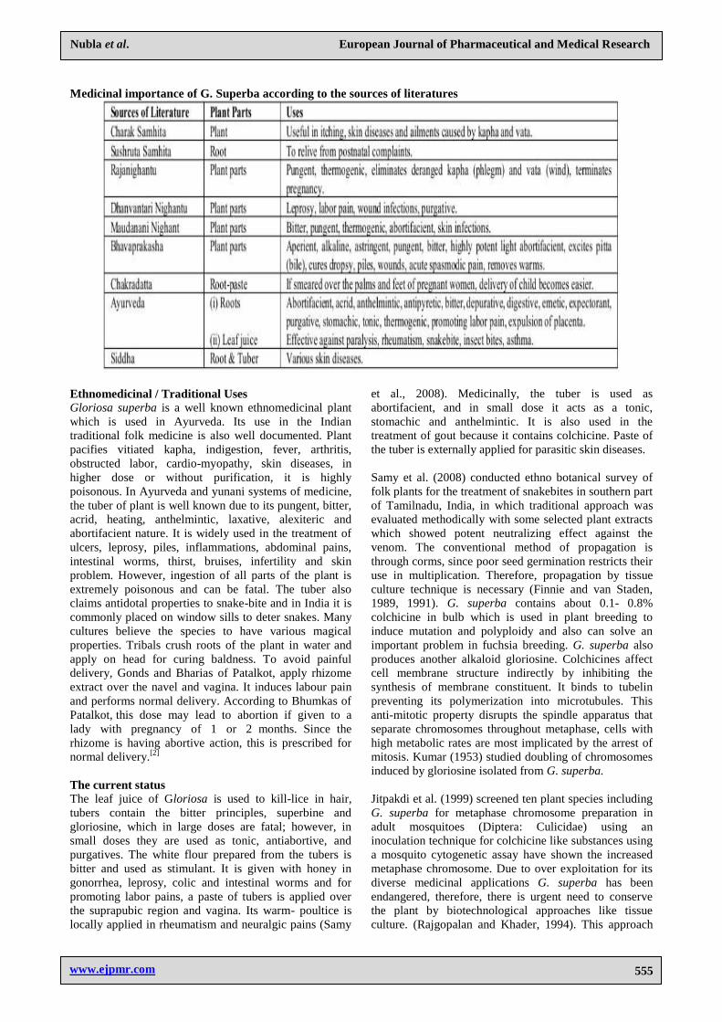
Medicinal importance of G. Superba according to the sources of literatures
Ethnomedicinal / Traditional Uses
Gloriosa superba is a well known ethnomedicinal plant
which is used in Ayurveda. Its use in the Indian
traditional folk medicine is also well documented. Plant
pacifies vitiated kapha, indigestion, fever, arthritis,
obstructed labor, cardio-myopathy, skin diseases, in
higher dose or without purification, it is highly
poisonous. In Ayurveda and yunani systems of medicine,
the tuber of plant is well known due to its pungent, bitter,
acrid, heating, anthelmintic, laxative, alexiteric and
abortifacient nature. It is widely used in the treatment of
ulcers, leprosy, piles, inflammations, abdominal pains,
intestinal worms, thirst, bruises, infertility and skin
problem. However, ingestion of all parts of the plant is
extremely poisonous and can be fatal. The tuber also
claims antidotal properties to snake-bite and in India it is
commonly placed on window sills to deter snakes. Many
cultures believe the species to have various magical
properties. Tribals crush roots of the plant in water and
apply on head for curing baldness. To avoid painful
delivery, Gonds and Bharias of Patalkot, apply rhizome
extract over the navel and vagina. It induces labour pain
and performs normal delivery. According to Bhumkas of
Patalkot, this dose may lead to abortion if given to a
lady with pregnancy of 1 or 2 months. Since the
rhizome is having abortive action, this is prescribed for
normal delivery.[2]
The current status
The leaf juice of Gloriosa is used to kill-lice in hair,
tubers contain the bitter principles, superbine and
gloriosine, which in large doses are fatal; however, in
small doses they are used as tonic, antiabortive, and
purgatives. The white flour prepared from the tubers is
bitter and used as stimulant. It is given with honey in
gonorrhea, leprosy, colic and intestinal worms and for
promoting labor pains, a paste of tubers is applied over
the suprapubic region and vagina. Its warm- poultice is
locally applied in rheumatism and neuralgic pains (Samy
et al., 2008). Medicinally, the tuber is used as
abortifacient, and in small dose it acts as a tonic,
stomachic and anthelmintic. It is also used in the
treatment of gout because it contains colchicine. Paste of
the tuber is externally applied for parasitic skin diseases.
Samy et al. (2008) conducted ethno botanical survey of
folk plants for the treatment of snakebites in southern part
of Tamilnadu, India, in which traditional approach was
evaluated methodically with some selected plant extracts
which showed potent neutralizing effect against the
venom. The conventional method of propagation is
through corms, since poor seed germination restricts their
use in multiplication. Therefore, propagation by tissue
culture technique is necessary (Finnie and van Staden,
1989, 1991). G. superba contains about 0.1- 0.8%
colchicine in bulb which is used in plant breeding to
induce mutation and polyploidy and also can solve an
important problem in fuchsia breeding. G. superba also
produces another alkaloid gloriosine. Colchicines affect
cell membrane structure indirectly by inhibiting the
synthesis of membrane constituent. It binds to tubelin
preventing its polymerization into microtubules. This
anti-mitotic property disrupts the spindle apparatus that
separate chromosomes throughout metaphase, cells with
high metabolic rates are most implicated by the arrest of
mitosis. Kumar (1953) studied doubling of chromosomes
induced by gloriosine isolated from G. superba.
Jitpakdi et al. (1999) screened ten plant species including
G. superba for metaphase chromosome preparation in
adult mosquitoes (Diptera: Culicidae) using an
inoculation technique for colchicine like substances using
a mosquito cytogenetic assay have shown the increased
metaphase chromosome. Due to over exploitation for its
diverse medicinal applications G. superba has been
endangered, therefore, there is urgent need to conserve
the plant by biotechnological approaches like tissue
culture. (Rajgopalan and Khader, 1994). This approach

www.ejpmr.com
Nubla et al. European Journal of Pharmaceutical and Medical Research
556
has been very important because it provides complete
sterile and virus-free plants by rapid multiplication. G.
superba is now promising as an industrial medicinal crop
in Asia particularly in South India for its high colchicine
content. For commercial production of colchicine and its
derivatives, natural production from in vitro methods of
the source plant are thus of great attention. In the past two
decades, focus has been on plant biotechnology as a
potential alternative production method, using cultured
cells rather than plants.[15]
Source of precious alkaloids
Gloriosa superba produces two important alkaloid
colchicine and gloriosine, which are present in seeds and
tubers while the other compounds such as lumi-
colchicine, 3-demethyl-N-deformyl-N-
deacetylcolchicine, 3- demethylcolchicine, N-formyl
deacetylcolchcine have been isolated from the plant
(Sugandhi, 2000; Suri et al., 2001). Suri et al. (2001)
reported new colchicine glycoside, 3-O-
demethylcolchicine-3-O-alpha-D-glucopyranoside in G.
superba seeds. Kaur et al. (2007) studied purification of
3-monomeric monocot mannose-binding lectins and their
evaluation for antipoxviral activity isolated from G.
superba. Alkaloids are structurally heterogeneous class of
secondary biomolecules derived from basically five
amino acids ornithine, lysine, phenylalanine, tyrosine and
tryptophan (Thakur et al., 1975). Thakur et al. (1975)
reported the substances from plant of the sub family
Wurmbaeoideae and their derivatives along with
alkaloids from the G. superba.[16]
Conservation by means of in vitro propagation
Gloriosa superba usually multiply by corm and seeds but
due to low germination capability it restricts for the
regeneration. Therefore, in order to safeguard and
preserve this important plant biotechnological approaches
would be very useful (Sivakumar and Krishnamurthy,
2002). The conventional method of propagation has many
disadvantages as 50% of the yield has to be set aside for
raising the next crop, transmittance of soil-borne diseases
from one crop to the next, and from one location to
another and during the 2-3 month storage period between
harvest and the raising of next crop (Mrudul et al.,
2001).[17]
Kala et al. (2004) studied the prioritization of medicinal
plants on the basis of available knowledge, existing
practices and use value status in Uttaranchal, India in
order to understand the pattern of indigenous uses of
medicinal plants available in the Uttaranchal state, India
and documented 300 species including G. superba.
Hassan and Roy (2005) reported 92% of the cultures of
apical and axillary buds of young sprout from naturally
grown G. superba plants regenerate four shoots per
culture in MS basal medium fortified with 1.5 mg/L BA
+ 0.5 mg/L NAA.[18]
Custers and Bergervoet (1994) reported
micropropagation of G. superba by shoot cuttings and
explants from node, internode, leaves, flowers, pedicels
and tubers. G. rothschildiana (duphur) vs. G.
rothschildiana (new accession) and G. rothschildiana vs.
G. superba were cultured on MS basal medium with 3%
w/v sucrose, 0-10 mg/L Benzyl Adenine (BA) and 0.1
mg lndole Acetic Acid (IAA) and maintained at 24 days
under 16 hours photoperiod. Addition of low level of
Benzyl Adenine (BA)(1 mg/L) improved plant growth,
whereas the high level of BA (10 mg/L) caused
proliferation of multiple shoots, from rhizome meristem,
by applying alternatively high and low BA level, a
method of continued propagation was achieved which
resulted in a 4-7 fold multiplication of qualitatively good
plantlets every 18 week. The resulting shoots were
incubated on MS medium, with 3% sucrose and 0-1 mg/L
IAA or NAA. Transplantation into soil was only possible
after the plants had formed. [19]
Samarajeewa et al. (1993) studied clonal propagation of
G. superba from apical bud and node segment of shoot
tip, cultured on solidified agar (0.8% w/v) Gamborg‘s B5
medium containing BA, IAA, Kinetin, NAA, IBA or 2,4-
D. The cultures were maintained under fluorescent light
at 25-27ºC. Primary cultures were initiated in solid B5
medium containing 0.5 to 1 mg/L BA and 0.01-0.5 mg/L
IAA, IBA, NAA when shoot tip of primary cultures were
transferred to shoot multiplication media, shoot
proliferation occurred via adventitious bud formation
within 4-8 weeks.[20]
Somani et al. (1989) reported in vitro propagation and
corm formation in G. superba. The fresh sproutswere
excised from corms of G. superba and dissected
propagules with shoot and root primordia were placed on
MS basal medium (Murashige and Skoog, 1962)
containing 3% sucrose and 0.6% agar. Explant
germinated on the MS medium producing shoot and root,
which formed new corm within one month. For shoot and
cormlet regeneration, 1-4 mg/L kinetin was added to the
medium. Cultures were maintained at 25ºC in white
fluorescent light (2500 lux) with an 8- h/day
photoperiod.[21]
Sivakumar and Krishnamurthy (2002) reported in vitro
organogenetic responses of G. superba. They used MS
medium supplemented with ADS and BA, 98%. The
callus induction occurred in non-dormant corm bud
explants. The maximum number of multiple shoot (57%)
was observed in corm-derived calluses.[22]
Gupta (1999) compared the production of different
colchicinic substances from G. superba and C.
autumnale. He reported extensive range of these
colchicinic compounds like colchicines (0.9%), dimethyl-
3-colchicine (0.19%), colchicoside (0.82%) and their
formyl derivatives from G. superba. While these values
were found to be less in case of C. autumnale which were
reported as 0.62%, 0.9%, and 0.39% respectively.
Sivakumar and Krishnamurthy (2002, 2004) studied
induction of embryoids from leaf tissue of G. superba.

www.ejpmr.com
Nubla et al. European Journal of Pharmaceutical and Medical Research
557
The nodular calli were observed on S.H. medium
supplemented with 2,4-D and 1 isopentyldene. Gupta et
al. (1999) found hepatoprotective activity of G.
superba.[23]
Jha et al. (2005) reported production of forskolin,
withanolides, colchicine and tylophorine from plant
source by using biotechnological approach.[24]
PHARMACOLOGICAL ACTIVITIES
Antimicrobial activity
Haroon et al., reported antibacterial and antifungal
activity of methanolic extract and its subsequent fractions
in different solvent systems. The study claimed that n-
butanol fraction showed excellent antifungal potential
against candida albicans and candida glaberata (up to
90%) and against trichophyton longifusus (78%) followed
by chloroform fraction against microsporum canis (80%).
The chloroform fraction demonstrated highest
antibacterial activity against staphylococcus aureus
(69.4%).[25]
Enzyme inhibition activity
Haroon et al., reported the enzyme inhibition activity of
alcoholic extract of G. superba Linn rhizomes. The
alcoholic extract and its subsequent fractions in
chloroform, ethyl acetate, n-butanol, and water were
investigated against lipoxygenase, acetylcholinesterase,
butyrylcholinesterase and urease. The chloroform extract
represented maximum inhibition potency (90%) on
lipoxygenase and 29.10% inhibition potency on
butyrylcholinesterase. The ethyl acetate fraction showed
highest inhibition potency (83.50%) on
acetylcholinesterase. However, urease was not inhibited
by any of the tested fractions.[26]
Treatment of snakebite
Ramar Perumal Samy et al., claimed the use of G.
superba tubers paste in the treatment of snakebite. The
study reported that purified fraction (2.4 mg/kg, body
weight) significantly inhibited the toxic effects of snake
venom induced changes in serum SOD and LPx levels in
mice.[27]
Analgesic and anti-inflammatory activity
Jomy C. John et al., reported the analgesic and anti-
inflammatory activity of hydroalcoholic extract obtained
from dried aerial parts of G. superba employing Eddy‘s
hot plate method and acetic acid induced writhing method
for determination of analgesic potential; cotton wool
granuloma and carrageenan induced paw edema model
for anti-inflammatory activity. The study claimed that the
treatment of mice at 100, 200, and 400 mg/kg body
weight exhibited significant (P<0.01) increase in reaction
time. The maximum percentage protection was observed
at 90 min for all the three doses. The % inhibition of
writhes were 64.09%, 78.56% and 81.45% at dose of 100,
200, and 400 mg/kg body weight. The dose of 200 and
400 mg/kg exhibited significant results in carrageenan
induced paw edema model (P<0.05) as compared to
control. The rats exhibited 9.59%, 28.72% and 45.8%
inhibition of granuloma mass formation after 7 days of
treatment with doseof 100, 200, and 400 mg/kg body
weight.[28]
Neuroprotective activity
V. Uma Rani et al., reported neuroprotective activity of
hydroalcoholic extract obtained from tubers of G.
superba. The study revealed that the extract of Gloriosa
superba Linn decreased the transfer latencies,
strengthened its memory improvement action in drug
treated rats. Hence showed decrease in muscle strength
measured by rota-rod test whereas, in hydroalcoholic
extract of Gloriosa superba treated group there was
improvement in muscle strength. The locomotor activity
assessed by actophotometer and open field test was
decreased in lead nitrate group compared with
hydroalcoholic extract of Gloriosa superba Linn treated
group. Biochemical analysis of brain revealed that the
chronic administration of lead nitrate significantly
increased lipid peroxidation and decreased levels of
catalase (CAT), reduced glutathione (GSH) and
glutathione reductase (GR), an
index of oxidative stress process. Administration of
hydroalcoholic extract of Gloriosa superba Linn
attenuated the lipid peroxidation and reversed the
decreased brain CAT and GSH levels. Lead exposed rats
showed increased levels of various serum parameters like
glucose, ALT, ALP, TG and TC.[29]
Anti-arthritic activity
K.P. Latha et al., reported the anti-arthritic activity of
chloroform extract obtained from tubers of G. superba
using Freund‘s complete adjuvant induced arthritis model
in rats. The study demonstrated that chloroform extract of
tubers of G superba has shown a dose dependent and
significantly decreased paw edema and ankle diameter in
treated groups as compared with arthritic group.[30]
Anticoagulant activity
Nalise Low Ah Kee et al., reported anticoagulant/anti-
thrombotic potential of methanolic extract obtained from
leaves of G. superba. The study proclaimed that leaf
extract of G. superba inhibited thrombin-induced with
IC50 values of
2.97 mg/ml.[31]
Anticancer activity
Samson Eugin Simon et al., reported the anticancer
activity of phytochemical extract obtained from tubes of
G. superba against Hep-G2 cancer cell line (Human liver
cancer cells) employing MTT assay. The study revealed
that concentration of 100µg of plant extract has
maximum inhibition value of 54.3% against Hep-G2
cancer cell line.[32]
Toxicity/Poisoning
The colchicine which is major component of G. superba
is mainly responsible for toxic effect.[21]
The commonest
clinical presentation of poisoning is severe gastroenteritis

www.ejpmr.com
Nubla et al. European Journal of Pharmaceutical and Medical Research
558
with nausea, vomiting, diarrhoea with blood leading to
dehydration, hypovolaemic shock and acute renal failure.
Muscle weakness, hypoventilation, ascending
polyneuropathy, bone marrow depression and coagulation
disorders are the other features of poisoning. Death in
severe poisoning occurs due to shock or respiratory
failure although haemorrhagic or infective complications
may cause death after the first day.[33]
CLINICAL EFFECTS IN POISONING
1. Cardiovascular: There is no direct effect on the heart,
but fluid and electrolyte loss, often causes
hypovolaemic shock manifested by hypotension and
tachycardia.
2. Respiratory: Respiratory failure is thought to be due
to the paralysis of intercostal muscles rather than the
direct depression of the respiratory centre by
colchicine.[34]
3. Central nervous system (CNS): There is progressive
paralysis of the central nervous system and
peripheral nervous system[35]
4. Peripheral nervous system: Ascending
polyneuropathy, weakness, loss of deep tendon
reflexes may be described.
5. Skeletal and smooth muscle: Colchicine could have a
direct toxic effect on skeletal muscles causing
muscular weakness. Rhabdomyolysis may occur
with significant increase in muscle enzymes and
myoglobinuria as a result of direct muscular damage.
Muscle weakness that may persist for many weeks
may contribute to respiratory deficiency.[36]
6. Gastrointestinal: Gastroenteritis including nausea,
vomiting, diarrhoea with blood accompanied by colic
and tenesmus. Loss of fluids and electrolytes leads to
hypovolaemia. Intestinal ileus may develop within
the first few days and may persist up to a week.[37]
7. Hepatic: Colchicine may exert direct hepatic toxicity
with moderate cytolysis.
8. Renal: Any direct toxic effect of the toxin on kidney
is not clear. Renal failure is probably secondary to
excess fluid loss or hypovolaemia and is preceded by
oliguria and haematuria. Proteinuria could also
occur[38].
9. Endocrine and reproductive systems: Vaginal
bleeding has been reported as a feature of
intoxication. Tubers are used as an abortifacient in
some countries.
10. Dermatological: Alopecia usually occurs one or two
weeks after the ingestion of G. superba. A case of
generalized depilation has also been reported Eye,
ear, nose, and throat: Subconjunctival haemorrhages
have been observed. Burning and rawness of the
throat may be early symptoms of toxicity.
11. Fluid and electrolyte disturbances: There is an
extensive fluid and electrolyte loss due to intense
vomiting and diarrhoea or sometimes due to
haemorrhages. Hypokalaemia, hypocalcaemia,
hypophosphataemia and hyponatraemia may occur.
12. Haematological: Colchicine has a depressant action
on the bone marrow which is characterized by a
transient leucocytosis followed by leucopenia. It
could also cause thrombocytopenia that may give
rise to various coagulation disorders resulting in
vaginal bleeding, conjunctival and gastrointestinal
haemorrhages. Severe thrombocytopenia occurring
within 6 hours of poisoning has been documented.
Anaemia may occur, mostly secondary to
haemorrhages.[39]
CASE REPORTS
Toxic encephalopathy due to colchicine-Gloriosa
superba poisoning CASE REPORT -1
A 28-year-old woman, previously healthy, presented with
abdominal pain, diarrhoea and profuse vomiting. Six
hours before, she had eaten G superba tubers when
attempting to end her life after a domes tic dispute. On
admission, she was stable; we gave activated charcoal
and treatedher symptomatically. After initially
improving, on the 5th day she developed generalised
tonic-clonic sei zures. She was hypocalcaemic, with
serum ionised calcium of 0.9 mmol/L (1.1–1.4). We gave
intravenous calcium and anticonvulsants and achieved
seizure control.
On day 7, her level of consciousness declined to a
Glasgow coma scale score of 6 out of 15. There were no
focal neurological signs. Her full blood count, arterial
blood gas tensions, urinalysis, erythro cyte sedimentation
rate, serum C-reactive protein, urea, electrolytes, glucose,
ammonia, calcium and magnesium were normal. Her
serum transaminases were transiently mildly elevated:
serum aspartate ami notransferase 278 U/L (1–31) and
alanine aminotrans ferase 216 U/L (5–35). CT scan of
head, lumbar puncture, ultrasound scan of pelvis and
abdomen were normal. Serology for Epstein-Barr virus,
cyto megalovirus, Japanese encephalitis (JE) virus, and
herpes simplex virus were negative. EEG showed bilat
eral diffuse slow waves, suggestive of encephalopathy.
MR scan of her brain (figure 3) showed bilateral T2
hyperintensities in the basal ganglia. We managed her
supportively in the intensive care unit. By day 15, her
Glasgow coma scale score had improved to 11/15 and
remained static up to the point of the current report. She
also developed alopecia.
DISCUSSION
This case highlights a delayed toxic encephalopathy
following Gloriosa poisoning. Based on the unequivo cal
history of ingestion of Gloriosa tubers, the extra central
nervous system clinical features supporting Gloriosa
toxicity, the temporal profile of the onset of
encephalopathy and the exclusion of other neoplastic,
immune, infective and metabolic causes for encephal
opathy, we conclude that her encephalopathy resulted
from colchicine toxicity following Gloriosa tuber
ingestion. However, we had no facilities to assess serum
levels of colchicine. Patients with colchicine poisoning
occasionally develop confusion and sei zures, but all such
cases have concomitant renal or liver dysfunction. By
contrast, our patient had no significant metabolic

www.ejpmr.com
Nubla et al. European Journal of Pharmaceutical and Medical Research
559
derangement. The other unusual feature of her
encephalopathy was the delayed onset, reminiscent of
that following anoxic encephalopathy and carbon
monoxide poisoning.MR brain scan findings of bilateral
T2 hyper inten sities in the caudate and lentiform nuclei
of the basal ganglia in this patient are consistent with a
toxic encephalopathy, further strengthening the case for a
Gloriosa- induced encephalopathy. The thalamic regions
were unaffected. The putamen and globus pallidus of the
basal ganglia have abundant mitochondria,
neurotransmitter and chemical content. They are also
extremely metabolically active and are exceedingly prone
to metabolic and toxic damage.
The basal ganglia lesions in toxic and metabolic
encephalopathies are global and symmetrical, as in this
lesions without concomitant thalamic involvement occur
with only a few causes of toxic encephalopathy, suchas
hyperammonaemia and hypoglycaemia:we excluded both
these in this patient. Infectious disease can also cause
basal ganglia abnormalities, but such lesions are usually
more asymmetrical and patchy. It is also possible that she
had ingested another neurotoxic agent along with the G
superbaGloriosa‘s active toxic compound is the alkaloid
col chicine, which binds to tubulin and inhibits its poly
merisation into microtubules. Colchicine‘s toxic effects
are mediated through microtubule disruption, which, in
neural cells, interferes with axonal and den dritic
transmission. In experimental and animal models,
colchicine causes the neuronal cell death of certain
neuronal populations, such as the granular cells of the
dentate gyrus and pyramidal cells of the hippocampus.
Colchicine may induce an autotoxic response, leading to
neuronal death of certain populations, including the basal
ganglia, due to the accumu lation of a toxic cellular
product which is normally transported by a microtubule-
dependent process.
This may be the explanation for the delayed onset ofthis
patient‘s encephalopathy, as a time lag may be evident
before a critical neuronal toxic concentration of these
substances is reached. The concentration of colchicine in
cerebral tissue is usually low following poisoning,
perhaps explaining why, despite these experimental
findings, clinical cases of colchicine poising are so
rare.[40,41,42,43]
Colchicine cardiotoxicity following ingestion of
Gloriosa superba tubers CASE REPORT-2
A 29 year old man who attempted suicide by ingesting
tubers of Gloriosa superba was admitted 4 hours later to
Teaching Hospital Peradeniya, complaining of burning in
the mouth and throat, intense thirst, nausea, vomiting and
abdominal colics. On examin ation he was restless,
afebrile and dehydrated. Pulse rate was 90 beats/min.
Blood pressure was 100/ 70 mmHg and respiratory rate
was 20 per minute. Gastric lavage was performed and he
was given intravenous fluids. Except for an elevated
haematocrit (PCV = 0.541/1) his biochemical and
haematological investigations were normal on admission.
He was oliguric for 24 hours. By the second day the
blood urea increased to 10.3 mmol/l and urinalysis
showed proteinuria and mild haematuria. He developed a
watery diarrhoea, complained of severe body ache and
generalized chest pain. An electrocard iogram done at
this stage was normal. Serum aspartate transaminase was
10IU/I (normal 0-12). Serum alanine transaminase was
20 IU/I (normal 0-11), serum bilirubin was 17 mmol/l,
plasma potassium was 4.2 mmol/l and plasma sodium
was 138 mmol/l. On the third day the patient complained
of severe pain all over the chest associated with difficulty
in breathing. His respiratory rate was 38/min, pulse was
100 beats/ min regular, and blood pressure 110/70
mmHg. He had a triple rhythm and bilateral basal
crepitations. The electrocardiogram (Figure 1) showed
ST eleva tion. Aspartate transaminase rose to over 60
IU/I (normal 0-12), creatine kinase was 40/IU/l (normal
0-12) and serum cholesterol was 5.0 mmol/l.
He was treated with analgesics and frusemide. He
continued to complain of chest pain on the fourth day.
The electrocardiogram showed further T elevation
(Figure 2). By the fifth day he developed bleeding gums
and subconjunctival haemorrhages and haematuria. At
this stage haematological investi gations showed
haemoglobin 9.5 g/dl, white cell count 2.8 x 99/1, platelet
count 20 x 109/1, bleeding time 3 minutes, clotting time
11 minutes, prothrombin time 18 seconds (control 15
seconds). He was transfused three pints of fresh blood in
the next 48 hours. Serum aspartate transaminase and
creatine kinase returned to normal by the ninth day. The
electrocardiogram showed only inverted T waves in V5.
His condition gradually improved and 3 weeks
afteradmission he was transferred to the Department of
Psychiatry for further management.
Biochemical and haematological investigations and the
electrocardio gram (resting and in response to exercise)
were normal.
DISCUSSION
There does not appear to be a clear separation of non-
toxic, toxic and lethal dosages of colchicine.2-5 This
patient had ingested about 100 grams of tuber. Dunuwille
and others' have estimated that 10 g of fresh tuber
contains approximately 6.0 mg of col chicine. Therefore
the amount of colchicine ingested by this patient is in the
region of 60 mg. Fatalities have been reported with doses
as small as 7 mg.[44,45,46]
Massive generalized alopecia after poisoning by
Gloriosa superba CASE REPORT-3
The patient, a married woman aged 21 years, was
admitted on 17 September 1964 at about 2 p.m. to the
University Medical Unit, General Hospital, Kandy. She
had fallen ill after an afternoon meal the day before
which consisted of a variety of boiled yams. The plant
and the ingested tubers were identified as Gloriosa
superba. The colchicine content of the tubers is 0.3%.
Therefore, as the patient had eaten about 125 g. of tubers,

www.ejpmr.com
Nubla et al. European Journal of Pharmaceutical and Medical Research
560
the total amount of colchinine taken was in the region of
350 mg. About two hours after this meal she had started
vomiting, and about eight hours later had profuse watery
diarrhoea, which continued throughout the night. She had
vomited 25 times that night and had 20 watery stools.
The patient had no children. Her past history revealed
nothing of note. She attained puberty at 13 and her
periods were regular.
She had no physical abnormalities. Her hair had never
been cut since childhood, as is the custom among
Ceylonese girls, and it came down to her knees. On
admission she was unconscious, restless, dehydrated, and
collapsed. Her pulse rate was 122/min., of moderate
volume and in sinus rhythm; blood-pressure was 95/70
mm. Hg, and respiratory rate was 18 per minute. Apart
from these findings, nothing abnormal was noted. She
had no cyanosis or dyspnoea. Her white-cell count was
8,800/c.mm., with a differential count of polymorphs
76%, lymphocytes 21%, eosinophils 3%. Her blood urea
was 37 mg./100 ml.; serum potassium 3.7 mEq/litre;
serum sodium 142 mEq/litre. She did not pass any urine
on the day of admission. On the day of admission she was
treated with a slow intravenous drip-infusion of 3 pints
(1.7 litres) normal saline and 1 pint (0.6 litres) 5%
dextrose with added vitamins. Her condition improved on
this regimen. Blood-pressure rose to 100/70 mm. Hg, and
the pulse rate fell to 1 10/min. The following morning she
collapsed again, the blood-pressure could not be
recorded, and the pulse was imperceptible.
Hydrocortisone hemisuccinate, methoxamine, and
noradrenaline were then added to the drip. Two days after
admission, the day after her collapse in the ward, her
general condition- improved, blood-pressure was 90/70
mm. Hg, pulse rate 104/min., of moderate volume, and in
sinus rhythm. No other abnormalities were detected
clinically. Examination of her urine showed " pus cells-
field full," probably because she was catheterized
previously. She was given sulphadimidine and the urine
report came back to normal in four days. Blood urea was
39 mg./100 ml. Five days after admission the patient was
found to have a subconjunctival haemorrhage in her left
eye. Her menstrual period, which was ending the day she
ate the tubers, continued for a further 20 days. A platelet
count carried out at this stage was 475,000/ c.mm.; white-
cell count was 5,000/c.mm., with polymorphs 50%,
lymphocytes 49%, eosinophils 1%, and packed cell
volume 35%. Haemoglobin was 11.3 g./100 ml., and
mean corpuscular haemo globin concentration 30
mg./100 ml. Twelve days after admission marked
alopecia was noticed, especially affecting the scalp hair
(Fig. J), and within two days most of the hair on her scalp
had dropped out, as had her axillarhair and part of the
pubic hair. She was seen in the clinic subse quently, and
within a week after her discharge from hospital, 23 days
after admission, she was completely bald. Two months -
later her scalp hair had regrown to half an inch (12.7
mm.) (Fig.2). Pubic hair showed regrowth. Her axillary
hair remained very scanty. After five months her scalp
hair was 2-3 in. (5.1-7.6 cm.) long.[47,48,49,50]
CONCLUSION
Application indigenous natural products has been
alternative way to replace synthetic medicine. Gloriosa
superba is a well known ethnomedicinal plant which is
used in Ayurveda. Photochemical studies of G. superba
showpresence of a highly active alkaloid, Colchicine.
FDA- approved use of colchicine is to treat gout (it is one
of the active ingredients of anti-gout tablets marketed by
Merck & Co.), though it is also occasionally used in
veterinary medicine totreat cancers in some animals. It is
also used as an antimicrobial, antifungal, anticoagulant,
antilipoxygenase agent and antidote in snake bite.
However, ingestion of all parts of the plant is extremely
poisonous and can be fatal. The commonest clinical
presentation of poisoning is severe gastroenteritis with
nausea, vomiting, diarrhoea with bleading to dehydration,
hypovolaemic shock and acute renal failure. Gloriosa
superba usually multiply by corm and seeds but due to
low germination capability it restricts for the
regeneration. Therefore, in order to safeguard and
preserve this important plant biotechnological approachs
would be very useful. Micropropagation of Gloriosa
superba meets ever increasing demands for colchicine.
The availability from both wild and cultivated sources
make the plant of Gloriosa superba a potential source of
Colchicine in India.
BIBLIOGRAPHY
1. Senthilkumar M, Phytochemical Screening and
Antibacterial Activity of Gloriosa superba Linn,
International Journal of Pharmacognosy and
Phytochemical Research, 2013; 5(1): 31-36.
2. Hemant Badwaik, Tapan Kumar Giri, D.K. Tripathi,
Mukesh Singh and Abdul Hanif Khan, A Review on
Pharmacological Profile for Phytomedicine Known
as Gloriosa superba Linn. Research J.
Pharmacognosy and Phytochemistry, 2011; 3(3):
103-107.
3. Shrivastava A. et.al. ‗Indigenous herbal medicines:
tribal formulations and tradinal herbal practices,
aavishkar publishers and distributors, 2008; 440.
4. Nair C.K. ‗Ethino medicinal plants to fight
Neoplastic diseses‘, Ethinomedicine:a source of
commentary therapeutics, 2010; 203-226.
5. Badola HK. Endangered medicinal plant species in
Himachal Pradesh. A report on the International
Workshop on ―Endangered Medicinal Plant Species
in Himachal Pradesh’, organized by G.B. Pant
Institute of Himalayan Environment and
Development at Himachal Unit, Mohal-Kullu during
18-19 March 2002,Curr. Sci., 2002; 83: 797-798.
6. Abhishek Mathur, Satish K Verma, Santosh K Singh,
Deepika Mathur, Prasad GBKS, and Dua VK,
Investigation Of Anti-Inflammatory Properties of
Swertia Chirayta And Gloriosa Superba, Recent
Research in Science and Technology, 2011, 3(3): 40-
43.
7. Padmapriya S, Rajamani K, Sathiyamurthy VA.

www.ejpmr.com
Nubla et al. European Journal of Pharmaceutical and Medical Research
561
Glory lily (Gloriosa superba L.)- A review.
International Journal of Current Pharmaceutical
Review and Research, 2015; 7(1): 43-49.
8. Sonali Jana, G.S. Shekhawat, Critical review on
medicinally potent plant species: Gloriosa superba,
Fitoterapia, 2011; 82: 293–301.
9. Thakur RS, Potesilova H, Santavy F., Substances
from plants of the subfamily Wurmbaeoideae and
their derivatives. Part LXXIX. Alkaloids of the plant
Gloriosa superba L. Planta Medica, 1975; 3: 201-
209.
10. Sugandhi, R., Biodiversity conservation and
patenting and property right of tribal medicine of
medicinal plants of India. 10th Asian Symposium on
Medicinal Plants, Spices and other Natural products
(ASOMPS X). Dhaka, Bangladesh, November, 2000;
18-23.
11. Kaur, A., S.S. Kamboj, and J. Singh; Purification of
3 monomeric monocot mannose-binding lectins and
their evaluation for antipoxviral activity: potential
application in multiple viral diseases caused by
enveloped viruses. Biochemistry and Cell Biology,
2007; 85: 88-95.
12. Bellet, P., and J.C. Gaignanlt; Gloriosa superba Linn
and the production of colchicinic substances.
Annales Pharmaceutiques Francaises. 1985; 43: 345-
347.
13. Ghose, B. et.al., Enhanced colchicin production in
root cultures of Glorisa superba by direct and indirect
precursors of the biosynthetic pathways.
Biotechnology Letters., 2002; 24: 231- 234.
14. Khan H, Khan MA, Mahmood T, Chaudhary MI.
Antimicrobial activities of Gloriosa superba Linn
(colchicaceae) extract. Journal of Enzyme Inhibition
and Medicinal Chemistry, 2008; 23(6): 855-859.
15. Ravindraade, Mahindra K. Rai,Current Advances in
Gloriosa superba L., Biodiversitas, 2009; 4(10): 210-
214
16. Aleem, H.M. Gloriosa superba poisoning. Journal of
Association of Physicians of India, 1992; 40: 541-2.
17. Mrudul, V., C.K. Shirgurkar, and R.S. John. Factors
affecting in vitro microrhizome production in
turmeric. Plant Cell, Tissue and Organ Culture,
2001; 64: 5-11.
18. Kala, C.P., N.A. Farooquee, and U. Dhar.
Prioritization of medicinal plants on the basis of
available knowledge, existing practices and use value
status in Uttaranchal, India. Biodiversity and
Conservation, 2004; 13(2): 453-469.
19. Custers, J.B.M. and J.H.W. Bergervoet.
Micropropagation of Gloriosa: towards a practical
protocol. Scientia Horticulturae. 1994; 57: 323-334.
20. Samarajeewa, P.K., M.D. Dassanayake, and S.D.G.
Jayawardena. Clonal propagation of Gloriosa
superba. Indian Journal of Experimental Biology,
1993; 31: 719-720.
21. Somani, V.J., C.K. John, and R.J. Thengane. In vitro
propagation and corm formation in Gloriosa superba.
Indian Journal of Experimental Biology, 1989; 27:
578-579.
22. Sivakumar, G. and K.V. Krishnamurthy. In vitro
organogenetic responses of Gloriosa superba.
Russian Journal of Plant Physiology, 2004; 51: 790-
798.
23. Gupta, B.K. 1999. Production of colchicine from G.
superba tubers in cultivation and Utilization of
medicinal plants. CSIR Journal, 270-278.
24. Jha, S., M. Bandypadhyay, and K.N. Chaudhary.
2005. Biotechnological approaches for the
production of farskolin, withanolides, colchicine and
tylophorine. Plant Genetic Resource, 3: 101-115.
25. Khan H, Khan MA, Hussan I. Enzyme inhibition
activities of the extract from rhizomes of Gloriosa
superba Linn (colchicaceae). Journal of Enzyme
Inhibition and Medicinal Chemistry, 2007; 2(6): 722-
725.
26. Samy RP, Thwin MM, Gopalkrishnakone P,
Ignacimuthu S. Ethnobotanical survey of folk plants
for the treatment of snakebites in southern part of
Tamilnadu, India. Journal of Ethnopharmacology,
2008; 115: 302-312.
27. John JC, Fernandes J, Nandgude T, Niphade SR,
Salva A, Deshmukh PT. Analgesic and anti-
inflammatory activities of the hydroalcoholic extract
from Gloriosa superba Linn. International Journal of
Green Chemistry, 2009; 3(3): 215- 219.
28. Rani VU, Sudhakar M, Ramesh A, Lakshmi BVS,
Srinivas Y. Protective effect of hydroalcoholic
extract of Gloriosa superba Linn in lead induced
neurotoxicity in rats. International Journal of
Pharmacy, 2016; 6(2): 257-266.
29. Latha KP, Girish HN, Kirana H. Anti-arthritis
activity of chloroform extract of tubers of Gloriosa
superba Linn. International Journal of Research in
Pharmacy and Chemistry, 2015; 5(4): 662-667.
30. Ah Kee NL, Mnonopi N, Davids H, Naude RJ, Frost
CL. Antithrombotic/ anticoagulant and anticancer
activities of selected medicinal plants from South
Africa. African Journal of Biotechnology, 2008;
7(3): 217-223
31. Simon SE, Jayakumar FA. Antioxidant activity and
anticancer study on phytochemicals extract from
tubers of Gloriosa superba against human cancer cell
(HEP-G2).Research and Reviews: Journal of
Pharmacognosy and Phytochemistry, 2016; 4(4): 7-
12.
32. Vishwanathan, N. And B.S. Joshi., Toxic constituent
of some Indian plants. Current Science, 1983; 52: 1-
8.
33. Mendis, S. Colchicine cardiotoxicity following
ingestion of Gloriosa superba tubers. Postgraduate
Medical Journal, 1989; 768: 752-755.
34. Angunawela RM and Fernando HA, Acute ascending
polyneuropathy & dermatitis following poisoning by
tubers of G. Superba. Ceylon Medical Journal, 1971;
16: 233-235.
35. Jayaweera, D.M.A., Medicinal Plants Used in
Ceylon, 1982; 3. Colombo: National Science Council
of Sri Lanka.
36. Wijesundere A, Plant poisons. Ceylon Medical
www.ejpmr.com
Nubla et al. European Journal of Pharmaceutical and Medical Research
562
Journal, 1986; 31(2): 89-91.
37. Ellenhorn MJ, Schonwald S, et.al., Ellenhornn s
Medical toxicology: diagnosis & treatment of human
poisoning, 2nd ed., 1996.
38. Murray SS, Kramlinger KG, McMichan JC and
Mohr DN, Acute toxicity after excessive ingestion of
colchicine. Mayo Clin Proc., 1983; 58: 528-532.
39. Saravanapavananthan T., Plant poisoning in Sri
Lanka. Jaffna Medical Journal, 1985; 20(1): 17-21.
40. Gooneratne IK, et al, Toxic encephalopathy due to
colchicine—Gloriosa superba poisoning, 2014; 14:
357–359
41. Jana S, Shekhawat GS. Critical review on
medicinally potent plant species: Gloriosa superba.
Fitoterapia, 2011; 82: 293–301.
42. Fernando R, Fernando DN. Poisoning with plants
and mushrooms in Sri Lanka: a retrospective hospital
based study. Vet Hum Toxicol, 1990; 32: 579–81.
43. Mendis S. Colchicine cardiotoxicity following
ingestion of Gloriosa superba tubers. Postgrad Med
J., 1989; 65: 752–5.
44. Shanthi Mendis, Colchicine cardiotoxicity following
ingestion of Gloriosa superba tubers, Postgraduate
Medical Journal, 1989; 65: 752-755.
45. Liu, Y.K., Hymowitz, R. & Carroll, M.G. Marrow
aplasia induced by colchicine. A case report.
Arthritis Rheum, 1978; 21: 731-735.
46. Jarvie, D., Park, J. & Stewart, M.J. Estimation of
colchicine in a poisoned patient by using high
perform ance chromatography. Clin Toxicol, 1979;
14: 375-381.
47. Amoroso, E. C, Massive Generalized Alopecia After
Poisoning by Gloriosa superba, Brit. med. J., 1966; 1:
1023-1024.
48. Steward O, Goldschmidt RB, Sutula T.
Neurotoxicity of colchicine and other tubulin-
binding agents: a selective vulnerability of certain
neurons to the disruption of microtubules. Life Sci.,
1984; 35: 43–51.
49. Hegde AN, Mohan L, Lath N, et al. Differential
diagnosis for bilateral abnormalities of the basal
ganglia and thalamus. Radiographics, 2011; 31: 5–30.
50. Stapczynski JS, Rothstein RJ, Gaye WA, et al.
Colchicine overdose: report of two cases and review
of the literature. Ann Emerg Med., 1981; 10: 364–9.