toxicity of silver nanoparticles in fish: a critical revie · which changes as the particles are...
TRANSCRIPT
J. Bio. & Env. Sci. 2015
211 | Khan et al.
REVIEW PAPER OPEN ACCESS
Toxicity of silver nanoparticles in fish: a critical review
Muhammad Saleem Khan1, Farhat Jabeen1*, Naureen Aziz Qureshi2, Muhammad
Saleem Asghar1, Muhammad Shakeel1 and Aasma Noureen
1Department of Zoology, GC University Faisalabad, Pakistan
2GC Women University Faisalabad, Pakistan
Article published on May 18, 2015
Key words: Silver, Nanoparticles, Uses, Toxicity, Fish model.
Abstract
The variable spectrum of applications largely depends upon silver physicochemical and biological properties
which changes as the particles are decreased to nano-scale. This unique behavior is responsible for the larger use
of silver in consumer product and industry. Since little information is available about toxicity to the organisms
practically in the aquatic environment, the predication of possible environmental hazards and remedy are the hot
topics of current research studies. Researchers are drawing more and more data from appropriate model
organisms. Fish being aquatic organism is badly affected by Ag-NPs, so concern of potential risk to aquatic
organism increases. The toxicity endpoints include growth and reproduction impairment, mortality and
biochemical changes in both adult fish and embryos. Being a healthy food for human, the researchers try to know
how Ag-NPs can affect the fish and its body when sizes decrease to nano-scale. Therefore, fish is extensively
studied model in the toxicological studies. It examined some of these studies which address the adverse effects of
Ag-NPs on biological systems of different fish group predicting the dose dependent toxicity. The organisms more
acutely sensitive have lower LC50 values. All the studies also indicated that silver ions released from Ag-NPs
surface contributes to toxicity. Therefore, it is suggested that emphasis should be placed on upcoming
investigations for the evaluation of environmental impact of nano-silver in both in vitro and in vivo studies. The
effect of long term exposure and bioaccumulation of silver through food web should be unmasked and discussed
in detail.
*Corresponding Author: Dr Farhat Jabeen [email protected]
Journal of Biodiversity and Environmental Sciences (JBES) ISSN: 2220-6663 (Print) 2222-3045 (Online)
Vol. 6, No. 5, p. 211-227, 2015
http://www.innspub.net
J. Bio. & Env. Sci. 2015
212 | Khan et al.
Introduction
The commercial applications of the nanomaterial
research are maximum in the present era. It is
estimated that approximately 60,000 tons of
nanomaterial is produced annually (Jovanovic et al.,
2011) with 1628 nano based products in 30 countries
(Woodrow Wilson Database, 2015). Among the nano
industry, silver nanoparticles is the most rapidly
growing class with 320 tons of production per year
(Nowack et al., 2011). Its importance is due to unique
properties and consumers demands. About 30% of all
the currently registered nanoproducts are claiming to
contain the nano-silver (Project on emerging
nanotechnologies, 2013). It is estimated that 383
nanoproducts are available in the market (Woodrow
Wilson Database, 2015).
The extensive use of Ag nanoproducts might increase
the discharge of these particles into aquatic
environment (Benn and Westerhoff, 2008; Taju et al.,
2014). It may be released into water and air through
different sources including weathering of rocks,
processing of ores, cement manufacturing and
burning of fossil fuel. Rain is responsible to release
the silver in the ground and water reservoirs
(Wijnhoven et al., 2009). About 68% of nano silver
load will be increased in the waste water due to
biocidal products from 2010 to 2015 (Blaser et al,
2008). In the aquatic environment, it exits in four
(Ag, Ag+, Ag+2 and Ag+3) oxidation states. But Ag and
Ag+ exist more commonly (Smith and Carson, 1997).
Metallic silver is insoluble whereas salts (AgCl,
AgNO3) are soluble in water (WHO, 2002). Silver
nanomaterial is found in the form of colloidal
particles in the aquatic medium.
In spite of existence in the aquatic environment,
limited information regarding the toxicity of Ag-NPs
is available (Wijnhoven et al., 2009). Depending
upon the existing literature, it can be hypothesized
that Ag-NPs are more toxic than other forms because
of it’s more readily absorbance than metallic silver
(Drake and Hazelwood, 2005). The fate and behavior
of silver nanomaterial is influenced by many factors
including; size, surface area, surface chemistry and
chemical composition (coatings and purity). The
other factors like water and lipid solubility, vapor
pressure and aggregation or coagulation state
(Wijnhoven et al., 2009).
In the aquatic environment, Ag-NPs most likely enter
the ecosystems produce a physiological response in
many animals, altering their fitness and population
densities (Luoma and Rainbow, 2008). On the other
hand, the detailed studies on the effect of these
particles on the target organisms and their
environment have just begun. Numbers of
toxicological studies have been performed but it
showed huge variations due to lack of proper particles
characterization (Gliga et al., 2014). The researchers
focus on toxicity of Ag-NPs in aquatic environment
including fish in the recent studies (Asharani et al.,
2008; Yeo and Kang 2008; Bar-Ilan et al., 2009;
Chae et al., 2009; Choi et al., 2009; Griffitt et al.,
2009; Bilberg et al., 2010; Powers et al., 2010; Wu et
al., 2010). Toxicity induced by Ag-NPs to vertebrate
cell line include generation of the reactive oxygen
species (Hussain et al., 2005; Schrand et al., 2008),
apoptosis (Braydich-Stolle et al., 2005; Park et al.,
2007), reduced mitochondrial function (Braydich-
Stolle et al., 2005; Hussain et al., 2005; Schrand et
al., 2008), increase lipid peroxidation (Arora et al.,
2008) and depletion of the oxidative stress markers
(Hussain et al., 2005; Arora et al., 2008). Further a
study by Larese et al. (2009) demonstrated that Ag-
NPs can pass through stratum corneum and the outer
layer of epidermis and even blood-brain barriers
causing damage to human skin, liver, lungs and
olfactory blabs (Braydich-Stolle et al., 2005; Hussain
et al., 2005; Arora et al. 2008; Sung et al., 2008). So
the current review will present the toxicity caused by
Ag-NPs on some groups of fish.
Silver uses
Silver is among basic elements that formed our
planet. Its size ranges 5-50 nm in many commercial
products so refers as nanosilver (Panyala et al.,
2008). It has a long history of use. Ancient Egypt,
J. Bio. & Env. Sci. 2015
213 | Khan et al.
Rome, Italy and Greece were aware about the use of
silver (Reidy et al., 2013). They use silver for the
preparation of storage vessels that keep the water
fresh (Russell and Hugo 1994). It was used in the
form of colloidal silver more than 150 years ago and
first time registered as biocidal material in 1954
(Nowack et al., 2011). In the present time, it is mostly
used in the fields of chemistry, material science and
physics (Syrvatka et al., 2014). It is used worldwide in
photography, batteries as coatings of solar energy
absorption, water treatment filters (Li et al., 2008),
washing machines (Jung et al., 2007), fabrics
(Perelshtein et al., 2008), Heat sink, sensors
(Schrand et al., 2008), catalysts (Kumar et al., 2008),
superconductors, cloud seeding, shampoo, food
packing, Electroplating, kitchen utensils, and odor
resistant textiles (Sondi and Sondi, 2004; Cohen et
al., 2007; Yon and Lead, 2008). Furthermore silver is
also combined with other substance to develop
combined functions.
More attention is devoted towards silver due to its
medical importance. It has been used in the fields of
biotechnology, medicine, environmental technology
and as a broad spectrum antimicrobial agent (Kim et
al., 2007; Kim et al., 2013). Table 1 provides the
comprehensive medical uses of naonosilver in the
current time. The silver containing biocidal products
has reached to 110 to 230 tons in the European
market and significant portion is consists of Ag-NPs
(Blaser et al., 2008).
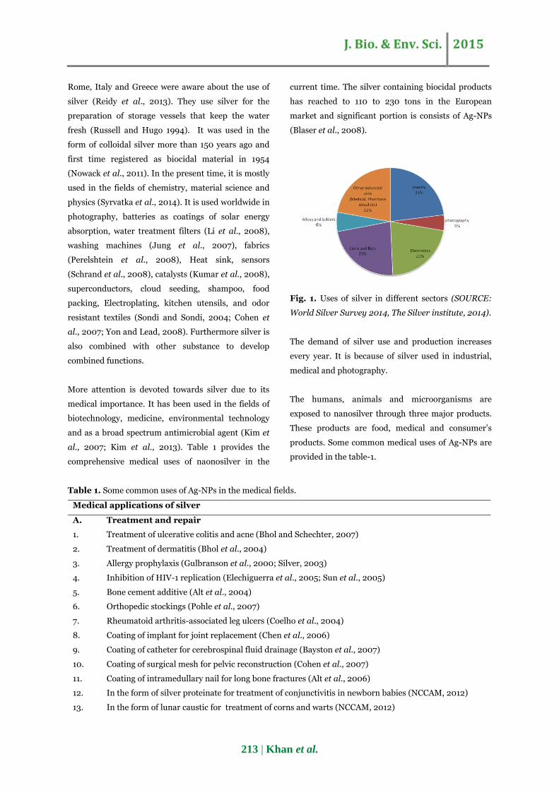
Fig. 1. Uses of silver in different sectors (SOURCE:
World Silver Survey 2014, The Silver institute, 2014).
The demand of silver use and production increases
every year. It is because of silver used in industrial,
medical and photography.
The humans, animals and microorganisms are
exposed to nanosilver through three major products.
These products are food, medical and consumer’s
products. Some common medical uses of Ag-NPs are
provided in the table-1.
Table 1. Some common uses of Ag-NPs in the medical fields.
Medical applications of silver
A. Treatment and repair
1. Treatment of ulcerative colitis and acne (Bhol and Schechter, 2007)
2. Treatment of dermatitis (Bhol et al., 2004)
3. Allergy prophylaxis (Gulbranson et al., 2000; Silver, 2003)
4. Inhibition of HIV-1 replication (Elechiguerra et al., 2005; Sun et al., 2005)
5. Bone cement additive (Alt et al., 2004)
6. Orthopedic stockings (Pohle et al., 2007)
7. Rheumatoid arthritis-associated leg ulcers (Coelho et al., 2004)
8. Coating of implant for joint replacement (Chen et al., 2006)
9. Coating of catheter for cerebrospinal fluid drainage (Bayston et al., 2007)
10. Coating of surgical mesh for pelvic reconstruction (Cohen et al., 2007)
11. Coating of intramedullary nail for long bone fractures (Alt et al., 2006)
12. In the form of silver proteinate for treatment of conjunctivitis in newborn babies (NCCAM, 2012)
13. In the form of lunar caustic for treatment of corns and warts (NCCAM, 2012)
J. Bio. & Env. Sci. 2015
214 | Khan et al.
Medical applications of silver
14. Anti-inflammatory medicine (Kirsner et al., 2001)
15. Modulate cytokines in wound healing (Tian et al., 2007)
16. Treatment of burns (Tredget et al.,1998)
17. Silver diamine fluoride to reduce tooth decay (Rosenblatt et al., 2009; Deery, 2009)
18. Silver acetate antismoking agent (Lancaster Stead, 2012)
B. Laboratory diagnosis
1. Detection of viral strain (SERS and silver nanorods) (Zhao et al., 2006)
2. Dendrimer nanocomposite for cell labeling (Lesniak et al., 2005)
3. Ag pyramids enhance bio-detection (Walt, 2005)
4. Sensitive diagnosis of myocardial infarction (Aslan and Geddes, 2006)
5. Fluorescence-based RNA sensing (Aslan et al., 2006)
6. Molecular imaging of cancer cells (Tai et al., 2007)
7. Protein biosensor any protein or any antibody (Ananth et al., 2011)
8. Clinical diagnosis of myocardial infraction (Aslan and Geddes, 2006)
9. Genosensors silver (I) and hydroquinone (He et al., 2009)
C. Antiseptic uses
1. Antimicrobial agent against infectious organisms (Yves and Philippe, 2012)
2. Hydrogel for wound dressing (silver-containing hydrocolloid) (Yu et al., 2007)
3. Antifungal uses (Wright et al., 1999)
4. Effective against yeast isolated from bovine mastitis (Kim et al., 2007)
D. Medical utensils
1. Coating of driveline for ventricular assist devices (Drake and Hazelwood, 2005)
2. Coating of endotracheal tube ventilatory support (Bouadma et al., 2012)
3. Coating of hospital textile (e.g., surgical gowns and face mask) (Lee et al., 2003)
4. Remote laser light-induced opening of microcapsules Skirtach et al., (2006)
5. Coating of breathing mask patent (Drake and Hazelwood, 2005)
6. Needles, catheters, dental amalgams (Drake and Hazelwood, 2005)
7. Surgical instruments production (Chen and Schluesener, 2008)
8. Coating of contact lens (Weisbarth et al., 2007)
9. Drug carrier in the medical products (Chen et al., 2015)
Causes of toxicity
According to the Thomson Reuters, the number of
papers on the toxicological aspects of Ag-NPs has
been increased since 1990. Currently more than 3500
research articles are published on this theme
annually. These research articles suggest that the
toxicity of Ag-NPs depends upon many factors such as
shape, size, surface area, chemical composition and
surface charges (Hedayati et al., 2012). In most of the
studies, it is observed that physical and chemical
properties change when we decrease the particle size
to nonoscale (Hedayati et al., 2012). This concluded
the nano-sized particles show variations of optical,
electrical and magnetic properties from large particles
of the same compounds (Dowling et al., 2009).
Further the toxicity may also affects due to particles
size (Nowack and Bucheli, 2007; Carlson et al., 2008;
Inoue et al., 2010). However, the relationship
between the biological effects and particle size of Ag-
NPs is still unclear (Ivask et al., 2014). One proposed
argument is that the smaller size of particles might
allow the Ag-NPs to enter an organism more readily
than larger particles (Hedayati et al., 2012; Ivask et
al., 2014). To prove this, Lvask et al. (2014) used four
J. Bio. & Env. Sci. 2015
215 | Khan et al.
different sizes of Ag-NPs (10, 20, 60, and 80 nm) on
different organisms. The analysis showed that 10nm
particles were more toxic than all the types. Ag-NPs
are coated with different organic compounds in the
synthesis process which can change fate, toxicity and
stability of the particles in the aqueous and biological
mediums. Several organic coated particles may
damage cell membrane directly, interfare in DNA
replication and ATP synthesis, cause alteration in the
expression of genes and produce reactive oxygen
species (Sherma et al., 2014).
Ag-NPs enter organism’s body through oral
absorption, inhalation, through damage skin (ATSDR,
1990; Drake and Hazelwood, 2005) even through
barrier of retina (Soderstjerna et al., 2014) in adult
and diffusion or endocytosis through the skin of
embryos (Asharani et al., 2008). Colloidal silver
nanoparticles and silver compounds enter in
organism s body through ingestion from food
containing silver preservatives, water and children s
toys. Inhalation of the dust or fumes having silver
occurs in the industrial and jewelry processing (Drake
and Hazelwood, 2005). It also enters in the body
through damage skin from the application of silver
containing burn creams (Wan et al., 1991) or through
cosmetics and textiles (Jones et al., 2010). It can also
enter through female genital tract as most of the
female use various hygienic products containing Ag-
NPs (West and Halas, 2003; Chen and Schluesener,
2008; Schrand et al., 2008). Other routes may
include through acupuncture needles, dental
amalgams (Drake and Hazelwood, 2005) and contact
of jewelry with body (Catsakis and Sulica, 1978).
Absorption of soluble silver compound is greater than
insoluble or metallic silver in this way causing adverse
health effects (Drake and, Hazelwood 2005). Is has
been reported that after the administration Ag-NPs
accumulate in the some organs and cause
hepatotoxicity or renal toxicity after administration
(Sung et al., 2009; Kim et al., 2010).
Fish being aquatic organisms is more venerable to
xenobiotic exposure containing the silver waste. Fish
gills are the primary site for Ag-NPs entrance in the
fish body and histological alterations occur very soon
(Hawkins et al., 2015). The possible effects of Ag-NPs
in different groups of fish are discussed in details.
Zebra fish (Danio rerio)
Zebra fish is extensively studied model in the Ag-NPs
toxicological studies (Asharani et al., 2008; Kanan et
al., 2011). The toxicity indicators may include, drop in
heart rate, hatching delay and higher mortality rate
(Asharani et al., 2008). The LC50 value is 250 mg L-1
in case of embryo (Choi et al., 2010). Bar-llan et al.
(2009) calculated the LC50 values of 3nm to 100nm of
Ag-NPs. The calculated values were 93.31 µM for 3nm
particle size and 137.26 µM for 100 nm. The higher
value of LC50 for lager particles suggest that the
toxicity increases as the particle size decreases. In
some studies free Ag+ also demonstrated the almost
same cytotoxicity as Ag-NPs with almost same LC50
values in zebra fish model (Kim et al., 2009). The Ag-
NPs also accumulated in the different organs like
intestine, gills, blood, liver and brain upon exposing
fish to particles (Handy et al., 2008). The
concentration of accumulated Ag in the liver tissues
was found 0.29 and 2.4ng/mg liver when treated with
30 and 120mgL-1 (Choi et al., 2010). The accumulated
Ag-NPs cause number of cellular alterations in the
liver. These alterations are haptic cell cords, apoptotic
changes, condensation of chromatin and pyknosis in
adult (Gonzalez et al., 2006) and circulatory and
morphological abnormalities in embryo (Asharani et
al., 2008; Bar-Ilan et al., 2009). 2 to 4 mgL-1
exposure for 14 days causes decrease the
Na(+)/K(+)ATPase activity in the gills and
erythrocytes acetylcholinestrase activity (Katuli et al.,
2014). The Ag-NPs treatment also causes oxidative
damage in the hepatic cells. The DNA damage
includes double strand breaks cause lesions in cells
(Rothkamm and Lobrich, 2003).
Toxicity to silver carp (Hypophthalmicthys molitrix)
Hedayati et al. (2012a) suggested Ag-NPs are very
toxic to the silver carp than the metallic silver. The
recorded LC50 value was 0.34 ppm in case of nanocid
J. Bio. & Env. Sci. 2015
216 | Khan et al.
(Hedayati et al., 2012a) and 66.4 ppm in case of
nanosil (Jahanbakhsi et al., 2012). The mortality also
increases as the time of exposure and concentration
increases. There was 100% mortality seen in case of
1ppm and 96 hours of exposure (Hedayati et al.,
2012). In different studies difference in toxicity was
also seen due to change in the size time, age and
condition of test organisms (Rathore and Khangarot,
2002). Shalui et al. (2013) found 0.810 mg L-1 LC50
value for 24h explore and 0.64, 0.383, 0.202 mg L-1
for 48, 72 and 96h respectively. The Ag-NPs also
decrease the RBC, hemoglobin and hematocrit level
in the silver carp (Shalui et al., 2013).
Common Carp (Cyprinus carpio)
The results of the comparative toxicities of Ag-NPs
and Ag ions suggested that Ag-NPs are slightly more
toxic than Ag ions (Hedayati et al., 2012b). Ag-NPs
alter the metabolic enzymes in the organs like gills,
kidney, brain and liver (Reddy et al., 2013). The liver
was found most susceptible to change in Ag-NPs
concentration among all the examined tissues (Lee et
al., 2012). Jung et al. (2014) found mean concentra-
tion of 5.61 mg kg-1 in the liver when exposed to
0.06±0.12 mgL-1 for 7 days. The other organs were
found to have concentration of 3.32 mg kg-1 in gills,
2.93 mg kg-1 in gastrointestinal tract, 0.48 mg kg-1 in
the skeletal muscle 0.48 mg kg-1 in skeletal muscle,
0.14 mg kg-1 in brain and 0.02 mg kg-1 in blood. The
localized Ag-NPs badly reduce the activities of the
metabolic enzymes (SOD, CAT and GST) in brain and
other tissues (Lee et al., 2012). Silver salts (AgNO3),
Nanocid and Nanosil are mostly used in the
toxicological studies of Ag-NPs in the case of the
juvenile common carp (Hedayati et al., 2012b). The
recorded values of LC50 for 96 hours exposure are
0.49±0.90 ppm (Nanocid), 73.8±0.38 (Nanosil) and
0.33± 0.3 ppm (AgNO3) (Hedayati et al., 2012b).
Thala (Catla catla)
Little work has been done for Ag-NPs toxicity in the
case of catla catla. Reddy et al. (2013) found a
significant change in the lipid peroxidation level in
the gills when fish was exposed to 1/5th concentration
of 100 µgL-1 of Ag-NPs for a period of 48 h. But after
the 96 h the lipid peroxidation levels in the gills were
declined at the same concentration. This is due to the
gills endogenous antioxidant system mitigating the
free radical generation (Diehl, 2000). Taju et al.
(2014) also found lipid peroxidation level, decrease in
level of antioxidant enzymatic level due to Ag-NPs
explore.
Rohu (Labeo rohita)
Chemically synthesized Ag-NPs show dose dependent
toxicity in the Labeo rohita. 500 mg kg-1 causes 100%
mortality and 50% mortality was observed at 100 mg
kg-1 in the studies of Rajkumar et al. (2015). The Ag-
NPs creates stressful condition which causes
alteration in the WBC, RBC and total protein level in
the serum. Acid phosphate (ACP) and alkaline
phosphate (ALP) level increases in the Ag-NPs treated
tissues. Orally administrated Ag-NPs also cause the
reduction of GST (glutathione-S-transferase), SOD
(superoxide dismutase) and catalase activities
(Rajkumar et al., 2015).
Crucian carp (Perca fluviatilis)
Exposure of nanosilver causes impairment of
tolerance to hypoxia in crucian carp. It affects gills
and causes reduction in diffusion of oxygen through
gills epithelium leading to hypoxia (Bilberg et al.,
2010). The exposure of 45mgL-1 also suppresses
olfactory responses. It hyperpolarized the olfactory
epithelium membrane interfere the odor detection
mechanism. The free Ag ions release from the surface
of Ag-NPs form complex with receptors and prevent
the odor to combine with olfactory receptors (Klaprat
et al., 1992).
Rainbow trout (Oncorhynchus mykiss)
In different studies, the sized effect of Ag-NPs on
rainbow trout has been studied. Scown et al. (2010)
for example, treated the rainbow trout with 10 nm, 35
nm and 600 to 1600 nm through water medium for
ten days. The uptake level was found very low. 10nm
Ag-NPs were found highest among all the other size
and concentrated more in the gills than liver and
J. Bio. & Env. Sci. 2015
217 | Khan et al.
kidney. Fish liver also showed significant decrease in
weight (p< 0.05). In hepatic parenchyma, local
congestion was decrease in size when exposed to Ag-
NPs (Monfared et al., 2013). The Ag-NPs coated with
PVP (Polyvinylpyrrolidone) and citrate also
accumulate in the gills transport through gill
epithelium and cause cytotoxicity (Farkas et al.,
2010).
The calculated LC50 values were 0.25 and 28.25 mgL-1
for colloidal and suspended powder respectively in
alevins for 96 hours exposure and 2.16 mgL-1 for
colloidal in juveniles. No mortality was seen in case of
powder Ag-NPs (Kalbassi et al., 2013). Johari et al.
(2013) also calculated the LC50 values for the colloidal
particles. Their calculated values were 0.25 mgL-1,
0.71 and 2.16 mgL-1 for eleutheroembryo, larvae and
juveniles, respectively. According to the European
Union Directive (EC, 1999) number 67/548/EEC
dated 27 June 1967 and European legislation (EC,
2008), any substance that has less than 1 mgL-1 LC50
value for 96 hours must be classified as very toxic. It
must have long term adverse effect on the aquatic
organisms. So according to the findings of Johari et
al. (2013), the colloidal Ag-NPs should be classified
very toxic for eleutheroembryo and larvae and toxic to
juveniles in rainbow trout.
The serum level of total protein decreases by
elevation of Ag-NPs concentration (Monfared et al.,
2013). Reduction of potassium and chloride ions and
increase of cortisol and cholinsterse present in blood
plasma were seen when exposed to Ag-NPs (Johari et
al., 2013). Glucose level was also increases (Webb and
Wood, 1998). These changes were dose-dependent
(Johari et al., 2013). Ag-NPs increase the lipid
peroxidation where Ag+ increase the DNA damage
(Massarsky et al., 2014) and inhibits the Na(+),K(+)-
ATPase activity (Schultz et al., 2013).
Medaka (Oryzias latipes)
Kim et al. (2013) demonstrated aged (old) Ag-NPs are
more toxic than fresh one as aged particles release
more Ag ions. They performed toxicity assay in
medaka (Japanese rice fish) and found lower LC50
value (1.44mgL-1) in case of aged Ag-NPs than (3.53
mgL-1) fresh Ag-NPs. Wu et al. (2010) found 100%
mortality at 2.0 mgL-1 in 48h toxicity test and no
mortality at 0.5 mgL-1.
In embryo development retardation, reduce
pigmentation and reduction of width of optic tectum
were seen at 400 µgL-1 concentration (Wu et al.,
2010). Other malfunctions include spinal card
abnormalities, heart deformation, edema and defects
in eyes development at different concentrations (Wu
et al., 2010). 0.8 mgL-1 concentration causes heart
beat retardation, where lower concentration also
causes dose dependent decrease in hatching rate and
body length posing same toxicity to embryo and adult
(Cho et al., 2013). Histological analysis found that Ag-
NPs can also penetrate through chorion of egg
(embryo) and skin membrane and then distributed to
the other tissues (Lee et al., 2014). The principal sites
of uptake are gills in Japanese medaka (Kwok et al.,
2012).
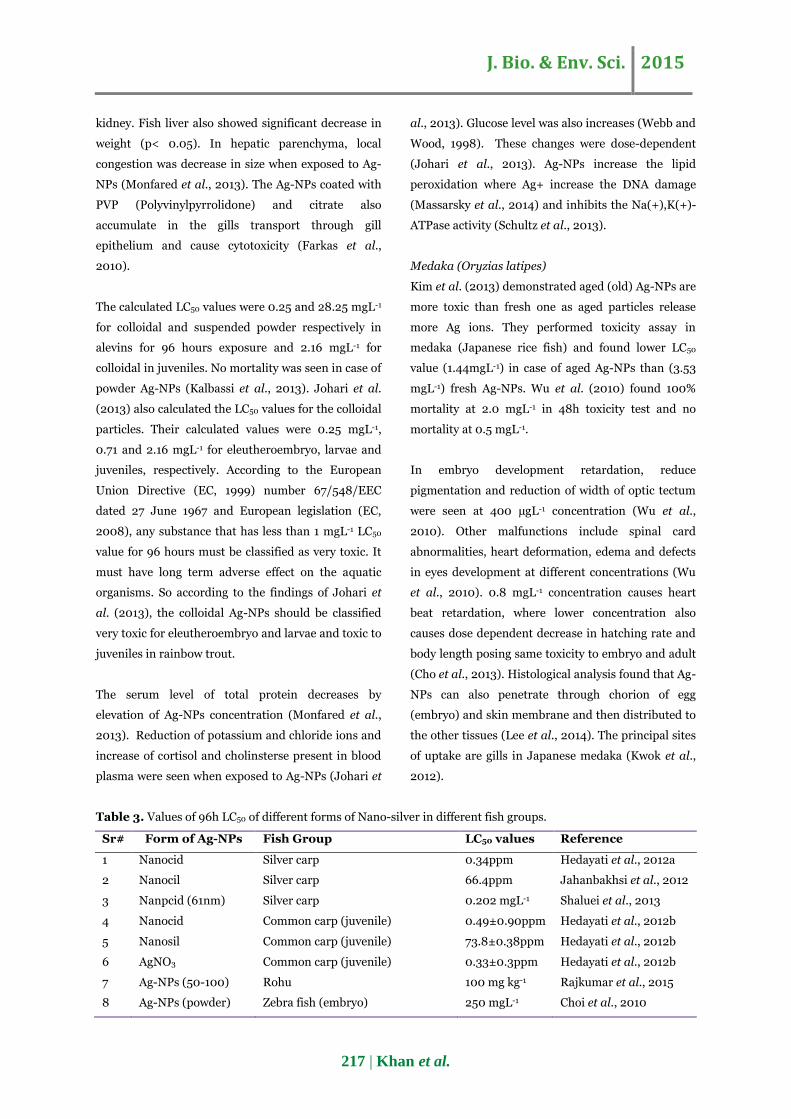
Table 3. Values of 96h LC50 of different forms of Nano-silver in different fish groups.
Sr# Form of Ag-NPs Fish Group LC50 values Reference
1 Nanocid Silver carp 0.34ppm Hedayati et al., 2012a
2 Nanocil Silver carp 66.4ppm Jahanbakhsi et al., 2012
3 Nanpcid (61nm) Silver carp 0.202 mgL-1 Shaluei et al., 2013
4 Nanocid Common carp (juvenile) 0.49±0.90ppm Hedayati et al., 2012b
5 Nanosil Common carp (juvenile) 73.8±0.38ppm Hedayati et al., 2012b
6 AgNO3 Common carp (juvenile) 0.33±0.3ppm Hedayati et al., 2012b
7 Ag-NPs (50-100) Rohu 100 mg kg-1 Rajkumar et al., 2015
8 Ag-NPs (powder) Zebra fish (embryo) 250 mgL-1 Choi et al., 2010
J. Bio. & Env. Sci. 2015
218 | Khan et al.
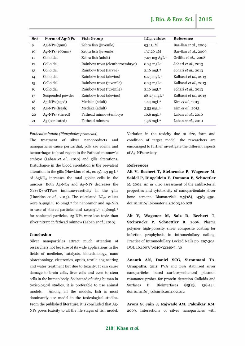
Sr# Form of Ag-NPs Fish Group LC50 values Reference
9 Ag-NPs (3nm) Zebra fish (juvenile) 93.11µM Bar-llan et al., 2009
10 Ag-NPs (100nm) Zebra fish (juvenile) 137.26 µM Bar-llan et al., 2009
11 Colloidal Zebra fish (adult) 7.07 mg AgL-1 Griffitt et al., 2008
12 Colloidal Rainbow trout (eleutheroembryo) 0.25 mgL-1 Johari et al., 2013
13 Colloidal Rainbow trout (larvae) 2.16 mgL-1 Johari et al., 2013
14 Colloidal Rainbow trout (alevins) 0.25 mgL-1 Kalbassi et al., 2013
15 Colloidal Rainbow trout (juvenile) 0.25 mgL-1 Kalbassi et al., 2013
16 Colloidal Rainbow trout (juvenile) 2.16 mgL-1 Johari et al., 2013
17 Suspended powder Rainbow trout (alevins) 28.25 mgL-1 Kalbassi et al., 2013
18 Ag-NPs (aged) Medaka (adult) 1.44 mgL-1 Kim et al., 2013
19 Ag-NPs (fresh) Medaka (adult) 3.53 mgL-1 Kim et al., 2013
20 Ag-NPs (stirred) Fathead minnow(embryo 10.6 mgL-1 Laban et al., 2010
21 Ag (sonicated) Fathead minnow 1.36 mgL-1 Laban et al., 2010
Fathead minnow (Pimephales promelas)
The treatment of silver nanoproducts and
nanoparticles cause pericardial, yolk sac edema and
hemorrhages to head region in the Fathead minnow’ s
embryo (Laban et al., 2010) and gills alterations.
Disturbance in the blood circulation is the prevalent
alteration in the gills (Hawkins et al., 2015). 1.3 µg L-1
of AgNO3 increases the total goblet cells in the
mucous. Both Ag-NO3 and Ag-NPs decreases the
Na+/K+-ATPase immune-reactivity in the gills
(Hawkins et al., 2015). The calculated LC50 values
were 9.4mgL-1, 10.6mgL-1 for nanoAmor and Ag-NPs
in case of stirred particles and 1.25mgL-1, 1.36mgL-1
for sonicated particles. Ag-NPs were less toxic than
silver nitrate in fathead minnow (Laban et al., 2010).
Conclusion
Silver nanoparticles attract much attention of
researchers not because of its wide applications in the
fields of medicine, catalysts, biotechnology, nano
biotechnology, electronics, optics, textile engineering
and water treatment but due to toxicity. It can cause
damage to brain cells, liver cells and even to stem
cells in the human body. So instead of using human in
toxicological studies, it is preferable to use animal
models. Among all the models, fish is most
dominantly use model in the toxicological studies.
From the published literature, it is concluded that Ag-
NPs poses toxicity to all the life stages of fish model.
Variation in the toxicity due to size, form and
condition of target model, the researchers are
encouraged to further investigate the different aspects
of Ag-NPs toxicity.
References
Alt V, Bechert T, Steinrucke P, Wagener M,
Seidel P, Dingeldein E, Domann E, Schnettler
R. 2004. An in vitro assessment of the antibacterial
properties and cytotoxicity of nanoparticulate silver
bone cement. Biomaterials 25(18), 4383-4391.
doi:10.1016/j.biomaterials.2003.10.078
Alt V, Wagener M, Salz D, Bechert T,
Steinrucke P, Schnettler R. 2006. Plasma
polymer high-porosity silver composite coating for
infection prophylaxis in intramedullary nailing.
Practice of Intramedullary Locked Nails pp. 297-303.
DOI: 10.1007/3-540-32345-7_30
Ananth AN, Daniel SCG, Sironmani TA,
Umapathi. 2011. PVA and BSA stabilized silver
nanoparticles based surface–enhanced plasmon
resonance probes for protein detection Colloids and
Surfaces B: Biointerfaces 85(2), 138-144.
doi:10.1016/ j.colsurfb.2011.02.012
Arora S, Jain J, Rajwade JM, Paknikar KM.
2009. Interactions of silver nanoparticles with
J. Bio. & Env. Sci. 2015
219 | Khan et al.
primary mouse fibroblasts and liver cells. Toxicology
and Applied Pharmacology 236(3), 310–318.
doi:10.1016/j.taap.2009.02.020.
Asharani PV, Wu YL, Gong Z, Valiyaveetti S.
2008. Toxicity of silver nanoparticles in zebrafish
models. Nanotechnology 19(25), 55-102.
-doi:10.1088/0957-4484/19/25/ 255102
Aslan K, Geddes CD. 2006. Microwave-accelerated
and metalenhanced fluorescence myoglobin detection
on silvered surfaces: Potential application to myo-
cardial infarction diagnosis. Plasmonics 1(1), 53-59.
DOI: 10.1007/s11468-006-9006-7
Aslan K, Huang J, Wilson GM, Geddes CD.
2006. Metalenhanced fluorescence-based RNA
sensing. Journal of American Chemical Society 128,
4206-4207.
ATSDR (Agency for Toxic Substances and
Disease Registry). 1990. Toxicological profile for
Silver. Prepared by Clement international
corporation, under Contract 205-88-0608). U.S.
public Health Service. ATSDR/TP-90-24.
Bar-Ilan O, Albrecht RM, Fako VE, Furgeson
DY. 2009. Toxicity assessments of multisized gold
and silver nanoparticles in Zebrafish embryos. Small
5(16), 1897-910. DOI: 10.1002/smll.200801716.
Bayston R, Ashraf W, Fisher L. 2007. Prevention
of infection in neurosurgery: Role of ‘antimicrobial’
catheters. Journal of Hospital Infection 65(2), 39-42.
DOI: http://dx.doi.org/10.1016/S0195-6701(07)60013-9.
Benn TM, Westerhoff P. 2008. Nanoparticle silver
released into water from commercially available sock
fabrics. Environmental Science and Technology
42(11), 4133–4139. DOI: 10.1021/es7032718.
Bhol KC, Alroy J, Schechter PJ. 2004. Anti-
inflammatory effect of topical nanocrystalline silver
cream on allergic contact dermatitis in a guinea pig
model. Clinical Experimental Dermatology 29(3),
282-287. DOI: 10.1111/j.1365-2230.2004.01515.x.
Bhol KC, Schechter PJ. 2007. Effects of
nanocrystalline silver (NPI 32101) in a rat model of
ulcerative colitis. Digestive Diseases and Sciences 52,
2732-2742. DOI: 10.1007/s10620-006-9738-4.
Bilberg K, Doving KB, Beedholm K, Baatrup
E. 2011. Silver nanoparticles disrupt olfaction in
Crucian carp (Carassius carassius) and Eurasian
perch (Perca fluviatilis). Aquatic Toxicology 104,
145–152. doi: 10.1016/j.aquatox.2011. 04.010.
Blaser SA, Scheringer M, MacLeod M,
Hungerbühler K. 2008. Estimation of cumulative
aquatic exposure and risk due to silver: Contribution
of nano-functionalized plastics and textiles. Science of
Total Environment 390 (2-3), 396–409.
DOI: 10.1016/ j.scitotenv. 2007.10.010.
Bouadma L, Wolff M, Lucet JC. 2012. Ventilator-
associated pneumonia and its prevention. Current
opinion in infectious diseases 25 (4), 395–404. doi:
10.1097/ QCO.0b013e328355a835.
Braydich-Stolle L, Hussain S, Schlager JJ,
Hofmann MC. 2005. In vitro cytotoxicity of
nanoparticles in mammalian germline stem cells.
Toxicological Science 88(2), 412–419.
doi: 10.1093/toxsci/kfi340.
Carlson C, Hussain S, Schrand A, Braydich-
Stolle L, Hess K, Jones R, Schlager J. 2008.
Unique cellular interaction of silver nanoparticles:
Size-dependent generation of reactive oxygen species.
The Journal of Physical Chemistry 112(43), 13608-
13619. DOI: 10.1021/jp712087m.
Catsakis LH, Sulica VI. 1978. Allergy to silver
amalgams. Oral Surgery Medicine Oral Pathology
Oral Radiology 46(3), 371-375.
DOI: http://dx.doi.org/ 10.1016/0030-4220(78)90402-4.
J. Bio. & Env. Sci. 2015
220 | Khan et al.
Chae YJ, Pham CH, Lee J, Bae E, Yi J, Gu MB.
2009. Evaluation of the toxic impact of silver
nanoparticles on Japanese medaka (Oryzias latipes).
Aquatic Toxicology 94(4), 320–327.
doi:10.1016/j.aquatox.2009.07.019
Chen LQ, Fang L, Ling J, Ding CZ, Kang B, Huang
CZ. 2015. Nanotoxicity of silver nanoparticles to red
blood cells: size dependent adsorption, uptake, and
hemolytic activity. Chemical Research in Toxicology
28(3), 501-9. doi: 10.1021/tx 500479m.
Chen W, Liu Y, Courtney HS, Bettenga M,
Agrawal CM, Bumgardner JD, Ong JL. 2006. In
vitro anti-bacterial and biological properties of
magnetron co-sputtered silver-containing hydroxyapatite
coating. Biomaterials 27(32), 5512-5517.
doi:10.1016/j.biomaterials.2006.07.003.
Chen X, Schluesener HJ. 2008. Nanosilver: A
nanoproduct in medical application. Toxicological
Letters 176(1), 1–12.
doi:10.1016/j.toxlet.2007.10.004.
Cho JG, Kim KT, Ryu TK, Lee JW, Kim JE,
Kim J, Lee BC, Jo EH, Yoon J, Eom IC, Choi
K, Kim P. 2013. Stepwise Embryonic Toxicity of
Silver Nanoparticles on Oryzias latipes. BioMed
Research International, Article ID 494671, 7 pages.
http:// dx. doi.org/10.1155/2013/494671.
Choi JE, Kim S, Ahn JH, Youn P, Kang JS,
Park K, Yi J, Ryu D. 2009. Induction of oxidative
stress and apoptosis by silver nanoparticles in the
liver of adult Zebrafish. Aquatic Toxicology 100(2),
151-159. doi: 10.1016/j.aquatox. 2009.12.012.
Coelho S, Amarelo M, Ryan S, Reddy M,
Sibbald RG. 2004. Rheumatoid arthritis-associated
inflammatory leg ulcers: A new treatment for
recalcitrant wounds. International Wound Journal
1(1), 81-84. DOI: 10.1111/j.1742-481x.2004. 0002.x.
Cohen MS, Stern JM, Vanni AJ, Kelley RS,
Baumgart E, Field D, Libertino JA, Summer-
hayes IC. 2007. In vitro analysis of a nanocrystalline
silver-coated surgical mesh. Surgical Infections
(Larchmt.) 8(3), 397-403. DOI: 10.1089/sur.
2006.032
Deery C. 2009. Silver lining for caries cloud?
Evidence-Based Dentistry 10(3), 68. doi:10.1038/ sj.
ebd.6400661
Diehl AM. 2000. Cytokine regulation of liver injury
and repair. Immunological Reviews 174(1), 160-171.
DOI: 10.1034/j.1600-0528.2002.017411.x
Dowling A, Clift R, Grobert N, Hutton D,
Oliver R, Neill O, Pethica J, Inoue KI, Takano
H, Yanagisawa R, Koike E, Shimada A. 2009.
Size effects of latex nanomaterials on lung
inflammation in mice. Toxicology and Applied
Pharmacology 234(1), 68-76.
doi:10.1016/j.taap.2008.09.012
Drake PL, Hazelwood KJ. 2005. Exposure-related
health effects of silver and silver compounds: A
review. The Annals of Occupational Hygiene 49(7),
575-585. doi: 10.1093/annhyg/mei019
EC. 1999. Annex VI of Directive 1999/45/EC to
consolidated version of directive 67/548/EEC.
General classification and labeling requirements for
dangerous substances and preparations.
ec.europa.eu/environment/archives/dansub/pdfs/an
nex6_ en.pdf
EC. 2008. Regulation (EC) No 1272/2008 of the
European Parliament and Council of 16 December
2008 on classification, labeling and packaging of
substances and mixtures, Official Journal of the
European Union, 31.12.2008.
http://eur-lex.europa.eu/legal-content/en/TXT/?uri
=CELEX:32008R1272.
J. Bio. & Env. Sci. 2015
221 | Khan et al.
Elechiguerra JL, Morones JR, Camacho A,
Holt K, Kouri JB, Ramirez JT, Yacaman MJ.
2005. Interaction of silver nanoparticles with HIV-1.
Journal of Nanotechnology 16, 23-46.
DOI: 10.1186/1477-3155-3-6
Farkas J, Christian P, Gallego JA, Urrea N,
Roos, Hassellöv M, Tollefsen KE, Thomas KV.
2010. Effects of silver and gold nanoparticles on
rainbow trout (Oncorhynchus mykiss) hepatocytes.
Aquatic Toxicology 96(1), 44-52. doi:10.1016/j.
aquatox.2009.09. 016
Gliga AR, Skoglund S, Wallinder IO, Fadeel B,
Karlsson HL. 2014. Size-dependent cytotoxicity of
silver nanoparticles in human lung cells: the role of
cellular uptake, agglomeration and Ag release.
Particle and Fibre Toxicology 11(11), 1-17 doi:
10.1186/1743-8977-11-11
Gonzalez P, Baudrimont M, Boudou A,
Bourdineaud JP. 2006. Comparative effects of
direct cadmium contamination on gene expression in
gills, liver, skeletal muscles and brain of the zebrafish
(Danio rerio). Biometals 19(3), 225–235. DOI:
10.1007/s10534-005-5670-x.
Griffitt RJ, Hyndman K, Denslow ND, Barber
DS. 2009. Comparison of molecular and histological
changes in zebrafish gills exposed to metallic
nanoparticles. Toxicological Science 107(2), 404-415.
doi: 10.1093/toxsci/kfn256
Griffitt RJ, Luo J, Gao J, Bonzongo JC, Barber
DS. 2008. Effects of particle composition and species
on toxicity of metallic nanomaterials in aquatic
organisms. Environmental Toxicology and Chemistry
27(9), 1972–1978. DOI: 10.1897/08-002.1
Gulbranson SH, Hud JA, Hansen RC. 2000.
Argyria following the use of dietary supplements
containing colloidal silver protein. Cutis 66, 373-376.
Handy RH, Owen R, Valsami-Jones E. 2008.
The ecotoxicology of nanoparticles and nanomaterials:
current status, knowledge gaps, challenges, and
future needs. Ecotoxicology 17(5), 315-325. doi:
10.1007/s10646-008-0206-0.
Hawkins AD, Thornton C, Kennedy AJ, Bu
K, Cizdziel J, Jones BW, Steevens JA, Willett
KL. 2015. Gill histopathologies following exposure to
nanosilver or silver nitrate. Journal of Toxicology and
Environmental Health A 78(5), 301-15.
doi: 10.1080/15287394.2014.971386.
He J, Lin L, Liu H, Zhang P, Lee M, Sankey OF,
Lindsay SM. 2009. A hydrogen-bounded electron-
tunneling circuit reads the base composition of
unmodified DNA. Nanotechnology 20(7), 075102.
doi: 10.1088/0957-4484/20/7/075102
Hedayati A, Kolangi H, Jahanbakhshi A,
Shaluei F. 2012a. Evaluation of Silver nanoparticles
Ecotoxicity in Silver carp (Hypophthalmicthys molitrix)
and Goldfish (Carassius auratus). Bulgarian Journal
of Veterinary Medicine 15(3), 172−177. Article id:
80158939
Hedayati A, Shaluei F, Jahanbakhshi A. 2012b.
Comparison of Toxicity Responses by Water Exposure to
Silver Nanoparticles and Silver Salt in Common Carp
(Cyprinus carpio). Global Veterinaria 8(2), 179-184.
Hussain SM, Hess KL, Gearhart JM, Geiss KT,
Schlager JJ. 2005. In vitro toxicity of nanoparticles
in BRL 3A rat liver cells. Toxicology in Vitro 19(7),
975–983. doi:10.1016/j.tiv.2005.06.034.
Inoue Y, Uota M, Torikai T, Watari T, Noda I,
Hotokebuchi T. 2010. Antibacterial properties of
nanostructured silver titanate thin films formed on a
titanium plate. Journal of Biomedical Materials
Research Part A 92A(3), 1171-1180.
doi: 10.1002/jbm.a.32456.
J. Bio. & Env. Sci. 2015
222 | Khan et al.
Ivask A, Kurvet I, Kasemets K, Blinova I,
Aruoja V. 2014. Size-Dependent Toxicity of Silver
Nanoparticles to Bacteria, Yeast, Algae, Crustaceans
and Mammalian Cells In Vitro. PLoS ONE 9(7),
e102108. doi:10.1371/journal.pone.0102108
Jahanbakhshi A, Shaluei F, Hedayati A. 2012b.
Detection of Silver Nanoparticles (Nanosil®) LC50 in
Silver Carp (Hypophthalmichthys molitrix) and
Goldfish (Carassius auratus). World Journal of
Zoology 7(2), 126-130. DOI: 10.5829/idosi.
wjz.2012.7.2.62129.
Jang MH, Kim WK, Lee SK, Henry TB, Park
JW. 2014. Uptake, tissue distribution, and
depuration of total silver in common carp (Cyprinus
carpio) after aqueous exposure to silver nanoparticles.
Environmental Science and Technology 48(19),
11568-74. doi: 10.1021/es5022813.
Johari SA, Kalbassi MR, Soltani M, Yu IJ.
2013. Toxicity comparison of colloidal silver
nanoparticles in various life stages of rainbow trout
(Oncorhynchus mykiss). Iranian Journal of Fisheries
Science 12(1), 76 -95.
Jones CM, Hoek EM. 2010. A review of the
antibacterial effects of silver nanomaterials and
potential implications for human health and the
environment. Journal of Nanoparticle Research 12,
1531–1551.
Jovanovic B, Anastasova L, Rowe EW, Zhang Y,
Clapp AR, Palic D. 2011. Effects of nanosized titanium
dioxide on innate immune system of fathead minnow
(Pimephales promelas Rafinesque, 1820). Ecotoxicology
and Environmental Safety 74(7), 675-683.
DOI: 10.1016/j.ecoenv.2010.10.017
Jung WK, Kim SH, Koo HC, Shin S, Kim JM,
Park YK, Hwang SY, Yang H, Park YH. 2007.
Antifungal activity of the silver ion against
contaminated fabric. Mycoses 50(4), 265–269.
DOI: 10.1111/j.1439-0507.2007.01372.x
Kalbassi MR, Johari SA, Soltani M, Yu LJ.
2013. Particle Size and Agglomeration Affect the
Toxicity Levels of Silver Nanoparticle Types in
Aquatic Environment. ECOPERSIA 1(3), 273-290.
Kannan R, Jerley A, Ranjani M, Prakash V.
2011. Antimicrobial silver nanoparticle induces organ
deformities in the developing Zebra fish (Danio rerio)
embryos. Journal of Biomedical Science and
Engineering 4, 248-254.
doi: 10.4236/ jbise.2011.44034.
Katuli KK, Massarsky A Hadadi A, Pourmehran
Z. 2014. Silver nanoparticles inhibit the gill Na⁺/K⁺-
ATPase and erythrocyte AChE activities and induce
the stress response in adult zebrafish (Danio rerio).
Ecotoxicology and Environmental Safety 106, 173-80
doi: 10.1016/j.ecoenv.2014.04.001.
Kim J, Kuk E, Yu K, Park S, Lee H, Kim S,
Park Y, Hwang C, Kim Y, Lee Y, Jeong D, Cho
M. 2007. Antimicrobial effects of silver nanoparticles.
Nanomedicine: Nanotechnology, Biology and Medicine
3(1), 95-101. doi:10.1016/j.nano. 2006.12.001
Kim J, Lee J, Kwon S, Jeong S. 2009.
Preparation of biodegradable polymer/silver nano-
particles composite and its antibacterial efficacy.
Journal of Nanoscience and Nanotechnology 9(2),
1098–1102. doi:10.1166/jnn.2009.C096
Kim JY, Kim KT, Lee BG, Lim BJ, Kim SD.
2013. Developmental toxicity of Japanese medaka
embryos by silver nanoparticles and released ions in
the presence of humic acid. Ecotoxicology and
Environmental Safety 92(1), 57-63. doi: 10.1016/j.
ecoenv.2013.02.004.
Kirsner RS, Orstead H, Wright JB. 2001. Matrix
metalloproteinases in normal and impaired wound
healing: a potential role for nanocrystalline silver.
Wounds 13(3), 5-12.
J. Bio. & Env. Sci. 2015
223 | Khan et al.
Klaprat DA, Evans RE, Hara TJ. 1992.
Environmental contaminants and chemoreception in
fishes. In Fish Chemoreception Fish and Fisheries
Series 6, 321-341. DOI: 10.1007/978-94-011-2332-
7_15.
Kumar PS, Sivakumar R, Anandan S,
Madhavan J, Maruthamuthu P, Ashokkumar
M. 2008. Photocatalytic degradation of Acid Red 88
using Au TiO2 nanoparticles in aqueous solutions.
Water Research 42(19), 4878–4884. doi:10.1016/j.
watres.2008.09.027
Kwok KW, Auffan M, Badireddy AR, Nelson
CM, Wiesner MR, Chilkoti A, Liu J, Marinakos
SM, Hinton DE. 2012. Uptake of silver nanoparticles
and toxicity to early life stages of Japanese medaka
(Oryzias latipes): effect of coating materials. Aquatic
Toxicology 120(121), 59-66.
doi: 10.1016/j.aquatox.2012.04.012.
Laban G, Nies LF, Turco RF, Bickham JW,
Sepulveda MS. 2010. The effects of silver
nanoparticles on fathead minnow (Pimephales
promelas) embryos. Ecotoxicology 19(1), 185-195.
DOI: 10.1007/s10646-009-0404-4
Lancaster T, Stead LF. 2012. Silver acetate for
smoking cessation. The Cochrane Collaboration.
(Online) 9, CD000191.
DOI: 10.1002/14651858.CD000191.
Lansdown A. 2006. Silver in health care:
antimicrobial effects and safety in use. Current
Problems in Dermatology 33, 17-34.
DOI: 10.1159/000093928.
Larese FF, Dagostin F, Crosera M, Adami G,
Renzi N, Bovenzi M, Maina G. 2009. Human
skin penetration of silver nanoparticles through intact
and damaged skin. Toxicology 255(1-2), 33–37.
doi:10.1016/j.tox.2008.09.025
Lee B, Duong C, Cho J, Lee J, Kim K, Seo Y,
Kim P, Choi K, Yoon J. 2012. Toxicity of Citrate-
Capped Silver Nanoparticles in Common Carp
(Cyprinus carpio). Journal of Biomedicine and
Biotechnology 2012, 262670.
doi: 10.1155/2012/262670.
Lee BC, Kim J, Cho JG, Lee JW, Duong CN, Bae
E, Yi J, Eom IC, Choi K, Kim P, Yoon J. 2014.
Effects of ionization on the toxicity of silver
nanoparticles to Japanese medaka (Oryzias latipes)
embryos. Toxic/Hazardous Substances and Environ-
mental Engineering 49(3), 287-93.
doi: 10.1080/10934529.2014. 846614.
Lee HJ, Yeo SY, Jeong SH. 2003. Antibacterial
effect of nanosized silver colloidal solution on textile
fabrics. Journal of Materials Science 38(10), 2199-
2204. DOI: 10.1023/A: 1023736416361.
Lesniak W, Bielinska AU, Sun K, Janczak KW,
Shi X, Baker JR, Balogh LP, 2005. Silver
/dendrimer nanocomposites as biomarkers:
Fabrication, characterization, in vitro toxicity, and
intracellular detection. Nano Letters 5(11), 2123-
2130. DOI: 10.1021/nl051077u.
Li Q, Mahendra S, Lyon, DY, Brunet L, Liga
MV, Li D, Alvarez PJJ. 2008. Antimicrobial
nanomaterials for water disinfection and microbial
control: potential applications and implications.
Water Research 42 (18), 4591–4602.
doi: 10.1016/j.watres.2008.08.015.
Luoma SN, Rainbow PS. 2008. Metal
contamination in aquatic environments: science and
lateral management. Journal of Fish Biology 75,
1911–1912.
DOI: 10.1111/j.1095-8649.2009.02440_4.x.
Massarsky A, Abraham R, Nguyen KC,
Rippstein P, Tayabali AF, Trudeau VL, Moon
TW. 2014. Nanosilver cytotoxicity in rainbow trout
(Oncorhynchus mykiss) erythrocytes and hepatocytes.
J. Bio. & Env. Sci. 2015
224 | Khan et al.
Comparative Biochemistry and Physiology Part C:
Pharmacology, Toxicology and Endocrinology; 159,
0-21. doi: 10.1016/j. cbpc.2013.09.008.
Moaddab S, Ahari H, Shahbazzadeh D,
Motallebi A, Anvar A, Rahman-Nya J,
Shokrgozar MA. 2011. Toxicity study of nanosilver
(Nanocid®) on osteoblast cancer cell Line.
International Nano Letters 1(1), 11-16.
Monfared AL, Soltani S. 2013. Effects of silver
nanoparticles administration on the liver of rainbow
trout (Oncorhynchus mykiss): histological and
biochemical studies. European Journal of
Exponential Biology 3(2), 285-289.
NCCAM. 2012. Colloidal Silver Products. National
Center for Complementary and Alternative Medicine.
February 2012.
https://nccih.nih.gov/health/providers/digest/topsu
pplements
Nowack B, Bucheli TD. 2007. Occurrence,
behavior and effects of nanoparticles in the
environment. Environmental Pollution 150(1), 5-22.
doi:10.1016/j. envpol.2007.06.006
Nowack B, Krug HF, Height M. 2011. 120 years of
nanosilver history: implications for policy makers.
Environmental Science and Technology 45(4), 1177–
1183. DOI: 10.1021/es103316q.
Pal S, Tak YK, Song JM. 2007. Does the
Antibacterial Activity of Silver Nanoparticles Depend
on the Shape of the Nanoparticle? A Study of the
Gram-Negative Bacterium Escherichia coli. Applied
and Environmental Microbiology 73(6), 1712-1720.
doi:10.1128/AEM.02218-06.
Panyala NR, Pena-Mendez EM, Havel J. 2008.
Silver or silver nanoparticles: a hazardous threat to
the environment and human health? Journal of
Applied Biomedicine 6, 117–129.
Perelshtein I, Applerot G, Perkas N, Guibert
G, Mikhailov S, Gedanken A. 2008. Sonochemical
coating of silver nanoparticles on textile fabrics
(nylon, polyester and cotton) and their antibacterial
activity. Nanotechnology 19, 245705.
doi:10.1088/0957-4484/19/24/245705
Pohle D, Damm C, Neuhof J, Rosch A,
Munstedt H. 2007. Antimicrobial properties of
orthopaedic textiles after in-situ deposition of silver
nanoparticles. Polymers & Polymer Composites
15(5), 357-363. Accession# 28655926.
Powers CM, Yen J, Linney EA, Seidler FJ,
Slotkin TA. 2010. Silver exposure in developing
Zebrafish (Danio rerio): Persistent effects on larval
behavior and survival. Neurotoxicology and
Teratology 32(3), 391-397.
doi:10.1016/j.ntt.2010.01.009
Project on emerging nanotechnologies. 2013.
Available online: http://www. nanotechproject. org/
inventories/consumer/ (accessed on 3 June 2013).
Rajkumar KS, Kanipandian N, Thirumurugan
R. 2015. Toxicity assessment on haemotology,
biochemical and histopathological alterations of silver
nanoparticles-exposed freshwater fish Labeo rohita.
Applied Nanoscience DOI 10.1007/s13204-015-0417-7.
Rathore RS, Khangarot BS. 2002. Effect of
temperature on the sensitivity of sludge worm Tubifex
tubifex (Muller) to selected heavy metals. Ecotoxi-
cology and Environmental Safety 53(1), 27–36.
doi:10.1006/eesa.2001.2100
Reddy TK, Reddy SJ, Prasad TNVKV. 2013.
Effect of Silver Nanoparticles on Energy Metabolism
in Selected Tissues of Aeromonas Hydrophila
Infected Indian Major Carp, Catla Catla. IOSR
Journal of Pharmacy 3(1), 49-55.
Reidy B, Haase A, Luch A, Dawson KA, Lynch
A. 2013. Mechanisms of Silver Nanoparticle Release,
J. Bio. & Env. Sci. 2015
225 | Khan et al.
Transformation and Toxicity: A Critical Review of
Current Knowledge and Recommendations for Future
Studies and Applications. Materials 6, 2295-2350.
doi: 10.3390/ma6062295.
Rivero P, Urrutia A, Goicoechea J, Zamarreno
C, Arregui F, Matias I. 2011. An antibacterial
coating based on a polymer/solgel hybrid matrix
loaded with silver nanoparticles. Nanoscale Research
Letters 6(305). doi:10.1186/1556-276X-6-305.
Rosenblatt A, Stamford TCM, Niederman R.
2009. Silver Diamine Fluoride: A Caries Silver-
Fluoride Bullet. Journal of Dental Research 88 (2),
116–125. DOI: 10.1177/0022034508329406
Rothkamm K, Lobrich M. 2003. Evidence for a
lack of DNA double-strand break repair in human
cells exposed to very low X-ray doses. Proceding of
National and Academy of Sciences U.S.A. 100, 5057-
5062. doi: 10.1073/pnas.0830918100
Russell AD, Hugo WB. 1994. Antimicrobial
activity and action of silver. Progress in Medicinal
Chemistry 31, 351-370.
Safaepour M, Shahverdi A, Shahverdi H,
Khorramizadeh M, Gohari A. 2009. Green
synthesis of small silver nanoparticles using geraniol and
its cytotoxicity against Fibro sarcoma-Wehi 164.
Avicenna Journal of Medical Biotechnology 1(2), 111-
115.
Samuel U, Guggenbichler J. 2004. Prevention of
catheter-related infections: the potential of a new
nano-silver impregnated catheter. International
Journal of Antimicrob Agents 23, 75-78.
DOI: 10.1016/j.ijantimicag.2003.12.004
Schrand AM, Braydich-Stolle LK, Schlager JJ,
Dai L, Hussain SM. 2008. Can silver nanoparticles
be useful as potential biological labels? Nanotech-
nology 19(2), 235104.
doi: 10.1088/0957-4484/19/23/235104
Schultz AG, Ong KJ, MacCormack T, Ma G,
Veinot JG, Goss GG. 2012. Silver nanoparticles
inhibit sodium uptake in juvenile rainbow trout
(Oncorhynchus mykiss). Environmental Science and
Technology 46(18), 10295-301.
doi: 10.1021/es3017717.
Scown TM, Santos EM, Johnston BD, Gaiser
B, Baalousha M, Mitov S, Lead JR, Stone V,
Fernandes TF, Jepson M, Van Aerle R, Tyler
CR. 2010. Effects of aqueous exposure to silver
nanoparticles of different sizes in rainbow trout.
Toxicological Science 115(2), 521–534. doi:
10.1093/toxsci/ kfq076.
Shaluei F, Hedayati A, Jahanbakhshi A, Kolangi
H, Fotovat M. 2013. Effect of subacute exposure
to silver nanoparticle on some hematological and
plasma biochemical indices insilver carp (Hypoph-
thalmichthys molitrix). Human& Experimental
Toxicology 32(12), 1270-7.
doi: 10.1177/0960327113485258.
Sharma VK, Siskova KM, Zboril R, Gardea-
Torresdey JL. 2014. Organic-coated silver
nanoparticles in biological and environmental
conditions: fate, stability and toxicity. Advances in
Colloid and Interface Science 204, 15-34. doi:
10.1016/j. cis.2013.12.002.
Silver Institute, 2014. World Silver Survey 2014.
https://www.silverinstitute.org/ site/supply- demand/
Silver S. 2003. Bacterial silver resistance: Molecular
biology and uses and misuses of silver compounds.
FEMS Microbiology Reviews 27, 341-353.
DOI: http://dx. doi.org/10.1016/S0168-6445(03) 000
47-0
Skirtach AG, Antipov AA, Shchukin DG,
Sukhorukov GB. 2004. Remote activation of
capsules containing Ag nanoparticles and IR dye by
laser light. Langmuir 20(17), 6988-6992.
DOI: 10.1021/la048873k
J. Bio. & Env. Sci. 2015
226 | Khan et al.
Smith I, Carson B. 1977. Trace metals in the
environment. Trace Metals in the Environment 469
pp. ISBN: 978-0-444-50352-7
Soderstjerna E, Bauer P, Cedervall T, Abdshill
H, Johansson F. 2014. Silver and Gold
Nanoparticles Exposure to In Vitro Cultured Retina
Studies on Nanoparticle Internalization, Apoptosis,
Oxidative Stress, Glial- and Microglial Activity. PLoS
ONE 9(8), e105359.
doi:10.1371/journal.pone.0105359.
Sondi I, Sondi BS. 2004. Silver nanoparticles as
antimicrobial agent: a case study on E. coli as a model
for Gram-negative bacteria. Journal of Colloid and
Interface Science 275(1), 177–182.
doi:10.1016/j.jcis.2004.02.012
Sung J, Ji J, Yoon J, Kim D, Song M, Jeong J,
Han B, Han J, Chung Y, Kim J, Kim T, Chang
H, Lee E, Lee J, Yu I. 2008. Lung function changes
in Sprague-Dawley rats after prolonged inhalation
exposure to silver nanoparticles. Inhalation
Toxicology 20(6), 567–574. doi:10.1080/089583707
01874671.
Syrvatka V, Rozgoni I, Slyvchuk Y, Milovanova
G, Hevkan I, Matyukha I. 2014. Effects of Silver
nanoparticles in Solution and liposomal form on
some blood Parameters in female rabbits during
fertilization and early embryonic development.
Journal of microbiology, biotechnology and food
sciences 3 (4), 274-278. ICID: 1092144
Tai SP, Wu Y, Shieh BD, Chen LJ, Lin KJ, Yu
CH, Chu SW, Chang CH, Shi XY, Wen YC, Lin
KH, Liu TM, Sun CK. 2007. Molecular imaging of
cancer cells using plasmonresonant- enhanced third-
harmonic-generation in silver nanoparticles. Advance
Materials 19, 4520-4523.
DOI: 10.1002/adma.200602213.
Taju G, Majeed AS, Nambi KS, Sahul Hameed
AS. 2014. In vitro assay for the toxicity
of silver nanoparticles using heart and gill cell lines of
Catla catla and gill cell line of Labeo rohita.
Comparative Biochemistry and Physiology Part C:
Pharmacology, Toxicology and Endocrinology 161,
41-52. doi: 10.1016/j.cbpc.2014.01.007.
Tian J, Wong KK, Ho CM, Lok CN, Yu WY, Che
CM, Chiu JF, Tam PK. 2007. Topical delivery of
silver nanoparticles promotes wound healing. Chem
Med Chem 2(1), 129-136.
DOI: 10.1002/cmdc.200600171
Tredget EE, Shankowsky HA, Groeneveld A,
Burrell R. 1998. A matched-pair randomized study
evaluating the efficacy and safety of Acticoat silver-
coated dressing for the treatment of burn wounds.
Journal of Burn Care & Rehabilitation 19, 531-537.
DOI: 10.1097/00004630-199811000-00013.
Walt DR. 2005. Miniature analytical methods for
medical diagnostics. Science 308(5719), 217-219.
DOI: 10.1126/science.1108161
Wan AT, Conyers RA, Coombs CJ, Masterton
JP. 1991. Determination of silver in blood, urine and
tissues of volunteers and burn patients. Clinical
Chemistry 37(10), 1683-1687.
Webb NA, Wood CM. 1998. Physiological analysis
of the stress response associated with acute silver
nitrate exposure in freshwater rainbow trout
(Oncorhynchus mykiss). Environmental Toxicology
and Chemistry 17(4), 579–588.
DOI: 10. 1002/etc.5620170408
Weisbarth RE, Gabriel MM, George M,
Rappon J, Miller M, Chalmers R, Winterton L.
2007. Creating antimicrobial surfaces and materials
for contact lenses and lens cases. Eye and Contact
Lens 33, 426-429.
West JL, Halas NJ. 2003. Engineered nanomaterials
for biophotonics applications: Improving sensing,
imaging, and therapeutics. Annual Review of
J. Bio. & Env. Sci. 2015
227 | Khan et al.
Biomedical Engineering 5, 285–292. doi: 10.1146/
annurev.bioeng.5.011303.120723.
Wijnhoven SWP, Peijnenburg WJGM,
Herberts CA, Hagens WI, Oomen AG,
Heugens EHW, Roszek B, Bisschops J, Gösens
I, Van de Meent D, Dekkers S, De Jong WH,
Van Zijverden M, Sips AJAM, Geertsma RE.
2009. Nano-silver - A review of available data and
knowledge gaps in human and environmental risk
assessment. Nanotoxicology 3(2), 109-138.
doi:10.1080/17435390902725914.
Woodrow Wilson Database. 2015. Nanotechnology
consumer product inventory
http://www.nanotechproject.org/cpi/about/analysis/.
World Health Organization. 2002. Silver and
silver compounds: Environmental aspects. (Concise
international chemical assessment document; 44). 1.
Silver _ adverse effects 2. Water pollutants, Chemical
3. Risk assessment 4. Environmental exposure I.
International Programme on Chemical Safety II.
Series ISBN 92 4 153044 8 (NLM Classification: QV
297). ISSN 1020 6167.
http://www.who.int/ipcs/publications/cicad /en/cicad
44.Pdf
Wright JB, Lam K, Hansen D, Burrell RE.
1999. Efficacy of topical silver against fungal burn
wound pathogens. American Journal of Infection
Control 27(4), 344-350. doi:10.1016/S0196-
6553(99)70055-6
Wu Y, Zhoua Q, Li H, Liua W, Wanga T,
Jianga G. 2010. Effects of silver nanoparticles on the
development and histopathology biomarkers of
Japanese medaka (Oryzias latipes) using the partial-
life test. Aquatic Toxicology 100(2), 160-167. doi:
10.1016/j.aquatox.2009.11.014
Yeo M, Kang M. 2008. Effects of nanometer sized
silver materials on biological toxicity during Zebra
fish embryogenesis. Bulletin of the Korean Chemical
Society 29(6), 1179-1184.
Yon JN, Lead JR. 2008. Manufactured
nanoparticles: An overview of their chemistry,
interactions and potential environmental
implications. Science of Total Environment 400(1-
3), 396–414. doi:10.1016/j.scitotenv.2008.06.042.
Yu H, Xu X, Chen X, Lu T, Zhang P, Jing X.
2007. Preparation and antibacterial effects of PVA-
PVP hydrogels containing silver nanoparticles.
Journal of Applied Polymer Science 103, 125-133.
DOI: 10.1002/app.24835
Yves MJ, Philippe H. 2012. Silver as an
antimicrobial: Facts and gaps in knowledge. Critical
Reviews in Microbiology 39(4), 373-83. doi:
10.3109/1040841X.2012. 713323.
Zhao Y, Shanmukh S, Liu Y, Jones L, Dluhy
RA, Tripp RA. 2006. Silver nanorod arrays can
distinguish virus strains. Nanotech SPIE Newsroom
DOI: 10.1117/2. 1200610.0438.