toxicological and pathological applications of
TRANSCRIPT

Toxicological and Pathological Applicationsof Proliferating Cell uclear ntigen (PCNA),A Novel Endogenous Marker for CellProliferation
Daniel R. DietrichInstitute of Toxicology, Swiss Federal Institute of Technology and University of Zürich, eH-86G3Schwerzenbach, Switzerland
ABSTRACT: A major stimulus to study cell proliferation, particularly in rodent carcinogenicity assays andhuman tumors, has been the belief that the quantification of this fundamental biological process will providethe toxicologist and pathologist with objective data allowing a better understanding of the mechanisms involvedin the toxicity and/or earcinogenicity of certain compounds as weil as guiding more effective management ofpatients afflieted with neoplasia. Among the markers used for cell proliferation measurement, PCNA has recentlygained much attention and holds much promise as it is intricately involved in the cell replication processes. Itnot only eould allow measurement of the replication rates without necessitating pretreatment of the animalltissuein prospective studies, but also would allow retrospective assessment of the proliferative rates in archival tissuesdue to the conservation 01' this marker in fixed and paraffin-embedded tissues. Finally, knowledge of the function01' PCNA in the cell cycle and its regulation by other factors may help us understand the advantages and!imitations 01' PCNA as a ceH proliferation marker in its application in toxicology and as a prognostic markerin human tumors.
KEY WORDS: PCNA, retrospective, cell cycle, pathology, immunohistochemistry, DNA synthesis, DNAexcision repair, prognostic marker.
I. INTRODUCTION
The fundamental biological process of celldivision, and thus of cell proliferation, has beeninvestigated from various viewpoints for a number of years. In the course of these investigations,numerous proteins intricately involved in themechanism of cell division have been discovered.Among these, the proliferating cell nuclear antigen (PCNA/cyclin) has been most intensivelyinvestigated not only due to its role in DNA synthesis and DNA repair mechanisms, but also asa result of its use as a cell proliferation markerand particularly as a prognostic tooI in surgicalpathology. Indeed, the existence of an endogenous cell proliferation marker that is preserved
1040-8444/93/$ .50© 1993 by CRC Press, [ne.
during tissue processing for pathological studiesmakes it tempting for toxicologists and pathologists to "go back" to studies that were completed some time in the past and measure retrospectively the cell proliferation rates resultingfrom the respective treatment regimens used ina particular study. However, the mere fact thatthis cell proliferation marker is a nuelear protein,the expression of which could be regulated bynumerous factors and which may be involved inmore than one mechanism of cell cyele control,should raise some doubt whether the "cell proliferation rates" measured via immunohistochemistry or flow cytometry can be taken as suchwithout additional knowledge of the effects ofthe compound in question on the expression lev-
77

eIs of PCNA or its regulating factors. Furthermore , although many lesions/tumors were shownto appear in conjunction with oncogene expression, growth factor overexpression or suppression, or loss oftumor suppressor genes, the question also must be raised whether these changescan influence PCNA expression levels and, if so,whether reliable .,cell proliferation rates" can bedetermined in these lesions/tumors with any degree of certainty. In addition, the designationPCNA/cyclin (i.e., cyclin due to the presumablycell cycle-dependent synthesis and marked presence during S-phase of PCNA) has caused confusion with the unrelated cyclins that have beendescribed in frogs, clams, sea urchins, yeast, andmammalian cells. Thus, it is the intention of thispaper to review the current literature on PCNAand to distinguish PCNA from other cell cycleassociated proteins with regard to its biochemicaland molecular characteristics as weIl as to itsfunction in the cell cycle. Moreover, the advantages and pitfaIls of current methodologies usingPCNA as a cell proliferation marker in toxicologyor as a prognostic tool in surgical pathology arediscussed.
11. DISCOVERY OF PCNA IN HUMANSAND ITS PRESENCE IN OTHEREUKARYOTES
PCNA was first described by Miyachi et al. 1as a nuclear antigen, restricted to proliferatingcells, that reacts with sera from some patientswith the autoimmune disorder systemic lupus erythematosus (SLE), hence the name proliferatingcell nuclear antigen. Tested via indirect immunofluorescence, these sera reacted with proliferating cells in a variety of tissues of the mouse,rabbit, and human, as weIl as with the dividingcell populations of baby hamster kidney, mousefibroblast, SP 2/0 mouse hybridoma, Wil-2 human B diploid lymphocyte, Hep-2 human hepatoma, MCF-7 human breast carcinoma, Raji,MOLT-4 T lymphocyte, and Ehrlich ascite tumorcells lines. 2 The presence of PCNA in varioustissues and animal species, as previously mentioned, not only led to speculation as to the importance of this antigen, but also raised the question of its presence in other eukaryotes and
78
possibly prokaryotes. Indeed, the developmentof monoclonal antibodies to PCNA3 and rat andhuman PCNA cDNA probes4-6 led to the discovery of PCNA-like proteins and homologousgenes not only in eukaryotes, e.g., amphibians,mammals, marsupials, fish, birds, insects, ciliated protozoa, plants, and yeast,7-13 but also inviruses. 14 Furthermore , amino acid sequence comparisons between human and rat PCNA revealedan extremely high degree of homology,4-6 withonly 4 amino acid substitutions in 261 aminoacids. Moreover, it was shown that the yeast typeof PCNA was able to functionaIly interact withmammalian DNA polymerase. 13 The presence ofa similar PCNA gene throughout eukaryotes and,in some cases, viruses implies that a primordialgene for PCNA evolved more than one billionyears aga at aperiod prior to the divergence intoPlanta and Animalia. 15 The latter observationsalso indicate that PCNA is, phylogenetically, astructurally and functionally highly conservedprotein and thus, based on the concept introducedby Kimura and Ohta16 on the principles governingmolecular evolution, PCNA must play an essential role in the cell cycle and in the maintenanceof species. However, prior to discussing the function(s) of PCNA in the cell cycle, it is of utmostimportance to define this protein biochemicallyand molecularly in order to clearly distinguish itfrom other cell cycle-associated proteins , i. e., thecyclins.
m. CHARACTERIZATION OF PCNA
A. Biochemical Characteristics
The gene for PCNA has been highly conserved throughout the course of evolution, as isquite impressively demonstrated by the fact thatrat PCNA cDNA probes have been successfullyused for the detection of homologous PCNA genesequences in Xenopus laevis, Drosophila melanogaster, two subspecies of rice, soybean, andtobacco. 7.10 ,12 However, despite this high conservation, some differences are found in the DNAsequence and length ofthe gene coding for PCNAin the various species and genera. It is interestingto note that while in all genera there appears tobe only one gene that codes for PCNA, disre-

garding the surpnsmg number of pseudogenesreported in mammalia whose function is not yetdear, the structure of the PCNA gene (DNA sequence and length and number of exons and introns) had undergone some modifications in thecourse of evolution among the genera, but hasremained highly conserved within the respectivegenera, e.g., mammalia (Table 1).17 This mayinlply that with increasing complexity of thehigher organisms, an enhanced need for the control of cell division and differentiation and thusfor the regulation of PCNA expression developed. Indeed, in higher eukaryotes such as D.melanogaster, rice (Oryza sativa), and mice, the5'-flanking regions of the respective PCNA genesappear to contain homeodomain protein-bindingsites in addition to the promoter region. 10,17,18Homeodomain proteins have been shown to havea regulatory function in gene expression and toregulate via the modulation of important mastergenes, thus playing an important role in cell di-
vision and differentiation. 19.2o Furthermore, thepresence of a similar number and nucleotide lengthof exons and introns in the PCNA gene of highereukaryotes, namely, in the human and mousePCNA gene,I7·21 as weIl as the observation thatintron 4 of the human PCNA gene codes for thatpart in the PCNA protein necessary for the correctregulation of PCNA levels in quiescent cells,22
further corroborate the assumption that enhancedpossibilities of regulating gene expression had tobe developed concurrently with the increasingcomplexity of higher eukaryotes.
The PCNA gene product is a nuclear nonhistone protein , as demonstrated by Takasaki etal. 23 It was shown by Almendral and co-workers4
to have a domain (amino acids 66-80) resembling the a-helix-tum-a-helix putative DNAbinding domain of several other DNA-bindingproteins. 24 Generally, the PCNA faund in thevarious species are all acidic proteins; however,more acidic and more basic variants can be dis-
TABLE 1Comparison of PCNA Genes and PCNA Gene Products in Various Species and Genera
Gene Protein
Mol.Exons/introns Pseudo- Amino acids wt. Homologya
Species per gene genes No. (Da) (%) Ret.
Human 6/5 2 261 2926b 100 4,22,171, 172
Rat n.d. 2 or 3 261 28748b 99.0 5, 6
Mouse 6/5 2 261 n.d. 98.5 17
Rabbit n.d. n.d. n.d. 36000° n.d. 47
Frog n.d. n.d. n.d. 36000° n.d. 7, 8
(Xenopus laevis)Fly 2/1 n.d. 260 28830b 70.4 10
(Drosophila melanogaster)Yeast n.d. 0 258 28916b 35.0 13,34
(Saccharomyces cerevisiae)Rice n.d. n.d. 263 29275b 61.0 12,173
(Oryza sativa)Periwinkle n.d. n.d. 268 29765b 85.0 174
(Cantharanthus raseus)Baculovirus n.d. n.d. 165 n.d. 42.0 14
(Autographa californica)
Note: n.d. = not determined.
a Amino acid sequence homology with human PCNA.b Mol. wt. calculated from the deduced amino acid sequence of the PCNA coding sequence.° Mol. wt. estimated via SDS-PAGE.
79
tinguished. Currently two "variants" are weIlcharacterized: (1) an acidic variant found in humans, rat, hamster, and potoroo, with an isoelectric point (IP) of 4.5 to 4.8;25-27 and (2) themore basic variant found in the mouse. 27 Accordingly, these species-specific variants havedifferent molecular weights (Table 1). It is important to note that when the molecular weightsare calculated according to the respective aminoacid sequence, the resultant weights (Table 1) arealways lower than the 33,000 to 37,000 Da estimated by SDS-PAGE and immunoblotting.23.25.2H.29 Sadaie and Mathews30 showed thatthe molecular weight of PCNA synthesized in anin vitro cell-free translation system is the sameas that of PCNA isolated from cells, indicatingthat PCNA does not undergo extensive posttranslational modification. Thus post-translational modification cannot account for the difference in calculated and experimentally determined molecular weights. A possible explanationfor this discrepancy is that PCNA migrates abnormally slowly in SDS-PAOE and the molecularweights have been overestimated. This phenomenon also has been observed with several otherproteins , such as adenovirus EI A and c-mycprotein. 31 .32
The functional unit of PCNA in mammaliancells, however, does not appear to be a proteinmonomer but rather a homodimer .25.33 In yeast,a tri- or tetramer is found. 13 The molecular weightsof the functional units of PCNA were calculatedfrom the glycerol gradient sedimentation coefficient of 5.0 sand the respective Stokes radii(36.5 Afor mammalian PCNA and 40 Afor theyeast analog) resulting in molecular weights of75,000 Da for the mammalian PCNA homodimer25 and approximately 82,000 Da for the trior tetrameric yeast PCNA-analog. 13 Thus, thefunctional units of the latter PCNAs are comparable in molecular weight and size. In addition,the observation that calf thymus PCNA can stimulate yeast polymerase Irr, the mammalian polymerase 8 analog of the yeast Saccharomycescerevisiae, or conversely that the yeast PCNAanalog can stimulate the DNA-synthesizing abilities of calf thymus polymerase 8,13 emphasizesthe apparent high conservation of PCNA in itsstructure and function throughout the course ofevolution. This function is conserved despite the
80
fact that the amino acid sequence of the PCNAsfound in mammals and yeast show very littlehomology. However, a sequence comparison between human, yeast, and baculovirus PCNA revealed that there are a few highly homologousdomains, which might be important for proteinprotein interaction with the 8 polymerases. 34
B. Expression of PCNA during the CellCycle
In order to determine at what time point during the cell cycle PCNA synthesis is initiated,the PCNA content of synchronized cells, e.g.,mouse 3T3 or human MOLT 4, was analyzed viaimmunofluorescence and flow cytometry, usingmonoclonal and/or polyclonal antibodies toPCNA.9.27.30.35-41 All investigators unanimouslyreported a maximum of staining intensity in theS-phase of cycling cells as weIl as the presenceof PCNA at sites of ongoing DNA replication,as shown by the colocalization of PCNA andtritiated thymidine in the nucleus of replicatingcells. 9 Further investigation showed that the concentration of PCNA increased starting in late GIphase, reaching its maximum during S-phase,which is approximately sevenfold the concentration found in quiescent cells,9,3o,35.38-41 and thento gradually decrease throughout O2 phase andmitosis. Using a full-Iength cDNA clone for thehuman PCNA, these observations have furtherbeen corroborated by Almendral and co-workers4
and laskulski and colleagues,42 who demonstrated that the expression of PCNA mRNA waslow to undetectable in quiescent cells, whereasincreased expression was detected 8 to lOh afterserum stimulation of quiescent 3T3 cells, reaching a maximum induction of tenfold at 18 to 20h, which also is the peak of DNA synthesis inthese cells. In addition, quiescent cells stimulatedwith fetal calf serum in the presence of 5-hydroxyurea, thus being inhibited from DNA synthesis, exhibited the same increase in PCNAmRNA as control cells without hydroxyurea.These experiments suggested that the inductionof PCNA mRNA expression is independent ofDNA synthesis. 40,43 Despite the good rapport between PCNA detection via immunofluorescenceand PCNA synthesis evidenced via mRNA lev-
eIs, the findings by Bravo and MacDonaldBrav036 evoked some doubt as to the reliabilityof PCNA detection via immunofluorescence. Indeed, when cells were fixed using organic solvents such as methanol, PCNA was detected atthe intranuclear sites where DNA synthesis wastaking place as shown by simultaneous[3H]thymidine (Tdr) incorporation. 35 .37 With thisfixation technique, PCNA had a very granulardistribution and was absent from the nucleoli inthe early S-phase, whereas more prominent nucleolar staining was observed in the later stagesof S-phase. On the other hand, when cells werefixed with aldehydes, the distribution of PCNAappeared different in that intense diffuse nuclearstaining was observed throughout the cell cycIe. 36
This discrepancy was explained with the hypothesis that there are two forms of PCNA: an organicsolvent insoluble form associated with the site ofongoing DNA synthesis, and a soluble form presumably not involved in DNA replication. 36 Thishypothesis was substantiated further by Kurki andco-workers,43 who found higher numbers of formaldehyde-fixed cells staining positive for PCNAthan for bromodeoxyuridine (BrdU). Furthermore, Morris and Mathews44 demonstrated, incontrast to earlier studies, that the total concentration of PCNA varied at most two- to threefoldduring the cell cycle, but that a greater fractionof PCNA is insoluble due to chromatin association during S-phase than in other phases of thecell cycle, and, in corroboration with earlier findings by Bravo and MacDonald-Bravo,36 that amaximum of 30% of the PCNA present duringS-phase was tightly associated with the nucleusand thus presumably present in replication complexes . Moreover, Morris and Mathews44 concIuded that the cyclic synthesis of PCNA in proliferating HeLa cells maintained PCNA in excessof the amount necessary for DNA replication. Ifthis were the case, the assessment of proliferatingcells using the commercially available antibodiesto PCNA 1•3 ,7,H,41,4S-4H would grossly overestimatethe number of proliferating ceIls, as the antibodies apriori would not be able to distinguish between chromatin-associated and nonchromatinassociated PCNA. Indeed, Richter and coworkers49 and Oaland and DegraefSO found anexcellent agreement between cell proliferationmeasurements obtained via PCNA and Tdr or
BrdU in tissues fixed with ethanol or methanol,whereas in tissues fixed with formalin or formaldehyde more PCNA than Tdr positive cellsalways were detected. 50 Coltrera and Gown,51 onthe other hand, found no agreement or any correlation between the number of BrdU and PCNApositive alcohol-fixed cells in a variety of celllines. However, prior to discussing the advantages and disadvantages of the various antibodiesand the feasibility of using PCNA for cell proliferation studies, it is important to understandthe regulation of PCNA mRNA expression andthe function(s) of PCNA within the cell cycle.
As mentioned earlier, the levels of PCNAmRNA appear to be cycling during the cell cycleof 3T3 cells.4,42 In these cells, the PCNA mRNAwas shown to be inducible only by platelet-derived growth factor (PDGF), not by platelet-poorplasma. The expression of PCNA mRNA is inhibited by low concentrations of cycloheximide. 42 In addition, PCNA mRNA was not expressed in serum-stimualted ts 13 cells at therestrictive temperature,42,52 ts13 cells being 01specific, temperature-sensitive mutants of the cellcycle originally derived from baby hamster kidney cells and made quiescent by serum deprivation. 42 The latter two findings suggest that thePCNA gene is growth factor regulated and, unlike early growth-regulated genes, PCNA requires the previous expression of other growthregulated genes (Figure 1). In contrast to othergrowth factor-regulated genes coding for proteinsinherent to DNA synthesis, such as thymidinekinase, increased expression of PCNA mRNAcan be induced by epidermal growth factor (BOF)or PDOP in the absence of other growth factors. 42,52 Gf the two most important pathways ofregulating PCNA mRNA expression levels (Figure 1), the transcriptional regulation of PCNAmRNA steady-state levels involving the promoterregion and intron 4 of the PCNA gene22,53 appearsto playa minor role, whereas post-transcriptionalregulation seems to predominate, as demonstrated in the latter experiments wherein the increase in mRNA levels that occurred in serumstimulated cells was largely post-transcriptionallyregulated (Figure 1),52,53 To make it even morecomplex, the regulation of PCNA mRNA levelsis different in continuously proliferating cells,wherein both the mRNA and the protein amounts
81

DNA RepUcation
@inases/ 2nd messenger0"'111!111~_/I G..1 S- S, G-2,
, and M-I Phase Phase
G-1 Phase
IRegulators \
a(genes~ / / "
Re~ators IPCNA gene\
~\
hnRNA ~ IhnRNA \
I · t / \VVVV ",VVVV 1\ :Y/
, 1 I" I \ PCNA
~ \" 1 \
... ; \,~ \,, \
\
V'Y'A "\/:VV\ ... PCNA/ \. '. \ PCNA mRNA
mRNA ~Proteins(gene products ofNUCLEUS tumor suppressor genes, proto
onco enes?)
Early growth factor regulated genes late growth factor regulated genes
_ -< Growth factors}- _ _
FIGURE 1. Schematic diagram of PCNA regulation during the cell cycle. Full arrowsindicate the various steps in PCNA expression starting with gene transcription and endingwith PCNA protein synthesis and the interaction of this protein during DNA synthesis. DottedIines with arrows depict how and at which steps PCNA expression can be regulated.
vary litde during the cell cycle. 44 ,45,54 Furthermore, an overexpression of PCNA mRNA wasfound in the R3230AC mammary tumor of therat, which was accompanied by an altered PCNAgene structure,54 emphasizing again that cautionmust be exercised when PCNA is used as a cellproliferation marker.
IV. FUNCTION OF PCNA IN THE CElLCYCLE
A. Cell Replication
Based on the distribtuion pattern of PCNAduring the cell cycle, the biochemical and struc-
82

tural characteristics (putative DNA-binding domain) of the protein suggest that PCNA is intricately involved in DNA replication and possiblyin cell cycle progression. Indeed, antibodies directed against PCNA reduced plasmid and chromosomal DNA replication in microinjected frogeggs,8 and inhibited the stimulation of DNA synthesis in MOLT-4 cells. 55 FUl1hermore, after exposuring exponentially growing Balb/c3T3 cellsto antisense oligodeoxynucleotides to PCNA,DNA synthesis and mitosis were both completelysuppressed. 56 In addition, Tan and co-workers25
noticed that physically PCNA closely resemblesa protein that regulates the activity of calf thymusDNA polymerase o. Further investigation revealed that PCNA and the auxiliary protein ofcalf thymus DNA polymerase 0 were one andthe same. 33 ,57 PCNA also was shown to be required for replication of an SV40 DNA templatein vitra in extracts from human 293 cells33 ,58 asweIl as for cell replication of HeLa cells.59 In itsinteraction with calf thymus DNA polymerase 8,PCNA increases the processivity of polymerase8 decisively.25 Within the concept of a two-polymerase hypothesis of eukaryotic replication,polymerase 0., with its tighly associated primaseactivity and semiprocessive mode of action, isideally suited for the synthesis of the laggingstrand. 60 Conversely, polymerase 0, lacking primase activity but possessing strand displacementactivity and being highly processive in conjunction with the presence of PCNA,25,33 is capableof synthesizing long stretches of DNA as wouldbe required of a leading strand polymerase (Figure 2).60-f>3 This hypothesis was tested using theSV40 replication system and the tests showed thatin the absence of PCNA the leading strand synthesis was virtuaIly abolishedM
-66 and that poly
merase 0. was responsible for both the initiationand the synthesis of the lagging strand. Furthermore, leading strand synthesis was not inhibitedwhen the SV40 replication system was treatedwith antibodies to polymerase 0.. 67 However, despite these clear indications that PCNA is direcdyinvolved in DNA synthesis and despite the factthat PCNA possesses a DNA-binding domain, noDNA-binding activities of PCNA could be detected. 25 Therefore, the role of PCNA in DNAsynthesis appears to be the increased binding ofpolymerase 0 to poly(dA)/oligo(dT) in conjunction with the RF-A protein complex, RF-C pro-
tein complex, and activator 1 protein complex,65-68 resulting in the stabilization ofthe polymerase-template/primer complex (Figure 2).
B. DNA Excision Repair
Besides the interaction of PCNA with polyluerase 8, PCNA can be detected in nuclei ofnon-S-phase cells following UV irradiation, suggesting an involvement of PCNA in the excisionrepair process.39,69 Inhibition of protein and DNAsynthesis via cycloheximide and aphidicolintreatment, respectively, revealed that upon UVirradiation DO new PCNA was synthesized, thePCNA observed via immunofluorescence was redistributed from an already existing pool withinthe nucleus, and this immunofluorescence staining was independent of DNA synthesis, thus suggesting that the relocation of PCNA was not triggered by DNA repair synthesis by itself butpossibly preceded it. 69 Furthermore, Toschi andBrav069 were able to show that the PCNA involved in excision repair was actually looselyattached to nuclear components and was in effectthe part of the PCNA population that could notbe detected in organic solvent fixed cells. Inkeeping with the hypothesis that the involvementof PCNA in the excision repair process precedesthe actual DNA synthesis step, Shivji and coworkers70 and Coverley and co-workers71 investigated the excision repair process via fractionation of cell extracts and UV-irradiated plasmidDNA, which allowed them to resolve the excisionrepair process into discrete incision and polymerization stages. They were able to show thatPCNA is required for the DNA synthesis thatconverts the nicked intermediates to complete repair events; however, this was only in conjunction with other proteins, e.g., xeroderma pigmentosum protein complement A (XP-A), humansingle-strand binding protein (HSSB), replicationfactor C (RF-C), and DNA polymerases 8 or E
(Figure 3). However, with respect to excisionrepair, it must be stated that no direct interactionbetween PCNA and polymerase 0 has so far beendemonstrated. On the contrary, Syvaoja andLinn72 and Nishida and co-workers73 described aPCNA-independent form ofpolymerase 8, whichappeared to be involved in the DNA repair process in UV-exposed Brij-58 cells.
83

51
Okazaki fragments
lagging strand
___________ 31
--------- 5'
DNA primase
1iC--'::::::::==~;;;:RF-A (H SSB; RPA)
Polymerase 8
Activator 1complex
FIGURE 2. Schematic representation of the rales of PCNA and DNA polymerases a and0, replication factor A (RF-A), also known as human single-strand binding pratein (HSSB)er replication protein A (RPA},68,71,175 replication factor C (RF-C),65,66 DNA primase, andactivator 1 complex68 in eukaryotic DNA replication. (Modified from Stillman, B., BioEssays,9, 56, 1988.)
The involvement of PCNA in DNA repairfollowing UV irradiation and thus the detectionof prior immunohistochemically undetectablePCNA forms must be taken into special consideration when using PCNA as a cell proliferationmarker in epidermal tumors such as melanomas.In these tumors, immunohistochemical methodsmay weIl detect PCNA involved in DNA synthesis; however, not all PCNA positive cells needto represent dividing cells, meaning that a fairnumber of cells may be undergoing DNA repair.This applies not only to melanomas, as was shownto be the case, for example, in patients with acutemyelogenous leukemia in which high levels of
84
PCNA correlated with DNA repair synthesis andwas associated with enhanced resistance to chemotherapy but did not correlate with increased cellproliferation. 74
c. Interaction with Tumor SuppressorGenes and Oncogenes
From the previous paragraphs, it should beclear that PCNA is involved in two mechanismsinherent to the cell cycle, Le., DNA replicationduring the S-phase and DNA excision repair during the 02 phase and in quiescent cells. How-

Ci) TITTlI (iv)
1incision proteins
1PCNA C+ RF-C?)
Cil)
1HSSBjRPA
DNA Iigase 1(iii) j]IDT!TllJI (vi)
FIGURE 3. Model for nucleotide excision repair in mammalian cells. The model makes use of the data reportedby Coverley et al. 71 and Shivji et aVo (i) Incisions are introduced about 20 nucleotides apart from the damage site176
byan unknown number of proteins, one of which is most probably the xeroderma pigmentosum A polypeptide.16B
(ii) The oligonucleotide containing DNA damage and the incision proteins are displaced, possibly by the humansingle-strand binding protein (HSSB) in concert with a DNA helicase. (iii) Degradation of the DNA gapped regionmay be prevented by HSSB. (iv) PCNA (in conjunction with RF-C?) binds to an incision site on the 5' side of thegap, possibly mediated by protein-protein interactions with HSSB. (v) Repair synthesis is carried out by DNApolymerases E or 8. (vi) The repaired area is completed and sealed bya DNA ligase. (From Coverley, 0., Kenny,M. K. t Lane, D. P., and Wood, R. 0., Nucleic Acids Res., 20(15), 3873, 1992. With permission.)
ever, in order to understand PCNA and its involvement in cell proliferation, it is necessary tounderstand not only how the expression of thePCNA gene and the level of the PCNA geneproduct are regulated, but also how PCNA geneexpression and PCNA gene product levels maybe affected by mutations, translocations, and allel loss in genes of cell proliferation and PCNAregulators.
Regarding altered PCNA RNA expression dueto genetic events, constantly high expression levels ofPCNA mRNA and gene product have beenfound in continuously proliferating cells,44.54 indicating that the gene(s) downregulating PCNAexpression has either been missing, nonfunctional due to alterations in the gene(s), or suppressed in its function by other proliferation reg-
ulators. Among the genes possibly regulatingPCNA expression are the p53 and the retinoblastoma [pI05(Rb)] gene products. Both of thesegene products have been shown to have tumorsuppressing capabilities in that they can inhibittransformation of cells to tumorigenic phenotypes. 75- 79 Furthermore, it was shown that thetwo tumor suppressor gene products in their underphosphorylated state keep cells from progressing from the GI phase into S-phase, and thusplay an important role in the control of the cellcycle (Figures 4 and 5). Phosphorylation of thesegene products by the cdc2(p34)-cyclinC complexlifts the Gl-S-phase baITier, allowing transitionof the cell into S-phase. 80
-85
In view of the fact that PCNA is a late growthfactor-regulated gene, its expression starting at
85

/
~:; / wt-p53
';1 "',tl! ..
FIGURE 4. Schematic model of the cyclic phosphorylation and dephosphorylation of the human wild-typep53 protein by the p34(cdc2)-cyclin complexes andphosphatases during the cell cycle. Although the p34cyclin complex is depicted as the phosphokinase involved in the phosphorylation of p53 in every cell cyclephase, this has been explicitely shown to be the casefor the p34-cyclin Bcomplex in only the G2- to M-phasetransition.B4 However, p34-cyclin complexes have beenimplicated as the phosphokinase complexes involvedin the G1- to S-phase, S- to G2-phase, and G2- to Mphase transitions in the human cell cycle.B5 The p53protein is depicted here with three phosphate groupsin the M-phase; however, the actual degree of p53phosphorylation during the cell cycle has not been determined yet, with the exception that p53 is underphosphorylated in the G1 phase, phosphorylated uponentry into S-phase, and additionally phosphorylatedduring the transition from the G2- to the M-phase.7B,B4In addition, the putative interaction possibilities of theDNA tumor virus gene products large T antigen (LTA)and adenovirus E1 B (E1 B) with the various p53 phosphorylation states are shown.94 ,177 However, there is noevidence at this time that E1 B can bind to phosphorylated p53. The association of E1 B with phosphorylated p53 is merely hypothetical, and was drawn in analogy to the known association of the E1 A protein withphosphoryalted p105(Rb) depicted in Figure 5.82,17B
the end of the GI phase (Figure 1), the quest ionarises whether p53 and/or pI 05(Rb) have a regulatory effect on PCNA expression. Indeed, bothp53 and pl05(Rb) have domains with DNA-binding abilities. 86
-X9 However, thus far there is evi
dence only for the p53 gene product demonstrating that the wild-type p53 protein selectivelydownregulates PCNA mRNA and protein expression (Figure 6) in conjunction with the inhibition
86
FIGURE 5. Schematic model of the cyclicphosphorylationBO and dephosphorylation of the humanp105(Rb) protein by the p34(cdc2)-cyclin complexesand phosphatases during the cell cycle. Although thep34-cyclin complex is depicted as the phosphokinaseinvolved in the phosphorylation of p105(Rb) in everycell cycle phase, it has not been explicitely shown thatcyclins are part of this phosphokinase complex implicated in p105(Rb) phosphorylation. However, thep34(cdc2) kinase has been implicated as the phosphokinase involved in p1 05(Rb) phosphorylation,179 andthe p34-cyclin complexes have been shown to be important in the G- to S-phase, S- to G2-phase, and G2to M-phase transitions of the human cell cycle.B5 Themodel also depicts the interaction possibilities of theDNA tumor virus gene products large T antigen (LTA)and adenovirus E1 A (E1 A) with the various p105(Rb)phosphorylation states.B2,178
of cell cycle progression. 90 Alteration of p53 bymutation, as often observed in human tumors,leads to gene products that are unable to bind toDNA,x6 thus raising the question whether mutated p53 can still regulate PCNA expression.Alteration or inactivation of p53 by mutation, orby its interaction with oncogene products of DNAtumor viruses, can lead to abrogated cell cyclecontrol and subsequently to cancer. 77,91 Althoughdirect evidence is lacking, experiments with SV40DNA virus-transformed keratinocytes show thatPCNA expression is increased in these transformed cells, irrespective of the cell cycle stage,thus suggesting that the PCNA expression controlby regulatory proteins is abrogated. 37 Furthermore, the conformational changes observed inthe gene products of mutated p53 92 also could

toxicological studies. Over/underestimation of cellproliferation could occur under circumstances inwhich PCNA protein is over/underexpressed asthe result of functional changes in genes regulating PCNA expression. Indeed, overexpressionof PCNA was found to con-elate with overexpression of wild-type and mutated p53 in human colorectal tumors. 102 Thus, more researchstudying tumor suppressor gene and oncogeneexpression in conjunction with PCNA (over/un
der)expression is clearly needed.
v. DIFFERENCES OF CYClIN(S) VS.PCNA
Numerous publications describe a protein involved in the cell cycle as PCNAlcyclin; however, just this designation can be quite misleadinginasmuch as PCNA and cyclin(s) are not one andthe same, although both PCNA and cyclin(s) appear to be characterized by a cyclic expressionduring the cell cycle, have been highly conservedthroughout evolution in many organisms, andseem to be intricately involved in ceIl replication.Indeed, cyclins describe a dass of proteins thatare found in viruses, 103 clams,104 frogs,105 seaurchins, 106,107 yeast,IOlU09 flies, H5.103 and humans,H5,103 are highly conserved,I()3 and have aapproximate molecular weight of 56 kDa. liD Onthe basis of sequence comparisons, cyclins havebeen divided into two classes (A and B) and mostorganisms contain both types, 110,111 However,most recent findings suggest further classes ofcyclins, i.e., the C, D, and E classes in humansas weIl as the Cigl and Mcs2 classes in yeast, H5,112Of importance is that cyclins are synthesized during interphase, associate into a complex with thep34cdc2 kinase (Figure 4 and 5), and are destroyedby cyclin-degrading enzymes after the ceIl entersS-phase (cyclins A, C, D, E), 02 phase (cyclinsA), or mitosis (cyclins A and B).III.l13 Cyclin Awas shown to playamajor role in the control ofDNA replication in that the microinjection ofmammalian cells with plasmids encoding antisense cyclin A cDNA or with affinity-purifiedanti-cyclin A antibodies during the GI phase ledto inhibition of DNA synthesis. 114 Although itwas demonstrated that the cyclin A_p33cdk2 kinasecomplex, i.e., the p33cdk2 kinase belonging to thep34cdc2 kinase family, has a sequence-specific
88
DNA-binding activity, 115 this DNA-binding activity was associated with the phosphorylation ofother DNA-bound substrates during S-phase(Figures 4 and 5) and was not, as is the case withPCNA, associated with the processes directly involved in DNA synthesis. Thus, cyclins are biochemically, structurally, and functionally different from PCNA and therefore the term PCNAIcyclin is erroneous and should be avoided.
VI. CEll PROLIFERATIONMEASUREMENTS USING PCNAANTIBODIES
There is increasing evidence that enhancedceU proliferation, whether induced by chemicals,UV or ionizing radiation, or genetic alterationsin cell cycle-regulating genes, may be a significant factor in the etiology of tumor development.116-120 Furthermore, the assessment of therate of cell proliferation in an organ or lesion hasbeen shown to be enormously useful for understanding at least some of the aspects of the underlying mechanisms involved in the development and progression of induced and spontaneously occun-ing lesions and tumors. 120-122Thus, several methods for measuring cell proliferation, such as flow cytometry and immunohistochemistry, have evolved in the last few decades. Among these, methods using exogenouslyapplied thymidine analogs (BrdU and 3[H]-thymidine [Tdr]) for marking the DNA synthesizedduring S-phase have seen widespread applicationand gained acceptance by the scientific community. However, the major disadvantages ofthese techniques is that postmortem (post-fixation) analysis of cell proliferation in organisms,organs, biopsies, or celliines is not possible without prior in vivo application of these S-phasemarkers. Thus, with the discovery of PCNA, withits presumably cyclic synthesis, involvement inDNA replication, and marked presence in S-phasecells, and the development of commerciallyavailable PCNA antibodies,I.3,41,46.48.123,124 much
attention has focused on PCNA as a new markerfor proliferating cells. In contrast to techniquesusing BrdU or Tdr, flow cytometric and immunohistochemical analysis using the endogenouslyformed PCNA could potentially allow retrospective assessment of cell proliferation in archived

material. 125-127 However, in order to achieve reliable results with this new cell proliferationmarker, it is not enough to understand the roleand function of this endogenous protein in thecell, but rather a thorough knowledge of the possibilities, limitations, and uncertainties involvedin the use of the techniques using this cell proliferation marker is aprerequisite.
A. Antibodies
As mentioned earlier, PCNA and the respective autoantibodies were discovered in patients presenting with SLE. These polyclonalautoantibodies were the first antibodies available for studying the role and function ofPCNA. 1,7,8,27,38.41,55,123,124 In the beginning, the
use of these polyclonal autoantibodies were problematic as these antibodies also recognized proteins other than PCNA.7,47 The preparation of amonospecific immunoglobulin G-type anti-PCNAvia absorption of serum from an SLE patient toimmobilized rabbit kidney extract, apparentlyeontaining negligible amounts of PCNA butabundant amounts of other autoantigens, solvedthe problem of unspeeific antigen reaetion. 3,7 Ina further step, a number of monoclonal antibodiesto PCNA were developed.3.46,48 Among these,three are commercially available: a murine IgMdesignated "19A2", a murine IgG designated" 19F4" ,3 ,46 and a genetically engineered murineIgG isotype designated "PCIO".48 Whereas thepolyclonal PCNA autoantibodies were demonstrated to recognize at least two different epitopesat the N- and the C-terminals of the PCNA protein, the epitopes recognized by the 19A2 and19F4 monoclonal antibodies appear to reside moreto the center of the protein. 46 The PCI0 antibodywas shown to have staining characteristics similarto those observed with the 19A2 and 19F4 antibodies when tested using immunofluorescenee,48 suggesting that the epitope recognizedby PCI0 also may reside in the center of theprotein. Epitope location and recognition by antibodies are important factors to be consideredwhenever PCNA is used as a eell proliferationmarker. Indeed, Waseem and Lane48 found thatamong their 11 genetically engineered PCNA antibodies one antibody (PC9) appeared to reeognize a completely discrete epitope, meaning thatwhen monkey kidney CV-1 cells were stained
with PC9 only the nucleoli were positIve forPCNA, thus suggesting that this specific epitope,not recognized by other PCNA antibodies, ispresent only on the nucleolar form and is absentor masked on the nucleoplasmic form of PCNA.This may indicate that the PCNA protein possiblyundergoes conformational changes, depending onits location within the nucleus and its functionduring the cell cycle. On the other hand, thesedifferences may reflect methodological discrepancies such as different fixation procedures, ete.The former hypothesis is corroborated by the observation that in studies in which the presenee ofPCNA was measured in proliferating MOLT-4cells via flow cytometry or immunofluorescence,polyclonal antibodies reacted with PCNA in cellsfrom late GI to G2/M phase of the cell cycle,40,123whereas the monoclonal antibodies 19A2 and19F4 behaved more like S-phase markers.41 Thelatter hypothesis is eontrasted by the observationthat the monoclonal antibodies 19A2 and PCIOused for immunohistological deteetion of PCNAin fonnalin-fixed paraffin-embedded tissues werefound to stain S-phase cells as weIl as cellsundergoing mitosis. 45 ,128 Thus, with regard to theuse of PCNA antibodies in cell proliferation measurement techniques, the question must be askedif indeed PCNA undergoes eonformationalchanges, and whether some of the epitopes maybe masked during specific phases of the cell cyc1eand thus are not readily detectable by PCNA antibodies; or whether epitope masking is inducedby the type of fixative used and the duration offixation and thus represents a methodological artifact. Clearly, more studies, such as were commeneed by Waseem and Lane,48 are needed thatare aimed at understanding changes in epitopeaecessibility during the cell cycle. A possiblefuture tool for such studies may be the use of anumber of PCNA antibodies recognizing different well-characterized epitopes.
B. Methodologies
1. Immunocytochemistry andImmunohistochemistry
a. Freshly Fixed Cells and Tissues
Standard techniques of immunofluorescentstaining or biotin-streptavidin-chromagen COffi-
89

plexation in conjunction with the appropriate microscopy have been used for the detection ofPCNA and subsequently for measurement of cellproliferation. 3,45,48.50,128~131 These techniques areapplicable not only to recently frozen or paraffinembedded sections or to cytospins from culturedcells,9.35,38-41.43 but also possibly to archival tissues. 126.127,132-136 However, as already mentionedin Section III.B, all of these methods have aserious flaw in that the quality of the PCNA stain,i.e., the number of cells and type of cell cyclephases positive for PCNA (Figure 7), may varydepending on the methods used for tissue/cellpreservation. Indeed, Garcia and co-workers 130
were not able to achieve acceptable staining intissues fixed with formalin and embedded in paraffin, whereas tissues fixed with alcohol or methacarn proved to be no problem. Fixation-relateddifferences in PCNA staining also were reported
for immunofluorescence stainings (see SectionIII.B),35,36.43,137 where indeed it was postulatedthat different PCNA "forms" could be identifiedpending the use of either alcohols or aldehydesas fixatives. These findings were corroboratedby Galand and Degraef,50 who found that tissuestaining with the 19A2 antibody following methanol fixation would allow the detection only ofS-phase cells, whereas in tissues fixed with aldehydes, the 19A2 antibody detected PCNA incells of all phases of cell replication, with theexception of quiescent cells. On the other hand,Rowlands and co-workers, 138 using the PCIO antibody, found no differences in the degree ofstaining between sections fixed with absolute ethanol, methanol, Carnoy's fluid, 10% formol-saline, or 10% neutral buffered formalin; however,they were unable to achieve adequate staining inBouin' s fixed sections. This stands in contrast to
2
ft4
4~·,2............
FIGURE 7. Liver section of a male F344 rat 24 h after partial hepatectomy stained with PCNA(19A2 antibody, biotin-streptavidin, AEC chromagen). This liver tissue was fixed with 10% formalinfor a maximum of 7 years and stored in paraffin for up to 18 months. Various phases of the cellcycle are positive for PCNA, Le., dark staining (1--.07) nuclei depicting S-phase cells, light stainingnuclei (2--.07) representing G1-S and G2 cells, and cells with cytoplasmic staining usually depictingmitoses (3-7), whereas nonstaining nuclei (4-7) represent quiescent (GO) cells. (Magnification x100.) (From Dietrich, D. R, Candrian, R, Marsman, D. S., Popp, J. A., Kaufmann, W. K., andSwenberg, J. A., in preparation.)
90

the findings by Hall and co-workers45 and Dietrich and Curtis,133 who achieved accpetablestaining with the PC 10 antibody in sections ofBouins' fixed human and rainbow trout tissues(Figure 8). To further confuse the matter, Halland co-workers45 found no PCNA immunoreactivity in normal liver sections assayed with thePCIO antibody, whereas Foley et al. , 125.128,139
Nakamura and co-workers, 140.141 and Dietrich andco-workersI26.132 found PCNA positive stainingin liver seetions of normal and treated young andadult rats (Figures 7, 9, and 10), mice,125 andrainbow trout 133 (Figure 8) using the I9A2 andthe PCIO antibody, respectively. Unfortunately,not only the chaice of antibody and type of fixative used but also the duration of tissue fixationcan influence the quality of the PCNA stain. Indeed, it was reported that staining in rat smallintestine and human colon is greatly reduced after48 h of fixation and is virtually abolished after72 h,45 a trait that most likely can be explained
by progressive protein-aldehyde crosslinking withincreasing fixation time l42 and thus with proteinconformational changes that consequently maskthe PCNA epitopes. Therefore the study protocolplays a critical role with regard to PCNA immunohistochemical staining, and many of thediscrepancies discussed previously may be related to study protocol differences as weIl as tothe choice of PCNA antibody and staining pracedure.
b. Archival Tissues
In view of the problems involved in the immunohistochemical detection of PCNA in recently fixed tissues, the question must be askedwhether it is at all possible to do any retrospectivecell proliferation studies in tissues that were fixeda lang time ago and in which the tissue fixationprotocol usually is unknown or in tissues that
FIGURE 8. Liver tumor of a rainbow trout treated with aflatoxin B1 • This section was fixed withBouin's, embedded in paraffin, sectioned, and stained with PCNA (PC10 antibody, biotin-streptavidin, Fast-Red™ chromogen) for assessment of cell proliferation. Dark staining nuclei depictS-phase cells; light staining nuclei represent G1-S and G2 cells; nonstaining nuclei representquiescent (GO) cells. (Magnification x 400.) (From Dietrich, D. R. and Curtis, L. R., unpublishedresults.)
91

rd an PCN Labeling Index inat Renal ubule ells
14 14
BrdUPCNA PCNA
12 NART ART 12• ._-~-_ . ............,,-.....
cf 10 10..........><Cl> 8 8"Cc:.......C'> 6 6c.--Q)
..0 4 T 4co
....J
2 2
0 00 6 8 10 12 14 16 18 20
Age (weeks)
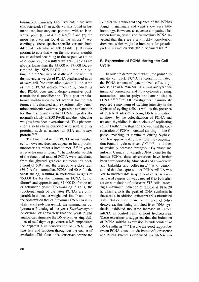
FIGURE 9. Comparison of BrdU and PCNA immunostaining for the assessment of cell proliferation in kidneysections of young male F344 rats at varying time points after birth, showing that PCNA and BrdU are comparablein older animals, whereas they vary decisively in very young animals. The LI were obtained by counting the numberof labeled nuclei in 1000 renal tubule epithelial cells. BrdU, 5-bromo-2'-deoxyuridine labeled nuclei; PCNA-NART,PCNA immunostained S-phase nuclei obtained without the use of the antigen retrieval technique (ART); PCNAART, PCNA immunostained S-phase nuclei obtained via the use of ART. (From Nakamura, J., Dietrich, D. R.,Schoonhaven, R., and Swenberg, J. A., in preparation.)
have been kept in fixatives for years. PCNAstaining was achieved in conventionally fixed andhistologically processed archival human tissues,usually encompassing a 4-h formalin fixation andparaffin embedding, using both the PC10 135 ,I43and 19A2 antibody130,l33,16 (Figure 11) and a conventional staining procedure. 45 ,128,134 For tissuesthat were fixed for longer than 48 h, a specialtissue treatment using the "Antigen Retrieval Solution™ " (ARS) and microwaving was developed,126,127,132,140-142 making it possible to carryout PCNA immunostaining in tissues that hadbeen preserved in formalin for up to 7 years (Figure 7).126 It is hypothesized that microwaving thetissues in conjunction with ARS results in breaking the protein-aldehyde crosslinks and in recon-
92
stitution of the protein, thus unmasking the epitopes for PCNA immunohistochemistry (Figures7, 9, and 10).142 Although different nuclear staining intensities possibly depicting the GO, G l-S,S, G2, and M phases of the cell cycle can beobserved (Figure 7), and despite the fact that itis generally accepted that the most intensivestaining nuclei depict cells in S-phase whereasnonstaining cells represent quiescent (GO)cells,126-128,132,139,144 the question needs to be an-swered whether or not the observed stained cellsrepresent all cells in the process of replication.Keeping in mind that PCNA is present, althoughin low concentrations, in quiescent cells,9 theapplication of techniques such as microwavingfor unmasking PCNA epitopes in archival tissues
BrdU and PCNA Labeling Index in Rat Hepatic Cells7 7
8 10 12Age (weeks)
6
~50........,)(
CD4'"0c0)3c.....--CD..c 2ca...J
1
0
0 2 4 6
BrdU
•PCNA PCNANART ART
·--A--· ..
14 16 18
6
5
4
3
2
1
FIGURE 10. Comparison of BrdU and PCNA immunostaining for the assessment of cell proliferation in Iiversections of young male F344 rats at varying time points after birth, showing that PCNA and BrdU are comparablein older animals, whereas they vary decisively in very young animals. The LI were obtained by counting the numberof labeled nuclei in 1000 hepatocytes. BrdU, 5-bromo-2' -deoxyuridine labeled nuclei; PCNA-NART, PCNA immunostained S-phase nuclei obtained without the use of the antigen retrival technique (ART); PCNA-ART, PCNAimmunostained S-phase nuclei obtained via the use of ART. (From Nakamura, J., Dietrich, D. R., Schoonhaven,R., and Swenberg, J. A., in preparation.)
also may unwantingly unravel PCNA epitopesusually masked in quiescent cells and thus couldgive the impression of a higher fraction of proliferating cells than is actually present (Figures9 and 10; Table 2). On the other hand, PCNAimmunohistochemical staining was shown to behighly variable from tissue to tissue and evenwithin sections of the same tissue,45,126,132mostlyas a result of tissue handling (preservation, fixation, embedding) and section thickness, indicating that cell proliferation measurements usingPCNA immunohistochemistry may underestimate the actual size of the proliferating cell population due to methodological artifacts (Figures9 and 10). One possible solution to this problemmay be the concurrent use of flow cytometricanalyses and PCNA immunohistochemistry, thus
measuring the actual proliferating cell populationand distinguishing different phases of the cellcycle in addition to determining the ploidy of thecells studied (Figure 12),145 while having simultaneously stained sections depicting the morphological characteristics of the tissue. If PCNA immunohistochemistry is to be used as a tool forassessing cell proliferating, the results obtainedwith this technique also must be comparable tothose obtained with established techniques suchas Tdr autoradiography or BrdU immunohistochemistry. Therefore, it will be necessary to investigate whether the PCNA proliferation index(LI), defined as labeled cellslunlabeled + labeled cells, should incorporate all PCNA positivecells or only those that are considered to be inS-phase.
93

FIGURE 11. PCNA immunostained section of an archival human melanoma, biopsied, fixedwith formalin, and embedded in paraffin in 1974 and sectioned and stained in 1992. Note thediversity of cellular morphology (nuclear size and form) and staining characteristics (light, granular,and dark stained nuclei). (Magnification x 400.) (From Woosley, J. T. and Dietrich, D. R., J.Cutan. Pathol., 19, 557, 1992. With permission.)
2. Flow Cytometry
Flow cytometry is a method that enables thesimultaneous quantitation of laser beam, xenon-,or mercury-arc-stimulated fluorescence of dyesand antibodies bound to cellular components inindividual cells. This method has been successfully used for the distinction of cell cycle phases,i.e., by the determination of the amount of DNApresent in the nucleus,146 an especially importantfactor that must be taken into consideration whenever cell proliferation is assessed in the liver whereheptocytes, especially in rats and mice, are knownto consist of several ploidy classes.145.147 Theamount of DNA can be measured indirectly viathe fluorescence intensity of propidium iodide, afluorescent dye that binds to DNA in a stoichiometric manner. 147 ,148 The use of fluorescein isothyocyanate (FITC)-conjugated secondary antibodies allowed quantitation of the presence ofcertain antigens, such as PCNA-antibodies andthus measurement of the PCNA content simultaneously with DNA content. 40,41,43,45,137 The
94
concentration of PCNA during the cell cycle andthe spatial distribution within the nucleus werestudied using various cell lines, e. g., mousespleenocytes, mouse hybridoma, SP2/0, MOLT4,40,41 human T lymphocytes,43 HeLa,45 and human breast carcinoma MCF-7. U7 Similar to theproblems of fixation encountered in immunohistochemistry and immunocytochemistry of tissuesections and cultured cells, respectively, alcoholfixation of cells resulted in the detection ofPCNAprimarily in S-phase cells and to a lesser extentin G2, M, or in GI-phase cells and was clearlyconfined to the nucleus,40,43 whereas paratormaldehyde fixation allowed not only the detection of PCNA in all phases of the cell cycle withthe exception of quiescent cells, but also thePCNA present in the cytoplasm,41 especially during mitosis, 137 a trait also observed in histochemically stained tissue seetions and immunocytochemically stained cytospins of culturedcells. 126,128,137 The presence of PCNA in the cy-toplasm of cells in G2 and M phase is assumedto be a direct consequence of the dissolution of
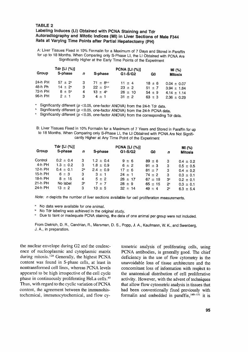
TABlE 2labeling Induces (LI) Obtained with PCNA Staining and TdrAutoradiography and Mitotic Indices (MI) in Llver Sections of Male F344Rats at Varying Time Points after Partial Hepatectomy (PH)
A: Liver Tissues Fixed in 10% Formalin for a Maximum of 7 Days and Stored in Paraffinfor up to 18 Months. When Comparing only S-Phase LI, the LI Obtained with PCNA Are
Significantly Higher at the Early Time Points of the Experiment
Tdr [LI (%)] PCNA [LI (%)] MI (%)Group S-phase n S-phase G1-S/G2 GO Mitosis
24-h PH 57 ± 2a 3 71 ± 8b,c 11 ± 4 18 ± 6 0.04 ± 0.0748-h Ph 14 ± 2a 3 22 ± 5b,c 23 ± 2 51 ± 7 3.94 ± 1.8472-h PH 8 ± 5a 4 13 ± 4b 26 ± 10 54 ± 9 4.14 ± 1.1496-h PH 2 ± 1 3 4 ± 1 31 ± 2 63 ± 3 2.36 ± 0.29
a Significantly different (p <0.05, one-factor ANOVA) from the 24-h Tdr data.b Significantly different (p <0.05, one-factor ANOVA) fram the 24-h PCNA data.c Significantly different (p <0.05, one-factor ANOVA) from the corresponding Tdr data.
B: Liver Tissues Fixed in 10% Formalin for a Maximum of 7 Years and Stored in Paraffin for upto 18 Months. When Comparing only S-Phase LI, the LI Obtained with PCNA Are Not Signifi-
cantly Higher at Any Time Point of the Experiment
Tdr [LI (%)] PCNA[LI (%)] MI (%)Group S-phase n S-phase G1-S/G2 GO n Mitosis
Contral 0.2 ± 0.4 3 1.2 ± 0.4 9 ± 6 89 ± 6 3 0.4 ± 0.24-h PH 1.3 ± 0.2 3 1.8 ± 0.9 6 ± 2 91 ± 3 3 0.5 ± 0.5
12-h PH 0.4 ± 0.1 2a 2.4 ± 0.9 17 ± 6 81 ± 7 3 0.4 ± 0.215-h PH 6 ± 3 3 3 ± 1 24 ± 1 74 ± 2 3 0.3 ± 0.118-h PH 8 ± 15 4 5 ± 2 28 ± 17 67 ± 19 3c 0.2 ± 0.121-h PH No label 3b 7 ± 7 28 ± 9 65 ± 15 2C 0.3 ± 0.124-h PH 13 ± 2 3 13 ± 5 32 ± 14 49 ± 4 2c 6.3 ± 5.4
Note: n depicts the number of Iiver sections available for cell proliferation measurements.
a No data were available for one anima!.b No Tdr labeling was achieved in the original study.c Due to faint or inadequate PCNA staining, the data of one animal per group were not included.
From Dietrich, D. R., Candrian, R., Marsman, D. S., Popp, J. A. , Kaufmann, W. K., and Swenberg,J. A., in preparation.
the nuclear envelope during 02 and the coalescence of nucleoplasmic and cytoplasmic matrixduring mitosis. 128 Oenerally, the highest PCNAcüntent was found in S-phase cells, at least innüntransformed cell lines, whereas PCNA levelsappeared to be high irrespective of the cell cyclephase in continuüusly proliferating HeLa cells.45Thus, with regard to the cyclic variation ofPCNAcüntent, the agreement between the immunohistochemical, immunocytochemical, and flow cy-
tometric analysis of proliferating cells, usingPCNA antibodies, is generally good. The chiefdeficiency in the use of flow cytometry is theunavoidable loss of tissue architecture and theconcomitant loss of information with respect tothe anatomical distribution of cell proliferativeactivity. However, with the advent of techniquesthat allow flow cytometric analysis in tissues thathad been conventionally fixed previously withformalin and embedded in paraffin,149-151 it is
95

regenerat/on ~N2N----.......2N 2N
FIGURE 12. Schematic diagram showing three possible effects induced afterxenobiotic insultthatcan leadto replicative DNA synthesis and that apriori are notdistinguishable trom one another solely by determiningthe LI via PCNA immunohistochemistry.
possible to compare directly the proliferativecompartment of alesion observed in a tissue section with the respective data on DNA contentobtained via flow cytometry. Such studies havebeen carried out with regard to the prognosticvalue of the proliferative index assessed via PCNAimmunostaining and flow cytometric measurement of DNA content in human gastrointestinallymphomas,152 gastric carcinomas,153 and hemangiopericytomas. 143 Interestingly, a good correlation between the immunohistochemically determined PCNA LI and the S + 02 +M phasefraction determined by flow cytometry was foundin the gastrointestinallymphomas, 152 whereas nosuch correlation could be demonstrated for thegastric carcinomas153 or the hemangiopericytomas. 143 Indeed, such comparisons are extremelyvaluable in cases where an increased expressionof PCNA is observed. However, no correlationof this expression to cell replication can be demonstrated, as was shown to be the case in humanacute myelogenous leukemias,74 thus indicatingthat the increased PCNA expression in this caseis possibly not related to its function in cell replicative DNA synthesis but rather to DNA ex-
96
cision repair. One disadvantage of retrospectiveflow cytometry yet to be resolved is that at present is is possible to measure only the DNA content, not the PCNA content, in the cells of therespective tissue. The reason for this may be thefact that a pepsin digestion step is needed in orderto render the tumor into the single cell moietynecessary for flow cytometric analysis, yet pepsin digestion as weIl as any other form of enzymedigestion has been shown to virtually abolishPCNA immunoreactivity.45 In addition, to validate retrospective flow cytometric analysis inparaffin-embedded tissues with regard to cellproliferation assessments, it would be useful tocompare the measured DNA contents to those ofa simultaneously analyzed exogenously appliedS-phase marker, such as BrdU. Although paraffin-embedded biopsy sampies of humans thathad been treated with BrdU prior to biopsy aremost likely not easily accessible, numerous sampIes from toxicology and carcinogenesis studiesin rodents, all with well-defined treatment protocols, are obtainable and therefore would allOWthe validation of cell proliferation measurementsvia retrospective flow cytometric analysis and itsuse in toxicology and pathology in conjunctionwith PCNA immunohistochemistry.
C. Comparison of Cell ProliferationMeasurement Using PCNA withExogenous and Endogenous CellProliferation Markers Such As BrdU,Tdr, and Ki-67
Evaluations of PCNA as a cell proliferationmarker via a comparison with the well-knownand established exogenously applied cell proliferation markers Tdr and BrdU were carried outusing immunocytochemistry on cultured cellsgrown on glass coverslips and on cytospins ofcultured cells such as human amnion, mouseNIH 3T3, HeLa, MCF-7 human breast cancer,human peripheral blood mononuclear, humanA-431 malignant carcinoma, human SK-5 nontransformed fibroblast, and HUVE (nontransformed human umbilical vein endothelial)cells. 36,.1X,45,51, 137 Not surprisingly, in synchro-
nized cell cultures, the proliferation measuredwith PCNA and Tdr or BrdU correlated ex-

tremely weIl. 36,3H,45 However, comparative cellcycle analysis of PCNA and BrdU distribution innonsynchronized MCF-7 cells indicated that replication patterns visualized by PCNA immunostaining were not a measure of replicative activityper se. 137 Similar observations were made by Coltrera and Gown,51 who found that the BrdU positive subpopulation of the SK-5 cellline was notidentical to or had any overlap with the PCNApositive subpopulation. Interestingly, a coimmunostain with another endogenous cell proliferation marker, Ki-67, gave similarresults in SK5 cells as did PCNA immunostaining, whereasthis was not the case for the three other cell linesstudied (Hela, A-431 , HUVE). In two other celllines (HeLa, A-431), the latter authors found thatBrdU positive cells formed inclusive subsets ofthe PCNA positive population. This suggests thatthe PCNA expression levels may be different incell lines with inherently different proliferationrates, and thus cannot uncritically be used as amarker for cell proliferation. This hypothesis wascorroborated by Hall and co-workers,45 whodemonstrated that the expression of PCNA remained constantly high and independent of thecell cycle phase in continuously proliferatingHeLa cells.
Studies comparing cell proliferation measurements obtained via immunohistochemicalstaining of tissue sections with PCNA and BrdU, I
Tdr, or Ki-67 have evolved recently and includefreshly fixed as weIl as archi val tissues. 49.50,126,132,139-14I,I44,154-159 In freshly fixed ratcolon, liver and kidney, and human colon, theagreement between cell proliferation measurements obtained via BrdU and PCNA appears tobe excellent (Figures 9 and 10),49,140,141,157 es-pecially if only S-phase cells were counted.Slightly higher labeling indices were obtainedwith PCNA in rodent and human liver and gastrointestinal tract;'56 however, this was shown tobe the result of the counting procedure, i.e., allstaining nuclei were counted, including non-Sphase cells. Similar results were reported by Galand and Degraef,50 who found the PCNA LI tomarkedly exceed the Tdr LI in formaldehydefixed tissue sections, whereas in methanol-fixedtissues, the PCNA LI agreed weIl with the TdrLI. These differences are due mainly to the factthat in methanol-fixed sections primarily S-phase
cells are PCNA positive, whereas in addition tothe S-phase cells, non-S-phase cells stain positivein formaldehyde-fixed tissues (see Seetion VLB).Thus, cell proliferation measurements in freshlyfixed tissues via PCNA immunohistochemistryappear to agree quite weIl with those obtainedusing the two weIl-known exogenously appliedS-phase markers, BrdU and Tdr, and this suggests that PCNA immunohistochemistry is a viable method for cell proliferation measurementin freshly fixed paraffin-embedded tissues. Yetmost of these studies mentioned were carried outin human tissues or in tissues of adult rodents ,with the exception of the experiments carried outby Nakamura and co-workers, 140,141 in which agerelated cell proliferation in liver and kidney wasstudied using PCNA and BrdU immunohistochemistry. These experiments showed that in animals aged 6 weeks and older there are no differences in PCNA LI and BrdU LI (Figures 9and 10). Surprisingly, the PCNA LI did not agreewith those achieved with BrdU in the kidney andliver of male rats up to 6 and 4 weeks of age,respectively (Figures 9 and 10). Using the "An-tigen Retrieval Solution™ " technique (ART)slightly improved the situation in that near agreement between PCNA LI and BrdU LI wasachieved in rats as young as 2 weeks of age.However, these experiments indicate that PCNAmay not be a suitable proliferation marker in veryyoung animals. The reasons for this "underexpression" ofPCNA in very young animals certainly merits further investigation.
Comparisons betweencell proliferation measurements via PCNA and BrdU immunohistochemistry or Tdr autoradiography also have beencarried out using archival tissues. 126,132,139,158,159All of these studies used rat or mouse liver tissuesarchived from earlier toxicological studies, withweIl-known treatment protocols, and the ARTtechnique for improved PCNA immunohistochemieal staining. Among these tissues, somehad been fixed very briefly, paraffin embedded,and then remained in paraffin blocks from 18 to26 months (Table 2A),132,139,158,159 while othershad been kept in the fixative for 7 years, paraffinembedded, and then remained in paraffin blocksfor 18 months (Table 2B).126 Generally, exeellentagreement was observed between S-phase PCNALI and BrdU LI or Tdr LI irrespective of the
97

duration of tissue fixation or paraffin storage,with the exception of one study in whieh thePCNA LI slightly exeeeded the LI determinedvia Tdr autoradiography (Table 2A). This diserepaney may be explained by the low numberof tissues analyzed and by the variability in staining intensities found to oeeur between and withintreatment groups of the latter study, thus makinga elear distinetion of S-phase from non-S-phasecells sometimes diffieult. 126 It has to be emphasized that in order to earry out retrospective eellproliferation measurements via PCNA immunohistochemistry it must be possible to distinguishclearly the S-phase from non-S-phase eells. However, despite the paueity of retrospeetive studiescomparing PCNA with other cell proliferationmarkers, the present data indicate that PCNA isa suitable marker for retrospecitve cell proliferation measurelnent in arehival tissues.
PCNA immunohistoehemistry also was eorrelated to Ki-67 immunohistochemistry in humanmalignant lymphomas, 154 brain tumors, 160 and tumor xenografts of the LoVo cell line. 155 Whilegood agreement between Ki-67 and PCNA wasfound in malignant lymphomas and low-gradegliomas, 154,160 little eorrelation to Ki-67 andgrowth fraction, estimated via fraction of labeledmitosis,155 was observed in astrocytomas, highgrade and mixed gliomas, Schwannomas, andxenograft tumors, 155.160 indicating that PCNA immunohistochemistry cannot be uncritically usedas a proliferation marker in tumors.
VII. PERSPECTIVES, FUTURE NEEDS,AND APPLICATION OF PCNA AS APROLIFERATION MARKER INTOXICOLOGY AND AS A PROGNOSTICMARKER IN SURGICAL PATHOLOGY
A. Toxicology
In its toxicological application, PCNA immunohistochemistry or, if even possibly, PCNAflow cytometry, should enable the measurementof cell proliferation in archival tissues thus preventing researchers from having to repeat eompleted studies currently lacking adequate proliferation data. So far, it has been established thatPCNA may be used as a cell proliferation marker
98
as it compares quite weIl with other weIl-knownS-phase markers such as BrdU or Tdr, and mayreflect chemically induced cell proliferation evenbetter than BrdU or Tdr especially if the proliferative index (PI), incorporating all PCNA positive cells, is used rather than the S-phase(LI). 158,159 In addition, PCNA analysis has thepotential to identify the specific cell populations(01, S, 02, M) that exist in the cell cycle and,if feasible, may lead to quantitating the effectsof a compound on the different cell populationsand thus to potentially critical information in understanding eompound-induced cell proliferation. 119 Indeed, the presence of cytoplasmic PCNAduring the late 02 and M phase of the cellcycle50 ,126.128 may provide further insight into theeffects of chemicals on the distribution of PCNAwithin the eell during the cell cycle. However,for many tissues with a normally low proliferation rate, e.g., liver, kidney, pancreas, etc., theuse of PCNA immunohistochemistry may proveto be problematic as only few cells will be positive for PCNA and of these very few will be inS-phase, meaning that the LIs generated withPCNA immunohistochemistry are more comparable to those generated by BrdU or Tdr administered as a pulse-dose rather than those fromBrdU or Tdr administered continuously.139 Furthermore, in contrast to the exogenously appliedproliferation markers BrdU or Tdr, the expression of PCNA, being a cell cycle-regulated protein, may be influenced by the compound theanimal was treated with, indicating that PCNAimmunohistochemistry eould under- or overestimate the actual proliferation rate. Indeed, theimmunosuppressants dexamethasone and cyclosporin were shown to inhibit PCNA expression asweIl as T-lymphocyte proliferation, whereas theDNA synthesis inhibitors cytarabin and hydroxyurea prevented lymphoeyte proliferation but notPCNA expression. 43 Furthermore, Foley and coworkers l39 reported similar LI of PCNA and Tdrup to 24 h after 4-acetylaminofluorene (4-AAF)treatment; however, 48 h after 4-AAF treatment,the PCNA LI remained increased while the TdrLI retumed to control values. These discrepancies could have stemmed from a potential induction of growth factors by the nongenotoxic 4AAF resulting in the overexpression of PCNA(Figure 1). Overexpression of PCNA also was

reported in conjunction with poligeenan-inducedcolonic cell proliferation in F344 rats.!61 In thisstudy, poligeenan, a nongenotoxic sulfated polysaccharide known to induce colorectal tumors,was fed in the diet for 64 days after which theanimals were returned to the NIH-07 diet alonefor 28 days. Despite removal of poligeenan fromthe diet, the PCNA levels in the upper third ofthe crypt remained ll-fold above control levelsfor 28 days, indicating either a decreased abilityof the colon crypt cells to adapt rates of cellproliferation or a deregulated expression or catabolism of PCNA resulting from poligeenantreatment. Deregulation of the cyclic expressionof PCNA was demonstrated earlier by Hall andco-workers,45 who found PCNA remained at highlevels, irrespective of the cell cycle phase, incontinuously proliferating HeLa cells. On theother hand, Ahnen and co-workers l62 found similar cell proliferation-associated staining patternsfor PCNA and Tdr in normal colon and colonictumors of rats treated with the known colon carcinogen dinlethlhydrazine, suggesting that in theirstudy PCNA expression was a reliable marker ofthe proliferative compartment in the rat colon.Interestingly, the cell proliferative response wasconfined to the lower third of the crypt in therectum, whereas in the proximal and mid-colon,staining extended into the mid to upper third ofthe crypt.
Regenerative cell proliferation determinedwith PCNA immunohistochemistry in mouse lungepithelia following acute injury with butylatedhydroxytoluene l3 ! showed that the increasedexpression of PCNA correlated weIl with increased Tdr incorporation, indicating that PCNAexpression is not altered during enhanced regenerative proliferation. Similarly, the effects of themitogenic hepatocarcinogenic agents Wy-14,643and l,4-dichlorobenzene on liver cell proliferation were measured using PCNA and BrdU immunohistochemistry as weIl as Trd autoradiography!58,15lJ and demonstrated that mitogeninduced cell proliferation can be reliably determined with PCNA immunohistochemistry and thatthe two hepatocarcinogenic agents do not induceoverexpression of PCNA.
However, in view of the paucity of data regarding compound-induced enhanced cell proliferation measured via PCNA analysis and keeping
in mind that the expression of PCNA is regulatedat several levels within the cell (Figure 1), thequestions need to be answered as to how andwhich genotoxic, mitogenic, and cytotoxic compounds (Figure 12), hormones, and growth factors influence not only the expression of PCNA,but also the stability/half-life of PCNA mRNAand its protein product and thus the reliability ofPCNA as a cell proliferation marker.
Such questions may be addressed by using awell-defined cell system, i.e., a cell line with awell-characterized and manipulatable cell cyclesuch as the Chinese hamster ovary cell, the V79chinese hamster lung fibroblast, 146 or T lymphocytes,43 and combining flow cytometry, immunocytochemistry, and biochemical techniques(Western blot analysis, etc.) for PCNA analysis.Furthermore, these experiments should focus notonly on the PCNA gene, mRNA, and gene product, but also on PCNA regulating factors such asp53, pI05(Rb), and TGF-ß (Figure 6), thus distinguishing between direct and indirect effects ofcompounds on PCNA expression. Indeed, withthe development of techniques such as PCR andthe availability of cDNA probes. for rat and human PCNA,5,6,21 it should be possible to analyzecompound-induced alterations in the PCNA gene.Using these techniques, Liu and Bambara54 demonstrated that PCNA is overexpressed in theR3230AC mammary tumor, which was accompanied by an altered PCNA gene structure. Inaddition, the experiments proposed earlier shouldbe able to demonstrate whether an increasedexpression of PCNA is assoeiated with replicative DNA synthesis or DNA excision repair (Figures 2 and 3). Although in vitro experiments,such as the ones proposed previously, are helpfulin understanding the effects of compounds on aspecific cell subpopulation of the cell cycle, theycannot replace studies in a whole tissue. Indeed,this is demonstrated by the study of Foley andco-workers,125 who found higher S-phase PCNALI in liver foei of alteration than in the surrounding normal hepatocytes of control and methylenechloride-exposed female B6C3Fl mice, thusdemonstrating a higher proliferative rate in theclonally expanded preneoplastic lesions. However, the combination of in vitro experimentswith retrospective cell proliferation measurements in archival tissues of completed studies
99

provides a powerful tool for studying the toxicityand/or carcinogenicity of the respective compounds. Furthermore, recent progress made inflow-cytometric analysis of archival tissues shouldallow distinguishing between PCNA associatedwith replicative DNA synthesis and PCNA involved in DNA excision repair, thus providingbetter insight into cell proliferation mechanismsand higher reliability of cell proliferation measurements.
Also, additional studies, e.g., analyzing cellproliferation measurement variability resultingfrom fixation, tissue handling, antibody, andstaining procedure related effects, are requiredfor the proper utilization and interpretation of thecell proliferation response as detected by PCNAanalysis. Provided these studies are carried outand result in an improvement of in the methodology and a better data base, PCNA immunohistochemistry may prove to be the method ofchoice not only for retrospective but also for prospective studies. In view of recent reports regarding the adverse effects of the well-established cell proliferation marker BrdU, whichsuggest that BrdU may be toxic and thereforeenhances the cell proliferative response, 163.164these studies are urgently needed. Although moststudies reported so far have been conducted inrodents and humans, PCNA immunohistochemistry and cytochemistry allow cell proliferationand cell cycle analysis to be conducted in a multitude of other organisms7,S.1O-12,133 with the ben-efit that the findings all have the same denominator and thus are readily comparable. This alsoshould make it possible to transfer the knowledgeon toxic and carcinogenic mechanisms obtainedin mammals to non-mammalian organisms important as bioindicators, such as fish or clams,for ecotoxicological risk assessment.
B. Surgical Pathology/Modern Medicine
The quest for more efficient management ofpatients afflicted with neoplasia and efforts tobeUer predict the progression of tumors have ledto a rather uncritical use of so-called prognosticmarkers in human tumors. Indeed, in many cases,such markers were readily applied without a clearunderstanding of the role or function of the marker
100
in the respective tumor. 165 PCNA immunohistochemistry has been called upon as a means forestimating the growth fraction 136.154,155 within agiven tumor as weIl as a means for trying topredict the progression of tumors based on theperhaps naive assumption that a high degree ofPCNA staining reflects high proliferative activityper se and thus automatically means a worseprognosis for the patient, irrespective of the tumor type. 129,130,160 Although the PI assessed viaPCNA immunohistochemistry appears to have aprognostic value in hemangiopericytomas, 143gastric carcinomas, 153 gastrointestinal lymphomas,152 colorectal cancer, 102 soft tissue sarcomas,l66 and melanomas,136 this was not the casein ovarian cancer,167 acute myelogenous leukemia,74 and, in contrast to the findings by Takahashi et al., 136 in melanomas with 8 or more yearsof clinical folIow-up (Figure 11 ).134,135 Wooselyand co-workers l34,135 statistically correlated theprognostic value of the proliferative fraction estimated via PCNA in melanomas with the clinicaloutcome (patient survival) and other prognosticindicators, e.g., anatomieal level, tumor thickness, mitotic frequency, tumor infiltrating 1ymphocytes, tumor regression, or sex, and demonstrated that PCNA did not correlate to eitherthe clinical outcome or any of the other prognostie indicators. Takahashi et al. ,136 on the otherhand, compared the growth fraction visualizedvia PCNA immunohistochemistry with tumorgrade only and found that PCNA-positive tumorcells increased in number and staining intensitywith increasing progression of the lesions towardmalignancy. To further understand the role ofPCNA in melanomas, they exposed normal skinto sunlight, found that mainly suprabasal keratinocytes stained positive for PCNA whereas melanocytes were PCNA negative, and concludedthat PCNA-positive staining in melanocytes wasclosely associated with malignant transformation. At first sight, the results of the studies byWoosley and co-workers 134 and Takahashi andco-workers 136 seem to contradict one another, butthis need not necessarily be the case. The mainproblem lies in the assumption that all positivePCNA staining is associated with replicating activity; however, PCNA expression may be alteredby changes in the structure of the PCNA gene,as was already shown to be the case in rat mam-

mary tumors,54 and potentially has nothing to dowith increased cell proliferation. Furthermore ,increased PCNA-positive staining clearly not associated with cell proliferation also was observedin patients with acute myelogenous leukemia inconjunction with increased resistance to chemotherapy,74 in which case this increased PCNAexpression was attributed to enhanced DNA excision repair. Indications of the latter observationalso can be found in the study by Takahashi andco-workers 136 in that presumably increased cellproliferation-associated PCNA staining was observed in keratinocytes of the suprabasal layerbut not in the basal layer of normal skin exposedto sunlight, although it is known that the basalcelllayer is proliferatively active, thus indicatingthat some of the PCNA reactivity observed mayhave been due to DNA excision repair demonstrated to occur in cells irradiated with UV light(see Section IV.B).69.168 Therefore, PCNA immunostaining in melanomas may reflect a cohortof several "PCNA populations:" PCNA expressed as a function of cell replication, PCNAredistributed as a function of DNA excision repair, and PCNA over/underexpressed due to analteration in the PCNA gene or deregulated PCNAtranscription and translation. Deregulated PCNAtranscription or translation also was suggested incases where increased immunohistochemicallydetectable PCNA was observed in histopathologically nornlal breast lobules adjacent to breasttumors as weIl as in pancreatic exocrine parenchyma adjacent to endocrine and exocrine tumorsof the pancreas. 45 In these cases, it was postulatedthat some of the tumors are actively secretinggrowth factors that are stabilizing the PCNAmRNA and thus inducing PCNA protein accumulation without actually inducing DNA synthesis. In this context, it also would be useful toanalyze whether deregulated PCNA expressioncould stern from structural changes in the PCNAgene in these tumors, as was shown to be thecase in rat matnmary tumors. 54 A further solutionto this dilema may be the use of retrospectiveflow cytometry in conjunction with PCNA immunohistochemistry, thus distinguishing proliferating from nonproliferating cell populations.This approach was chosen by Woods and coworkers l52 in their evaluation of PCNA inlmunohistochemistry in primary gastrointestinallym-
phomas. Their study showed that there is a goodcorrelation between the PCNA index and the histological grade of the tumors as weIl as a significant relationship between the PCNA indexand the S +02 +M-phase fraction as measuredby retrospective flow-cytometric analysis. A highPCNA index was significantly correlated withpoor patient survival, thus indicating that a highPCNA index is an adverse prognostic factor inprimary gastrointestinal tumors. Using the sameapproach, Jain and co-workers l53 and Yu et al. 143
evaluated the prognostic value of PCNA in gastric carcinomas and hemangiopericytomas, respectively. Although it was demonstrated that thePCNA index correlated weIl with patient survivalprobability in both retrospective studies, a positive correlation between PCNA index and histological grade was demonstrated only in hemangiopericytomas. In gastric carcinomas, thePCNA index did not correlate with histologicalgrade, nor was there any correlation betweenPCNA index and tumor stage or lymph node metastasis. Furthermore, in both tumor types, thecomparison between flow-cytometric measurement of cell proliferation and PCNA index, histological grade, or patient survival revealed thatthe results obtained with flow cytometry did notcorrelate with any of the latter parameters. Autocrine and paracrine growth factor-mediatedregulation of PCNA expression also may explainthe excess of PCNA immunoreactive cells observed in the above-mentioned tumors, indicatingthat not all PCNA staining observed in the different tumor types is functionally associated withcell replication, 160 thus emphasizing that PCNAimmunohistochemistry in conjunction with retrospective flow cytometry may be a helpful toolfor understanding tumor etiology and progression, but that the PCNA index should not beuncritically used as a prognostic marker.
PCNA immunohistochemistry also has beenused to visualize regenerating hepatocytes in patients with acute viral hepatitis, hepatic cirrhosis,or hepatocellular carcinomas, 169 and to measurecell proliferation in hyperplastic, preneoplastic,and neoplastic lesions of intrahepatic bile ductsin livers with hepatoliths. 170 In the former study,the respective lesions were characterized by lesion-specific PCNA staining patterns and proliferation rates, while in the latter study it was shown
101

that the PCNA LI increased with increasing malignant progression of the lesion, i.e., lowest inhyperplasias and highest in invasive adenocarcinomas. Simultaneous determination of the meannumber of argyrophylic nucleolar organizer regions (AgNORs) demonstrated a significant positive correlation between PCNA LI and AgNORscount in alllesions, 170 thus suggesting that PCNAimmunohistochemistry can be used for cell proliferation measurement in certain human tissuesand lesions.
VIII. SUMMARY
The cell cycle-associated protein PCNA wasreviewed here with regard to its regulation,expression, and function during the cell cycle aswell as its use as a proliferation marker in toxicology and pathology and as a prognostic indicator in surgical pathology, i. e., patient management, particularly those afflicted withneoplasia. Despite numerous studies analyzingthe function of PCNA in the cell, only two functions have been clearly elucidated to date, itsassociation with replicative DNA synthesis andwith DNA excision repair. Due to the varyingexpression levels and the varying distribution (10calization) of PCNA during the cell cycle, it maybe hypothesized that PCNA could have otherfunctions besides the ones stated above. Futurestudies should focus on determining whether ornot such additional functions exist. Moreover,additional studies are required that will providedata on the regulation/deregulation 01' PCNAexpression by chemicals, hormones, growth factors, protooncogenes, and tumor suppressorgenes. Only then will it be possible to adequatelyinterprete the experimental data that used PCNAas a proliferation marker either by immunohistochemistry, immunocytochemistry, or flow cytometry. Furthermore, it will be necessary to understand and to be able to distinguishmethodological effects related to tissue treatment, fixation, storage, and staining from compound-related effects on PCNA expression.However, despite these caveats, the present dataclearly demonstrate that PCNA can be used forcell proliferation measurements in prospective asweIl as in retrospective studies. A more critical
102
approach must be taken for the use 01' PCNA asa prognostic marker in human tumors. The highvariability 01' PCNA staining observed within tumors and among different tumor types limits itsuse as a prognostic marker per se for many tumortypes. However, additional clinicopathologicalstudies, whether prospective or retrospective, thatencompass excellent patient selection, largenumbers of patients, multiparameter analysis,documented clinical outcome, and comparablepatient treatment protocols should be able to elucidate the role of PCNA in the context of humandisease.
ACKNOWlEDGMENTS
I would like to thank Dr. Christian Sengstagand Prof. Fritz Würgler for critically reviewingthe manuscript and Nicola Wittekindt for helpfulcomments regarding the illustrations.
REFERENCES
1. Miyachi, K., Fritzler, Mo Jo, and Tan, Eo Mo,Autoantibody to a nuclear antigen in proliferatingcens, J. Immuno!., 121(6),2228, 1978.
2. Doolittle, Do Jo, McKarns, S. Co, Ayres, Po Ho,and Bombick, D. Wo, Molecular approaches forquantifying DNA synthesis and cen proliferation during rodent bioassays, Toxico!. Methods, 1(4), 215,1991.
3. Ogata, Ko, Kurki, Po, Celis, Eo Jo, Nakamura,R. Mo, and Tan, E. Mo, Monoclonal antibodies toa nuclear protein (PCNA/cyclin) associated with DNAreplication, Exp. Cello Res., 168,475, 1987.
4. Almendral, J. Mo, Heubsch, D., Blundell, P. Ao,MacDonald-Bravo, Ho, and Bravo, Ro, Cloningand sequence of the human nuclear protein cyclin:Homology with DNA-binding proteins, Proc. Natl.Acad. Sei. U.S.A., 84, 1575, 1987.
5. Moriuchi, T., Matsumoto, Ko, Koji, T., andNakane, P. K., Molecular cloning and nucleotidesequence analysis of rat PCNA/cyclin cDNA, NucleicAcids Res., 17,117,1986.
6. Matsumoto, K., Moriuchi, To, Koji, To, andNakane, Po Ko, Molecular cloning of cDNA codingfor rat proliferating ceH nuclear antigen (PCNA/cyclin), EMBO J., 6(3),637, 1987.
7. Zuber, Mo, Tan, E. Mo, and Ryoji, Mo, Involvement of proliferating cen nuclear antigen (cyclin) in

DNA replication in living ceIls, Mol. Cello BioI.,9(1),57, 1989.
8. Zuber, M., Yasui, W., Tan, E. M., and Ryoji,M., Quantitation and subcellular localization of proliferating ceIl nuclear antigen (PCNA) in oocytes andeggs of Xenopus laevis, Exp. Cel! Res., 182, 384,1989.
9. Celis, J. E., Madsen, P., Celis, A., Nieisen, H. V.,and Gesser, B., Cyclin (PCNA, auxiliary protein ofDNA polymerase 8) is a central cümponent of thepathway(s) leading to DNA replication and ceIl division, FEBS Leu., 220(1), 1, 1987.
10. Yamaguchi, M., Nishida, Y., Moriuchi, T., Hirose,F., Hui, C., Suzuki, Y., and Matsutake, A., Drosophila proliferating ceIl nuclear antigen (cyclin) gene:structure, expression during development, and specific binding ofhomeodomain proteins to its 5/-flanking region, Mol. Cello BioI., 10(3), 872, 1990.
11. Olins, D. E., Olins, A. L., Cacheiro, L. H., andTan, E. M., Proliferating cell nu1cear antigen/cyclinin ciliate Euplotes eurystemus: localization in thc replication band and micronuclei, J. Ceil BioI., 109,1399, 1989.
12. Suzuka, 1., Daidoji, H., Matsuoka, M., Kadowaki,K., Takasaki, Y., Nakane, P. K., and Moriuchi,T., Gene für proliferating cell nuclear antigen (DNApolymerase 8 auxiliary protein) is present in bothmammalian and higher plant genomes, Proc. Natl.Acad. Sei. U.S.A., 86,3189,1989.
13. Bauer, G. A. and Burger, P. M. J., The yeastanalog of mammalian cyclin/proliferating cell nuclearantigen interacts with mammalian DNA polymerase8, Proc. Natl. Acad. Sei. U.S.A., 85, 7506, 1988.
14. O'ReiHy, D. R., Crawford, A. M., and Miller,L. K., Viral proliferating ceIl nuclear antigen,Nature, 337,606, 1989.
15. Hori, H. and Osawa, S., Evolutionary change in55 RNA secondary structure and a phylogenie treeof 5455 RNA species, Proc. Natl. Acad. Sei. U.S.A.,76( 1), 381, 1979.
16. Kimura, M. and Obta, T., On some principles governing l110lecular evolution, Proc. Natl. Acad. Sei.U.S.A., 71 (7), 2848, 1974.
17. Yamaguchi, M., Hayashi, Y., Hirose, F., Matsuka,S., Moriuchi, T., Shiroishi, T., Moriwaki, K., andMatsukage, A., Molecular cloning und structuralanalysis of mouse gene and pseudogenes for proliferating cell nuclear antigen, Nucleic Acids Res., 19(9),2403, 1991.
18. Kosugi, S., Suzuka, 1., Ohashi, Y., Marakami,T., and Arai, Y., Upstream sequences of rice proliferating ceIl nuclear antigen (PCNA) gene mediateexpression of PCNA-GUS chimeric gene in meristems of transgenic tobacco plants, Nucleic Acids Res.,19(7), 1571, 1991.
19. Desplan, C., Theis, J., and O'FarreH, P. H., Theseqllence specificity 01' homeodomain-DNA interaction, Cell, 54, 1081, 1988.
20. Chowdhury, K., Deutsch, V., and Grllss, P., Amllltigene 1'amily encoding several "finger" struc-
tures is present and differentially active in mammalian genomes, Cell, 48, 771, 1987.
21. Travali, S., Ku, D.-H., Rizzo, M.-G., Ottavio, L.,Baserga, R., and Calabretta, B., Structure of thehuman gene for the proliferating cell nuclear antigen,J.Biol. Chern., 264(13),7466, 1989.
22. Ottavio, L., Chang, C.-D., Rizzo, M.~G., Travali,S., Casadevali, C., and Baserga, R., Importanceof introns in the growth regulation of mRNA levelof the proliferating cell nuclear antigen, Mol. CelloBioI., 10(1),303, 1990.
23. Takasaki, Y., Fishwild, D., and Tan, E. M., Characterization of proliferating cell nuclear antigen recognized by autoantibodies in lupus sera, J. Exp. Med..159, 981, 1984.
24. Laughon, A. and Scott, M. P., Sequence of a Drosophila segmentation gene: protein structure homology with DNA-binding proteins, Nature, 310, 25,1984.
25. Tan, C.-K., Casti11o, C., So, A. G., and Downey,K. M., An auxiliary protein for DNA polymerase 8from fetal calf thymus, J. Biol. Chem., 261 (26),12310, 1986.
26. Ogata, K., Celis, J. E., and Tan, E. M., Proliferating cell nuclear antigen: cyclin, Methods EIlzymol., 150, 147, 1987.
27. Celis, J. E., Bravo, R., Larsen, P. M., and Fey,S., Cyclin: a nuclear protein whose level correlatesdirectly with the proliferative state of normal as weIlas transformedcells, LeukerniaRes., 8(2), 143,1984.
28. Bravo, R., Frey, S. J., BeUantin, J., Larsen, P. M.,Arevalo, J., and Celis, J. E., Identification of anuclear and of a cytoplasmic polypeptide whose relative proportions are sensitive to changes in the rateof cell proliferation, Exp. Cello Res.. 136,311, 1981.
29. Mathews, M. B., Bernstein, R. M., Franza, B. R.,and Garreis, J. 1., Identity of the proliferating cellnllclear antigen and cyclin, Nature, 309, 374, 1984.
30. Sadaie, M. R. and Mathews, M. B., Immunoehemical and biochemical analysis 01' the proliferatingcell nuclear antigen (PCNA) in HeLa cells, Exp. CelloRes.. 163, 423, 1986.
31. Smart, J. E., Lewis, J. B., Mathews, M. B.,Harter, M. L., and Anderson, C. W., Adenovirustype 2 early proteins: assignment of the early regionlA proteins synthesized in vivo and in vitro to speeifie mRNAs, Virology, 112, 703, 1981.
32. Persson, H., Hennighallsen, L., Taub, R.,DeGrado, W., and Leder, P., Antibodies to humanc-myc oncogene product: evidence of an evolutionaryconserved protein induced during cell proliferation,Science, 225,687, 1984.
33. Prelich, G., Kostura, M., Marshak, D. R.,Mathews, M. B., and Stillman, B., The cell-cycleregulated proliferating cell nuclear antigen is requiredfor SV40 DNA replication in vitro, Nature, 326,471,1987.
34. Bauer, G. A. and Burgers, P. M. J., Molecularcloning, structure and expression of the yeast prolif-
103

erating cell nuclear antigen gene, Nudeie Acids Res..18(2), 261, 1990.
35. Bravo, R. and MacDonald-Bravo, H., Changes inthe nuclear distribution 01' cylin (PCNA) but not itssynthesis depend on DNA rep1ication, EMBO J .. 4(3),655, 1985.
36. Bravo, R. and MacDonald-Bravo, H., Existence01' two populations 01' cyclin/pro1iferating cell nuclearantigen during the ceIl cycle: association with DNAreplication sites, J. Cell Bioi., 105, 1549, 1987.
37. Celis, J. E., Fey, S. J., Larsen, P. M., and Celis,A., Expression of the transformation-sensitive protein "cyclin" in nonnal human epidermal basal cellsand simian virus 40-transformed keratinocytes, Proc.Natl. Acad. Sci. U.S.A., 81,3128, 1984.
38. Celis, J. E. and Celis, A., Ccll cycle-dependentvariations in the distribution of thc nuclear proteincyclin proliferating cell nuclear antigen in culturedcells: subdivision of S phase, Proc. Natl. Acad. Sei.U.S.A., 82, 3262, 1985.
39. Celis, J. E. and Madsen, P., Increased nuclear cyclin/PCNA antigen staining of non S-phase transfonned human amnion cells engaged in nucleotideexcision DNA repair, FEBS Lett., 209(2),277, 1986.
40. Kurki, P., Vanderlaan, M., Dolbeare, F., Gray,J., and Tan, E. M., Expression of proliferating cellnuclear antigen (PCNA/cyclin) during the cell cycle,Exp. Cello Res., 166, 209, 1986.
41. Kurki, P., Ogata, K., and Tan, E. M., Monoclonalantibodies to proliferating cell nuclear antigen (PCNA/cyclin) as probes far proliferating cells by immunofluorescence microscopy and flow cytometry, J. Immunol. Methods.. 109, 49, 1988.
42. Jaskulski, D., Gatti, C., Travali, S., Calabretta,B., and Baserga, R., Regulation of the proliferatingceH nuclear antigen cyclin and thymidine kinasemRNA levels by growth factors, J. Biol. Chem.,263(21), 10175, 1988.
43. Kurki, P., Lotz, M., Ogata, K., and Tan, E. M.,Proliferating cell nuclear antigen (PCNA)/cyclin inactivated human T lymphocytes, J. Immunol.,138(12),4114, 1987.
44. Morris, G. F. and Mathews, M. B., Regulation 01'proliferating ceH nuclear antigen during the cell cycle,J. Biol. Chem., 264(23), 13856, 1989.
45. Hall, P. A., Levinson, D. A., Woods, A. L., Yu,C. C.-W., Kellock, D. B., Watkins, J. A., Barnes,D. M., Gillett, C. E., Camplejohn, R., Dover, R.,Waseem, N. H., and Lane, D. P., Proliferating cellnuclear antigen (PCNA) immunolocaIization in paraffin sections: an index of cell proliferation with evidence of deregulated expression in some neoplasms,J. Pathol., 162,285, 1990.
46. Ogata, K., Ogata, Y., Takasaki, Y., and Tan,E. M., Epitopes on proliferating cell nuclear antigenrecognized by human lupus autoantibody and murinemonoclonal antibody, J. Immunol., 139(9), 2942,1987.
47. Ogata, K., Ogata, Y., Nakamura, R. M., and Tan,E. M., Purification and N-terminal amino acid se-
104
guence 01' proliferating cell nuclear antigen (PCNA)/cyclin and development 01' ELISA for anti-PCNAantibodies, J. Immunol., 135(4), 2623, 1985.
48. Waseem, N. H. and Lane, D. P., Monoclonal antibody analysis of the proliferating ceH nuclear antigen (PCNA): stmctural conservation and the detection of a nucleolar form, J. Cell Sci., 96, 121, 1990.
49. Richter, F., Richter, A., Yang, K., and Lipkin,M., CeH proliferation in rat colon measured withBrdU, PCNA, and [.JH]dThd, Proc. Am. Assoc. Cancer Res., 32, 154, 1991.
50. Galand, P. and Degraef, C., Cyclin/PCNA immunostaining as an alternative to triated thymidinepulse labeling tor marking S-phase cells in paraffinsections from animal and human tissues, Cell TissueKinet .. 22, 383, 1989.
51. Coltrera, M. D. and Gown, A. M., PCNA/Cyclinexpression and BrdU uptake define different subpopulations in different celliines, J. Histochem. Cywchem., 39(1),23, 1991.
52. Baserga, R., Growth regulation of the PCNA gene,J. Cell Sei., 98,433, 1991.
53. Chang, C.-D., Ottavio, L., Travali, S., Lipson,K., and Baserga, R., Transcriptional and posttranscriptional regulation of the proliferating cell nuclearantigen gene, Mol. Cello Bioi., 10(7), 3289, 1990.
54. Liu, Y.-C. and Bambara, R. A., Gene expression01' PCNA/cyclin in adult tissues and the R3230ACmammary tumor of the rat, Biochem. Biophys. Res.Commun., 161(2),873, 1989.
55. Wong, R. L., Katz, M. E., Ogata, K., Tan, E. M.,and Cohen, S., Inhibition 01' nuclear DNA synthesisby an autoantibody to proliferating ceH nuclear antigen/cyclin, Cello Immunol., 110,443, 1987.
56. Jaskulski, D., de Riel, J. K., Mercer, W. E.,Calabretta, B., and Baserga, R., Inhibition 01' cellular proliferation by antisense oligodeoxynucleotidesto PCNA/cyclin, Science, 240, 1544, 1988.
57. Bravo, R., Frank, R., BlundeIl, P. A., andMacDonald-Bravo, H., Cyclin/PCNA is the auxiliary protein of DNA polymerase 0, Nature, 326,515,1987.
58. Prelich, G., Tan, C.-K., Kostura, M., Mathews,M. B., So, A. G., Downey, K. M., and Stillman,B., Functional identity 01' proliferating ceH nuclearantigen and DNA polymerase 0 auxiliary protein,Nature, 326,517, 1987.
59. Wolds, M. S., Li, J. J., Weinberg, D. H., Virshup,D. M., Sherley, J. L., Verheyen, E., and Kelly,T., CeHular proteins reguired far SV40 DNA replication in vitro, Cancer Cells, 6, 133,1988.
60. Fairman, M. P., DNA polymerase o/PCNA: actionsand interactions, J. Cell Sei., 95, 1, 1990.
61. Downey, K. M., Tan, C.-K., Andrews, D. M., Li,X., and So, A. G., Proposed roles for DNA polymerases a and 0 at the replication fork, Cancer Cells,6, 403, 1988.
62. So, A. G. and Downey, K. M., Mammalian DNApolymerases 0'. and 0: current status in DNA replication, Biochemistry, 27(13), 4591, 1988.

63. Stillman, B., Initiation of eukaryotic DNA replication in vitra, BioEssays, 9, 56, 1988.
64. Prelich, G. and Stillman, B., Coordinated leadingand lagging strand synthesis during SV40 DNA replication in vitra requires PCNA, Cell, 53, 117, 1988.
65. Tsurimoto, T. and StiHman, B., Replication factorsrequired for SV40 DNA replication in vitra. I, J.Bio!. Chern., 266(3), 1950, 1991.
66. Tsurimoto, T. and StiHman, B., Replication factorsrequired for SV40 DNA replication in vitra. II, J.Bio!. Chern., 266(3), 1961, 1991.
67. Melendy, T. and Stillman, B., Purification ofDNApolymerase 3 as an essential simian virus 40 DNAreplication factor, 1. Bio!. Chern., 266(3), 1942, 1991.
68. Lee, S.-H., Kwong, A. D., Pan, Z.-Q., andHurwitz, J., Studies on the activator 1 protein complex, an accessory factor for proliferating cell nuclearantigen-dependent polymerase 5, J. Bio!. Chern.,266(1),549, 1991.
69. Toschi, L. and Bravo, R., Changes in cyclin/praliferating cell nuclear antigen distribution dudng DNArepair synthesis, J. CeIlBiol., 107,1623,1988.
70. Shivji, M. K. K., Kenny, M. K., and Wood, R. 0.,Proliferating cell nuclear antigen is rcquircd for DNAcxcision repair, Ceil. 69, 367, 1992.
71. Coverley, 0., Kenny, M. K., Lane, O. P., andWood, R. D., A rale for the human single-strandedDNA binding pratein HSSB/RPA in an early stageof nucleotide excision repair, Nucleic Acids Res.,20(15), 3873, 1992.
72. Syvaoja, J. and Linn, S., Characterization of a largeform of DNA polymerase 3 from HeLa cells that isinsensitive to proliferating cell nuclear antigen, J.Bio!. Chern., 264(5), 2489, 1989.
73. Nishida, C., Reinhard, P., and Linn, S., DNArepair synthesis in human fibroblasts requires DNApolymerase 8, J. Biol. Chem., 263( 1), 501, 1989.
74. dei Giglio, A., Khetan, R., Saya, H., Johnston,D., Hester, .1., EI-Naggar, A., and Deisseroth, A.,High levels of PCNA in adult acute myelogenousleukemia (AML) are assoeiated to increased resistance to chemotherapy und not to cell proliferation,Proe. Am. Assoe. Cancer Res., 33, 1589, 1992.
75. Finlay, C. A., Hinds, P. W., and Levine, A. J.,Thc p53 proto-oncogene can aet as a suppressor oftransformation, Cell, 57, 1083, 1989.
76. Mercer, W. E., Shields, M. T., Amin, M., Sauve,G. J., Appella, E., Romano, J. W., and Ullrich,S. J., Negative grawth regulation in a glioblastomatumor cell I ine that conditionally expresses humanwild-type pS3, Proe. Nat!. Acad. Sei. V.S.A., 87,6166, 1990.
77. Levine, A. .1., Momad, J., and Finlay, C. A., Thep53 tumor suppressor gene, Nature, 351,453, 1991.
78. Marshall, D. J., Tumor suppressor genes, Cell, 64,313, 1991.
79. Harlow, E., For our eyes only, Nature, 359, 270,1992.
80. Buchkovich, K., Duffy, L. A., and Harlow, E.,Thc retinoblastoma protein is phosphorylated during
specific phases of the cell cycJe, Cell, 59, 1097,1989.
81. Mihara, K., Cao, X.-R., Yen, A., Chandler, S.,Discroll, B., Murphree, L., T'Ang, A., and Fung,Y.-K. T., Cell cycle-dependent regulation of phosphorylation of the human retinoblastoma gene product, Science, 246, 1300, 1989.
82. DeCaprio, J. A., Ludlow, J. W., Lynch, D.,Furukawa, Y., Griffin, J., Piwnica-Worms, H.,Huang, C.-M., and Livingston, D. M., The productof the retinoblastoma susceptibility gene has properties of a cell cycle regulatory element, Cell, 58,1085, 1989.
83. Chen, P.-L., ScuUy, P., Shew, J.-Y., Wang,J. Y. J., and Lee, W.-H., Phosphorylation of theretinoblastoma gene product is modulated during thecell cycle and cellulardifferentiation, Cell, 58, 1193,1989.
84. Bischoff, J. R., Friedman, P. N., Marshak, D. R.,Prives, C., and Beach, D., Human p53 is phosphorylated by p60-cdc2 and cyclin B-cdc2, Proc.Natl. Acad. Sei. U.S.A., 87, 4766, 1990.
85. Hunter, T. and Pines, J., Cyclins and cancer, Cell,66, 1071, 1991.
86. Kern, S. E., Kinzier, K. W., Bruskin, A., Jarosz,0., Friedman, P., Prives, C., and Vogelstein, B.,Identification of p53 as a sequence-specific DNAbinding protein, Sdence, 252, 1078, 1991.
87. Fields, S. and Jang, S. K., Presence of a potenttranscription activating sequence in the p53 protein,Science, 249, 1046, 1990.
88. Raycroft, L., Wu, H., and Lozano, G., Transcriptional activation by wild-type but not transformingmutants ofthe p53 anti-oncogene, Science, 249, 1049,1990.
89. Lee, W.-H., Shew, J.-S., Hong, F. D., Sery, T. W.,Donoso, L. A., Young, L.-J., Bookstein, R., andLee, E. Y.-H. P., The retinoblastoma susceptibilitygene encodes a nuclear phosphoprotein associatedwith DNA binding activity, Nature, 329,642, 1987.
90. Mercer, W. E., Shields, M. T., Lin, 0., AppeUa,E., and Ullrich, S. J., Growth suppression inducedby wild-type p53 protein is accompanied by selectivedown-regulation of proliferating cell nuclear antigenexpression, Proc. Nat!. Acad. Sei. U.S.A., 88, 1958,1991.
91. Kastan, M. B., Onyekwere, 0., Sidransky, 0.,Vogelstein, B., and Craig, R. W., Participation ofp53 protein in the cellular response to DNA damage,Cancer Res., 51,6304, 1991.
92. Gannon, J. V., Greaves, R., Iggo, R., and Lane,D. P., Activating mutations in p53 produce a common conformational effect. A monoclonal antibodyspecific for the mutant form, EMBO J., 9(5), 1595,1990.
93. Koniecki, J., Nugent, P., Kordowska, J., andBaserga, R., Effect of the SV40 T antigen on theposttranscriptional regulation of the proliferating cellnuclear antigen and DNA polymerase-cx genes, Cancer, Res., 51, 1465, 1991.
105

94. Lane, D. P. and Crawford, L. V., T antigen isbound to a host protein in SV 40-transformed ceHs,Nature, 278, 261, 1979.
95. Horowitz, J. M., Yandell, D. W., Park, S.-H.,Canning, S., Whyte, P., Buchkovich, K., Harlow,E., Weinberg, R. A., and Dryja, T. P., Point mutational inactivation of the retinoblastoma antioncogene, Seience, 243, 937, 1989.
96. DeCaprio, J. A., Ludlow, J. W., Figge, J., Shew,J.-Y., Huang, C.-M., Lee, W.-H., Marsilio, E.,Paucha, E., and Livingston, D. M., SV40 largetumor antigen forms a specific complex with theproduct of the retinoblastoma susceptibility gene, Cell,54, 275, 1988.
97. Pietenpol, J. A., Holt, J. T., Stein, R. W., andMoses, H. L., Transforming growth factor ß 1suppression of c-myc gene transcription: role in inhibition of keratinocyte proliferation, Proc. Natl.Acad, Sei. U.S.A., 87,3758, 1990.
98. Pietenpol, J. A., Stein, R. W., Moran, E., Yaciuk,P., Schlegel, R., Lyons, R. M., Pittelkow, M. R.,Münger, K., Howley, P. M., and Moses, H. L.,TGF-ß I inhibition of c-myc transcription and growthin keratinocytes is abrogated by viral transformingproteins with pRB binding domains, Cell, 61,777,1990.
99. Stanbridge, E. J., Human tumor suppressor genes,Annu. Rev. Genet., 24,615, 1990.
100. Jelsma, T. N., Howe, J. A., Mymryk, J. S.,Evelegh, C. M., Cunniff, N. F. A., and BayJey,S. T., Sequences in EI A proteins of human adenovirus 5 required for cell transformation, repressionof a transcriptional enhancer, and induction of proliferating cell nuclear antigen, Virology, 171, 120,1989.
101. Morris, G. F. and Mathews, M. B., The adenovirusEI A transforming protein activates the proliferatingcell nuclear antigen promoter via an activating transcription factor site, J. Virol .. 65(12), 6397, 1991.
102. Darmon, E. and Wargovich, M. J., Immunohistochemical detection of p53 and PCNA overexpression in colorectal tumors, Proc. Am. Assoe. CancerRes., 33, 1712, 1992.
103. Nicholas, J., Cameron, K. R., and Honess, R. W.,Herpesvirus saimiri encodes homologues of G protein-coupled receptors and cyclins, Nature. 355,362,1992.
104. Swenson, K. 1., Farren, K. M., and Ruderman,J. V., The clam embryo protein cyclin A inducesentry into M phase and the resumption of meiosis inXenopus oocytes, Cell, 47, 861, 1986.
105. Dunphy, W. G. and Newport, J. W., Unravelingof mitotic control mechanisms, Cell, 55, 925, 1988.
lO6. Evans, T., Rosenthai, E. T., Youngblom, J., Distel,D., and Hunt, T., Cyclin: a protein specified bymatemal mRNA in sea urchin eggs that is destroyedat each cleavage division, Cello 33, 389, 1983.
107. Pines, J. and Hunt, T., Molecular cloning and characterization of the mRNA for cyclin from sea urchineggs, EMBO .I., 6(10),2987, 1987.
106
108. Booher, R. and Beach, D., Involvement of cdc 13 +
in mitotic control in Schizosaccharomyces pombe:possible interaction of the gene product with microtubules, EMBO .I., 7(8), 2321, 1988.
109. Solomon, M., Booher, R., Kirschner, M., andBeach, D., Cyclin in fission yeast, Cell, 54, 738,1988.
110. Murray, A. W. and Kirschner, M. W., Dominoesand docks: the union of two views of the cell cycle,Science, 246, 614, 1989.
111. Pines, J. and Hunter, T., Isolation of a human cyclincDNA: evidence for cyclin mRNA and protein regulation in the cell cycle and for interaction with p34c'k2 ,
Cell, 58, 833, 1989.112. Lu, X. P., Koch, K. S., Lew, D. J., Dulic, V.,
Pines, J., Reed, S. 1., Hunter, T., and Leffert,H. L., Induction of cyclin mRNA and cyclin-associated histone H1 kinase during liver regeneration,J. eiol. Chem., 267(5), 2841, 1992.
113. Murray, A. W. and Kirschner, M. W., What controls the cell cyc1e?, Sei. Am., 264(3),34, 1991.
114. Girard, F., Strausfeld, V., Fernandez, A., andLamb, N. J. C., Cyclin A is required for the onsetof DNA replication in mummalian fibroblasts, Cell,67, 1169, 1991.
115. Devoto, S. H., Mudryi, M., Pines, J., Hunter, T.,and Nevins, J., A cyclin A-protein kinase complexpossesses sequence-specific DNA binding activity:p33nlcK is a component of the E2F-cyclin A complex,Cell, 68, 167, 1992.
116. Ames, B. N., Profet, M., and Gold, L. S., Nature'schemieals and synthetic chemieals: comparative toxicology, Proc. Natl. Acad. Sei. U.S.A., 87, 7782,1990.
117. Moolgavkar, S. H. and Leubeck, G., Two-eventmodel carcinogenesis: biological, mathematical, andstatistical considerations, Risk Anal., 10(2),323, 1990.
118. Cohen, S. M. and Ellwein, L. B., Cell proliferationin carcinogenesis, Science. 249 1007, 1990.
119. Goldsworthy, T. L., Butterworth, B. E., andMaronpot, R. R., Concepts, labeling procedures,and design of cell proliferation studies relating tocarcinogenesis, Environ. Health Perspect., in press.
120. Dietrich, D. R. and Swenberg, J. A., The presenceof (X21" globulin is necessary for d-Iimonene promotionof male rat kidney tumors, Cancer Res.. 51, 3512,1991.
121. Dietrich, D. R. and Swenberg, J. A., Preneoplasticlesions in rodent kidney induced spontaneously 01' bynon-genotoxic agents: predictive nature and comparison to lesions induced by genotoxic carcinogens,Mutat. Res., 248,239, 1991.
122. SJaga, T., Cellular proliferation in multistage carcinogenesis, Proc. Int. Symp. Cell Proliferation andChemicaJ Carcinogenesis, NlEHS, January, 14-16,1992.
123. Takasaki, Y., Deng, J.-S., and Tan, E. M., Anuclear antigen associated with cell proliferation andblast transformation, J. Exp. Med., 154, 1899, 1981.

124. Takasaki, Y., Robinson, W. A., and Tan, E. M.,Proliferating cell nuclear antigen in blast crisis cellsof patients with chronic myeloid leukemia, J. Natl.Cancer Inst., 73(3), 655, 1984.
125. Foley, J. F., Tuck, P. D., Ton, T.-V. '1'., Frost,M., Kari, F., Anderson, M. W., and Maronpot,R. R., Inhalation exposure to a hepatocarcinogenicconcentration of methylene chloride does not indueesustained replicative DNA synthesis in hepatocytesof female B6C3Fl mice, Environ. Health Perspect.,in press.
126. Dietrich, D. R., Candrian, R., Marsman, D. S.,Popp, J. A., Kaufmann, W. K., and Swenberg,J. A., in preparation.
127. GreenweIl, A., Foley, J. F., and Maronpot, R. R.,An enhanced method for immunohistoehemical staining of proliferating cell nuclear antigen in archivalrodent tissues, Cancer Leu., 59, 251, 1991.
128. Foley, J. F., Dietrich, D. R., Swenberg, J. A., andMaronpot, R. R., Detection and evaluation of proliferating cell nuclear antigen (PCNA) in rat tissueby an improved immunohistochemical procedure, J.Histotechnol., 14(4), 237, 1991.
129. Robbins, B. A., de la Vega, D., Ogata, K., Tan,E. M., and Nakamura, R. M., lmmunohistochemical detection of proliferating cell nuclear antigen insolid malignancies, Arch. Pathol. Lab. Med., 111,841, 1987.
130. Garcia, R. L., Coltrera, M. D., and Gown, A. M.,Analysis of proliferative grade using anti-PCNA/cydin monoclonal antibodies in fixed, embedded tissues, Am. J. Pathol., 134,733, 1989.
131. Thaete, L. G., Ahnen, D. J., and Malkinson,A. M., Proliferating ceH nuclear antigen (PCNA/cyclin) immunocytochemistry as a labeling index inmouse lung tisslles, CellTissue Res., 256, 167, 1989.
132. Dietrich, D. R., Marsman, D. S., Popp, J. A.,and Swenberg, J. A., Retrospecitve assessment 01'liver ceH proliferation: Comparison of two proliferation markers, PCNA and tritated thymidine,Toxicologist, 12, 1007, 1992.
t.:D. Dietrich, D. R. and Curtis, L. R., unpllblishcdresults.
134. Woosley, J. T. and Dietrich, D. R., J. Cutan. Pllt!lo/., in press.
135. Woosley, J. T. and Dietrich, D. R., Prognostic signiricance of PCNA grade in malignant melanoma. J.Cutan. PatllO!., )1.). 557, 11.)92.
136. Takahashi, H., Strutton, G. M., and Parsons,P. G., Determination or proliferating fractions in malignant melanomas by anti-PCNA/cyclin !11onoc!onalantibody. HistopatllO/ogy, 18,221,1991.
137. van Diel'endollck, J. H., Wijsman, J. H., Keijzer,R., van de Velde, C. J. H., and Corllclisse, C. J.,Cell-cycle related staining patterns 01' anti-proliferating ccllnucJear antigen monoc!onal antibodies. Am../. Pet/ho!., 138(5),1165.1991.
138. Rowlands, D. C., Brown, H. E., Barber, P. C.,and Jones, E. L., The effect 01' tissue fixation onimmllnostaining für proliferating cel1 nuclear antigen
with the monoclonal antibody PCIO, J. Pathol., 165,356, 1991.
139. Foley, J., Ton, '1'., Maronpot, R. R., Butterworth,B., and Goldswol'thy, T. L., Proliferating cell nuclear antigen (PCNA): comparison to tritiated thymidine ([3H]_Tdr) as a marker of proliferating hepatoeytes in rats, Environ. Health Perspeet., in press.
140. Nakamura, J., Dietrich, D. R., and Swenberg,J. A" Age-related changes in cell proliferation in ratliver and kidney, Toxicologist, 12, 1014, 1992.
141. Nakamura, J., Dietrich, D. R., Schoonhaven, R.,and Swenberg, J. A., In preparation.
142. Shi, S.-R., Key, M. E., and Kaka, K. L., Antigenretrieval in formalin-fixed, paraffin-embedded tissues: an enhancement method for immunohistochemieal staining based on microwave oven heating oftissue sections, J. Histoehem. Cytochem., 39(6), 741,1991.
143. Yu, C. C.-W., Hall, P. A., Fletcher, C. D. M.,Camplejohn, R. S., Waseem, N. H. W., Lane,D. P., and Levison, D. A., Haemangiopericytomas:the prognostic vaille of immunohistochemical staining with a monoclonal antibody to proliferating eellnuclear antigen (PCNA), Histopathology, 19, 29,1991.
144. GreenweIl, A., Foley, J. F., andMaronpot, R. R.,Proliferating cell nuclear antigen immunohistochemistry: an enhanced method for archival rodent tissue,Toxicologist, 12, 1008, 1992.
145. Maier, P., Schawalder, H., and Eisner, J., Singlecell analysis in toxicity testing: the mitogenic activity01' thioacetamide in cultured rat hepatocytes analyzedby DNAIprotein flow cytometry, Arch. Toxicol., 65,454, 1991.
146. Maier, P., Kränzlin, R., and Fasciati, R., Identification and characterization of aneup1oidy-inducingagents by DNA-protein flow cytometry in V79 chinesehamster cells, Toxicol. Methods, in press.
147. Styles, J., Measurement of ploidy and cell proliferation in the rodent liver, Proe. lnt. Symp. CellProliferation and Chemieal Carcinogenesis, NIEHS,January, 14-16, 1992.
148. Hall, P. A. and Levison, D. A., Review: assessmentof cell proliferation in histological material, J. Clin.Pathol., 43, 184, 1990.
149. Hedley, D. W., Friedlander, M. L., Taylor, I. W.,Rugg, C. A., and Musgrove, E. A., Method foranalysis of cellular DNA content of paraffin-embedded pathological material using flow cytometry, J.Histochem. Cytochem., 31(11), 1333, 1983.
150. Hedley, D. W., Flow cytometry using paraffinembedded tissue: five years on, Cytometry, 10, 229,1989.
151. Vindelov, L. L., Christensen, 1. J., and Nissen,N. 1., A detergent-trypsin method for the preparation01' nuclei for flow cytometric DNA analysis, Cytometry, 3(5),323, 1983 .
152. Woods, A. L., Hall, P. A., Shepherd, N. A., Hanby,A. M., Waseem, N. H., Lane, D. P., and Levison,D. A., The assessment of the proliferatingeeIl nu-
107

dear antigen (PCNA) immunostaining in primarygastrointestinallymphomas and its relationship to histological grade, S+G2 +M phase fraction (flow cytometrie analysis) and prognosis, Histopathology, 19,21, 1991.
153. Jain, S., Filipe, M. J., Hall, P. A., Lane, D. P.,and Levison, D. A., Prognostie value ofproliferatingceH nuc1ear antigen in gastric carcinomas, J. Clin.Pathol., 44, 655, 1991.
154. Kamel, O. W., LeBrun, D. P., Davis, R. E., Gerry,G. J., and Warnke, R. A., Growth fraction estimation of malignant lymphomas in formalin-fixedparaffin embedded tissue using anti-PCNA/cyc1in19A2, Am. J. Pathol., 138(6), 1472, 1991.
155. Scott, R. J., Hall, P. A., Haldane, J. S., VanNoorden, S., Price, Y., Lane, D. P., and Wright,N. A., A comparison ofimmunohistochemical markers of eell proliferation with experimentaHy determined growth fraction, J. Pathol., 165, 173, 1991.
156. Sarraf, C. E., McCormkk, C. S. F., Brown, G. R.,Price, Y. E., Hall, P. A., Lane, D. P., and Alison,M. R., Proliferating ceH nuclear antigen immunolocalization in gastro-intestinal epithelia, Digestion,50, 85, 1991.
157. Paganelli, G. M., Biasco, G., Santucci, R., Brandi,G., Di Febo, G., Miglioli, M., and Barbara, L.,Labeling patterns 01' bromodeoxyuridine and PCNAin human rectal biopsies, Prol'. Am. Assol'. CancerRes., 33,222, 1992.
158. Eldridge, S. R., Lyght, 0., Butterworth, B. E.,and Goldsworthy, T. L., Comparison of proliferating celI nuclear antigen and bromodeoxyuridine formeasurements 01' hepatocellular proliferation in miceand rats, Proc. Am. Assoc. Cancer Res., 33, 868,1992.
159. Eldridge, S. R., Butterworth, B. E., andGoldsworthy, T. L., Proliferating ceH nuclear antigen (PCNA): a marker for hepatoceHular proliferation in rodents, Environ. Health Per~pe('t., in press.
160. Louis, D. N., Edgerton, S., Thor, A. D., andHedley-Whyte, E. T., Proliferating ceH nuclear antigen and Ki-67 immunohistochemistry in brain tumors: a comparative study, Acta Neuropathol., 81,675, 1991.
161. Wilcox, D. K. and Bertram, T. A., Colonic epithelial cell proliferation in a rat model of carcinogenesis induced by a nongenotoxin, Toxicologist, 12,1020, 1992.
162. Ahnen, D. J., Yoshitake, K., Kinoshita, K., andBrown, W., Proliferating cell nuclear antigen expression is a relaible marker of the proliferative compartment in the rat colon, Gastroenterology, 92, 1291,1987.
163. Goldsworthy, T. L., Dunn, C. S., and Popp, J. A.,Dose effects of bromodeoxyuridine (BrdU) on rodemhepatocyte proliferation measurements, Toxicologist,12,265, 1992.
164. Weghorst, C. M., Henneman, J. R., and Ward,J. M., Dose response of hepatic and renal DNAsynthetic rates to continuous exposure of bromode-
108
oxyuridine (BrdU) via slow-release pellets or osmoticminipumps in male B6C3Fl mice, 1. Histochem.Cytochem., 39, 177, 1991.
165. Hilsenbeck, S. G., Clark, G. M., and McGuire,W. L., Why do so many promising prognostic factorsfail to pan out?, Proc. Am. Assoe. Cancer Res., 33,1211, 1992.
166. Vilor, M., Khalek, Y., Sorrentino, J., Brown, M.,and Herrera, L., Uptake ofproliferating ceH nuclearantigen as a prognostic factor in soft-tissue sarcomas,Proc. Am. Assoc. Cancer Res., 33, 1210, 1992.
167. Thomas, H., Nasim, M. M., Sarraf, C. E., Alisoß,M. R., Lambert, H. E., and Price, P. M., Quantitation of proliferation in ovarian cancer by proliferating ceH nuclear antigen (PCNA) immunostaining,Proc. Am. Assoe. Cancer Res., 33, 1554, 1992.
168. Robins, P., Jones, C. J., Biggerstaff, M., Lindahl,T., and Wood, R. D., Complementation 01' DNArepair in xeroderma pigmentosum group A ceH extracts by a protein with affinity for damaged DNA,EMBO 1., 10(12),3913, 1991.
169. Kawakita, N., Seki, S., Sakaguchi, H., Yanai, A.,Kuroki, T., Mizoguchi, Y., Kobayashi, K., andMonna, T., Analysis 01' proliferating hepatocytes using a monoclonal antibody against proliferating ceHnuclear antigen/cyclin in embedded tissues from varous liver diseases fixed in formaldehyde, Am. 1. PathaI, 140(2),513, 1992.
170. Tereda, T. and Nakunama, Y., Cell kinetic analysesand expression 01' carcinoembryonic antigen, carbohydrate antigen 19-9 and DU-PAN-2 in hyperplastic,preneoplastic and neoplastic lesions of intrahepaticbile ducts in livers with hepatoliths, Virchows Arch.A Pathol. Anat., 420, 327, 1992.
171. Rao, V. V. N. G., Schßittger, S., and Hansmann,1., Chromosomal localization 01' the human proliferating ceH nuclear antigen (PCNA) gene to or closeto 20pl2 by in situ hybridization, Cytogenet. CellGenet., 56, 169, 1991.
172. Ku, D.-H., Travali, S., Calabretta, B., Huebner,K., and Baserga, R., Human gene for proliferatingcell nuclear antigen has pseudogenes and localizes tochromosome 20, Somat. Cell Mol. Genet., 15(4),297, 1989.
173. Suzuka, 1., Hata, S., Matsuoka, M., Kosugi, S.,and Hashimoto, J., Highly conserved structure 01'proliferating cell nulcear antigen (DNA polymeraseoauxiliary protein) gene in plants, Eur. J. Biochem.,195,571, 1991.
174. Kodama, H., Ho, M., Ohnishi, N., SuzlIka, 1.,and Komamine, A., Molecular cloning of the genefor plant proliferating ceH nuclear antigen and expression ofthis gene during the ceH cycle in synchronizedcultures 01' Cantharanthus roseus ceHs, Eur. J.Biochem., 197,495, 1991.
175. Coverley, D., Kenny, M. K., Munn, M., Rupp,W. D., Lane, D. P., and Wood, R. D., Requirementfor the replication protein SSB in human DNA excision repair, Nature, 349, 538, 1991.

176. Huang, J.-C., Svoboda, D. L., Reardon, J. T.,and Sancar, A., Human nucleotide excision nucleaseremoves thymine dimers from DNA by incising the22nd phosphodiester bond 5' and the 6th phosphodiester bond 3' to the photodimer, Proe. Natl. Aead.Sei. U.S.A., 89,3664, 1992.
177. Sarnow, P., Ho, Y. S., Williams, J., and Levine,A. J., Adenovirus EI b-58kd tumor antigen and SV40large tumor antigen are physically associated with thesame 54 kd cellular protein in transformed cells, Cell,28,387, 1982.
178. Whyte, P., Buchkovich, K. J., Horowitz, J. M.,Friend, S. H., Raybuck, M., Weinberg, R. A.,
and Harlow, E., Association between an oncogeneand an anti-oncogene: the adenovirus EI A proteinsbind to the retinoblastoma gene product, Nature, 334,124, 1988.
179. Taya, Y., Yasuda, H., Kamijo, M., Nakaya, K.,Nakamura, Y., Ohba, Y., and Nishimura, S., Invitro phosphorylation of the tumor suppressor geneRb protein by mitosis-specific histone H I kinase ,Siochem. Biophys. Res. Commun., 164(1),580, 1989.
180. Eliyahu, D., Michalovitz, D., Eliyahu, S., PinhasiKimhi, 0., and üren, M., Wild-type p53 can inhibitoncogene-mediated foeus formation, Proe. Natl.Aead. Sei. U.S.A .. 86, 8763, 1989.
109