toxicology analytical methods - idaho state police...toxicology analytical methods revision 9...
TRANSCRIPT
Idaho State Police Forensic Services
TOXICOLOGY ANALYTICAL METHODS
Toxicology Analytical Methods Revision 9Issue Date: 08/13/2019Table of Contents
Page 2 of 143 Issuing Authority: Quality ManagerAll printed copies are uncontrolled
Table of ContentsTable of Contents .........................................................................................................................................2
Revision History ............................................................................................................................................6
Revision #......................................................................................................................................................6
Description of Changes.................................................................................................................................6
Toxicology AM #1: Enzyme Immunoassay Screening for Drugs-of-Abuse in Urine......................................8
1.0 Background/References .....................................................................................................................8
2.0 Scope ................................................................................................................................................10
3.0 Equipment/Reagents ........................................................................................................................11
4.0 Procedure .........................................................................................................................................12
Toxicology AM #2: General Extraction of Urine for Basic/Neutral or Acidic/Neutral Compounds ............16
1.0 Background/References ...................................................................................................................16
2.0 Scope ................................................................................................................................................16
3.0 Equipment/Reagents ........................................................................................................................16
4.0 Procedure .........................................................................................................................................17
Toxicology AM #3: Qualitative 11-nor-9-THC-D9-COOH (Carboxy-THC) in Urine.......................................20
1.0 Background/References ...................................................................................................................20
2.0 Scope ................................................................................................................................................21
3.0 Equipment/Reagents ........................................................................................................................21
4.0 Procedure .........................................................................................................................................22
Toxicology AM #4: Cocaine and Cocaine Metabolites in Urine .................................................................26
1.0 Background/References ...................................................................................................................26
2.0 Scope ................................................................................................................................................27
3.0 Equipment/Reagents ........................................................................................................................27
4.0 Procedure .........................................................................................................................................29
5.0 Work Instructions .............................................................................................................................32
Toxicology AM #5: Qualitative Benzodiazepines and Ancillary Compounds in Urine ...............................33
1.0 Background/References ...................................................................................................................33
2.0 Scope ................................................................................................................................................35
3.0 Equipment/Reagents ........................................................................................................................35
4.0 Procedure .........................................................................................................................................37
5.0 Work Instructions .............................................................................................................................40
Toxicology Analytical Methods Revision 9Issue Date: 08/13/2019Table of Contents
Page 3 of 143 Issuing Authority: Quality ManagerAll printed copies are uncontrolled
6.0 Comments.........................................................................................................................................41
Toxicology AM #6: Screening for Gamma-Hydroxybutyrate (GHB) in Urine .............................................42
1.0 Background/References ...................................................................................................................42
2.0 Scope ................................................................................................................................................44
3.0 Equipment/Reagents ........................................................................................................................44
4.0 Procedure .........................................................................................................................................45
Toxicology AM #7: Enzyme-Linked Immunosorbent Assay (ELISA) Screening for Drugs of Abuse............48
1.0 Background/References ...................................................................................................................48
2.0 Scope ................................................................................................................................................49
3.0 Equipment/Reagents ........................................................................................................................50
4.0 Procedure .........................................................................................................................................53
5.0 Comments.........................................................................................................................................58
Toxicology AM #8: Basic and Neutrals Drugs in Blood ..............................................................................60
1.0 Background/References ...................................................................................................................60
2.0 Scope ................................................................................................................................................60
3.0 Equipment/Reagents ........................................................................................................................61
4.0 Procedure .........................................................................................................................................62
Toxicology AM #9: Acidic and Neutral Drugs in Blood...............................................................................66
1.0 Background/References .............................................................................................................66
2.0 Scope ..........................................................................................................................................66
3.0 Equipment/Reagents ..................................................................................................................66
4.0 Procedure ...................................................................................................................................68
5.0 Work Instructions .......................................................................................................................70
Toxicology AM #13: LCMS-QQQ Confirmation of Benzodiazepines and Z drugs in Blood and Urine........71
1.0 Background/References ...................................................................................................................71
2.0 Scope ................................................................................................................................................72
3.0 Equipment/Reagents ........................................................................................................................72
4.0 Procedure .........................................................................................................................................75
5.0 Comments.........................................................................................................................................79
Toxicology AM #16: Pipette Calibration and Intermediate Checks ...........................................................80
1.0 Background/References ...................................................................................................................80
2.0 Scope ................................................................................................................................................80

Toxicology Analytical Methods Revision 9Issue Date: 08/13/2019Table of Contents
Page 4 of 143 Issuing Authority: Quality ManagerAll printed copies are uncontrolled
3.0 Equipment/Reagents ........................................................................................................................80
4.0 Procedure .........................................................................................................................................80
Toxicology AM #17: Balance Calibration and Intermediate Checks ..........................................................83
1.0 Background/References ...................................................................................................................83
2.0 Scope ................................................................................................................................................83
3.0 Equipment/Reagents ........................................................................................................................83
4.0 Procedure .........................................................................................................................................83
Toxicology AM #18: Toxicology Proficiency Tests ......................................................................................85
1.0 Background/References ...................................................................................................................85
2.0 Scope ................................................................................................................................................85
3.0 Equipment/Reagents ........................................................................................................................85
4.0 Procedure .........................................................................................................................................85
Toxicology AM #19: Quality Assurance Measures.....................................................................................86
1.0 Background/References ...................................................................................................................86
2.0 Scope ................................................................................................................................................86
3.0 Equipment/Reagents ........................................................................................................................86
4.0 Procedure .........................................................................................................................................86
Toxicology AM #20: Testing Guidelines and Reporting Criteria ................................................................89
1.0 Background/References ...................................................................................................................89
2.0 Scope ................................................................................................................................................89
3.0 Equipment/Reagents ........................................................................................................................89
4.0 Procedure .........................................................................................................................................89
Toxicology AM #21: Authentication of Reference Materials.....................................................................95
1.0 Background/References ...................................................................................................................95
2.0 Scope ................................................................................................................................................95
3.0 Equipment/Reagents ........................................................................................................................95
4.0 Procedure .........................................................................................................................................95
Toxicology AM #23: Solution Preparation ..................................................................................................99
1.0 Background/References ...................................................................................................................99
2.0 Scope ................................................................................................................................................99
3.0 Equipment/Reagents ........................................................................................................................99
4.0 Procedure .......................................................................................................................................100
Toxicology Analytical Methods Revision 9Issue Date: 08/13/2019
Page 5 of 143 Issuing Authority: Quality ManagerAll printed copies are uncontrolled
Toxicology AM #24: LCMS-QQQ Instrument Maintenance and Operation .............................................107
1.0 Background/References ...........................................................................................................107
2.0 Scope ..............................................................................................................................................107
3.0 Equipment/Reagents ......................................................................................................................107
4.0 Procedure .......................................................................................................................................108
Toxicology AM #25: Blood Multi-Drug Screen by LCMS-QQQ .................................................................112
1.0 Background/References .................................................................................................................112
2.0 Scope ..............................................................................................................................................112
3.0 Equipment/Reagents ......................................................................................................................114
4.0 Procedure .......................................................................................................................................116
Toxicology AM #26: Blood THC and Metabolites Screen by LCMS-QQQ.................................................121
1.0 Background/References ...........................................................................................................121
2.0 Scope ..............................................................................................................................................122
3.0 Equipment/Reagents ......................................................................................................................122
4.0 Procedure .......................................................................................................................................123
Toxicology AM #27: Quantitative Analysis of THC and Metabolites in Blood by LCMS-QQQ ..................127
1.0 Background/References ...........................................................................................................127
2.0 Scope ..............................................................................................................................................128
3.0 Equipment/Reagents ......................................................................................................................128
4.0 Procedure .......................................................................................................................................129
Toxicology AM #28: Blood Multi-Drug Confirmatory Analysis by LCMS-QQQ .........................................134
1.0 Background/References .................................................................................................................134
2.0 Scope ..............................................................................................................................................134
3.0 Equipment/Reagents ......................................................................................................................138
4.0 Procedure .......................................................................................................................................139
Toxicology Analytical Methods Revision 9Issue Date: 08/13/2019Revision History
Page 6 of 143 Issuing Authority: Quality ManagerAll printed copies are uncontrolled
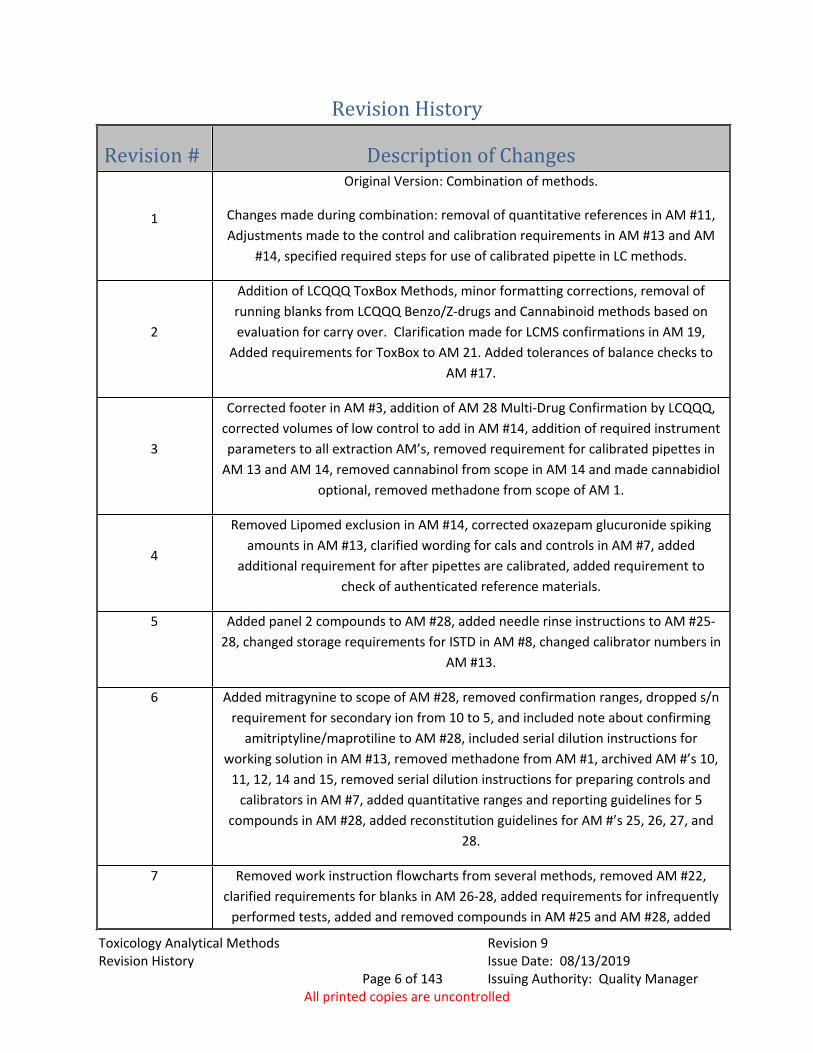
Revision History
Revision # Description of Changes
1
Original Version: Combination of methods.
Changes made during combination: removal of quantitative references in AM #11, Adjustments made to the control and calibration requirements in AM #13 and AM
#14, specified required steps for use of calibrated pipette in LC methods.
2
Addition of LCQQQ ToxBox Methods, minor formatting corrections, removal of running blanks from LCQQQ Benzo/Z-drugs and Cannabinoid methods based on evaluation for carry over. Clarification made for LCMS confirmations in AM 19,
Added requirements for ToxBox to AM 21. Added tolerances of balance checks to AM #17.
3
Corrected footer in AM #3, addition of AM 28 Multi-Drug Confirmation by LCQQQ, corrected volumes of low control to add in AM #14, addition of required instrument parameters to all extraction AM’s, removed requirement for calibrated pipettes in
AM 13 and AM 14, removed cannabinol from scope in AM 14 and made cannabidiol optional, removed methadone from scope of AM 1.
4
Removed Lipomed exclusion in AM #14, corrected oxazepam glucuronide spiking amounts in AM #13, clarified wording for cals and controls in AM #7, added
additional requirement for after pipettes are calibrated, added requirement to check of authenticated reference materials.
5 Added panel 2 compounds to AM #28, added needle rinse instructions to AM #25-28, changed storage requirements for ISTD in AM #8, changed calibrator numbers in
AM #13.
6 Added mitragynine to scope of AM #28, removed confirmation ranges, dropped s/n requirement for secondary ion from 10 to 5, and included note about confirming
amitriptyline/maprotiline to AM #28, included serial dilution instructions for working solution in AM #13, removed methadone from AM #1, archived AM #’s 10,
11, 12, 14 and 15, removed serial dilution instructions for preparing controls and calibrators in AM #7, added quantitative ranges and reporting guidelines for 5
compounds in AM #28, added reconstitution guidelines for AM #’s 25, 26, 27, and 28.
7 Removed work instruction flowcharts from several methods, removed AM #22, clarified requirements for blanks in AM 26-28, added requirements for infrequently
performed tests, added and removed compounds in AM #25 and AM #28, added
Toxicology Analytical Methods Revision 9Issue Date: 08/13/2019Description of Changes
Page 7 of 143 Issuing Authority: Quality ManagerAll printed copies are uncontrolled
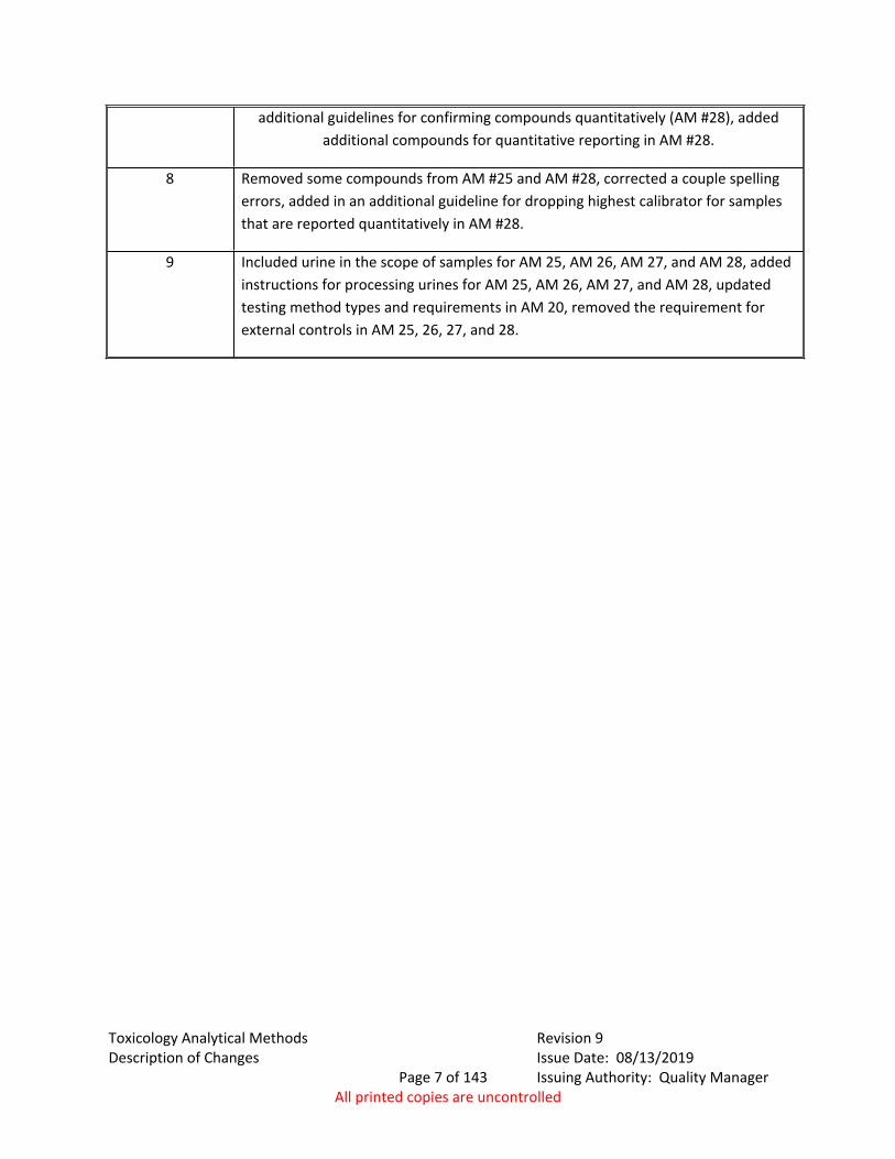
additional guidelines for confirming compounds quantitatively (AM #28), added additional compounds for quantitative reporting in AM #28.
8 Removed some compounds from AM #25 and AM #28, corrected a couple spelling errors, added in an additional guideline for dropping highest calibrator for samples that are reported quantitatively in AM #28.
9 Included urine in the scope of samples for AM 25, AM 26, AM 27, and AM 28, added instructions for processing urines for AM 25, AM 26, AM 27, and AM 28, updated testing method types and requirements in AM 20, removed the requirement for external controls in AM 25, 26, 27, and 28.
Toxicology Analytical Methods Revision 9Issue Date: 08/13/2019Toxicology AM #1: Enzyme
Immunoassay Screening for Drugs-of-Abuse in Urine
Page 8 of 143 Issuing Authority: Quality Manager
All printed copies are uncontrolled
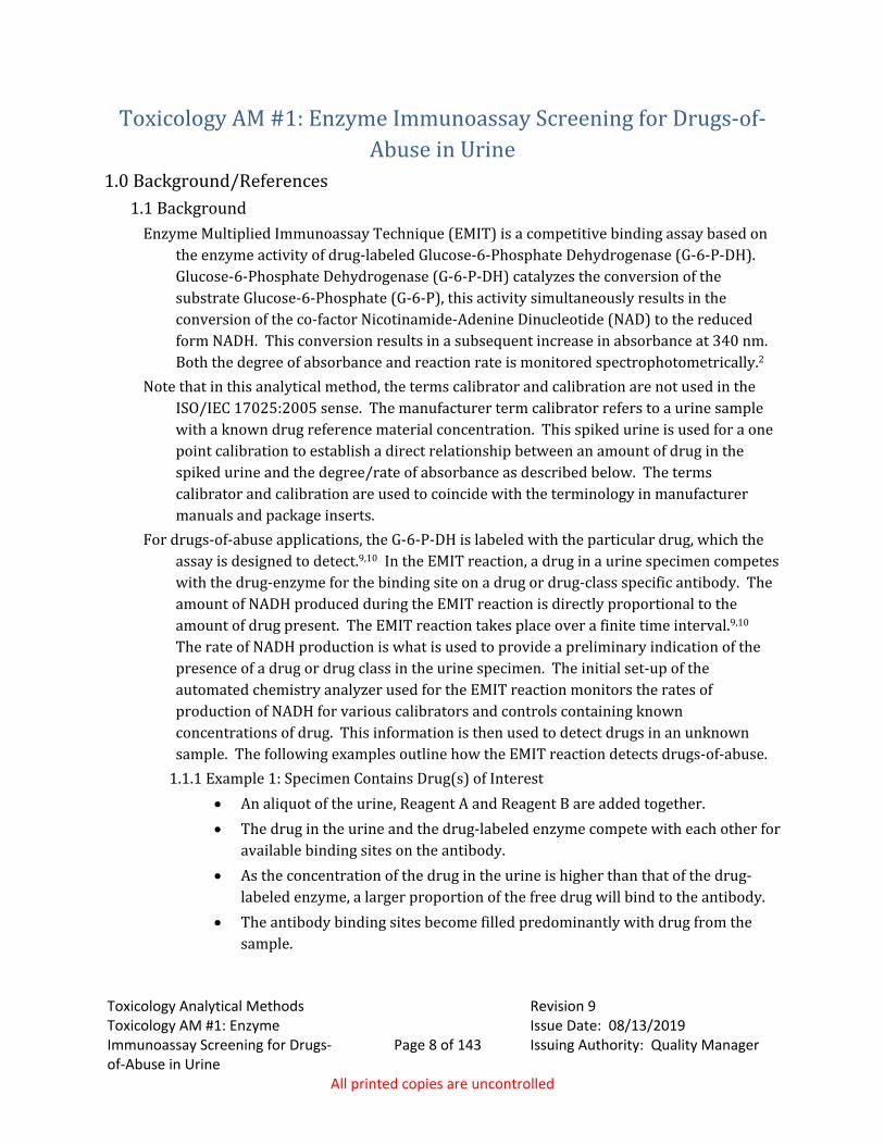
Toxicology AM #1: Enzyme Immunoassay Screening for Drugs-of-Abuse in Urine
1.0 Background/References1.1 Background
Enzyme Multiplied Immunoassay Technique (EMIT) is a competitive binding assay based on the enzyme activity of drug-labeled Glucose-6-Phosphate Dehydrogenase (G-6-P-DH). Glucose-6-Phosphate Dehydrogenase (G-6-P-DH) catalyzes the conversion of the substrate Glucose-6-Phosphate (G-6-P), this activity simultaneously results in the conversion of the co-factor Nicotinamide-Adenine Dinucleotide (NAD) to the reduced form NADH. This conversion results in a subsequent increase in absorbance at 340 nm. Both the degree of absorbance and reaction rate is monitored spectrophotometrically.2
Note that in this analytical method, the terms calibrator and calibration are not used in the ISO/IEC 17025:2005 sense. The manufacturer term calibrator refers to a urine sample with a known drug reference material concentration. This spiked urine is used for a one point calibration to establish a direct relationship between an amount of drug in the spiked urine and the degree/rate of absorbance as described below. The terms calibrator and calibration are used to coincide with the terminology in manufacturer manuals and package inserts.
For drugs-of-abuse applications, the G-6-P-DH is labeled with the particular drug, which the assay is designed to detect.9,10 In the EMIT reaction, a drug in a urine specimen competes with the drug-enzyme for the binding site on a drug or drug-class specific antibody. The amount of NADH produced during the EMIT reaction is directly proportional to the amount of drug present. The EMIT reaction takes place over a finite time interval.9,10 The rate of NADH production is what is used to provide a preliminary indication of the presence of a drug or drug class in the urine specimen. The initial set-up of the automated chemistry analyzer used for the EMIT reaction monitors the rates of production of NADH for various calibrators and controls containing known concentrations of drug. This information is then used to detect drugs in an unknown sample. The following examples outline how the EMIT reaction detects drugs-of-abuse.
1.1.1 Example 1: Specimen Contains Drug(s) of Interest An aliquot of the urine, Reagent A and Reagent B are added together. The drug in the urine and the drug-labeled enzyme compete with each other for
available binding sites on the antibody. As the concentration of the drug in the urine is higher than that of the drug-
labeled enzyme, a larger proportion of the free drug will bind to the antibody. The antibody binding sites become filled predominantly with drug from the
sample.
Toxicology Analytical Methods Revision 9Issue Date: 08/13/2019Toxicology AM #1: Enzyme
Immunoassay Screening for Drugs-of-Abuse in Urine
Page 9 of 143 Issuing Authority: Quality Manager
All printed copies are uncontrolled
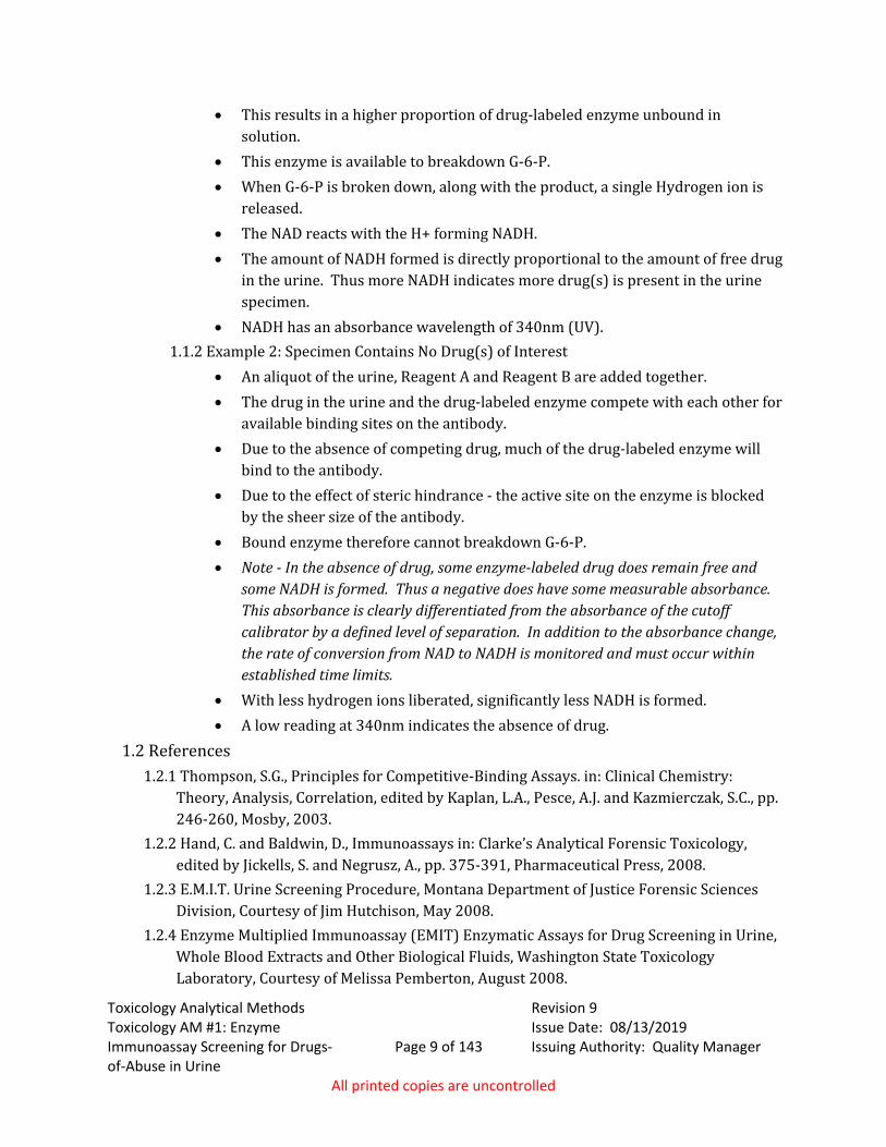
This results in a higher proportion of drug-labeled enzyme unbound in solution.
This enzyme is available to breakdown G-6-P. When G-6-P is broken down, along with the product, a single Hydrogen ion is
released. The NAD reacts with the H+ forming NADH. The amount of NADH formed is directly proportional to the amount of free drug
in the urine. Thus more NADH indicates more drug(s) is present in the urine specimen.
NADH has an absorbance wavelength of 340nm (UV).1.1.2 Example 2: Specimen Contains No Drug(s) of Interest
An aliquot of the urine, Reagent A and Reagent B are added together. The drug in the urine and the drug-labeled enzyme compete with each other for
available binding sites on the antibody. Due to the absence of competing drug, much of the drug-labeled enzyme will
bind to the antibody. Due to the effect of steric hindrance - the active site on the enzyme is blocked
by the sheer size of the antibody. Bound enzyme therefore cannot breakdown G-6-P. Note - In the absence of drug, some enzyme-labeled drug does remain free and
some NADH is formed. Thus a negative does have some measurable absorbance. This absorbance is clearly differentiated from the absorbance of the cutoff calibrator by a defined level of separation. In addition to the absorbance change, the rate of conversion from NAD to NADH is monitored and must occur within established time limits.
With less hydrogen ions liberated, significantly less NADH is formed. A low reading at 340nm indicates the absence of drug.
1.2 References1.2.1 Thompson, S.G., Principles for Competitive-Binding Assays. in: Clinical Chemistry:
Theory, Analysis, Correlation, edited by Kaplan, L.A., Pesce, A.J. and Kazmierczak, S.C., pp. 246-260, Mosby, 2003.
1.2.2 Hand, C. and Baldwin, D., Immunoassays in: Clarke’s Analytical Forensic Toxicology, edited by Jickells, S. and Negrusz, A., pp. 375-391, Pharmaceutical Press, 2008.
1.2.3 E.M.I.T. Urine Screening Procedure, Montana Department of Justice Forensic Sciences Division, Courtesy of Jim Hutchison, May 2008.
1.2.4 Enzyme Multiplied Immunoassay (EMIT) Enzymatic Assays for Drug Screening in Urine, Whole Blood Extracts and Other Biological Fluids, Washington State Toxicology Laboratory, Courtesy of Melissa Pemberton, August 2008.
Toxicology Analytical Methods Revision 9Issue Date: 08/13/2019Toxicology AM #1: Enzyme
Immunoassay Screening for Drugs-of-Abuse in Urine
Page 10 of 143 Issuing Authority: Quality Manager
All printed copies are uncontrolled
1.2.5 Viva-JrTM Operator's Manual, Article No.: 6002-940-410, Version number: 01/04-06.1.2.6 Viva-Jr System Operations Guide, T268, 6/25/07, D01373.1.2.7 Viva-E Operator's Manual, Article No.: 6002-380-410-01, Version number: 1.0/08-041.2.8 Viva-E System Operations Guide, T216, 6/4/07, D01320. 1.2.9 Leedam, D.C., EMIT Basic Power Point Presentation, February 1997 (Provided by
Siemens during training October 16, 2008.)1.2.10 Syva Package Inserts for Emit II Plus Assays
o Amphetamines: 9C122UL.4DS_Ao Benzodiazepine: 9F022UL.10DS_Bo Cannabinoid: 9N022UL.9DS_Ao Cocaine: 9H522UL.4DS_Ao Opiate: 9B322UL.10DS_A
2.0 Scope2.1 This analytical method employs EMIT for the qualitative screening for drugs-of-
abuse in urine specimens. EMIT is commonly used for the detection of drugs-of- abuse in urine. The EMIT assays are run on a microprocessor-controlled automatic chemistry analyzer. The assay results are intended as only a preliminary analytical test result. Confirmatory analysis is performed with an instrument such as a gas chromatograph or liquid chromatograph equipped with a mass selective detector. If EMIT results are reported out, the report must clearly state that the results are from initial screening and confirmatory testing may be requested.
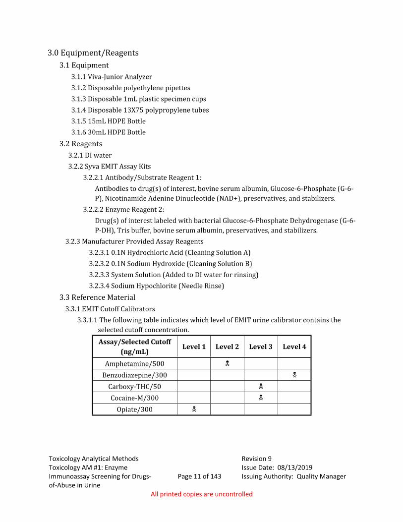
As indicated in the table below, each assay in use has an established administrative threshold or cutoff. For this reason, a negative result does not indicate that no drug is present; the concentration of the drug may be less than the administrative cutoff, or a drug may have poor cross-reactivity to the assay. For this reason, there may be situations where confirmation of an analyte may be pursued even if a negative result is indicated for the compound or a class of compounds in question.
Assay Calibrator Urine Cutoff
Amphetamines d-Methamphetamine 500ng/mLBenzodiazepines Lormetazepam 300ng/mL
Cannabinoids 11-Nor-9-Carboxy-THC 50ng/mLCocaine Metabolite/-M Benzoylecgonine 300ng/mL
Opiates Morphine 300ng/mL
Toxicology Analytical Methods Revision 9Issue Date: 08/13/2019Toxicology AM #1: Enzyme
Immunoassay Screening for Drugs-of-Abuse in Urine
Page 11 of 143 Issuing Authority: Quality Manager
All printed copies are uncontrolled
3.0 Equipment/Reagents3.1 Equipment
3.1.1 Viva-Junior Analyzer3.1.2 Disposable polyethylene pipettes3.1.3 Disposable 1mL plastic specimen cups3.1.4 Disposable 13X75 polypropylene tubes3.1.5 15mL HDPE Bottle 3.1.6 30mL HDPE Bottle
3.2 Reagents3.2.1 DI water3.2.2 Syva EMIT Assay Kits
3.2.2.1 Antibody/Substrate Reagent 1: Antibodies to drug(s) of interest, bovine serum albumin, Glucose-6-Phosphate (G-6-P), Nicotinamide Adenine Dinucleotide (NAD+), preservatives, and stabilizers.
3.2.2.2 Enzyme Reagent 2: Drug(s) of interest labeled with bacterial Glucose-6-Phosphate Dehydrogenase (G-6-P-DH), Tris buffer, bovine serum albumin, preservatives, and stabilizers.
3.2.3 Manufacturer Provided Assay Reagents3.2.3.1 0.1N Hydrochloric Acid (Cleaning Solution A)3.2.3.2 0.1N Sodium Hydroxide (Cleaning Solution B)3.2.3.3 System Solution (Added to DI water for rinsing)3.2.3.4 Sodium Hypochlorite (Needle Rinse)
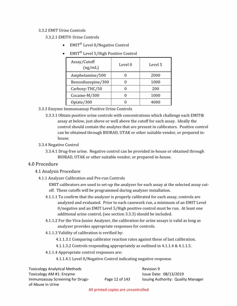
3.3 Reference Material3.3.1 EMIT Cutoff Calibrators
3.3.1.1 The following table indicates which level of EMIT urine calibrator contains the selected cutoff concentration.Assay/Selected Cutoff
(ng/mL)Level 1 Level 2 Level 3 Level 4
Amphetamine/500
Benzodiazepine/300
Carboxy-THC/50
Cocaine-M/300
Opiate/300
Toxicology Analytical Methods Revision 9Issue Date: 08/13/2019Toxicology AM #1: Enzyme
Immunoassay Screening for Drugs-of-Abuse in Urine
Page 12 of 143 Issuing Authority: Quality Manager
All printed copies are uncontrolled
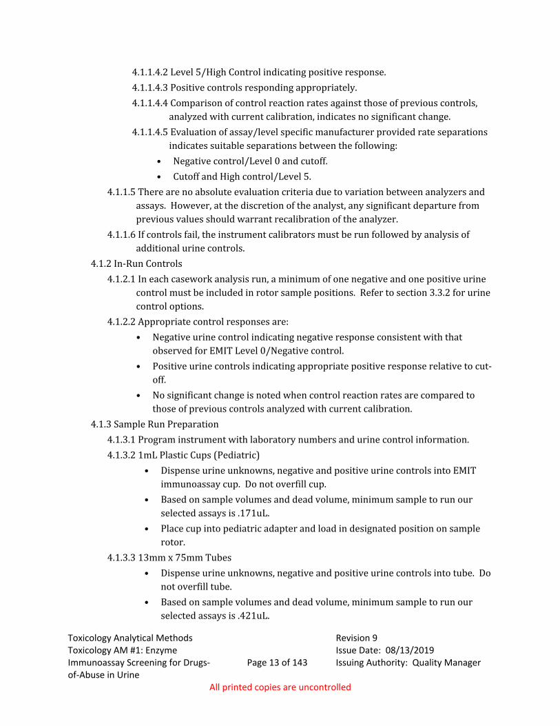
3.3.2 EMIT Urine Controls3.3.2.1 EMIT Urine Controls
EMIT Level 0/Negative Control
EMIT Level 5/High Positive Control
Assay/Cutoff (ng/mL)
Level 0 Level 5
Amphetamine/500 0 2000Benzodiazepine/300 0 1000Carboxy-THC/50 0 200Cocaine-M/300 0 1000Opiate/300 0 4000
3.3.3 Enzyme Immunoassay Positive Urine Controls 3.3.3.1 Obtain positive urine controls with concentrations which challenge each EMIT®
assay at below, just above or well above the cutoff for each assay. Ideally the control should contain the analytes that are present in calibrators. Positive control can be obtained through BIORAD, UTAK or other suitable vendor, or prepared in- house.
3.3.4 Negative Control3.3.4.1 Drug-free urine. Negative control can be provided in-house or obtained through
BIORAD, UTAK or other suitable vendor, or prepared in-house.
4.0 Procedure4.1 Analysis Procedure
4.1.1 Analyzer Calibration and Pre-run ControlsEMIT calibrators are used to set-up the analyzer for each assay at the selected assay cut-off. These cutoffs will be programmed during analyzer installation.
4.1.1.1 To confirm that the analyzer is properly calibrated for each assay, controls are analyzed and evaluated. Prior to each casework run, a minimum of an EMIT Level 0/negative and an EMIT Level 5/High positive control must be run. At least one additional urine control, (see section 3.3.3) should be included.
4.1.1.2 For the Viva-Junior Analyzer, the calibration for urine assays is valid as long as analyzer provides appropriate responses for controls.
4.1.1.3 Validity of calibration is verified by:4.1.1.3.1 Comparing calibrator reaction rates against those of last calibration.4.1.1.3.2 Controls responding appropriately as outlined in 4.1.1.4 & 4.1.1.5.
4.1.1.4 Appropriate control responses are:4.1.1.4.1 Level 0/Negative Control indicating negative response.
Toxicology Analytical Methods Revision 9Issue Date: 08/13/2019Toxicology AM #1: Enzyme
Immunoassay Screening for Drugs-of-Abuse in Urine
Page 13 of 143 Issuing Authority: Quality Manager
All printed copies are uncontrolled
4.1.1.4.2 Level 5/High Control indicating positive response.4.1.1.4.3 Positive controls responding appropriately.4.1.1.4.4 Comparison of control reaction rates against those of previous controls,
analyzed with current calibration, indicates no significant change.4.1.1.4.5 Evaluation of assay/level specific manufacturer provided rate separations
indicates suitable separations between the following: • Negative control/Level 0 and cutoff.• Cutoff and High control/Level 5.
4.1.1.5 There are no absolute evaluation criteria due to variation between analyzers and assays. However, at the discretion of the analyst, any significant departure from previous values should warrant recalibration of the analyzer.
4.1.1.6 If controls fail, the instrument calibrators must be run followed by analysis of additional urine controls.
4.1.2 In-Run Controls4.1.2.1 In each casework analysis run, a minimum of one negative and one positive urine
control must be included in rotor sample positions. Refer to section 3.3.2 for urine control options.
4.1.2.2 Appropriate control responses are:• Negative urine control indicating negative response consistent with that
observed for EMIT Level 0/Negative control.• Positive urine controls indicating appropriate positive response relative to cut-
off.• No significant change is noted when control reaction rates are compared to
those of previous controls analyzed with current calibration.4.1.3 Sample Run Preparation
4.1.3.1 Program instrument with laboratory numbers and urine control information. 4.1.3.2 1mL Plastic Cups (Pediatric)
• Dispense urine unknowns, negative and positive urine controls into EMIT immunoassay cup. Do not overfill cup.
• Based on sample volumes and dead volume, minimum sample to run our selected assays is .171uL.
• Place cup into pediatric adapter and load in designated position on sample rotor.
4.1.3.3 13mm x 75mm Tubes• Dispense urine unknowns, negative and positive urine controls into tube. Do
not overfill tube.• Based on sample volumes and dead volume, minimum sample to run our
selected assays is .421uL.
Toxicology Analytical Methods Revision 9Issue Date: 08/13/2019Toxicology AM #1: Enzyme
Immunoassay Screening for Drugs-of-Abuse in Urine
Page 14 of 143 Issuing Authority: Quality Manager
All printed copies are uncontrolled
• Place tube into designated position of sample rotor.4.2 Viva Junior Operation and Maintenance5,6
4.2.1 Daily required Maintenance: 4.2.1.1 Fill Water reservoir and add 15mL Siemens System Solution4.2.1.2 Check for air bubbles in lines4.2.1.3 Run Needle Rinse4.2.1.4 Run Blank Rotor
4.2.2 Monthly/Quarterly Required Maintenance:Note: Documentation is not required for Monthly, Quarterly Maintenance or as needed
maintenance. This maintenance does not affect the results of analysis.4.2.2.1 Rinse and dry Water Reservoir with .1N Sodium Hydroxide
4.3 Detection Criteria4.3.1 Positive Case Sample Result
4.3.1.1 Provided that calibration and control evaluation indicate that analyzer has quality assurance conditions suitable for use, a positive result for a sample is indicated by a change in absorbance at a rate value (dABS/m) of equal to or greater than the Cutoff Calibrator.
4.3.2 Elevated Absorbance4.3.2.1 At the discretion of an analyst, confirmatory techniques may be applied to samples
that exhibit an elevated absorbance rate. An elevated absorbance rate is that greater than that of the negative control/Level 0 but less than the cutoff calibrator. If data for confirmatory techniques supports the presence of an analyte, the analyte may be reported as present. In addition, samples with compounds that have low cross reactivity may be confirmed and reported with a negative screen result.
4.3.3 Negative Result4.3.3.1 A negative result for a sample is indicated by a change in absorbance at a rate that
is less than the Cutoff Calibrator. Special considerations may apply as outlined above (4.2).
4.4 Distribution of Assay Information4.4.1 Electronic copy of EIA analysis report must be attached to the case in LIMS. Case results
are also to be recorded in the LIMS system.4.4.2 A copy of data for calibrators and controls may be stored electronically in a central
location. 4.4.3 Original data for calibration and controls for each analysis will be stored centrally in the
laboratory, until archiving or destruction. If an electronic copy is created the hard copies need not be retained, if electronic copies are maintained on a network drive.
4.5 Quality Assurance Requirements4.5.1 Refer to Toxicology AM# 19 for storage requirements.
Toxicology Analytical Methods Revision 9Issue Date: 08/13/2019Toxicology AM #1: Enzyme
Immunoassay Screening for Drugs-of-Abuse in Urine
Page 15 of 143 Issuing Authority: Quality Manager
All printed copies are uncontrolled
4.5.2 Refer to Toxicology AM #21 for authentication of reference material requirements.
Toxicology Analytical Methods Revision 9Issue Date: 08/13/2019Toxicology AM #2: General
Extraction of Urine for Basic/Neutral or Acidic/Neutral Compounds
Page 16 of 143 Issuing Authority: Quality Manager
All printed copies are uncontrolled
Toxicology AM #2: General Extraction of Urine for Basic/Neutral or Acidic/Neutral Compounds
1.0 Background/References1.1 Background
These extractions are extensions of the TOXI-LAB TOXI-A and TOXI-B thin layer chromatography (TLC) drug detection systems. The samples are extracted as with the TLC system, however, instead of concentrating the extract onto a disc, the solvent extract is concentrated and placed into an automated liquid sampler (ALS) vial for analysis by a gas chromatograph equipped with a mass selective detector (GC/MSD). Discussions of TLC and GC/MS theory can be found in most college-level chemistry and/or instrumental texts. In 2013 the TOXI-LAB line was discontinued. An equivalent product, De-Tox Tubes by Dyna-Tek, were evaluated and found to be a suitable replacement.
2.0 Scope2.1 This procedure describes the extraction of drug compounds from urine. Depending
upon the pKa of a drug compound, either De-Tox Tubes A or B (or verified equivalents) are used. Basic and neutral compounds are extracted with an A tube. Addition of urine to the De-Tox A tube results in the sample becoming alkaline and basic and neutral drugs thus extract into a solvent mixture. The B tube is used for acidic and neutral compounds. Urine placed into the De-Tox B tube becomes acidic resulting in acidic and neutral compounds being extracted into a solvent mixture. Either resulting extract is analyzed by full scan GC/MS in EI mode.
3.0 Equipment/Reagents3.1 Equipment and Supplies
3.1.1 Tube Rocker3.1.2 Evaporative concentrator and appropriate concentration cups or tubes3.1.3 Laboratory Centrifuge capable of 3000 rpm3.1.4 Laboratory oven or waterbath3.1.5 Fixed and/or adjustable volume pipettes, and appropriate tips.3.1.6 Automated Liquid Sampler (ALS) vials 3.1.7 GC/MS Vial Microinserts3.1.8 Gas Chromatograph equipped with a mass selective detector and a low bleed (5%-
Diphenyl-95%-Dimethylsiloxane copolymer) capillary column.3.2 Reagents
3.2.1 De-Tox Tubes A and B (or equivalent Toxi Tubes)3.2.2 b-Glucuronidase Solution
Toxicology Analytical Methods Revision 9Issue Date: 08/13/2019Toxicology AM #2: General
Extraction of Urine for Basic/Neutral or Acidic/Neutral Compounds
Page 17 of 143 Issuing Authority: Quality Manager
All printed copies are uncontrolled
3.2.3 2M Acetate buffer, pH 4.83.3 Qualitative Controls
3.3.1 Positive control3.3.1.1 Tube A positive control may be commercially obtained or prepared in-house. At a
minimum, the control must contain at least one phenethylamine at an approximate concentration between 500 and 3000 ng/mL, and one opiate at an approximate concentration between 300 and 3000 ng/mL.
3.1.1.2 Tube B positive control may also be commercially obtained or prepared in-house. At a minimum, the control must contain two barbiturates at an approximate concentration between 300 and 1000 ng/mL.
3.3.2 Negative Urine 3.3.2.1 Negative urine can be commercially obtained or in-house urine verified to be
negative for drugs of interest. Refer to Toxicology AM#19 for additional details.3.3.3 Morphine-Glucuronide Positive and Negative Controls for Optional Enzymatic
Hydrolysis Step3.3.3.1 Commercially-obtained control or in-house spiked urine containing morphine-
glucuronide should be used. The same negative urine must be used to prepare both the positive and negative glucuronide controls for in-house spiking. Morphine-glucuronide should be used for these controls and must be at a minimum concentration of 375ng/mL. The positive and negative glucuronide controls are used to demonstrate the glucuronidase cleavage was effective.
3.4 Qualitative Non-Extracted Reference Material3.4.1 Run necessary reference material as indicated by examination of GC/MSD data.
Reference material mixes may be used.3.4.2 Dilute reference material as necessary. A suggested dilution for a 1mg/mL solution is 1
in 3 parts of appropriate solvent.
4.0 Procedure4.1 Extraction Procedure
4.1.1 De-Tox Tubes-A Extraction (Basic and Neutral Compounds)4.1.1.1 Label DE-TOX TUBE A and ALS vials with micro-inserts for negative control,
positive control and appropriate laboratory case numbers. 4.1.1.2 Transfer approx. 5mL of casework, negative and positive control urine to
appropriate DE-TOX TUBE A (pH=9). If case sample volume is limited, less than 5mL may be used.
4.1.1.3 Agitate the DE-TOX tube to break up salts. This assists in reducing the occurrence of emulsions.
4.1.1.4 Rock DE-TOX TUBE A for at least 10 minutes.4.1.1.5 Centrifuge tube at ~2500-3000 rpm for ~10 minutes.
Toxicology Analytical Methods Revision 9Issue Date: 08/13/2019Toxicology AM #2: General
Extraction of Urine for Basic/Neutral or Acidic/Neutral Compounds
Page 18 of 143 Issuing Authority: Quality Manager
All printed copies are uncontrolled
4.1.1.6 Transfer solvent and evaporate to ~100-300uL.4.1.1.7 Transfer solvent to labeled GC/MS ALS vial with micro-insert.4.1.1.8 OPTIONAL: Analyst may, at their discretion, perform an enzymatic hydrolysis on a
sample aliquot prior to the above De-Tox Tube A extraction. If done, this must be done in addition to the regular (non-hydrolyzed) extraction of the sample. Positive and negative morphine glucuronide controls (see section 3.3.3 of this method) should be run in addition to the regular controls required by the method.
4.1.1.8.1 Optional Enzyme Hydrolysis: To 4.5mL of urine, add 150μL of 2M acetate buffer and vortex. To all but the glucuronidase negative control, add 100μL of Kura BG-100 β-glucuronidase solution. Cap and vortex gently to mix. Place in a 60°C laboratory oven or water bath for 2 hours. Allow sample to cool before proceeding with steps 4.1.1.2 through 4.1.1.7.
4.1.2 De-Tox Tubes-B Extraction (Acidic and Neutral Compounds)4.1.2.1 Label DE-TOX TUBES B and ALS vials with microinserts for negative control,
positive control and appropriate laboratory numbers.4.1.2.2 Transfer approx. 4.5mL of casework, negative and positive control urine to
appropriate DE-TOX TUBE B (pH=4.5). If case sample volume is limited, less than 4.5mL may be used.
4.1.2.3 Rock DE-TOX TUBE B for at least 10 minutes.4.1.2.4 Centrifuge tube at ~2500-3000 rpm for ~10 minutes.4.1.2.5 Transfer solvent and evaporate to ~100-300 μL.4.1.2.6 Transfer solvent to labeled GC/MS ALS vial with microinsert.
4.2 Preparation for Analysis Run4.2.1 Into Sequence log table, enter the sample case numbers, blanks and controls.4.2.2 Load samples, reference materials, blanks and controls into the quadrant rack as noted
in the sequence table.4.2.3 GC-MSD Analysis Parameters
4.2.3.1 Refer to instrument method for current analysis parameters. 4.2.3.1.1 Instrument Run Parameters
Inlet at 280, splitless injection, injection volume: 1μL Oven at 80 degrees Celsius, hold 2.5 minutes Ramp 25 degrees Celsius Final temperature: 300 degrees Celsius, hold at least 7.5 minutes. Additional sample runs may be done with adjustments to these parameters,
for example if there is a coeluting peak or a late eluting compound.
Toxicology Analytical Methods Revision 9Issue Date: 08/13/2019Toxicology AM #2: General
Extraction of Urine for Basic/Neutral or Acidic/Neutral Compounds
Page 19 of 143 Issuing Authority: Quality Manager
All printed copies are uncontrolled
4.2.3.2 Current analysis method must be stored centrally as a hard or electronic copy. Setting the instrument parameters to store with the data file is also approved.
4.2.4 Detection and Identification Criteria4.2.4.1 The presence of a drug compound is indicated if the retention time for the sample
versus applicable reference material does not differ by more than ±0.2 minutes and there are no significant differences in the mass spectral data.
NOTE: Early eluting drugs, as well as drugs known to have similar retention times and mass spectral fragmentation patterns (e.g. Phentermine and Methamphetamine), may not differ from the retention time of the applicable reference material by more than ±0.1 minutes.
4.3 Quality Assurance Requirements4.3.1 Refer to applicable sections of Toxicology AM #19 and Toxicology AM #21 for additional
quality assurance and reference material authentication requirements.4.4 Analysis Documentation
4.4.1 The printed results for each case sample and accompanying blank will be included with the analysts’ notes. Case results are to be recorded in the iLIMS system.
4.4.2 Original data for controls will be prepared for each analysis run and stored centrally in the laboratory where the analysis was performed until archiving, or electronically on a shared drive.
4.4.3 A copy of controls may be stored electronically in a central location and need not be included in individual case files. When necessary, a copy of control printouts can be prepared from the centrally stored document.
Toxicology Analytical Methods Revision 9Issue Date: 08/13/2019Toxicology AM #3: Qualitative 11-
nor-9-THC-D9-COOH (Carboxy-THC) in Urine
Page 20 of 143 Issuing Authority: Quality Manager
All printed copies are uncontrolled
Toxicology AM #3: Qualitative 11-nor-9-THC-D9-COOH (Carboxy-THC) in Urine
1.0 Background/References1.1 Background
Cannabis sativa use dates back to 2700 B.C.2,5 Marijuana (MJ) refers to a mixture of the leaves and flowering tops.3 The smoke from burning cannabis includes 61 different cannabinoids.2,6 The major active ingredient in marijuana is delta–9-tetrahydrocannabinol (D9-THC). The D9-THC content varies from 2 to 10% with an average of four to five percent. The quality of marijuana is reported to have improved over the last 20 years due to superior cultivation practices. The medicinal effects of MJ include anti-nausea, muscle relaxing, anticonvulsant and reduction of intraocular pressure.6 Cannabis therefore has found use as an antiemetic to deal with the nausea associated with anticancer chemotherapy and for relief for those suffering from glaucoma. The debate continues on medical use and the complete legalization of the drug.
Several factors come into play when considering the behavioral effects of (D9)-THC. These include the route of administration (smoked or ingested), THC concentration of the plant (dose), the experience of the user, the user’s vulnerability to psychoactive effects, and the setting of the use.5,6 The desirable effects of MJ include an increased sense of well-being, mild euphoria, relaxation and a mild sedative-hypnotic effect.5,6 Its clinical effects are similar to those of alcohol and the anti-anxiety agents.5 The side-effects of MJ use include impairment of cognitive functions, alteration of the user’s perception of time and distance, reaction time, learning and short-term memory.2,5,6 MJ has been shown to interfere with a person’s ability or willingness to concentrate. Cannabis causes temporal disintegration such that the individual loses the ability to store information in the short term and is easily distracted.2 Impairment from use is thought to last from 4 to 8-hours with more recent studies reporting 3 to 6 hours. Dr. Huestis reported that most behavioral and physiological effects return to baseline within three to six hours after use with residual effects in specific behaviors for up to 24 hours.
Impairment of coordination and tracking behavior has been reported to persist several hours beyond the perception of the high.6 Due to the variable period of impairment, the relating of urine Carboxy-THC to the time of use, and thus impairment, requires the development of the scenario surrounding the stop for DUI. The presence of Carboxy-THC in urine only indicates exposure to MJ at some previous, indeterminate time.
The physiological effects may include an increase in heart rate and blood pressure, conjunctival suffusion, vasodilation, dry mouth and throat and a decrease in respiratory rate. The individual may also experience increased hunger (munchies).
Toxicology Analytical Methods Revision 9Issue Date: 08/13/2019Toxicology AM #3: Qualitative 11-
nor-9-THC-D9-COOH (Carboxy-THC) in Urine
Page 21 of 143 Issuing Authority: Quality Manager
All printed copies are uncontrolled
D9-THC is rapidly metabolized to the inactive metabolite, Carboxy-THC.1,4,5,6 In urine, this major metabolite, Carboxy-THC is pursued due to D9-THC only being present in minute quantities.6 Carboxy-THC in urine has been conjugated with glucuronic acid to improve excretion. The detection time of Carboxy-THC in urine following marijuana use varies dependent upon various pharmacological factors such as the dose obtained, the route of administration, and the rates of metabolism and excretion.1 D9-THC is deposited in body fat due to its high lipid solubility. It is slowly released from this storage depot over time.1 The amount of D9-THC stored in fat is a function of the amount, frequency and potency of drug exposure. The detection time can therefore vary from days to months.
1.2 References1.2.1 Huestis, M.A., Mitchell, J.M. and Cone, E.J. Detection Times of Marijuana Metabolites in
Urine by Immunoassay and GC-MS J. Anal. Tox. 19:443-449, 1995.1.2.2 Huestis, M. Marijuana. pp. 269-304. in: Principles of Forensic Toxicology, Third Edition.
Levine, B. ed., AACC, 2010.1.2.3 Cannabis. in: Clark’s Isolation and Identification of Drugs pp. 423-425, Moffat, A.C. ed.,
Pharmaceutical Press:London, 1986.1.2.4 Drug Evaluation and Classification Training Manual, U.S. Dept. of Transportation, 1993.1.2.5 Julien, R.M. Marijuana: A Unique Sedative-Euphoriant-Psychedelic Drug. in: A Primer of
Drug Action. pp. 319-349, W.H. Freeman and Company: NewYork, 1998.1.2.6 O’Brien, C.P. Drug Addiction and Drug Abuse. pp. 572-573. In: Goodman & Gilman’s The
Pharmacological Basis of Therapeutics, Ninth edition, Hardman, J.G. ed., McGraw-Hill, 1996.
2.0 Scope2.1 This method is to qualitatively confirm the presence of a major metabolite of
marijuana, Carboxy-THC, in urine specimens.3.0 Equipment/Reagents
3.1 Equipment and Supplies3.1.1 Tube Rocker 3.1.2 Laboratory Centrifuge capable of 3500 rpm3.1.3 Waterbath 3.1.4 Drybath 3.1.5 Evaporative Concentrator equipped with nitrogen tank.3.1.6 pH Indicator Strips 3.1.7 Glassware
3.1.7.1 16X100mm tubes 3.1.7.2 16X144mm tapered tip centrifuge tubes 3.1.7.3 Caps for 16mm OD tubes 3.1.7.4 GC/MS ALS vials
Toxicology Analytical Methods Revision 9Issue Date: 08/13/2019Toxicology AM #3: Qualitative 11-
nor-9-THC-D9-COOH (Carboxy-THC) in Urine
Page 22 of 143 Issuing Authority: Quality Manager
All printed copies are uncontrolled
3.1.7.5 GC/MS vial microinserts 3.1.8 Gas Chromatograph equipped with a mass selective detector and a nonpolar capillary
column (e.g. 100%-dimethylpolysiloxane or 95%-dimethyl-polysiloxane with 5%diphenyl).
3.2 ReagentsRefer to Toxicology AM#23 for solution preparation instructions. Purity of chemicals must be
ACS Grade or equivalent.3.2.1 1N KOH3.2.2 Saturated Potassium Phosphate Monobasic pH approx. 1.83.2.3 87:13 Hexane with Ethyl Acetate (v/v)3.2.4 Ethyl acetate 3.2.5 Silylating Agent (select from): BSTFA/1% TMCS or MSTFA
3.3 Standards/Reference Material3.3.1 Stock Standard Solution
100µg/mL (+) 11-nor-9-carboxy-D9-THC 3.3.2 Working Standard Solution (1800ng/mL)
3.3.2.1 Add 180µL Stock Solution to 9.82mL Methanol. Other volumes may be prepared. Document preparation on appropriate log sheet. Solution is stable for 1-year (or at the date of the earliest expiring Stock solution) when stored under refrigeration.
3.4 Qualitative Controls3.4.1 Positive Controls
3.4.1.1 A minimum of one spiked 60ng/mL and one commercial Carboxy-THC containing control must be analyzed in each batch of samples.
3.4.1.2 60ng/mL Carboxy-THC Spiked Control3.4.1.2.1 Add 3mL of the same lot of negative urine used to prepare the negative
control to extraction tube. Add 100µL of working standard solution, and vortex.
3.4.1.3 Suitable nominal concentration range for commercial control is 15ng/mL to 150ng/mL.
3.4.2 Negative Control 3.4.2.1 Negative urine commercially obtained or in-house urine verified to be negative for
drugs of interest.
4.0 Procedure4.1 Extraction Method
4.1.1 Initial set-up
Toxicology Analytical Methods Revision 9Issue Date: 08/13/2019Toxicology AM #3: Qualitative 11-
nor-9-THC-D9-COOH (Carboxy-THC) in Urine
Page 23 of 143 Issuing Authority: Quality Manager
All printed copies are uncontrolled
4.1.1.1 Label extraction tubes, tapered bottom derivatization tubes and GC/MS vials with microinserts for the negative control, spiked positive control, commercial positive control(s), and casework samples.
4.1.2 Sample Preparation4.1.2.1 Transfer 3mL urine specimen, negative urine, spiked positive control and
commercial positive control(s) to extraction tubes. 4.1.3 Sample Hydrolysis
4.1.3.1 Add 0.5mL 1.0N KOH to each extraction tube.4.1.3.2 Vortex gently to mix.4.1.3.3 Check resulting pH.4.1.3.4 pH must be > 12. If pH <12, add additional 0.5mL of KOH.4.1.3.5 Place in 40C water bath for 15 minutes.4.1.3.6 Allow samples to cool before proceeding with solvent extraction.
4.1.4 Extraction4.1.4.1 If original pH was > 12:
4.1.4.1.1 Add 1.5mL Saturated Phosphate Buffer (pH 1.8). 4.1.4.1.2 Add 3mL Hexane/Ethyl Acetate (87:13). 4.1.4.1.3 Rock for 10 minutes.
4.1.4.2 If original pH was < 12:4.1.4.2.1 Add 3.0mL Saturated Phosphate Buffer (pH 1.8). 4.1.4.2.2 Add 4mL Hexane/Ethyl Acetate (87:13). 4.1.4.2.3 Rock for 10 minutes.
4.1.4.3 Centrifuge tubes at ~3500 rpm for 10 minutes.4.1.4.4 Transfer upper organic phase from tube into labeled tapered bottom tube. 4.1.4.5 Evaporate solvent to dryness, under a gentle stream of nitrogen, at ~37C.
4.1.5 Derivatization4.1.5.1 To dried extract in tapered bottom tubes, add 50µL ethyl acetate and 50µL
silylating reagent. 4.1.5.2 Cap tubes.4.1.5.3 Vortex.4.1.5.4 Heat tube for 15 minutes in 95C drybath. 4.1.5.5 Remove from heat and allow samples to cool.4.1.5.6 Transfer derivative to labeled GC/MS ALS vial with microinsert.
4.2 Gas Chromatograph/Mass Spectrometry (GC/MS) 4.2.1 Preparation for Analysis Run
Toxicology Analytical Methods Revision 9Issue Date: 08/13/2019Toxicology AM #3: Qualitative 11-
nor-9-THC-D9-COOH (Carboxy-THC) in Urine
Page 24 of 143 Issuing Authority: Quality Manager
All printed copies are uncontrolled
4.2.1.1 Into Sequence log table, enter information for case samples, controls and pre-sample solvent blanks. A 60ng/mL spiked positive control should run both early and late in the sequence.
4.2.1.2 Load case samples, controls and solvent blanks into the quadrant rack(s) as noted in the sequence table.
4.2.2 GC-MSD Acquisition Parameters 4.2.2.1 Refer to instrument method for current acquisition parameters.
4.2.2.1.1 Instrument Run Parameters Inlet at 280 degrees Celsius, splitless injection, injection volume: 1μL Oven at 185 degrees Celsius Ramp 30 degrees Celsius Final temperature: 285 degrees Celsius, hold 4 minutes
4.2.2.2 Current acquisition method must be stored centrally as a hard or electronic copy.4.2.2.3 Acquire sample data in SIM (selected ion monitoring) utilizing the ions 371, 473
and 488.4.2.3 Detection and Identification Criteria
4.2.3.1 Retention Time4.2.3.1.1 Identification requires a peak within ±0.1 minutes of the retention time
established for Carboxy-THC with the in-run control(s).4.2.3.2 Ion ratios - Selective Ion Monitoring (SIM)
4.2.3.2.1 Carboxy-THC Ion ratio for the early and late 60 ng/mL control must be calculated and averaged. This mean ratio must be compared to ratio obtained from casework and the mean of the 60ng/mL control samples. Ratio between monitored ions, 371:473 and 371:488, must agree within ±20%.
4.2.3.2.1.1 Incorrect Ratios4.2.3.2.1.1.1 If the casework or control sample ion ratios do not agree
within ~20% due to high concentration of c-THC in the sample, the extract may be diluted, and run again. Alternatively, carboxy-THC in the sample may be confirmed using full scan data, provided that a derivatized reference material is also run in full scan mode. The analyte may be confirmed from full scan data if there are no significant differences in the mass spectral data as compared to the appropriate reference material and the retention time is within ±0.1 minutes of the appropriate reference material.
Toxicology Analytical Methods Revision 9Issue Date: 08/13/2019Toxicology AM #3: Qualitative 11-
nor-9-THC-D9-COOH (Carboxy-THC) in Urine
Page 25 of 143 Issuing Authority: Quality Manager
All printed copies are uncontrolled
4.2.3.2.1.1.2 Assessment of relative strength of case sample to 60 ng/mL control. The response of case samples will be compared to a 60 ng/mL control sample. The analyst will pick either of the responses from the positive control and divide the response of the 371 ion by 5; this will be defined as the approximate minimum response. The approximate minimum response will be documented in the analyst’s notes. The analyst will compare this response to the response for each case sample.
4.2.3.2.1.1.3 If the response for the case sample is less than the approximate minimum response established by the control. Carboxy-THC will generally not be confirmed. If it is below the minimum response, it is at the analyst’s discretion whether or not to call the drug. Other factors such as enzyme screen results and the sample response in relation to the baseline must be considered and noted in the analyst’s notes.
4.3 Quality Assurance Requirements 4.3.1 Refer to relevant sections of Toxicology AM #19 and Toxicology AM #21 for additional
quality assurance and reference material authentication requirements.4.4 Analysis Documentation
4.4.1 The printed results for each case sample and accompanying blank will be included with the analysts’ notes. Case results are to be recorded in the iLIMS system.
4.4.2 Original data for controls will be compiled for each analysis run and must be stored centrally in the laboratory where the analysis was performed, or electronically.
Toxicology Analytical Methods Revision 9Issue Date: 08/13/2019Toxicology AM #4:
Cocaine and Cocaine Metabolites in Urine
Page 26 of 143 Issuing Authority: Quality Manager
All printed copies are uncontrolled
Toxicology AM #4: Cocaine and Cocaine Metabolites in Urine
1.0 Background/References1.1 Background
Cocaine is a naturally occurring alkaloid derived from leaves of the South American shrub, Erythroxylon coca. Cocaine also can be produced synthetically. Cocaine is one of the most potent stimulants of the central nervous system due to its mechanism of action, which involves blocking reuptake of stimulatory neurotransmitters. Cocaine is used licitly as a local anesthetic in ophthalmology and health care settings (e.g. biopsy, wound care). The positive effects of cocaine include an increased mental awareness and alertness, a sense of clarity and feelings of elation. The fictional detective Sherlock Holmes used cocaine for its transcendently stimulating and mind clarifying properties, to the displeasure of Doctor Watson. As with all drugs, the effects of cocaine depend on the dosage, the form in which it is taken, and the route of administration. Other significant factors include the setting or circumstances in which the drug is used and the expectations of the user. Side effects can include pupillary dilation, restlessness, dizziness, dyskinesia, tremor, dysphoria, and paranoia. Additional major side effects of cocaine use are a consequence of discontinued use. If the user does not re-administer the drug, they may experience increased anxiety, agitation, restlessness and the disturbance of normal sleep patterns, which leads to fatigue. Due to these effects following cocaine use, an individual's ability to operate a motor vehicle may be impaired both during and following cocaine use.
Routes of administration include snorting, injection and smoking. The metabolism of cocaine and its metabolites involves hydrolysis, transesterification and N-demethylation. Cocaine metabolites detectable in urine include benzoylecgonine, ecgonine methyl ester, norcocaine and various arylhydroxy- and arylhydroxymethoxy- metabolites. The duration of action of cocaine is limited by its rate of metabolism since its major metabolites are inactive.
1.2 References1.2.1 UCT CLEAN SCREEN® Extraction Columns Application Manual.1.2.2 Telepchak, M.J., August, T.F. and Chaney, G., Drug Methods for the Toxicology Lab, pp.
204 - 209. In: Forensic and Clinical Applications of Solid Phase Extraction, Humana Press: New Jersey, 2004.
1.2.3 Platoff, G.E., Gere, J.A. Solid Phase Extraction of Abuse Drugs from Urine, For. Sci. Review, 3 (2):117-132; 1991.
Toxicology Analytical Methods Revision 9Issue Date: 08/13/2019Toxicology AM #4:
Cocaine and Cocaine Metabolites in Urine
Page 27 of 143 Issuing Authority: Quality Manager
All printed copies are uncontrolled
2.0 Scope2.1 This procedure outlines the use of the 200mg CLEAN SCREEN® DAU SPE column
for the extraction of the cocaine metabolite Benzoylecgonine along with Cocaine and additional metabolite Ecgonine Methyl Ester, from urine. The CLEAN SCREEN® DAU column utilizes a copolymeric sorbent which combines a cationic exchanger and a hydrophobic functionality (reverse phase) to interact effectively, physically and chemically, with analytes of interest and minimally with interfering substances.
The cation exchanger will allow the anionic sorbent ( - ) to bind to cations. Additional retention mechanisms include hydrophobic interactions and polar adsorption. The nonpolar aspect of the column serves to extract nonpolar compounds from a polar sample matrix.2 The cation exchanger component of the phase is effective for compounds which are present in the urine sample in a cationic form bonding ionically to the sorbent.
To maximize the ionic character of analytes, the urine is adjusted with a pH 6 100mM phosphate buffer, and loaded onto a pre-conditioned SPE column. The conditioning creates an environment which allows for optimal interaction between the sorbent and the analytes of interest. Analytes are retained by ionic interaction of the amine functional groups present on the drug and the anionic sulfonic acid exchanger on the sorbent. The column is subsequently washed with water and a weak aqueous buffer, to selectively remove matrix components and interfering substances from the column. The wash also disrupts the hydrophobic and adsorption interactions but not the ionically bound material. Next, the column is dried to remove traces of aqueous and organic solvents. When the column is dry the analytes of interest are recovered from the column with a basic organic solvent mixture. Following elution from the SPE column, the extract is derivatized for qualitative confirmation on a gas chromatograph equipped with a mass selective detector (GC/MSD).
3.0 Equipment/Reagents3.1 Equipment and Supplies
3.1.1 200 mg CLEAN SCREEN® Extraction Column3.1.2 Disposable inserts for SPE manifold ports (optional)3.1.3 Tube Rocker3.1.4 Vortex Mixer 3.1.5 Dry-bath or Laboratory Oven 3.1.6 Evaporative concentrator equipped with nitrogen tank3.1.7 Vacuum Manifold/pump3.1.8 Fixed and adjustable volume single channel air displacement pipettes, and appropriate
tips, capable of accurate and precise dispensing of volumes indicated
Toxicology Analytical Methods Revision 9Issue Date: 08/13/2019Toxicology AM #4:
Cocaine and Cocaine Metabolites in Urine
Page 28 of 143 Issuing Authority: Quality Manager
All printed copies are uncontrolled
3.1.9 pH indicator strips3.1.10 16 x 100mm Screw-top Glass Tube3.1.11 Screw Cap for 16mm O.D. tube3.1.12 {Optional} 16X144mm tapered tip centrifuge tubes 3.1.13 Automated Liquid Sample (ALS) vials3.1.14 GC/MS Vial Micro-insert3.1.15 Gas Chromatograph equipped with a mass selective detector and a nonpolar capillary
column with a phase composition comparable to 100%-dimethylpolysiloxane or 95%-dimethyl-polysiloxane with 5%-diphenyl
3.2 Reagents Refer to Toxicology AM# 23 for Solution Preparation Instructions3.2.1 Methylene Chloride (Certified ACS Grade)3.2.2 Isopropanol (Certified ACS Grade)3.2.3 Ammonium Hydroxide (Certified ACS Grade)3.2.4 Methanol (Certified ACS Grade)3.2.5 Ethyl Acetate (Certified ACS Grade)3.2.6 Deionized/Distilled (DI) water 3.2.7 100mM Phosphate buffer, pH 6.03.2.8 100mM Monobasic Sodium Phosphate3.2.9 100mM Dibasic Sodium Phosphate3.2.10 100mM HCl3.2.11 Elution Solvent 3.2.11.1 Mix 20mL isopropanol with 2mL ammonium hydroxide, QS to 100mL with methylene
chloride.3.2.12 BSTFA + 1% TMCS
3.3 Quality Assurance Materials 3.3.1 Positive Control
NOTE: Positive Control can be prepared with the working solution described below and/or obtained commercially.
3.3.1.1 Positive Control Stock Solution3.3.1.1.1 Obtain 1mg/mL (1µg/µL) stock drug reference material solutions through
Cerilliant, Grace, Sigma or other appropriate vendor. 3.3.1.2 Positive Control Working Solution
3.3.1.2.1 Add the designated volume of stock solution to 10mL volumetric flask partially filled with methanol. QS with methanol. Solution is stable for 1 year (or at the date of the earliest expiring Stock solution) when stored under refrigeration.
Toxicology Analytical Methods Revision 9Issue Date: 08/13/2019Toxicology AM #4:
Cocaine and Cocaine Metabolites in Urine
Page 29 of 143 Issuing Authority: Quality Manager
All printed copies are uncontrolled
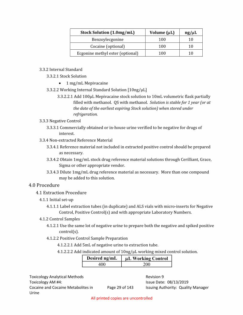
Stock Solution (1.0mg/mL) Volume (L) ng/LBenzoylecgonine 100 10
Cocaine (optional) 100 10Ecgonine methyl ester (optional) 100 10
3.3.2 Internal Standard3.3.2.1 Stock Solution
1 mg/mL Mepivacaine3.3.2.2 Working Internal Standard Solution [10ng/µL]
3.3.2.2.1 Add 100µL Mepivacaine stock solution to 10mL volumetric flask partially filled with methanol. QS with methanol. Solution is stable for 1 year (or at the date of the earliest expiring Stock solution) when stored under refrigeration.
3.3.3 Negative Control3.3.3.1 Commercially obtained or in-house urine verified to be negative for drugs of
interest.3.3.4 Non-extracted Reference Material
3.3.4.1 Reference material not included in extracted positive control should be prepared as necessary.
3.3.4.2 Obtain 1mg/mL stock drug reference material solutions through Cerilliant, Grace, Sigma or other appropriate vendor.
3.3.4.3 Dilute 1mg/mL drug reference material as necessary. More than one compound may be added to this solution.
4.0 Procedure4.1 Extraction Procedure
4.1.1 Initial set-up4.1.1.1 Label extraction tubes (in duplicate) and ALS vials with micro-inserts for Negative
Control, Positive Control(s) and with appropriate Laboratory Numbers.4.1.2 Control Samples
4.1.2.1 Use the same lot of negative urine to prepare both the negative and spiked positive control(s).
4.1.2.2 Positive Control Sample Preparation 4.1.2.2.1 Add 5mL of negative urine to extraction tube.4.1.2.2.2 Add indicated amount of 10ng/µL working mixed control solution.
Desired ng/mL L Working Control 400 200
Toxicology Analytical Methods Revision 9Issue Date: 08/13/2019Toxicology AM #4:
Cocaine and Cocaine Metabolites in Urine
Page 30 of 143 Issuing Authority: Quality Manager
All printed copies are uncontrolled
4.1.2.2.3 Additional concentrations may be used at the discretion of the analyst. 4.1.2.3 Negative Control Sample Preparation
4.1.2.3.1 Add 5mL of negative urine to extraction tube.4.1.3 Case Sample Preparation
4.1.3.1 Based on enzyme immunoassay screen results, samples may be diluted with negative urine prior to analysis.
4.1.3.2 The total volume of urine or diluted urine should be 5mL. 4.1.3.3 Add 5mL neat or diluted sample to labeled extraction tube.4.1.3.4 Internal Standard Addition
4.1.3.4.1 Add 250µL of internal standard to controls and case samples. This results in an internal standard concentration of 500ng/mL.
4.1.4 SPE4.1.4.1 All aspirations must be at ~3 inches Hg to prevent sorbent drying. Ideally, gravity
flow should be used. 4.1.4.2 To 5mL prepared Casework and Control samples, add 2mL pH 6 100mM
phosphate buffer. Vortex. 4.1.4.3 Check pH. If pH is not 6.0 +/- 0.5, adjust as necessary with 100mM monobasic or
dibasic sodium phosphate.4.1.4.4 Insert labeled CLEAN SCREEN® extraction column into vacuum manifold. 4.1.4.5 Add 3mL of methanol to column. 4.1.4.6 After methanol has flowed through, add 3mL of DI H2O to column. 4.1.4.7 After water has flowed through, add 1mL 100mM phosphate buffer (pH 6.0) to
column. 4.1.4.8 After buffer has flowed through, add buffered urine. Load sample onto column at
~2mL/minute.4.1.4.9 Wash column with 2mL DI H2O. 4.1.4.10 Wash column with 2mL of 100mM hydrochloric acid. 4.1.4.11 Wash column with 3mL of methanol. 4.1.4.12 Dry column by aspirating at ~ 10 in. Hg for about 5 minutes.4.1.4.13 Open vacuum manifold, wipe collection tips, and insert collection rack containing
collection tubes.4.1.4.14 Add 3mL of elution solvent to column and allow to gravity-flow through. Once
elution appears complete, aspirate slowly, < 3 in. Hg (10kPa), to finish recovery.4.1.4.15 Remove collection tubes with eluates from rack and place into evaporative
concentrator.4.1.4.16 Evaporate to dryness under a gentle stream of nitrogen at ~37C.
4.1.5 Derivatization 4.1.5.1 Add 50µL ethyl acetate, vortex.
Toxicology Analytical Methods Revision 9Issue Date: 08/13/2019Toxicology AM #4:
Cocaine and Cocaine Metabolites in Urine
Page 31 of 143 Issuing Authority: Quality Manager
All printed copies are uncontrolled
4.1.5.2 Add 50µL BSTFA + 1% TMCS.4.1.5.3 Cap and vortex. 4.1.5.4 Heat tubes for 20 minutes at 70C.4.1.5.5 Remove tubes from dry heat. Allow to cool to room temperature.4.1.5.6 Transfer extract to the appropriately labeled ALS vial with microinsert.
4.2 Preparation for Analysis Run4.2.1 Into Sequence log table, enter the sample case numbers, blanks and controls.4.2.2 Load samples, reference material, blanks and controls into the quadrant rack as noted in
the sequence table.4.2.3 GC-MSD Analysis Parameters
4.2.3.1 Refer to instrument METHOD printout for current analysis parameters. 4.2.3.1.1 Instrument Run Parameters
Inlet at 280 degrees Celsius, splitless injection, injection volume: 1μL Oven at 80 degrees Celsius, hold 2.5 minutes Ramp 25 degrees Celsius Final temperature: 300 degrees Celsius, hold at least 7.5 minutes.
4.2.3.2 Current analysis method must be stored centrally as a hard or electronic copy.4.3 Detection and Identification Criteria
4.3.1 The presence of a drug compound is indicated if the retention time for the sample versus applicable reference material does not differ by more than ±0.1 minutes and there are no significant differences in the mass spectral data.
4.4 Quality Assurance Requirements4.4.1 Urine samples should be stored frozen or refrigerated prior to analysis. 4.4.2 Urine samples are to be stored under refrigeration while analysis is in process. 4.4.3 Post analysis, urine samples are to be stored frozen until returned to submitting agency. 4.4.4 Refer to relevant sections of Toxicology AM#19 and AM#21 for additional quality
assurance and reference material authentication requirements.4.5 Analysis Documentation
4.5.1 The printed results for each case sample and accompanying blank will be included with the analysts’ notes. Case results are to be recorded in the LIMS system.
4.5.2 Original data for controls will be prepared for each analysis run and stored centrally in the laboratory where the analysis was performed, until archiving.
4.5.3 A copy of control data may be stored electronically in a central location and need not be included in individual case files. When necessary, a copy of control printouts can be prepared from the centrally stored document.
Toxicology Analytical Methods Revision 9Issue Date: 08/13/2019Toxicology AM #4:
Cocaine and Cocaine Metabolites in Urine
Page 32 of 143 Issuing Authority: Quality Manager
All printed copies are uncontrolled
5.0 Work Instructions 5.1 Reference Material
5.1.1 Stock Solutions 1mg/mL Benzoylecgonine, Cocaine, Ecgonine Methyl Ester and Mepivacaine.
5.1.2 Working Control Solution Add designated volume of Stock Solutions to 10mL Methanol. Solution is stable for 1
year (or at the date of the earliest expiring Stock solution) when stored under refrigeration.
Stock Solution Volume (L) ng/LBenzoylecgonine 100 10
Cocaine (optional) 100 10Ecgonine methyl ester (optional) 100 10
5.1.3 Working Internal Standard Solution5.1.3.1 Add 100L Mepivacaine stock solution to 10mL Methanol. Solution is stable for 1
year (or at the date of the earliest expiring Stock solution) when stored under refrigeration.
5.1.4 Elution Solvent 5.1.4.1 Elution solvent must be prepared fresh. Mix 20mL 2-Propanol with 2mL Ammonia
Hydroxide in 100mL ball flask. Bring up to volume with Methylene Chloride and mix well.
Toxicology Analytical Methods Revision 9Issue Date: 08/13/2019Toxicology AM #5:
Qualitative Benzodiazepines and Ancillary Compounds in Urine
Page 33 of 143 Issuing Authority: Quality Manager
All printed copies are uncontrolled
Toxicology AM #5: Qualitative Benzodiazepines and Ancillary Compounds in Urine
1.0 Background/References1.1 Background
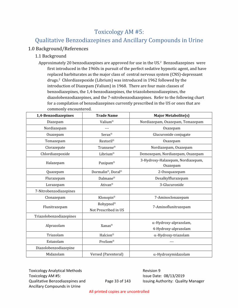
Approximately 20 benzodiazepines are approved for use in the US.2 Benzodiazepines were first introduced in the 1960s in pursuit of the perfect sedative hypnotic agent, and have replaced barbiturates as the major class of central nervous system (CNS)-depressant drugs.2 Chlordiazepoxide (Librium) was introduced in 1962 followed by the introduction of Diazepam (Valium) in 1968. There are four main classes of benzodiazepines, the 1,4-benzodiazepines, the triazolobenzodiazepines, the diazolobenzodiazepines, and the 7-nitrobenzodiazepines. Refer to the following chart for a compilation of benzodiazepines currently prescribed in the US or ones that are commonly encountered.
1,4-Benzodiazepines Trade Name Major Metabolite(s)Diazepam Valium Nordiazepam, Oxazepam, Temazepam
Nordiazepam --- OxazepamOxazepam Serax Glucuronide conjugate
Temazepam Restoril OxazepamClorazepate Tranxene Nordiazepam, Oxazepam
Chlordiazepoxide Librium Demoxepam, Nordiazepam, Oxazepam
Halazepam Paxipam 3-Hydroxy-Halazepam, Nordiazepam, Oxazepam
Quazepam Dormalin, Doral 2-OxoquazepamFlurazepam Dalmane DesalkylflurazepamLorazepam Ativan 3-Glucuronide
7-NitrobenzodiazepinesClonazepam Klonopin 7-Aminoclonazepam
FlunitrazepamRohypnol
Not Prescribed in US7-Aminoflunitrazepam
Triazolobenzodiazepines
Alprazolam Xanax-Hydroxy-alprazolam,4-Hydroxy-alprazolam
Triazolam Halcion -Hydroxy-triazolamEstazolam ProSom ---
DiazolobenzodiazepineMidazolam Versed (Parenteral) -Hydroxymidazolam
Toxicology Analytical Methods Revision 9Issue Date: 08/13/2019Toxicology AM #5:
Qualitative Benzodiazepines and Ancillary Compounds in Urine
Page 34 of 143 Issuing Authority: Quality Manager
All printed copies are uncontrolled
Benzodiazepines are used primarily as anti-epileptics in the treatment of seizure disorders, as anxiolytics for the short-term relief of anxiety disorders, as sedative-hypnotics for the treatment of sleep disorders, and as muscle relaxants to relieve spasticity. The primary side effects that accompany their use include dose-related extensions of the intended actions, including sedation and sleepiness/drowsiness. In addition, other undesired effects that will influence the outcome of field sobriety tests include ataxia, a blocked ability to coordinate movements, a staggering walk and poor balance, lethargy/apathy, indifference or sluggishness, mental confusion, disorientation, slurred speech and amnesia. Impairment of motor abilities, especially a person's ability to drive an automobile, is common. This impairment is compounded by the drug-induced suppression of ones' ability to assess their own level of physical and mental impairment. Alcohol and other CNS depressants (e.g., barbiturates, antidepressants, etc.) will increase CNS depressant effects, such as sedation and impairment of psychomotor function, in an additive manner.4-6
The benzodiazepines are lipid soluble and are absorbed well from the GI tract with good distribution to the brain. They are metabolized primarily in the liver. Their CNS active metabolites extend their duration of action. The benzodiazepines work by enhancing, facilitating or potentiating the action of the inhibitory neurotransmitter GABA. They serve to increase the frequency of GABA-mediated chloride ion channel opening.
Benzodiazepines are metabolized primarily in the liver via several different microsomal enzyme systems.6 Many products of their metabolism are active. Since many of the active metabolites have been marketed as therapeutic agents, it is difficult to ascertain which drug was ingested solely upon the basis of the results of analysis. Current drug therapy will assist in determining the source of a particular compound. The detection of a particular agent is determined partly by whether its metabolism yields active metabolites. Excretion of benzodiazepines is predominantly in the urine. Depending upon the particular benzodiazepine, the urine may contain parent compounds, N-dealkylation and oxidative (hydroxylation) metabolism products, and/or glucuronide conjugates.
1.1.1 At the analyst’s discretion, the samples may be extracted with or without derivatizing, and there are two options in the method for derivatization.
1.2 References1.2.1 Valentine, J.L., Middleton, R., Sparks, C. Identification of Urinary Benzodiazepines and
their Metabolites: Comparison of Automated HPLC and GC-MS after Immunoassay Screening of Clinical Specimens. J. Anal. Tox. 20:416-424, 1996.
1.2.2 Levine, B. Central Nervous System Depressants. pp. 191-197. In: Principles of Forensic Toxicology. Levine, B. ed., AACC, 1999.
1.2.3 Huang, W. and Moody, D.E. Immunoassay Detection of Benzodiazepines and Benzodiazepine Metabolites in Blood. J. Anal. Tox. 19:333-342, 1995.
1.2.4 Drug Facts and Comparisons Prescription Drug Information Binder, Updated monthly.
Toxicology Analytical Methods Revision 9Issue Date: 08/13/2019Toxicology AM #5:
Qualitative Benzodiazepines and Ancillary Compounds in Urine
Page 35 of 143 Issuing Authority: Quality Manager
All printed copies are uncontrolled
1.2.5 Julien, R.M. A Primer of Drug Action. pp. 95-107, W.H. Freeman and Company: NewYork, 1998.
1.2.6 Hobbs, W.R., Rall, T.W. and Verdoorn, T.A. Hypnotics and Sedatives. pp. 362-373. In: Goodman & Gilman’s The Pharmacological Basis of Therapeutics, 9th edition, Hardman, J.G. ed., McGraw-Hill, 1996.
2.0 Scope2.1 This extraction method is a modification of the method developed by Valentine, et
al., for the extraction of benzodiazepines from urine.1 This method has also been found to be effective in the extraction of opiates and various other drugs such as Zolpidem, Buprenorphine and Trazodone.
3.0 Equipment/Reagents3.1 Equipment and Supplies
3.1.1 Tube Rocker3.1.2 Laboratory oven or water bath 3.1.3 Laboratory Centrifuge capable of 3500 rpm3.1.4 Fixed and adjustable volume single channel air displacement pipettes, and appropriate
tips, capable of accurate and precise dispensing of volumes indicated.3.1.5 Dry bath 3.1.6 Evaporative Concentrator equipped with nitrogen tank.3.1.7 Threaded-end 16X100 Round Bottom Glass Tubes and/or 16X114 Tapered Bottom
Glass Centrifuge Tubes3.1.8 Screw caps for 16mm O.D. Tubes3.1.9 pH Indicator Strips3.1.10 ALS Vials3.1.11 ALS Vial Microinserts3.1.12 Gas Chromatograph equipped with a mass selective detector and a nonpolar capillary
column with a phase composition capable of efficiently separating amines, alkaloids, drug compounds and other analytes encountered in toxicological specimens (e.g. 100%-dimethylpolysiloxane or 95%-dimethyl-polysiloxane with 5% diphenyl).
3.2 Reagents Refer to Toxicology AM#23 for solution preparation instructions.3.2.1 BG100 b-Glucuronidase Solution (Kura Biotec)3.1.2 2M Acetate buffer, pH 4.83.1.3 50mM Sodium Bicarbonate, pH 113.1.4 Chloroform/Isopropanol 9:1 (Each Certified ACS Grade)3.1.5 Ethyl Acetate (Certified ACS Grade)3.1.6 Silylating agent: MSFTA or BSTFA with 1% TMCS
Toxicology Analytical Methods Revision 9Issue Date: 08/13/2019Toxicology AM #5:
Qualitative Benzodiazepines and Ancillary Compounds in Urine
Page 36 of 143 Issuing Authority: Quality Manager
All printed copies are uncontrolled
3.3 Qualitative Reference Material and Controls3.3.1 Positive Control
3.3.1.1 Positive Control can be prepared with single or multi-component working solutions and/or obtained commercially.
3.3.2 Non-extracted Reference Material3.3.2.1 Run necessary reference material as indicated by examination of GC/MSD data.
Reference material mixes may be employed.3.3.3 Non-extracted Derivatized Reference Material
3.3.3.1 Prepare derivatized reference material as necessary based on current drug therapy and examination of GC/MSD data.
3.3.3.2 Add ~3-5µL (1mg/mL) stock reference material to labeled centrifuge tube. Derivatize as described in 4.1.2.2
3.3.4 Internal Standard3.3.4.1 Stock Solutions
1 mg/mL Prazepam3.3.4.2 Working Internal Standard Solution [10ng/μL]
3.3.4.2.1 Add 100µL Prazepam stock solution to 10mL volumetric ball flask. QS with methanol.
NOTE: Working Solution is stable for 1 year (or at the date of the earliest expiring Stock solution) when stored under refrigeration.
3.3.5 Required Extracted Controls (inclusive of all options for method)3.3.5.1 Extracted Negative Control
NOTE: Commercially obtained or in-house urine verified to be negative for drugs of interest.
3.3.5.2 Positive Control Positive Control can be prepared with single or multi-component working solutions
and/or obtained commercially. The positive control must have at least one compound in it that is appropriate for demonstrating that each chosen extraction and derivatization (if applicable) is working. The preferred concentration range of this control is 300–600ng/mL. Examples of preparation and dilutions for an in-house control are given at the end of this method in Section 6.0: Comments.
Toxicology Analytical Methods Revision 9Issue Date: 08/13/2019Toxicology AM #5:
Qualitative Benzodiazepines and Ancillary Compounds in Urine
Page 37 of 143 Issuing Authority: Quality Manager
All printed copies are uncontrolled
3.3.5.3 Positive and Negative Glucuronide Controls. 3.3.5.3.1 These controls may be obtained commercially or prepared in-house by
spiking negative urine. The same negative urine must be used to prepare both the positive and negative glucuronide controls. Oxazepam glucuronide, lorazepam glucuronide or morphine glucuronide may be used for these controls and must be at a minimum concentration of 375ng/mL. Examples of preparation are given in Appendix III. Derivatization will be required for the controls prepared in-house, even if there are no case samples requiring derivatization. The positive and negative glucuronide controls are used to demonstrate the glucuronidase was effective (if these samples are run in conjunction with samples that are not derivatized or are derivatized using option 2, one set of glucuronide controls can be used for both.)
4.0 Procedure4.1 Extraction Procedure
NOTE: This method provides three options for the analyst. The method describes the preparation of an ethyl acetate extract and two options for a derivatized extract. Based on compounds of interest, both extracts need not be prepared and only the corresponding control material must be included.
4.1.1 Non-Derivatized Ethyl Acetate Samples 4.1.1.1 Casework and Control samples
4.1.1.1.1 Transfer 6mL casework samples and controls to screw top extraction tubes.
4.1.1.2 Internal Standard Addition4.1.1.2.1 To each prepared sample, add 300µL of internal standard (10ng/µL
working solution) or 3µL of 1mg/mL stock solution. Vortex to mix.4.1.1.3 Sample Hydrolysis
4.1.1.3.1 Add 200µL 2M acetate buffer to each tube, vortex.4.1.1.3.2 To all but the glucuronidase negative, add 150µL BG100 b-Glucuronidase
Solution. Cap and vortex gently to mix.4.1.1.3.3 Place all tubes in 60C laboratory oven or water bath for one hour. 4.1.1.3.4 Allow samples to cool before proceeding with solvent extraction.
4.1.1.4 Extraction4.1.1.4.1 Add 2mL 50mM sodium bicarbonate to each sample tube. Vortex. 4.1.1.4.2 Check pH. If necessary, adjust pH to approximately pH 9 with 1N NaOH or
KOH.4.1.1.4.3 Add 4mL of chloroform/isopropanol {9:1}. 4.1.1.4.4 Rock for approximately 15 minutes.
Toxicology Analytical Methods Revision 9Issue Date: 08/13/2019Toxicology AM #5:
Qualitative Benzodiazepines and Ancillary Compounds in Urine
Page 38 of 143 Issuing Authority: Quality Manager
All printed copies are uncontrolled
4.1.1.4.5 Centrifuge (~3300-3500 for about 10-15 minutes)4.1.1.4.6 Transfer lower organic phase from tube into labeled tapered bottom tube.4.1.1.4.7 Evaporate solvent to dryness under a gentle stream of nitrogen at ~37C.
Proceed to 4.1.1.5 if not derivatizing, or to 4.1.2 if derivatizing before running on GC/MS.
4.1.1.5 Reconstitution with Ethyl Acetate (No Derivatization)4.1.1.5.1 Add 50µL ethyl acetate. Vortex.4.1.1.5.2 Transfer extract to labeled ALS vial with micro-insert.
4.1.1.6 Preparation for Analysis Run4.1.1.6.1 Into Sequence log table, enter the sample case numbers, blanks and
controls.4.1.1.6.2 Load samples, reference material, blanks and controls into the quadrant
rack(s) as noted in the sequence table.4.1.2 Derivatization of Samples: Option 1
4.1.2.1 Follow Ethyl acetate sample preparation steps included in 4.1.1.2 - 4.1.1.44.1.2.2 Derivatization
4.1.2.2.1 To the tapered-bottom tubes add 20µL ethyl acetate and 30µL of silylating agent.
4.1.2.2.2 Cap tubes. Vortex.4.1.2.2.3 Heat tube for 15 minutes in 75C drybath. 4.1.2.2.4 Remove from heat and allow samples to cool. Transfer derivative to
labeled ALS vial with micro-insert. 4.1.2.3 Preparation for Analysis Run
4.1.2.3.1 Into Sequence log table, enter the sample case numbers, blanks and controls.
4.1.2.3.2 Load samples, reference material, blanks and controls into the quadrant rack(s) as noted in the sequence table.
4.1.3 Derivatization of Samples: Option 2 (Complete ethyl acetate sample Extraction Procedure (4.1.1.2-4.1.1.5), run samples on
GC/MS, then complete derivatization).4.1.3.1 Derivatization
4.1.3.1.1 Once the ethyl acetate extracts have run on the GC/MS, add 20µL of silylating agent to remaining extract in the autosampler insert and vortex.
4.1.3.1.2 Heat vials for 15 minutes at 75C. 4.1.3.1.3 Remove from heat and allow to cool.
4.1.3.2 Preparation for Analysis Run4.1.3.2.1 Into Sequence log table, enter the sample case numbers, blanks and
controls.
Toxicology Analytical Methods Revision 9Issue Date: 08/13/2019Toxicology AM #5:
Qualitative Benzodiazepines and Ancillary Compounds in Urine
Page 39 of 143 Issuing Authority: Quality Manager
All printed copies are uncontrolled
4.1.3.2.2 Load samples, reference material, blanks and controls into the quadrant rack(s) as noted in the sequence table.
4.1.3.3 GC-MSD Analysis Parameters 4.1.3.3.1 Refer to instrument method for current analysis parameters.
4.1.3.3.1.1 Instrument Run Parameters Inlet at 280 degrees Celsius, splitless injection, injection volume: 1μL Oven at 80 degrees Celsius, hold 2.5 minutes Ramp 25 degrees Celsius Final temperature: 300 degrees Celsius, hold at least 7.5 minutes.
4.1.3.3.2 Current analysis method must be stored centrally as a hard or electronic copy.
4.1.4 Detection and Identification Criteria4.1.4.1 The presence of a drug compound is indicated if the retention time for the sample,
versus applicable reference material, does not differ by more than ±0.2 minutes and there are no significant differences in the mass spectral data.
4.2 Methods Limitations and Application to Other Analytes4.2.1 This method is applicable to other compounds, which require an enzymatic hydrolysis
to liberate the compound of interest. Both the ethyl acetate extraction and the TMS derivative can be applied toward the identification of these compounds.
4.2.2 This method has proven useful in the identification of opiate class compounds such as codeine, morphine, 6-monoacetylmorphine, hydrocodone, and buprenorphine.
4.2.3 Care should be taken when estazolam is detected, particularly in samples containing alprazolam and/or alpha-hydroxyalprazolam. For samples containing alprazolam and/or alpha-hydroxyalprazolam, estazolam must be detected in both underivatized and derivatized GC/MSD data to be considered reportable. Estazolam shall not be reported if alprazolam and/or alpha-hydroxyalprazolam are detectable in the sample and derivatized estazolam is not detected.
4.3 Quality Assurance Requirements4.3.1 Refer to Toxicology AM #19 for additional quality assurance and Toxicology AM #21 for
reference material authentication requirements.4.4 Analysis Documentation
4.4.1 The printed results for each case sample and accompanying blank will be included with the analysts’ notes. Case results are to be recorded in the LIMS system.
4.4.2 Original data for controls will be prepared for each analysis run and stored centrally in the laboratory where the analysis was performed, until archiving or destruction.
4.4.3 A copy of controls need not be included in individual case files. When necessary, a copy of control printouts can be prepared from the centrally stored document.
Toxicology Analytical Methods Revision 9Issue Date: 08/13/2019Toxicology AM #5:
Qualitative Benzodiazepines and Ancillary Compounds in Urine
Page 40 of 143 Issuing Authority: Quality Manager
All printed copies are uncontrolled
5.0 Work Instructions5.1 Reference Material
5.1.1 Benzodiazepine Positive Control 5.1.1.1 Stock Solutions
o 1 mg/mL single component or 250μg/mL multi-component benzodiazepine class reference solutions.
5.1.1.2 Direct spikingo Spike negative urine with 3μL of each single component stock or 12μL of
250μg/mL stock solution. 5.1.1.3 Working Control Solution (10ng/μL)
o Add 100L each 1mg/mL or 400L 250μg/mL Stock Solution to 9mL MeOH in 10mL ball flask. Bring up to volume with MeOH. Solution is stable for one-year when stored under refrigeration. (Note: A minimum of two benzodiazepine class compounds must be included in the control solution. One of these compounds must form a TMS derivative.)
5.1.2 Conjugated Controls5.1.2.1 Commercially obtained conjugated control (example: Bio-Rad C3)
5.1.2.2 Stock Solution5.1.2.2.1 1 mg/mL Oxazepam Glucuronide or 100μg/mL Lorazepam Glucuronide or
100μg/mL Morphine Glucuronide5.1.2.2.1 Direct spiking
5.1.2.2.1.1 Spike negative urine with 3μL of 1mg/mL stock solution or 30μL of 100μg/mL stock solution.
5.1.2.2.2 Working Glucuronide Solution (10ng/μL)5.1.2.2.2.1 Add 100L of 1mg/mL Stock Solution (or 1mL 100μg/mL Stock Solution)
to 10mL MeOH. Solution is stable for one year when stored under refrigeration.
5.1.3 Internal Standard5.1.3.1 Stock Solution
5.1.3.1.1 1mg/mL Prazepam. 5.1.3.1.1 Direct addition
5.1.3.1.1.1 Add 3μL of 1mg/mL stock solution to each 6mL urine sample and control.
5.1.3.1.2 Working Internal Standard Solution5.1.3.1.2.1 Add 100L Stock Solution to 9mL MeOH in 10mL ball flask. Bring
up to volume with MeOH. Solution is stable for one year when stored under refrigeration.
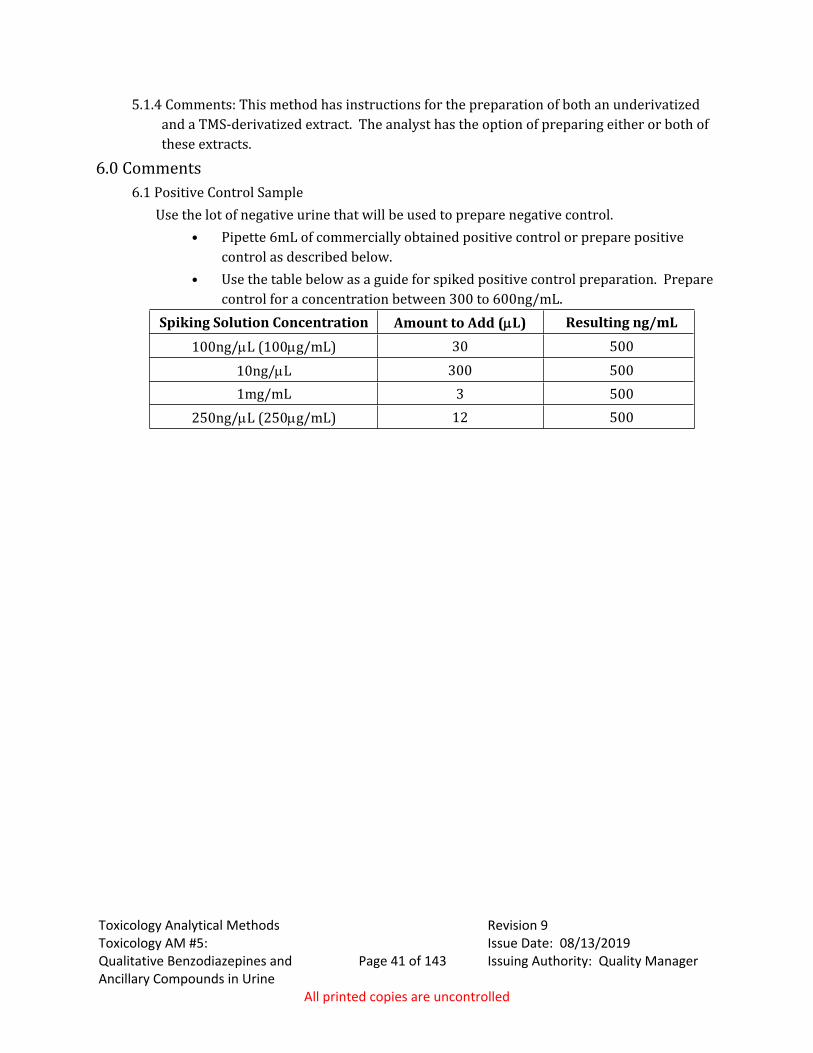
Toxicology Analytical Methods Revision 9Issue Date: 08/13/2019Toxicology AM #5:
Qualitative Benzodiazepines and Ancillary Compounds in Urine
Page 41 of 143 Issuing Authority: Quality Manager
All printed copies are uncontrolled
5.1.4 Comments: This method has instructions for the preparation of both an underivatized and a TMS-derivatized extract. The analyst has the option of preparing either or both of these extracts.
6.0 Comments6.1 Positive Control Sample
Use the lot of negative urine that will be used to prepare negative control.• Pipette 6mL of commercially obtained positive control or prepare positive
control as described below. • Use the table below as a guide for spiked positive control preparation. Prepare
control for a concentration between 300 to 600ng/mL.Spiking Solution Concentration Amount to Add (L) Resulting ng/mL
100ng/L (100g/mL) 30 50010ng/L 300 5001mg/mL 3 500
250ng/L (250g/mL) 12 500
Toxicology Analytical Methods Revision 9Issue Date: 08/13/2019Toxicology AM #6:
Screening for Gamma-Hydroxybutyrate (GHB) in Urine
Page 42 of 143 Issuing Authority: Quality Manager
All printed copies are uncontrolled
Toxicology AM #6: Screening for Gamma-Hydroxybutyrate (GHB) in Urine
1.0 Background/References1.1 Background:
GHB occurs naturally in minute quantities as a result of the metabolism of the inhibitory neurotransmitter, GABA.6,8 The drug GHB is a potent tranquilizer that was previously used as an anesthetic and as a treatment for major depressive illnesses, alcohol withdrawal, and narcolepsy.1-8 Legitimate use is limited due to the side effects associated with the drug. GHB can produce visual disturbances, nausea, vomiting, drowsiness, dizziness, severe respiratory depression, unconsciousness and involuntary muscle spasms.1,5,6 Overdoses can require emergency medical treatment including intensive care due to the respiratory depression, bradycardia and coma.5,6,8
The use of GHB has been increasing since the 1980s and currently is popular among body builders, teenagers and on the club/dance scene. Body builders use the drug due to its alleged role as a growth hormone releasing agent to enhance muscle growth.5,6,8 GHB has not been proven to possess any anabolic effects.6 GHB has gained notoriety as a date rape drug due to its ability to produce short-term amnesia and decreased inhibitions.1-8
The FDA banned the use of GHB in 1990 except for FDA approved physician supervised protocols.6,8
A dose of GHB is anywhere from 2.5 to 4.0 grams in approximately 1 teaspoon ("capful") of liquid. The taste of GHB has been described as salty or soapy, the odor is said to be mothball-like.5,6 Due to the short half-life of GHB (0.3 to 1.0 hours5, 27 5 minutes7) the person will re-administer every 45 minutes to 1 hour. The onset of effects is 15 to 60 minutes. The effects of the drug will be detectable during a DRE exam for 4 to 6 hours. GHB is classified as a central nervous system depressant. The observed effects include horizontal and vertical nystagmus, lack of convergence, body tremors, and slowed breathing. The person will also exhibit a lowered pulse, blood pressure, and body temperature. In addition, the muscle tone will exhibit flaccidity and the person may be in a trance-like state, the pupils will exhibit a lack of reaction to light.5 Lower doses will promote an agitated, combative state however their pulse and other vitals will be depressed.5,6 Combining GHB with alcohol plus a stimulant or marihuana allows the user to remain conscious during use. This allows them to experience the euphoric “buzz” that is the desired effect of its abuse. The desired effect is a state of relaxation and tranquility, a pleasant drowsiness, mild euphoria, hallucinations and a release of inhibitions. Combining GHB with alcohol or other central nervous system depressant will provide an additive depressant effect.5 GHB is detectable in blood for up to eight hours and in urine for up to 12 hours.5,6,7 Peak plasma levels are obtained in 20 to 45 minutes.7 Peak urine GHB concentrations on the order of 1100 µg/mL are observed within the first four hours after a 100 mg/kg oral dose.3,7
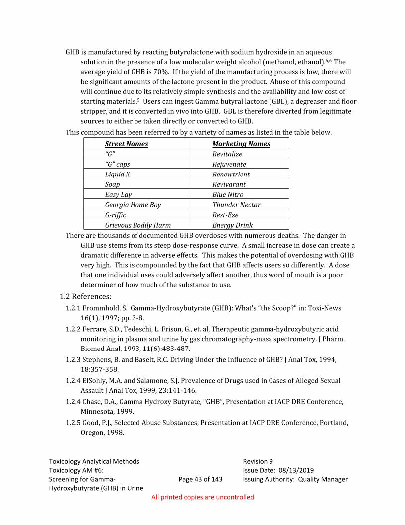
Toxicology Analytical Methods Revision 9Issue Date: 08/13/2019Toxicology AM #6:
Screening for Gamma-Hydroxybutyrate (GHB) in Urine
Page 43 of 143 Issuing Authority: Quality Manager
All printed copies are uncontrolled
GHB is manufactured by reacting butyrolactone with sodium hydroxide in an aqueous solution in the presence of a low molecular weight alcohol (methanol, ethanol).5,6 The average yield of GHB is 70%. If the yield of the manufacturing process is low, there will be significant amounts of the lactone present in the product. Abuse of this compound will continue due to its relatively simple synthesis and the availability and low cost of starting materials.5 Users can ingest Gamma butyral lactone (GBL), a degreaser and floor stripper, and it is converted in vivo into GHB. GBL is therefore diverted from legitimate sources to either be taken directly or converted to GHB.
This compound has been referred to by a variety of names as listed in the table below.Street Names Marketing Names“G” Revitalize“G” caps RejuvenateLiquid X RenewtrientSoap RevivarantEasy Lay Blue NitroGeorgia Home Boy Thunder NectarG-riffic Rest-EzeGrievous Bodily Harm Energy Drink
There are thousands of documented GHB overdoses with numerous deaths. The danger in GHB use stems from its steep dose-response curve. A small increase in dose can create a dramatic difference in adverse effects. This makes the potential of overdosing with GHB very high. This is compounded by the fact that GHB affects users so differently. A dose that one individual uses could adversely affect another, thus word of mouth is a poor determiner of how much of the substance to use.
1.2 References:1.2.1 Frommhold, S. Gamma-Hydroxybutyrate (GHB): What’s “the Scoop?” in: Toxi-News
16(1), 1997; pp. 3-8.1.2.2 Ferrare, S.D., Tedeschi, L. Frison, G., et. al, Therapeutic gamma-hydroxybutyric acid
monitoring in plasma and urine by gas chromatography-mass spectrometry. J Pharm. Biomed Anal, 1993, 11(6):483-487.
1.2.3 Stephens, B. and Baselt, R.C. Driving Under the Influence of GHB? J Anal Tox, 1994, 18:357-358.
1.2.4 ElSohly, M.A. and Salamone, S.J. Prevalence of Drugs used in Cases of Alleged Sexual Assault J Anal Tox, 1999, 23:141-146.
1.2.4 Chase, D.A., Gamma Hydroxy Butyrate, “GHB”, Presentation at IACP DRE Conference, Minnesota, 1999.
1.2.5 Good, P.J., Selected Abuse Substances, Presentation at IACP DRE Conference, Portland, Oregon, 1998.
Toxicology Analytical Methods Revision 9Issue Date: 08/13/2019Toxicology AM #6:
Screening for Gamma-Hydroxybutyrate (GHB) in Urine
Page 44 of 143 Issuing Authority: Quality Manager
All printed copies are uncontrolled
1.2.6 Determination of Gamma-Hydroxybutyric Acid by GC/MS, Dade County Medical Examiner’s Toxicology Lab SOP.
1.2.7 Microgram, Volume XXXI, No. 3, March 1998.1.2.8 Couper, F.J. and Logan, B.K. Determination of gamma-Hydroxybutyrate (GHB) in
Biological Specimens by Gas Chromatograph-Mass Spectrometry, J Anal Tox, 2000, 24:1-7.
1.2.9 SOFT/AAFS Forensic Toxicology Laboratory Guidelines, 1997.1.2.10 Goldberger, B.A., Huestis, M.A., Wilkins, D.G. Commonly Practiced Quality Control and
Quality Assurance Procedures for Gas Chromatography/Mass Spectrometry Analysis in Forensic Urine Drug-Testing Laboratories, Forensic Sci Rev, 1997, 9(2):59-79.
2.0 Scope2.1 This method provides an efficient qualitative analysis option for the liquid-liquid
extraction of urine samples suspected of containing Gamma-Hydroxybutyrate/ gamma-Hydroxybutyric Acid (GHB). GHB is isolated from an acidified solution into methylene chloride and heptane with zinc chloride to facilitate the extraction process. The extraction is achieved with a De-Tox B extraction tube. The extraction is followed by the creation of a di-TMS derivative of GHB. The derivative is analyzed by full scan GC/MS in EI mode. This method may not provide adequate sensitivity for weaker concentrations of GHB. This method should only be used for driving under the influence of drugs (DUID) situations where GHB is suspected or for screening purposes for drug-facilitated sexual assault (DFSA). Presently DFSA case urine that indicates a positive result by this method must be outsourced for quantitative confirmation.
3.0 Equipment/Reagents3.1 Equipment
3.1.1 Tube Rocker (Fisher Scientific or equivalent)3.1.2 Evaporative Concentrator equipped with a nitrogen tank3.1.3 Laboratory Centrifuge capable of 3000 rpm3.1.4 Drybath3.1.5 Fixed and/or adjustable volume pipettes, and appropriate tips, capable of accurate and
precise dispensing of volumes indicated.3.1.6 16X100mm centrifuge tubes 3.1.7 {Optional} 16X144mm tapered-end centrifuge tubes3.1.8 Caps for 16mm O.D. Tubes 3.1.9 Automated Liquid Sampler (ALS) vials 3.1.10 ALS vial microinserts
Toxicology Analytical Methods Revision 9Issue Date: 08/13/2019Toxicology AM #6:
Screening for Gamma-Hydroxybutyrate (GHB) in Urine
Page 45 of 143 Issuing Authority: Quality Manager
All printed copies are uncontrolled
3.1.11 Gas Chromatograph equipped with a mass selective detector and a nonpolar capillary column with a phase composition capable of efficiently separating GHB and its analogs in toxicological specimens (e.g. 100%-dimethylpolysiloxane or 95%-dimethyl-polysiloxane with 5% diphenyl)
3.2 Reagents 3.2.1 De-Tox B Tubes (or equivalent Toxi-B tube) 3.2.2 MSFTA or BSTFA with 1% TMCS
3.3 Reference Material3.3.1 Stock Solution
1mg/mL (1000ng/μL) GHB 3.3.2 GHB Spiked Urine Positive Controls (One required)
Use the same lot of negative urine to prepare both the negative and spiked positive controls.
3.3.2.1 Preparation of 100ug/mL Positive Control3.3.2.1.1 Add 450µL of GHB 1mg/mL stock to 4050µL negative urine. Vortex.
3.3.2.2 Preparation of 200ug/mL Positive Control3.3.2.2.1 Add 900µL of GHB 1mg/mL stock to 3600µL negative urine. Vortex.
3.3.3 Non-Extracted GHB Reference Material [10ug]3.3.3.1 Place 10µL of GHB stock into tube.
3.3.4 Negative Control 3.3.4.1 Negative Urine can be commercially obtained or in-house urine verified to be
negative for drugs of interest.
4.0 Procedure4.1 Extraction Procedure
4.1.1 Initial set-up4.1.1.1 Label De-Tox B Tubes for positive controls, negative control and case samples. 4.1.1.2 Label tubes and ALS vials for positive controls, negative control, case samples and
non-extracted reference material.4.1.2 Extraction Procedure
4.1.2.1 Extract 4.5mL of specimen, negative and spiked positive control in De-Tox B Tube (acidic extraction @pH=4.5). Less than 4.5mL may be used if sample is limited.
4.1.2.2 Rock De-Tox tube for ~10 minutes.4.1.2.3 Centrifuge De-Tox Tube at ~2500-3000 rpm for ~10 minutes.4.1.2.4 Transfer solvent from De-Tox Tube into tapered-end centrifuge tube.4.1.2.5 Evaporate solvent to approximately 50µL with nitrogen at ~40C. Non-extracted
reference material must be evaporated to dryness.4.1.3 Derivatization Procedure
Toxicology Analytical Methods Revision 9Issue Date: 08/13/2019Toxicology AM #6:
Screening for Gamma-Hydroxybutyrate (GHB) in Urine
Page 46 of 143 Issuing Authority: Quality Manager
All printed copies are uncontrolled
4.1.3.1 Add 40µL MSFTA or BSTFA with 1%TMCS to evaporated extracted samples, spiked control(s) and non-extracted reference material. Cap tube. Vortex.
4.1.3.2 Place tube in 60C drybath for 15 minutes.4.1.3.3 Remove tube from drybath. Allow sample to cool. 4.1.3.4 Transfer derivative to labeled GC/MS ALS vial for analysis.
4.1.4 Gas Chromatography/Mass Spectrometry (GC/MS) Parameters4.1.4.1 Key parameters are specified below. Parameters not specified are at the discretion
of the analyst and should be optimized for both the gas chromatographic and mass spectral characteristics of an instrument. Refer to GC/MS method for current parameters for analysis. Each laboratory shall maintain a centrally stored current method printout or electronic copy.
4.1.4.2 Instrument Run Parameters Inlet at 250 degrees Celsius, splitless injection, injection volume: 1μL Oven at 65 degrees Celsius, hold 1 minute Ramp 15 degrees Celsius Final temperature: 300 degrees Celsius, hold 2 minutes.
4.1.4.3 Acquisition Mode4.1.4.3.1 Sample must be analyzed in full scan acquisition mode.
4.1.5 Detection and Identification Criteria4.1.5.1 Chromatographic Criteria
The retention time of the analyte should fall within ±2% of the retention time exhibited by GHB reference material and control(s).
4.1.5.2 Mass Spectral Criteria Full scan mass spectral data should be compared against within-run GHB
reference material and control(s). No significant differences should be apparent.
4.2 Quality Assurance4.2.1 General
4.2.1.1 Refer to Toxicology AM #19 for additional quality assurance and Toxicology AM #21 for reference material authentication requirements.
4.2.2 Per Analysis Run Control and Reference Material Requirement Each run should include, at a minimum, a 100ug/mL and/or 200ug/mL GHB
control, a negative control and a non-extracted GHB reference material.4.3 Analysis Documentation
4.3.1 The printed results for each case sample and accompanying blank will be included with the analysts’ notes. Case results are to be recorded in the LIMS system.
Toxicology Analytical Methods Revision 9Issue Date: 08/13/2019Toxicology AM #6:
Screening for Gamma-Hydroxybutyrate (GHB) in Urine
Page 47 of 143 Issuing Authority: Quality Manager
All printed copies are uncontrolled
4.3.2 Original data for controls will be compiled for each analysis run and stored centrally in the laboratory where the analysis was performed until archiving.
4.3.3 A copy of controls need not be included in individual case files. When necessary, a copy of control printouts can be prepared from the centrally stored document.
Toxicology Analytical Methods Revision 9Issue Date: 08/13/2019Toxicology AM #7:
Enzyme-Linked Immunosorbent Assay (ELISA) Screening for Drugs of Abuse
Page 48 of 143 Issuing Authority: Quality Manager
All printed copies are uncontrolled
Toxicology AM #7: Enzyme-Linked Immunosorbent Assay (ELISA) Screening for Drugs
of Abuse1.0 Background/References
1.1 BackgroundELISA is an acronym for enzyme-linked immunosorbent assay. An ELISA is an enzyme
immunoassay (EIA) in which one reactant is immobilized on a solid phase and the signal generator is an enzyme. The enzyme delivers a signal to indicate to what extent a particular antigen-antibody reaction has occurred. This reaction takes place inside of a polystyrene microtiter plate well. Horseradish peroxidase is an enzyme commonly employed as a signal generator. The small size of HRP, the ease with which small conjugates can be produced through oxidation of its carbohydrate moieties to reactive aldehyde, its rapid kinetics, and reasonable price, make it popular in ELISA.
For the qualitative determination of a specific drug, or class of drugs in blood and urine this method utilized competitive micro-plate immunoassay. Each of the assays requires a predilution step for samples, controls and calibrators. This brings the analytes into an acceptable range for optimum performance of the bound microplate antibodies. Dilutions are either performed manually, with an air displacement pipette or a dilutor, or using the automated dilution capability of the instrument. Samples, calibrators or controls are added to individual wells of the microplate along with the conjugate, which is the drug or hapten labeled with the enzyme horseradish peroxidase (HRP).
There is a competition between the free drug in the matrix sample (blood or urine) and drug bound to enzyme (conjugate) for antibody (sheep or rabbit) fixed on the well. The wells are washed with DI water, the substrate (3,3´,5,5´-tetramethylbenzidine (TMB) with peroxide (H2O2) is added, and a color is produced. HRP catalyzes H2O2 oxidation of the substrate by transferring one electron from the TMB to the peroxide to yield a blue colored product. The reaction is stopped when 2.0N sulfuric acid is added to the well. This acidic environment provides the necessary conditions for the loss of an additional electron to produce the final yellow color. The acidic environment also serves to inactivate the enzymatic activity of the HRP. The resulting absorbance at 450nm is inversely proportional to the amount of drug present in the sample or standard. Consequently, a more intense yellow color results in a greater absorbance and indicates a lower concentration of drug in the sample.
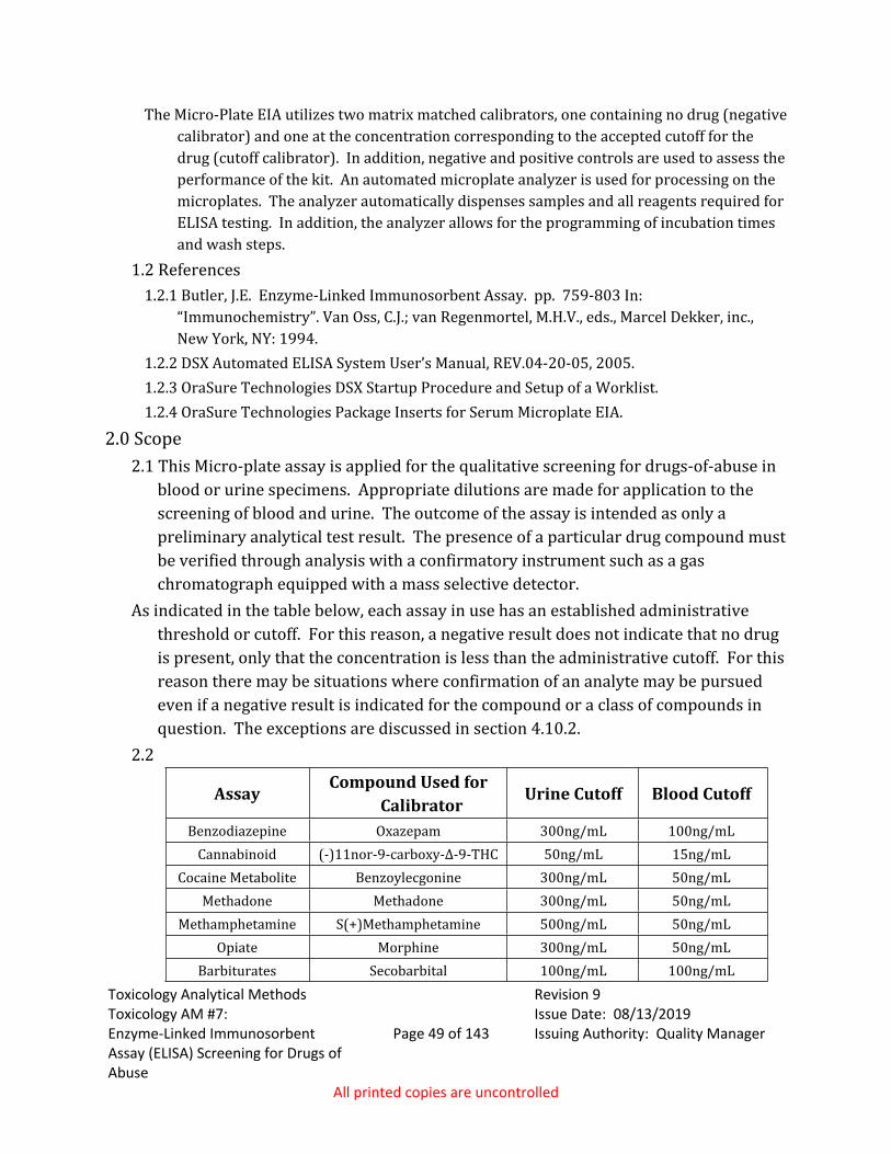
Toxicology Analytical Methods Revision 9Issue Date: 08/13/2019Toxicology AM #7:
Enzyme-Linked Immunosorbent Assay (ELISA) Screening for Drugs of Abuse
Page 49 of 143 Issuing Authority: Quality Manager
All printed copies are uncontrolled
The Micro-Plate EIA utilizes two matrix matched calibrators, one containing no drug (negative calibrator) and one at the concentration corresponding to the accepted cutoff for the drug (cutoff calibrator). In addition, negative and positive controls are used to assess the performance of the kit. An automated microplate analyzer is used for processing on the microplates. The analyzer automatically dispenses samples and all reagents required for ELISA testing. In addition, the analyzer allows for the programming of incubation times and wash steps.
1.2 References1.2.1 Butler, J.E. Enzyme-Linked Immunosorbent Assay. pp. 759-803 In:
“Immunochemistry”. Van Oss, C.J.; van Regenmortel, M.H.V., eds., Marcel Dekker, inc., New York, NY: 1994.
1.2.2 DSX Automated ELISA System User’s Manual, REV.04-20-05, 2005.1.2.3 OraSure Technologies DSX Startup Procedure and Setup of a Worklist.1.2.4 OraSure Technologies Package Inserts for Serum Microplate EIA.
2.0 Scope2.1 This Micro-plate assay is applied for the qualitative screening for drugs-of-abuse in
blood or urine specimens. Appropriate dilutions are made for application to the screening of blood and urine. The outcome of the assay is intended as only a preliminary analytical test result. The presence of a particular drug compound must be verified through analysis with a confirmatory instrument such as a gas chromatograph equipped with a mass selective detector.
As indicated in the table below, each assay in use has an established administrative threshold or cutoff. For this reason, a negative result does not indicate that no drug is present, only that the concentration is less than the administrative cutoff. For this reason there may be situations where confirmation of an analyte may be pursued even if a negative result is indicated for the compound or a class of compounds in question. The exceptions are discussed in section 4.10.2.
2.2
AssayCompound Used for
CalibratorUrine Cutoff Blood Cutoff
Benzodiazepine Oxazepam 300ng/mL 100ng/mLCannabinoid (-)11nor-9-carboxy-Δ-9-THC 50ng/mL 15ng/mL
Cocaine Metabolite Benzoylecgonine 300ng/mL 50ng/mLMethadone Methadone 300ng/mL 50ng/mL
Methamphetamine S(+)Methamphetamine 500ng/mL 50ng/mLOpiate Morphine 300ng/mL 50ng/mL
Barbiturates Secobarbital 100ng/mL 100ng/mL
Toxicology Analytical Methods Revision 9Issue Date: 08/13/2019Toxicology AM #7:
Enzyme-Linked Immunosorbent Assay (ELISA) Screening for Drugs of Abuse
Page 50 of 143 Issuing Authority: Quality Manager
All printed copies are uncontrolled
3.0 Equipment/Reagents3.1 Equipment
3.1.1 Sample Diluting Supplies3.1.1.1 Air-displacement pipettes and appropriate tips.3.1.1.2 Repeater Pipette and appropriate tips.3.1.1.3 Automatic Dilutor equipped with appropriate syringes.3.1.1.4 Screening instrument programmed for automatic dilutions.
3.1.2 Plasticware3.1.2.1 2.0mL control vials with caps3.1.2.2 25mL reagent tubes with caps 3.1.2.3 Disposable 13x75 polypropylene tubes3.1.2.4 Disposable transfer pipettes3.1.2.5 Deep-well strips/plates for automated dilution
3.1.3 DSX Automated ELISA Instrument3.1.4 Tube Rocker 3.1.5 Vortex Mixer
3.2 Reagents3.2.1 Forensic Specimen Diluent (Remember to check expiration date prior to use.)
3.2.1.1 Assay Kits:o Micro-plates coated with anti-drug antibodies.o Enzyme conjugate for specific drug/drug class.o TMB Substrate reagent (universal).o 2N H2SO4 Stopping reagent (universal).
3.2.2 Processing of New Assay Supplies3.2.2.1 When a new kit is opened, note the expiration date of all components listed on
each assay's specification sheet. 3.2.2.1.1 The manufacturer’s kit expiration date may be based on a component not
used for the analysis of blood or urine. Only the expiration of the conjugate and plate involve the use of the assay kit since the expiration date of the substrate and stop always far exceeds the expiration date of the conjugate and the plates.
3.2.2.2 Cocaine Assay Conjugate Preparation3.2.2.2.1 Using a pipette, to the vial containing Benzoylecgonine Lypophilized Stock
Enzyme Conjugate, add 2mL Conjugate Diluent. 3.2.2.2.2 Place vial on tube rocker for a minimum of 10 minutes.
Toxicology Analytical Methods Revision 9Issue Date: 08/13/2019Toxicology AM #7:
Enzyme-Linked Immunosorbent Assay (ELISA) Screening for Drugs of Abuse
Page 51 of 143 Issuing Authority: Quality Manager
All printed copies are uncontrolled
3.2.2.2.3 Using a pipette, add the volume of reconstituted Stock Enzyme Conjugate listed on the kit package insert to appropriate volume of Conjugate Diluent. Prepare only necessary volume of conjugate. (Note: The Stock Enzyme Conjugate is lot specific.)
3.2.2.2.4 Gently mix Conjugate Diluent bottle on tube rocker for a minimum of 1 minute.
3.2.2.2.5 Prior to use, allow bottle to equilibrate for a minimum of 30 minutes at room temperature or overnight under refrigeration.
3.3 Reference MaterialNOTE: For both urine and blood, the following calibrators and controls must be included in each
analysis run. In-house calibrators and controls are stable for approximately 1-2 months. If a screen fails, preparation of fresh calibrators/controls is a good troubleshooting step.
3.3.1 Urine 3.3.1.1 Stock Reference Material Solutions
3.3.1.1.1 Obtain Methamphetamine (1mg/mL), Benzoylecgonine (1mg/mL), Methadone (1mg/mL), Morphine (1mg/mL), 11-nor-9-Carboxy-D9-THC (100µg/mL), Oxazepam (1mg/mL), and Secobarbital (1mg/mL) drug reference material from an appropriate vendor. Different vendors should be used to make up the Calibrator and Control Working Solutions whenever possible. If this is not possible, different lots from the same vendor may be used. Certificates of analysis must be stored centrally.
3.3.1.2 Working Standard Solution3.3.1.2.1 Add ~9mL methanol to 10mL volumetric flask. Add 50µL each of
methamphetamine and c-THC stock. Add 30µL each of benzoylecgonine, methadone, morphine and oxazepam stock. Add 10μL of secobarbital stock. QS to 10mL with methanol. Record lot numbers of stock reference material on reagent log.
(Solution is stable for up to 12 months when stored at 4C.)3.3.1.3 Urine Calibrators:
NOTE: May be commercially obtained or prepared in-house as described in Section 6 of this method.
• Negative Urine Calibrator • Cutoff Urine Calibrator
3.3.1.4 Platform Urine ControlsNOTE: May be commercially obtained or prepared in-house as described in Section 6 of
this method. • Negative Urine Control• Positive Urine Control
Toxicology Analytical Methods Revision 9Issue Date: 08/13/2019Toxicology AM #7:
Enzyme-Linked Immunosorbent Assay (ELISA) Screening for Drugs of Abuse
Page 52 of 143 Issuing Authority: Quality Manager
All printed copies are uncontrolled
3.3.1.5 Sample Rack Urine Controls3.3.1.5.1 Negative Urine Control
Negative Urine3.3.1.5.2 Positive Urine Controls
3.3.1.5.2.1 25% Above Cutoff Positive Control Each run must include a control at 25% above the cutoff calibrator.
To prepare, add 125µL working standard solution to 1mL of negative urine.
3.3.1.5.2.2 Drugs-of-Abuse Positive Control Each run must include a commercially obtained drugs-of-abuse urine
control. The concentration of analytes may be varied.3.3.1.5.3 Urine Cannabinoid Cutoff Control
3.3.1.5.3.1 The last sample run on a urine cannabinoid screen will be an aliquot of the cutoff calibrator. This sample will be used for evaluating results.
3.3.2 Blood3.3.2.1 Stock Reference Material Solutions
3.3.2.1.1 Obtain Methamphetamine (1mg/mL), Benzoylecgonine (1mg/mL), Methadone (1mg/mL), Morphine (1mg/mL), 11-nor-9-Carboxy-D9-THC (100µg/mL), Oxazepam (1mg/mL), and Secobarbital (1mg/mL) drug reference material from an appropriate vendor. Different vendors should be used to make up the Calibrator and Control Working Solutions whenever possible. If this is not possible, different lots from the same vendor may be used. Certificates of analysis must be stored centrally.
3.3.2.2 Working Standard Solution3.3.2.2.1 Add ~9mL methanol to 10mL volumetric flask. Add 50µL each of stock
methamphetamine, benzoylecgonine, methadone and morphine. Add 100µL each of stock oxazepam and secobarbital. Add 150µL of stock c-THC. QS to 10mL with methanol. Record lot numbers of stock reference material on reagent log.
(Solution is stable for up to 12 months when stored at 4C.)3.3.2.3 Blood Calibrators
NOTE: May be commercially obtained or prepared in-house as described in Section 5 of this Method.
• Negative Blood Calibrator • Cutoff Blood Calibrator
3.3.2.4 Platform Blood Controls
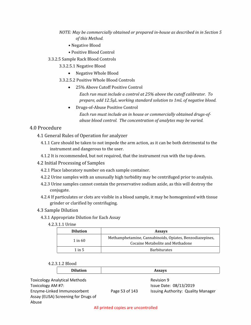
Toxicology Analytical Methods Revision 9Issue Date: 08/13/2019Toxicology AM #7:
Enzyme-Linked Immunosorbent Assay (ELISA) Screening for Drugs of Abuse
Page 53 of 143 Issuing Authority: Quality Manager
All printed copies are uncontrolled
NOTE: May be commercially obtained or prepared in-house as described in in Section 5 of this Method.
• Negative Blood • Positive Blood Control
3.3.2.5 Sample Rack Blood Controls3.3.2.5.1 Negative Blood
Negative Whole Blood3.3.2.5.2 Positive Whole Blood Controls
25% Above Cutoff Positive ControlEach run must include a control at 25% above the cutoff calibrator. To prepare, add 12.5µL working standard solution to 1mL of negative blood.
Drugs-of-Abuse Positive ControlEach run must include an in house or commercially obtained drugs-of-abuse blood control. The concentration of analytes may be varied.
4.0 Procedure4.1 General Rules of Operation for analyzer
4.1.1 Care should be taken to not impede the arm action, as it can be both detrimental to the instrument and dangerous to the user.
4.1.2 It is recommended, but not required, that the instrument run with the top down. 4.2 Initial Processing of Samples
4.2.1 Place laboratory number on each sample container. 4.2.2 Urine samples with an unusually high turbidity may be centrifuged prior to analysis.4.2.3 Urine samples cannot contain the preservative sodium azide, as this will destroy the
conjugate.4.2.4 If particulates or clots are visible in a blood sample, it may be homogenized with tissue
grinder or clarified by centrifuging.4.3 Sample Dilution
4.3.1 Appropriate Dilution for Each Assay4.2.3.1.1 Urine
Dilution Assays
1 in 60Methamphetamine, Cannabinoids, Opiates, Benzodiazepines,
Cocaine Metabolite and Methadone1 in 5 Barbiturates
4.2.3.1.2 BloodDilution Assays
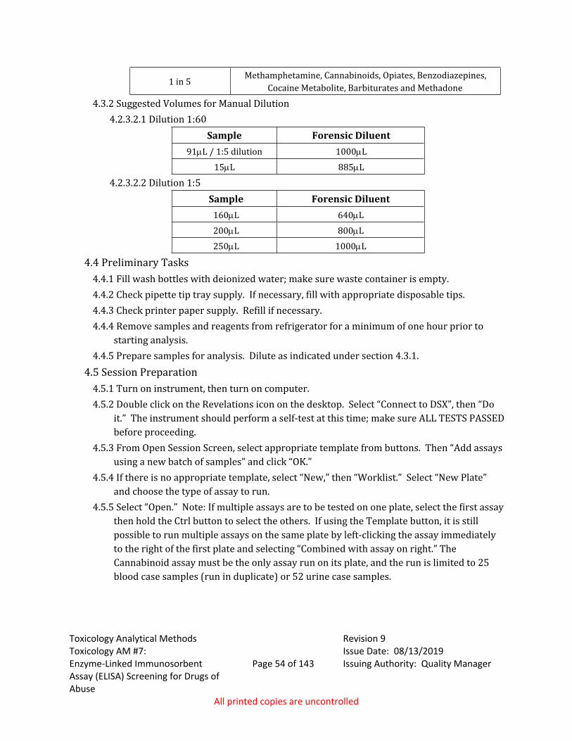
Toxicology Analytical Methods Revision 9Issue Date: 08/13/2019Toxicology AM #7:
Enzyme-Linked Immunosorbent Assay (ELISA) Screening for Drugs of Abuse
Page 54 of 143 Issuing Authority: Quality Manager
All printed copies are uncontrolled
1 in 5Methamphetamine, Cannabinoids, Opiates, Benzodiazepines,
Cocaine Metabolite, Barbiturates and Methadone
4.3.2 Suggested Volumes for Manual Dilution4.2.3.2.1 Dilution 1:60
Sample Forensic Diluent91L / 1:5 dilution 1000L
15L 885L
4.2.3.2.2 Dilution 1:5 Sample Forensic Diluent
160L 640L200L 800L250L 1000L
4.4 Preliminary Tasks 4.4.1 Fill wash bottles with deionized water; make sure waste container is empty.4.4.2 Check pipette tip tray supply. If necessary, fill with appropriate disposable tips. 4.4.3 Check printer paper supply. Refill if necessary.4.4.4 Remove samples and reagents from refrigerator for a minimum of one hour prior to
starting analysis.4.4.5 Prepare samples for analysis. Dilute as indicated under section 4.3.1.
4.5 Session Preparation4.5.1 Turn on instrument, then turn on computer.4.5.2 Double click on the Revelations icon on the desktop. Select “Connect to DSX”, then “Do
it.” The instrument should perform a self-test at this time; make sure ALL TESTS PASSED before proceeding.
4.5.3 From Open Session Screen, select appropriate template from buttons. Then “Add assays using a new batch of samples” and click “OK.”
4.5.4 If there is no appropriate template, select “New,” then “Worklist.” Select “New Plate” and choose the type of assay to run.
4.5.5 Select “Open.” Note: If multiple assays are to be tested on one plate, select the first assay then hold the Ctrl button to select the others. If using the Template button, it is still possible to run multiple assays on the same plate by left-clicking the assay immediately to the right of the first plate and selecting “Combined with assay on right.” The Cannabinoid assay must be the only assay run on its plate, and the run is limited to 25 blood case samples (run in duplicate) or 52 urine case samples.
Toxicology Analytical Methods Revision 9Issue Date: 08/13/2019Toxicology AM #7:
Enzyme-Linked Immunosorbent Assay (ELISA) Screening for Drugs of Abuse
Page 55 of 143 Issuing Authority: Quality Manager
All printed copies are uncontrolled
4.5.6 Click and drag under the “Test” column to select the assays for the samples. Enter ID numbers for the samples, then click “OK.” NOTE: Blood samples are to be run in duplicate. The assays are programmed to do this automatically, depending on the matrix selected.
4.5.7 Select the “PLAY” button (green triangle). The timeline will now be built and displayed. NOTE: To view the timeline, click the down (↓) arrow button; to hide the timeline, click the up (↑) arrow button. To view the entire timeline, click the minus (marked as a dash) button.
4.5.8 When ready to load the reagents and consumables, click the “FAST FORWARD” button (two green triangles followed by a green vertical line).
4.5.9 Follow directions on the screen to load all reagents and consumables. Note: when asked to enter “Lot Name,” enter the name of the assay (e.g. METHAMPHETAMINE, or METH), followed by the kit lot number and kit expiration date. Click green check mark when done.
4.5.10 Load all samples into sample caddy, as instructed on the screen, then click the green check mark.
4.5.11 Once the plate tray is ejected, remove the plate holder, insert assay plate (with the correct number of strips in place) into the plate holder, and place the plate holder back into the tray, as instructed on the screen. Change “Plate Identifier” to the assay being run for that plate, followed by the date (e.g. COC011212). Click green check mark to proceed to next plate. Repeat for each plate being loaded, making sure to change the plate identifier for each.
4.5.12 Load all calibrator and control fluids as instructed on the screen. Make sure to load at least the minimum volumes required for each, as the software instructs. Click green check mark.
4.5.13 Make sure the washer bottle(s) contain sufficient DI water. Click green check mark.4.5.14 Make sure sufficient sample and reagent tips are loaded. Click green check mark.4.5.15 Make sure there is sufficient space for waste in tip disposal and waste disposal
containers. Click “OK.” Note: once you click “OK” the instrument cycle will start, so make sure all objects are out of the way of the arm.
4.5.16 Once all runs are complete, click the “STOP” button (red square). This will cause the plate tray to be ejected. Once the plate tray is ejected, click on the “EJECT” button (a square with an arrow pointing down), to fully eject the tray. Once the tray is fully ejected, the plate holders will be unlocked and easily removed.
4.5.17 Remove the plate holders, dispose of the strips, then return the plate holders to the tray. Push the “IN” button (a square with an arrow pointing up) to make the tray return to its “IN” position.
Toxicology Analytical Methods Revision 9Issue Date: 08/13/2019Toxicology AM #7:
Enzyme-Linked Immunosorbent Assay (ELISA) Screening for Drugs of Abuse
Page 56 of 143 Issuing Authority: Quality Manager
All printed copies are uncontrolled
4.6 Obtaining Results - Post-Run4.6.1 To display the results, click the blue “UP” arrow. Select the pop-up window displaying
assay results.4.7 Obtaining Archived Results
4.7.1 Results from past runs can be viewed by clicking “Open,” then clicking on the “Plates” folder. From here, you can select the appropriate document.
4.7.2 Text files of all runs are also stored. To access the text files, click “Open,” then click on the “Text” folder. From here, you can select the appropriate document. These documents can be then be opened in Excel.
4.8 Post-run Tasks4.8.1 General Clean-up
4.8.1.1 Return conjugates, stop, and diluent reservoirs to refrigerator.4.8.1.2 Dispose of used calibrators, controls, samples, and tips into appropriate biohazard
waste container.4.8.2 Instrument Shut-down
4.8.2.1 Close out of Revelations, and then shut down computer.4.8.2.2 Power off the instrument.
4.9 Run Acceptance Criteria4.9.1 Calibrators and Controls
4.9.1.1 The individual replicates for the absorbance of the negative calibrator must be less than 1.2 times the mean negative calibrator and greater than 0.8 times the mean negative calibrator.
4.9.1.2 The individual replicates of the cutoff calibrator must be less than 1.2 times the mean cutoff calibrator and greater than 0.8 times the mean cutoff calibrator.
4.9.1.3 The mean absorbance for the negative urine or blood is greater than the absorbance for the negative calibrator.
4.9.1.4 The absorbance for the negative calibrator is greater than the mean absorbance for the cutoff calibrator.
4.9.1.5 The mean absorbance for the cutoff calibrator is greater than the absorbance for the positive control.
4.9.1.6 The quality control equations (((KIT1-KIT2)/KIT1)*100)>20 and (((NEG-CUT)/NEG)*100)>30 are default settings allowing tracking of the OraSure immunoassay kits’ performance through calculation of percent displacement. These criteria are not critical; they are used for tracking the performance of the specific assay. The default settings may be adjusted as appropriate.
4.9.2 Urine and Blood Sample Controls
Toxicology Analytical Methods Revision 9Issue Date: 08/13/2019Toxicology AM #7:
Enzyme-Linked Immunosorbent Assay (ELISA) Screening for Drugs of Abuse
Page 57 of 143 Issuing Authority: Quality Manager
All printed copies are uncontrolled
4.9.2.1 Matrix matched urine and blood controls, analyzed as samples, should indicate an appropriate positive or negative response.
4.9.2.2 For purposes of this criterion, a significantly depressed absorbance qualifies as a positive result.
4.10 Interpretation of Results4.10.1 Positive Result
4.10.1.1 A positive result for a sample is indicated by an absorbance less than or equal to the Cutoff Calibrator. Urine samples that have an absorbance less than or equal to the cut off control run at the end of the assay will be considered positive. Blood samples are run in duplicate; if the coefficient of variation is over 10% for a sample, the analyst will use the value with the lowest absorbance for evaluating the sample result.
4.10.2 Depressed Absorbance4.10.2.1 At the discretion of the analyst, confirmatory techniques may be applied to
samples that exhibit depressed absorbance. For purposes of this exception, a depressed absorbance is a response which fall between the values observed for the cutoff calibrator and the negative urine or blood. If data for confirmatory techniques supports the presence of an analyte, it may be reported as present. In addition, samples with compounds that have low cross reactivity may be confirmed and reported with a negative screen result.
4.10.2.2 Blood samples are run in duplicate; if the coefficient of variation is greater than 10% for a sample, the analyst will use the value with the lowest absorbance for evaluating the sample result.
4.10.3 Negative ResultA negative result for a sample is indicated by an absorbance that is greater than the Cut-
off Calibrator. Special considerations may apply as outlined above (see 4.10.2). 4.11 Distribution of Assay Information
4.11.1 Assay results are to be recorded in the LIMS system. 4.11.2 The original data printouts will be stored centrally in the laboratory where the analysis
was performed. 4.11.3 A copy of assay results may be stored electronically in a central location and need not
be included in individual case files. When necessary, a copy of the control and standard printouts may be prepared from the centrally stored document.
4.12 DSX Maintenance 4.12.1 When-in-use Daily Maintenance/Tasks
4.12.1.1 Run an optional “Wash” cycle before doing sample runs. It is also recommended that the “Wash” cycle be run after sample runs as well. If any wash ports are clogged, perform the periodic maintenance procedure for unclogging the ports.
Toxicology Analytical Methods Revision 9Issue Date: 08/13/2019Toxicology AM #7:
Enzyme-Linked Immunosorbent Assay (ELISA) Screening for Drugs of Abuse
Page 58 of 143 Issuing Authority: Quality Manager
All printed copies are uncontrolled
4.12.2 Periodic MaintenanceMaintenance to promote cleanliness of the instrument (including washing and air drying
the wash and waste tanks and cleaning the tip tray) can be done on an as-needed basis and does not need to be documented. Please note that the tip tray should be disinfected with 70% isopropanol, and not bleach, as bleach can interfere with the assays.
4.12.2.1 Six-Month Maintenance (Approximately every 6 months- does not need to be documented)
4.12.2.1.1 Replace the dispense tubing as needed.4.12.2.1.2 Replace the aspiration tubing as needed.
4.12.2.2 Preventive Observations (Does not need to be documented)4.12.2.2.1 During wash cycle, check the washer dispensing apparatus to ensure each
port is correctly dispensing water. This may be most effectively accomplished when aided by a flashlight.
4.12.2.2.2 After wash cycle, check plate to make sure most water is removed from wells and remaining water is uniform among the wells.
5.0 Comments 5.1 Appendix I:
5.1.1 Urine Control In-House Preparation Guide5.1.1.1 Negative Urine Calibrator (NEG)
Negative urine only5.1.1.2 Below Cutoff Urine Calibrator (1/2x)
Add 50L working standard solution to 1mL negative urine. 5.1.1.3 Cutoff Urine Calibrator (x)
Add 100L working standard solution to 1mL negative urine. 5.1.1.4 Positive Urine Calibrator (2x)
Add 200L working standard solution to 1mL negative urine.5.1.1.5 Negative Urine Control (NEG)
Negative urine only5.1.1.6 Positive Urine Control (POS)
Undiluted commercial control (Biorad C3, S3 or similar)5.1.1.7 Urine Cannabinoid Cutoff Control
Undiluted aliquot of calibrator run for cannabinoid assay only5.1.2 Blood Control In-House Preparation Guide
5.1.2.1 Negative Blood (NEG) Negative blood only
Toxicology Analytical Methods Revision 9Issue Date: 08/13/2019Toxicology AM #7:
Enzyme-Linked Immunosorbent Assay (ELISA) Screening for Drugs of Abuse
Page 59 of 143 Issuing Authority: Quality Manager
All printed copies are uncontrolled
5.1.2.2 Below Cutoff Blood Calibrator (1/2x) Add 5L working standard solution to 1mL negative blood.
5.1.2.3 Cutoff Blood Calibrator (x) Add 10L working standard solution to 1mL negative blood.
5.1.1.4 Positive Blood Calibrator (2x) Add 20L working standard solution to 1mL negative blood.
5.1.2.5 Negative Blood Control (NEG) Negative blood only
5.1.2.6 Above Cutoff Blood Control (1.25X) Add 12.5L working standard solution to 1mL negative blood.
5.1.2.7 Positive Blood Control (3X) Add 30L working standard solution to 1mL negative blood.
Toxicology Analytical Methods Revision 9Issue Date: 08/13/2019Toxicology AM #8: Basic and
Neutrals Drugs in Blood Page 60 of 143 Issuing Authority: Quality ManagerAll printed copies are uncontrolled
Toxicology AM #8: Basic and Neutrals Drugs in Blood1.0 Background/References
1.1 BackgroundThis method is a general blood extraction procedure for a variety of commonly encountered
neutral and basic drugs along with their metabolites. The method is based upon the principle of liquid/liquid extraction. Positive controls are spiked for a resulting concentration of 200ng/mL or 500ng/mL of drugs of interest. The sample pH is adjusted with a pH 9.2 borate buffer and extracted with n-butyl chloride. An optional back extraction procedure removes most frequently encountered interfering substances. Two internal standards are used to monitor extraction efficiency and chromatographic performance. Gas chromatography in conjunction with full scan mass spectrometry is used to confirm the presence of basic and neutral analytes of interest.
1.2 References1.2.1 Procedure for Basic Drug Analysis, Courtesy of Jim Hutchison, Montana Department of
Justice, Forensic Services Division, 2005.1.2.2 Procedure for Back Extraction, Courtesy of Jim Hutchison, Montana Department of
Justice, Forensic Services Division, 2006.1.2.3 Strong Bases Extractions - Screening SOP, Courtesy of Dr. Graham Jones, Office of the
Chief Medical Examiner, Edmonton, Canada, 2003.1.2.4 Jones, G., Postmortem Toxicology. pp. 98-102, in: Clarke's Analysis of Drugs and Poisons,
3rd Edition, Moffat, A.C, Osselton, M.D. and Widdop, B., eds., Pharmaceutical Press, 2004.1.2.5 Hearn, W.L. and Walls, H.C., Strategies for Postmortem Toxicology Investigation. pp. 937-
939. In: Drug Abuse Handbook, S.B. Karch, ed., CRC Press, Boca Raton, FL, 1998.
2.0 Scope2.1 This method is a general blood extraction procedure for a variety of commonly
encountered neutral and basic drugs, along with their metabolites. This method has also been shown capable of extracting some acidic drugs (e.g. butalbital) if sufficient quantity is present in the sample. This method does not efficiently extract some basic compounds, such as morphine and hydromorphone, due to pKa/pH considerations. The method allows for the analyst to use either methanol or ethyl acetate as a reconstitution solvent. Some benzodiazepines are more efficiently extracted using ethyl acetate than methanol as a reconstitution solvent. In addition, samples reconstituted in ethyl acetate can also be derivatized to increase sensitivity and detection of some compounds. Some drugs are more efficiently extracted using methanol as a reconstitution solvent. It is at the analyst’s discretion to determine which solvent to use.
Toxicology Analytical Methods Revision 9Issue Date: 08/13/2019Toxicology AM #8: Basic and
Neutrals Drugs in Blood Page 61 of 143 Issuing Authority: Quality ManagerAll printed copies are uncontrolled
3.0 Equipment/Reagents3.1 Equipment and Supplies
3.1.1 Tube Rocker 3.1.2 Vortex Mixer3.1.3 Evaporative concentrator equipped with nitrogen tank.3.1.4 Laboratory centrifuge capable of 3400rpm3.1.5 Fixed and adjustable single channel pipettes, and appropriate tips3.1.6 16X100mm round bottom glass screw-top tubes 3.1.7 Screw Cap for 16mm O.D. tubes 3.1.8 GC/MS Automated Liquid Sampler (ALS) vials 3.1.9 GC/MS Vial Microinsert 3.1.10 Gas Chromatograph equipped with a Mass Selective Detector 3.1.11 5%-Diphenyl-95%-Dimethyl-siloxane copolymer capillary GC column, 12.5 to 30M.
3.2 ReagentsRefer to Toxicology AM #23 for Solution Preparation instructions.3.2.1 Methanol (ACS Grade)3.2.2 n-Butyl chloride (ACS Grade)3.2.3 Borate Buffer (pH 9.2)3.2.4 Deionized/Distilled (DI) Water3.2.5 1% Hydrochloric Acid in Methanol3.2.6 100mM Hydrochloric Acid3.2.7 Ethyl Acetate (ACS Grade)3.2.8 Silylating Agent (select from):
BSTFA/1% TMCS MSTFA
3.3 Reference Materials3.3.1 Positive Control
NOTE: Positive Control can be prepared with the working solution described below and/or obtained commercially.
3.3.1.1 Positive Control Stock Solution3.3.1.1.1 Obtain 1mg/mL stock drug standard solutions through Cerilliant, Grace,
Sigma or other appropriate vendor. 3.3.1.2 Positive Control Working Solution
3.3.1.2.1 Add the designated volume of stock solution to 10mL methanol. A minimum of four compounds must be used.
NOTE: Solution is stable for 6-months when stored at room temperature or 12-months when stored under refrigeration. Re-make solution if deterioration is noted.
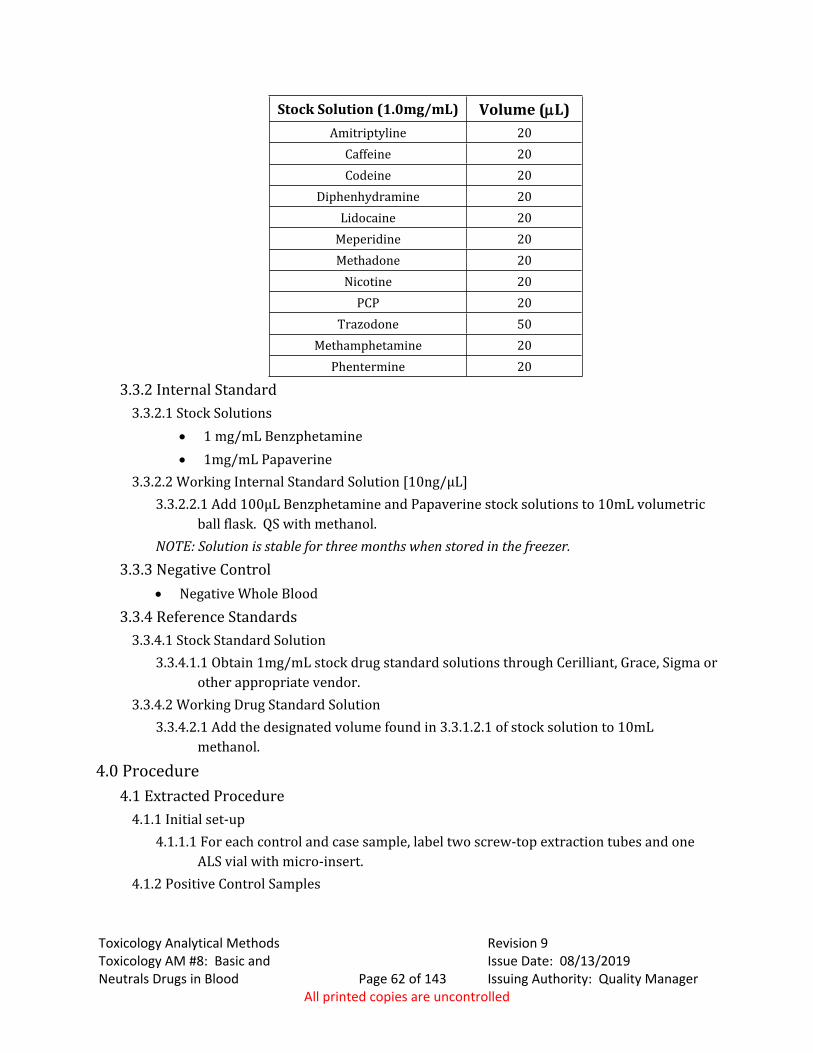
Toxicology Analytical Methods Revision 9Issue Date: 08/13/2019Toxicology AM #8: Basic and
Neutrals Drugs in Blood Page 62 of 143 Issuing Authority: Quality ManagerAll printed copies are uncontrolled
Stock Solution (1.0mg/mL) Volume (L)Amitriptyline 20
Caffeine 20Codeine 20
Diphenhydramine 20Lidocaine 20
Meperidine 20Methadone 20
Nicotine 20PCP 20
Trazodone 50Methamphetamine 20
Phentermine 20
3.3.2 Internal Standard3.3.2.1 Stock Solutions
1 mg/mL Benzphetamine 1mg/mL Papaverine
3.3.2.2 Working Internal Standard Solution [10ng/μL]3.3.2.2.1 Add 100µL Benzphetamine and Papaverine stock solutions to 10mL volumetric
ball flask. QS with methanol. NOTE: Solution is stable for three months when stored in the freezer.
3.3.3 Negative Control Negative Whole Blood
3.3.4 Reference Standards3.3.4.1 Stock Standard Solution
3.3.4.1.1 Obtain 1mg/mL stock drug standard solutions through Cerilliant, Grace, Sigma or other appropriate vendor.
3.3.4.2 Working Drug Standard Solution3.3.4.2.1 Add the designated volume found in 3.3.1.2.1 of stock solution to 10mL
methanol.
4.0 Procedure4.1 Extracted Procedure
4.1.1 Initial set-up4.1.1.1 For each control and case sample, label two screw-top extraction tubes and one
ALS vial with micro-insert.4.1.2 Positive Control Samples
Toxicology Analytical Methods Revision 9Issue Date: 08/13/2019Toxicology AM #8: Basic and
Neutrals Drugs in Blood Page 63 of 143 Issuing Authority: Quality ManagerAll printed copies are uncontrolled
NOTE: The same lot of negative blood must be used for the preparation of both negative and positive spiked controls.
4.1.2.1 Prepare control sample by adding 200µL mixed working control solution to 2mL negative whole blood or pipette a 2mL sample of commercially-obtained whole blood positive control.
4.1.2.2 When the optional back extraction is used, prepare an additional positive control to parallel the back extraction process.
4.1.2.3 When some samples in a batch are going to be reconstituted with methanol and others with ethyl acetate, a separate positive control must be run for each reconstitution solvent.
4.1.3 Casework Samples4.1.3.1 Transfer 2mL casework sample to screw top extraction tube.
4.1.4 Negative Control Sample4.1.4.1 Transfer 2mL negative whole blood to extraction tube. If some samples are going
to be run with ethyl acetate and some with methanol as a reconstitution solvent, a negative control must be prepared and run for each reconstitution solvent. If the optional back extraction is used, prepare an additional negative control to parallel the back extraction process.
4.1.5 Internal Standard4.1.5.1 Add 20µL of internal standard mixture and vortex. If benzodiazepines are of
interest, 3µL of Prazepam (1mg/mL) may also be spiked into the sample. NOTE: If the analyst has reason to suspect the sample may contain one or more of
these internal standard compounds, it is permissible for the analyst to eliminate one of the internal standard compounds (e.g. papaverine) or replace one of the internal standard compounds with an appropriate alternative (prazepam may be used). Clear notation of the replacement, along with justification, must be included in the analysis notes. If the analyst prefers to use a different internal standard than those listed here, s/he must confer with the toxicology discipline leader in the selection of said internal standard.
4.1.5.2 Allow sample to stand 10 minutes.4.1.6 Initial Extraction
4.1.6.1 Add 2mL borate buffer. Vortex.4.1.6.2 Pipet 4mL n-butyl chloride into each tube, cap.4.1.6.3 Place tube on rocker for 10 minutes.4.1.6.4 Centrifuge for 10 minutes at 3400 rpm.4.1.6.5 Transfer the n-butyl chloride layer (upper) to second screw-top tube. 4.1.6.6 Add 50µL 1% HCl in Methanol.4.1.6.7 Evaporate to dryness under N2 at ≤37°C.
Toxicology Analytical Methods Revision 9Issue Date: 08/13/2019Toxicology AM #8: Basic and
Neutrals Drugs in Blood Page 64 of 143 Issuing Authority: Quality ManagerAll printed copies are uncontrolled
4.1.6.7.1 If no clean-up proceed to 4.1.8.4.1.7 Optional Sample Clean-up (Back Extraction Procedure)
4.1.7.1 Reconstitute with 50µL 100mM HCl. 4.1.7.2 Add 1mL of n-Butyl Chloride. Vortex.4.1.7.3 Rock for 5 minutes. 4.1.7.4 Centrifuge for 5 minutes at 3400 rpm.4.1.7.5 Discard upper n-Butyl Chloride layer.4.1.7.6 Add 2mL of pH 9.2 borate solution. Vortex 4.1.7.7 Add 4mL of n-Butyl Chloride. 4.1.7.8 Rock for 5 minutes.4.1.7.9 Centrifuge for 5 minutes at 3400 rpm.4.1.7.10 Transfer upper n-Butyl Chloride layer into screw-top tube.4.1.7.11 Evaporate to just dryness under N2 at ≤45°C.
4.1.8 Reconstitution4.1.8.1 Add 50µL Methanol or Ethyl Acetate to the residue, vortex.4.1.8.2 Transfer extract to labeled ALS vial with micro-insert.
4.1.9 Preparation for Analysis Run4.1.9.1 Into Sequence log table, enter the case samples, blanks and controls.4.1.9.2 Load samples, standards, blank and controls into the quadrant rack as noted in the
sequence table.4.1.10 Optional Derivatization (samples that are extracted with ethyl acetate may be
derivatized at the analyst’s discretion)4.1.10.1 After ethyl acetate extraction samples have run on the GC-MSD, add 20µL
silylating agent to the sample. In addition to the case samples, the extracted positive and negative controls must also be derivatized and run.
4.1.10.2 Heat at about 75°C for ~15 minutes.4.1.10.3 Allow samples to cool; run on GC-MSD.
4.1.11 GC-MSD Acquisition Parameters 4.1.11.1 Refer to instrument method printout for current acquisition parameters.
4.1.11.1.1 Instrument Run Parameters (Short Run) Inlet at 250 degrees Celsius, splitless injection, injection volume: 1μL Oven at 70 degrees Celsius, hold 2 minutes Ramp 35 degrees Celsius to 290 degrees Celsius, hold 3 minutes Ramp 40 degrees Celsius to 300 degrees Celsius, hold at least 6 minutes Additional sample runs may be done with adjustments to these parameters,
for example if there is a coeluting peak or a late eluting compound.
Toxicology Analytical Methods Revision 9Issue Date: 08/13/2019Toxicology AM #8: Basic and
Neutrals Drugs in Blood Page 65 of 143 Issuing Authority: Quality ManagerAll printed copies are uncontrolled
4.1.11.1.2 Instrument Run Parameters (Longer Run) Inlet at 250 degrees Celsius, splitless injection, injection volume: 1μL Oven at 90 degrees Celsius, hold 4 minutes Ramp 15 degrees Celsius Final temperature: 290 degrees Celsius, hold at least 10.667 minutes Additional sample runs may be done with adjustments to these parameters,
for example if there is a co-eluting peak or a late eluting compound.4.1.11.2 Current acquisition method must be stored centrally as a hard or electronic copy.
4.1.12 GC-MSD Qualitative Detection and Identification Criteria4.1.12.1 For the confirmation/identification of compounds, analyze appropriate non-
extracted reference materials.4.1.12.2 The presence of a drug compound is indicated if the retention time for the sample
versus applicable reference material does not differ by more than ±0.2 minutes and there are no significant differences in the mass spectral data. NOTE: early eluting drugs, as well as drugs known to have similar retention times and mass spectral fragmentation patterns (e.g. phentermine and methamphetamine), may not differ from the retention time of the applicable reference material by more than ±0.1 minutes.
4.2 Quality Assurance Requirements4.2.1 General
4.2.1.1 Blood samples are to be stored under refrigeration after aliquots are removed for analysis.
4.2.1.2 Refer to Toxicology AM #17, AM #19 and AM #21 for quality assurance and reference material authentication requirements.
4.3 Analysis Documentation4.3.1 The printed results for each case sample and accompanying blank will be included with
the analysts’ notes. Case results are to be recorded in the LIMS system.4.3.2 A packet containing original data for controls will be prepared for each analysis run and
stored centrally in the laboratory where the analysis was performed, until archiving or destruction.
4.3.3 A copy of controls need not be included in individual case files. When necessary, a copy of control printouts can be prepared from the centrally stored document.
Toxicology Analytical Methods Revision 9Issue Date: 08/13/2019Toxicology AM #9:
Acidic and Neutral Drugs in Blood Page 66 of 143 Issuing Authority: Quality ManagerAll printed copies are uncontrolled
Toxicology AM #9: Acidic and Neutral Drugs in Blood
1.0 Background/References1.1 This method is a general liquid-liquid procedure to extract a variety of commonly
encountered acidic and neutral drugs along with their metabolites from blood. Drug compounds are extracted from blood by a liquid-liquid extraction process. Blood pH is adjusted with saturated ammonium chloride followed by extraction with ethyl acetate. After evaporation and a hexane wash, the final extract is subjected to analysis by GC-MSD. Two internal standards are used to monitor extraction efficiency and chromatographic performance. This extraction yields excellent recovery of most acidic and neutral drugs, and can generally be accomplished in under one hour. The extraction is designed to yield fewer and lower levels of endogenous compounds that can interfere with drug detection.
1.2 Reference1.2.1 Procedure for Acid/Neutral Drug Analysis, Courtesy of Jim Hutchison, Montana
Department of Justice, Forensic Services Division, 2005.1.2.2 Foerster, E.H., Dempsey, J., and Garriott, J.D., A Gas Chromatography Screening
Procedure for Acid and Neutral Drugs in Blood, J Anal Tox, 3:87-91, 1979. 1.2.3 Jones, G., Postmortem Toxicology. pp. 98-102, in: Clarke's Analysis of Drugs and Poisons,
3rd Edition, Moffat, A.C, Osselton, M.D. and Widdop, B., eds., Pharmaceutical Press, 2004.1.2.4 Hearn, W.L. and Walls, H.C. Strategies for Postmortem Toxicology Investigation. pp. 937-
939. In: “Drug Abuse Handbook” S.B. Karch, ed., CRC Press, Boca Raton, FL: 1998.
2.0 Scope2.1 This method is used to extract a variety of commonly encountered acidic and
neutral drugs, and their metabolites, from blood. Some basic drug compounds (e.g. diazepam), if in sufficient quantity in the sample, have been successfully extracted with this method.
3.0 Equipment/Reagents3.1 Equipment and Supplies
3.1.1 Tube rocker3.1.2 Evaporative concentrator equipped with nitrogen tank.3.1.3 Vortex mixer3.1.4 Laboratory centrifuge capable of 3400rpm.3.1.5 Fixed and adjustable volume single channel pipettes, and appropriate tips3.1.6 16X100mm round bottom glass screw-top tubes 3.1.7 Screw Cap for 16mm O.D. tubes 3.1.8 GC/MS Automated Liquid Sample (ALS) vials
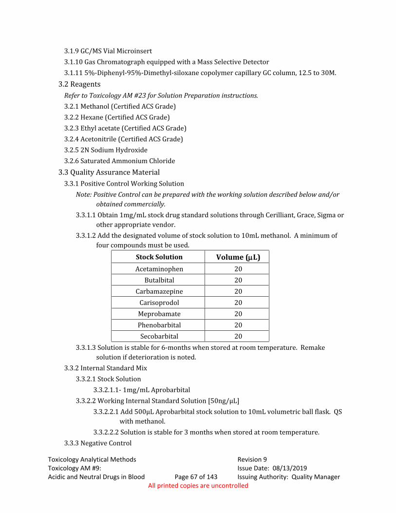
Toxicology Analytical Methods Revision 9Issue Date: 08/13/2019Toxicology AM #9:
Acidic and Neutral Drugs in Blood Page 67 of 143 Issuing Authority: Quality ManagerAll printed copies are uncontrolled
3.1.9 GC/MS Vial Microinsert 3.1.10 Gas Chromatograph equipped with a Mass Selective Detector 3.1.11 5%-Diphenyl-95%-Dimethyl-siloxane copolymer capillary GC column, 12.5 to 30M.
3.2 ReagentsRefer to Toxicology AM #23 for Solution Preparation instructions.3.2.1 Methanol (Certified ACS Grade)3.2.2 Hexane (Certified ACS Grade)3.2.3 Ethyl acetate (Certified ACS Grade)3.2.4 Acetonitrile (Certified ACS Grade)3.2.5 2N Sodium Hydroxide3.2.6 Saturated Ammonium Chloride
3.3 Quality Assurance Material3.3.1 Positive Control Working Solution
Note: Positive Control can be prepared with the working solution described below and/or obtained commercially.
3.3.1.1 Obtain 1mg/mL stock drug standard solutions through Cerilliant, Grace, Sigma or other appropriate vendor.
3.3.1.2 Add the designated volume of stock solution to 10mL methanol. A minimum of four compounds must be used.
Stock Solution Volume (L)Acetaminophen 20
Butalbital 20Carbamazepine 20
Carisoprodol 20Meprobamate 20Phenobarbital 20Secobarbital 20
3.3.1.3 Solution is stable for 6-months when stored at room temperature. Remake solution if deterioration is noted.
3.3.2 Internal Standard Mix3.3.2.1 Stock Solution
3.3.2.1.1- 1mg/mL Aprobarbital3.3.2.2 Working Internal Standard Solution [50ng/μL]
3.3.2.2.1 Add 500µL Aprobarbital stock solution to 10mL volumetric ball flask. QS with methanol.
3.3.2.2.2 Solution is stable for 3 months when stored at room temperature.3.3.3 Negative Control
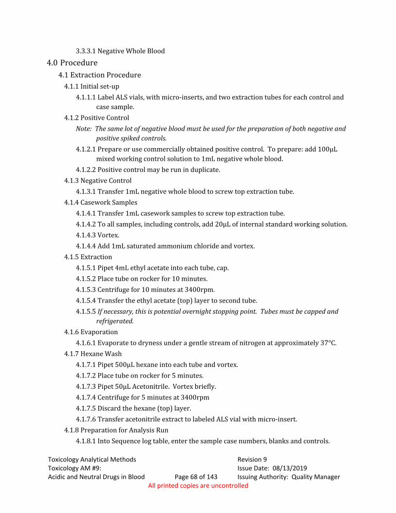
Toxicology Analytical Methods Revision 9Issue Date: 08/13/2019Toxicology AM #9:
Acidic and Neutral Drugs in Blood Page 68 of 143 Issuing Authority: Quality ManagerAll printed copies are uncontrolled
3.3.3.1 Negative Whole Blood
4.0 Procedure4.1 Extraction Procedure
4.1.1 Initial set-up4.1.1.1 Label ALS vials, with micro-inserts, and two extraction tubes for each control and
case sample.4.1.2 Positive Control
Note: The same lot of negative blood must be used for the preparation of both negative and positive spiked controls.
4.1.2.1 Prepare or use commercially obtained positive control. To prepare: add 100µL mixed working control solution to 1mL negative whole blood.
4.1.2.2 Positive control may be run in duplicate.4.1.3 Negative Control
4.1.3.1 Transfer 1mL negative whole blood to screw top extraction tube.4.1.4 Casework Samples
4.1.4.1 Transfer 1mL casework samples to screw top extraction tube.4.1.4.2 To all samples, including controls, add 20µL of internal standard working solution.4.1.4.3 Vortex.4.1.4.4 Add 1mL saturated ammonium chloride and vortex.
4.1.5 Extraction4.1.5.1 Pipet 4mL ethyl acetate into each tube, cap.4.1.5.2 Place tube on rocker for 10 minutes.4.1.5.3 Centrifuge for 10 minutes at 3400rpm.4.1.5.4 Transfer the ethyl acetate (top) layer to second tube. 4.1.5.5 If necessary, this is potential overnight stopping point. Tubes must be capped and
refrigerated.4.1.6 Evaporation
4.1.6.1 Evaporate to dryness under a gentle stream of nitrogen at approximately 37°C.4.1.7 Hexane Wash
4.1.7.1 Pipet 500µL hexane into each tube and vortex.4.1.7.2 Place tube on rocker for 5 minutes.4.1.7.3 Pipet 50µL Acetonitrile. Vortex briefly. 4.1.7.4 Centrifuge for 5 minutes at 3400rpm4.1.7.5 Discard the hexane (top) layer. 4.1.7.6 Transfer acetonitrile extract to labeled ALS vial with micro-insert.
4.1.8 Preparation for Analysis Run4.1.8.1 Into Sequence log table, enter the sample case numbers, blanks and controls.
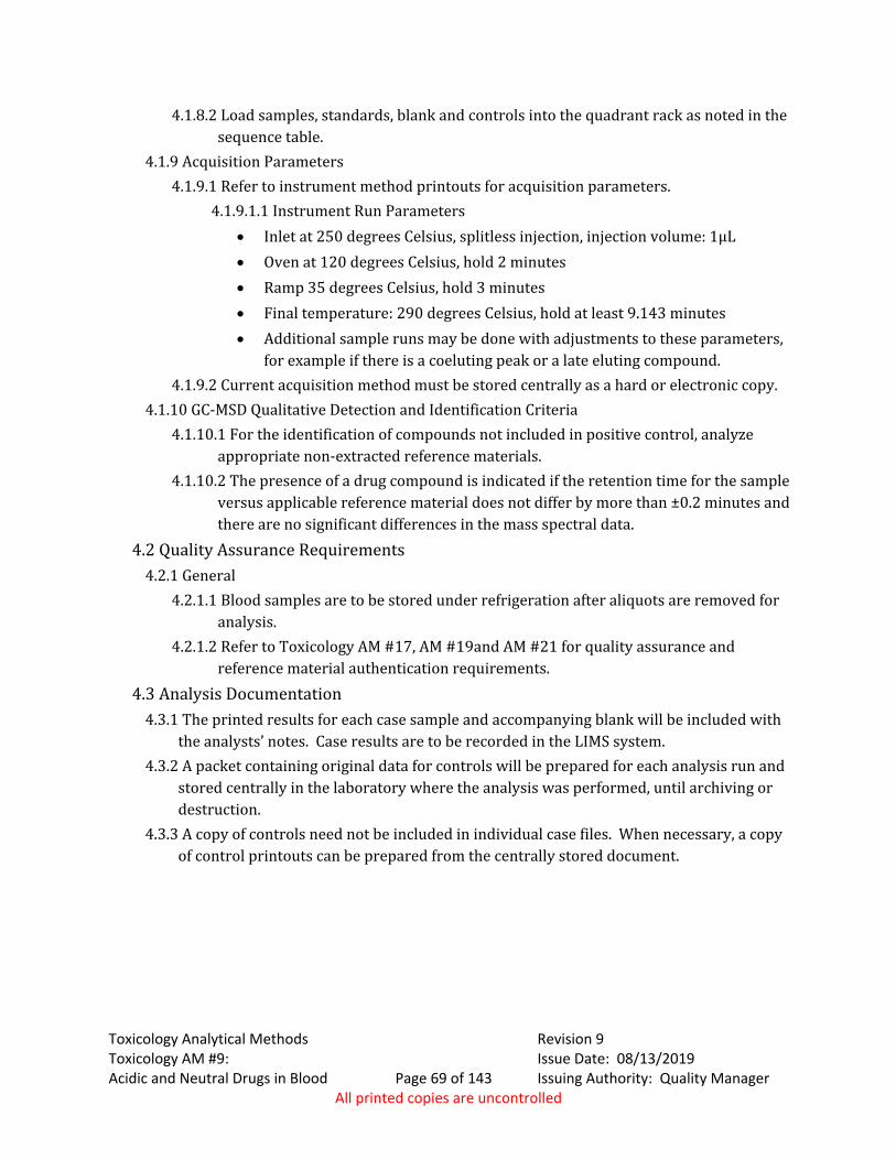
Toxicology Analytical Methods Revision 9Issue Date: 08/13/2019Toxicology AM #9:
Acidic and Neutral Drugs in Blood Page 69 of 143 Issuing Authority: Quality ManagerAll printed copies are uncontrolled
4.1.8.2 Load samples, standards, blank and controls into the quadrant rack as noted in the sequence table.
4.1.9 Acquisition Parameters 4.1.9.1 Refer to instrument method printouts for acquisition parameters.
4.1.9.1.1 Instrument Run Parameters Inlet at 250 degrees Celsius, splitless injection, injection volume: 1μL Oven at 120 degrees Celsius, hold 2 minutes Ramp 35 degrees Celsius, hold 3 minutes Final temperature: 290 degrees Celsius, hold at least 9.143 minutes Additional sample runs may be done with adjustments to these parameters,
for example if there is a coeluting peak or a late eluting compound.4.1.9.2 Current acquisition method must be stored centrally as a hard or electronic copy.
4.1.10 GC-MSD Qualitative Detection and Identification Criteria4.1.10.1 For the identification of compounds not included in positive control, analyze
appropriate non-extracted reference materials.4.1.10.2 The presence of a drug compound is indicated if the retention time for the sample
versus applicable reference material does not differ by more than ±0.2 minutes and there are no significant differences in the mass spectral data.
4.2 Quality Assurance Requirements4.2.1 General
4.2.1.1 Blood samples are to be stored under refrigeration after aliquots are removed for analysis.
4.2.1.2 Refer to Toxicology AM #17, AM #19and AM #21 for quality assurance and reference material authentication requirements.
4.3 Analysis Documentation4.3.1 The printed results for each case sample and accompanying blank will be included with
the analysts’ notes. Case results are to be recorded in the LIMS system.4.3.2 A packet containing original data for controls will be prepared for each analysis run and
stored centrally in the laboratory where the analysis was performed, until archiving or destruction.
4.3.3 A copy of controls need not be included in individual case files. When necessary, a copy of control printouts can be prepared from the centrally stored document.
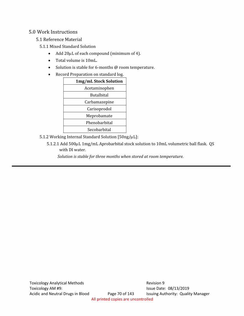
Toxicology Analytical Methods Revision 9Issue Date: 08/13/2019Toxicology AM #9:
Acidic and Neutral Drugs in Blood Page 70 of 143 Issuing Authority: Quality ManagerAll printed copies are uncontrolled
5.0 Work Instructions 5.1 Reference Material
5.1.1 Mixed Standard Solution Add 20L of each compound (minimum of 4). Total volume is 10mL. Solution is stable for 6-months @ room temperature. Record Preparation on standard log.
1mg/mL Stock SolutionAcetaminophen
ButalbitalCarbamazepine
CarisoprodolMeprobamatePhenobarbitalSecobarbital
5.1.2 Working Internal Standard Solution [50ng/L]: 5.1.2.1 Add 500L 1mg/mL Aprobarbital stock solution to 10mL volumetric ball flask. QS
with DI water. Solution is stable for three months when stored at room temperature.
Toxicology Analytical Methods Revision 9Issue Date: 08/13/2019Toxicology AM #13: LCMS-QQQ
Confirmation of Benzodiazepines and Z drugs in Blood and Urine
Page 71 of 143 Issuing Authority: Quality Manager
All printed copies are uncontrolled
Toxicology AM #13: LCMS-QQQ Confirmation of Benzodiazepines and Z drugs in Blood and Urine
1.0 Background/References1.1 Background
Approximately 20 benzodiazepines are approved for use in the US.2 Benzodiazepines were first introduced in the 1960s in pursuit of the perfect sedative hypnotic agent, and have replaced barbiturates as the major class of central nervous system (CNS)-depressant drugs.2 In 1962, Chlordiazepoxide (Librium) was introduced, followed by the introduction of Diazepam (Valium) in 1968. There are four main classes of benzodiazepines, the 1,4-benzodiazepines, the triazolobenzodiazepines, the diazolobenzodiazepines, and the 7-nitrobenzodiazepines.
Benzodiazepines are used primarily as anti-epileptics in the treatment of seizure disorders, as anxiolytics for the short-term relief of anxiety disorders, as sedative-hypnotics for the treatment of sleep disorders, and as muscle relaxants to relieve spasticity. The primary side effects that accompany their use include dose-related extensions of the intended actions, including sedation and sleepiness/drowsiness. In addition, other undesired effects that will influence the outcome of field sobriety tests include ataxia, a blocked ability to coordinate movements, a staggering walk and/or poor balance, lethargy/apathy, indifference or sluggishness, mental confusion, disorientation, slurred speech, and amnesia. Impairment of motor abilities, especially a person's ability to drive an automobile, is common. This impairment is compounded by the drug-induced suppression of one’s' ability to assess their own level of physical and mental impairment. Alcohol combined with other CNS depressants (e.g., barbiturates antidepressants, etc.) will increase CNS depressant effects, such as impairment of psychomotor function and sedation, in an additive manner.4-6
Z drugs (zolpidem, zopiclone), prescribed as sleep aids, and quetiapine which is used in the treatment of mental disorders act in a similar manner to benzodiazepines, but are not included in that particular class of drugs.
The benzodiazepines are lipid soluble and are absorbed well from the GI tract with good distribution to the brain. They are metabolized primarily in the liver. Their CNS active metabolites extend their duration of action. The benzodiazepines work by enhancing, facilitating or potentiating the action of the inhibitory neurotransmitter GABA. They serve to increase the frequency of GABA-mediated chloride ion channel opening.
Toxicology Analytical Methods Revision 9Issue Date: 08/13/2019Toxicology AM #13: LCMS-QQQ
Confirmation of Benzodiazepines and Z drugs in Blood and Urine
Page 72 of 143 Issuing Authority: Quality Manager
All printed copies are uncontrolled
Benzodiazepines are metabolized primarily in the liver via several different microsomal enzyme systems.6 Many products of their metabolism are active. Since many of the active metabolites have been marketed as therapeutic agents, it may be difficult to ascertain which drug was ingested based solely upon the results of analysis. Current drug therapy will assist in determining the source of a particular compound. The detection of a particular agent is determined partly by whether its metabolism yields active metabolites. Excretion of the benzodiazepines is predominantly in the urine. Depending upon the particular benzodiazepine, the urine may contain parent compounds, N-dealkylation and oxidative (hydroxylation) metabolism products and/or glucuronide conjugates.
1.2 References1.2.1 This method was developed in conjunction with Agilent. Patrick Friel from Agilent came
to the Idaho State Police Forensic lab located in Coeur d’Alene and provided application training July 23-26, 2012.
1.2.2 Williamson S.C, ISP Toxicology Analytical Method 2.4.3 (Archived- See Toxicology AM1.2.3 Levine, B. Central Nervous System Depressants. pp. 191-197. In: Principles of Forensic
Toxicology. Levine, B. ed., AACC, 1999.1.2.4 Huang, W. and Moody, D.E. Immunoassay Detection of Benzodiazepines and
Benzodiazepine Metabolites in Blood. J. Anal. Tox. 19:333-342, 1995.1.2.5 Fu, S. Molnar, A. Bowen, P. Lewis J. Wang H. Reduction of Temazepam to Diazepam and
Lorazepam to Delorazepam During Enzyme Hydrolysis. Anal Bioanal Chem 400: 153-164, 2011.
1.2.6 Julien, R.M. A Primer of Drug Action. pp. 95-107, W.H. Freeman and Company: NewYork, 1998.
1.2.7 Hobbs, W.R., Rall, T.W. and Verdoorn, T.A. Hypnotics and Sedatives. pp. 362-373. In: Goodman & Gilman’s The Pharmacological Basis of Therapeutics, 9th edition, Hardman, J.G. ed., McGraw-Hill, 1996.
2.0 Scope2.1 This method is used for the confirmation of 7-aminoclonazepam, 7-
aminoflunitrazepam, zopiclone, zolpidem, chlordiazepoxide, quetiapine, midazolam, flurazepam, nitrazepam, alpha-hydroxyalprazolam, alpha-hydroxytriazolam, oxazepam, nordiazepam, clonazepam, lorazepam, alprazolam, flunitrazepam, temazepam, and diazepam in blood and urine. The words calibrator and calibration are used to coincide with the terminology in instrument software and manufacturer manuals. The manufacturer’s term calibrator refers to what is considered by ISP-FS as reference material that has a certified concentration of drug present.
3.0 Equipment/Reagents3.1 Equipment and Supplies
Toxicology Analytical Methods Revision 9Issue Date: 08/13/2019Toxicology AM #13: LCMS-QQQ
Confirmation of Benzodiazepines and Z drugs in Blood and Urine
Page 73 of 143 Issuing Authority: Quality Manager
All printed copies are uncontrolled
3.1.1 Agilent 6410B or equivalent LC/MS/MS system and MassHunter software3.1.2 De-Tox A Tubes (or equivalent Toxi A tubes)3.1.3 Tapered or round-bottom glass tubes for evaporation and reconstitution3.1.4 Transfer pipettes3.1.5 Pipettes for accurate dispensing of volumes 10µL to 4mL3.1.6 Auto-sampler vials with snap-caps for Agilent 1260 ALS3.1.7 Test tube rocker or rotator3.1.8 Centrifuge capable of 3000 rpm3.1.9 Oven capable of 60oC
3.2 ReagentsRefer to AM#23 for Solution Preparation instructions.3.2.1 BG100 -Glucuronidase Solution (Kura Biotec)3.2.2 2M Acetate buffer, pH 4.83.2.3 0.1% formic acid in water (mobile phase A)3.2.4 0.1% formic acid in acetonitrile (mobile phase B)3.2.5 Deionized water3.2.6 LC/MS grade water3.2.7 LC/MS grade acetonitrile3.2.8 LC/MS grade methanol 3.2.9 LC/MS grade formic acid 3.2.10 Extract reconstitution solvent: 9:1 mobile phase A to mobile phase B
3.3 Qualitative Assurance: Reference Materials and Controls3.3.1 Calibrator and Control SolutionsNOTE: Corresponding calibrator and control reference material must be obtained from different
vendors, or be from different lot numbers if suitable second vendors are not available. NOTE: Stock solution concentrations other than those listed here may be obtained, but
appropriate addition volume adjustments must be made when direct spiking or preparing working solutions. Stock solutions should be stored as recommended by vendor.
3.3.1.1 Reference Material Stock Solutions3.3.1.1.1 1mg/mL single component benzodiazepine-class reference solutions. A
multi-component benzodiazepines mix (250 μg/mL) may be obtained for use in controls.
3.3.1.1.2 Refer to section 5 of this method for the preparation instructions and stability of the working solutions.
3.3.1.2 Reference Material Working Solutions3.3.1.2.1 Refer to section 5 of this method for the preparation instructions and
stability of the working solutions.
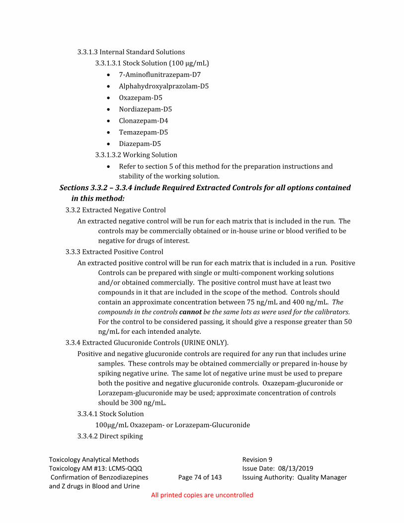
Toxicology Analytical Methods Revision 9Issue Date: 08/13/2019Toxicology AM #13: LCMS-QQQ
Confirmation of Benzodiazepines and Z drugs in Blood and Urine
Page 74 of 143 Issuing Authority: Quality Manager
All printed copies are uncontrolled
3.3.1.3 Internal Standard Solutions3.3.1.3.1 Stock Solution (100 μg/mL)
7-Aminoflunitrazepam-D7 Alphahydroxyalprazolam-D5 Oxazepam-D5 Nordiazepam-D5 Clonazepam-D4 Temazepam-D5 Diazepam-D5
3.3.1.3.2 Working Solution Refer to section 5 of this method for the preparation instructions and
stability of the working solution.Sections 3.3.2 – 3.3.4 include Required Extracted Controls for all options contained
in this method:3.3.2 Extracted Negative Control
An extracted negative control will be run for each matrix that is included in the run. The controls may be commercially obtained or in-house urine or blood verified to be negative for drugs of interest.
3.3.3 Extracted Positive Control An extracted positive control will be run for each matrix that is included in a run. Positive
Controls can be prepared with single or multi-component working solutions and/or obtained commercially. The positive control must have at least two compounds in it that are included in the scope of the method. Controls should contain an approximate concentration between 75 ng/mL and 400 ng/mL. The compounds in the controls cannot be the same lots as were used for the calibrators. For the control to be considered passing, it should give a response greater than 50 ng/mL for each intended analyte.
3.3.4 Extracted Glucuronide Controls (URINE ONLY). Positive and negative glucuronide controls are required for any run that includes urine
samples. These controls may be obtained commercially or prepared in-house by spiking negative urine. The same lot of negative urine must be used to prepare both the positive and negative glucuronide controls. Oxazepam-glucuronide or Lorazepam-glucuronide may be used; approximate concentration of controls should be 300 ng/mL.
3.3.4.1 Stock Solution100μg/mL Oxazepam- or Lorazepam-Glucuronide
3.3.4.2 Direct spiking
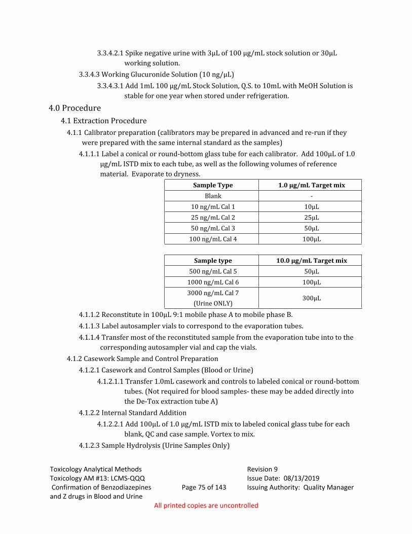
Toxicology Analytical Methods Revision 9Issue Date: 08/13/2019Toxicology AM #13: LCMS-QQQ
Confirmation of Benzodiazepines and Z drugs in Blood and Urine
Page 75 of 143 Issuing Authority: Quality Manager
All printed copies are uncontrolled
3.3.4.2.1 Spike negative urine with 3μL of 100 μg/mL stock solution or 30μL working solution.
3.3.4.3 Working Glucuronide Solution (10 ng/μL)3.3.4.3.1 Add 1mL 100 μg/mL Stock Solution, Q.S. to 10mL with MeOH Solution is
stable for one year when stored under refrigeration.
4.0 Procedure4.1 Extraction Procedure
4.1.1 Calibrator preparation (calibrators may be prepared in advanced and re-run if they were prepared with the same internal standard as the samples)
4.1.1.1 Label a conical or round-bottom glass tube for each calibrator. Add 100µL of 1.0 µg/mL ISTD mix to each tube, as well as the following volumes of reference material. Evaporate to dryness.
Sample Type 1.0 µg/mL Target mixBlank -
10 ng/mL Cal 1 10µL 25 ng/mL Cal 2 25µL 50 ng/mL Cal 3 50µL
100 ng/mL Cal 4 100µL
Sample type 10.0 µg/mL Target mix500 ng/mL Cal 5 50µL
1000 ng/mL Cal 6 100µL 3000 ng/mL Cal 7
(Urine ONLY)300µL
4.1.1.2 Reconstitute in 100µL 9:1 mobile phase A to mobile phase B.4.1.1.3 Label autosampler vials to correspond to the evaporation tubes.4.1.1.4 Transfer most of the reconstituted sample from the evaporation tube into to the
corresponding autosampler vial and cap the vials.4.1.2 Casework Sample and Control Preparation
4.1.2.1 Casework and Control Samples (Blood or Urine)4.1.2.1.1 Transfer 1.0mL casework and controls to labeled conical or round-bottom
tubes. (Not required for blood samples- these may be added directly into the De-Tox extraction tube A)
4.1.2.2 Internal Standard Addition4.1.2.2.1 Add 100µL of 1.0 µg/mL ISTD mix to labeled conical glass tube for each
blank, QC and case sample. Vortex to mix.4.1.2.3 Sample Hydrolysis (Urine Samples Only)
Toxicology Analytical Methods Revision 9Issue Date: 08/13/2019Toxicology AM #13: LCMS-QQQ
Confirmation of Benzodiazepines and Z drugs in Blood and Urine
Page 76 of 143 Issuing Authority: Quality Manager
All printed copies are uncontrolled
4.1.2.3.1 Enzyme hydrolysis: add 20µL 2M acetate buffer to all controls and case samples, and 76μl BG100 β-glucuronidase to calibrators, controls, and casework samples (except the negative glucuronidase control sample). Cap and gently vortex the samples. Incubate at approximately 60°C in an oven for 30 minutes. Remove from oven.
4.1.2.4 Extraction 4.1.2.4.1 Label a De-Tox Tube A for each QC, blank, and case sample. 4.1.2.4.2 To the De-Tox Tubes, add ~4mL of deionized water to each or add the
4mLs to the conical tubes with the samples.4.1.2.4.3 Transfer the casework and control samples with added ISTD from the
labeled conical or round-bottom tube to the corresponding De-Tox Tube (for blood samples, the ISTD and sample may be added directly to the De-Tox tube. There is no requirement to place it in a conical or round-bottom tube first.)
4.1.2.4.4 Cap the De-Tox Tubes and mix by inverting.4.1.2.4.5 Rotate or rock the tubes gently for ~5 minutes.4.1.2.4.6 Centrifuge the tubes at approximately 2000-2500 rpm for ~ 5 minutes.
NOTE: If an emulsion occurs, it may be broken up with a disposable transfer pipette and the tube re-centrifuged at approximately 3000 rpm for ~5 minutes. Care should be taken that no solvent is lost to the disposable pipette when the emulsion is broken up.
4.1.2.4.7 Transfer most (~2mL) of the upper organic layer from each De- Tox Tube to the corresponding labeled evaporation tube. Avoid transferring any solids.
4.1.2.4.8 Evaporate to dryness under nitrogen at ~ 40 degrees Celsius. It is critical that the extracts are evaporated completely to dryness.
4.1.2.5 Reconstitution 4.1.3.5.1 Reconstitute in 100µL 9:1 mobile phase A to mobile phase B.4.1.3.5.2 Transfer the reconstituted sample from the evaporation tube into to the
corresponding autosampler vial and cap.4.1.3 Instrument/Run set up
4.1.3.1 Refer to Toxicology AM #24 for general LCMS instrument operation and maintenance.
4.1.3.2 Cycle time for the method is approximately 13 minutes.4.1.3.3 Instrument acquisition method will be printed and stored near the instrument
(this may also be done electronically and stored in a central location)4.1.3.3.1 Acquisition Required Settings
Column Temperature - 40*C Injection volume – 0.5µL– 5µL
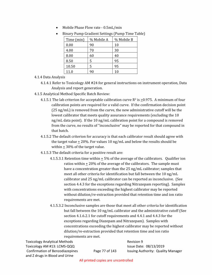
Toxicology Analytical Methods Revision 9Issue Date: 08/13/2019Toxicology AM #13: LCMS-QQQ
Confirmation of Benzodiazepines and Z drugs in Blood and Urine
Page 77 of 143 Issuing Authority: Quality Manager
All printed copies are uncontrolled
Mobile Phase Flow rate - 0.5mL/min Binary Pump Gradient Settings (Pump Time Table)
Time (min) % Mobile A % Mobile B0.00 90 104.00 70 308.00 60 408.50 5 9510.50 5 9511.0 90 10
4.1.4 Data Analysis4.1.4.1 Refer to Toxicology AM #24 for general instructions on instrument operation, Data
Analysis and report generation.4.1.5 Analytical Method Specific Batch Review:
4.1.5.1 The lab criterion for acceptable calibration curve R2 is >0.975. A minimum of four calibration points are required for a valid curve. If the confirmation decision point (25 ng/mL) is removed from the curve, the new administrative cutoff will be the lowest calibrator that meets quality assurance requirements (excluding the 10 ng/mL data point). If the 10 ng/mL calibration point for a compound is removed from the curve, no results of “inconclusive” may be reported for that compound in that batch.
4.1.5.2 The default criterion for accuracy is that each calibrator result should agree with the target value + 20%. For values 10 ng/mL and below the results should be within + 30% of the target value.
4.1.5.3 The default criteria for a positive result are:4.1.5.3.1 Retention time within + 5% of the average of the calibrators. Qualifier ion
ratios within + 20% of the average of the calibrators. The sample must have a concentration greater than the 25 ng/mL calibrator; samples that meet all other criteria for identification but fall between the 10 ng/mL calibrator and 25 ng/mL calibrator can be reported as inconclusive. (See section 4.4.3 for the exceptions regarding Nitrazepam reporting). Samples with concentrations exceeding the highest calibrator may be reported without dilution/re-extraction provided that retention time and ion ratio requirements are met.
4.1.5.3.2 Inconclusive samples are those that meet all other criteria for identification but fall between the 10 ng/mL calibrator and the administrative cutoff (See section 4.1.6.2.1 for cutoff requirements and 4.4.1 and 4.4.3 for the exceptions regarding Diazepam and Nitrazepam). Samples with concentrations exceeding the highest calibrator may be reported without dilution/re-extraction provided that retention time and ion ratio requirements are met.
Toxicology Analytical Methods Revision 9Issue Date: 08/13/2019Toxicology AM #13: LCMS-QQQ
Confirmation of Benzodiazepines and Z drugs in Blood and Urine
Page 78 of 143 Issuing Authority: Quality Manager
All printed copies are uncontrolled
4.2 Quality Assurance Requirements:4.2.1 Refer to Toxicology AM #19 and AM #21 for additional quality assurance and reference
material authentication requirements.4.3 Analysis Documentation
4.3.1 The printed results for each case sample will be included with the analysts’ notes. Case results are to be recorded in the LIMS system.
4.3.2 Reports for the batch, including calibration curves and controls, if printed, will be stored centrally in the lab in which the analysis was performed. A copy of data for controls may be stored electronically in a central location and need not be included in individual case files. When necessary, a copy of control printouts can be prepared from the centrally stored document.
4.3.3 The data from the run will be stored electronically, and if it is on a computer, will be backed up at least every two months.
4.4 Limitations of Method4.4.1 The hydrolysis process for glucuronides in urine has limited efficiency; based on the
validation study, the estimated conversion is about 60-70 percent. There is potential for a small amount of temazepam to convert to diazepam in the hydrolysis process. If both diazepam and temazepam are detected in a urine sample, the diazepam will not be reported unless it has a response that is greater than 5% of the temazepam response.
4.4.2 This method is only approved for qualitative identification of the listed compounds in urine and blood. The uncertainty associated with the quantitative values has not been established; therefore, no values shall be referenced or reported.
4.4.3 Nitrazepam has been found to have significant variability in concentration responses with this method, though no false positives have been observed. If a case sample gives a nitrazepam response that is >5 ng/mL and <50 ng/mL, it will be reported as “inconclusive for nitrazepam due to method limitations.”
Toxicology Analytical Methods Revision 9Issue Date: 08/13/2019Toxicology AM #13: LCMS-QQQ
Confirmation of Benzodiazepines and Z drugs in Blood and Urine
Page 79 of 143 Issuing Authority: Quality Manager
All printed copies are uncontrolled
5.0 Comments5.1 Appendix Working Solutions:
5.1.1 -10µg/mL Target Working Solution in methanol (Document on a prep sheet with an expiration of one year, store under refrigeration)
In a 25mL volumetric flask fill the flask about half full with methanol add 250µL of 1mg/mL stock solution of the following compounds.
7-aminoclonazepam, 7-aminoflunitrazepam, zopiclone, zolpidem, chlordiazepoxide, quetiapine, midazolam, flurazepam, nitrazepam, alpha-hydroxyalprazolam, alpha-hydroxytriazolam, oxazepam, nordiazepam, clonazepam, lorazepam, alprazolam, flunitrazepam, temazepam, and diazepam
QS with methanol and ensure it is thoroughly mixed.5.1.2 - 1µg/mL Target Working Solution in methanol
(Document on a prep sheet with an expiration of the earliest expiring reference material or one year whichever is first, store under refrigeration)
In a 10mL volumetric flask fill the flask about half full with methanol, add 1mL of the 10µg/mL Target Working Solution
QS with methanol and ensure it is thoroughly mixed.6.1.3 - 1µg/mL ISTD mix in methanol (Document on a prep sheet with an expiration of one year, store under refrigeration)
Fill a 10mL volumetric flask about half full with methanol, add 100µL of 100 µg/mL stock solution of the following compounds. (If the stock solution is a different concentration you will need to adjust addition volumes.)
7-aminoflunitrazepam-D7, alphahydroxyalprazolam-D5, oxazepam-D5, nordiazepam-D5, clonazepam-D4, temazepam-D5, diazepam-D5
QS with methanol and ensure it is thoroughly mixed.5.1.3 - 1µg/mL Control mix in methanol
(Document on a prep sheet with an expiration of the earliest expiring reference material or one year whichever is first, store under refrigeration)
Add 10 L each 1 mg/mL or 40 L 250 μg/mL Stock Solution to 6mL MeOH (LCMS grade) in 10mL ball flask. QS with LC/MS grade MeOH.
Toxicology Analytical Methods Revision 9Issue Date: 08/13/2019Toxicology AM #16:
Pipette Calibration and Intermediate Checks
Page 80 of 143 Issuing Authority: Quality Manager
All printed copies are uncontrolled
Toxicology AM #16: Pipette Calibration and Intermediate Checks
1.0 Background/References1.1 Background
The initial calibration of piston or plunger operated volumetric apparatus (POVA) is performed by the manufacturer. Upon receipt of a newly obtained pipette or syringe and thereafter periodically, the calibration must be verified to substantiate that the volume delivered is both accurate and precise. This is accomplished by determining the mass of a volume of liquid of known density that has been delivered into a closed vessel.
1.2 References1.2.1 ASTM Method E-1154-89 (reapproved 2003), Standard Specification for Piston or
Plunger Operated Volumetric Apparatus.1.2.2 Curtis, R.H., Performance Verification of Manual Action Pipets: Part I, Am. Clin. Lab.
12(7):8-9; 1994.1.2.3 Curtis, R.H., Performance Verification of Manual Action Pipets: Part II, Am. Clin. Lab.
12(9):16-17; 1994.1.2.4 Byer, B.J., How to Use and Check Pipetting Equipment, Scientific Newsletters, Inc., 1977.1.2.5 ISO 8655-6:2002, Piston-operated volumetric apparatus - Part 6: Gravimetric method
for the determination of measurement error.
2.0 Scope2.1 Scope
The reliability of the volume delivered by a pipette is dependent upon verification of calibration. This method sets forth the requirements for both intermediate checks and calibration. The intermediate check is performed to maintain confidence in calibration.
3.0 Equipment/Reagents3.1 Analytical Balance
• Capable of accurately weighing volumes of interest. 3.2 Thermometer
• Subdivisions of ≤0.5 degree• Capable of reading 20° - 28°C
3.3 Weighing Vessel 3.4 Appropriate disposable pipette tips3.5 Water
4.0 Procedure4.1 Calibration
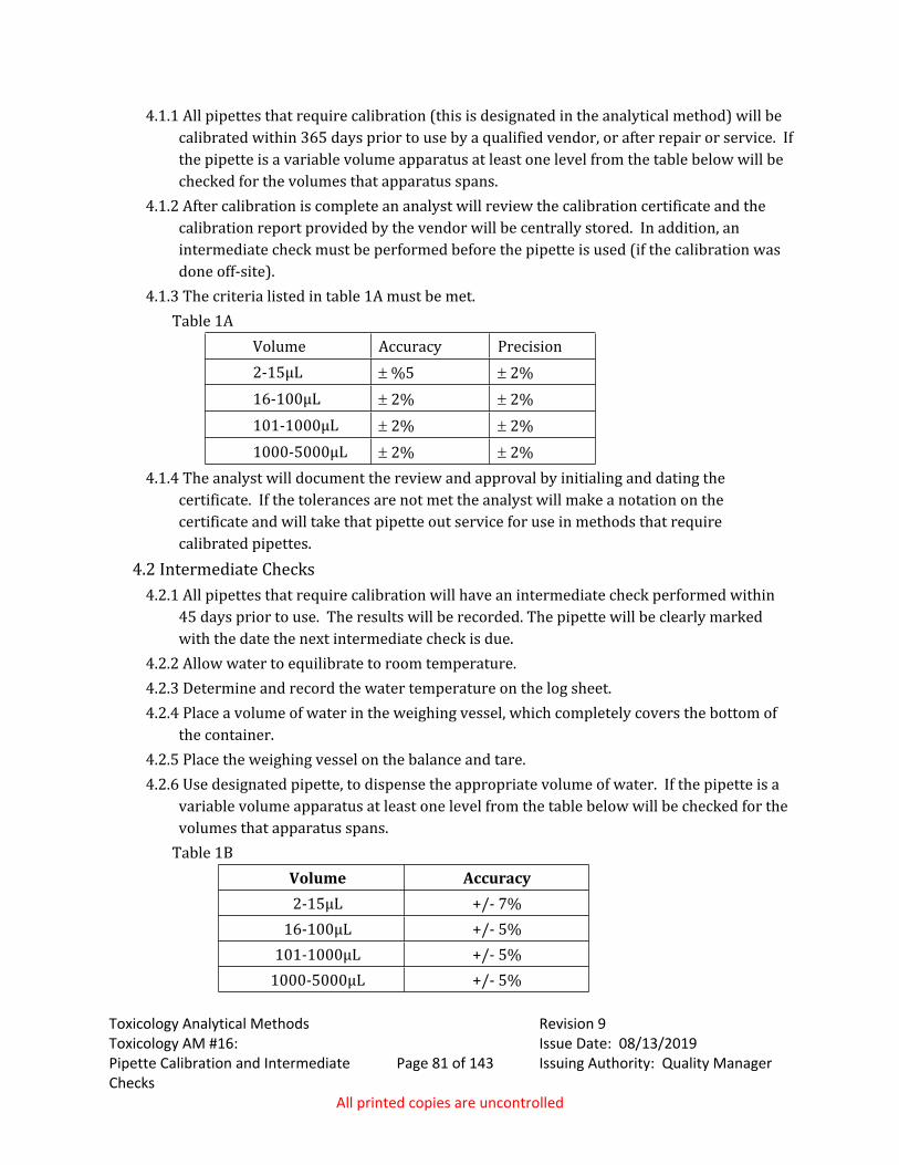
Toxicology Analytical Methods Revision 9Issue Date: 08/13/2019Toxicology AM #16:
Pipette Calibration and Intermediate Checks
Page 81 of 143 Issuing Authority: Quality Manager
All printed copies are uncontrolled
4.1.1 All pipettes that require calibration (this is designated in the analytical method) will be calibrated within 365 days prior to use by a qualified vendor, or after repair or service. If the pipette is a variable volume apparatus at least one level from the table below will be checked for the volumes that apparatus spans.
4.1.2 After calibration is complete an analyst will review the calibration certificate and the calibration report provided by the vendor will be centrally stored. In addition, an intermediate check must be performed before the pipette is used (if the calibration was done off-site).
4.1.3 The criteria listed in table 1A must be met. Table 1A
Volume Accuracy Precision2-15μL %5 2%16-100μL 2% 2%101-1000μL 2% 2%1000-5000μL 2% 2%
4.1.4 The analyst will document the review and approval by initialing and dating the certificate. If the tolerances are not met the analyst will make a notation on the certificate and will take that pipette out service for use in methods that require calibrated pipettes.
4.2 Intermediate Checks4.2.1 All pipettes that require calibration will have an intermediate check performed within
45 days prior to use. The results will be recorded. The pipette will be clearly marked with the date the next intermediate check is due.
4.2.2 Allow water to equilibrate to room temperature.4.2.3 Determine and record the water temperature on the log sheet. 4.2.4 Place a volume of water in the weighing vessel, which completely covers the bottom of
the container. 4.2.5 Place the weighing vessel on the balance and tare.4.2.6 Use designated pipette, to dispense the appropriate volume of water. If the pipette is a
variable volume apparatus at least one level from the table below will be checked for the volumes that apparatus spans.
Table 1BVolume Accuracy2-15μL +/- 7%
16-100μL +/- 5%101-1000μL +/- 5%
1000-5000μL +/- 5%
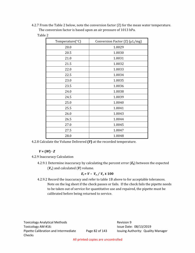
Toxicology Analytical Methods Revision 9Issue Date: 08/13/2019Toxicology AM #16:
Pipette Calibration and Intermediate Checks
Page 82 of 143 Issuing Authority: Quality Manager
All printed copies are uncontrolled
4.2.7 From the Table 2 below, note the conversion factor (Z) for the mean water temperature. The conversion factor is based upon an air pressure of 1013 hPa.
Table 2
Temperature(C) Conversion Factor (Z) (L/mg)
20.0 1.002920.5 1.003021.0 1.003121.5 1.003222.0 1.003322.5 1.003423.0 1.003523.5 1.003624.0 1.003824.5 1.003925.0 1.004025.5 1.004126.0 1.004326.5 1.004427.0 1.004527.5 1.004728.0 1.0048
4.2.8 Calculate the Volume Delivered (V) at the recorded temperature.
V = (W) · Z4.2.9 Inaccuracy Calculation
4.2.9.1 Determine inaccuracy by calculating the percent error (Et) between the expected (Vo) and calculated (V) volume.
Et = V – Vo / Vo x 1004.2.9.2 Record the inaccuracy and refer to table 1B above to for acceptable tolerances.
Note on the log sheet if the check passes or fails. If the check fails the pipette needs to be taken out of service for quantitative use and repaired, the pipette must be calibrated before being returned to service.
Toxicology Analytical Methods Revision 9Issue Date: 08/13/2019Toxicology AM #17:
Balance Calibration and Intermediate Checks
Page 83 of 143 Issuing Authority: Quality Manager
All printed copies are uncontrolled
Toxicology AM #17: Balance Calibration and Intermediate Checks
1.0 Background/References1.1 Background: Refer to manufacturer’s manual for balance specific procedures.1.2 References
1.2.1 ASTM Method E-617-97, Standard Specification for Laboratory Weights and Precision Mass Standards.
2.0 Scope2.1 The procedure is to ensure accuracy of measurement when an analytical or top-
loading balance is used to prepare solutions or reference material for application to toxicology methods.
3.0 Equipment/Reagents3.1 ANSI/ASTM Type I, Class 1 or Class 2 laboratory weights3.2 Analytical and/or Top-Loading, Direct-Reading Laboratory Balances
4.0 Procedure4.1 Intermediate Check Procedure
4.1.1 Inspect balance pan, clean if necessary.4.1.2 Inspect level bubble, level if necessary.4.1.3 Tare balance with weighing paper or weigh boat.4.1.4 Place weight on balance.4.15 Record weight on balance logsheet.4.1.6 The allowable deviation from the standard weights will be 0.01g or 0.1%, whichever is
greater.4.1.7 The verification procedure should be repeated if the value does not fall within the
acceptable range. If value is still out of range, contact service vendor to set up a service call. A note must be placed on the balance to indicate that it is not in range and must not be used.
4.2 Quality Assurance Requirements4.2.1 Within 45 days prior to use, the balance is to have its calibration checked (intermediate
check is acceptable) against an appropriate set of NIST traceable weights. Results are to be recorded for future reference. All balances will be calibrated yearly by a qualified vendor. Calibration certificates will be checked for compliance with ISO/IEC 17025:2005, and initialed before the balance is placed back in use.
Toxicology Analytical Methods Revision 9Issue Date: 08/13/2019Toxicology AM #17:
Balance Calibration and Intermediate Checks
Page 84 of 143 Issuing Authority: Quality Manager
All printed copies are uncontrolled
4.2.2 Weights used for intermediate checks of calibration will be NIST traceable and will be calibrated every 5 years by a qualified vendor. Upon return to the laboratory and before the weights are placed back into service, the calibration will be verified by weighing them on an analytical balance the weights must be with .005 g of their expected weight. Calibration Certificates will be checked for compliance with ISO/IEC 17025:2005 and the weights are within .005 g of their expected weight, and initialed.
4.2.3 An in-house intermediate check of balances in use will be performed within 45 days prior to use. Results are to be recorded in logbook.
4.2.4 The weights used for the intermediate check should depend on the application of the balance. Three weights must be used to represent the weight range in question. For instance, if the balance is being used to prepare buffer solutions, then perhaps 100g, 500g and 1000g weights would characterize the weight range. If the balance is used to prepare mg/mL reference material then 1mg, 10mg and 100mg weights may be appropriate.
4.2.5 The type of balance employed should be a consideration. For weights less than 100mg, an analytical balance should be used. For the preparation of a solution involving gram quantities, a top-loading balance should be used.
4.2.6 Do not touch weights with bare hands. Weights should be transferred with forceps or gloves. The weights will be stored and transported in their case.
Toxicology Analytical Methods Revision 9Issue Date: 08/13/2019Toxicology AM #18: Toxicology
Proficiency Tests Page 85 of 143 Issuing Authority: Quality ManagerAll printed copies are uncontrolled
Toxicology AM #18: Toxicology Proficiency Tests 1.0 Background/References
1.1 BackgroundAs set forth in the Idaho State Police Forensic Services Procedure and Quality Manual,
proficiency testing is an integral part of a quality program.
2.0 Scope2.1 This method describes the criteria to be applied to the evaluation of results
obtained for proficiency testing for the toxicology discipline.3.0 Equipment/Reagents
3.1 Not Applicable4.0 Procedure
4.1 Urine and Blood Analysis Proficiency Testing4.1.1 Only analytes that are routinely tested for with current ISP-FS analytical methods will be
evaluated. If the parent drug or a metabolite of a drug is reported, additional metabolites of that drug are not required to be reported.
4.1.2 When reported, quantitative values must be within the estimated uncertainty of measurement.
4.1.3 If the analyst does not correctly identify all target analytes and/or quantitative values do not fall within range, the analyst's training will be reviewed and additional training may be required as deemed appropriate by the Toxicology Program Discipline Leader. The analyst may be required to complete a competency test prior to resuming casework. The number of samples will be determined by the nature of the discrepancy.

Toxicology Analytical Methods Revision 9Issue Date: 08/13/2019Toxicology AM #19:
Quality Assurance Measures Page 86 of 143 Issuing Authority: Quality ManagerAll printed copies are uncontrolled
Toxicology AM #19: Quality Assurance Measures
1.0 Background/References1.1 Background
The quality assurance measures applied towards analysis of toxicological samples promote confidence in results.
1.2 References 1.2.1 Wu Chen, N.B. Cody, J.T., Garriott, J.C., Foltz, R.L., and et al., Report of the Ad hoc
Committee on Forensic GC/MS: Recommended guidelines for forensic GC/MS procedures in toxicology laboratory associated with offices of medical examiners and/or coroners, J. Foren. Sci, 236 (35): 236-242, 1990.
1.2.2 Goldberger, B.A., Huestis, M.A., Wilkins, D.G., Commonly practiced quality control and quality assurance procedures for gas chromatograph/mass spectrometry analysis in forensic urine drug-testing laboratories, For Sci Review, 9(2): 60-79, 1997.
1.2.3 SOFT/AAFS Forensic Toxicology Laboratory Guidelines, 2002
2.0 Scope2.1 This analytical method addresses general acceptance requirements for qualitative
and quantitative analysis data obtained through analysis by gas chromatography equipped with a mass selective detective (MSD). Requirements for analysis with other instrumentation are addressed in relevant analytical methods.
3.0 Equipment/Reagents3.1 Refer to specific analytical method for necessary equipment and supplies.3.2 Refer to appropriate analytical method for required reagents as well as Toxicology
AM #23 for Solution Preparation instructions.4.0 Procedure
4.1 Instrument Quality Assurance GCMS 4.1.1 Instrument Maintenance
NOTE: Refer to manufacturer’s hardcopy or electronic instrument manuals for maintenance indicators and instructions.
4.1.2 MSD Tuning4.1.2.1 A Tune must be run within a week prior to analysis of casework. 4.1.2.2 A Tune Evaluation should be completed and parameters should be reviewed for
acceptance based on the report’s predefined criteria, set by the manufacturer. Note: H2O and N2 values should be monitored for possible instrumentation leaks (acceptance parameters are listed in parentheses on the tune evaluation report).
Toxicology Analytical Methods Revision 9Issue Date: 08/13/2019Toxicology AM #19:
Quality Assurance Measures Page 87 of 143 Issuing Authority: Quality ManagerAll printed copies are uncontrolled
4.1.3 Instrument Performance Monitoring4.1.3.1 Instrument performance will be monitored through evaluation of the positive and
negative controls run with every extraction batch. The control requirements for an extraction are found in the applicable analytical methods. The instrument will be determined as working properly if the expected responses are obtained for the controls evaluated.
4.1.3.2 Analysts may use the control data to determine if instrument maintenance is needed.
4.1.3.3 Troubleshooting, repairs and maintenance that takes place will be documented.4.1.4 Data file back up
4.1.4.1 Data files will be retained and backed up to external media at least once every two months.
4.2 Instrument Quality Assurance LC/MS- Refer to Toxicology AM# 244.3 Sample Preparation Quality Assurance
4.3.1 Qualitative Analysis4.3.1.1 Matrix Controls
4.3.1.1.1 Quality controls are to be prepared and analyzed as designated in the appropriate analytical method.
4.3.1.1.2 Positive controls should exhibit proper retention time and mass spectral characteristics for compounds of interest.
4.3.1.1.3 Negative controls should be examined for compound(s) of interest and interfering substances prior to the completion of analysis, the negative control can be included in the batch.
4.3.1.1.4 Commercially-obtained controls may be utilized for qualitative analysis after the manufacturer’s expiration date provided all method control requirements are met AND an additional in-run control that is not expired be run. This limited permission is not applicable to expired controls of a unique nature (e.g. EMIT level 0 pre-run control).
4.3.1.1.5 It is applicable, for example, to the use of an expired commercially-obtained c-THC control when performing Toxicology AM# 3; this method requires one (non-expired) spiked controls be run in addition to the commercially-obtained control.
4.3.1.2 GC/MS Solvent Blanks 4.3.1.2.1 An appropriate solvent blank will be run before case sample extracts. If the
same sample is injected for more than one instrument method, only one blank needs to be run preceding the sample injections.
4.3.1.2.2 If the solvent blank contains a reportable analyte of interest, the corrected area of the analyte peak in the sample data must be a minimum of 10 times stronger than the corresponding peak in the blank preceding it. Ideally, no contamination should be apparent.
Toxicology Analytical Methods Revision 9Issue Date: 08/13/2019Toxicology AM #19:
Quality Assurance Measures Page 88 of 143 Issuing Authority: Quality ManagerAll printed copies are uncontrolled
4.3.1.2.3 Reportable is defined as a complete fragmentation pattern at the appropriate retention time. Analytes of interest include, but are not limited to, analytes routinely reported.
4.3.1.2.4 If significant contamination is present, as discussed in 4.2.1.2.2, rerun the solvent blank that was used in that analysis and the sample extract in question. If the contamination is still apparent, or the original samples are no longer available, troubleshoot the instrument to determine the source of contamination. In addition, the sample in question must be re-extracted prior to reanalysis on the rectified instrument.
4.3.2 Quantitative Analysis4.3.2.1 Quality measures are optimized for the analytes in question and are addressed in
each individual quantitative analytical method.4.3.3 Distribution of Quality Data
4.3.3.1 Original data for matrix controls will be stored in a designated central location in the laboratory where the analysis was performed, or electronically on a shared network drive.
4.3.3.2 Copies of all quality assurance control data need not be placed in each case file, except those required under 4.2.3.3.
4.3.3.3 Copies of analytical reference material used to substantiate the identification of each drug compound must be included in each case file, if not otherwise indicated in the relevant analytical method.
4.4 Sample Storage4.4.1 Blood samples should be stored under refrigeration; this includes Combo Collection Kits.4.4.2 Urine samples should be stored under refrigeration or frozen. If samples are going to be
stored longer than two weeks prior to analysis, they should be frozen. Once analysis is complete, samples should be frozen until they are returned to the agency.
Toxicology Analytical Methods Revision 9Issue Date: 08/13/2019Toxicology AM #20:
Testing Guidelines and Reporting Criteria
Page 89 of 143 Issuing Authority: Quality Manager
All printed copies are uncontrolled
Toxicology AM #20: Testing Guidelines and Reporting Criteria
1.0 Background/References1.1 Background
To best utilize the resources available to support the ISP-FS toxicology discipline, the degree of analysis pursued should be guided by all available information. It may not always be necessary and/or appropriate to confirm all drug compounds present. With urine analysis, when a subject has admitted to use of prescription and/or over-the counter drugs that may impair driving, confirmation of all drugs present may not serve to strengthen pending charges. With drugs-of-abuse, confirming the presence of all drug compounds may not be necessary, depending on the circumstances. For instance, for Probation and Parole cases, prescription pharmaceuticals are most likely not a consideration. This method also covers reporting criteria.
2.0 Scope2.1 This method addresses requirements to confirm a drug as well as reporting the indication
of drugs. It also covers the factors to consider when determining the extent of analysis a toxicology case sample requires. It is intended to provide guidance to analysts; however, the decision to pursue testing remains at the discretion of each analyst. The goal of these considerations is for the efficient utilization of resources in order to provide timely analysis results to user agencies. This method covers reporting criteria to ensure consistent reporting in the lab system and to ensure limitations are properly expressed.
3.0 Equipment/Reagents3.1 Not applicable
4.0 Procedure4.1 Assignment of classifications for analytical methods and instrumental techniques.
4.1.1 Class A (indicative/screening test) Enzyme Immunoassay (analytical methods 1 and 7) Full scan GCMS mass spectrum comparison to a library (no
comparison of retention time or to standard run on instrument) GCMS full scan compared to an instrument run standard with
acceptable retention time but mass spectral information is too weak. LCMSMS targeted screen (analytical methods 25 and 26) LCMSMS targeted confirmation below limit of confirmation with
defined indicative range in method.Class B (confirmatory test, or indicative test- provides basic structural information)
GCMS methods in SIM (selective ion monitoring mode)
Toxicology Analytical Methods Revision 9Issue Date: 08/13/2019Toxicology AM #20:
Testing Guidelines and Reporting Criteria
Page 90 of 143 Issuing Authority: Quality Manager
All printed copies are uncontrolled
LCMSMS targeted confirmation methods (analytical methods 13, 27, and 28)
Class C (confirmatory test- provides detailed structural information) GCMS full scan mass spectrum with comparison of retention time
and spectrum to reference material.4.2 Testing requirements to confirm a drug.
4.2.1 To confirm a drug, positive test results with the following test combinations must be obtained: (the testing may be performed by different analysts)
Class A and Class B Class A and Class C Two different Class B methods Class B and Class C Class C with two sampling events (may be same method)
4.3 Testing requirements to report a drug was indicated.4.3.1 To report a positive screening test, it must be clear on the report that the drug was indicated but not confirmed. For example, “preliminary testing indicates the possible presence of (indicated drug/drug class), not confirmed due to (reason)”. The indicative testing may be reported when further testing is not pursued, is not possible, or will be pursued at a later date.
4.4 Positive Class A, and Class B, and single sample Class C tests may be reported as indicative.
4.5 Guidelines for method selection and reporting4.5.1 Reporting confirmed drugs
4.5.1.1 When a drug produces positive tests required for confirmatory testing, it will be reported. It will be reported as Drugs Confirmed: any drugs confirmed will be listed. If no drugs are confirmed it will be reported Drugs Confirmed: None.
4.5.2 Reporting indicative drug results4.5.2.1 When a sample has indicative screening drug testing results, it is at
the analyst’s discretion whether or not to report it. Indicative results must be clearly qualified, see 4.5.5 for additional requirements for targeted screen and EIA reporting.
4.5.2.2 If a sample is positive on one indicative test but does not give an indication on a more sensitive valid test, it will not be reported. For example, a sample has a positive result for tramadol using AM 25, AM 28 is then run and tramadol meets all the criteria for evaluation but gives a response below the AM 28 limit of confirmation, it will not be reported indicatively.
4.5.2.3 When a drug is indicated in a confirmatory test but does not meet the criteria for identification and the analyst chooses to report it, it will be reported as “Inconclusive for (drug), as it does not meet ISP
Toxicology Analytical Methods Revision 9Issue Date: 08/13/2019Toxicology AM #20:
Testing Guidelines and Reporting Criteria
Page 91 of 143 Issuing Authority: Quality Manager
All printed copies are uncontrolled
Forensic Services toxicology criteria for identification. This is due to (reason)”.
4.5.2.4 For example: Inconclusive for zolpidem, as it does not meet ISP Forensic Services toxicology criteria for identification. This is due to mass spectral differences between sample and reference material.
4.4.5 Method selection4.5.1 When available, the type of case associated with a toxicology sample should
be determined.4.5.2 The extent of analysis should be based on background information and the
charges pending.4.5.3 If no background information is provided, it is at the discretion of the
analyst to perform only basic testing. 4.5.4 The typical basic drug testing protocol will start with a targeted screen
using LCMSMS, LC-QTOF, EIA or a combination of these tests to cover general drugs of abuse. The toxicology supplemental report will identify the drugs or classes covered in the screen.
4.5.5 The analyst will pursue indicated drugs as appropriate. Not all drugs indicated are required to have confirmation pursued. When EIA or LCMS targeted screening results are positive but confirmatory testing is not done, the following comment may be added to the report: “Preliminary testing indicates the possible presence of a (drug class or drug); confirmatory testing not pursued due to (reason)”. If the parent drug is indicated and confirmed it is not necessary to pursue confirmatory testing for metabolites or to report the metabolite was indicated.
Example 1. A targeted LCMSMS screen is run and is positive for zolpidem and zopiclone. Zolpidem is confirmed by LCMSMS. Additional confirmatory testing for zopiclone is not required. The analyst would report. Drug(s) confirmed: Zolpidem. Preliminary testing indicates the possible presences of zopiclone; confirmatory testing not pursued due to the presence of other drugs.
Example 2. A targeted LCMSMS screen is run and is positive for hydrocodone, norhydrocodone, and dihydrocodeine. An LCMSMS confirmation is run and is positive for hydrocodone but the norhydrocodone is too weak and dihydrocodeine could not be evaluated. The analyst would report Drug(s) confirmed Hydrocodone.
4.5.6 Drugs that are indicated in general extractions such as a basic GCMS extraction but not confirmed are at the analyst’s discretion to report. For
Toxicology Analytical Methods Revision 9Issue Date: 08/13/2019Toxicology AM #20:
Testing Guidelines and Reporting Criteria
Page 92 of 143 Issuing Authority: Quality Manager
All printed copies are uncontrolled
example: in a DUI case where the subject presented with symptoms consistent with a CNS-stimulant, a targeted drug screen using LCMSMS is run, and is positive for methamphetamine and amphetamine. A basic extraction with full scan GCMS is then run. Methamphetamine and amphetamine are confirmed, and lidocaine is indicated. The analyst may report out just the amphetamine and methamphetamine with no mention of the lidocaine on the report, or the analyst may report it out as preliminary testing indicated the possible presences of lidocaine confirmatory testing not pursued due to the presence of other drugs.
4.6.6 Tests in addition to the typical screening tests may be run if information is provided that merits running the additional tests. For example, in a suspected drug facilitated sexual assault case, where urine was collected 3 hours after the suspected dosing, and no other drugs were detected, a test for GHB may be run. Another example would be a DUI case where the subject presented with symptoms consistent with a CNS-depressant and an alcohol level of 0.04; prescription therapy of butalbital is listed, an acidic extraction with full scan GCMS may be run.
4.6.7 Specific reporting criteria and limitations are listed in the appropriate analytical method.
4.6.8 Reporting listed Rx therapy will be at the analyst’s discretion, but is recommended for cases where it could alleviate confusion of where a drug came from. For example if Oxazepam was detected in urine, the comment could read: Prescription drug therapy is said to include Valium (diazepam), oxazepam is an active metabolite of diazepam.
4.6.9 All examples of reporting criteria are guidelines and can be altered to fit the circumstance. The exact wording specified does not need to be followed but is merely a guideline.
Toxicology Analytical Methods Revision 9Issue Date: 08/13/2019Toxicology AM #20:
Testing Guidelines and Reporting Criteria
Page 93 of 143 Issuing Authority: Quality Manager
All printed copies are uncontrolled
4.7 Testing Guidelines: Post-Blood Alcohol or Breath Testing Analysis4.7.1 When the ethanol concentration is 0.10g/100cc, or greater, further testing for
additional drugs, in either blood or urine, should not be pursued unless justified by case-related circumstances. This is in consideration that the legal limit for ethanol is 0.08 grams per 100 cc blood.
4.7.3 Extenuating circumstances may include the following: Fatality or injury accidents. Death investigations. Sexual assaults.
4.7.3.1 In the case of crashes where the subject is the driver and is deceased and further toxicology testing is requested, testing will be performed on samples that have a blood alcohol content of less than 0.20 grams per 100 cc of blood, accident victim “AV” samples that are for statistical purposes only will be run for both alcohol and toxicology, it will be at the analysts discretion whether or not confirmatory or indicative screening test only are run.
4.7.4 The submitting officer or agency is responsible for providing justification for additional testing. Justification could take the form of a note on the submittal information, memo, e-mail or letter outlining the situation, or a case report.
4.7.5 If the ethanol concentration is 0.10 g/100cc or higher future testing for other impairing drugs will not be pursued if additional testing is not requested. Analysts are encouraged to contact the agency if it is believed that further analysis is recommended. Additional analysis may be prudent if impairment described cannot reasonably be explained by ethanol/other volatiles results.
Toxicology Analytical Methods Revision 9Issue Date: 08/13/2019Toxicology AM #20:
Testing Guidelines and Reporting Criteria
Page 94 of 143 Issuing Authority: Quality Manager
All printed copies are uncontrolled
4.8 Infrequently Used Methods4.8.1 If a particular type of analysis is not performed by any analyst in the toxicology section
at a specific lab within the previous 6-month period, it will be considered an infrequently used method.
4.8.1.1 If any of the analytical methods of the same analysis type have been completed in the previous 6 months, the extraction/analysis without case samples is not required.
4.8.1.2 If it has been longer than 6 months, then prior to the use of the method for case samples the analytical method will be completed using only the calibrators/controls specified in the method. This will ensure that any controls, calibrators, solvents, etc. used in the method are still viable. Once it has been verified that the method is working as intended, it may be used for case samples, a new set of calibrators/controls will be used during the extraction with the case samples. The results from the first extraction will be included in the central file
4.8.1.3 Methods will be grouped based on instrumentation and extraction techniques used. The analysis types are listed below:
Immunoassay methods Methods that utilize the GC/MS and liquid/liquid extraction methods Methods that utilize the LC/MS/MS and SLE or liquid/liquid extraction
Toxicology Analytical Methods Revision 9Issue Date: 08/13/2019Toxicology AM #21:
Authentication of Reference Materials
Page 95 of 143 Issuing Authority: Quality Manager
All printed copies are uncontrolled
Toxicology AM #21: Authentication of Reference Materials
1.0 Background/References1.1 Background
The quality assurance measures applied towards analysis promote confidence in results. This analytical method was created so that the shared requirements did not have to be included in every toxicology discipline analytical method.
1.2 References1.2.1 Wu Chen, N.B. Cody, J.T., Garriott, J.C., Foltz, R.L., et al., Report of the Ad Hoc Committee
on Forensic GC/MS: Recommended guidelines for forensic GC/MS procedures in toxicology laboratory associated with offices of medical examiners and/or coroners, J. Foren. Sci, 236 (35): 236-242, 1990.
1.2.2 Goldberger, B.A., Huestis, M.A., Wilkins, D.G., Commonly practiced quality control and quality assurance procedures for gas chromatograph/mass spectrometry analysis in forensic urine drug-testing laboratories, For Sci Review, 9(2): 60-79, 1997.
1.2.3 SOFT/AAFS Forensic Toxicology Laboratory Guidelines, 2002.
2.0 Scope2.1 This analytical method addresses qualitative and quantitative authentication of
reference materials. Reference materials include both standards and controls. 3.0 Equipment/Reagents
3.1 Refer to appropriate analytical method for necessary equipment and reagents.5.1 Refer to appropriate analytical method for solution preparation instructions.
4.0 Procedure4.1 General
4.1.1 Appropriate authentication must be documented for reference materials prior to an analyst reporting a conclusion in casework in which that reference material was used. Authentication data should be stored centrally. If more than one laboratory within the ISP Forensic Services system will use the same lot of a commercial control or reference material, authentication at each individual laboratory is not required, provided personnel in each laboratory have ready access to authentication data.
4.1.2 When a reference material or control contains more than one constituent, only the compound(s) of interest need be authenticated. It should be clearly marked what compounds are authenticated.
Toxicology Analytical Methods Revision 9Issue Date: 08/13/2019Toxicology AM #21:
Authentication of Reference Materials
Page 96 of 143 Issuing Authority: Quality Manager
All printed copies are uncontrolled
4.1.3 Whenever possible, the source of reference material used to prepare matrix controls must differ from that used to prepare a quantitative response curve. If different vendors are not available, a different lot number should be used. As a last resort, if different lot numbers are not available, a second qualified analyst may prepare one of the working solutions.
4.1.4 Unauthenticated reference material must be stored in a designated area or clearly marked that authentication is needed.
4.1.5 It is the responsibility of each analyst to verify that each standard or control used has been properly authenticated. If an analyst uses a previously authenticated reference material, the analyst will initial the authentication data cover sheet indicating that he/she has looked at the authentication paperwork.
4.2 Qualitative Reference Material Authentication4.2.1 Certified reference material may be obtained from ISO Guide 34 approved providers that
are included on the supply service list. These reference materials may be authenticated by a review of the Certificate of Analysis (COA) associated with the reference material. The COA will be centrally filed (in the laboratory or electronically). The review and approval by the analyst will be noted on the first page of the COA. The analyst will ensure the compound was tested and evaluated as positive for containing the anticipated compound by the provider.
4.2.1.1 Qualitative standards that are not obtained by an approved ISO Guide 34 provider will be authenticated by an instrument that provides structural information (such as GCMS or FTIR) and has been validated and approved for use in the lab. A standard will be considered authenticated when the match (Q) is at least 85%, as compared to a library search and the analyst confirms that the spectra matches with no significant differences. If the spectra does not have a library match of 85% or greater, the spectra may be authenticated by comparing it to a peer reviewed scientific journal, reference standard compendium or a library match that is less than 85%. For these three options, two analysts trained to use the authentication instrumentation must initial the documentation signifying that it is an appropriate match.4.2.1.1.1 Reference material used for LCMS analytical methods may be authenticated
using the appropriate acquisition method. Documentation of the authentication will be the calibration curve print out for the compound of interest. The calibration curve and COA should be filed.
4.2.1.2 When comparison to a journal, compendium or other document is not an option, mass spectral interpretation may be used in conjunction with the COA. This would apply in cases where instrumental data for a drug metabolite is not yet published, but a structurally similar compound is available to assist with interpretation. A second trained analyst must also review and initial the printout verifying the interpretation.
Toxicology Analytical Methods Revision 9Issue Date: 08/13/2019Toxicology AM #21:
Authentication of Reference Materials
Page 97 of 143 Issuing Authority: Quality Manager
All printed copies are uncontrolled
4.2.1.3 A coversheet providing the information necessary for authentication will be prepared and placed with the MSD or FTIR data and a copy of the reference spectra. The coversheet must, at a minimum, list the lot number, vendor, date of analysis, analyst name, and mode of authentication.
4.2.2 Reference materials used for qualitative purposes do not have expiration dates; if the compound breaks down and is no longer performing as intended, the reference material will be discarded (or clearly marked invalid for casework since it may be used for training purposes).
4.3 Quantitative Reference Materials Authentication4.3.1 The qualitative properties of these reference materials will be evaluated using the
procedures described in 4.2.4.3.2 The quantitative values on the COA will be accepted. The COA will be centrally stored
for quantitative reference materials. Quantitative reference materials will be marked or stored in a designated location to prevent those that are only authenticated for qualitative use from being inadvertently used in quantitative applications.
4.3.3 The manufacturer of reference materials used for quantitative purposes must either utilize balances calibrated with weights traceable to National Institute of Standards and Technology (NIST) standards or be 17025 certified to produce reference materials. The certificate of analysis or manufacturer’s accreditation certificate(s) and scope must be consulted to verify compliance with this requirement.
4.3.4 Quantitative reference materials will be discarded or designated for qualitative use only after they expire.
4.4 Qualitative and Quantitative Matrix Control Authentication4.4.1 Matrix controls are analyzed in parallel with casework samples to demonstrate that a
procedure performed as intended.4.4.2 Quantitative Matrix controls also serve to verify the accuracy of a response curve.4.4.3 Matrix controls may be prepared with authenticated reference materials or obtained
through a vendor. The quantitative and qualitative properties of these controls will be based on the certificate of analysis, or the in-house preparation information. In addition, controls used in qualitative analysis may be authenticated following 4.2, if a certificate of analysis is not available.
4.4.4 The qualitative identity and quantitative values of component(s) in a commercially obtained matrix control will be based on the package insert or certificate of analysis. Certificates of Analysis (COA) and package inserts for commercially obtained matrix controls will be stored centrally in the laboratory in which they are used.
Toxicology Analytical Methods Revision 9Issue Date: 08/13/2019Toxicology AM #21:
Authentication of Reference Materials
Page 98 of 143 Issuing Authority: Quality Manager
All printed copies are uncontrolled
4.4.5 To authenticate the qualitative presence of components when the manufacturer does not provide a certificate of analysis or package insert, the analyst will authenticate each compound in the same way a qualitative reference material is authenticated (see 5.10.5.2). If a previous lot of that control has been authenticated with this process, the analyst may simply compare the new lot to the previously authenticated lot.
4.5 Internal Standard Authentication4.5.1 Internal standards can be used to demonstrate the efficiency of an extraction, that the
injection on the instrument worked properly, and for quantitation.4.5.2 The qualitative identity and quantitative values of component(s) used as internal
standards will be based on the package insert or certificate of analysis. Certificate of Analysis (COA) and package inserts for internal standards will be stored centrally in the laboratory in which they are used.
4.5.3 If the Certificate of Analysis is not available for an internal standard that is only used in qualitative analysis, it may be authenticated the same way a qualitative reference material is authenticated (see 4.2).
4.6 Qualitative and Quantitative ToxBox Internal Standard, Reference Material and Control Authentication.
4.6.1 The qualitative identity and quantitative values of component(s) in the ToxBox kits from Cayman Chemical or PinPoint Testing will be based on the certificate of analysis. The Certificates of Analysis (COA) will be stored electronically on a common drive.
Toxicology Analytical Methods Revision 9Issue Date: 08/13/2019Toxicology AM #23: Solution
Preparation Page 99 of 143 Issuing Authority: Quality ManagerAll printed copies are uncontrolled
Toxicology AM #23: Solution Preparation1.0 Background/References
1.1 Background: Refer to references1.2 References
1.2.1 Shugar, G.J., Shugar, R.A. and Bauman, L. Grades of Purity of Chemicals pp. 145-154, pH Measurement. pp. 232-234. In: Chemical Technicians' Ready Reference Handbook, McGraw Hill: New York, 1973.
1.2.2 Ansys, Inc. SPEC Extraction Methods 1.2.3 United Chemical Technologies, Inc. Applications Manual.
2.0 Scope2.1 This section describes the proper preparation of solutions and buffers used in ISP
Forensic Services toxicology methods for the extraction of drug compounds from blood and urine specimens.
3.0 Equipment/Reagents3.1 Equipment
3.1.1 Glasswareo Adequately sized beakers, volumetric flasks, graduated cylinders and volumetric
pipettes3.1.2 Laboratory balance3.1.3 pH Meter and/or Indicator Strips3.1.4 Appropriate buffer solutions for pH meter3.1.5 Stirring hotplate3.1.6 Magnetic stirrers3.1.7 Safety Equipment
o Chemical Fume Hoodo Acid Resistant Aprono Laboratory Coato Safety Goggles and/or face Shieldo Laboratory Gloves
3.2 ReagentsAll chemicals must be ACS Grade or better.3.2.1 Acids
Acetic, Glacial Hydrochloric Phosphoric Sulfuric
Toxicology Analytical Methods Revision 9Issue Date: 08/13/2019Toxicology AM #23: Solution
Preparation Page 100 of 143 Issuing Authority: Quality ManagerAll printed copies are uncontrolled
Formic (this may be ACS or LCMS grade)3.2.2 Salts
Ammonium Acetate Ammonium Chloride Potassium Hydroxide Potassium Phosphate Monobasic Potassium Phosphate Dibasic Sodium Acetate Trihydrate Sodium Bicarbonate Sodium Hydroxide Sodium Phosphate Monobasic Sodium Phosphate Dibasic Sodium Tetraborate Decahydrate
3.2.3 Solvents Methanol
4.0 Procedure4.1 Preparation of the following solutions must be recorded on corresponding
preparation logs. Solutions may be made in different volumes by adjusting reagent ratios.
Note: Appropriate safety equipment must be worn during the preparation of solutions to minimize exposure to caustic/corrosive solutions. The order of the addition of chemicals may be crucial to minimize exothermic reactions. Refer to appropriate MSDS sheets for more information on handling chemicals.
4.1.1 Acetic Acid4.1.1.1 1.0M Acetic Acid (500mL)
Place approximately 400mL DI water into a 500mL volumetric flask. Add 29mL glacial acetic acid, mix. QS to 500mL.
A positive and negative control will be run with each use. Remake as indicated by control data.
4.1.1.2 20% Acetic Acid (500mL) Place approximately 300mL DI water into a 500mL volumetric flask. Add 100mL
glacial acetic acid, mix. QS to 500mL. A positive and negative control will be run with each use. Remake as indicated by
control data.4.1.1.3 10% Acetic Acid (100mL)
Place 90mL DI water into a 100mL graduated cylinder. Add 10mL glacial acetic acid, mix.
Toxicology Analytical Methods Revision 9Issue Date: 08/13/2019Toxicology AM #23: Solution
Preparation Page 101 of 143 Issuing Authority: Quality ManagerAll printed copies are uncontrolled
A positive and negative control will be run with each use. Remake as indicated by control data.
4.1.2 Ammonium Acetate4.1.2.1 0.1M Ammonium Acetate
Add 7.7 grams ammonium acetate to ~990mL of DI water (in a volumetric flask). pH adjust with glacial acetic acid to reach a pH of 5.0 1.0. QS to 1L with DI water.
4.1.3 Ammonium Chloride4.1.3.1 Saturated Ammonium Chloride (500mL)
Place approximately 300mL DI water in a beaker and heat/stir over low heat. Add ammonium chloride until the solution is saturated. QS to 500mL.
A positive and negative control will be run with each use. Remake as indicated by control data.
4.1.34 Ammonium Formate4.1.4.1 5mM Ammonium Formate +0.01% Formic Acid in LCMS Water
Add 0.316 grams ammonium formate to 1000mL of LCMS grade water, spike with 100µL Formic Acid. Mix thoroughly.
4.1.4.2 10mM Ammonium Formate in LCMS Water Add 0.631 grams ammonium formate to 1000mL LCMS grade water, mix
thoroughly. 4.1.4.3 Concentrated Ammonium Formate in LCMS Water
A concentrated ammonium formate solution may also be prepared and spiked into LCMS water for mobile phase preparation.
4.1.3.1 For instance, make a 2M solution by adding 25.22 grams of ammonium formate to 200mL of LCMS grade water. For 10mM mobile phase, spike 5mL of 2M ammonium formate into 1000mL of LCMS grade water.
4.1.5 Ammonium Hydroxide4.1.5.1 0.5M Ammonium Hydroxide
Add 33.6mL Stock (28-30%w/w) ammonium hydroxide to 466.4mL of DI water.
4.1.6 Borate Buffers4.1.6.1 Borate Buffer, pH 9.2
Place approximately 500mL DI water into a 1000mL beaker. Heat and stir while adding 50g sodium tetraborate (Na2B4O7·10 H2O). Once dissolved, allow to cool. Bring volume up to approximately 950mL with DI water. Verify pH and adjust as necessary to pH 9.2 0.2 with 1N KOH or 100mm HCl. Place solution in 1000mL volumetric flask and QS with DI water.
Toxicology Analytical Methods Revision 9Issue Date: 08/13/2019Toxicology AM #23: Solution
Preparation Page 102 of 143 Issuing Authority: Quality ManagerAll printed copies are uncontrolled
Solution is stable for at least six months. After six months, analyst is to verify pH prior to each use. If pH is outside preparation tolerance, the buffer should be remade. Further, a positive and negative control will be run with each use. Remake as indicated by control data.
Toxicology Analytical Methods Revision 9Issue Date: 08/13/2019Toxicology AM #23: Solution
Preparation Page 103 of 143 Issuing Authority: Quality ManagerAll printed copies are uncontrolled
4.1.6.2 Borate Buffer, pH 12 Place approximately 500mL DI water into a 1000mL beaker. Heat and stir while
adding 50g sodium tetraborate (Na2B4O7·10 H2O). Once dissolved, allow to cool. Bring volume up to approximately 900mL with DI water. Add 25mL 10N NaOH and stir. Verify pH and adjust as necessary to pH 12 0.2 with 10N NaOH or 6N HCl. Place solution in 1000mL volumetric flask and QS with DI water. Solution is stable for at least six months. After six months, analyst is to verify pH prior to each use. If pH is outside preparation tolerance, the buffer should be remade. Further, a positive and negative control will be run with each use. Remake as indicated by control data.
4.1.7 Formic Acid for LC/MS Mobile Phases4.1.7.1 0.1% Formic Acid in LCMS Grade Water (500mL)
Place approximately 300mL LCMS Grade Water into instrument solvent bottle. Add 500µL of Formic Acid, mix. QS to approximately 500mL.
Make appropriate volume adjustments if needed, or purchase from an approved vendor.
4.1.7.2 0.1% Formic Acid in LCMS Acetonitrile (500mL) Place approximately 300mL LCMS Grade Acetonitrile into instrument solvent
bottle. Add 500µL of Formic Acid, mix. QS to approximately 500mL. Make appropriate volume adjustments if needed, or purchase from an approved
vendor.4.1.7.3 0.01% Formic Acid in LCMS Grade Methanol (1000mL)
Spike 100µL of formic acid into 1000mL methanol, prepare in an instrument solvent bottle. Make appropriate volume adjustments if needed, or purchase from an approved vendor (if available).
4.1.7.4 0.1% Formic Acid in LCMS Grade Methanol (1000mL) Spike 1mL of formic acid into 1000mL LCMS grade methanol, prepare in an
instrument solvent bottle. Make appropriate volume adjustments if needed, or purchase from an approved vendor (if available).
4.1.8 Hydrochloric Acid4.1.8.1 0.1M/100mM Hydrochloric Acid (500mL)
Place approximately 300mL DI water into a 500mL volumetric flask. Add 4.2mL concentrated hydrochloric acid, mix. QS to 500mL.
A positive and negative control will be run with each use. Remake as indicated by control data.
4.1.8.2 1% HCl in Methanol (10mL) Add approximately 5mL of methanol to a 10mL volumetric flask. Pipet 100L of
concentrated HCl, QS and mix. Store in a brown glass bottle.
Toxicology Analytical Methods Revision 9Issue Date: 08/13/2019Toxicology AM #23: Solution
Preparation Page 104 of 143 Issuing Authority: Quality ManagerAll printed copies are uncontrolled
Solution is stable for six-months. A positive and negative control will be run with each use. Remake as indicated by control data, or every 6 months.
4.1.9 Potassium Hydroxide (KOH)Note: As the addition of KOH to water will generate significant heat, exercise due caution.4.1.9.1 1M/1N Potassium Hydroxide (100mL)
Dissolve 5.6g potassium hydroxide in approximately 80mL DI water in a 100mL volumetric flask. QS to 100mL.
A positive and negative control will be run with each use. Remake as indicated by control data.
4.1.9.2 11.8N Potassium Hydroxide (1000mL) Gradually (!) add 662g potassium hydroxide to approximately 600mL DI water,
stir on stir plate to dissolve. Allow to cool (this takes a while) and QS in a 1000mL volumetric flask.
A positive and negative control will be run with each use. Remake as indicated by control data.
4.1.10 Potassium Phosphate Buffers4.1.10.1 Saturated Potassium Phosphate Buffer (1000mL)
Place approximately 1000mL DI water in a beaker and heat/stir over low heat. Add potassium phosphate monobasic until the solution is saturated. Allow solution to cool. Adjust pH to approximately 1.8 with concentrated phosphoric acid.
A positive and negative control will be run with each use. Remake as indicated by control data.
4.1.10.2 0.1M/100mM Potassium Phosphate Buffer (100mL) - Adjusted to pH 6 Dissolve 1.36g potassium phosphate monobasic in approximately 90mL DI water
in a 150mL beaker. Adjust to pH 6.0 with 1 M potassium hydroxide. QS in a 100mL volumetric flask.
Store in colored-glass container (red or brown). Solution is stable for at least six months. After six months, analyst is to verify pH prior to each use. If pH is outside preparation tolerance, the buffer should be remade. Further, a positive and negative control will be run with each use. Remake as indicated by control data.
4.1.11 Sodium Acetate Buffers4.1.11.1 0.1M/100mM Acetate Buffer, pH 4.5 (500mL)
Dissolve 2.93g sodium acetate trihydrate in 400mL DI water in a 600mL beaker. Add 1.62mL glacial acetic acid, and mix well. Adjust to pH 4.50.1 with glacial acetic acid or 100mM acetic acid. QS to 500mL in a 500mL volumetric flask.
A positive and negative control will be run with each use. Remake as indicated by control data.
4.1.11.2 0.1M/100mM Acetate Buffer, pH 5.0 (500mL)
Toxicology Analytical Methods Revision 9Issue Date: 08/13/2019Toxicology AM #23: Solution
Preparation Page 105 of 143 Issuing Authority: Quality ManagerAll printed copies are uncontrolled
Prepare as with pH 4.5 buffer (4.1.10.1). Adjust pH to 5.0 0.1. A positive and negative control will be run with each use. Remake as indicated by
control data.4.1.11.3 0.2M Acetate Buffer, pH 4.8 (1000mL)
Dissolve 14.14g sodium acetate trihydrate in approximately 800mL DI water. Add 55.2mL glacial acetic acid. Adjust to pH 4.8 and QS to 1000mL.
A positive and negative control will be run with each use. Remake as indicated by control data.
4.1.11.4 2.0M Acetate Buffer, pH 4.8 (1000mL) Dissolve 141.4g sodium acetate trihydrate in approximately 800mL DI water. Add
55.2mL glacial acetic acid. Adjust to pH 4.8 and QS to 1000mL. A positive and negative control will be run with each use. Remake as indicated by
control data.4.1.12 Sodium Bicarbonate
4.1.12.1 50mM Sodium Bicarbonate, pH 11 (500mL) Dissolve 2.1g sodium bicarbonate in 500mL DI water. Adjust to pH 11 as needed. A positive and negative control will be run with each use. Remake as indicated by
control data.4.1.13 Sodium Hydroxide (NaOH)
Note: As the addition of NaOH to water will generate heat, exercise due caution.4.1.13.1 2N NaOH (1000mL)
Place approximately 250mL DI water into a 1000mL beaker. Gradually add 80g NaOH. Transfer to 500mL volumetric flask and QS to 500mL. (Caution: Exothermic)
A positive and negative control will be run with each use. Remake as indicated by control data.
4.1.13.2 10N NaOH (500mL) Place approximately 400mL DI water into a 1000mL beaker. Gradually add 200g
NaOH. Transfer to 500mL volumetric flask and QS to 500mL. (Caution: Exothermic)
This reagent is used in the preparation of other reagents; those reagents are checked with each use.
4.1.14 Sodium Phosphate 4.1.14.1 100mM Sodium Phosphate Dibasic (200mL) Dissolve 2.84g sodium phosphate dibasic in approximately 160mL DI water. QS to
200mL and mix. Store in glass container. A positive and negative control will be run with each use.
Remake as indicated by control data.
Toxicology Analytical Methods Revision 9Issue Date: 08/13/2019Toxicology AM #23: Solution
Preparation Page 106 of 143 Issuing Authority: Quality ManagerAll printed copies are uncontrolled
4.1.14.2 100mM Sodium Phosphate Monobasic (200mL) Dissolve 2.76g sodium phosphate monobasic in approximately 160mL DI water.
QS to 200mL and mix. Store in glass container. A positive and negative control will be run with each use.
Remake as indicated by control data.4.1.15 Sodium Phosphate Buffers
4.1.15.1 0.1M/100mM Sodium Phosphate Buffer (1000mL) (pH 6) Dissolve 1.70g sodium phosphate dibasic (Na2HPO4) and 12.14
sodium phosphate monobasic (NaH2PO4H20) in approximately 800mL DI water in a 1000mL volumetric flask. QS to 1000mL. Adjust to pH 6.0 0.1 with 100mM monobasic sodium phosphate (to lower pH) or 100mM dibasic sodium phosphate (to raise the pH).
Check pH prior to use for blood toxicology casework; if pH outside preparation tolerance, remake buffer. Store in colored-glass container (red or brown). A positive and negative control will be run with each use. Remake as indicated by control data.
4.1.16 Sulfuric Acid4.1.16.1 0.05M/0.1N Sulfuric Acid
Place approximately 800mL distilled/deionized (DI) water into a 1L volumetric flask. Add 2.7mL concentrated sulfuric acid, mix. QS to 1L.
A positive and negative control will be run with each use. Remake as indicated by control data.
4.2 Quality Assurance4.2.1 Refer to Toxicology AM #17 for balance intermediate check and calibration
requirements.Note: Balances properly monitored by drug discipline analysts fulfill quality assurance
requirements. Additional check need not be performed.
Toxicology Analytical Methods Revision 9Issue Date: 08/13/2019Toxicology AM #24:
LCMS-QQQ Instrument Maintenance and Operation
Page 107 of 143 Issuing Authority: Quality Manager
All printed copies are uncontrolled
Toxicology AM #24: LCMS-QQQ Instrument Maintenance and Operation
1.0 Background/References1.1 Background
Recent instrument improvements have led to the accepted use of Liquid Chromatography Mass Spectrometry Mass Spectrometry use in the toxicology field. This instrument utilizes theory of separation of analytes using liquid chromatography, coupled with identification of analytes based on specified fragmentation and transitional patterns.
1.2 References1.2.1 This method was developed in conjunction with Patrick Friel from Agilent during
application training July 23-26, 2012. Additional information was added during validations of new methods completed by ISPFS Toxicologists in conjunction with PinPoint Testing, LLC in 2016.
2.0 Scope2.1 This method is used for maintenance and operational instructions for Liquid
Chromatograph Mass Spectrometer Mass Spectrometers (LC-QQQ or LC Triple Quad) used by the Idaho State Police Forensic Services Toxicology Section.
3.0 Equipment/Reagents3.1 Equipment and Supplies
3.1.3 Agilent 6410B or Equivalent LC/MS/MS system and MassHunter software3.2 Reagents
3.2.1 LC/MS Low Tuning Solution 3.2.2 LC/MS Grade Methanol 3.2.3 LC/MS Needle Rinse (40/40/20: Water/Methanol/Isopropanol or 75% MeOH in water)3.2.4 LC/MS Seal Wash Solution (if applicable) (90% Water, 10% isopropanol)

Toxicology Analytical Methods Revision 9Issue Date: 08/13/2019Toxicology AM #24:
LCMS-QQQ Instrument Maintenance and Operation
Page 108 of 143 Issuing Authority: Quality Manager
All printed copies are uncontrolled
4.0 Procedure4.1 Instrument Maintenance
4.1.1 Refer to Manufacturer’s Recommendation for Scheduled Preventative Maintenance. (Note: Preventative Maintenance is not required as long as response and successful tuning is completed)
4.2 Instrument Operation4.2.1 Instrument Start-up
4.2.1.1 If necessary, clean the Electrospray Ion Source using lint free cloth and LCMS Grade Methanol, the use of ultrafine grit cleaning cloth may be necessary (ensure thorough rinse with LCMS grade methanol after this type of cleaning). A drop in response from one checktune to the next may indicate the need to clean the electrospray ion source.
4.2.1.2 Select the appropriate mobile phase set for the desired method being used. Ensure that the aqueous mobile phase is connected to channel A and the organic mobile phase is connected to channel B. Reset the solvent levels in the acquisition software.
4.2.1.3 Turn LC/MS/MS on, using the prepare pump function, purge the LC pump and set flow to 1ml/min. Let run for approximately 10-15 minutes, this will remove any bubbles that may have accumulated since last use. Monitor the pressure curves. If it appears there may be any solvent bubbles, the analyst may use the prime or condition function.
4.2.1.4 Verify the Column to be used for the desired method. 4.2.1.4.1 If a column change is needed, remove the column that is installed on the
instrument, and replace with the dead volume connector. 4.2.1.4.2 Run correct mobile phases at a 50/50 ratio with 1mL/min flow for
approximately 5 minutes to flush any previous mobile phase from the system.
4.2.1.4.3 Install the desired column, set solvent flow to appropriate method flow rate and solvent ratio, then let run for approximately 5-10 minutes.
4.2.1.5 Select Tune in MassHunter Acquisition and run a checktune, review tune report. If successful continue with pre-run instrument start-up. If unsuccessful perform Autotune, then re-run checktune.
4.2.1.5.1 NOTE: A successful checktune must have been run within one week prior to running case samples.
4.2.1.5.2 If analysis mode for samples to run is Positive mode, only a checktune in Positive mode is necessary.
4.2.2 Analytical Run Set-up
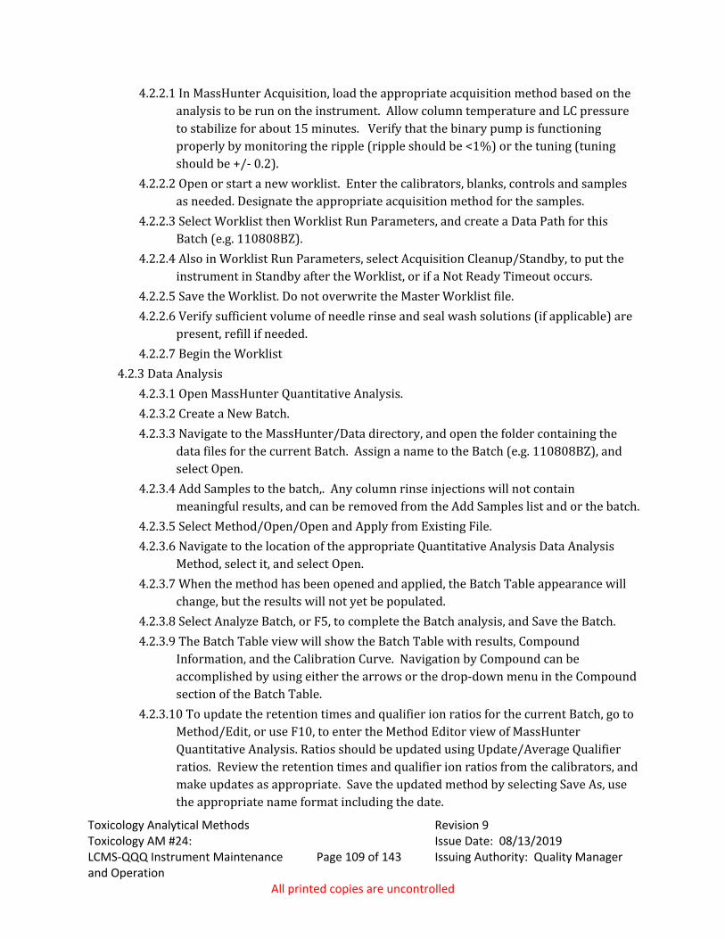
Toxicology Analytical Methods Revision 9Issue Date: 08/13/2019Toxicology AM #24:
LCMS-QQQ Instrument Maintenance and Operation
Page 109 of 143 Issuing Authority: Quality Manager
All printed copies are uncontrolled
4.2.2.1 In MassHunter Acquisition, load the appropriate acquisition method based on the analysis to be run on the instrument. Allow column temperature and LC pressure to stabilize for about 15 minutes. Verify that the binary pump is functioning properly by monitoring the ripple (ripple should be <1%) or the tuning (tuning should be +/- 0.2).
4.2.2.2 Open or start a new worklist. Enter the calibrators, blanks, controls and samples as needed. Designate the appropriate acquisition method for the samples.
4.2.2.3 Select Worklist then Worklist Run Parameters, and create a Data Path for this Batch (e.g. 110808BZ).
4.2.2.4 Also in Worklist Run Parameters, select Acquisition Cleanup/Standby, to put the instrument in Standby after the Worklist, or if a Not Ready Timeout occurs.
4.2.2.5 Save the Worklist. Do not overwrite the Master Worklist file. 4.2.2.6 Verify sufficient volume of needle rinse and seal wash solutions (if applicable) are
present, refill if needed.4.2.2.7 Begin the Worklist
4.2.3 Data Analysis4.2.3.1 Open MassHunter Quantitative Analysis. 4.2.3.2 Create a New Batch.4.2.3.3 Navigate to the MassHunter/Data directory, and open the folder containing the
data files for the current Batch. Assign a name to the Batch (e.g. 110808BZ), and select Open.
4.2.3.4 Add Samples to the batch,. Any column rinse injections will not contain meaningful results, and can be removed from the Add Samples list and or the batch.
4.2.3.5 Select Method/Open/Open and Apply from Existing File.4.2.3.6 Navigate to the location of the appropriate Quantitative Analysis Data Analysis
Method, select it, and select Open. 4.2.3.7 When the method has been opened and applied, the Batch Table appearance will
change, but the results will not yet be populated.4.2.3.8 Select Analyze Batch, or F5, to complete the Batch analysis, and Save the Batch.4.2.3.9 The Batch Table view will show the Batch Table with results, Compound
Information, and the Calibration Curve. Navigation by Compound can be accomplished by using either the arrows or the drop-down menu in the Compound section of the Batch Table.
4.2.3.10 To update the retention times and qualifier ion ratios for the current Batch, go to Method/Edit, or use F10, to enter the Method Editor view of MassHunter Quantitative Analysis. Ratios should be updated using Update/Average Qualifier ratios. Review the retention times and qualifier ion ratios from the calibrators, and make updates as appropriate. Save the updated method by selecting Save As, use the appropriate name format including the date.
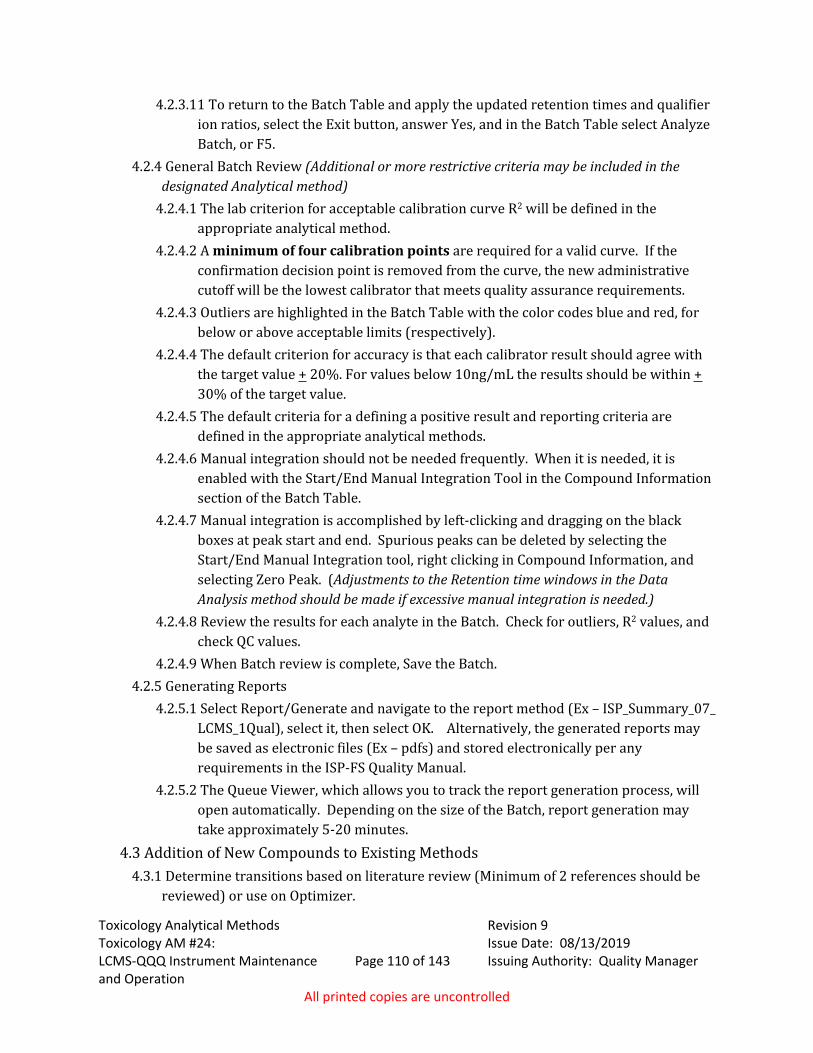
Toxicology Analytical Methods Revision 9Issue Date: 08/13/2019Toxicology AM #24:
LCMS-QQQ Instrument Maintenance and Operation
Page 110 of 143 Issuing Authority: Quality Manager
All printed copies are uncontrolled
4.2.3.11 To return to the Batch Table and apply the updated retention times and qualifier ion ratios, select the Exit button, answer Yes, and in the Batch Table select Analyze Batch, or F5.
4.2.4 General Batch Review (Additional or more restrictive criteria may be included in the designated Analytical method)
4.2.4.1 The lab criterion for acceptable calibration curve R2 will be defined in the appropriate analytical method.
4.2.4.2 A minimum of four calibration points are required for a valid curve. If the confirmation decision point is removed from the curve, the new administrative cutoff will be the lowest calibrator that meets quality assurance requirements.
4.2.4.3 Outliers are highlighted in the Batch Table with the color codes blue and red, for below or above acceptable limits (respectively).
4.2.4.4 The default criterion for accuracy is that each calibrator result should agree with the target value + 20%. For values below 10ng/mL the results should be within + 30% of the target value.
4.2.4.5 The default criteria for a defining a positive result and reporting criteria are defined in the appropriate analytical methods.
4.2.4.6 Manual integration should not be needed frequently. When it is needed, it is enabled with the Start/End Manual Integration Tool in the Compound Information section of the Batch Table.
4.2.4.7 Manual integration is accomplished by left-clicking and dragging on the black boxes at peak start and end. Spurious peaks can be deleted by selecting the Start/End Manual Integration tool, right clicking in Compound Information, and selecting Zero Peak. (Adjustments to the Retention time windows in the Data Analysis method should be made if excessive manual integration is needed.)
4.2.4.8 Review the results for each analyte in the Batch. Check for outliers, R2 values, and check QC values.
4.2.4.9 When Batch review is complete, Save the Batch.4.2.5 Generating Reports
4.2.5.1 Select Report/Generate and navigate to the report method (Ex – ISP_Summary_07_ LCMS_1Qual), select it, then select OK. Alternatively, the generated reports may be saved as electronic files (Ex – pdfs) and stored electronically per any requirements in the ISP-FS Quality Manual.
4.2.5.2 The Queue Viewer, which allows you to track the report generation process, will open automatically. Depending on the size of the Batch, report generation may take approximately 5-20 minutes.
4.3 Addition of New Compounds to Existing Methods4.3.1 Determine transitions based on literature review (Minimum of 2 references should be
reviewed) or use on Optimizer.
Toxicology Analytical Methods Revision 9Issue Date: 08/13/2019Toxicology AM #24:
LCMS-QQQ Instrument Maintenance and Operation
Page 111 of 143 Issuing Authority: Quality Manager
All printed copies are uncontrolled
4.3.2 Dilute appropriately (for example ~ 10µL of a 1mg/mL solution standard in 1mL of methanol.)
4.3.3 Prep Instrument for acquisition as generally performed.4.3.4 Load or create a new analyte MRM method using the current acquisition parameters for
all but the QQQ tab. Set the analyte transitions in the appropriate location within the acquisition method
4.3.5 Run each diluted analyte vial using the MRM method with the Optimizer or literature transitions.
4.3.6 Review data using Qualitative Data Analysis to determine Retention time of the analyte.4.3.7 Add transitions into the currently used Acquisition method.4.3.8 Request addition of new analyte to the ToxBox plate order (Should only be completed by
the person designated to order the plates).4.3.9 Evaluate the calibration Curves of each analyte, if acceptance criteria is met over a
minimum of 5 batches, the new analytes may be added to the Analytical method and the documentation shall be centrally stored by the Discipline lead.
4.3.10 At no point should analytes being verified for addition be included in the analytical notes/documentation for case work. Note: A separate data analysis method should be used in the evaluation of new analytes.
4.4 Removing Analyte(s)4.4.1 Review all batch data for the analytical method completed on all instruments utilized by
the ISPFS Toxicology discipline performed within the last 12 calendar month. 4.4.2 If detection of the analyte has not been seen over the specified time frame,
recommendation for removal should be sent to the discipline lead. 4.4.3 The Discipline lead shall review all documentation associated with the analyte removal
and update the corresponding analytical method accordingly. 4.5 Quality Assurance Requirements
4.5.1 Refer to Toxicology AM #19 and AM #21 for additional quality assurance and reference material authentication requirements.
4.6 Analysis Documentation4.6.1 Case results are to be recorded in the iLIMS system.4.6.2 Reports for the batch and controls, if printed, will be stored centrally in the lab in which
the analysis was performed. Electronic storage is also permitted in lieu of printed copies. A copy of data for controls may be stored electronically in a central location and need not be included in individual case files.
4.6.3 The data from the run will be stored electronically, and if it is on a computer, will be backed up at least every two months.
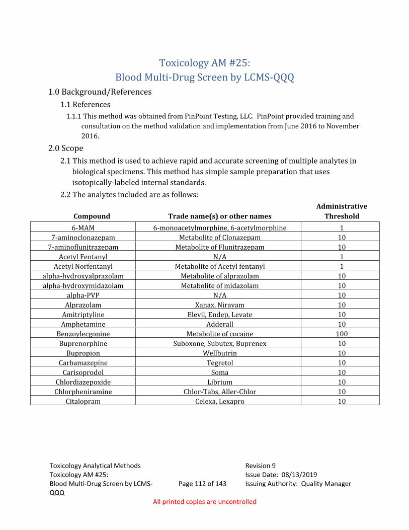
Toxicology Analytical Methods Revision 9Issue Date: 08/13/2019Toxicology AM #25:
Blood Multi-Drug Screen by LCMS-QQQ
Page 112 of 143 Issuing Authority: Quality Manager
All printed copies are uncontrolled
Toxicology AM #25: Blood Multi-Drug Screen by LCMS-QQQ
1.0 Background/References1.1 References
1.1.1 This method was obtained from PinPoint Testing, LLC. PinPoint provided training and consultation on the method validation and implementation from June 2016 to November 2016.
2.0 Scope2.1 This method is used to achieve rapid and accurate screening of multiple analytes in
biological specimens. This method has simple sample preparation that uses isotopically-labeled internal standards.
2.2 The analytes included are as follows:
Compound Trade name(s) or other namesAdministrative
Threshold6-MAM 6-monoacetylmorphine, 6-acetylmorphine 1
7-aminoclonazepam Metabolite of Clonazepam 107-aminoflunitrazepam Metabolite of Flunitrazepam 10
Acetyl Fentanyl N/A 1Acetyl Norfentanyl Metabolite of Acetyl fentanyl 1
alpha-hydroxyalprazolam Metabolite of alprazolam 10alpha-hydroxymidazolam Metabolite of midazolam 10
alpha-PVP N/A 10Alprazolam Xanax, Niravam 10
Amitriptyline Elevil, Endep, Levate 10Amphetamine Adderall 10
Benzoylecgonine Metabolite of cocaine 100Buprenorphine Suboxone, Subutex, Buprenex 10
Bupropion Wellbutrin 10Carbamazepine Tegretol 10
Carisoprodol Soma 10Chlordiazepoxide Librium 10Chlorpheniramine Chlor-Tabs, Aller-Chlor 10
Citalopram Celexa, Lexapro 10
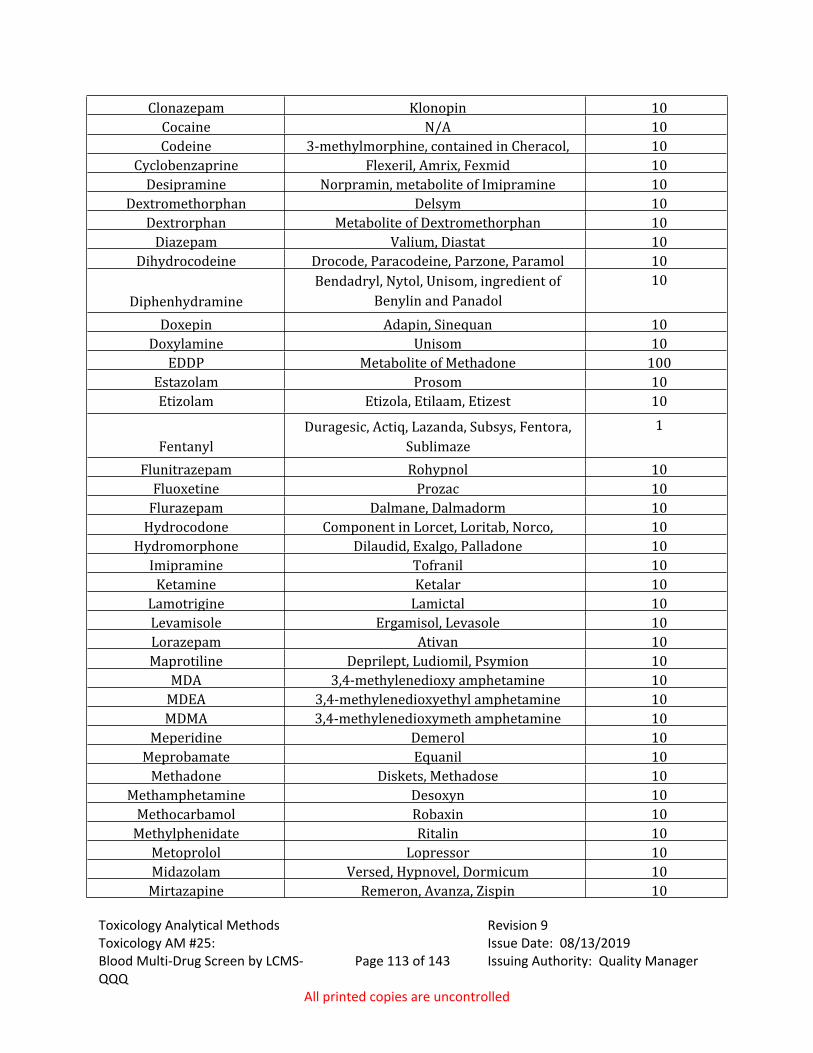
Toxicology Analytical Methods Revision 9Issue Date: 08/13/2019Toxicology AM #25:
Blood Multi-Drug Screen by LCMS-QQQ
Page 113 of 143 Issuing Authority: Quality Manager
All printed copies are uncontrolled
Clonazepam Klonopin 10Cocaine N/A 10Codeine 3-methylmorphine, contained in Cheracol,
Robitussin A-C,10
Cyclobenzaprine Flexeril, Amrix, Fexmid 10Desipramine Norpramin, metabolite of Imipramine 10
Dextromethorphan Delsym 10Dextrorphan Metabolite of Dextromethorphan 10
Diazepam Valium, Diastat 10Dihydrocodeine Drocode, Paracodeine, Parzone, Paramol 10
DiphenhydramineBendadryl, Nytol, Unisom, ingredient of
Benylin and Panadol10
Doxepin Adapin, Sinequan 10Doxylamine Unisom 10
EDDP Metabolite of Methadone 100Estazolam Prosom 10Etizolam Etizola, Etilaam, Etizest 10
FentanylDuragesic, Actiq, Lazanda, Subsys, Fentora,
Sublimaze1
Flunitrazepam Rohypnol 10Fluoxetine Prozac 10
Flurazepam Dalmane, Dalmadorm 10Hydrocodone Component in Lorcet, Loritab, Norco,
Vicodin, Hycodan, Tussionex10
Hydromorphone Dilaudid, Exalgo, Palladone 10Imipramine Tofranil 10
Ketamine Ketalar 10Lamotrigine Lamictal 10Levamisole Ergamisol, Levasole 10Lorazepam Ativan 10Maprotiline Deprilept, Ludiomil, Psymion 10
MDA 3,4-methylenedioxy amphetamine 10MDEA 3,4-methylenedioxyethyl amphetamine 10MDMA 3,4-methylenedioxymeth amphetamine 10
Meperidine Demerol 10Meprobamate Equanil 10
Methadone Diskets, Methadose 10Methamphetamine Desoxyn 10
Methocarbamol Robaxin 10Methylphenidate Ritalin 10
Metoprolol Lopressor 10Midazolam Versed, Hypnovel, Dormicum 10
Mirtazapine Remeron, Avanza, Zispin 10
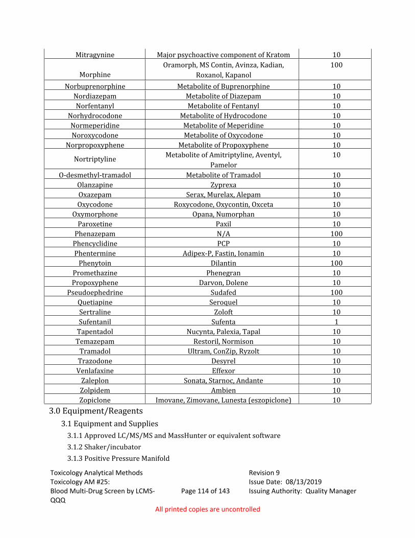
Toxicology Analytical Methods Revision 9Issue Date: 08/13/2019Toxicology AM #25:
Blood Multi-Drug Screen by LCMS-QQQ
Page 114 of 143 Issuing Authority: Quality Manager
All printed copies are uncontrolled
Mitragynine Major psychoactive component of Kratom 10
MorphineOramorph, MS Contin, Avinza, Kadian,
Roxanol, Kapanol100
Norbuprenorphine Metabolite of Buprenorphine 10Nordiazepam Metabolite of Diazepam 10Norfentanyl Metabolite of Fentanyl 10
Norhydrocodone Metabolite of Hydrocodone 10Normeperidine Metabolite of Meperidine 10Noroxycodone Metabolite of Oxycodone 10
Norpropoxyphene Metabolite of Propoxyphene 10
Nortriptyline Metabolite of Amitriptyline, Aventyl, Pamelor
10
O-desmethyl-tramadol Metabolite of Tramadol 10Olanzapine Zyprexa 10Oxazepam Serax, Murelax, Alepam 10Oxycodone Roxycodone, Oxycontin, Oxceta 10
Oxymorphone Opana, Numorphan 10Paroxetine Paxil 10
Phenazepam N/A 100Phencyclidine PCP 10Phentermine Adipex-P, Fastin, Ionamin 10
Phenytoin Dilantin 100Promethazine Phenegran 10Propoxyphene Darvon, Dolene 10
Pseudoephedrine Sudafed 100Quetiapine Seroquel 10Sertraline Zoloft 10Sufentanil Sufenta 1
Tapentadol Nucynta, Palexia, Tapal 10Temazepam Restoril, Normison 10
Tramadol Ultram, ConZip, Ryzolt 10Trazodone Desyrel 10Venlafaxine Effexor 10
Zaleplon Sonata, Starnoc, Andante 10Zolpidem Ambien 10Zopiclone Imovane, Zimovane, Lunesta (eszopiclone) 10
3.0 Equipment/Reagents3.1 Equipment and Supplies
3.1.1 Approved LC/MS/MS and MassHunter or equivalent software 3.1.2 Shaker/incubator 3.1.3 Positive Pressure Manifold
Toxicology Analytical Methods Revision 9Issue Date: 08/13/2019Toxicology AM #25:
Blood Multi-Drug Screen by LCMS-QQQ
Page 115 of 143 Issuing Authority: Quality Manager
All printed copies are uncontrolled
3.1.4 SPE dry 3.1.5 Phenomenex Phenyl Hexyl (4.6 x 50 mm; 2.6 µm)3.1.6 Calibrated pipette for dispensing blood and/or urine samples3.1.7 Single or Multi-channel pipettes for all other transfers and additions 3.1.10 ToxBox kit with 96 well plate containing internal standards, and controls, 96 well SLE+
plate, and 96 well blank sample preparation plate.3.1.11 Appropriate sized pipette tips3.1.12 Heat sealing foil covers for 96 well plate3.1.13 ACT plate cover for use during drying step with SPE dry (if run contains urine samples)3.1.14 Additional 48 or 96-well plate (if run contains urine samples)
3.2 ReagentsSee AM# 23 for Solution Preparation instructions3.2.1 10mM Ammonium Formate in LCMS Water (mobile phase A)3.2.2 0.1% Formic Acid in LCMS Methanol (mobile phase B)3.2.3 0.5 M Ammonium Hydroxide3.2.4 Formic Acid (LCMS grade)3.2.5 Water (LCMS grade)3.2.6 Methanol (LCMS grade)3.2.7 Ammonium Formate (LCMS)3.2.8 Ammonium Hydroxide (ACS or higher)3.2.9 Ethyl Acetate (ACS or higher)3.2.9.10 100% LCMS Methanol in LCMS Water (Reconstitution Solvent)3.2.9.11 LC/MS Needle Rinse (40/40/20: Water/Methanol/Isopropanol or 75% MeOH in
water)3.2.10 Beta-glucuronidase (Kura BG-100 or BGTurbo) (if run contains urine samples)3.2.11 0.1M ammonium acetate buffer (pH 5) (if using BG-100)3.2.12 Instant Buffer I (provided with BGTurbo) (if using BGTurbo)
3.3 Quality Assurance: Reference Materials and Controls3.3.1 Internal Controls, and Internal Standards.
3.3.1.1 Internal controls and internal standards are prepared by the ToxBox plate manufacturer and contained on the 96 well plate. A minimum of one internal positive control that has a concentration near the administrative threshold level for each compound will be run with each extraction.
Toxicology Analytical Methods Revision 9Issue Date: 08/13/2019Toxicology AM #25:
Blood Multi-Drug Screen by LCMS-QQQ
Page 116 of 143 Issuing Authority: Quality Manager
All printed copies are uncontrolled
3.3.2 A negative control will be run with each extraction. If the run contains urine samples, both a blood and urine negative control must be included.
4.0 Procedure4.1 Extraction Procedure
4.1.1 Allow refrigerated or frozen samples and frozen components to reach ambient temperature.
4.1.2 Urine samples must be hydrolyzed prior to extraction. For urine samples/controls only: Add 250 µl of blank urine to a clean 48 or 96 well plate. If using BG-100, add 50 µl of the BG-100 and 200 µl of 0.1 M ammonium acetate buffer (pH 5) to each well containing urine. Place a cover on the plate and shake at 900 rpm for approximately 180 minutes at 60 degrees Celsius. If using BGTurbo, add 40 µl of BGTurbo and 100 µl of 500mM sodium phosphate (pH 6.8) to the wells containing urine. Place a cover on the plate and shake for a minimum of 5 minutes at ambient temperature.
Toxicology Analytical Methods Revision 9Issue Date: 08/13/2019Toxicology AM #25:
Blood Multi-Drug Screen by LCMS-QQQ
Page 117 of 143 Issuing Authority: Quality Manager
All printed copies are uncontrolled
4.1.3 Using a calibrated single channel pipette, add 250 µL of blank blood into the appropriate wells of the analytical (standards) plate (wells containing internal positive and negative controls). Pipette the appropriate sample/case blood into the appropriate wells containing only internal standards. For urine samples/controls: after shaking, transfer 250 µl of the urine/buffer/BG-100 or urine/buffer/BGTurbo to the appropriate wells containing only internal standard on the analytical plate.
4.1.4 Place plate on shaking incubator at approximately 900 rpm for approximately 15 minutes.
4.1.5 Pipette 250 µL of 0.5 M ammonium hydroxide buffer into all wells in use on the analytical (standards) plate.
4.1.6 Place plate on shaking incubator at approximately 900 rpm for approximately 15 minutes.
4.1.7 Transfer 300 µL of blood+base and urine + base mixture (if applicable) to the corresponding wells of SLE+ plate.
4.1.8 Apply positive pressure for approximately 10-15 seconds or until no liquid remains on top of sorbent. Wait 5 minutes. (Positive Pressure manifold setting for this loading step is between 85-100 PSI)
4.1.9 Add 900 µL of ethyl acetate and allow to flow through for approximately 5 minutes under gravity.
4.1.10 Apply positive pressure for approximately 10-15 seconds. (Positive Pressure manifold setting for this step is between 12-15 PSI).
4.1.11 Add 900 µL of ethyl acetate and allow to flow through for approximately 5 minutes under gravity.
4.1.12 Apply positive pressure for approximately 10-15 seconds. (Positive Pressure manifold setting for this step is between 12-15 PSI).
4.1.13 Remove plate containing eluate. Place on SPE Dry and evaporate to dryness at approximately 35 degrees Celsius. If run contains urine samples, place ACT cover on top of plate prior to drying.
4.1.14 Reconstitute in 100 µL LCMS grade methanol and heat seal plate with foil.4.1.15 Analyze samples immediately or freeze for future analysis.
Toxicology Analytical Methods Revision 9Issue Date: 08/13/2019Toxicology AM #25:
Blood Multi-Drug Screen by LCMS-QQQ
Page 118 of 143 Issuing Authority: Quality Manager
All printed copies are uncontrolled
4.2 Instrument and Run set up.4.2.1 See Toxicology AM #24 for instrument maintenance and operation.4.2.2 Instrument run parameters and the acquisition methods for this analytical method will
be printed out (paper or electronically) and stored centrally in the lab or in a common file folder electronically.
4.2.2.1 Acquisition Required Settings 4.2.2.1.1 Column Temperature - 35⁰C4.2.2.1.2 Injection volume – 0.5-10µL 4.2.2.1.3 Mobile Phase Flow rate - 0.5mL/min4.2.2.1.3: Binary Pump Gradient Settings (Pump Time Table) (Note: the gradient
parameters may be adjusted slightly but adequate separation of isobaric compounds must be demonstrated).
Time (min) % Mobile A % Mobile B0 95 54 0 1005 0 1005.1 95 59 95 5
4.2.3 Worklist Set up should include internal control, negative control(s) and case samples. 4.3 Evaluation of Results
4.3.1 Minimum Criteria:4.3.1.1 Signal to noise of primary transition greater than 5.4.3.1.2 Signal to noise of secondary transition greater than 3. 4.3.1.3 In cases that do not meet the criteria above, the analyst may also evaluate peak
Symmetry/Resolution and it is at their discretion to determine if the sample exhibits a positive response for that compound. Correct peak shape (as compared with QCs) and the sufficient resolution of potential interferents from the analyte of interest can be considered.
4.3.1.4 To ensure the response is adequate, each compound in the internal control will be evaluated using the criteria listed above. If a compound does not meet these standards, samples cannot be evaluated for that compound. At the analyst’s discretion, that compound can be removed from the list of compounds that the sample was evaluated for, or the sample can be reinjected or re-extracted. If a sample needs to be reinjected, the analyst may add more of the reconstitution solvent to facilitate this. The internal standards must give an adequate response in order for the sample to be evaluated.
Toxicology Analytical Methods Revision 9Issue Date: 08/13/2019Toxicology AM #25:
Blood Multi-Drug Screen by LCMS-QQQ
Page 119 of 143 Issuing Authority: Quality Manager
All printed copies are uncontrolled
4.3.1.5 Retention time criterion for peak identification is a ±2% or +/-0.100 min, whichever is greater, retention time window relative to the internal control and/or internal standards around the analytes retention time. At the analyst’s discretion when the analyte peak falls outside the retention time window and the internal standard shifts comparably that sample may be evaluated as positive if the other criteria are met.
4.3.1.6 Using the internal plate control: a 1-point calibration curve will be established. The curve will be set to linear, non-weighted and origin set to force. The concentration will be set to 10 for all compounds. This is not the actual concentration of the compounds but a number used to compare sample responses.
4.3.1.7 Case Samples and negative controls will generally be considered negative if the calculated concentration is less than 5. Samples with a calculated concentration between 2 and 5 may be evaluated as positive at the analyst’s discretion. If calculated concentration is less than 10 for methamphetamine it may be evaluated as negative.
4.3.1.8 Samples should have a significant internal standard response, if a sample has little to no internal standard response for a compound, an evaluation for that sample cannot be made. The analyst will need to reinject the sample, remove that drug from the list of drugs that sample was screened for or re-extract the sample.
4.3.2 Limitations of Method4.3.2.1 Samples flagged as positive for Maprotiline and Amitriptyline will be reported as
Amitriptyline/Maprotiline. 4.3.2.2 Samples that indicate as positive for Benzoylecgonine will be reported as
Benzoylecgonine/Cocaine break-down product (or other wording that indicates that the response may be from a breakdown of cocaine).
4.4 Quality Assurance Requirements4.4.1 Refer to Toxicology AM #19 and AM #21 for additional quality assurance and reference
material authentication requirements.4.5 Analysis Documentation
4.5.1 A print out showing compound name, retention time, calculated concentration, internal standard retention time and response will be printed out for the case sample and included in the analyst’s notes.
4.5.2 The print out (paper or electronic) for the negative control and internal control will be stored centrally in the lab in which they were performed or stored on a network drive.
4.5.3 The data from the run will be stored electronically, and if it is on a computer, will be backed up at least every two months.
Toxicology Analytical Methods Revision 9Issue Date: 08/13/2019Toxicology AM #25:
Blood Multi-Drug Screen by LCMS-QQQ
Page 120 of 143 Issuing Authority: Quality Manager
All printed copies are uncontrolled
4.6 Limitation of method4.6.1 This method is a preliminary screen. The screen results are an indication of drugs that
may be present in a sample. The weight or confidence in screening results cannot be given the same as a confirmatory test. If these results are reported it must be clear on the report that it is an indicator the drug is present in the sample, not a confirmation of the drug in the sample.
Toxicology Analytical Methods Revision 9Issue Date: 08/13/2019Toxicology AM #26: Blood THC and
Metabolites Screen by LCMS-QQQ
Page 121 of 143 Issuing Authority: Quality Manager
All printed copies are uncontrolled
Toxicology AM #26: Blood THC and Metabolites Screen by LCMS-QQQ
1.0 Background/References1.1 Background9-THC (Figure 1) is the chief psychoactive cannabinoid resulting from exposure to marijuana.
9-THC has a peak blood concentration within 5 to 15 minutes following smoking of a marijuana cigarette.4,5,6 This blood concentration drops rapidly after cessation of smoking.4,5 The level may fall to less than 5 ng within 30 to 60 minutes, although longer detection times have been reported.4,5 Detection of low dose (1.75%) post-smoking 9-THC has been reported to vary from 3 to 12 hours.5 This detection window was based on a limit of quantitation of 0.5 ng/mL. The number, duration, spacing of puffs, hold time, and inhalation volume all impact the degree of drug exposure and thus bioavailability.5 Longer detection times have been observed for frequent users. 9-THC is metabolized to an active metabolite, 11-hydroxy-tetrahydrocannabinol (Hydroxy-THC, THC-OH), and an inactive metabolite, 11-nor-9-carboxy-tetrahydrocannabinol (carboxy-THC). The concentration of the 9-THC inactive metabolite, carboxy-THC, gradually increases and may plateau for several hours.5 There is poor correlation between blood 9-THC and psychoactive effects since the 9-THC concentrations begin to decline prior to the time of peak effects.4,5,6 The detection window for the active analytes is much shorter than that of carboxy-THC.7 Cannabinol and cannabidiol are minor cannabinoids also found in marijuana.8,9 Due to their low abundance, rapid clearance from blood, and similar pharmacokinetic profiles in chronic versus occasional smokers, the presence of cannabidiol and cannabinol in blood has been suggested to be a sufficient but unnecessary marker for recent marijuana use.8
Negative behavioral effects reported from exposure to marijuana include altered time perception, lack of concentration, and impaired learning and memory which can lead to impairment of cognitive and performance tasks.5 Establishing impairment in an individual is based on evaluation of all available information in conjunction with quantitative blood levels.
For additional background, refer to Toxicology AM #3, obsolete analytical method 3.10.1, and the provided references.
1.2 References1.2.1 This method was obtained from PinPoint Testing, LLC. The method validation and
implementation was completed by ISPFS personnel from October 2016 to November 2016.
1.2.2 (4)Standard Operating Procedure for Blood SPE THC and Carboxy-THC GC/MSD Assay, Edmonton, Canada Office of the Chief Medical Examiners, 2003.
Toxicology Analytical Methods Revision 9Issue Date: 08/13/2019Toxicology AM #26: Blood THC and
Metabolites Screen by LCMS-QQQ
Page 122 of 143 Issuing Authority: Quality Manager
All printed copies are uncontrolled
1.2.3 (5) Huestis, M.A., Cannabis (Marijuana) - Effects on Human Behavior and Performance, Forensic Science Rev. 14(1/2): 16-60, 2002.
1.2.4 (6)Drummer, O.H., Cannabis, pp. 178-212. In: The Forensic Pharmacology of Drugs of Abuse, Arnold: London, 2001.
1.2.5 (7)Huestis, M. Marijuana. pp. 229-244. In: Principles of Forensic Toxicology, Second Edition. Levine, B. ed., AACC, 2003.
1.2.6 (8)Desrosiers, N.A.; Himes, S.K.; Scheidweiler, K.B.; Concheiro-Guisan, M.; Huestis, M.A. Phase I and II Cannbinoid Disposition in Blood and Plasma of Occasional and Frequent Smokers Following Controlled Smoked Cannabis. Clinical Chemistry, 60:4, pp. 631-643, 2014.
1.2.7 (9)Nadulski, T., et al. Simultaneous and Sensitive Analysis of THC, 11-OH0THC, THC-COOH, CBD, and CBN by GC-MS in Plasma after Oral Application of Small Doses of THC and Cannabis Extract. Journal of Analytical Toxicology, Vol 29, pp. 782-789, November/December 2005.
2.0 Scope2.1 This method is used to achieve rapid and accurate screening of cannabinoids in
biological specimens. This method has simple sample preparation that uses isotopically-labeled internal standards.
2.2 The analytes included are as follows: Analyte Administrative Threshold
THC 3 ng/mLCarboxy-THC 10 ng/mL
THC-OH 3 ng/mL
3.0 Equipment/Reagents3.1 Equipment and Supplies
3.1.1 Approved LC/MS/MS and MassHunter or equivalent software 3.1.2 Shaking incubator3.1.3 ToxBox kit with 48 Well plate containing internal standards and controls, 48 well SLE+
plate, and 48 well blank sample collection plate.3.1.4 Test tube rocker3.1.5 Calibrated pipette for dispensing blood and/or urine samples3.1.6 Single or Multi-channel pipettes for all other transfers and additions3.1.7 Positive Pressure Manifold3.1.8 SPE sample evaporator concentrator3.1.9 ACT plate cover for use during drying step with SPE dry (if run contains urine samples)3.1.10 Additional 48-well plate (if run contains urine samples)

Toxicology Analytical Methods Revision 9Issue Date: 08/13/2019Toxicology AM #26: Blood THC and
Metabolites Screen by LCMS-QQQ
Page 123 of 143 Issuing Authority: Quality Manager
All printed copies are uncontrolled
3.2 ReagentsRefer to Toxicology AM #23 for Solution Preparation instructions.3.2.1 0.1% Formic Acid in Water (LCMS grade)3.2.2 0.1% Formic Acid in Methanol (LCMS grade) 3.2.3 Methyl Tert-Butyl Ether (MTBE) 99.9%3.2.4 Hexanes (ACS)3.2.5 Methanol (LCMS grade)3.2.6 10 mM Ammonium Formate3.2.7 LC/MS Needle Rinse (40/40/20: Water/Methanol/Isopropanol or 75% MeOH in water)3.2.8 1 N KOH (if run includes urine samples)
3.3 Quality Assurance: Reference Materials, Controls, and Blanks3.3.1 Plate controls and internal standards are prepared by the ToxBox Manufacturer and are
contained on the 48 well plate. A minimum of one internal positive plate control will be included in each run. A minimum of three calibrators will be run to establish a calculated response value for the case samples (one must be between the nominal value of 3 and 5) control will be included in each run.
3.3.2 A negative control will be run with each extraction. If the run contains urine samples, a negative urine control must also be included.
4.0 Procedure4.1 Extraction Procedure
4.1.1 Allow refrigerated or frozen samples and frozen components to reach ambient temperature.
4.1.2 Urine samples must be hydrolyzed prior to extraction. For urine samples/controls only: Add 1.5 mL of blank urine to a clean 48-well plate. Add 250 µL of 1 N KOH to the wells containing urine. Place a cover on the plate and shake at 900 rpm for approximately 15 minutes at 40 degrees Celsius.
4.1.3 Using a calibrated single channel pipette, add 1.0 mL of blank blood to the appropriate sample wells. Of the analytical (standards) plate (wells containing internal positive and negative controls). Pipette the appropriate sample/case blood into the appropriate wells containing only internal standards. For urine samples/controls: after shaking, transfer 1.0 mL of the urine/buffer to the appropriate wells containing internal standard on the analytical plate.
4.1.4 Place plate on shaking incubator at approximately 900 rpm for approximately 15 minutes.
4.1.5 Add 0.5 mL 0.1% formic acid in LCMS water to all of the wells containing blood. Add 0.5 mL of saturated phosphate buffer to all of the wells containing urine.
4.1.6 Place plate on shaking incubator at approximately 900 rpm for approximately 15 minutes.
Toxicology Analytical Methods Revision 9Issue Date: 08/13/2019Toxicology AM #26: Blood THC and
Metabolites Screen by LCMS-QQQ
Page 124 of 143 Issuing Authority: Quality Manager
All printed copies are uncontrolled
4.1.7 Transfer 800 µL of blood+acid or urine+acid mixture (if applicable) to the corresponding wells of the SLE+ plate.
4.1.8 Apply positive pressure for approximately 10-15 seconds or until no liquid remains on top of sorbent. (Recommended setting for Positive Pressure manifold for this loading step is between 80-95 PSI)
4.1.9 Wait 5 minutes for sample to completely absorb.4.1.10 Add 2.25 mL MTBE and allow to flow for 5 minutes under gravity. This should be
completed in increments, as the sample well accommodates a maximum volume of 1 mL. (Recommended for this step is: three (3) repetitions of adding 750 µL)
4.1.11 Apply positive pressure to complete elution- approximately 15 seconds. (Positive Pressure manifold set between 12-15 PSI)
4.1.12 Add 2.25 mL hexane and allow to flow for 5 minutes under gravity. This should be completed in increments, as the sample well accommodates a maximum volume of 1mL. (Recommended for this step is: three (3) repetitions of adding 750 µL)
4.1.13 Apply positive pressure to complete elution- approximately 15 seconds. (Positive Pressure manifold set between 12-15 PSI)
4.1.14 Remove plate containing eluate. Place on SPE Dry and evaporate to dryness at approximately 35 degrees Celsius. If run contains urine samples, place ACT cover on top of plate prior to drying. Temperatures for this step are not critical and it is appropriate to use the equipment temperature readings. The temperatures does not need to be monitored or verified with a traceable thermometer.
4.1.15 Reconstitute each sample with 100 µL LCMS Grade Methanol. Heat seal plate with foil to prevent evaporation.
4.1.16 Analyze samples immediately or freeze for future analysis.4.2 Instrument and Run set up.
4.2.1 See Toxicology AM #24 for instrument maintenance and operation.4.2.2 Instrument run parameters and the acquisition methods for this Analytical Method will
be printed out (paper or electronically) and stored centrally in the lab or in a common file folder electronically.
4.2.2.1 Acquisition Required Settings 4.2.2.1.1 Column Temperature - 35°C4.2.2.1.2 Injection volume – 0.5-10µL 4.2.2.1.3 Mobile Phase Flow rate - 0.6mL/min4.2.2.1.4 Binary Pump Gradient Settings (Pump Time Table) (Note: the gradient
parameters may be adjusted slightly but adequate separation of the compounds must be demonstrated).
Time (min) % Mobile A % Mobile B0 55 450.5 0 100
Toxicology Analytical Methods Revision 9Issue Date: 08/13/2019Toxicology AM #26: Blood THC and
Metabolites Screen by LCMS-QQQ
Page 125 of 143 Issuing Authority: Quality Manager
All printed copies are uncontrolled
2.0 0 1002.5 25 753.5 55 455.0 55 45
4.2.3 Worklist Set up should include a positive control, calibrators, negative control, and case samples.
4.3 Evaluation of Results4.3.1 Minimum Criteria:
4.3.1.1 A sample or control with a concentration of 3 ng/mL or greater for THC or THC-OH, or 10
ng/mL or greater for carboxy-THC will be considered positive. Samples and controls with a carboxy-THC response between 5 and 10 ng/mL may be evaluated as positive at the analyst’s discretion.
4.3.1.2 Retention time within +/- 2% or +/-0.100 min whichever is greater of the average retention time of the calibrators. At the analyst’s discretion when the analyte peak falls outside the retention time window and the internal standard shifts comparably that sample may be evaluated as positive if the other criteria are met.
4.3.1.4 To ensure the response is adequate, each compound in the internal control will be evaluated using the criteria listed above. If a compound does not meet these standards, samples cannot be evaluated for that compound. At the analyst’s discretion, that compound can be removed from the list of compounds that the sample was evaluated for, or the sample can be reinjected or re-extracted. If a sample needs to be reinjected, the analyst may add more of the reconstitution solvent to facilitate this. The internal standards must give an adequate response in order for the sample to be evaluated.
4.3.1.7 Samples should have a significant internal standard response, if a sample has little to no internal standard response for a compound, an evaluation for that sample cannot be made. The analyst will need to re-inject or re-extract that sample.
4.4 Quality Assurance Requirements4.4.1 Refer to Toxicology AM #19 and AM #21 for additional quality assurance and reference
material authentication requirements.4.5 Analysis Documentation
4.5.1 The printed results for each case sample will be included with the analyst’s notes. Case results are to be recorded in the iLIMS system.
4.5.2 The print out (paper or electronic) for the calibrators and controls will be stored centrally in the lab in which they were performed or stored on a network drive.
4.5.3 The data from the run will be stored electronically, and if it is on a computer, will be backed up at least every two months.
4.6 Limitation of method
Toxicology Analytical Methods Revision 9Issue Date: 08/13/2019Toxicology AM #26: Blood THC and
Metabolites Screen by LCMS-QQQ
Page 126 of 143 Issuing Authority: Quality Manager
All printed copies are uncontrolled
4.6.1 This method is a preliminary screen, the screen results are an indication of drugs that may be present in a sample. The weight or confidence in screening results cannot be given the same as a confirmatory test. This is not a quantitative method and quantitative results cannot be reported from this method. The quantitative aspects of this method are only for establishing a minimum response threshold. If these results are reported it must be clear on the report that it is an indicator the drug is present in the sample, not a confirmation of the drug in the sample.
Toxicology Analytical Methods Revision 9Issue Date: 08/13/2019Toxicology AM #27: Quantitative
Analysis of THC and Metabolites in Blood by LCMS-QQQ
Page 127 of 143 Issuing Authority: Quality Manager
All printed copies are uncontrolled
Toxicology AM #27: Quantitative Analysis of THC and Metabolites in Blood by LCMS-QQQ
1.0 Background/References1.1 Background
9-THC (Figure 1) is the chief psychoactive cannabinoid resulting from exposure to marijuana. 9-THC has a peak blood concentration within 5 to 15 minutes following smoking of a marijuana cigarette.4,5,6 This blood concentration drops rapidly after cessation of smoking.4,5 The level may fall to less than 5 ng within 30 to 60 minutes, although longer detection times have been reported.4,5 Detection of low dose (1.75%) post-smoking 9-THC has been reported to vary from 3 to 12 hours.5 This detection window was based on a limit of quantitation of 0.5 ng/mL. The number, duration, spacing of puffs, hold time, and inhalation volume all impact the degree of drug exposure and thus bioavailability.5 Longer detection times have been observed for frequent users. 9-THC is metabolized to an active metabolite, 11-hydroxy-tetrahydrocannabinol (Hydroxy-THC, THC-OH), and an inactive metabolite, 11-nor-9-carboxy-tetrahydrocannabinol (carboxy-THC). The concentration of the 9-THC inactive metabolite, carboxy-THC, gradually increases and may plateau for several hours.5 There is poor correlation between blood 9-THC and psychoactive effects since the 9-THC concentrations begin to decline prior to the time of peak effects.4,5,6 The detection window for the active analytes is much shorter than that of carboxy-THC.7 Cannabinol and cannabidiol are minor cannabinoids also found in marijuana.8,9 Due to their low abundance, rapid clearance from blood, and similar pharmacokinetic profiles in chronic versus occasional smokers, the presence of cannabidiol and cannabinol in blood has been suggested to be a sufficient but unnecessary marker for recent marijuana use.8
Negative behavioral effects reported from exposure to marijuana include altered time perception, lack of concentration, and impaired learning and memory which can lead to impairment of cognitive and performance tasks.5 Establishing impairment in an individual is based on evaluation of all available information in conjunction with quantitative blood levels.
For additional background, refer to Toxicology AM #3 and obsolete analytical methods 3.10.1, as well as provided references.
1.2 References1.2.1 This method was obtained from PinPoint Testing, LLC. PinPoint provided training and
consultation on the method validation and implementation from June 2016 to October 2016.
1.2.2(4) Standard Operating Procedure for Blood SPE THC and Carboxy-THC GC/MSD Assay, Edmonton, Canada Office of the Chief Medical Examiners, 2003.
Toxicology Analytical Methods Revision 9Issue Date: 08/13/2019Toxicology AM #27: Quantitative
Analysis of THC and Metabolites in Blood by LCMS-QQQ
Page 128 of 143 Issuing Authority: Quality Manager
All printed copies are uncontrolled
1.2.3(5) Huestis, M.A., Cannabis (Marijuana) - Effects on Human Behavior and Performance, Forensic Science Rev. 14(1/2): 16-60, 2002.
1.2.4 (6)Drummer, O.H., Cannabis, pp. 178-212. in: The Forensic Pharmacology of Drugs of Abuse, Arnold: London, 2001.
1.2.5(7) Huestis, M. Marijuana. pp. 229-244. in: Principles of Forensic Toxicology, Second Edition. Levine, B. ed., AACC, 2003.
1.2.6 (8)Desrosiers, N.A.; Himes, S.K.; Scheidweiler, K.B.; Concheiro-Guisan, M.; Huestis, M.A. Phase I and II Cannbinoid Disposition in Blood and Plasma of Occasional and Frequent Smokers Following Controlled Smoked Cannabis. Clinical Chemistry, 60:4, pp. 631-643, 2014.
1.2.7 (9)Nadulski, T., et al. Simultaneous and Sensitive Analysis of THC, 11-OH0THC, THC-COOH, CBD, and CBN by GC-MS in Plasma after Oral Application of Small Doses of THC and Cannabis Extract. Journal of Analytical Toxicology, Vol 29, pp. 782-789, November/December 2005.
2.0 Scope2.1 This method is used for the confirmation of 9-THC, 11-nor-9-THC-9-COOH (Carboxy-THC)
and 11-hydroxy-9-THC (Hydroxy-THC) in blood and urine. The words calibrator and calibration are used to coincide with the terminology in instrument software and manufacturer manuals. The manufacturer’s term calibrator refers to what is considered by ISPFS as reference material that has a certified concentration of drug present.
3.0 Equipment/Reagents3.1 Equipment and Supplies
3.1.1 Approved LC/MS/MS and MassHunter or equivalent software 3.1.2 Shaking incubator3.1.3 ToxBox kit with 48 Well plate containing internal standards and controls, 48 well SLE+
plate, and 48 well blank sample collection plate.3.1.4 Test tube rocker3.1.5 Calibrated pipettes for dispensing blood and/or urine samples.3.1.6 Pipettes for all other transfers and additions.3.1.7 Positive Pressure Manifold3.1.8 SPE sample evaporator concentrator3.1.9 UCT Selectra DA 100 x 2.1 mm, 3um LC Column3.1.10 ACT plate cover for use during drying step with SPE dry (if run contains urine samples)3.1.11 Additional 48-well plate (if run contains urine samples)
Toxicology Analytical Methods Revision 9Issue Date: 08/13/2019Toxicology AM #27: Quantitative
Analysis of THC and Metabolites in Blood by LCMS-QQQ
Page 129 of 143 Issuing Authority: Quality Manager
All printed copies are uncontrolled
3.2 ReagentsRefer to Toxicology AM #23 for Solution Preparation instructions.3.2.1 0.1% Formic Acid in Water (LCMS grade)3.2.2 0.1% Formic Acid in Acetonitrile (LCMS grade) 3.2.3 Methyl Tert-Butyl Ether (MTBE) 99.9%3.2.4 Hexanes (ACS)3.2.5 Methanol (LCMS grade)3.2.6 LC/MS Needle Rinse (40/40/20: Water/Methanol/Isopropanol or 75% MeOH in water)3.2.7 1 N KOH (if run includes urine samples)
3.3 Quality Assurance: Reference Materials and Controls3.3.1 Plate controls and internal standards are prepared by the ToxBox vendor and are
contained on the 48 well plate. A minimum of one positive plate control for each drug compound will be included in each run.
3.3.2 A negative control will be run with each extraction. If the run contains urine samples, a negative urine control will also be included in the run.
3.3.3 A non-extracted blank containing internal standard will be run directly before each case sample to rule out carryover. If confirmation criteria (e.g. ion ratios, RT, S/N) are not met, the analyte is not considered present. If confirmation criteria is met, the response of the blank will be considered. The area response of the blank preceding a sample must be at least 10 times less than any compound confirmed in the case sample, and must be below the limit of confirmation for any analyte confirmed in the case sample to be considered negative. If a blank does contain a compound, the blank and the sample can be reinjected.
3.3.3.1 To prepare blank, dilute 1 part ISTD (obtained from a blank well in the extraction plate) in 4-5 parts LCMS grade methanol.
4.0 Procedure4.1 Extraction Procedure
4.1.1 Allow refrigerated or frozen samples and frozen components to reach ambient temperature.
4.1.2 Urine samples must be hydrolyzed prior to extraction. For urine samples/controls only: Add 1.5 mL of blank urine to a clean 48-well plate. Add 250 µL of 1 N KOH to the wells containing urine. Place a cover on the plate and shake at 900 rpm for approximately 15 minutes at 40 degrees Celsius.
Toxicology Analytical Methods Revision 9Issue Date: 08/13/2019Toxicology AM #27: Quantitative
Analysis of THC and Metabolites in Blood by LCMS-QQQ
Page 130 of 143 Issuing Authority: Quality Manager
All printed copies are uncontrolled
4.1.3 Using a calibrated single channel pipette, add 1.0 mL of blank blood to the appropriate sample wells. Of the analytical (standards) plate (wells containing internal positive and negative controls). Pipette the appropriate sample/case blood into the appropriate wells containing only internal standards. For urine samples/controls: after shaking, transfer 1.0 mL of the urine/buffer to the appropriate wells containing internal standard on the analytical plate.
4.1.4 Place plate on shaking incubator at approximately 900 rpm for approximately 15 minutes.
4.1.5 Add 0.5 mL 0.1% formic acid in LCMS water to all of the wells containing blood. Add 0.5 mL of saturated phosphate buffer to all of the wells containing urine.
4.1.6 Place plate on shaking incubator at approximately 900 rpm for approximately 15 minutes.
4.1.7 Transfer 800 µL of blood+acid or urine+acid mixture (if applicable) to the corresponding wells of the SLE+ plate.
4.1.8 Apply positive pressure for approximately 10-15 seconds or until no liquid remains on top of sorbent. (Recommended setting for Positive Pressure manifold for this loading step is between 80-95 PSI)
4.1.9 Wait 5 minutes for sample to completely absorb.4.1.10 Add 2.25 mL MTBE and allow to flow for 5 minutes under gravity. This should be
completed in increments, as the sample well accommodates a maximum volume of 1mL. (Recommended for this step is: three (3) repetitions of adding 750 μL)
4.1.11 Apply positive pressure to complete elution- approximately 10-15 seconds. (Positive Pressure manifold set between 12-15 PSI)
4.1.12 Add 2.25 mL hexane and allow to flow for 5 minutes under gravity. This should be completed in increments, as the sample well accommodates a maximum volume of 1 mL. (Recommended for this step is: three (3) repetitions of adding 750 μL)
4.1.13 Apply positive pressure to complete elution- approximately 10-15 seconds. (Positive Pressure manifold set between 12-15 PSI)
4.1.14 Remove plate containing eluate. Place on SPE Dry and evaporate to dryness at approximately 35 degrees Celsius. If run contains urine samples, place ACT cover on top of plate prior to drying. Temperatures for this step are not critical and it is appropriate to use the equipment temperature readings. The temperatures do not need to be monitored or verified with a traceable thermometer.
4.1.15 Reconstitute each sample with 100 µL LCMS grade Methanol. Heat-seal plate with foil to prevent evaporation.
4.1.16 Analyze samples immediately or freeze for future analysis.4.2 Instrument and Run set up
4.2.1 See Toxicology AM #24 for LCMS instrument maintenance and operation.4.2.2 Set up the worklist in MassHunter.
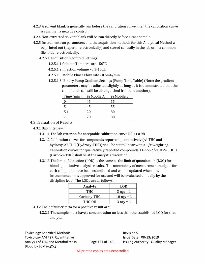
Toxicology Analytical Methods Revision 9Issue Date: 08/13/2019Toxicology AM #27: Quantitative
Analysis of THC and Metabolites in Blood by LCMS-QQQ
Page 131 of 143 Issuing Authority: Quality Manager
All printed copies are uncontrolled
4.2.3 A solvent blank is generally run before the calibration curve, then the calibration curve is run, then a negative control.
4.2.4 Non-extracted solvent blank will be run directly before a case sample.4.2.5 Instrument run parameters and the acquisition methods for this Analytical Method will
be printed out (paper or electronically) and stored centrally in the lab or in a common file folder electronically.
4.2.5.1 Acquisition Required Settings 4.2.5.1.1 Column Temperature - 50⁰C4.2.5.1.2 Injection volume –0.5-10µL 4.2.5.1.3 Mobile Phase Flow rate - 0.6mL/min4.2.5.1.3: Binary Pump Gradient Settings (Pump Time Table) (Note: the gradient
parameters may be adjusted slightly as long as it is demonstrated that the compounds can still be distinguished from one another).
Time (min) % Mobile A % Mobile B0 45 555 45 555.1 20 807 20 80
4.3 Evaluation of Results4.3.1 Batch Review
4.3.1.1 The lab criterion for acceptable calibration curve R2 is >0.984.3.1.2 Calibration curves for compounds reported quantitatively (9-THC and 11-
hydroxy-9-THC (Hydroxy-THC)) shall be set to linear with a 1/x weighting. Calibration curves for qualitatively reported compounds 11-nor-9-THC-9-COOH (Carboxy-THC) shall be at the analyst’s discretion.
4.3.1.3 The limit of detection (LOD) is the same as the limit of quantitation (LOQ) for blood quantitative analysis results. The uncertainty of measurement budgets for each compound have been established and will be updated when new instrumentation is approved for use and will be evaluated annually by the discipline lead. The LODs are as follows:
Analyte LODTHC 3 ng/mL
Carboxy-THC 10 ng/mLTHC-OH 3 ng/mL
4.3.2 The default criteria for a positive result are:4.3.2.1 The sample must have a concentration no less than the established LOD for that
analyte.
Toxicology Analytical Methods Revision 9Issue Date: 08/13/2019Toxicology AM #27: Quantitative
Analysis of THC and Metabolites in Blood by LCMS-QQQ
Page 132 of 143 Issuing Authority: Quality Manager
All printed copies are uncontrolled
4.3.2.2 Samples with a higher response than the 50 ng/mL will be reported out as greater than 50 ng/mL. The uncertainty of measurement has currently only been evaluated between 3 and 50 ng/mL for each compound. The estimated expanded uncertainty will only be reported when it impacts evaluation of a statute, legal requirement, or upon customer request. When the measurement uncertainty is reported it will be on the report in the same units as the measurement and a statement regarding the coverage probability of 99.73%(k=3) will also be on the report. The current expanded uncertainty is published as a protected document is in Qualtrax and is available to all analysts.
4.3.2.3 The analyte retention time must be within +/- 3% or +/- 0.100 min whichever is greater of the average retention time of the calibrators for that analyte. At the analyst’s discretion when the analyte peak falls outside the retention time window and the internal standard shifts comparably that sample may be evaluated as positive if the other criteria are met.
4.3.2.4 For calibrators and controls 10 ng and below the accuracy must be within 30%, for calibrators and controls greater than 10 ng/mL the accuracy must be within 20%. If the control falls outside the accuracy range, at the analyst’s discretion, the compound may be reported qualitatively.
4.3.2.5 The ion ratios for selected transitions must fall within 20% of the averaged calibrators for each run.
4.3.2.6 S/N of detected analyte must be greater than 10.4.3.2.7 The negative control will be evaluated as passing if the compound of interest does
not meet all of the above listed criteria and the internal standard gives an anticipated response to demonstrate the sample extracted and injected correctly.
4.3.2.8 To ensure the response is adequate, each compound in the internal control will be evaluated using the criteria listed above. If a compound does not meet these standards, samples cannot be evaluated for that compound. At the analyst’s discretion, that compound can be removed from the list of compounds that the sample was evaluated for, or the sample can be reinjected or re-extracted. If a sample needs to be reinjected, the analyst may add more of the reconstitution solvent to facilitate this. The internal standards must give an adequate response in order for the sample to be evaluated.
4.3.2.9 No quantitative values will be reported for urine cases.4.4 Quality Assurance Requirements
4.4.1 Refer to Toxicology AM #19 and AM #21 for additional quality assurance and reference material authentication.
4.5 Analysis Documentation4.5.1 The printed results for each case sample and accompanying blank will be included with
the analysts’ notes. Case results will be recorded in the LIMS system.
Toxicology Analytical Methods Revision 9Issue Date: 08/13/2019Toxicology AM #27: Quantitative
Analysis of THC and Metabolites in Blood by LCMS-QQQ
Page 133 of 143 Issuing Authority: Quality Manager
All printed copies are uncontrolled
4.5.2 The reports for the batch, including calibration curves and controls will be stored centrally in the lab or electronically on a common drive.
4.5.3 The raw data from the run will be stored electronically and will be backed up at least every two months.
4.5.4 Results of the controls will be entered onto the Control Monitoring Chart.
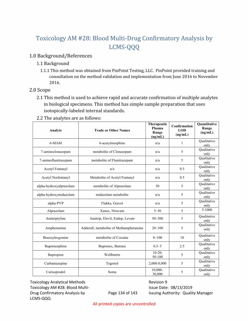
Toxicology Analytical Methods Revision 9Issue Date: 08/13/2019Toxicology AM #28: Blood Multi-
Drug Confirmatory Analysis by LCMS-QQQ
Page 134 of 143 Issuing Authority: Quality Manager
All printed copies are uncontrolled
Toxicology AM #28: Blood Multi-Drug Confirmatory Analysis by LCMS-QQQ
1.0 Background/References1.1 Background
1.1.1 This method was obtained from PinPoint Testing, LLC. PinPoint provided training and consultation on the method validation and implementation from June 2016 to November 2016.
2.0 Scope2.1 This method is used to achieve rapid and accurate confirmation of multiple analytes
in biological specimens. This method has simple sample preparation that uses isotopically-labeled internal standards.
2.2 The analytes are as follows:
Analyte Trade or Other Names
Therapeutic Plasma Range
(ng/mL)
Confirmation LOD
(ng/mL)
QuantitativeRange
(ng/mL)
6-MAM 6-acetylmorphine n/a 1 Qualitative only
7-aminoclonazepam metabolite of Clonazepam n/a 5 Qualitative only
7-aminoflunitrazepam metabolite of Flunitrazepam n/a 5 Qualitative only
Acetyl Fentanyl n/a n/a 0.5 Qualitative only
Acetyl Norfentanyl Metabolite of Acetyl Fentanyl n/a 0.5 Qualitative only
alpha-hydroxyalprazolam metabolite of Alprazolam 50 5 Qualitative only
alpha-hydroxymidazolam midazolam metabolite n/a 5 Qualitative only
alpha-PVP Flakka, Gravel n/a 5 Qualitative only
Alprazolam Xanax, Niravam 5–50 5 5-1000
Amitriptyline Amitrip, Elevil, Endep, Levate 50–300 5 Qualitative only
Amphetamine Adderall, metabolite of Methamphetamine 20–100 5Qualitative
only
Benzoylecgonine metabolite of Cocaine 0–100 10 Qualitative only
Buprenorphine Buprenex, Butrans 0.5–5 2.5 Qualitative only
Bupropion Wellbutrin 10-20; 50-100 5 Qualitative
only
Carbamazepine Tegretol 2,000-8,000 5 Qualitative only
Carisoprodol Soma 10,000–30,000 5 Qualitative
only
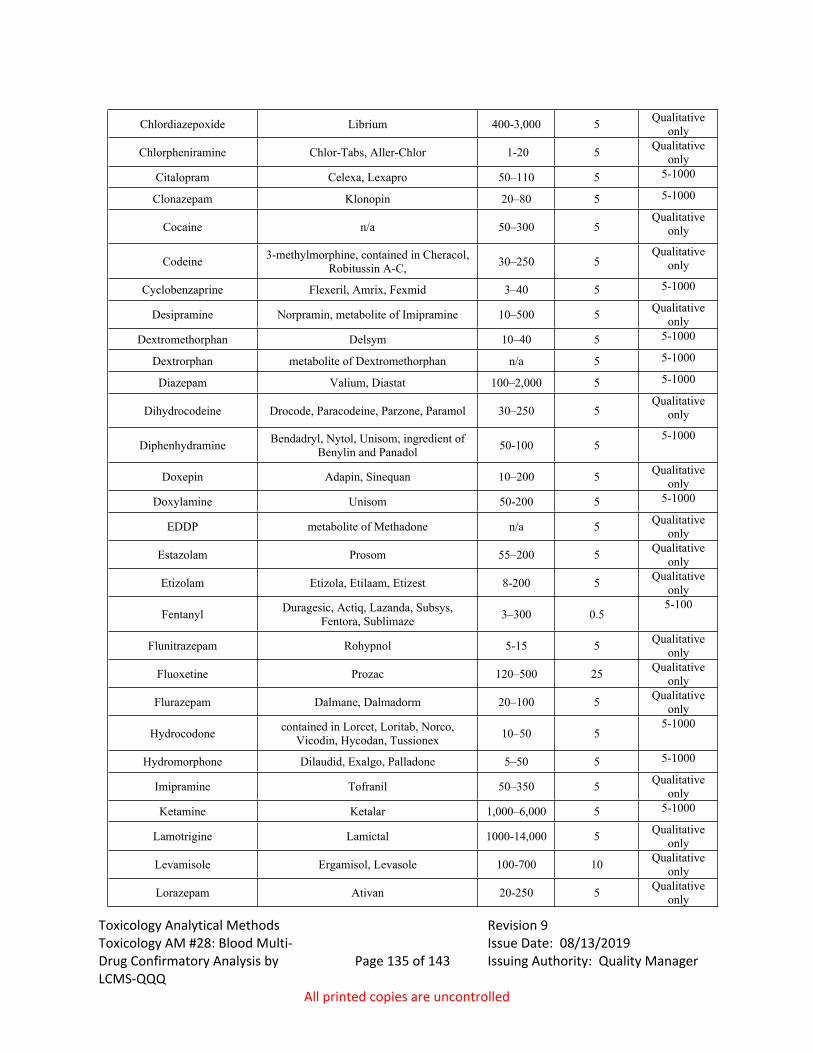
Toxicology Analytical Methods Revision 9Issue Date: 08/13/2019Toxicology AM #28: Blood Multi-
Drug Confirmatory Analysis by LCMS-QQQ
Page 135 of 143 Issuing Authority: Quality Manager
All printed copies are uncontrolled
Chlordiazepoxide Librium 400-3,000 5 Qualitative only
Chlorpheniramine Chlor-Tabs, Aller-Chlor 1-20 5 Qualitative only
Citalopram Celexa, Lexapro 50–110 5 5-1000
Clonazepam Klonopin 20–80 5 5-1000
Cocaine n/a 50–300 5Qualitative
only
Codeine 3-methylmorphine, contained in Cheracol, Robitussin A-C, 30–250 5
Qualitative only
Cyclobenzaprine Flexeril, Amrix, Fexmid 3–40 5 5-1000
Desipramine Norpramin, metabolite of Imipramine 10–500 5 Qualitative only
Dextromethorphan Delsym 10–40 5 5-1000
Dextrorphan metabolite of Dextromethorphan n/a 5 5-1000
Diazepam Valium, Diastat 100–2,000 5 5-1000
Dihydrocodeine Drocode, Paracodeine, Parzone, Paramol 30–250 5Qualitative
only
Diphenhydramine Bendadryl, Nytol, Unisom, ingredient of Benylin and Panadol 50-100 5
5-1000
Doxepin Adapin, Sinequan 10–200 5 Qualitative only
Doxylamine Unisom 50-200 5 5-1000
EDDP metabolite of Methadone n/a 5 Qualitative only
Estazolam Prosom 55–200 5 Qualitative only
Etizolam Etizola, Etilaam, Etizest 8-200 5 Qualitative only
Fentanyl Duragesic, Actiq, Lazanda, Subsys, Fentora, Sublimaze 3–300 0.5
5-100
Flunitrazepam Rohypnol 5-15 5 Qualitative only
Fluoxetine Prozac 120–500 25 Qualitative only
Flurazepam Dalmane, Dalmadorm 20–100 5 Qualitative only
Hydrocodone contained in Lorcet, Loritab, Norco, Vicodin, Hycodan, Tussionex 10–50 5
5-1000
Hydromorphone Dilaudid, Exalgo, Palladone 5–50 5 5-1000
Imipramine Tofranil 50–350 5 Qualitative only
Ketamine Ketalar 1,000–6,000 5 5-1000
Lamotrigine Lamictal 1000-14,000 5 Qualitative only
Levamisole Ergamisol, Levasole 100-700 10 Qualitative only
Lorazepam Ativan 20-250 5 Qualitative only
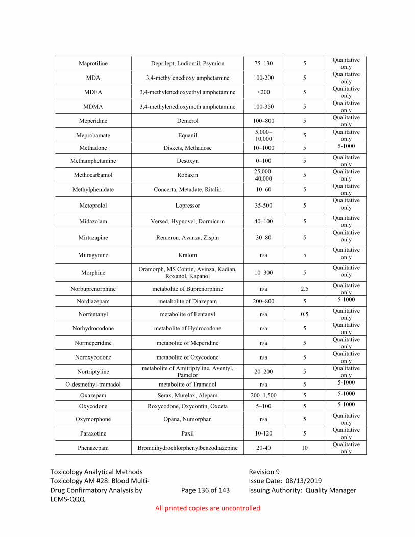
Toxicology Analytical Methods Revision 9Issue Date: 08/13/2019Toxicology AM #28: Blood Multi-
Drug Confirmatory Analysis by LCMS-QQQ
Page 136 of 143 Issuing Authority: Quality Manager
All printed copies are uncontrolled
Maprotiline Deprilept, Ludiomil, Psymion 75–130 5 Qualitative only
MDA 3,4-methylenedioxy amphetamine 100-200 5 Qualitative only
MDEA 3,4-methylenedioxyethyl amphetamine <200 5 Qualitative only
MDMA 3,4-methylenedioxymeth amphetamine 100-350 5 Qualitative only
Meperidine Demerol 100–800 5 Qualitative only
Meprobamate Equanil 5,000–10,000 5 Qualitative
onlyMethadone Diskets, Methadose 10–1000 5 5-1000
Methamphetamine Desoxyn 0–100 5 Qualitative only
Methocarbamol Robaxin 25,000-40,000 5 Qualitative
only
Methylphenidate Concerta, Metadate, Ritalin 10–60 5 Qualitative only
Metoprolol Lopressor 35-500 5Qualitative
only
Midazolam Versed, Hypnovel, Dormicum 40–100 5 Qualitative only
Mirtazapine Remeron, Avanza, Zispin 30–80 5Qualitative
only
Mitragynine Kratom n/a 5Qualitative
only
Morphine Oramorph, MS Contin, Avinza, Kadian, Roxanol, Kapanol 10–300 5
Qualitative only
Norbuprenorphine metabolite of Buprenorphine n/a 2.5 Qualitative only
Nordiazepam metabolite of Diazepam 200–800 5 5-1000
Norfentanyl metabolite of Fentanyl n/a 0.5 Qualitative only
Norhydrocodone metabolite of Hydrocodone n/a 5 Qualitative only
Normeperidine metabolite of Meperidine n/a 5 Qualitative only
Noroxycodone metabolite of Oxycodone n/a 5 Qualitative only
Nortriptyline metabolite of Amitriptyline, Aventyl, Pamelor 20–200 5 Qualitative
onlyO-desmethyl-tramadol metabolite of Tramadol n/a 5 5-1000
Oxazepam Serax, Murelax, Alepam 200–1,500 5 5-1000
Oxycodone Roxycodone, Oxycontin, Oxceta 5–100 5 5-1000
Oxymorphone Opana, Numorphan n/a 5 Qualitative only
Paraxotine Paxil 10-120 5 Qualitative only
Phenazepam Bromdihydrochlorphenylbenzodiazepine 20-40 10 Qualitative only
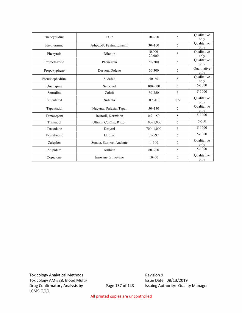
Toxicology Analytical Methods Revision 9Issue Date: 08/13/2019Toxicology AM #28: Blood Multi-
Drug Confirmatory Analysis by LCMS-QQQ
Page 137 of 143 Issuing Authority: Quality Manager
All printed copies are uncontrolled
Phencyclidine PCP 10–200 5 Qualitative only
Phentermine Adipex-P, Fastin, Ionamin 30–100 5 Qualitative only
Phenytoin Dilantin 10,000-20,000 5 Qualitative
only
Promethazine Phenegran 50-200 5 Qualitative only
Propoxyphene Darvon, Dolene 50-300 5 Qualtitative only
Pseudoephedrine Sudafed 50–80 5 Qualitative only
Quetiapine Seroquel 100–500 5 5-1000
Sertraline Zoloft 50-250 5 5-1000
Sufentanyl Sufenta 0.5-10 0.5 Qualitative only
Tapentadol Nucynta, Palexia, Tapal 50–130 5 Qualitative only
Temazepam Restoril, Normison 0.2–150 5 5-1000
Tramadol Ultram, ConZip, Ryzolt 100–1,000 5 5-500
Trazodone Desyrel 700–1,000 5 5-1000
Venlafaxine Effexor 35-597 5 5-1000
Zaleplon Sonata, Starnoc, Andante 1–100 5 Qualitative only
Zolpidem Ambien 80–200 5 5-1000
Zopiclone Imovane, Zimovane 10–50 5 Qualitative only
Toxicology Analytical Methods Revision 9Issue Date: 08/13/2019Toxicology AM #28: Blood Multi-
Drug Confirmatory Analysis by LCMS-QQQ
Page 138 of 143 Issuing Authority: Quality Manager
All printed copies are uncontrolled
3.0 Equipment/Reagents3.1 Equipment and Supplies
3.1.1 Approved LC/MS/MS and MassHunter or equivalent software3.1.2 Shaker/incubator 3.1.3 Positive Pressure Manifold 3.1.4 SPE dry 3.1.5 Agilent 120 EC-C18 (2.1 x 100-4 um) or equivalent3.1.6 Calibrated pipettes for dispensing blood and/or urine samples.3.1.7 Pipettes for all other transfers and additions. 3.1.10 ToxBox kit with 96 well plate
containing internal standards, calibrators, and controls, 96 well SLE+ plate and 96 well blank sample preparation plate.
3.1.11 Appropriate sized pipette tips3.1.12 Heat sealing foil covers 3.1.13 ACT plate cover for use during drying step with SPE dry (if run contains urine samples)3.1.14 Additional 48 or 96-well plate (if run contains urine samples)
3.2 ReagentsSee AM# 23 for Solution Preparation instructions3.2.1 5 mM Ammonium Formate + 0.01% Formic Acid in Water (mobile phase A)3.2.2 0.01% Formic Acid in Methanol (mobile phase B)3.2.3 0.5 M Ammonium Hydroxide3.2.4 Formic Acid (LCMS grade)3.2.5 Water (LCMS grade)3.2.6 Methanol (LCMS grade)3.2.7 Ammonium Formate (LCMS grade)3.2.8 Ammonium Hydroxide (ACS grade or higher)3.2.9 Ethyl Acetate (ACS grade or higher)3.2.9.10 20% LCMS Methanol in LCMS Water (Reconstitution Solvent)3.2.9.11 LC/MS Needle Rinse (40/40/20: LCMS Water/Methanol/Isopropanol or 75% MeOH
in water)3.2.10 Beta-glucuronidase (Kura BG-100 or BGTurbo) (if run includes urine samples)3.2.11 0.1 M ammonium acetate buffer (pH 5) (if using BG-100)3.2.12 Instant Buffer I (provided with BGTurbo) (if using BGTurbo)
3.3 Quality Assurance: Reference materials and Controls3.3.1 Internal Controls, Calibrators, Internal Standards, and Blanks.
Toxicology Analytical Methods Revision 9Issue Date: 08/13/2019Toxicology AM #28: Blood Multi-
Drug Confirmatory Analysis by LCMS-QQQ
Page 139 of 143 Issuing Authority: Quality Manager
All printed copies are uncontrolled
3.3.1.1 Internal controls, calibrators, and internal standards are prepared by the ToxBox manufacturer and contained on the 96 well plate. A minimum of one internal positive control that has a concentration near the administrative cut off level for each compound will be run with each extraction.
3.3.2 Negative Control3.3.2.1 A negative control will be run with each panel extraction. If the run contains urine
samples, a negative urine will also be included in the run.3.3.4 Blanks
3.3.4.1 A non-extracted blank containing internal standard will be run directly before each case sample to rule out carryover if the sample injected before it contains the same compound. If confirmation criteria (e.g. ion ratios, RT, S/N) are not met, the analyte is not considered present. If confirmation criteria is met, the response of the blank will be considered. The area response of the blank preceding a sample must be at least 10 times less than any compound confirmed in the case sample, and must be below the limit of confirmation for any analyte confirmed in the case sample to be considered negative. If a blank does contain a compound, the blank and the sample can be reinjected.
3.3.4.2 It is recommended that a blank follow any samples that exhibit a very strong response in the screen.
3.3.4.3 To prepare blank, dilute 1 part ISTD (obtained from a blank well in the extraction plate) in 4-5 parts LCMS grade methanol.
4.0 Procedure4.1 Extraction Procedure
4.1.1 Allow refrigerated or frozen samples and frozen components to reach ambient temperature.
4.1.2 Urine samples must be hydrolyzed prior to extraction. For urine samples/controls only: Add 250 µl of blank urine to a clean 48 or 96-well plate. If using BG-100, add 50 µl of the BG-100 and 200 µl of 0.1 M ammonium acetate buffer (pH 5) to each well containing urine. Place a cover on the plate and shake at 900 rpm for approximately 180 minutes at 60 degrees Celsius. If using BGTurbo, add 40 µl of BGTurbo and 100 µl of 500 mM sodium phosphate (pH 6.8) to the wells containing urine. Place a cover on the plate and shake for a minimum of 5 minutes.
4.1.3 Using a calibrated single channel pipette, add 250 µL of blank blood into the appropriate wells of the analytical (standards) plate (wells containing internal positive and negative controls). Pipette the appropriate sample/case blood into the appropriate wells containing only internal standards. For urine samples/controls: after shaking, transfer 250 µl of the urine/buffer/BG-100 or urine/buffer/BGTurbo to the appropriate wells containing internal standard on the analytical plate.
Toxicology Analytical Methods Revision 9Issue Date: 08/13/2019Toxicology AM #28: Blood Multi-
Drug Confirmatory Analysis by LCMS-QQQ
Page 140 of 143 Issuing Authority: Quality Manager
All printed copies are uncontrolled
4.1.4 Place plate on shaking incubator at approximately 900 rpm for approximately 15 minutes.
4.1.5 Add 250 µL of 0.5 M ammonium hydroxide buffer into all wells in use on the analytical (standards) plate.
4.1.6 Place plate on shaking incubator at approximately 900 rpm for approximately 15 minutes.
4.1.7 Transfer 300 µL of blood+base or urine+base mixture (if applicable) to corresponding wells of SLE+ plate.
4.1.8 Apply positive pressure for approximately 10-15 seconds or until no liquid remains on top of sorbent. Wait 5 minutes. (Positive Pressure manifold setting for this loading step is between 85-100 PSI)
4.1.9 Add 900 µL of ethyl acetate and allow to flow through for approximately 5 minutes under gravity.
4.1.10 Apply positive pressure for approximately 10-15 seconds. (Positive Pressure manifold setting for this step is between 12-15 PSI).
4.1.11 Add 900 µL of ethyl acetate and allow to flow through for approximately 5 minutes under gravity.
4.1.12 Apply positive pressure for approximately 10-15 seconds. (Positive Pressure manifold setting for this step is between 12-15 PSI).
4.1.13 Remove plate containing eluate. Place on SPE Dry and evaporate to dryness at approximately 35 degrees Celsius. If run contains urine samples, place ACT cover on top of plate prior to drying.
4.1.14 Reconstitute in 100 µL 20% LCMS grade methanol in LCMS Water and heat seal plate with foil.
4.2 Instrument and Run set up.4.2.1 See Toxicology AM #24 for instrument maintenance and operation.4.2.2 Set up the worklist in MassHunter.4.2.3 A solvent blank is generally run before the calibration curve, then the calibration curve
is run, then a negative control. At a minimum, one internal positive control will be run with each run.
4.2.4 Instrument run parameters and the acquisition methods for this Analytical Method will be printed out (paper or electronically) and stored centrally in the lab or in a common file folder electronically.
4.2.4.1 Acquisition Required Settings 4.2.4.1.1 Column Temperature - 55⁰C4.2.4.1.2 Injection volume – 0.5-10 µL 4.2.4.1.3 Mobile Phase Flow rate - 0.5 mL/min
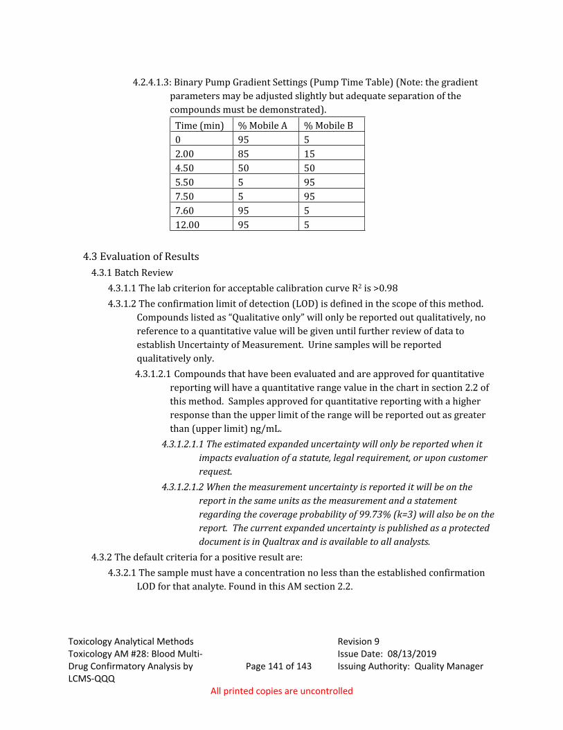
Toxicology Analytical Methods Revision 9Issue Date: 08/13/2019Toxicology AM #28: Blood Multi-
Drug Confirmatory Analysis by LCMS-QQQ
Page 141 of 143 Issuing Authority: Quality Manager
All printed copies are uncontrolled
4.2.4.1.3: Binary Pump Gradient Settings (Pump Time Table) (Note: the gradient parameters may be adjusted slightly but adequate separation of the compounds must be demonstrated).
Time (min) % Mobile A % Mobile B0 95 52.00 85 154.50 50 505.50 5 957.50 5 957.60 95 512.00 95 5
4.3 Evaluation of Results4.3.1 Batch Review
4.3.1.1 The lab criterion for acceptable calibration curve R2 is >0.984.3.1.2 The confirmation limit of detection (LOD) is defined in the scope of this method.
Compounds listed as “Qualitative only” will only be reported out qualitatively, no reference to a quantitative value will be given until further review of data to establish Uncertainty of Measurement. Urine samples will be reported qualitatively only.
4.3.1.2.1 Compounds that have been evaluated and are approved for quantitative reporting will have a quantitative range value in the chart in section 2.2 of this method. Samples approved for quantitative reporting with a higher response than the upper limit of the range will be reported out as greater than (upper limit) ng/mL.
4.3.1.2.1.1 The estimated expanded uncertainty will only be reported when it impacts evaluation of a statute, legal requirement, or upon customer request.
4.3.1.2.1.2 When the measurement uncertainty is reported it will be on the report in the same units as the measurement and a statement regarding the coverage probability of 99.73% (k=3) will also be on the report. The current expanded uncertainty is published as a protected document is in Qualtrax and is available to all analysts.
4.3.2 The default criteria for a positive result are:4.3.2.1 The sample must have a concentration no less than the established confirmation
LOD for that analyte. Found in this AM section 2.2.
Toxicology Analytical Methods Revision 9Issue Date: 08/13/2019Toxicology AM #28: Blood Multi-
Drug Confirmatory Analysis by LCMS-QQQ
Page 142 of 143 Issuing Authority: Quality Manager
All printed copies are uncontrolled
4.3.2.1.1 For any qualitatively reported compound, if a point(s) is dropped at the low end of a calibration curve, the lowest used point that is greater or equal to that compounds LOD listed in 2.2 will be the LOD for that compound for that run.
4.3.2.1.2 For quantitative reporting: All points in the approved range defined in section 2.2 of this method must be used. In addition, the accuracy for all controls (QC’s) for that compound must be within the appropriate tolerances (+/- 30% for 10ng/ml and lower and +/- 20% for above 10ng/ml). If a QC fails to meet the requirements, the compound will be reported qualitatively (see exceptions below).
4.3.2.1.2.1 If any points are dropped from the approved quantitative range of the curve, the compound will be reported qualitatively. The exception is if the highest point is dropped from the curve due to exceeding the instrument’s detection capacity, linearity, or due to column overload. In addition, if the highest control (QC) fails due to exceeding the instrument’s detection capacity, or linearity; the high point on the calibration curve may be dropped. The quantitative range for that compound in that range will be adjusted to the next highest calibrator level. Any samples exceeding that level will be reported as greater than (upper limit) ng/ml.
4.3.2.1.2.2 The quantitative range for fentanyl is 5-100. Samples with concentrations between 0.5 and 5 will be reported qualitatively.
4.3.2.2 The analyte retention time must be within +/- 5% or 0.100 min whichever is greater of the average retention time of the calibrators for that analyte. The retention time for tramadol must be within +/-2% of the average retention time of the calibrators. At the analyst’s discretion when the analyte peak falls outside the retention time window and the internal standard shifts comparably that sample may be evaluated as positive if the other criteria are met.
4.3.2.3 For calibrators 10 ng and below the accuracy must be within 30%, for calibrators greater than 10 ng/mL the accuracy must be within 20%.
4.3.2.4 The ion ratios for transitions must fall within 20% of the averaged calibrators for each run.
4.3.2.5 S/N for each primary transition of detected analyte must be greater than 10. The S/N for the secondary transition must be greater than 5.
4.3.2.6 The negative control will be evaluated as passing if the compound of interest does not meet all of the above listed criteria and the internal standard gives an anticipated response to demonstrate the sample extracted and injected correctly.
Toxicology Analytical Methods Revision 9Issue Date: 08/13/2019Toxicology AM #28: Blood Multi-
Drug Confirmatory Analysis by LCMS-QQQ
Page 143 of 143 Issuing Authority: Quality Manager
All printed copies are uncontrolled
4.3.2.7 Controls must give a positive response for each compound if the concentration of the control is greater than the LOD for any given compound. It is not necessary for the control to give a positive response if a particular compound is not being evaluated in a run. In those instances, a notation will be made on the control indicating that compound is not being evaluated. For compounds that are approved for quantitative reporting controls 10 ng and below the accuracy must be within 30%, for controls greater than 10 ng/mL the accuracy must be within 20%. If any control within the evaluated quantitative range falls outside the accuracy range, at the analyst’s discretion, the compound may be reported qualitatively.
4.3.2.8 To ensure the response is adequate, each compound in the internal control will be evaluated using the criteria listed above. If a compound does not meet these standards, samples cannot be evaluated for that compound. At the analyst’s discretion, that compound can be removed from the list of compounds that the sample was evaluated for, or the sample can be reinjected or re-extracted. If a sample needs to be reinjected, the analyst may add more of the reconstitution solvent to facilitate this. The internal standards must give an adequate response in order for the sample to be evaluated.
4.3.2.9 Any limitations to curve ranges and or evaluation of compounds should be noted. 4.3.3 Limitations of Method
4.3.3.1 Samples that indicate as positive for Benzoylecgonine will be reported as Benzoylecgonine/Cocaine break-down product (or other wording that indicates that the response may be from a breakdown of cocaine).
4.4 Quality Assurance Requirements4.4.1 Refer to Toxicology AM #19 and AM #21 for additional quality assurance and reference
material authentication.4.5 Analysis Documentation
4.5.1 The printed results for each case sample and (when applicable) accompanying blank will be included with the analyst’s notes. Case results will be recorded in the LIMS system.
4.5.2 The reports for the batch, including calibration curves and controls will be stored centrally in the lab or electronically on a common drive. Calibration curves and control data only need to be evaluated and printed for compounds being evaluated for that run.
4.5.3 The raw data from the run will be stored electronically and will be backed up at least every two months.
4.5.4 Results of the controls will be entered onto the Control Monitoring Chart.