training on medical waste management - kuwait university workshop lectures/lecture 3.pdf ·...
TRANSCRIPT

Training on Medical Waste Management
in Collaboration with Al-Essa Medical and Scientific Equipment Co. W.L.L
Kuwait University Health Science Center 29 January – 1 February, 2012
Risk Assessments – Biosafety Levels
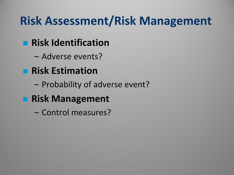
Risk Assessment/Risk Management Risk Identification
– Adverse events?
Risk Estimation – Probability of adverse event?
Risk Management – Control measures?

Risk (Definitions)
“Possibility of loss, injury, disease, or death.” Webster's Medical Desk Dictionary (1986)
“The probability that exposure to a hazard will lead to a negative consequence.”
David Ropeik, George Gray (2002)
“To risk living is to risk dying.” Anonymous

Risk Assessment
The emergent science based on toxicology, epidemiology and statistics that utilizes qualitative and quantitative hazard analysis to provide the public with a reasonable estimate of probability of harm.
“Not a scalpel, but a crude tool that allows you to make estimates.” Peter Preuss, US EPA


Risk Assessment
Difficult process
(expertise of many fields needed)
Involves uncertainty
Range provided (not a specific number)
Estimates for society (individual risk may vary)
“Reasonable worst-case estimate” (better to overestimate than underestimate risk)
Costs and benefits of proposed actions helpful

4 Steps in Risk Assessment (Jeff Wheelwright, 1996)
1) Identify health hazard
2) Quantify hazard
3) Exposure assessment
(from source to at risk person)
4) Determine probability of disease
(based on exposure estimate and potency of agent)

Biohazard Epidemiology
Incidence of Hepatitis among Danish clinical chemistry workers 7X higher than general population
(Skinholj, 1974)
Risk of acquiring TB 5X greater among medical lab workers in England than general population
(Harrington & Shannon, 1976)

Hierarchy of Controls
Anticipation
Recognition
Evaluation
Control
– substitution
– administrative
– engineering
– work practices
– personal protective clothing
– facility features

Infectious Agents are Classified by Level of Hazard
4 Agent Risk Group Classifications
RG1 RG2 RG3 RG4
No risk to
community Low risk to community High risk to
community
Moderate individual risk
High individual risk Low individual risk


Risk Groups (RG) RG1
• Not infectious to healthy adults
• e.g. E. coli K12 strains, B. subtilis, S. cerevisiae
RG2 • Infectious agents of varying severity, treatment
usually available, predominantly bloodborne, ingestion, and mucous membrane routes of exposure
• e.g. Salmonella, Shigella, Vibrio, Plasmodium, Hepatitis B Virus, Cryptococcus neoformans
– Both RG1/RG2 can be used in a basic lab • containment equipment to contain aerosols

Risk Groups (RG)
RG3 • potential to cause serious or lethal disease,
airborne route of exposure (and others), treatment generally not available, lower infectious dose.
• Containment Lab 2 doors off general corridor, dedicated air handler, controlled airflow, all work contained.
• e.g. TB, Vesicular Stomatitis Virus, Yellow Fever Virus, Coxiella burneti, Francisella tularensis.

Risk Groups (RG)
RG4 • Dangerous, exotic agents with high risk to
individual and community. Aerosol transmission along with all other routes. Very low infectious dose, high mortality rates.
• Building within building approach for research purposes.
• e.g. Ebola virus, Marburg virus, Junin, Lassa, Machupo, Sabia, Equine Morbillivirus (Hendra-like viruses), Tick-Borne Encephalitis Viruses

Laboratory Safety Containment Levels
4 Laboratory Biosafety Levels
BSL1 BSL2 BSL3 BSL4
Basic laboratory, confine aerosols in biosafety cabinet if needed
Containment lab, 2 door separation from general traffic, negative air flow, alarms
Maximum containment lab, building w/in building, all features isolated, pos. pressure suits, glove box type isolation


Hybrid Biosafety Level
BSL2/BSL3 (BL2+) – Creutzfeld Jacob
– HIV
– High risk clinical specimens
BSL3-Enhanced (HEPA filtered exhaust Lab) – Yellow Fever, Rift Valley Fever Virus, VEE
– Rickettsia rickettsii

Unknown Specimens
Facility Evaluation (highest level of protection available)
“B.A.R.E” – Block All Routes
of Exposure

Containment achieved with:
Good Microbiological practices
Safety Equipment
Facility Design

Risk Assessment & Risk Management
Prior Planning Prevents Poor Performance

Risk Assessment & Risk Management
Pathogen (Agent)
Procedures (Protocol)
Personnel
Protective Equipment
Place (Proposed lab facility)

Immunizations
– Vaccinia
– Tetanus
– Meningococcal
– Typhoid
– Botulinum
– Hepatitis B virus, Hepatitis A virus
– Yellow Fever, EEE
– Rabies

Protective Equipment
Personal Protective Equipment (clothing)
Containment Equipment – Biological safety cabinets
– Safety centrifuges
– Sealed sonicators, blenders, homogenizers
– Sealed tubes, transport carriers
– Safe sharps, needleboxes, medical waste bags, tongs, forceps, etc.


Personal Protective Equipment
Protect: skin clothing mucous membranes respiratory system
Use: gloves (double, kevlar) lab coats, solid-front gowns sleeve covers full face protection respiratory protection


Personal Protective Equipment
Disposable
Decontamination
Dedicated to area
Donning/Doffing – Compromised (wet/contaminated/torn)
– Respiratory Protection Program

Place (Facility Design)
Restricted access/Door sign
Easily cleanable
Hand washing sinks (near exit door)
Eye wash
Autoclave
Vacuum system protection
Biosafety Cabinet

Place (Facility Design)
Anteroom
Negative pressure gradient
Airflow monitor
Air changes per hour (10-15)
Sealed penetrations, coved flooring
Facility alarms/interlocks
Communication outside the lab

Procedures
Develop standard written practices (SOP’s) for handling pathogens
Job Safety Analysis (JSA) – identify each task
– describe all steps
– hazard assessment at each step
– incorporate safety
Focus on containing aerosol generating procedures and equipment

Aerosols
– Procedures that impart energy into a microbial suspension are a potential source of aerosol (Chatigny, 1974)
– Many common lab procedures and accidents have capability of releasing aerosols • homogenization, sonication, blending, mixing,
grinding, shaking, vortexing, spills, opening vials, pipetting, animals excreting agent, opening vials under pressure, etc.

Viable Particles Recovered from Air (Chatigny, 1974)
Procedure – sonic oscillator
– mixing w/ pipette
– overflow from mixer
– opening lyophilized vial
– top removed after blending
– dropping flask of culture
– dropping lyophilized culture
# Particles/ft3 of air – 6
– 7
– 9
– 135
– 1500
– 1551
– 4839

Procedures - Sharps Hazards
Syringe/Needle – adjusting volume
– withdrawal from stopper
– separation from syringe
– leaking syringe
– leakage from injection site
– inappropriate disposal
– poor work practices

Metal
Glass
Plastic

Procedures - Sharps Precautions
Syringe/Needle – use needle-locking syringes
– cover with disinfectant soaked gauze
– animal restraints
– cleanse inoculation site
– safe needle practices
– immediate collection/disposal

BD Eclipse Shielding Hypodermic Needle

Procedures - Sharps Precautions
Needle / Syringe – removal of needle from syringe (hemostat)
– no recapping, bending, breaking, etc.
– immediate disposal of intact needle/syringe
– location of needlebox (vicinity, height)
– replacement of needleboxes
– eliminate/minimize use/safe sharp devices
– avoid glass Pasteur pipettes

Procedures presenting risk
Microbiological loop – streaking plates
– spreading material on slides
– cooling loop in media
– heating loop in an open flame

Precautions in Bacteriology
Microbiological loop – smooth plates
– disposable plastic loops
– well formed loops with short staff
– glass spreaders
– electric (walled) micro-incinerators
– work within a biosafety cabinet


Procedures with general risk
Pipetting – mouth pipetting
– glass Pasteur pipettes
– blow-out pipettes
– mixing suspensions
– spill of droplets onto hard surfaces
Eating, drinking, smoking, applying cosmetics

Procedures
Pipetting – no mouth pipetting
– disposable plastic pipettes
– mark to mark pipettes
– collect within biosafety cabinet
– work over disinfectant-wet pad
Restrict consumption of food or beverage to well defined break areas

Procedures
Centrifugation – broken/leaking tubes
– microfuge tubes (snap caps)
– (flawed/overfilled)
Protective Measures – check O-rings on rotors (use O-ring tubes)
– safety cups/sealed rotors
– load/unload in a biosafety cabinet


HPV


Issues with Infectious/Clinical Waste
Transmission of Mycobacterium tuberculosis From Medical Waste Kammy R. Johnson, DVM, PhD; et al
JAMA. 2000; 284:1683-1688.
ABSTRACT Context Washington State has a relatively low incidence
rate of tuberculosis (TB) infection. However, from May to September 1997, 3 cases of pulmonary TB were reported among medical waste treatment workers at 1 facility in Washington. There is no previous documentation of Mycobacterium tuberculosis transmission as a result of processing medical waste.

LOS ANGELES (KABC) -- A new report by the California Division of Occupational Safety and Health blames a UCLA professor and the university for a lab accident in 2008 that killed a staff research assistant.
The criminal investigation report obtained by the Los Angeles Times stated that 23-year-old Sheharbano "Sheri" Sangji wasn't experienced or well-trained, if not trained at all, to handle the chemicals that killed her.
Sangji was severely burned over nearly half her body when air-sensitive chemicals burst into flames and ignited her clothes at a campus lab in December 2008. She died 18 days later.
CalOSHA report faults UCLA, professor in lab death January 12, 2012

http://www.cdc.gov/mmwr/pdf/other/su6101.pdf