transcription factor is not a zinc finger butformsa zn(ii ... · gal4transcription factor is not...
TRANSCRIPT

Proc. Nati. Acad. Sci. USAVol. 87, pp. 2077-2081, March 1990Biochemistry
GAL4 transcription factor is not a "zinc finger" but forms aZn(II)2Cys6 binuclear cluster
(IH NMR/metalloproteins)
TAO PAN AND JOSEPH E. COLEMANThe Department of Molecular Biophysics and Biochemistry, Yale University, New Haven, CT 06510
Communicated by Bert L. Vallee, December 26, 1989
ABSTRACT The DNA-binding domain of the transcrip-tion factor GAL4, consisting of the 62 N-terminal residues anddenoted GAL4(62*), contains a Cys-Xaa2-Cys-Xaa6-Cys-Xaa6-Cys-Xaa2-Cys-Xaa6-Cys motif, which has been shownpreviously to bind two Zn(ll) or Cd(il) ions. Binding of Zn(II)or Cd(II) is essential for the recognition by GALA of the specificpalindromic DNA sequence to which it binds upstream ofgenes for galactose-metabolizing enzymes, the UASG sequence.On the basis of the `3'Cd NMR chemical shifts of the two bound"13Cd(II) ions, we propose a binuclear cluster model for thisZn(ll)-binding subdomain. IH-113Cd heteronuclear multiple-quantum NMR spectroscopy and phase-sensitive double-quantum filtered 'H correlation spectroscopy of the "12Cd(II)-and "13Cd(ll)-substituted GALA(62*) derivatives provide directevidence that the two bound "13Cd(II) ions are coordinated onlyby the six cysteine residues, two of which form bridging ligandsbetween the two "13Cd(II) ions. The latter can be identifiedfrom the pattern of 'H-"3Cd J coupling. Thus a binuclearmetal ion cluster rather than a "zinc finger" is formed by thesix cysteine residues of the GALA DNA-binding domain. Thismodel can be directly applied to eight other fungal transcrip-tion factors which have been shown to contain similarly spacedCys6 clusters. 'H NMR spectra of apo-GAL4(62*) suggestconformational fluctuation of the metal-binding subdomainupon removal of Zn(II) or Cd(II). Both Cd(ll)2- and Zn(ll)2-containing species of GALA can be formed, and the similar 'HNMR spectra suggest similar conformations.
The GAL4 transcription factor from Saccharomyces cerevi-siae is a protein of 881 amino acids required for the tran-scriptional activation, in the presence of galactose, of thegenes coding for galactose-metabolizing enzymes (1). TheDNA-binding domain, which recognizes a specific nucleotidesequence (UASG) upstream of the promoters for these genes,has been localized to the N-terminal 62 amino acids (2-5).This domain of GAL4 contains a cysteine-rich amino acidsequence, Cys'1-Xaa2-Cys14-Xaa6-Cys21-Xaa6-Cys28-Xaa2-Cys31-Xaa6-Cys38, conserved among a group of nine tran-scription factors isolated from fungi (6). The cysteine clusterin GAL4 and related transcription factors has been proposedto form a single "zinc finger" containing a tetrahedral Zn(II)coordination complex (7), similar to that described for tran-scription factor TFIIIA from Xenopus oocytes (8). The GAL4"finger" would have 4 cysteine residues as ligands ratherthan the 2 cysteine and 2 histidine residues found in TFIIIAand related proteins (7, 8).We have shown previously that cloned N-terminal frag-
ments ofGAL4 consisting of 149 and 62 amino acids, denotedGAL4(149*) and GAL4(62*), contain Zn(II) (4,5). The Zn(II)is essential for the binding of these fragments to the UASGDNA sequence, and Zn(II) can be replaced by Cd(II) without
loss of specific DNA binding (4, 5). A single zinc-finger modelof GAL4 predicts that the protein contains 1 Zn(II) ion permolecule, but both GAL4(149*) and GAL4(62*) can bind 2Zn(II) or Cd(II) ions per molecule (4, 5). 113Cd(II) substitutionof Zn(II)-binding sites in metalloproteins has served as apowerful probe of the structure of these Zn(II) sites, primar-ily because of the great sensitivity of 113Cd NMR signals tothe nature of the ligands to the metal ion. 113Cd NMR incombination with two-dimensional (2D) 1H NMR has led tothe determination of the complete solution structure of thesmall Zn(II)- and Cd(II)-binding protein metallothionein (9,10). This protein is the only example of a Zn(II) or Cd(II)cluster complex found in nature in which several of thecysteine residues act as bridging ligands between two metalions (10). Metallothionein contains 7 metal ions located intwo clusters (11).Our initial 113Cd NMR study of Cd2GAL4 confirmed the
presence of two Cd(II)-binding sites in both GAL4(149*) andGAL4(62*) which induce 113Cd chemical shifts of 707 and 669ppm, consistent with ligation ofeach tt3Cd(II) to at least 3 andprobably 4 sulfur atoms (4, 5). We have now used 2D 1HNMR techniques to demonstrate that the only ligands to the2 113Cd(II) ions in 1l3Cd2GAL4(62*) are the 6 cysteine resi-dues. The 1H-113Cd J coupling patterns of the cysteine ,Bprotons are best explained by a binuclear cluster in which twoof the cysteine residues are bridging ligands between the twometal ions. The NMR data supporting this model of theDNA-binding domain of GAL4 are presented in this paper.
MATERIALS AND METHODSCloning, Overproduction, and Purification of GAL4(62*).
The GAL4(62*) clone was obtained by insertion of a Spe Istop-codon linker, 5'-CCCGGCTAGACTAGTCTAGC-CGGG, into the Hinfl site of codon 59 in pTPT7G1 (4). TheSpe I linker replaces the natural sequence Leu-Glu at resi-dues 61-62 by Leu-Asp. We have termed our constructGAL4(62*). Overproduction and purification of GAL4(62*)was as described for GAL4(149*) (4) except that GAL4(62*)was eluted at standard column buffer plus 250 mM NaCl froma Bluegel agarose column (Bio-Rad).Preparation of Apo- and Cd(II)GAL4(62*). Purified
GAL4(62*) contains 1-2 atoms of Zn(II) per molecule. Apo-GAL4(62*) can be obtained as described for apo-GAL4(149*)(4). Cd(II)GAL4(62*) can be prepared by addition of Cd(II)to the apoprotein at pH 8.0 in the presence of excess2-mercaptoethanol. Alternatively, Zn(II) can be exchangedwith Cd(II) as follows: A mixture of 3-fold molar excess ofCd(II) over Zn(II)GAL4(62*) in the presence of excess 2-mercaptoethanol is incubated at room temperature (22°C) for12 hr. Dialysis against metal-free buffer then removes morethan 90% of the Zn(II), which has become free. The exchange
Abbreviations: 2D, two-dimensional; HMQC, heteronuclear multi-ple-quantum correlated spectroscopy; COSY, correlated spectros-copy; DQF-'H-COSY, 2D double-quantum filtered proton COSY.
2077
The publication costs of this article were defrayed in part by page chargepayment. This article must therefore be hereby marked "advertisement"in accordance with 18 U.S.C. §1734 solely to indicate this fact.
2078 Biochemistry: Pan and Coleman
process can be repeated to yield GAL4(62*) with two tightlybound Cd(II) ions.'H NMR. The NMR spectroscopy was performed on a
Bruker (Billerica, MA) AM-500 spectrometer at 350C. Allsamples were stored under nitrogen to prevent oxidation ofSH groups. 112Cd2- and ll3Cd2GAL4(62*) were obtained asdescribed above, lyophilized, and redissolved in 2H20. Theconcentrations for both species were about 3 mM. Apo- andZn(II)GAL4(62*) were exchanged into buffer in 2H20 (50 mMsodium phosphate/100 mM NaCl, pH 7.8) by a SephadexG-25 size-exclusion spun column. The acquisition parame-ters are listed in the figure captions. Chemical shifts areplotted relative to sodium trimethylsilyltetradeuteriopropi-onate. The Fourier transform NMR program by Dennis Hare(Hare Research, Woodinville, WA) was used for processingthe 2D NMR spectra.
RESULTS'H-"3Cd Heteronuclear Multiple-Quantum Correlated
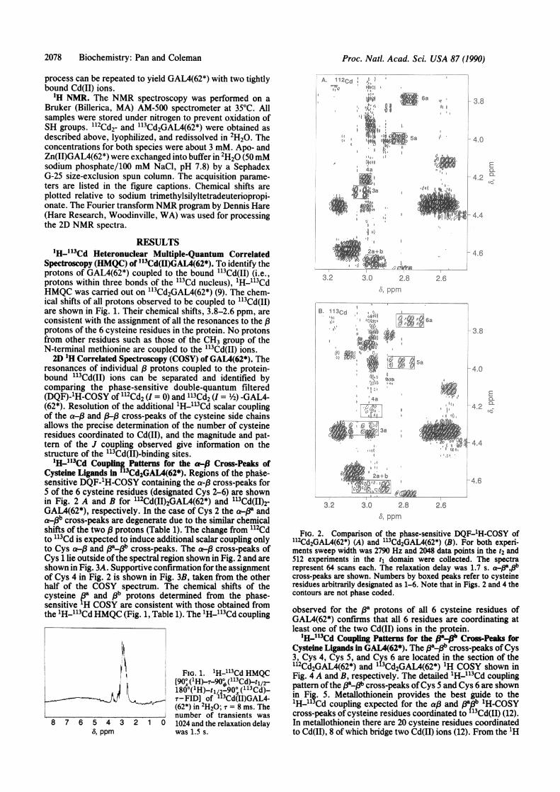
Spectroscopy (HMQC) of "3Cd(H)GAL4(62*). To identify theprotons of GAL4(62*) coupled to the bound 1"3Cd(II) (i.e.,protons within three bonds of the 113Cd nucleus), 'H-113CdHMQC was carried out on Ll3Cd2GAL4(62*) (9). The chem-ical shifts of all protons observed to be coupled to 113Cd(II)are shown in Fig. 1. Their chemical shifts, 3.8-2.6 ppm, areconsistent with the assignment of all the resonances to the /3protons of the 6 cysteine residues in the protein. No protonsfrom other residues such as those of the CH3 group of theN-terminal methionine are coupled to the 113Cd(II) ions.2D 'H Correlated Spectroscopy (COSY) of GAL4(62*). The
resonances of individual f protons coupled to the protein-bound "13Cd(II) ions can be separated and identified bycomparing the phase-sensitive double-quantum filtered(DQF)-'H-COSY of 112Cd2 (I = 0) and '13Cd2 (I = 1/2) -GAL4-(62*). Resolution of the additional 'H-"13Cd scalar couplingof the a-d8 and P,-/ cross-peaks of the cysteine side chainsallows the precise determination of the number of cysteineresidues coordinated to Cd(II), and the magnitude and pat-tern of the J coupling observed give information on thestructure of the "13Cd(II)-binding sites.'H-"3Cd Coupling Patterns for the A-(3 Cross-Peaks of
Cysteine Ligands in 3Cd2GAL4(62*). Regions of the phase-sensitive DQF-'H-COSY containing the a-,8 cross-peaks for5 of the 6 cysteine residues (designated Cys 2-6) are shownin Fig. 2 A and B for l12Cd(II)2GAL4(62*) and 113Cd(II)2-GAL4(62*), respectively. In the case of Cys 2 the a-Pa anda-pb cross-peaks are degenerate due to the similar chemicalshifts of the two 13 protons (Table 1). The change from "12Cdto 113Cd is expected to induce additional scalar coupling onlyto Cys a,-( and ,3a13b cross-peaks. The at-/ cross-peaks ofCys 1 lie outside of the spectral region shown in Fig. 2 and areshown in Fig. 3A. Supportive confirmation for the assignmentof Cys 4 in Fig. 2 is shown in Fig. 3B, taken from the otherhalf of the COSY spectrum. The chemical shifts of thecysteine ea and fib protons determined from the phase-sensitive H COSY are consistent with those obtained fromthe 1H-"13Cd HMQC (Fig. 1, Table 1). The 'H-"13Cd coupling
FIG. 1. H-"113Cd HMQC[90X ('H)--T90° (113Cd)-ti2-180°('H)-tl/2-900x (113Cd)-T-FID] of "'Cd(II)GAL4-(62*) in 2H20; T = 8 Ins. Thenumber of transients was1024 and the relaxation delaywas 1.5 s.
A. 112Cdi 4toI,
46.,p
44a
2g2a -b
3.2 30o 2.8i) p 1
B. 113Cd
55a
3.2
3.8
4 0
E
4.2 a!,-
4.6
2.6
3.8
4.0
Eal4.2i ~~I
|~~~~~~~~~~'4.4.
4.6.-k oi
3.0 2.8 2.625! ppm
FIG. 2. Comparison of the phase-sensitive DQF-'H-COSY ofll2Cd2GAL4(62*) (A) and ll3Cd2GAL4(62*) (B). For both experi-ments sweep width was 2790 Hz and 2048 data points in the t2 and512 experiments in the tj domain were collected. The spectrarepresent 64 scans each. The relaxation delay was 1.7 s. a.#,pbcross-peaks are shown. Numbers by boxed peaks refer to cysteineresidues arbitrarily designated as 1-6. Note that in Figs. 2 and 4 thecontours are not phase coded.
observed for the 8a protons of all 6 cysteine residues ofGAL4(62*) confrms that all 6 residues are coordinating atleast one of the two Cd(II) ions in the protein.
'H-"13Cd Coupling Patterns for the (glWpb Cross-Peaks forCysteine Ligands in GAL4(62*). The ,3aPpb cross-peaks ofCys3, Cys 4, Cys 5, and Cys 6 are located in the section of thel12Cd2GAL4(62*) and l 3Cd2GAL4(62*) 1H COSY shown inFig. 4 A and B, respectively. The detailed 'H-"13Cd couplingpattern ofthe 1381-(b cross-peaks ofCys 5 and Cys 6 are shownin Fig. 5. Metallothionein provides the best guide to the'H-'1 Cd coupling expected for the ai3 and 1a13b 'H-COSYcross-peaks of cysteine residues coordinated to"3Cd(II) (12).In metallothionein there are 20 cysteine residues coordinatedto Cd(II), 8 of which bridge two Cd(II) ions (12). From the 1H
8 7 6 5 4 35, ppm
2 1 0
Proc. Natl. Acad. Sci. USA 87 (1990)
,e-
I:$ 4.4
0-k,4a
I- ---
.-I1 3r 3a

Proc. Natl. Acad. Sci. USA 87 (1990) 2079
Table 1. Chemical shifts and 'H-'H and 'H-"3Cd coupling constants for six cysteine residues of GAL4(62*)ll3Cd2GAL4(62*) at 350C, pH 8.0 Zn2GAL4(62*) at 350C, pH 5.4
No. of Chemical shift, 'H-'H coupling 'H-"3Cd coupling Chemical shift,spin ppm constants, Hz constants, Hz ppm
system Ha Hpa Hpb JIM JambJbJppt J4acdl JobCdI JPaCd2 JbCd2 Hag Hp. Heb1 4.94 3.73 3.60 11 10 15 25 10 4.67 3.60 3.502 4.64 2.95 2.95 - =z52 - 4.65 2.84 2.843 4.33t 3.04 2.67 11 <2 22 55 26 4.29 3.04 2.554 4.22 2.99 3.14 7 <2 16 20 45 4.12 2.885 3.98 2.82 3.35 10 <2 14 28 14 5t 3 3.98 2.75 3.256 3.75 2.77 3.26 10 <2 14 29 16 2* 3 3.73 2.73 3.17Note that by definition pa is the proton with the larger homonuclear spin-spin coupling (12). A - indicates not obtainable due to lack of
resonances or degeneracy of the chemical shifts.tChemical shift differs from that of ll2Cd2GAL4(62*) (Ha = 4.29). The reason for such differences is not clear.*From adPa cross-peaks.
COSY of "3Cd7 metallothionein, it is apparent that in thepresence of a bridging cysteine, coupling of the 13 protons toone of the coordinated "3Cd nuclei is generally of muchgreater magnitude than to the other. In fact, only two of theshared cysteine residues in metallothionein can be readilyidentified in the 1H COSY of the lu3Cd7 derivative (12). The13a. b cross-peaks of Cys 5 and Cys 6 of GAL4(62*) showsplitting patterns similar to the pattern shown by the one"classical" shared cysteine (C14 = Cys" in the sequence) inmetallothionein. However, the discernible L3Cd2-,3 couplingin ll3Cd(II)GAL4(62*), expressed as increased vertical sepa-ration of the multiplet on shifting from "12Cd to 113Cd, is quitesmall, 3-4 Hz for both Cys S and Cys 6. Although a JH a113Cd2is not indicated for either Cys 5 or Cys 6 in Fig. 5 (JHPa,13Cd, =28 and 29 Hz, respectively), the splitting of the a-/a cross-peaks of these same cysteine residues is =33 and 31 Hz (Fig.2B). This difference in coupling of 113Cd' to
,as measured
from the pa1b vs. the a-,& cross-peak suggests that significantcoupling (=2-5 Hz) of f, to 1"3Cd2 is present. In addition,partial cancellation of the multiplets in the a-,B cross-peaks ofCys 5 and Cys 6 is also suggestive offinite coupling constantsto a second 113Cd ion. Such coupling is more directly manifestin the a-Pa than in the f cross-peak. The 'H-113Cdcoupling constants for GAL4(62*) derived from the phase-sensitive 1H COSY vary from 2 to 55 Hz (Table 1). The rangeof values is comparable to that observed for the 113Cd7derivative of metallothionein (12). The fla-P" cross-peak ofCys 1 is located downfield of the section of the COSYspectrum shown in Fig. 4 and has been used to measure the"3Cd-1H0 coupling constants given in Table 1. There is no fl-,3cross-peak for Cys 2 due to the degeneracy of the chemicalshifts of the 8 protons (Table 1).
A.Cys 1 a
112Cd25 Hz
+ - It -±+ -
We have shown previously by circular dichroism thatremoval of Zn(II) causes substantial conformational changein GAL4(63) (5). Considerable chemical shift changes andreduction of linewidth of several signals are observed whenZn(II) or Cd(II) is bound to apo-GAL4(62*) (Fig. 6). Theprotons of Zn(II)- and Cd(II)GAL4(62*) have very similarchemical shifts, suggestive of nearly identical conformationsfor both species.
DISCUSSIONThe N-terminal fragments of the transcription factor GAL4,either 147 or 63 amino acids in length, form domains whichfold independently and bind to the specific DNA sequence,UASG, recognized by GAL4 (4, 5). Both fragments bind 1 to2 Zn(II) ions, and they maintain the binding of the secondZn(II) when the free Zn(II) concentration is 5 ,uM (5). Whenthe Zn(II) is replaced with 113Cd(II), the '13Cd NMR of bothfragments shows two '"Cd NMR signals, each of whichintegrates to one 1"3Cd(II) ion (4, 5). Thus there are two metal-ion-binding sites on the DNA-binding domain of GAL4.GAL4(62*) has a high-resolution proton NMR spectrum
uncomplicated by the oligomerization shown by GAL4(149*)when it is present at the concentrations ofprotein required for1H NMR (5). The GAL4(62*) clone produces a proteinstructurally and functionally identical to GAL4(63) obtainedfrom GAL4(149*) by partial proteolysis (5). GAL4(62*) issoluble to concentrations of several millimolar and thus idealfor 2D 'H-NMR studies. The fact that cloned GAL4(62*) ishighly soluble, while GAL4-(1-74) is not (5), also suggeststhat GAL4(62*) forms an independent domain. The 'H-113CdHMQC establishes that only the six cysteine residues appearto be ligands to metal ions (Fig. 1). The DQF-'H-COSY
113Cd
+- -4- 4.92
j If."1I,. 4.96
3.76 3.72 3.68 3.64 3.60 3.56fw2, ppm
B.4.25 Cys4 a
_ - -,--: +
4.20
3.04 3.00 2.96anl. ppm
2.92
-I_-+±- +
- 4.92E
I- 4.96
3.76 3.72 3.68 3.64 3.60 3.56w2, ppm
4.25 - H7± - -
4.20 -
3.04 3.00 2.96 2.92(ol ppm
FIG. 3. Phase-sensitive DQF-'H-COSY regions showing a-MOab cross-peaks of Cys 1 (A)and a-#a cross-peak ofCys 4 (B)(see text). For Cys 1 and 113Cd,additional splitting makes reso-nance lb of low intensity.
E
CL
Biochemistry: Pan and Coleman

2080 Biochemistry: Pan and Coleman
I
A. 112Cd 3
;10@l 1,00l .; @°2t:
0iiIA4
,v. .. .. £ ... A
3.4 3.2 3.06, ppm
3
4'1
I; t
01 15 1J
2.8
2.6
2.8
3.0 aa_
3-.iii
3.2
6
34
2.6
EQ-Ca-
a-:
29 HzrI
B. 113Cd + -
6~~~~~~ ~ ~ -3.25
3HzQ + 3~~~~-.3014Hz
I 1 -~~~~~3.402.88 2.84 2.80 2.76
8, ppm2.72
, ppm
FIG. 4. Phase-sensitive DQF-1H-COSY as in Fig. 2. #a_.Bb cross-peaks of Cys 3, Cys 4, Cys 5, and Cys 6 are shown. (A)l12Cd2GAL4(62*). (B) ll3Cd2GAL4(62*). The cross-peak for Cys 4 inboth derivatives and half the cross-peak of Cys 3 in the "13Cdderivative are badly overlapped with other resonances, but they canbe separated in expanded and phase-coded plots. The expanded plotswere used to measure the coupling constants given in Table 1.
shows that two of these cysteine residues (designated 5 and6) appear to be bridging ligands between the two "13Cd(II)ions (Figs. 4 and 5). Among the six 113Cd(II)-coordinatedcysteine residues of GAL4(62*), Cys 5 and 6 must occupyalmost identical and unique coordination environments,since they both have nearly identical and relatively largecoupling of both p protons to one of the 113Cd(II) ions (Fig.5), a pattern which is also observed for the two well-resolved(3apb cross-peaks of bridging cysteine in metallothionein(12). One can conclude that the torsional angles describingthe configuration of the three-bond fragment between theprotons and the coordinated 113Cd(II) ions must be the samefor Cys 5 and Cys 6. A possible model of the binuclearcomplex (cloverleaf) formed by GAL4(62*) is shown in Fig.7. This model assumes Cys21 and Cys38 are the bridgingligands, which places them in equivalent configurations, as
FIG. 5. Expanded region from Fig. 4 showing the couplingpatterns derived from the pa_8b cross-peaks of Cys 5 and Cys 6.
their J coupling suggests. The model building suggests thatthe five peptide loops necessitated by the cluster structurepack most easily with this choice of bridging ligands. Apreliminary sequential assignment of Zn(II)GAL4(62*) sup-
ports the assignment of Cys 5 and 6 to residues 38 and 21,respectively, but this result must be considered tentative.
Sequential assignment of the GAL4(62*) spectrum by 2DNMR techniques is not yet complete. While the 1H-113Cdheteronuclear J coupling will help establish landmarks in thespectra of this sequence, which has an abundance of lysineand arginine residues, at present its most important aspect isthe establishment of the two-metal-ion cluster structure.From the complete COSY and nuclear Overhauser enhance-ment (NOE) spectroscopy (NOESY) spectra in H20, all theNH-Ha connectivities can be identified. Strong NH,-NHi+lNOEs in the NOESY spectrum of Zn(II)GAL4(62*) haveestablished two separate short stretches of a-helix (unpub-lished results). Thus a large part of the backbone ofGAL4(62*) must be unstructured or P-sheet-like, not incom-patible with the model shown in Fig. 7. The regions N-terminal (residues 1-10) and C-terminal (residues 49-62) tothe cluster subdomain are likely to form the a-helices sug-gested to be present by the NOESY data on Zn(II)GAL4(62*)and the earlier circular dichroism studies (5).The model of GAL4 in Fig. 7 places the preponderance of
positive charge, six lysine and one arginine residues, on oneside of the cluster formed by the two loops Cys14-Cys21 andCys21-Cys'. Comparison of the sequences shows that thepositive charge distribution in the Cys14-Cys21 loop is main-
A. 112Cd v
(i @0 a (@cy
'f 0Cys50~ ~~ys
Cys528 X
_+2.88 2.84 2.80 2.76 2.72
3.20
-3.25
E3.30 aM
3.35
3.40
E0.
so
Proc. Natl. Acad. Sci. USA 87 (1990)

Proc. Natl. Acad. Sci. USA 87 (1990) 2081
A. y ApoGAL4(62*)
TSP
8, ppm
FIG. 6. 1H NMR spectra of a 0.9 mM solution of apo-GAL4(62*)(A), Zn(II)GAL4(62*) (B), and l13Cd(II)GAL4(62*) (C). The numberof transients is 16 and the relaxation delay was 2.5 s. TSP, sodiumtrimethylsilyltetradeuteriopropionate.
tained in all nine fungal transcription factors of this class; infact, the positions ofthree ofthe lysine residues are absolutelyconserved in this loop. This suggests that these residues withinthe cluster (Cysl-Cys'8) may function in nonspecific DNAbinding and that the recognition determinants may be outsidethis region. The specific contact region could be provided bya turn and a-helix following the most C-terminal cysteineresidue. The specific DNA recognition could involve a clo-verleaf-turn-helix structure with turns in the region of Pro42and Pro'8. Genetic studies already suggest that amino acid
42 60
-D-P-A-T-G-K-D-V-P-R-C-Y-V-F-F-L-E-D-R(PPRI)-P-Q-V-V-R-T-P-L-T-R-A-H-L-T-E-M-E-Q-R(LAC9)
NH2 P-K-T-K-R-S-P-L-T-R-A-H-L-T-E-V-E-S-R(GAL4)7)1 <+31
'L 14 K T SLK L 21 'Ii% % % P P PK K K_ K - L- K --- K-E-K}\- K W-K --S -K- TV- V-\-_K- I- K D-Q-E- F
I *V 11
FIG. 7. Model of the Zn(II)2Cys6 or Cd(II)2Cys6 cluster (clover-leaf) of the DNA-binding domain of GAL4. Portions of the aminoacid sequences of transcription factors LAC9 and PPR1 are alsoshown. Loops I, II, and III each contain six amino acid residues.
residues within the Zn(II)2Cys6 cluster of GAL4 are notdirectly involved in specific DNA recognition (13).
Partial amino acid sequences oftwo other fungal transcrip-tion factors in the regions of the DNA-binding domainscorresponding to that of GAL4(62*) are compared in themodel (Fig. 7). One of them, LAC9, must be homologous toGAL4, since they both recognize the UASG sequence (14,15). The second, PPR1, does not recognize this sequence butmaintains the Cys6 motif (16). All three factors maintainsimilar charge and amino acid sequences within the metal ioncluster. As shown for PPR1, the most marked deviation ofsequence in this group of transcription factors occurs in theregion immediately downstream from the cluster. Ofthe eightother factors, only the more closely related LAC9 has asequence in this region similar to that ofGAL4 (Fig. 7). Thisregion of the DNA-binding domain may participate in thesequence-speciflic DNA interactions.A binuclear zinc cluster complex is a structure different
from those previously proposed for the zinc-finger transcrip-tion factors. The highly conserved arrangement of cysteineresidues in GAL4 and the other fungal transcription factorsalmost certainly means that this structure is also present inthe others. Whether such a binuclear complex or a variant ofit is present in other so-called Cys2Cys2 zinc-finger transcrip-tion factors, such as the steroid receptor proteins (containinga total of nine conserved cysteine residues), remains specu-lative. Cd(II) forms a stable binuclear cluster with GAL4 andthe binding is highly cooperative (4, 5). Not surprisingly, theZn(II) analogue is more labile, and one of the two Zn(II) ionsdissociates when free Zn(II) is less than micromolar. Thisraises the interesting possibility that DNA-binding affinityand therefore transcriptional regulation might depend undersome circumstances on the cellular Zn(II) concentration.Whether the structural changes as a function of Zn(II)content necessary for such a postulate are a feature of GAL4requires further investigation.
This work was supported by National Institutes of Health GrantsDK09070 and GM21919. The 500-MHz NMR was supported byNational Institutes of Health Grant RR03475, National ScienceFoundation Grant DMB-8610557, and American Cancer SocietyGrant RD259. This work is in partial fulfillment of the requirementsfor the Ph.D. degree (T.P.).
1. Oshima, Y. (1982) in Molecular Biology of the Yeast Saccharomy-ces, eds. Strathern, J., Jones, E. & Broach, J. K. (Cold SpringHarbor Lab., Cold Spring Harbor, NY), Vol. 1, pp. 159-180.
2. Johnston, M. & Dover, J. (1987) Proc. Nati. Acad. Sci. USA 84,2401-2405.
3. Keegan, L., Gill, G. & Ptashne, M. (1986) Science 231, 699-704.4. Pan, T. & Coleman, J. E. (1989) Proc. Natl. Acad. Sci. USA 86,
3145-3149.5. Pan, T. & Coleman, J. E. (1990) Biochemistry 29, in press.6. Pfeifer, K., Kim, K.-S., Kogan, S. & Guarante, L. (1989) Cell 56,
291-301.7. Johnston, M. (1987) Nature (London) 328, 353-355.8. Miller, J., Mclachlan, A. D. & Klug, A. (1985) EMBO J. 4, 1609-
1614.9. Frey, M. H., Wagner, G., Vasak, M., Sorensen, 0. W., Neuhaus,
D., Worg6tter, E., Kagi, J. H., Ernst, R. R. & Wuthrich, K. (1985)J. Am. Chem. Soc. 107, 6847-6851.
10. Schultze, P., Worgotter, E., Braun, W., Wagner, G., Vasak, M.,Kagi, J. H. R. & Wuthrich, K. (1988) J. Mol. Biol. 203, 251-268.
11. Kagi, J. H. & Kojima, Y., eds. (1987) Metallothionein Ii (Birkhae-user, Basel).
12. Neuhaus, D., Wagner, G., Vasak, M., Kagi, J. H. & Wuthrich, K.(1984) Eur. J. Biochem. 143, 659-667.
13. Corton, J. C. & Johnston, S. A. (1989) Nature (London) 340,724-727.
14. Salmeron, J. M., Jr., & Johnston, S. A. (1986) Nucleic Acids Res.14, 7767-7781.
15. Wray, L. V., Jr., Witte, M. M., Dickson, R. C. & Riley, M. I.(1987) Mol. Cell. Biol. 7, 1111-1121.
16. Kammerer, B., Guyonvarch, A. & Hubert, J. C. (1984) J. Mol. Biol.180, 239-250.
Biochemistry: Pan and Coleman