transcription,translation and genetic code(cell biology)by welfredo yu
TRANSCRIPT
Cells are governed by a cellular chain of
command
DNA RNA protein
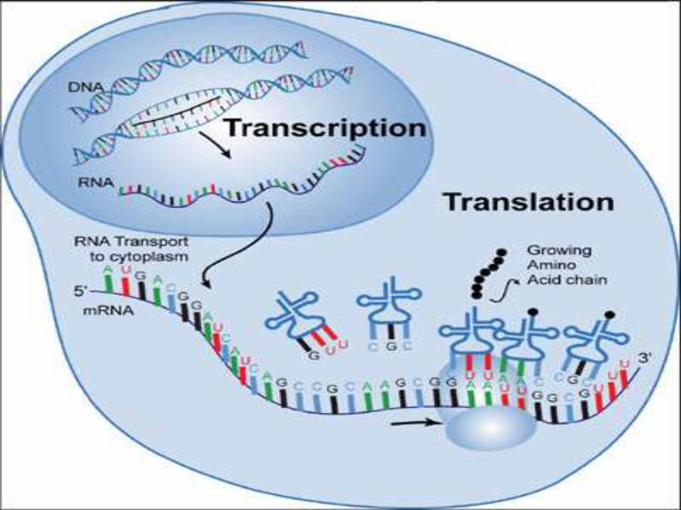
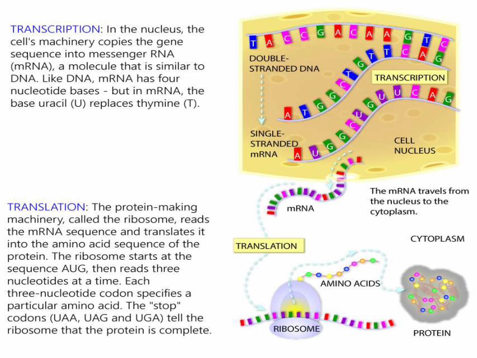
Transcription
Is the synthesis of RNA under the direction of
DNA
Produces messenger RNA (mRNA)
Translation
Is the actual synthesis of a polypeptide which
occurs under the direction of mRNA
Occurs on ribosomes
The Genetic Code
a non-overlapping sequence with each amino
acid plus polypeptide initiation and termination
specified by RNA codons composed of three
nucleotides
Genes can be expressed at different efficiencies
bull Gene A is transcribed much more efficiently than gene B
bull This allows the amount of protein A in the cell to be
greater than protein B
bull The lower expression of gene B is a reason behind
incomplete dominance
BIOL211
RNA is the bridge between
genes and the proteins for
which they code
what is RNA
Monomers of proteins are amino acids
what are proteins made of
Codons
bull A codon is three nucleotides in a row on an RNA
molecule that codes for a single amino acid
bull A specific three-nucleotide sequence encodes for each
amino acid
Template Strand
bull During transcription one of the two DNA
strands called the template strand provides
a template for ordering the sequence of
complementary nucleotides in an RNA
transcript
ndash The template strand is always the same strand for
a given gene
ndash However different genes may be on opposite
strands
bull The genetic code is nearly
universal shared by the simplest
bacteria to the most complex
animals
ndash Some species prefer certain
codons (codon bias)
bull Genes can be transcribed and
translated after being
transplanted from one species
to another
EVOLUTION OF THE CODE
History linking genes and proteins
1900rsquos Archibald Garrod Inborn errors of metabolism inherited human
metabolic diseases (more information) Genes are the inherited factors
Enzymes are the biological molecules that drive metabolic reactions
Enzymes are proteins
Question
How do the inherited factors the genes control the structure and activity of enzymes (proteins)
History linking genes and proteins
Beadle and Tatum (1941) PNAS USA 27 499ndash506
Hypothesis If genes control structure and activity of metabolic enzymes
then mutations in genes should disrupt production of required nutrients and that disruption should be heritable
Method Isolated ~2000 strains from single irradiate spores
(Neurospora) that grew on rich but not minimal medium Examples defects in B1 B6 synthesis
Conclusion Genes govern the ability to synthesize amino acids purines
and vitamins
History linking genes and proteins
1950s sickle-cell anemia Glu to Val change in hemoglobin
Sequence of nucleotides in gene determines sequence of amino acids in protein
Single amino acid change can alter the function of the protein
Tryptophan synthase gene in E coli Mutations resulted in single amino acid change
Order of mutations in gene same as order of affected amino acids
Ribosomal structure
EP A
Large subunit
Peptidyl-tRNA binding site
Aminoacyl-tRNA binding site
mRNA
5rsquo
Exit
site
Small subunit
3rsquo
From gene to protein transcription
Gene sequence (DNA) recopied or transcribed to RNA sequence
Product of transcription is a messenger molecule that delivers the genetic instructions to the protein synthesis machinery messenger RNA (mRNA)
Transcription evidence for mRNA
Brenner S Jacob F and Meselson M (1961) Nature190 576ndash81
Question How do genes work
Does each one encode a different type of ribosome which in turn synthesizes a different protein OR
Are all ribosomes alike receiving the genetic information to create each different protein via some kind of messenger molecule
Transcription evidence for mRNA
E coli cells switch from making bacterial proteins to phage proteins when infected with bacteriophage T4
Grow bacteria on medium containing ldquoheavyrdquo nitrogen (15N) and carbon (13C)
Infect with phage T4
Immediately transfer to ldquolightrdquo medium containing radioactive uracil
Transcription evidence for mRNA
If genes encode different ribosomes the newly synthesized phage ribosomes will be ldquolightrdquo
If genes direct new RNA synthesis the RNA will contain radiolabeled uracil
Results Ribosomes from phage-infected cells were ldquoheavyrdquo
banding at the same density on a CsCl gradient as the original ribosomes
Newly synthesized RNA was associated with the heavy ribosomes
New RNA hybridized with viral ssDNA not bacterial ssDNA
Transcription evidence for mRNA
Conclusion
Expression of phage DNA results in new phage-specific RNA molecules (mRNA)
These mRNA molecules are temporarily associated with ribosomes
Ribosomes do not themselves contain the genetic directions for assembling individual proteins
Transcription overview
Transcription requires
ribonucleoside 5acute triphosphates
ATP GTP CTP and UTP
bases are adenine guanine cytosine and uracil
sugar is ribose (not deoxyribose)
DNA-dependent RNA polymerase
Template (sense) DNA strand
Animation of transcription
Transcription overview
Features of transcription
RNA polymerase catalyzes sugar-phosphate bond between 3acute-OH of ribose and the 5acute-PO4
Order of bases in DNA template strand determines order of bases in transcript
Nucleotides are added to the 3acute-OH of the growing chain
RNA synthesis does not require a primer
Transcription overview
In prokaryotes transcription and translation are coupled Proteins are synthesized directly from the primary transcript as it is made
In eukaryotes transcription and translation are separated Transcription occurs in the nucleus and translation occurs in the cytoplasm on ribosomes
Figure comparing eukaryotic and prokaryotic transcription and translation
Transcription RNA Polymerase
DNA-dependent
DNA template ribonucleoside 5acute triphosphates and Mg2+
Synthesizes RNA in 5acute to 3acute direction
E coli RNA polymerase consists of 5 subunits
Eukaryotes have three RNA polymerases
RNA polymerase II is responsible for transcription of protein-coding genes and some snRNA molecules
RNA polymerase II has 12 subunits
Requires accessory proteins (transcription factors)
Does not require a primer
Stages of Transcription
Transcription promoter recognition
Transcription factors bind to promoter sequences and recruit RNA polymerase
DNA is bound first in a closed complex Then RNA polymerase denatures a 12ndash15 bp segment of the DNA (open complex)
The site where the first base is incorporated into the transcription is numbered ldquo+1rdquo and is called the transcription start site
Transcription factors that are required at every promoter site for RNA polymerase interaction are called basal transcription factors
Promoter recognition promoter sequences
Promoter sequences vary considerably
RNA polymerase binds to different promoters with different strengths binding strength relates to the level of gene expression
There are some common consensus sequences for promoters Example E coli ndash35 sequence (found 35 bases 5acute to the
start of transcription)
Example E coli TATA box (found 10 bases 5acute to the start of transcription)
Promoter recognition enhancers
Eukaryotic genes may also have enhancers
Enhancers can be located at great distances from the gene they regulate either 5acute or 3acute of the transcription start in introns or even on the noncoding strand
One of the most common ways to identify promoters and enhancers is to use a reporter gene
Promoter recognition other players
Many proteins can regulate gene expression by modulating the strength of interaction between the promoter and RNA polymerase
Some proteins can activate transcription (upregulate gene expression)
Some proteins can inhibit transcription by blocking polymerase activity
Some proteins can act both as repressors and activators of transcription
Transcription chain initiation
Chain initiation
RNA polymerase locally denatures the DNA
The first base of the new RNA strand is placed complementary to the +1 site
RNA polymerase does not require a primer
The first 8 or 9 bases of the transcript are linked Transcription factors are released and the polymerase leaves the promoter region
Figure of bacterial transcription initiation
Transcription chain elongation
Chain elongation
RNA polymerase moves along the transcribed or template DNA strand
The new RNA molecule (primary transcript) forms a short RNA-DNA hybrid molecule with the DNA template
Transcription chain termination
Most known about bacterial chain termination
Termination is signaled by a sequence that can form a hairpin loop
The polymerase and the new RNA molecule are released upon formation of the loop
Review the transcription animation
Transcription mRNA synthesisprocessing
Prokaryotes mRNA transcribed directly from DNA template and used immediately in protein synthesis
Eukaryotes primary transcript must be processed to produce the mRNA
Noncoding sequences (introns) are removed
Coding sequences (exons) spliced together
5acute-methylguanosine cap added
3acute-polyadenosine tail added
Transcription mRNA synthesisprocessing
Removal of introns and splicing of exons can occur several ways For introns within a nuclear transcript a spliceosome is
required
Splicesomes protein and small nuclear RNA (snRNA)
Specificity of splicing comes from the snRNA some of which contain sequences complementary to the splice junctions between introns and exons
Alternative splicing can produce different forms of a protein from the same gene
Mutations at the splice sites can cause disease
Thalassemia bull Breast cancer (BRCA 1)
Transcription mRNA synthesisprocessing
RNA splicing inside the nucleus on particles called spliceosomes
Splicesomes are composed of proteins and small RNA molecules (100ndash200 bp snRNA)
Both proteins and RNA are required but some suggesting that RNA can catalyze the splicing reaction
Self-splicing in Tetrahymena the RNA catalyzes its own splicing
Catalytic RNA ribozymes
From gene to protein genetic code
Central Dogma
Information travels from DNA to RNA to Protein
Is there a one-to-one correspondence between DNA RNA and Protein
DNA and RNA each have four nucleotides that can form them so yes there is a one-to-one correspondence between DNA and RNA
Proteins can be composed of a potential 20 amino acids only four RNA nucleotides no one-to-one correspondence
How then does RNA direct the order and number of amino acids in a protein
From gene to protein genetic code
How many bases are required for each amino acid (4 bases)2basesaa = 16 amino acidsmdashnot enough
(4 bases)3basesaa = 64 amino acid possibilities
Minimum of 3 basesaa required
What is the nature of the code Does it have punctuation Is it overlapping
Crick FH et al (1961) Nature 192 1227ndash32 (httpprofilesnlmnihgovSCBCBJ )
3-base nonoverlapping code that is read from a fixed point
From gene to protein genetic code
Nirenberg and Matthaei in vitro protein translation
Found that adding rRNA prolonged cell-free protein synthesis
Adding artificial RNA synthesized by polynucleotide phosphorylase (no template UUUUUUUUU) stimulated protein synthesis more
The protein that came out of this reaction was polyphenylalanine (UUU = Phe)
Other artificial RNAs AAA = Lys CCC =Pro
From gene to protein genetic code
Nirenberg
Triplet binding assay add triplet RNA ribosomes binding factors GTP and radiolabeled charged tRNA (figure)
UUU trinucleotide binds to Phe-tRNA
UGU trinucleotide binds to CYS-tRNA
By fits and starts the triplet genetic code was worked out
Each three-letter ldquowordrdquo (codon) specifies an amino acid or directions to stop translation
The code is redundant or degenerate more than one way to encode an amino acid
From gene to protein Translation
Components required for translation
mRNA
Ribosomes
tRNA
Aminoacyl tRNA synthetases
Initiation elongation and termination factors
Animation of translation
Translation initiation
Ribosome small subunit binds to mRNA
Charged tRNA anticodon forms base pairs with the mRNA codon
Small subunit interacts with initiation factors and special initiator tRNA that is charged with methionine
mRNA-small subunit-tRNA complex recruits the large subunit
Eukaryotic and prokaryotic initiation differ slightly
Translation initiation
The large subunit of the ribosome contains three binding sites
Amino acyl (A site)
Peptidyl (P site)
Exit (E site)
At initiation
The tRNAfMet occupies the P site
A second charged tRNA complementary to the next codon binds the A site
Translation elongation
Elongation
Ribosome translocates by three bases after peptide bond formed
New charged tRNA aligns in the A site
Peptide bond between amino acids in A and P sites is formed
Ribosome translocates by three more bases
The uncharged tRNA in the A site is moved to the E site
Translation elongation
EF-Tu recruits charged tRNA to A site Requires hydrolysis of GTP
Peptidyl transferase catalyzes peptide bond formation (bond between aa and tRNA in the P site converted to peptide bond between the two amino acids)
Peptide bond formation requires RNA and may be a ribozyme-catalyzed reaction
Translation termination
Termination
Elongation proceeds until STOP codon reached UAA UAG UGA
No tRNA normally exists that can form base pairing with a STOP codon recognized by a release factor
tRNA charged with last amino acid will remain at P site
Release factors cleave the amino acid from the tRNA
Ribosome subunits dissociate from each other
Review the animation of translation
48
Genetic code
Def Genetic code is the nucleotide base sequence on DNA ( and
subsequently on mRNA by transcription) which will be translated into a
sequence of amino acids of the protein to be synthesized
The code is composed of codons
Codon is composed of 3 bases ( eg ACG or UAG) Each codon is
translated into one amino acid
The 4 nucleotide bases (AGC and U) in mRNA are used to produce the
three base codons There are therefore 64 codons code for the 20 amino
acids and since each codon code for only one amino acids this means
that there are more than one cone for the same amino acid
How to translate a codon (see table)
This table or dictionary can be used to translate any codon sequence
Each triplet is read from 5prime rarr 3prime direction so the first base is 5prime base
followed by the middle base then the last base which is 3prime base
49
Examples 5prime- A UG- 3prime codes for methionine
5prime- UCU- 3prime codes for serine
5prime - CCA- 3prime codes for proline
Termination (stop or nonsense) codons
Three of the 64 codons UAA UAG UGA do not code for any amino
acid They are termination codes which when one of them appear in
mRNA sequence it indicates finishing of protein synthesis
Characters of the genetic code
1- Specificity the genetic code is specific that is a specific codon
always code for the same amino acid
2- Universality the genetic code is universal that is the same codon is
used in all living organisms procaryotics and eucaryotics
3- Degeneracy the genetic code is degenerate ie although each codon
corresponds to a single amino acidone amino acid may have more than
one codons eg arginine has 6 different codons (give more examples
from the table)
50
51
Gene mutation (altering the nucleotide sequence)
1- Point mutation changing in a single nucleotide base on the mRNA
can lead to any of the following 3 results
i- Silent mutation ie the codon containg the changed base may code
for the same amino acid For example in serine codon UCA if A is
changed to U giving the codon UCU it still code for serine See table
ii- Missense mutation the codon containing the changed base may code
for a different amino acid For example if the serine codon UCA is
changed to be CCA ( U is replaced by C) it will code for proline not
serine leading to insertion of incorrect amino acid into polypeptide chain
iii- Non sense mutation the codon containing the changed base may
become a termination codon For example serine codon UCA becomes
UAA if C is changed to A UAA is a stop codon leading to termination
of translation at that point
52
53
Types of point mutation
U A A (termination codon) Nonsense mutation
uarr
U C A rarr U C U Silent mutation
(codon for serine) (codon for serine)
darr
C C A ( codon for proline) Missense mutation
Give other examples on missense mutation which leads to some Hb
disease
54
2- Frame- shift mutation
deletion or addition of one or two base to
message sequence leading to change in
reading frame (reading sequence) and the
resulting amino acid seuence may become
completely different from this point
55
TranslationComponents required for protein synthesis1- Amino acids all amino acids involved in thefinished protein must be present at the time ofprotein synthesis2- Ribosomes the site of protein synthesis They arelarge complexes of protein and rRNA In humanthey consist of two subunits one large (60S) and onesmall (40S)
3- tRNA at least one specific type of tRNA is required to transfer
one amino acid There about 50 tRNA in human for the 20 amino
acids this means some amino acids have more than one specific
tRNA The role of tRNA in protein synthesis is discussed before
(amino acid attachment and anticodon loop)
4- aminoacyl-tRNA synthetase This is the enzyme that catalyzes the
attachment of amino acid with its corresponding tRNA forming
aminoacyl tRNA
56
5- mRNA that carry code for the protein to be synthesized
6- protein factors Initiation elongation and termination (or release)
factors are required for peptide synthesis
7- ATP and GTP are required as source of energy
Steps (movie)
1- Initiation
Initiation (start) codon is usually AUG which is the codon of
methionine so the initiator tRNA is methionnyl tRNA (Met tRNA)
a- The initiation factors (IF-1 IF-2 and IF-3) binds the Met tRNA with
small ribosomal subunit then to mRNA containing the code of the
protein to be synthesized IFs recognizes mRNA from its 5 cap
57
b-This complex binds to large ribosomal subunit forming initiation complex in which Met tRNA is present in P- site of 60 ribosomal subunitNB- tRNA bind with mRNA by base pairing between codon on mRNA and anticodon
on tRNA - mRNA is read from 5prime rarr 3prime direction
P-site is the peptidyl site of the ribosome to which methionyl tRNA is placed (enter)
2- Elongation elongation factors (EFs) stimulate the stepwise elongation of polypeptide chain as follow
a- The next aminoacyl tRNA (tRNA which carry the next amino acid specified by recognition of the next codon on mRNA) will enter A site of ribosome
A site or acceptor site or aminoacyl tRNA siteIs the site of ribosome to which each new incoming aminoacyl tRNA will enter
b) ribosomal peptidyl transferase enzyme will transfer methionine
from methionyl tRNA into A site to form a peptide bond between
methionine and the new incoming amino acid to form dipeptidyl
tRNA
c) Elongation factor-2 (EF-2) (called also translocase) moves
mRNA and dipeptidyl tRNA from A site to P site leaving A site free to
allow entrance of another new aminoacyl tRNA
The figure shows the repetitive cycle of elongation of chain Each
cycle is consisting of
1) codon recognition and the entrance of the new aminoacyl tRNA
acid ( amino acid carried on tRNA) into A site
2) The growing chain in P site will moved to A site with peptide
bond formation with the new amino acid
3) Translocation of growing chain to P site allowing A site free for
enterance of new amino acid an so onhelliphelliphelliphelliphelliphelliphellip Resulting in
elongation of poly peptide chain
59repetitive cycle of elongation
60
3- Termination occurs when one of the three stop codons (UAA UAG orUGA) enters A site of the ribosome These codons are recognized by releasefactors (RFs) which are RF-1 RF-2 RF-3 RFs cause the newly synthesizedprotein to be released from the ribosomal complex and dissociation ofribosomes from mRNA (ie cause dissolution of the complex)
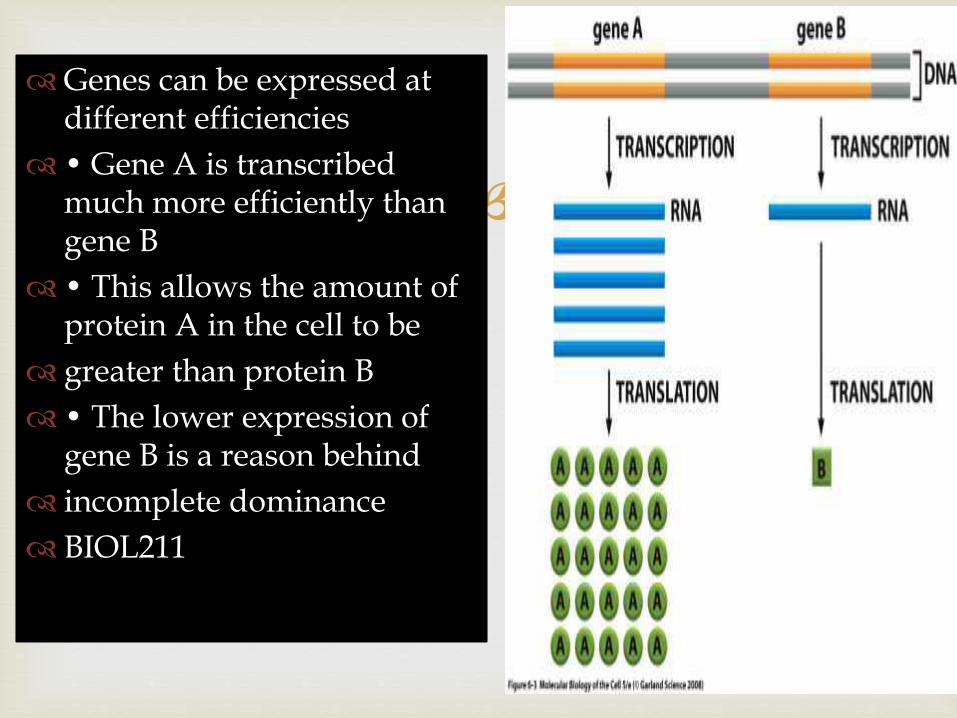
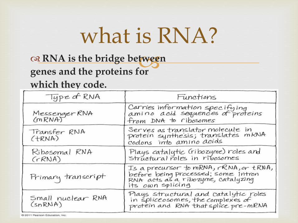
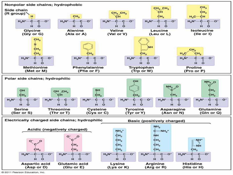
History linking genes and proteins
1900rsquos Archibald Garrod Inborn errors of metabolism inherited human
metabolic diseases (more information) Genes are the inherited factors
Enzymes are the biological molecules that drive metabolic reactions
Enzymes are proteins
Question
How do the inherited factors the genes control the structure and activity of enzymes (proteins)
History linking genes and proteins
Beadle and Tatum (1941) PNAS USA 27 499ndash506
Hypothesis If genes control structure and activity of metabolic enzymes
then mutations in genes should disrupt production of required nutrients and that disruption should be heritable
Method Isolated ~2000 strains from single irradiate spores
(Neurospora) that grew on rich but not minimal medium Examples defects in B1 B6 synthesis
Conclusion Genes govern the ability to synthesize amino acids purines
and vitamins
History linking genes and proteins
1950s sickle-cell anemia Glu to Val change in hemoglobin
Sequence of nucleotides in gene determines sequence of amino acids in protein
Single amino acid change can alter the function of the protein
Tryptophan synthase gene in E coli Mutations resulted in single amino acid change
Order of mutations in gene same as order of affected amino acids
Ribosomal structure
EP A
Large subunit
Peptidyl-tRNA binding site
Aminoacyl-tRNA binding site
mRNA
5rsquo
Exit
site
Small subunit
3rsquo
From gene to protein transcription
Gene sequence (DNA) recopied or transcribed to RNA sequence
Product of transcription is a messenger molecule that delivers the genetic instructions to the protein synthesis machinery messenger RNA (mRNA)
Transcription evidence for mRNA
Brenner S Jacob F and Meselson M (1961) Nature190 576ndash81
Question How do genes work
Does each one encode a different type of ribosome which in turn synthesizes a different protein OR
Are all ribosomes alike receiving the genetic information to create each different protein via some kind of messenger molecule
Transcription evidence for mRNA
E coli cells switch from making bacterial proteins to phage proteins when infected with bacteriophage T4
Grow bacteria on medium containing ldquoheavyrdquo nitrogen (15N) and carbon (13C)
Infect with phage T4
Immediately transfer to ldquolightrdquo medium containing radioactive uracil
Transcription evidence for mRNA
If genes encode different ribosomes the newly synthesized phage ribosomes will be ldquolightrdquo
If genes direct new RNA synthesis the RNA will contain radiolabeled uracil
Results Ribosomes from phage-infected cells were ldquoheavyrdquo
banding at the same density on a CsCl gradient as the original ribosomes
Newly synthesized RNA was associated with the heavy ribosomes
New RNA hybridized with viral ssDNA not bacterial ssDNA
Transcription evidence for mRNA
Conclusion
Expression of phage DNA results in new phage-specific RNA molecules (mRNA)
These mRNA molecules are temporarily associated with ribosomes
Ribosomes do not themselves contain the genetic directions for assembling individual proteins
Transcription overview
Transcription requires
ribonucleoside 5acute triphosphates
ATP GTP CTP and UTP
bases are adenine guanine cytosine and uracil
sugar is ribose (not deoxyribose)
DNA-dependent RNA polymerase
Template (sense) DNA strand
Animation of transcription
Transcription overview
Features of transcription
RNA polymerase catalyzes sugar-phosphate bond between 3acute-OH of ribose and the 5acute-PO4
Order of bases in DNA template strand determines order of bases in transcript
Nucleotides are added to the 3acute-OH of the growing chain
RNA synthesis does not require a primer
Transcription overview
In prokaryotes transcription and translation are coupled Proteins are synthesized directly from the primary transcript as it is made
In eukaryotes transcription and translation are separated Transcription occurs in the nucleus and translation occurs in the cytoplasm on ribosomes
Figure comparing eukaryotic and prokaryotic transcription and translation
Transcription RNA Polymerase
DNA-dependent
DNA template ribonucleoside 5acute triphosphates and Mg2+
Synthesizes RNA in 5acute to 3acute direction
E coli RNA polymerase consists of 5 subunits
Eukaryotes have three RNA polymerases
RNA polymerase II is responsible for transcription of protein-coding genes and some snRNA molecules
RNA polymerase II has 12 subunits
Requires accessory proteins (transcription factors)
Does not require a primer
Stages of Transcription
Transcription promoter recognition
Transcription factors bind to promoter sequences and recruit RNA polymerase
DNA is bound first in a closed complex Then RNA polymerase denatures a 12ndash15 bp segment of the DNA (open complex)
The site where the first base is incorporated into the transcription is numbered ldquo+1rdquo and is called the transcription start site
Transcription factors that are required at every promoter site for RNA polymerase interaction are called basal transcription factors
Promoter recognition promoter sequences
Promoter sequences vary considerably
RNA polymerase binds to different promoters with different strengths binding strength relates to the level of gene expression
There are some common consensus sequences for promoters Example E coli ndash35 sequence (found 35 bases 5acute to the
start of transcription)
Example E coli TATA box (found 10 bases 5acute to the start of transcription)
Promoter recognition enhancers
Eukaryotic genes may also have enhancers
Enhancers can be located at great distances from the gene they regulate either 5acute or 3acute of the transcription start in introns or even on the noncoding strand
One of the most common ways to identify promoters and enhancers is to use a reporter gene
Promoter recognition other players
Many proteins can regulate gene expression by modulating the strength of interaction between the promoter and RNA polymerase
Some proteins can activate transcription (upregulate gene expression)
Some proteins can inhibit transcription by blocking polymerase activity
Some proteins can act both as repressors and activators of transcription
Transcription chain initiation
Chain initiation
RNA polymerase locally denatures the DNA
The first base of the new RNA strand is placed complementary to the +1 site
RNA polymerase does not require a primer
The first 8 or 9 bases of the transcript are linked Transcription factors are released and the polymerase leaves the promoter region
Figure of bacterial transcription initiation
Transcription chain elongation
Chain elongation
RNA polymerase moves along the transcribed or template DNA strand
The new RNA molecule (primary transcript) forms a short RNA-DNA hybrid molecule with the DNA template
Transcription chain termination
Most known about bacterial chain termination
Termination is signaled by a sequence that can form a hairpin loop
The polymerase and the new RNA molecule are released upon formation of the loop
Review the transcription animation
Transcription mRNA synthesisprocessing
Prokaryotes mRNA transcribed directly from DNA template and used immediately in protein synthesis
Eukaryotes primary transcript must be processed to produce the mRNA
Noncoding sequences (introns) are removed
Coding sequences (exons) spliced together
5acute-methylguanosine cap added
3acute-polyadenosine tail added
Transcription mRNA synthesisprocessing
Removal of introns and splicing of exons can occur several ways For introns within a nuclear transcript a spliceosome is
required
Splicesomes protein and small nuclear RNA (snRNA)
Specificity of splicing comes from the snRNA some of which contain sequences complementary to the splice junctions between introns and exons
Alternative splicing can produce different forms of a protein from the same gene
Mutations at the splice sites can cause disease
Thalassemia bull Breast cancer (BRCA 1)
Transcription mRNA synthesisprocessing
RNA splicing inside the nucleus on particles called spliceosomes
Splicesomes are composed of proteins and small RNA molecules (100ndash200 bp snRNA)
Both proteins and RNA are required but some suggesting that RNA can catalyze the splicing reaction
Self-splicing in Tetrahymena the RNA catalyzes its own splicing
Catalytic RNA ribozymes
From gene to protein genetic code
Central Dogma
Information travels from DNA to RNA to Protein
Is there a one-to-one correspondence between DNA RNA and Protein
DNA and RNA each have four nucleotides that can form them so yes there is a one-to-one correspondence between DNA and RNA
Proteins can be composed of a potential 20 amino acids only four RNA nucleotides no one-to-one correspondence
How then does RNA direct the order and number of amino acids in a protein
From gene to protein genetic code
How many bases are required for each amino acid (4 bases)2basesaa = 16 amino acidsmdashnot enough
(4 bases)3basesaa = 64 amino acid possibilities
Minimum of 3 basesaa required
What is the nature of the code Does it have punctuation Is it overlapping
Crick FH et al (1961) Nature 192 1227ndash32 (httpprofilesnlmnihgovSCBCBJ )
3-base nonoverlapping code that is read from a fixed point
From gene to protein genetic code
Nirenberg and Matthaei in vitro protein translation
Found that adding rRNA prolonged cell-free protein synthesis
Adding artificial RNA synthesized by polynucleotide phosphorylase (no template UUUUUUUUU) stimulated protein synthesis more
The protein that came out of this reaction was polyphenylalanine (UUU = Phe)
Other artificial RNAs AAA = Lys CCC =Pro
From gene to protein genetic code
Nirenberg
Triplet binding assay add triplet RNA ribosomes binding factors GTP and radiolabeled charged tRNA (figure)
UUU trinucleotide binds to Phe-tRNA
UGU trinucleotide binds to CYS-tRNA
By fits and starts the triplet genetic code was worked out
Each three-letter ldquowordrdquo (codon) specifies an amino acid or directions to stop translation
The code is redundant or degenerate more than one way to encode an amino acid
From gene to protein Translation
Components required for translation
mRNA
Ribosomes
tRNA
Aminoacyl tRNA synthetases
Initiation elongation and termination factors
Animation of translation
Translation initiation
Ribosome small subunit binds to mRNA
Charged tRNA anticodon forms base pairs with the mRNA codon
Small subunit interacts with initiation factors and special initiator tRNA that is charged with methionine
mRNA-small subunit-tRNA complex recruits the large subunit
Eukaryotic and prokaryotic initiation differ slightly
Translation initiation
The large subunit of the ribosome contains three binding sites
Amino acyl (A site)
Peptidyl (P site)
Exit (E site)
At initiation
The tRNAfMet occupies the P site
A second charged tRNA complementary to the next codon binds the A site
Translation elongation
Elongation
Ribosome translocates by three bases after peptide bond formed
New charged tRNA aligns in the A site
Peptide bond between amino acids in A and P sites is formed
Ribosome translocates by three more bases
The uncharged tRNA in the A site is moved to the E site
Translation elongation
EF-Tu recruits charged tRNA to A site Requires hydrolysis of GTP
Peptidyl transferase catalyzes peptide bond formation (bond between aa and tRNA in the P site converted to peptide bond between the two amino acids)
Peptide bond formation requires RNA and may be a ribozyme-catalyzed reaction
Translation termination
Termination
Elongation proceeds until STOP codon reached UAA UAG UGA
No tRNA normally exists that can form base pairing with a STOP codon recognized by a release factor
tRNA charged with last amino acid will remain at P site
Release factors cleave the amino acid from the tRNA
Ribosome subunits dissociate from each other
Review the animation of translation
48
Genetic code
Def Genetic code is the nucleotide base sequence on DNA ( and
subsequently on mRNA by transcription) which will be translated into a
sequence of amino acids of the protein to be synthesized
The code is composed of codons
Codon is composed of 3 bases ( eg ACG or UAG) Each codon is
translated into one amino acid
The 4 nucleotide bases (AGC and U) in mRNA are used to produce the
three base codons There are therefore 64 codons code for the 20 amino
acids and since each codon code for only one amino acids this means
that there are more than one cone for the same amino acid
How to translate a codon (see table)
This table or dictionary can be used to translate any codon sequence
Each triplet is read from 5prime rarr 3prime direction so the first base is 5prime base
followed by the middle base then the last base which is 3prime base
49
Examples 5prime- A UG- 3prime codes for methionine
5prime- UCU- 3prime codes for serine
5prime - CCA- 3prime codes for proline
Termination (stop or nonsense) codons
Three of the 64 codons UAA UAG UGA do not code for any amino
acid They are termination codes which when one of them appear in
mRNA sequence it indicates finishing of protein synthesis
Characters of the genetic code
1- Specificity the genetic code is specific that is a specific codon
always code for the same amino acid
2- Universality the genetic code is universal that is the same codon is
used in all living organisms procaryotics and eucaryotics
3- Degeneracy the genetic code is degenerate ie although each codon
corresponds to a single amino acidone amino acid may have more than
one codons eg arginine has 6 different codons (give more examples
from the table)
50
51
Gene mutation (altering the nucleotide sequence)
1- Point mutation changing in a single nucleotide base on the mRNA
can lead to any of the following 3 results
i- Silent mutation ie the codon containg the changed base may code
for the same amino acid For example in serine codon UCA if A is
changed to U giving the codon UCU it still code for serine See table
ii- Missense mutation the codon containing the changed base may code
for a different amino acid For example if the serine codon UCA is
changed to be CCA ( U is replaced by C) it will code for proline not
serine leading to insertion of incorrect amino acid into polypeptide chain
iii- Non sense mutation the codon containing the changed base may
become a termination codon For example serine codon UCA becomes
UAA if C is changed to A UAA is a stop codon leading to termination
of translation at that point
52
53
Types of point mutation
U A A (termination codon) Nonsense mutation
uarr
U C A rarr U C U Silent mutation
(codon for serine) (codon for serine)
darr
C C A ( codon for proline) Missense mutation
Give other examples on missense mutation which leads to some Hb
disease
54
2- Frame- shift mutation
deletion or addition of one or two base to
message sequence leading to change in
reading frame (reading sequence) and the
resulting amino acid seuence may become
completely different from this point
55
TranslationComponents required for protein synthesis1- Amino acids all amino acids involved in thefinished protein must be present at the time ofprotein synthesis2- Ribosomes the site of protein synthesis They arelarge complexes of protein and rRNA In humanthey consist of two subunits one large (60S) and onesmall (40S)
3- tRNA at least one specific type of tRNA is required to transfer
one amino acid There about 50 tRNA in human for the 20 amino
acids this means some amino acids have more than one specific
tRNA The role of tRNA in protein synthesis is discussed before
(amino acid attachment and anticodon loop)
4- aminoacyl-tRNA synthetase This is the enzyme that catalyzes the
attachment of amino acid with its corresponding tRNA forming
aminoacyl tRNA
56
5- mRNA that carry code for the protein to be synthesized
6- protein factors Initiation elongation and termination (or release)
factors are required for peptide synthesis
7- ATP and GTP are required as source of energy
Steps (movie)
1- Initiation
Initiation (start) codon is usually AUG which is the codon of
methionine so the initiator tRNA is methionnyl tRNA (Met tRNA)
a- The initiation factors (IF-1 IF-2 and IF-3) binds the Met tRNA with
small ribosomal subunit then to mRNA containing the code of the
protein to be synthesized IFs recognizes mRNA from its 5 cap
57
b-This complex binds to large ribosomal subunit forming initiation complex in which Met tRNA is present in P- site of 60 ribosomal subunitNB- tRNA bind with mRNA by base pairing between codon on mRNA and anticodon
on tRNA - mRNA is read from 5prime rarr 3prime direction
P-site is the peptidyl site of the ribosome to which methionyl tRNA is placed (enter)
2- Elongation elongation factors (EFs) stimulate the stepwise elongation of polypeptide chain as follow
a- The next aminoacyl tRNA (tRNA which carry the next amino acid specified by recognition of the next codon on mRNA) will enter A site of ribosome
A site or acceptor site or aminoacyl tRNA siteIs the site of ribosome to which each new incoming aminoacyl tRNA will enter
b) ribosomal peptidyl transferase enzyme will transfer methionine
from methionyl tRNA into A site to form a peptide bond between
methionine and the new incoming amino acid to form dipeptidyl
tRNA
c) Elongation factor-2 (EF-2) (called also translocase) moves
mRNA and dipeptidyl tRNA from A site to P site leaving A site free to
allow entrance of another new aminoacyl tRNA
The figure shows the repetitive cycle of elongation of chain Each
cycle is consisting of
1) codon recognition and the entrance of the new aminoacyl tRNA
acid ( amino acid carried on tRNA) into A site
2) The growing chain in P site will moved to A site with peptide
bond formation with the new amino acid
3) Translocation of growing chain to P site allowing A site free for
enterance of new amino acid an so onhelliphelliphelliphelliphelliphelliphellip Resulting in
elongation of poly peptide chain
59repetitive cycle of elongation
60
3- Termination occurs when one of the three stop codons (UAA UAG orUGA) enters A site of the ribosome These codons are recognized by releasefactors (RFs) which are RF-1 RF-2 RF-3 RFs cause the newly synthesizedprotein to be released from the ribosomal complex and dissociation ofribosomes from mRNA (ie cause dissolution of the complex)
History linking genes and proteins
Beadle and Tatum (1941) PNAS USA 27 499ndash506
Hypothesis If genes control structure and activity of metabolic enzymes
then mutations in genes should disrupt production of required nutrients and that disruption should be heritable
Method Isolated ~2000 strains from single irradiate spores
(Neurospora) that grew on rich but not minimal medium Examples defects in B1 B6 synthesis
Conclusion Genes govern the ability to synthesize amino acids purines
and vitamins
History linking genes and proteins
1950s sickle-cell anemia Glu to Val change in hemoglobin
Sequence of nucleotides in gene determines sequence of amino acids in protein
Single amino acid change can alter the function of the protein
Tryptophan synthase gene in E coli Mutations resulted in single amino acid change
Order of mutations in gene same as order of affected amino acids
Ribosomal structure
EP A
Large subunit
Peptidyl-tRNA binding site
Aminoacyl-tRNA binding site
mRNA
5rsquo
Exit
site
Small subunit
3rsquo
From gene to protein transcription
Gene sequence (DNA) recopied or transcribed to RNA sequence
Product of transcription is a messenger molecule that delivers the genetic instructions to the protein synthesis machinery messenger RNA (mRNA)
Transcription evidence for mRNA
Brenner S Jacob F and Meselson M (1961) Nature190 576ndash81
Question How do genes work
Does each one encode a different type of ribosome which in turn synthesizes a different protein OR
Are all ribosomes alike receiving the genetic information to create each different protein via some kind of messenger molecule
Transcription evidence for mRNA
E coli cells switch from making bacterial proteins to phage proteins when infected with bacteriophage T4
Grow bacteria on medium containing ldquoheavyrdquo nitrogen (15N) and carbon (13C)
Infect with phage T4
Immediately transfer to ldquolightrdquo medium containing radioactive uracil
Transcription evidence for mRNA
If genes encode different ribosomes the newly synthesized phage ribosomes will be ldquolightrdquo
If genes direct new RNA synthesis the RNA will contain radiolabeled uracil
Results Ribosomes from phage-infected cells were ldquoheavyrdquo
banding at the same density on a CsCl gradient as the original ribosomes
Newly synthesized RNA was associated with the heavy ribosomes
New RNA hybridized with viral ssDNA not bacterial ssDNA
Transcription evidence for mRNA
Conclusion
Expression of phage DNA results in new phage-specific RNA molecules (mRNA)
These mRNA molecules are temporarily associated with ribosomes
Ribosomes do not themselves contain the genetic directions for assembling individual proteins
Transcription overview
Transcription requires
ribonucleoside 5acute triphosphates
ATP GTP CTP and UTP
bases are adenine guanine cytosine and uracil
sugar is ribose (not deoxyribose)
DNA-dependent RNA polymerase
Template (sense) DNA strand
Animation of transcription
Transcription overview
Features of transcription
RNA polymerase catalyzes sugar-phosphate bond between 3acute-OH of ribose and the 5acute-PO4
Order of bases in DNA template strand determines order of bases in transcript
Nucleotides are added to the 3acute-OH of the growing chain
RNA synthesis does not require a primer
Transcription overview
In prokaryotes transcription and translation are coupled Proteins are synthesized directly from the primary transcript as it is made
In eukaryotes transcription and translation are separated Transcription occurs in the nucleus and translation occurs in the cytoplasm on ribosomes
Figure comparing eukaryotic and prokaryotic transcription and translation
Transcription RNA Polymerase
DNA-dependent
DNA template ribonucleoside 5acute triphosphates and Mg2+
Synthesizes RNA in 5acute to 3acute direction
E coli RNA polymerase consists of 5 subunits
Eukaryotes have three RNA polymerases
RNA polymerase II is responsible for transcription of protein-coding genes and some snRNA molecules
RNA polymerase II has 12 subunits
Requires accessory proteins (transcription factors)
Does not require a primer
Stages of Transcription
Transcription promoter recognition
Transcription factors bind to promoter sequences and recruit RNA polymerase
DNA is bound first in a closed complex Then RNA polymerase denatures a 12ndash15 bp segment of the DNA (open complex)
The site where the first base is incorporated into the transcription is numbered ldquo+1rdquo and is called the transcription start site
Transcription factors that are required at every promoter site for RNA polymerase interaction are called basal transcription factors
Promoter recognition promoter sequences
Promoter sequences vary considerably
RNA polymerase binds to different promoters with different strengths binding strength relates to the level of gene expression
There are some common consensus sequences for promoters Example E coli ndash35 sequence (found 35 bases 5acute to the
start of transcription)
Example E coli TATA box (found 10 bases 5acute to the start of transcription)
Promoter recognition enhancers
Eukaryotic genes may also have enhancers
Enhancers can be located at great distances from the gene they regulate either 5acute or 3acute of the transcription start in introns or even on the noncoding strand
One of the most common ways to identify promoters and enhancers is to use a reporter gene
Promoter recognition other players
Many proteins can regulate gene expression by modulating the strength of interaction between the promoter and RNA polymerase
Some proteins can activate transcription (upregulate gene expression)
Some proteins can inhibit transcription by blocking polymerase activity
Some proteins can act both as repressors and activators of transcription
Transcription chain initiation
Chain initiation
RNA polymerase locally denatures the DNA
The first base of the new RNA strand is placed complementary to the +1 site
RNA polymerase does not require a primer
The first 8 or 9 bases of the transcript are linked Transcription factors are released and the polymerase leaves the promoter region
Figure of bacterial transcription initiation
Transcription chain elongation
Chain elongation
RNA polymerase moves along the transcribed or template DNA strand
The new RNA molecule (primary transcript) forms a short RNA-DNA hybrid molecule with the DNA template
Transcription chain termination
Most known about bacterial chain termination
Termination is signaled by a sequence that can form a hairpin loop
The polymerase and the new RNA molecule are released upon formation of the loop
Review the transcription animation
Transcription mRNA synthesisprocessing
Prokaryotes mRNA transcribed directly from DNA template and used immediately in protein synthesis
Eukaryotes primary transcript must be processed to produce the mRNA
Noncoding sequences (introns) are removed
Coding sequences (exons) spliced together
5acute-methylguanosine cap added
3acute-polyadenosine tail added
Transcription mRNA synthesisprocessing
Removal of introns and splicing of exons can occur several ways For introns within a nuclear transcript a spliceosome is
required
Splicesomes protein and small nuclear RNA (snRNA)
Specificity of splicing comes from the snRNA some of which contain sequences complementary to the splice junctions between introns and exons
Alternative splicing can produce different forms of a protein from the same gene
Mutations at the splice sites can cause disease
Thalassemia bull Breast cancer (BRCA 1)
Transcription mRNA synthesisprocessing
RNA splicing inside the nucleus on particles called spliceosomes
Splicesomes are composed of proteins and small RNA molecules (100ndash200 bp snRNA)
Both proteins and RNA are required but some suggesting that RNA can catalyze the splicing reaction
Self-splicing in Tetrahymena the RNA catalyzes its own splicing
Catalytic RNA ribozymes
From gene to protein genetic code
Central Dogma
Information travels from DNA to RNA to Protein
Is there a one-to-one correspondence between DNA RNA and Protein
DNA and RNA each have four nucleotides that can form them so yes there is a one-to-one correspondence between DNA and RNA
Proteins can be composed of a potential 20 amino acids only four RNA nucleotides no one-to-one correspondence
How then does RNA direct the order and number of amino acids in a protein
From gene to protein genetic code
How many bases are required for each amino acid (4 bases)2basesaa = 16 amino acidsmdashnot enough
(4 bases)3basesaa = 64 amino acid possibilities
Minimum of 3 basesaa required
What is the nature of the code Does it have punctuation Is it overlapping
Crick FH et al (1961) Nature 192 1227ndash32 (httpprofilesnlmnihgovSCBCBJ )
3-base nonoverlapping code that is read from a fixed point
From gene to protein genetic code
Nirenberg and Matthaei in vitro protein translation
Found that adding rRNA prolonged cell-free protein synthesis
Adding artificial RNA synthesized by polynucleotide phosphorylase (no template UUUUUUUUU) stimulated protein synthesis more
The protein that came out of this reaction was polyphenylalanine (UUU = Phe)
Other artificial RNAs AAA = Lys CCC =Pro
From gene to protein genetic code
Nirenberg
Triplet binding assay add triplet RNA ribosomes binding factors GTP and radiolabeled charged tRNA (figure)
UUU trinucleotide binds to Phe-tRNA
UGU trinucleotide binds to CYS-tRNA
By fits and starts the triplet genetic code was worked out
Each three-letter ldquowordrdquo (codon) specifies an amino acid or directions to stop translation
The code is redundant or degenerate more than one way to encode an amino acid
From gene to protein Translation
Components required for translation
mRNA
Ribosomes
tRNA
Aminoacyl tRNA synthetases
Initiation elongation and termination factors
Animation of translation
Translation initiation
Ribosome small subunit binds to mRNA
Charged tRNA anticodon forms base pairs with the mRNA codon
Small subunit interacts with initiation factors and special initiator tRNA that is charged with methionine
mRNA-small subunit-tRNA complex recruits the large subunit
Eukaryotic and prokaryotic initiation differ slightly
Translation initiation
The large subunit of the ribosome contains three binding sites
Amino acyl (A site)
Peptidyl (P site)
Exit (E site)
At initiation
The tRNAfMet occupies the P site
A second charged tRNA complementary to the next codon binds the A site
Translation elongation
Elongation
Ribosome translocates by three bases after peptide bond formed
New charged tRNA aligns in the A site
Peptide bond between amino acids in A and P sites is formed
Ribosome translocates by three more bases
The uncharged tRNA in the A site is moved to the E site
Translation elongation
EF-Tu recruits charged tRNA to A site Requires hydrolysis of GTP
Peptidyl transferase catalyzes peptide bond formation (bond between aa and tRNA in the P site converted to peptide bond between the two amino acids)
Peptide bond formation requires RNA and may be a ribozyme-catalyzed reaction
Translation termination
Termination
Elongation proceeds until STOP codon reached UAA UAG UGA
No tRNA normally exists that can form base pairing with a STOP codon recognized by a release factor
tRNA charged with last amino acid will remain at P site
Release factors cleave the amino acid from the tRNA
Ribosome subunits dissociate from each other
Review the animation of translation
48
Genetic code
Def Genetic code is the nucleotide base sequence on DNA ( and
subsequently on mRNA by transcription) which will be translated into a
sequence of amino acids of the protein to be synthesized
The code is composed of codons
Codon is composed of 3 bases ( eg ACG or UAG) Each codon is
translated into one amino acid
The 4 nucleotide bases (AGC and U) in mRNA are used to produce the
three base codons There are therefore 64 codons code for the 20 amino
acids and since each codon code for only one amino acids this means
that there are more than one cone for the same amino acid
How to translate a codon (see table)
This table or dictionary can be used to translate any codon sequence
Each triplet is read from 5prime rarr 3prime direction so the first base is 5prime base
followed by the middle base then the last base which is 3prime base
49
Examples 5prime- A UG- 3prime codes for methionine
5prime- UCU- 3prime codes for serine
5prime - CCA- 3prime codes for proline
Termination (stop or nonsense) codons
Three of the 64 codons UAA UAG UGA do not code for any amino
acid They are termination codes which when one of them appear in
mRNA sequence it indicates finishing of protein synthesis
Characters of the genetic code
1- Specificity the genetic code is specific that is a specific codon
always code for the same amino acid
2- Universality the genetic code is universal that is the same codon is
used in all living organisms procaryotics and eucaryotics
3- Degeneracy the genetic code is degenerate ie although each codon
corresponds to a single amino acidone amino acid may have more than
one codons eg arginine has 6 different codons (give more examples
from the table)
50
51
Gene mutation (altering the nucleotide sequence)
1- Point mutation changing in a single nucleotide base on the mRNA
can lead to any of the following 3 results
i- Silent mutation ie the codon containg the changed base may code
for the same amino acid For example in serine codon UCA if A is
changed to U giving the codon UCU it still code for serine See table
ii- Missense mutation the codon containing the changed base may code
for a different amino acid For example if the serine codon UCA is
changed to be CCA ( U is replaced by C) it will code for proline not
serine leading to insertion of incorrect amino acid into polypeptide chain
iii- Non sense mutation the codon containing the changed base may
become a termination codon For example serine codon UCA becomes
UAA if C is changed to A UAA is a stop codon leading to termination
of translation at that point
52
53
Types of point mutation
U A A (termination codon) Nonsense mutation
uarr
U C A rarr U C U Silent mutation
(codon for serine) (codon for serine)
darr
C C A ( codon for proline) Missense mutation
Give other examples on missense mutation which leads to some Hb
disease
54
2- Frame- shift mutation
deletion or addition of one or two base to
message sequence leading to change in
reading frame (reading sequence) and the
resulting amino acid seuence may become
completely different from this point
55
TranslationComponents required for protein synthesis1- Amino acids all amino acids involved in thefinished protein must be present at the time ofprotein synthesis2- Ribosomes the site of protein synthesis They arelarge complexes of protein and rRNA In humanthey consist of two subunits one large (60S) and onesmall (40S)
3- tRNA at least one specific type of tRNA is required to transfer
one amino acid There about 50 tRNA in human for the 20 amino
acids this means some amino acids have more than one specific
tRNA The role of tRNA in protein synthesis is discussed before
(amino acid attachment and anticodon loop)
4- aminoacyl-tRNA synthetase This is the enzyme that catalyzes the
attachment of amino acid with its corresponding tRNA forming
aminoacyl tRNA
56
5- mRNA that carry code for the protein to be synthesized
6- protein factors Initiation elongation and termination (or release)
factors are required for peptide synthesis
7- ATP and GTP are required as source of energy
Steps (movie)
1- Initiation
Initiation (start) codon is usually AUG which is the codon of
methionine so the initiator tRNA is methionnyl tRNA (Met tRNA)
a- The initiation factors (IF-1 IF-2 and IF-3) binds the Met tRNA with
small ribosomal subunit then to mRNA containing the code of the
protein to be synthesized IFs recognizes mRNA from its 5 cap
57
b-This complex binds to large ribosomal subunit forming initiation complex in which Met tRNA is present in P- site of 60 ribosomal subunitNB- tRNA bind with mRNA by base pairing between codon on mRNA and anticodon
on tRNA - mRNA is read from 5prime rarr 3prime direction
P-site is the peptidyl site of the ribosome to which methionyl tRNA is placed (enter)
2- Elongation elongation factors (EFs) stimulate the stepwise elongation of polypeptide chain as follow
a- The next aminoacyl tRNA (tRNA which carry the next amino acid specified by recognition of the next codon on mRNA) will enter A site of ribosome
A site or acceptor site or aminoacyl tRNA siteIs the site of ribosome to which each new incoming aminoacyl tRNA will enter
b) ribosomal peptidyl transferase enzyme will transfer methionine
from methionyl tRNA into A site to form a peptide bond between
methionine and the new incoming amino acid to form dipeptidyl
tRNA
c) Elongation factor-2 (EF-2) (called also translocase) moves
mRNA and dipeptidyl tRNA from A site to P site leaving A site free to
allow entrance of another new aminoacyl tRNA
The figure shows the repetitive cycle of elongation of chain Each
cycle is consisting of
1) codon recognition and the entrance of the new aminoacyl tRNA
acid ( amino acid carried on tRNA) into A site
2) The growing chain in P site will moved to A site with peptide
bond formation with the new amino acid
3) Translocation of growing chain to P site allowing A site free for
enterance of new amino acid an so onhelliphelliphelliphelliphelliphelliphellip Resulting in
elongation of poly peptide chain
59repetitive cycle of elongation
60
3- Termination occurs when one of the three stop codons (UAA UAG orUGA) enters A site of the ribosome These codons are recognized by releasefactors (RFs) which are RF-1 RF-2 RF-3 RFs cause the newly synthesizedprotein to be released from the ribosomal complex and dissociation ofribosomes from mRNA (ie cause dissolution of the complex)
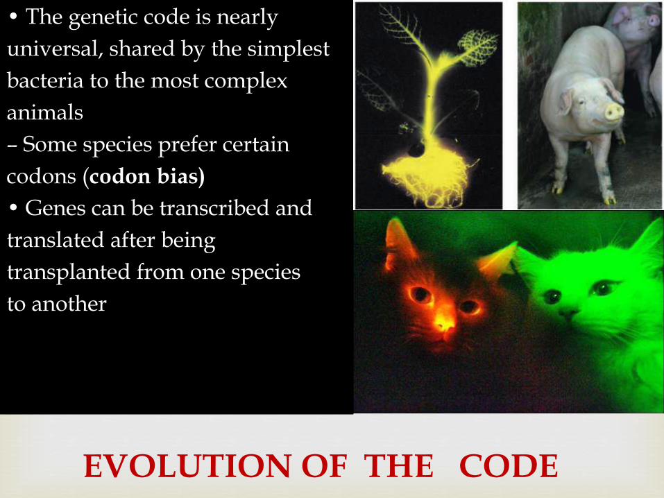
Transcription evidence for mRNA
Brenner S Jacob F and Meselson M (1961) Nature190 576ndash81
Question How do genes work
Does each one encode a different type of ribosome which in turn synthesizes a different protein OR
Are all ribosomes alike receiving the genetic information to create each different protein via some kind of messenger molecule
Transcription evidence for mRNA
E coli cells switch from making bacterial proteins to phage proteins when infected with bacteriophage T4
Grow bacteria on medium containing ldquoheavyrdquo nitrogen (15N) and carbon (13C)
Infect with phage T4
Immediately transfer to ldquolightrdquo medium containing radioactive uracil
Transcription evidence for mRNA
If genes encode different ribosomes the newly synthesized phage ribosomes will be ldquolightrdquo
If genes direct new RNA synthesis the RNA will contain radiolabeled uracil
Results Ribosomes from phage-infected cells were ldquoheavyrdquo
banding at the same density on a CsCl gradient as the original ribosomes
Newly synthesized RNA was associated with the heavy ribosomes
New RNA hybridized with viral ssDNA not bacterial ssDNA
Transcription evidence for mRNA
Conclusion
Expression of phage DNA results in new phage-specific RNA molecules (mRNA)
These mRNA molecules are temporarily associated with ribosomes
Ribosomes do not themselves contain the genetic directions for assembling individual proteins
Transcription overview
Transcription requires
ribonucleoside 5acute triphosphates
ATP GTP CTP and UTP
bases are adenine guanine cytosine and uracil
sugar is ribose (not deoxyribose)
DNA-dependent RNA polymerase
Template (sense) DNA strand
Animation of transcription
Transcription overview
Features of transcription
RNA polymerase catalyzes sugar-phosphate bond between 3acute-OH of ribose and the 5acute-PO4
Order of bases in DNA template strand determines order of bases in transcript
Nucleotides are added to the 3acute-OH of the growing chain
RNA synthesis does not require a primer
Transcription overview
In prokaryotes transcription and translation are coupled Proteins are synthesized directly from the primary transcript as it is made
In eukaryotes transcription and translation are separated Transcription occurs in the nucleus and translation occurs in the cytoplasm on ribosomes
Figure comparing eukaryotic and prokaryotic transcription and translation
Transcription RNA Polymerase
DNA-dependent
DNA template ribonucleoside 5acute triphosphates and Mg2+
Synthesizes RNA in 5acute to 3acute direction
E coli RNA polymerase consists of 5 subunits
Eukaryotes have three RNA polymerases
RNA polymerase II is responsible for transcription of protein-coding genes and some snRNA molecules
RNA polymerase II has 12 subunits
Requires accessory proteins (transcription factors)
Does not require a primer
Stages of Transcription
Transcription promoter recognition
Transcription factors bind to promoter sequences and recruit RNA polymerase
DNA is bound first in a closed complex Then RNA polymerase denatures a 12ndash15 bp segment of the DNA (open complex)
The site where the first base is incorporated into the transcription is numbered ldquo+1rdquo and is called the transcription start site
Transcription factors that are required at every promoter site for RNA polymerase interaction are called basal transcription factors
Promoter recognition promoter sequences
Promoter sequences vary considerably
RNA polymerase binds to different promoters with different strengths binding strength relates to the level of gene expression
There are some common consensus sequences for promoters Example E coli ndash35 sequence (found 35 bases 5acute to the
start of transcription)
Example E coli TATA box (found 10 bases 5acute to the start of transcription)
Promoter recognition enhancers
Eukaryotic genes may also have enhancers
Enhancers can be located at great distances from the gene they regulate either 5acute or 3acute of the transcription start in introns or even on the noncoding strand
One of the most common ways to identify promoters and enhancers is to use a reporter gene
Promoter recognition other players
Many proteins can regulate gene expression by modulating the strength of interaction between the promoter and RNA polymerase
Some proteins can activate transcription (upregulate gene expression)
Some proteins can inhibit transcription by blocking polymerase activity
Some proteins can act both as repressors and activators of transcription
Transcription chain initiation
Chain initiation
RNA polymerase locally denatures the DNA
The first base of the new RNA strand is placed complementary to the +1 site
RNA polymerase does not require a primer
The first 8 or 9 bases of the transcript are linked Transcription factors are released and the polymerase leaves the promoter region
Figure of bacterial transcription initiation
Transcription chain elongation
Chain elongation
RNA polymerase moves along the transcribed or template DNA strand
The new RNA molecule (primary transcript) forms a short RNA-DNA hybrid molecule with the DNA template
Transcription chain termination
Most known about bacterial chain termination
Termination is signaled by a sequence that can form a hairpin loop
The polymerase and the new RNA molecule are released upon formation of the loop
Review the transcription animation
Transcription mRNA synthesisprocessing
Prokaryotes mRNA transcribed directly from DNA template and used immediately in protein synthesis
Eukaryotes primary transcript must be processed to produce the mRNA
Noncoding sequences (introns) are removed
Coding sequences (exons) spliced together
5acute-methylguanosine cap added
3acute-polyadenosine tail added
Transcription mRNA synthesisprocessing
Removal of introns and splicing of exons can occur several ways For introns within a nuclear transcript a spliceosome is
required
Splicesomes protein and small nuclear RNA (snRNA)
Specificity of splicing comes from the snRNA some of which contain sequences complementary to the splice junctions between introns and exons
Alternative splicing can produce different forms of a protein from the same gene
Mutations at the splice sites can cause disease
Thalassemia bull Breast cancer (BRCA 1)
Transcription mRNA synthesisprocessing
RNA splicing inside the nucleus on particles called spliceosomes
Splicesomes are composed of proteins and small RNA molecules (100ndash200 bp snRNA)
Both proteins and RNA are required but some suggesting that RNA can catalyze the splicing reaction
Self-splicing in Tetrahymena the RNA catalyzes its own splicing
Catalytic RNA ribozymes
From gene to protein genetic code
Central Dogma
Information travels from DNA to RNA to Protein
Is there a one-to-one correspondence between DNA RNA and Protein
DNA and RNA each have four nucleotides that can form them so yes there is a one-to-one correspondence between DNA and RNA
Proteins can be composed of a potential 20 amino acids only four RNA nucleotides no one-to-one correspondence
How then does RNA direct the order and number of amino acids in a protein
From gene to protein genetic code
How many bases are required for each amino acid (4 bases)2basesaa = 16 amino acidsmdashnot enough
(4 bases)3basesaa = 64 amino acid possibilities
Minimum of 3 basesaa required
What is the nature of the code Does it have punctuation Is it overlapping
Crick FH et al (1961) Nature 192 1227ndash32 (httpprofilesnlmnihgovSCBCBJ )
3-base nonoverlapping code that is read from a fixed point
From gene to protein genetic code
Nirenberg and Matthaei in vitro protein translation
Found that adding rRNA prolonged cell-free protein synthesis
Adding artificial RNA synthesized by polynucleotide phosphorylase (no template UUUUUUUUU) stimulated protein synthesis more
The protein that came out of this reaction was polyphenylalanine (UUU = Phe)
Other artificial RNAs AAA = Lys CCC =Pro
From gene to protein genetic code
Nirenberg
Triplet binding assay add triplet RNA ribosomes binding factors GTP and radiolabeled charged tRNA (figure)
UUU trinucleotide binds to Phe-tRNA
UGU trinucleotide binds to CYS-tRNA
By fits and starts the triplet genetic code was worked out
Each three-letter ldquowordrdquo (codon) specifies an amino acid or directions to stop translation
The code is redundant or degenerate more than one way to encode an amino acid
From gene to protein Translation
Components required for translation
mRNA
Ribosomes
tRNA
Aminoacyl tRNA synthetases
Initiation elongation and termination factors
Animation of translation
Translation initiation
Ribosome small subunit binds to mRNA
Charged tRNA anticodon forms base pairs with the mRNA codon
Small subunit interacts with initiation factors and special initiator tRNA that is charged with methionine
mRNA-small subunit-tRNA complex recruits the large subunit
Eukaryotic and prokaryotic initiation differ slightly
Translation initiation
The large subunit of the ribosome contains three binding sites
Amino acyl (A site)
Peptidyl (P site)
Exit (E site)
At initiation
The tRNAfMet occupies the P site
A second charged tRNA complementary to the next codon binds the A site
Translation elongation
Elongation
Ribosome translocates by three bases after peptide bond formed
New charged tRNA aligns in the A site
Peptide bond between amino acids in A and P sites is formed
Ribosome translocates by three more bases
The uncharged tRNA in the A site is moved to the E site
Translation elongation
EF-Tu recruits charged tRNA to A site Requires hydrolysis of GTP
Peptidyl transferase catalyzes peptide bond formation (bond between aa and tRNA in the P site converted to peptide bond between the two amino acids)
Peptide bond formation requires RNA and may be a ribozyme-catalyzed reaction
Translation termination
Termination
Elongation proceeds until STOP codon reached UAA UAG UGA
No tRNA normally exists that can form base pairing with a STOP codon recognized by a release factor
tRNA charged with last amino acid will remain at P site
Release factors cleave the amino acid from the tRNA
Ribosome subunits dissociate from each other
Review the animation of translation
48
Genetic code
Def Genetic code is the nucleotide base sequence on DNA ( and
subsequently on mRNA by transcription) which will be translated into a
sequence of amino acids of the protein to be synthesized
The code is composed of codons
Codon is composed of 3 bases ( eg ACG or UAG) Each codon is
translated into one amino acid
The 4 nucleotide bases (AGC and U) in mRNA are used to produce the
three base codons There are therefore 64 codons code for the 20 amino
acids and since each codon code for only one amino acids this means
that there are more than one cone for the same amino acid
How to translate a codon (see table)
This table or dictionary can be used to translate any codon sequence
Each triplet is read from 5prime rarr 3prime direction so the first base is 5prime base
followed by the middle base then the last base which is 3prime base
49
Examples 5prime- A UG- 3prime codes for methionine
5prime- UCU- 3prime codes for serine
5prime - CCA- 3prime codes for proline
Termination (stop or nonsense) codons
Three of the 64 codons UAA UAG UGA do not code for any amino
acid They are termination codes which when one of them appear in
mRNA sequence it indicates finishing of protein synthesis
Characters of the genetic code
1- Specificity the genetic code is specific that is a specific codon
always code for the same amino acid
2- Universality the genetic code is universal that is the same codon is
used in all living organisms procaryotics and eucaryotics
3- Degeneracy the genetic code is degenerate ie although each codon
corresponds to a single amino acidone amino acid may have more than
one codons eg arginine has 6 different codons (give more examples
from the table)
50
51
Gene mutation (altering the nucleotide sequence)
1- Point mutation changing in a single nucleotide base on the mRNA
can lead to any of the following 3 results
i- Silent mutation ie the codon containg the changed base may code
for the same amino acid For example in serine codon UCA if A is
changed to U giving the codon UCU it still code for serine See table
ii- Missense mutation the codon containing the changed base may code
for a different amino acid For example if the serine codon UCA is
changed to be CCA ( U is replaced by C) it will code for proline not
serine leading to insertion of incorrect amino acid into polypeptide chain
iii- Non sense mutation the codon containing the changed base may
become a termination codon For example serine codon UCA becomes
UAA if C is changed to A UAA is a stop codon leading to termination
of translation at that point
52
53
Types of point mutation
U A A (termination codon) Nonsense mutation
uarr
U C A rarr U C U Silent mutation
(codon for serine) (codon for serine)
darr
C C A ( codon for proline) Missense mutation
Give other examples on missense mutation which leads to some Hb
disease
54
2- Frame- shift mutation
deletion or addition of one or two base to
message sequence leading to change in
reading frame (reading sequence) and the
resulting amino acid seuence may become
completely different from this point
55
TranslationComponents required for protein synthesis1- Amino acids all amino acids involved in thefinished protein must be present at the time ofprotein synthesis2- Ribosomes the site of protein synthesis They arelarge complexes of protein and rRNA In humanthey consist of two subunits one large (60S) and onesmall (40S)
3- tRNA at least one specific type of tRNA is required to transfer
one amino acid There about 50 tRNA in human for the 20 amino
acids this means some amino acids have more than one specific
tRNA The role of tRNA in protein synthesis is discussed before
(amino acid attachment and anticodon loop)
4- aminoacyl-tRNA synthetase This is the enzyme that catalyzes the
attachment of amino acid with its corresponding tRNA forming
aminoacyl tRNA
56
5- mRNA that carry code for the protein to be synthesized
6- protein factors Initiation elongation and termination (or release)
factors are required for peptide synthesis
7- ATP and GTP are required as source of energy
Steps (movie)
1- Initiation
Initiation (start) codon is usually AUG which is the codon of
methionine so the initiator tRNA is methionnyl tRNA (Met tRNA)
a- The initiation factors (IF-1 IF-2 and IF-3) binds the Met tRNA with
small ribosomal subunit then to mRNA containing the code of the
protein to be synthesized IFs recognizes mRNA from its 5 cap
57
b-This complex binds to large ribosomal subunit forming initiation complex in which Met tRNA is present in P- site of 60 ribosomal subunitNB- tRNA bind with mRNA by base pairing between codon on mRNA and anticodon
on tRNA - mRNA is read from 5prime rarr 3prime direction
P-site is the peptidyl site of the ribosome to which methionyl tRNA is placed (enter)
2- Elongation elongation factors (EFs) stimulate the stepwise elongation of polypeptide chain as follow
a- The next aminoacyl tRNA (tRNA which carry the next amino acid specified by recognition of the next codon on mRNA) will enter A site of ribosome
A site or acceptor site or aminoacyl tRNA siteIs the site of ribosome to which each new incoming aminoacyl tRNA will enter
b) ribosomal peptidyl transferase enzyme will transfer methionine
from methionyl tRNA into A site to form a peptide bond between
methionine and the new incoming amino acid to form dipeptidyl
tRNA
c) Elongation factor-2 (EF-2) (called also translocase) moves
mRNA and dipeptidyl tRNA from A site to P site leaving A site free to
allow entrance of another new aminoacyl tRNA
The figure shows the repetitive cycle of elongation of chain Each
cycle is consisting of
1) codon recognition and the entrance of the new aminoacyl tRNA
acid ( amino acid carried on tRNA) into A site
2) The growing chain in P site will moved to A site with peptide
bond formation with the new amino acid
3) Translocation of growing chain to P site allowing A site free for
enterance of new amino acid an so onhelliphelliphelliphelliphelliphelliphellip Resulting in
elongation of poly peptide chain
59repetitive cycle of elongation
60
3- Termination occurs when one of the three stop codons (UAA UAG orUGA) enters A site of the ribosome These codons are recognized by releasefactors (RFs) which are RF-1 RF-2 RF-3 RFs cause the newly synthesizedprotein to be released from the ribosomal complex and dissociation ofribosomes from mRNA (ie cause dissolution of the complex)
Transcription evidence for mRNA
E coli cells switch from making bacterial proteins to phage proteins when infected with bacteriophage T4
Grow bacteria on medium containing ldquoheavyrdquo nitrogen (15N) and carbon (13C)
Infect with phage T4
Immediately transfer to ldquolightrdquo medium containing radioactive uracil
Transcription evidence for mRNA
If genes encode different ribosomes the newly synthesized phage ribosomes will be ldquolightrdquo
If genes direct new RNA synthesis the RNA will contain radiolabeled uracil
Results Ribosomes from phage-infected cells were ldquoheavyrdquo
banding at the same density on a CsCl gradient as the original ribosomes
Newly synthesized RNA was associated with the heavy ribosomes
New RNA hybridized with viral ssDNA not bacterial ssDNA
Transcription evidence for mRNA
Conclusion
Expression of phage DNA results in new phage-specific RNA molecules (mRNA)
These mRNA molecules are temporarily associated with ribosomes
Ribosomes do not themselves contain the genetic directions for assembling individual proteins
Transcription overview
Transcription requires
ribonucleoside 5acute triphosphates
ATP GTP CTP and UTP
bases are adenine guanine cytosine and uracil
sugar is ribose (not deoxyribose)
DNA-dependent RNA polymerase
Template (sense) DNA strand
Animation of transcription
Transcription overview
Features of transcription
RNA polymerase catalyzes sugar-phosphate bond between 3acute-OH of ribose and the 5acute-PO4
Order of bases in DNA template strand determines order of bases in transcript
Nucleotides are added to the 3acute-OH of the growing chain
RNA synthesis does not require a primer
Transcription overview
In prokaryotes transcription and translation are coupled Proteins are synthesized directly from the primary transcript as it is made
In eukaryotes transcription and translation are separated Transcription occurs in the nucleus and translation occurs in the cytoplasm on ribosomes
Figure comparing eukaryotic and prokaryotic transcription and translation
Transcription RNA Polymerase
DNA-dependent
DNA template ribonucleoside 5acute triphosphates and Mg2+
Synthesizes RNA in 5acute to 3acute direction
E coli RNA polymerase consists of 5 subunits
Eukaryotes have three RNA polymerases
RNA polymerase II is responsible for transcription of protein-coding genes and some snRNA molecules
RNA polymerase II has 12 subunits
Requires accessory proteins (transcription factors)
Does not require a primer
Stages of Transcription
Transcription promoter recognition
Transcription factors bind to promoter sequences and recruit RNA polymerase
DNA is bound first in a closed complex Then RNA polymerase denatures a 12ndash15 bp segment of the DNA (open complex)
The site where the first base is incorporated into the transcription is numbered ldquo+1rdquo and is called the transcription start site
Transcription factors that are required at every promoter site for RNA polymerase interaction are called basal transcription factors
Promoter recognition promoter sequences
Promoter sequences vary considerably
RNA polymerase binds to different promoters with different strengths binding strength relates to the level of gene expression
There are some common consensus sequences for promoters Example E coli ndash35 sequence (found 35 bases 5acute to the
start of transcription)
Example E coli TATA box (found 10 bases 5acute to the start of transcription)
Promoter recognition enhancers
Eukaryotic genes may also have enhancers
Enhancers can be located at great distances from the gene they regulate either 5acute or 3acute of the transcription start in introns or even on the noncoding strand
One of the most common ways to identify promoters and enhancers is to use a reporter gene
Promoter recognition other players
Many proteins can regulate gene expression by modulating the strength of interaction between the promoter and RNA polymerase
Some proteins can activate transcription (upregulate gene expression)
Some proteins can inhibit transcription by blocking polymerase activity
Some proteins can act both as repressors and activators of transcription
Transcription chain initiation
Chain initiation
RNA polymerase locally denatures the DNA
The first base of the new RNA strand is placed complementary to the +1 site
RNA polymerase does not require a primer
The first 8 or 9 bases of the transcript are linked Transcription factors are released and the polymerase leaves the promoter region
Figure of bacterial transcription initiation
Transcription chain elongation
Chain elongation
RNA polymerase moves along the transcribed or template DNA strand
The new RNA molecule (primary transcript) forms a short RNA-DNA hybrid molecule with the DNA template
Transcription chain termination
Most known about bacterial chain termination
Termination is signaled by a sequence that can form a hairpin loop
The polymerase and the new RNA molecule are released upon formation of the loop
Review the transcription animation
Transcription mRNA synthesisprocessing
Prokaryotes mRNA transcribed directly from DNA template and used immediately in protein synthesis
Eukaryotes primary transcript must be processed to produce the mRNA
Noncoding sequences (introns) are removed
Coding sequences (exons) spliced together
5acute-methylguanosine cap added
3acute-polyadenosine tail added
Transcription mRNA synthesisprocessing
Removal of introns and splicing of exons can occur several ways For introns within a nuclear transcript a spliceosome is
required
Splicesomes protein and small nuclear RNA (snRNA)
Specificity of splicing comes from the snRNA some of which contain sequences complementary to the splice junctions between introns and exons
Alternative splicing can produce different forms of a protein from the same gene
Mutations at the splice sites can cause disease
Thalassemia bull Breast cancer (BRCA 1)
Transcription mRNA synthesisprocessing
RNA splicing inside the nucleus on particles called spliceosomes
Splicesomes are composed of proteins and small RNA molecules (100ndash200 bp snRNA)
Both proteins and RNA are required but some suggesting that RNA can catalyze the splicing reaction
Self-splicing in Tetrahymena the RNA catalyzes its own splicing
Catalytic RNA ribozymes
From gene to protein genetic code
Central Dogma
Information travels from DNA to RNA to Protein
Is there a one-to-one correspondence between DNA RNA and Protein
DNA and RNA each have four nucleotides that can form them so yes there is a one-to-one correspondence between DNA and RNA
Proteins can be composed of a potential 20 amino acids only four RNA nucleotides no one-to-one correspondence
How then does RNA direct the order and number of amino acids in a protein
From gene to protein genetic code
How many bases are required for each amino acid (4 bases)2basesaa = 16 amino acidsmdashnot enough
(4 bases)3basesaa = 64 amino acid possibilities
Minimum of 3 basesaa required
What is the nature of the code Does it have punctuation Is it overlapping
Crick FH et al (1961) Nature 192 1227ndash32 (httpprofilesnlmnihgovSCBCBJ )
3-base nonoverlapping code that is read from a fixed point
From gene to protein genetic code
Nirenberg and Matthaei in vitro protein translation
Found that adding rRNA prolonged cell-free protein synthesis
Adding artificial RNA synthesized by polynucleotide phosphorylase (no template UUUUUUUUU) stimulated protein synthesis more
The protein that came out of this reaction was polyphenylalanine (UUU = Phe)
Other artificial RNAs AAA = Lys CCC =Pro
From gene to protein genetic code
Nirenberg
Triplet binding assay add triplet RNA ribosomes binding factors GTP and radiolabeled charged tRNA (figure)
UUU trinucleotide binds to Phe-tRNA
UGU trinucleotide binds to CYS-tRNA
By fits and starts the triplet genetic code was worked out
Each three-letter ldquowordrdquo (codon) specifies an amino acid or directions to stop translation
The code is redundant or degenerate more than one way to encode an amino acid
From gene to protein Translation
Components required for translation
mRNA
Ribosomes
tRNA
Aminoacyl tRNA synthetases
Initiation elongation and termination factors
Animation of translation
Translation initiation
Ribosome small subunit binds to mRNA
Charged tRNA anticodon forms base pairs with the mRNA codon
Small subunit interacts with initiation factors and special initiator tRNA that is charged with methionine
mRNA-small subunit-tRNA complex recruits the large subunit
Eukaryotic and prokaryotic initiation differ slightly
Translation initiation
The large subunit of the ribosome contains three binding sites
Amino acyl (A site)
Peptidyl (P site)
Exit (E site)
At initiation
The tRNAfMet occupies the P site
A second charged tRNA complementary to the next codon binds the A site
Translation elongation
Elongation
Ribosome translocates by three bases after peptide bond formed
New charged tRNA aligns in the A site
Peptide bond between amino acids in A and P sites is formed
Ribosome translocates by three more bases
The uncharged tRNA in the A site is moved to the E site
Translation elongation
EF-Tu recruits charged tRNA to A site Requires hydrolysis of GTP
Peptidyl transferase catalyzes peptide bond formation (bond between aa and tRNA in the P site converted to peptide bond between the two amino acids)
Peptide bond formation requires RNA and may be a ribozyme-catalyzed reaction
Translation termination
Termination
Elongation proceeds until STOP codon reached UAA UAG UGA
No tRNA normally exists that can form base pairing with a STOP codon recognized by a release factor
tRNA charged with last amino acid will remain at P site
Release factors cleave the amino acid from the tRNA
Ribosome subunits dissociate from each other
Review the animation of translation
48
Genetic code
Def Genetic code is the nucleotide base sequence on DNA ( and
subsequently on mRNA by transcription) which will be translated into a
sequence of amino acids of the protein to be synthesized
The code is composed of codons
Codon is composed of 3 bases ( eg ACG or UAG) Each codon is
translated into one amino acid
The 4 nucleotide bases (AGC and U) in mRNA are used to produce the
three base codons There are therefore 64 codons code for the 20 amino
acids and since each codon code for only one amino acids this means
that there are more than one cone for the same amino acid
How to translate a codon (see table)
This table or dictionary can be used to translate any codon sequence
Each triplet is read from 5prime rarr 3prime direction so the first base is 5prime base
followed by the middle base then the last base which is 3prime base
49
Examples 5prime- A UG- 3prime codes for methionine
5prime- UCU- 3prime codes for serine
5prime - CCA- 3prime codes for proline
Termination (stop or nonsense) codons
Three of the 64 codons UAA UAG UGA do not code for any amino
acid They are termination codes which when one of them appear in
mRNA sequence it indicates finishing of protein synthesis
Characters of the genetic code
1- Specificity the genetic code is specific that is a specific codon
always code for the same amino acid
2- Universality the genetic code is universal that is the same codon is
used in all living organisms procaryotics and eucaryotics
3- Degeneracy the genetic code is degenerate ie although each codon
corresponds to a single amino acidone amino acid may have more than
one codons eg arginine has 6 different codons (give more examples
from the table)
50
51
Gene mutation (altering the nucleotide sequence)
1- Point mutation changing in a single nucleotide base on the mRNA
can lead to any of the following 3 results
i- Silent mutation ie the codon containg the changed base may code
for the same amino acid For example in serine codon UCA if A is
changed to U giving the codon UCU it still code for serine See table
ii- Missense mutation the codon containing the changed base may code
for a different amino acid For example if the serine codon UCA is
changed to be CCA ( U is replaced by C) it will code for proline not
serine leading to insertion of incorrect amino acid into polypeptide chain
iii- Non sense mutation the codon containing the changed base may
become a termination codon For example serine codon UCA becomes
UAA if C is changed to A UAA is a stop codon leading to termination
of translation at that point
52
53
Types of point mutation
U A A (termination codon) Nonsense mutation
uarr
U C A rarr U C U Silent mutation
(codon for serine) (codon for serine)
darr
C C A ( codon for proline) Missense mutation
Give other examples on missense mutation which leads to some Hb
disease
54
2- Frame- shift mutation
deletion or addition of one or two base to
message sequence leading to change in
reading frame (reading sequence) and the
resulting amino acid seuence may become
completely different from this point
55
TranslationComponents required for protein synthesis1- Amino acids all amino acids involved in thefinished protein must be present at the time ofprotein synthesis2- Ribosomes the site of protein synthesis They arelarge complexes of protein and rRNA In humanthey consist of two subunits one large (60S) and onesmall (40S)
3- tRNA at least one specific type of tRNA is required to transfer
one amino acid There about 50 tRNA in human for the 20 amino
acids this means some amino acids have more than one specific
tRNA The role of tRNA in protein synthesis is discussed before
(amino acid attachment and anticodon loop)
4- aminoacyl-tRNA synthetase This is the enzyme that catalyzes the
attachment of amino acid with its corresponding tRNA forming
aminoacyl tRNA
56
5- mRNA that carry code for the protein to be synthesized
6- protein factors Initiation elongation and termination (or release)
factors are required for peptide synthesis
7- ATP and GTP are required as source of energy
Steps (movie)
1- Initiation
Initiation (start) codon is usually AUG which is the codon of
methionine so the initiator tRNA is methionnyl tRNA (Met tRNA)
a- The initiation factors (IF-1 IF-2 and IF-3) binds the Met tRNA with
small ribosomal subunit then to mRNA containing the code of the
protein to be synthesized IFs recognizes mRNA from its 5 cap
57
b-This complex binds to large ribosomal subunit forming initiation complex in which Met tRNA is present in P- site of 60 ribosomal subunitNB- tRNA bind with mRNA by base pairing between codon on mRNA and anticodon
on tRNA - mRNA is read from 5prime rarr 3prime direction
P-site is the peptidyl site of the ribosome to which methionyl tRNA is placed (enter)
2- Elongation elongation factors (EFs) stimulate the stepwise elongation of polypeptide chain as follow
a- The next aminoacyl tRNA (tRNA which carry the next amino acid specified by recognition of the next codon on mRNA) will enter A site of ribosome
A site or acceptor site or aminoacyl tRNA siteIs the site of ribosome to which each new incoming aminoacyl tRNA will enter
b) ribosomal peptidyl transferase enzyme will transfer methionine
from methionyl tRNA into A site to form a peptide bond between
methionine and the new incoming amino acid to form dipeptidyl
tRNA
c) Elongation factor-2 (EF-2) (called also translocase) moves
mRNA and dipeptidyl tRNA from A site to P site leaving A site free to
allow entrance of another new aminoacyl tRNA
The figure shows the repetitive cycle of elongation of chain Each
cycle is consisting of
1) codon recognition and the entrance of the new aminoacyl tRNA
acid ( amino acid carried on tRNA) into A site
2) The growing chain in P site will moved to A site with peptide
bond formation with the new amino acid
3) Translocation of growing chain to P site allowing A site free for
enterance of new amino acid an so onhelliphelliphelliphelliphelliphelliphellip Resulting in
elongation of poly peptide chain
59repetitive cycle of elongation
60
3- Termination occurs when one of the three stop codons (UAA UAG orUGA) enters A site of the ribosome These codons are recognized by releasefactors (RFs) which are RF-1 RF-2 RF-3 RFs cause the newly synthesizedprotein to be released from the ribosomal complex and dissociation ofribosomes from mRNA (ie cause dissolution of the complex)

Transcription evidence for mRNA
Conclusion
Expression of phage DNA results in new phage-specific RNA molecules (mRNA)
These mRNA molecules are temporarily associated with ribosomes
Ribosomes do not themselves contain the genetic directions for assembling individual proteins
Transcription overview
Transcription requires
ribonucleoside 5acute triphosphates
ATP GTP CTP and UTP
bases are adenine guanine cytosine and uracil
sugar is ribose (not deoxyribose)
DNA-dependent RNA polymerase
Template (sense) DNA strand
Animation of transcription
Transcription overview
Features of transcription
RNA polymerase catalyzes sugar-phosphate bond between 3acute-OH of ribose and the 5acute-PO4
Order of bases in DNA template strand determines order of bases in transcript
Nucleotides are added to the 3acute-OH of the growing chain
RNA synthesis does not require a primer
Transcription overview
In prokaryotes transcription and translation are coupled Proteins are synthesized directly from the primary transcript as it is made
In eukaryotes transcription and translation are separated Transcription occurs in the nucleus and translation occurs in the cytoplasm on ribosomes
Figure comparing eukaryotic and prokaryotic transcription and translation
Transcription RNA Polymerase
DNA-dependent
DNA template ribonucleoside 5acute triphosphates and Mg2+
Synthesizes RNA in 5acute to 3acute direction
E coli RNA polymerase consists of 5 subunits
Eukaryotes have three RNA polymerases
RNA polymerase II is responsible for transcription of protein-coding genes and some snRNA molecules
RNA polymerase II has 12 subunits
Requires accessory proteins (transcription factors)
Does not require a primer
Stages of Transcription
Transcription promoter recognition
Transcription factors bind to promoter sequences and recruit RNA polymerase
DNA is bound first in a closed complex Then RNA polymerase denatures a 12ndash15 bp segment of the DNA (open complex)
The site where the first base is incorporated into the transcription is numbered ldquo+1rdquo and is called the transcription start site
Transcription factors that are required at every promoter site for RNA polymerase interaction are called basal transcription factors
Promoter recognition promoter sequences
Promoter sequences vary considerably
RNA polymerase binds to different promoters with different strengths binding strength relates to the level of gene expression
There are some common consensus sequences for promoters Example E coli ndash35 sequence (found 35 bases 5acute to the
start of transcription)
Example E coli TATA box (found 10 bases 5acute to the start of transcription)
Promoter recognition enhancers
Eukaryotic genes may also have enhancers
Enhancers can be located at great distances from the gene they regulate either 5acute or 3acute of the transcription start in introns or even on the noncoding strand
One of the most common ways to identify promoters and enhancers is to use a reporter gene
Promoter recognition other players
Many proteins can regulate gene expression by modulating the strength of interaction between the promoter and RNA polymerase
Some proteins can activate transcription (upregulate gene expression)
Some proteins can inhibit transcription by blocking polymerase activity
Some proteins can act both as repressors and activators of transcription
Transcription chain initiation
Chain initiation
RNA polymerase locally denatures the DNA
The first base of the new RNA strand is placed complementary to the +1 site
RNA polymerase does not require a primer
The first 8 or 9 bases of the transcript are linked Transcription factors are released and the polymerase leaves the promoter region
Figure of bacterial transcription initiation
Transcription chain elongation
Chain elongation
RNA polymerase moves along the transcribed or template DNA strand
The new RNA molecule (primary transcript) forms a short RNA-DNA hybrid molecule with the DNA template
Transcription chain termination
Most known about bacterial chain termination
Termination is signaled by a sequence that can form a hairpin loop
The polymerase and the new RNA molecule are released upon formation of the loop
Review the transcription animation
Transcription mRNA synthesisprocessing
Prokaryotes mRNA transcribed directly from DNA template and used immediately in protein synthesis
Eukaryotes primary transcript must be processed to produce the mRNA
Noncoding sequences (introns) are removed
Coding sequences (exons) spliced together
5acute-methylguanosine cap added
3acute-polyadenosine tail added
Transcription mRNA synthesisprocessing
Removal of introns and splicing of exons can occur several ways For introns within a nuclear transcript a spliceosome is
required
Splicesomes protein and small nuclear RNA (snRNA)
Specificity of splicing comes from the snRNA some of which contain sequences complementary to the splice junctions between introns and exons
Alternative splicing can produce different forms of a protein from the same gene
Mutations at the splice sites can cause disease
Thalassemia bull Breast cancer (BRCA 1)
Transcription mRNA synthesisprocessing
RNA splicing inside the nucleus on particles called spliceosomes
Splicesomes are composed of proteins and small RNA molecules (100ndash200 bp snRNA)
Both proteins and RNA are required but some suggesting that RNA can catalyze the splicing reaction
Self-splicing in Tetrahymena the RNA catalyzes its own splicing
Catalytic RNA ribozymes
From gene to protein genetic code
Central Dogma
Information travels from DNA to RNA to Protein
Is there a one-to-one correspondence between DNA RNA and Protein
DNA and RNA each have four nucleotides that can form them so yes there is a one-to-one correspondence between DNA and RNA
Proteins can be composed of a potential 20 amino acids only four RNA nucleotides no one-to-one correspondence
How then does RNA direct the order and number of amino acids in a protein
From gene to protein genetic code
How many bases are required for each amino acid (4 bases)2basesaa = 16 amino acidsmdashnot enough
(4 bases)3basesaa = 64 amino acid possibilities
Minimum of 3 basesaa required
What is the nature of the code Does it have punctuation Is it overlapping
Crick FH et al (1961) Nature 192 1227ndash32 (httpprofilesnlmnihgovSCBCBJ )
3-base nonoverlapping code that is read from a fixed point
From gene to protein genetic code
Nirenberg and Matthaei in vitro protein translation
Found that adding rRNA prolonged cell-free protein synthesis
Adding artificial RNA synthesized by polynucleotide phosphorylase (no template UUUUUUUUU) stimulated protein synthesis more
The protein that came out of this reaction was polyphenylalanine (UUU = Phe)
Other artificial RNAs AAA = Lys CCC =Pro
From gene to protein genetic code
Nirenberg
Triplet binding assay add triplet RNA ribosomes binding factors GTP and radiolabeled charged tRNA (figure)
UUU trinucleotide binds to Phe-tRNA
UGU trinucleotide binds to CYS-tRNA
By fits and starts the triplet genetic code was worked out
Each three-letter ldquowordrdquo (codon) specifies an amino acid or directions to stop translation
The code is redundant or degenerate more than one way to encode an amino acid
From gene to protein Translation
Components required for translation
mRNA
Ribosomes
tRNA
Aminoacyl tRNA synthetases
Initiation elongation and termination factors
Animation of translation
Translation initiation
Ribosome small subunit binds to mRNA
Charged tRNA anticodon forms base pairs with the mRNA codon
Small subunit interacts with initiation factors and special initiator tRNA that is charged with methionine
mRNA-small subunit-tRNA complex recruits the large subunit
Eukaryotic and prokaryotic initiation differ slightly
Translation initiation
The large subunit of the ribosome contains three binding sites
Amino acyl (A site)
Peptidyl (P site)
Exit (E site)
At initiation
The tRNAfMet occupies the P site
A second charged tRNA complementary to the next codon binds the A site
Translation elongation
Elongation
Ribosome translocates by three bases after peptide bond formed
New charged tRNA aligns in the A site
Peptide bond between amino acids in A and P sites is formed
Ribosome translocates by three more bases
The uncharged tRNA in the A site is moved to the E site
Translation elongation
EF-Tu recruits charged tRNA to A site Requires hydrolysis of GTP
Peptidyl transferase catalyzes peptide bond formation (bond between aa and tRNA in the P site converted to peptide bond between the two amino acids)
Peptide bond formation requires RNA and may be a ribozyme-catalyzed reaction
Translation termination
Termination
Elongation proceeds until STOP codon reached UAA UAG UGA
No tRNA normally exists that can form base pairing with a STOP codon recognized by a release factor
tRNA charged with last amino acid will remain at P site
Release factors cleave the amino acid from the tRNA
Ribosome subunits dissociate from each other
Review the animation of translation
48
Genetic code
Def Genetic code is the nucleotide base sequence on DNA ( and
subsequently on mRNA by transcription) which will be translated into a
sequence of amino acids of the protein to be synthesized
The code is composed of codons
Codon is composed of 3 bases ( eg ACG or UAG) Each codon is
translated into one amino acid
The 4 nucleotide bases (AGC and U) in mRNA are used to produce the
three base codons There are therefore 64 codons code for the 20 amino
acids and since each codon code for only one amino acids this means
that there are more than one cone for the same amino acid
How to translate a codon (see table)
This table or dictionary can be used to translate any codon sequence
Each triplet is read from 5prime rarr 3prime direction so the first base is 5prime base
followed by the middle base then the last base which is 3prime base
49
Examples 5prime- A UG- 3prime codes for methionine
5prime- UCU- 3prime codes for serine
5prime - CCA- 3prime codes for proline
Termination (stop or nonsense) codons
Three of the 64 codons UAA UAG UGA do not code for any amino
acid They are termination codes which when one of them appear in
mRNA sequence it indicates finishing of protein synthesis
Characters of the genetic code
1- Specificity the genetic code is specific that is a specific codon
always code for the same amino acid
2- Universality the genetic code is universal that is the same codon is
used in all living organisms procaryotics and eucaryotics
3- Degeneracy the genetic code is degenerate ie although each codon
corresponds to a single amino acidone amino acid may have more than
one codons eg arginine has 6 different codons (give more examples
from the table)
50
51
Gene mutation (altering the nucleotide sequence)
1- Point mutation changing in a single nucleotide base on the mRNA
can lead to any of the following 3 results
i- Silent mutation ie the codon containg the changed base may code
for the same amino acid For example in serine codon UCA if A is
changed to U giving the codon UCU it still code for serine See table
ii- Missense mutation the codon containing the changed base may code
for a different amino acid For example if the serine codon UCA is
changed to be CCA ( U is replaced by C) it will code for proline not
serine leading to insertion of incorrect amino acid into polypeptide chain
iii- Non sense mutation the codon containing the changed base may
become a termination codon For example serine codon UCA becomes
UAA if C is changed to A UAA is a stop codon leading to termination
of translation at that point
52
53
Types of point mutation
U A A (termination codon) Nonsense mutation
uarr
U C A rarr U C U Silent mutation
(codon for serine) (codon for serine)
darr
C C A ( codon for proline) Missense mutation
Give other examples on missense mutation which leads to some Hb
disease
54
2- Frame- shift mutation
deletion or addition of one or two base to
message sequence leading to change in
reading frame (reading sequence) and the
resulting amino acid seuence may become
completely different from this point
55
TranslationComponents required for protein synthesis1- Amino acids all amino acids involved in thefinished protein must be present at the time ofprotein synthesis2- Ribosomes the site of protein synthesis They arelarge complexes of protein and rRNA In humanthey consist of two subunits one large (60S) and onesmall (40S)
3- tRNA at least one specific type of tRNA is required to transfer
one amino acid There about 50 tRNA in human for the 20 amino
acids this means some amino acids have more than one specific
tRNA The role of tRNA in protein synthesis is discussed before
(amino acid attachment and anticodon loop)
4- aminoacyl-tRNA synthetase This is the enzyme that catalyzes the
attachment of amino acid with its corresponding tRNA forming
aminoacyl tRNA
56
5- mRNA that carry code for the protein to be synthesized
6- protein factors Initiation elongation and termination (or release)
factors are required for peptide synthesis
7- ATP and GTP are required as source of energy
Steps (movie)
1- Initiation
Initiation (start) codon is usually AUG which is the codon of
methionine so the initiator tRNA is methionnyl tRNA (Met tRNA)
a- The initiation factors (IF-1 IF-2 and IF-3) binds the Met tRNA with
small ribosomal subunit then to mRNA containing the code of the
protein to be synthesized IFs recognizes mRNA from its 5 cap
57
b-This complex binds to large ribosomal subunit forming initiation complex in which Met tRNA is present in P- site of 60 ribosomal subunitNB- tRNA bind with mRNA by base pairing between codon on mRNA and anticodon
on tRNA - mRNA is read from 5prime rarr 3prime direction
P-site is the peptidyl site of the ribosome to which methionyl tRNA is placed (enter)
2- Elongation elongation factors (EFs) stimulate the stepwise elongation of polypeptide chain as follow
a- The next aminoacyl tRNA (tRNA which carry the next amino acid specified by recognition of the next codon on mRNA) will enter A site of ribosome
A site or acceptor site or aminoacyl tRNA siteIs the site of ribosome to which each new incoming aminoacyl tRNA will enter
b) ribosomal peptidyl transferase enzyme will transfer methionine
from methionyl tRNA into A site to form a peptide bond between
methionine and the new incoming amino acid to form dipeptidyl
tRNA
c) Elongation factor-2 (EF-2) (called also translocase) moves
mRNA and dipeptidyl tRNA from A site to P site leaving A site free to
allow entrance of another new aminoacyl tRNA
The figure shows the repetitive cycle of elongation of chain Each
cycle is consisting of
1) codon recognition and the entrance of the new aminoacyl tRNA
acid ( amino acid carried on tRNA) into A site
2) The growing chain in P site will moved to A site with peptide
bond formation with the new amino acid
3) Translocation of growing chain to P site allowing A site free for
enterance of new amino acid an so onhelliphelliphelliphelliphelliphelliphellip Resulting in
elongation of poly peptide chain
59repetitive cycle of elongation
60
3- Termination occurs when one of the three stop codons (UAA UAG orUGA) enters A site of the ribosome These codons are recognized by releasefactors (RFs) which are RF-1 RF-2 RF-3 RFs cause the newly synthesizedprotein to be released from the ribosomal complex and dissociation ofribosomes from mRNA (ie cause dissolution of the complex)
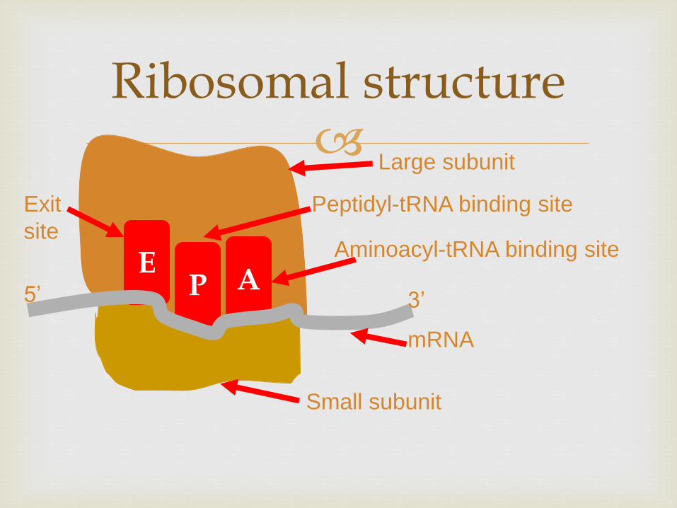
Transcription overview
Transcription requires
ribonucleoside 5acute triphosphates
ATP GTP CTP and UTP
bases are adenine guanine cytosine and uracil
sugar is ribose (not deoxyribose)
DNA-dependent RNA polymerase
Template (sense) DNA strand
Animation of transcription
Transcription overview
Features of transcription
RNA polymerase catalyzes sugar-phosphate bond between 3acute-OH of ribose and the 5acute-PO4
Order of bases in DNA template strand determines order of bases in transcript
Nucleotides are added to the 3acute-OH of the growing chain
RNA synthesis does not require a primer
Transcription overview
In prokaryotes transcription and translation are coupled Proteins are synthesized directly from the primary transcript as it is made
In eukaryotes transcription and translation are separated Transcription occurs in the nucleus and translation occurs in the cytoplasm on ribosomes
Figure comparing eukaryotic and prokaryotic transcription and translation
Transcription RNA Polymerase
DNA-dependent
DNA template ribonucleoside 5acute triphosphates and Mg2+
Synthesizes RNA in 5acute to 3acute direction
E coli RNA polymerase consists of 5 subunits
Eukaryotes have three RNA polymerases
RNA polymerase II is responsible for transcription of protein-coding genes and some snRNA molecules
RNA polymerase II has 12 subunits
Requires accessory proteins (transcription factors)
Does not require a primer
Stages of Transcription
Transcription promoter recognition
Transcription factors bind to promoter sequences and recruit RNA polymerase
DNA is bound first in a closed complex Then RNA polymerase denatures a 12ndash15 bp segment of the DNA (open complex)
The site where the first base is incorporated into the transcription is numbered ldquo+1rdquo and is called the transcription start site
Transcription factors that are required at every promoter site for RNA polymerase interaction are called basal transcription factors
Promoter recognition promoter sequences
Promoter sequences vary considerably
RNA polymerase binds to different promoters with different strengths binding strength relates to the level of gene expression
There are some common consensus sequences for promoters Example E coli ndash35 sequence (found 35 bases 5acute to the
start of transcription)
Example E coli TATA box (found 10 bases 5acute to the start of transcription)
Promoter recognition enhancers
Eukaryotic genes may also have enhancers
Enhancers can be located at great distances from the gene they regulate either 5acute or 3acute of the transcription start in introns or even on the noncoding strand
One of the most common ways to identify promoters and enhancers is to use a reporter gene
Promoter recognition other players
Many proteins can regulate gene expression by modulating the strength of interaction between the promoter and RNA polymerase
Some proteins can activate transcription (upregulate gene expression)
Some proteins can inhibit transcription by blocking polymerase activity
Some proteins can act both as repressors and activators of transcription
Transcription chain initiation
Chain initiation
RNA polymerase locally denatures the DNA
The first base of the new RNA strand is placed complementary to the +1 site
RNA polymerase does not require a primer
The first 8 or 9 bases of the transcript are linked Transcription factors are released and the polymerase leaves the promoter region
Figure of bacterial transcription initiation
Transcription chain elongation
Chain elongation
RNA polymerase moves along the transcribed or template DNA strand
The new RNA molecule (primary transcript) forms a short RNA-DNA hybrid molecule with the DNA template
Transcription chain termination
Most known about bacterial chain termination
Termination is signaled by a sequence that can form a hairpin loop
The polymerase and the new RNA molecule are released upon formation of the loop
Review the transcription animation
Transcription mRNA synthesisprocessing
Prokaryotes mRNA transcribed directly from DNA template and used immediately in protein synthesis
Eukaryotes primary transcript must be processed to produce the mRNA
Noncoding sequences (introns) are removed
Coding sequences (exons) spliced together
5acute-methylguanosine cap added
3acute-polyadenosine tail added
Transcription mRNA synthesisprocessing
Removal of introns and splicing of exons can occur several ways For introns within a nuclear transcript a spliceosome is
required
Splicesomes protein and small nuclear RNA (snRNA)
Specificity of splicing comes from the snRNA some of which contain sequences complementary to the splice junctions between introns and exons
Alternative splicing can produce different forms of a protein from the same gene
Mutations at the splice sites can cause disease
Thalassemia bull Breast cancer (BRCA 1)
Transcription mRNA synthesisprocessing
RNA splicing inside the nucleus on particles called spliceosomes
Splicesomes are composed of proteins and small RNA molecules (100ndash200 bp snRNA)
Both proteins and RNA are required but some suggesting that RNA can catalyze the splicing reaction
Self-splicing in Tetrahymena the RNA catalyzes its own splicing
Catalytic RNA ribozymes
From gene to protein genetic code
Central Dogma
Information travels from DNA to RNA to Protein
Is there a one-to-one correspondence between DNA RNA and Protein
DNA and RNA each have four nucleotides that can form them so yes there is a one-to-one correspondence between DNA and RNA
Proteins can be composed of a potential 20 amino acids only four RNA nucleotides no one-to-one correspondence
How then does RNA direct the order and number of amino acids in a protein
From gene to protein genetic code
How many bases are required for each amino acid (4 bases)2basesaa = 16 amino acidsmdashnot enough
(4 bases)3basesaa = 64 amino acid possibilities
Minimum of 3 basesaa required
What is the nature of the code Does it have punctuation Is it overlapping
Crick FH et al (1961) Nature 192 1227ndash32 (httpprofilesnlmnihgovSCBCBJ )
3-base nonoverlapping code that is read from a fixed point
From gene to protein genetic code
Nirenberg and Matthaei in vitro protein translation
Found that adding rRNA prolonged cell-free protein synthesis
Adding artificial RNA synthesized by polynucleotide phosphorylase (no template UUUUUUUUU) stimulated protein synthesis more
The protein that came out of this reaction was polyphenylalanine (UUU = Phe)
Other artificial RNAs AAA = Lys CCC =Pro
From gene to protein genetic code
Nirenberg
Triplet binding assay add triplet RNA ribosomes binding factors GTP and radiolabeled charged tRNA (figure)
UUU trinucleotide binds to Phe-tRNA
UGU trinucleotide binds to CYS-tRNA
By fits and starts the triplet genetic code was worked out
Each three-letter ldquowordrdquo (codon) specifies an amino acid or directions to stop translation
The code is redundant or degenerate more than one way to encode an amino acid
From gene to protein Translation
Components required for translation
mRNA
Ribosomes
tRNA
Aminoacyl tRNA synthetases
Initiation elongation and termination factors
Animation of translation
Translation initiation
Ribosome small subunit binds to mRNA
Charged tRNA anticodon forms base pairs with the mRNA codon
Small subunit interacts with initiation factors and special initiator tRNA that is charged with methionine
mRNA-small subunit-tRNA complex recruits the large subunit
Eukaryotic and prokaryotic initiation differ slightly
Translation initiation
The large subunit of the ribosome contains three binding sites
Amino acyl (A site)
Peptidyl (P site)
Exit (E site)
At initiation
The tRNAfMet occupies the P site
A second charged tRNA complementary to the next codon binds the A site
Translation elongation
Elongation
Ribosome translocates by three bases after peptide bond formed
New charged tRNA aligns in the A site
Peptide bond between amino acids in A and P sites is formed
Ribosome translocates by three more bases
The uncharged tRNA in the A site is moved to the E site
Translation elongation
EF-Tu recruits charged tRNA to A site Requires hydrolysis of GTP
Peptidyl transferase catalyzes peptide bond formation (bond between aa and tRNA in the P site converted to peptide bond between the two amino acids)
Peptide bond formation requires RNA and may be a ribozyme-catalyzed reaction
Translation termination
Termination
Elongation proceeds until STOP codon reached UAA UAG UGA
No tRNA normally exists that can form base pairing with a STOP codon recognized by a release factor
tRNA charged with last amino acid will remain at P site
Release factors cleave the amino acid from the tRNA
Ribosome subunits dissociate from each other
Review the animation of translation
48
Genetic code
Def Genetic code is the nucleotide base sequence on DNA ( and
subsequently on mRNA by transcription) which will be translated into a
sequence of amino acids of the protein to be synthesized
The code is composed of codons
Codon is composed of 3 bases ( eg ACG or UAG) Each codon is
translated into one amino acid
The 4 nucleotide bases (AGC and U) in mRNA are used to produce the
three base codons There are therefore 64 codons code for the 20 amino
acids and since each codon code for only one amino acids this means
that there are more than one cone for the same amino acid
How to translate a codon (see table)
This table or dictionary can be used to translate any codon sequence
Each triplet is read from 5prime rarr 3prime direction so the first base is 5prime base
followed by the middle base then the last base which is 3prime base
49
Examples 5prime- A UG- 3prime codes for methionine
5prime- UCU- 3prime codes for serine
5prime - CCA- 3prime codes for proline
Termination (stop or nonsense) codons
Three of the 64 codons UAA UAG UGA do not code for any amino
acid They are termination codes which when one of them appear in
mRNA sequence it indicates finishing of protein synthesis
Characters of the genetic code
1- Specificity the genetic code is specific that is a specific codon
always code for the same amino acid
2- Universality the genetic code is universal that is the same codon is
used in all living organisms procaryotics and eucaryotics
3- Degeneracy the genetic code is degenerate ie although each codon
corresponds to a single amino acidone amino acid may have more than
one codons eg arginine has 6 different codons (give more examples
from the table)
50
51
Gene mutation (altering the nucleotide sequence)
1- Point mutation changing in a single nucleotide base on the mRNA
can lead to any of the following 3 results
i- Silent mutation ie the codon containg the changed base may code
for the same amino acid For example in serine codon UCA if A is
changed to U giving the codon UCU it still code for serine See table
ii- Missense mutation the codon containing the changed base may code
for a different amino acid For example if the serine codon UCA is
changed to be CCA ( U is replaced by C) it will code for proline not
serine leading to insertion of incorrect amino acid into polypeptide chain
iii- Non sense mutation the codon containing the changed base may
become a termination codon For example serine codon UCA becomes
UAA if C is changed to A UAA is a stop codon leading to termination
of translation at that point
52
53
Types of point mutation
U A A (termination codon) Nonsense mutation
uarr
U C A rarr U C U Silent mutation
(codon for serine) (codon for serine)
darr
C C A ( codon for proline) Missense mutation
Give other examples on missense mutation which leads to some Hb
disease
54
2- Frame- shift mutation
deletion or addition of one or two base to
message sequence leading to change in
reading frame (reading sequence) and the
resulting amino acid seuence may become
completely different from this point
55
TranslationComponents required for protein synthesis1- Amino acids all amino acids involved in thefinished protein must be present at the time ofprotein synthesis2- Ribosomes the site of protein synthesis They arelarge complexes of protein and rRNA In humanthey consist of two subunits one large (60S) and onesmall (40S)
3- tRNA at least one specific type of tRNA is required to transfer
one amino acid There about 50 tRNA in human for the 20 amino
acids this means some amino acids have more than one specific
tRNA The role of tRNA in protein synthesis is discussed before
(amino acid attachment and anticodon loop)
4- aminoacyl-tRNA synthetase This is the enzyme that catalyzes the
attachment of amino acid with its corresponding tRNA forming
aminoacyl tRNA
56
5- mRNA that carry code for the protein to be synthesized
6- protein factors Initiation elongation and termination (or release)
factors are required for peptide synthesis
7- ATP and GTP are required as source of energy
Steps (movie)
1- Initiation
Initiation (start) codon is usually AUG which is the codon of
methionine so the initiator tRNA is methionnyl tRNA (Met tRNA)
a- The initiation factors (IF-1 IF-2 and IF-3) binds the Met tRNA with
small ribosomal subunit then to mRNA containing the code of the
protein to be synthesized IFs recognizes mRNA from its 5 cap
57
b-This complex binds to large ribosomal subunit forming initiation complex in which Met tRNA is present in P- site of 60 ribosomal subunitNB- tRNA bind with mRNA by base pairing between codon on mRNA and anticodon
on tRNA - mRNA is read from 5prime rarr 3prime direction
P-site is the peptidyl site of the ribosome to which methionyl tRNA is placed (enter)
2- Elongation elongation factors (EFs) stimulate the stepwise elongation of polypeptide chain as follow
a- The next aminoacyl tRNA (tRNA which carry the next amino acid specified by recognition of the next codon on mRNA) will enter A site of ribosome
A site or acceptor site or aminoacyl tRNA siteIs the site of ribosome to which each new incoming aminoacyl tRNA will enter
b) ribosomal peptidyl transferase enzyme will transfer methionine
from methionyl tRNA into A site to form a peptide bond between
methionine and the new incoming amino acid to form dipeptidyl
tRNA
c) Elongation factor-2 (EF-2) (called also translocase) moves
mRNA and dipeptidyl tRNA from A site to P site leaving A site free to
allow entrance of another new aminoacyl tRNA
The figure shows the repetitive cycle of elongation of chain Each
cycle is consisting of
1) codon recognition and the entrance of the new aminoacyl tRNA
acid ( amino acid carried on tRNA) into A site
2) The growing chain in P site will moved to A site with peptide
bond formation with the new amino acid
3) Translocation of growing chain to P site allowing A site free for
enterance of new amino acid an so onhelliphelliphelliphelliphelliphelliphellip Resulting in
elongation of poly peptide chain
59repetitive cycle of elongation
60
3- Termination occurs when one of the three stop codons (UAA UAG orUGA) enters A site of the ribosome These codons are recognized by releasefactors (RFs) which are RF-1 RF-2 RF-3 RFs cause the newly synthesizedprotein to be released from the ribosomal complex and dissociation ofribosomes from mRNA (ie cause dissolution of the complex)
Transcription overview
Features of transcription
RNA polymerase catalyzes sugar-phosphate bond between 3acute-OH of ribose and the 5acute-PO4
Order of bases in DNA template strand determines order of bases in transcript
Nucleotides are added to the 3acute-OH of the growing chain
RNA synthesis does not require a primer
Transcription overview
In prokaryotes transcription and translation are coupled Proteins are synthesized directly from the primary transcript as it is made
In eukaryotes transcription and translation are separated Transcription occurs in the nucleus and translation occurs in the cytoplasm on ribosomes
Figure comparing eukaryotic and prokaryotic transcription and translation
Transcription RNA Polymerase
DNA-dependent
DNA template ribonucleoside 5acute triphosphates and Mg2+
Synthesizes RNA in 5acute to 3acute direction
E coli RNA polymerase consists of 5 subunits
Eukaryotes have three RNA polymerases
RNA polymerase II is responsible for transcription of protein-coding genes and some snRNA molecules
RNA polymerase II has 12 subunits
Requires accessory proteins (transcription factors)
Does not require a primer
Stages of Transcription
Transcription promoter recognition
Transcription factors bind to promoter sequences and recruit RNA polymerase
DNA is bound first in a closed complex Then RNA polymerase denatures a 12ndash15 bp segment of the DNA (open complex)
The site where the first base is incorporated into the transcription is numbered ldquo+1rdquo and is called the transcription start site
Transcription factors that are required at every promoter site for RNA polymerase interaction are called basal transcription factors
Promoter recognition promoter sequences
Promoter sequences vary considerably
RNA polymerase binds to different promoters with different strengths binding strength relates to the level of gene expression
There are some common consensus sequences for promoters Example E coli ndash35 sequence (found 35 bases 5acute to the
start of transcription)
Example E coli TATA box (found 10 bases 5acute to the start of transcription)
Promoter recognition enhancers
Eukaryotic genes may also have enhancers
Enhancers can be located at great distances from the gene they regulate either 5acute or 3acute of the transcription start in introns or even on the noncoding strand
One of the most common ways to identify promoters and enhancers is to use a reporter gene
Promoter recognition other players
Many proteins can regulate gene expression by modulating the strength of interaction between the promoter and RNA polymerase
Some proteins can activate transcription (upregulate gene expression)
Some proteins can inhibit transcription by blocking polymerase activity
Some proteins can act both as repressors and activators of transcription
Transcription chain initiation
Chain initiation
RNA polymerase locally denatures the DNA
The first base of the new RNA strand is placed complementary to the +1 site
RNA polymerase does not require a primer
The first 8 or 9 bases of the transcript are linked Transcription factors are released and the polymerase leaves the promoter region
Figure of bacterial transcription initiation
Transcription chain elongation
Chain elongation
RNA polymerase moves along the transcribed or template DNA strand
The new RNA molecule (primary transcript) forms a short RNA-DNA hybrid molecule with the DNA template
Transcription chain termination
Most known about bacterial chain termination
Termination is signaled by a sequence that can form a hairpin loop
The polymerase and the new RNA molecule are released upon formation of the loop
Review the transcription animation
Transcription mRNA synthesisprocessing
Prokaryotes mRNA transcribed directly from DNA template and used immediately in protein synthesis
Eukaryotes primary transcript must be processed to produce the mRNA
Noncoding sequences (introns) are removed
Coding sequences (exons) spliced together
5acute-methylguanosine cap added
3acute-polyadenosine tail added
Transcription mRNA synthesisprocessing
Removal of introns and splicing of exons can occur several ways For introns within a nuclear transcript a spliceosome is
required
Splicesomes protein and small nuclear RNA (snRNA)
Specificity of splicing comes from the snRNA some of which contain sequences complementary to the splice junctions between introns and exons
Alternative splicing can produce different forms of a protein from the same gene
Mutations at the splice sites can cause disease
Thalassemia bull Breast cancer (BRCA 1)
Transcription mRNA synthesisprocessing
RNA splicing inside the nucleus on particles called spliceosomes
Splicesomes are composed of proteins and small RNA molecules (100ndash200 bp snRNA)
Both proteins and RNA are required but some suggesting that RNA can catalyze the splicing reaction
Self-splicing in Tetrahymena the RNA catalyzes its own splicing
Catalytic RNA ribozymes
From gene to protein genetic code
Central Dogma
Information travels from DNA to RNA to Protein
Is there a one-to-one correspondence between DNA RNA and Protein
DNA and RNA each have four nucleotides that can form them so yes there is a one-to-one correspondence between DNA and RNA
Proteins can be composed of a potential 20 amino acids only four RNA nucleotides no one-to-one correspondence
How then does RNA direct the order and number of amino acids in a protein
From gene to protein genetic code
How many bases are required for each amino acid (4 bases)2basesaa = 16 amino acidsmdashnot enough
(4 bases)3basesaa = 64 amino acid possibilities
Minimum of 3 basesaa required
What is the nature of the code Does it have punctuation Is it overlapping
Crick FH et al (1961) Nature 192 1227ndash32 (httpprofilesnlmnihgovSCBCBJ )
3-base nonoverlapping code that is read from a fixed point
From gene to protein genetic code
Nirenberg and Matthaei in vitro protein translation
Found that adding rRNA prolonged cell-free protein synthesis
Adding artificial RNA synthesized by polynucleotide phosphorylase (no template UUUUUUUUU) stimulated protein synthesis more
The protein that came out of this reaction was polyphenylalanine (UUU = Phe)
Other artificial RNAs AAA = Lys CCC =Pro
From gene to protein genetic code
Nirenberg
Triplet binding assay add triplet RNA ribosomes binding factors GTP and radiolabeled charged tRNA (figure)
UUU trinucleotide binds to Phe-tRNA
UGU trinucleotide binds to CYS-tRNA
By fits and starts the triplet genetic code was worked out
Each three-letter ldquowordrdquo (codon) specifies an amino acid or directions to stop translation
The code is redundant or degenerate more than one way to encode an amino acid
From gene to protein Translation
Components required for translation
mRNA
Ribosomes
tRNA
Aminoacyl tRNA synthetases
Initiation elongation and termination factors
Animation of translation
Translation initiation
Ribosome small subunit binds to mRNA
Charged tRNA anticodon forms base pairs with the mRNA codon
Small subunit interacts with initiation factors and special initiator tRNA that is charged with methionine
mRNA-small subunit-tRNA complex recruits the large subunit
Eukaryotic and prokaryotic initiation differ slightly
Translation initiation
The large subunit of the ribosome contains three binding sites
Amino acyl (A site)
Peptidyl (P site)
Exit (E site)
At initiation
The tRNAfMet occupies the P site
A second charged tRNA complementary to the next codon binds the A site
Translation elongation
Elongation
Ribosome translocates by three bases after peptide bond formed
New charged tRNA aligns in the A site
Peptide bond between amino acids in A and P sites is formed
Ribosome translocates by three more bases
The uncharged tRNA in the A site is moved to the E site
Translation elongation
EF-Tu recruits charged tRNA to A site Requires hydrolysis of GTP
Peptidyl transferase catalyzes peptide bond formation (bond between aa and tRNA in the P site converted to peptide bond between the two amino acids)
Peptide bond formation requires RNA and may be a ribozyme-catalyzed reaction
Translation termination
Termination
Elongation proceeds until STOP codon reached UAA UAG UGA
No tRNA normally exists that can form base pairing with a STOP codon recognized by a release factor
tRNA charged with last amino acid will remain at P site
Release factors cleave the amino acid from the tRNA
Ribosome subunits dissociate from each other
Review the animation of translation
48
Genetic code
Def Genetic code is the nucleotide base sequence on DNA ( and
subsequently on mRNA by transcription) which will be translated into a
sequence of amino acids of the protein to be synthesized
The code is composed of codons
Codon is composed of 3 bases ( eg ACG or UAG) Each codon is
translated into one amino acid
The 4 nucleotide bases (AGC and U) in mRNA are used to produce the
three base codons There are therefore 64 codons code for the 20 amino
acids and since each codon code for only one amino acids this means
that there are more than one cone for the same amino acid
How to translate a codon (see table)
This table or dictionary can be used to translate any codon sequence
Each triplet is read from 5prime rarr 3prime direction so the first base is 5prime base
followed by the middle base then the last base which is 3prime base
49
Examples 5prime- A UG- 3prime codes for methionine
5prime- UCU- 3prime codes for serine
5prime - CCA- 3prime codes for proline
Termination (stop or nonsense) codons
Three of the 64 codons UAA UAG UGA do not code for any amino
acid They are termination codes which when one of them appear in
mRNA sequence it indicates finishing of protein synthesis
Characters of the genetic code
1- Specificity the genetic code is specific that is a specific codon
always code for the same amino acid
2- Universality the genetic code is universal that is the same codon is
used in all living organisms procaryotics and eucaryotics
3- Degeneracy the genetic code is degenerate ie although each codon
corresponds to a single amino acidone amino acid may have more than
one codons eg arginine has 6 different codons (give more examples
from the table)
50
51
Gene mutation (altering the nucleotide sequence)
1- Point mutation changing in a single nucleotide base on the mRNA
can lead to any of the following 3 results
i- Silent mutation ie the codon containg the changed base may code
for the same amino acid For example in serine codon UCA if A is
changed to U giving the codon UCU it still code for serine See table
ii- Missense mutation the codon containing the changed base may code
for a different amino acid For example if the serine codon UCA is
changed to be CCA ( U is replaced by C) it will code for proline not
serine leading to insertion of incorrect amino acid into polypeptide chain
iii- Non sense mutation the codon containing the changed base may
become a termination codon For example serine codon UCA becomes
UAA if C is changed to A UAA is a stop codon leading to termination
of translation at that point
52
53
Types of point mutation
U A A (termination codon) Nonsense mutation
uarr
U C A rarr U C U Silent mutation
(codon for serine) (codon for serine)
darr
C C A ( codon for proline) Missense mutation
Give other examples on missense mutation which leads to some Hb
disease
54
2- Frame- shift mutation
deletion or addition of one or two base to
message sequence leading to change in
reading frame (reading sequence) and the
resulting amino acid seuence may become
completely different from this point
55
TranslationComponents required for protein synthesis1- Amino acids all amino acids involved in thefinished protein must be present at the time ofprotein synthesis2- Ribosomes the site of protein synthesis They arelarge complexes of protein and rRNA In humanthey consist of two subunits one large (60S) and onesmall (40S)
3- tRNA at least one specific type of tRNA is required to transfer
one amino acid There about 50 tRNA in human for the 20 amino
acids this means some amino acids have more than one specific
tRNA The role of tRNA in protein synthesis is discussed before
(amino acid attachment and anticodon loop)
4- aminoacyl-tRNA synthetase This is the enzyme that catalyzes the
attachment of amino acid with its corresponding tRNA forming
aminoacyl tRNA
56
5- mRNA that carry code for the protein to be synthesized
6- protein factors Initiation elongation and termination (or release)
factors are required for peptide synthesis
7- ATP and GTP are required as source of energy
Steps (movie)
1- Initiation
Initiation (start) codon is usually AUG which is the codon of
methionine so the initiator tRNA is methionnyl tRNA (Met tRNA)
a- The initiation factors (IF-1 IF-2 and IF-3) binds the Met tRNA with
small ribosomal subunit then to mRNA containing the code of the
protein to be synthesized IFs recognizes mRNA from its 5 cap
57
b-This complex binds to large ribosomal subunit forming initiation complex in which Met tRNA is present in P- site of 60 ribosomal subunitNB- tRNA bind with mRNA by base pairing between codon on mRNA and anticodon
on tRNA - mRNA is read from 5prime rarr 3prime direction
P-site is the peptidyl site of the ribosome to which methionyl tRNA is placed (enter)
2- Elongation elongation factors (EFs) stimulate the stepwise elongation of polypeptide chain as follow
a- The next aminoacyl tRNA (tRNA which carry the next amino acid specified by recognition of the next codon on mRNA) will enter A site of ribosome
A site or acceptor site or aminoacyl tRNA siteIs the site of ribosome to which each new incoming aminoacyl tRNA will enter
b) ribosomal peptidyl transferase enzyme will transfer methionine
from methionyl tRNA into A site to form a peptide bond between
methionine and the new incoming amino acid to form dipeptidyl
tRNA
c) Elongation factor-2 (EF-2) (called also translocase) moves
mRNA and dipeptidyl tRNA from A site to P site leaving A site free to
allow entrance of another new aminoacyl tRNA
The figure shows the repetitive cycle of elongation of chain Each
cycle is consisting of
1) codon recognition and the entrance of the new aminoacyl tRNA
acid ( amino acid carried on tRNA) into A site
2) The growing chain in P site will moved to A site with peptide
bond formation with the new amino acid
3) Translocation of growing chain to P site allowing A site free for
enterance of new amino acid an so onhelliphelliphelliphelliphelliphelliphellip Resulting in
elongation of poly peptide chain
59repetitive cycle of elongation
60
3- Termination occurs when one of the three stop codons (UAA UAG orUGA) enters A site of the ribosome These codons are recognized by releasefactors (RFs) which are RF-1 RF-2 RF-3 RFs cause the newly synthesizedprotein to be released from the ribosomal complex and dissociation ofribosomes from mRNA (ie cause dissolution of the complex)

Transcription mRNA synthesisprocessing
Removal of introns and splicing of exons can occur several ways For introns within a nuclear transcript a spliceosome is
required
Splicesomes protein and small nuclear RNA (snRNA)
Specificity of splicing comes from the snRNA some of which contain sequences complementary to the splice junctions between introns and exons
Alternative splicing can produce different forms of a protein from the same gene
Mutations at the splice sites can cause disease
Thalassemia bull Breast cancer (BRCA 1)
Transcription mRNA synthesisprocessing
RNA splicing inside the nucleus on particles called spliceosomes
Splicesomes are composed of proteins and small RNA molecules (100ndash200 bp snRNA)
Both proteins and RNA are required but some suggesting that RNA can catalyze the splicing reaction
Self-splicing in Tetrahymena the RNA catalyzes its own splicing
Catalytic RNA ribozymes
From gene to protein genetic code
Central Dogma
Information travels from DNA to RNA to Protein
Is there a one-to-one correspondence between DNA RNA and Protein
DNA and RNA each have four nucleotides that can form them so yes there is a one-to-one correspondence between DNA and RNA
Proteins can be composed of a potential 20 amino acids only four RNA nucleotides no one-to-one correspondence
How then does RNA direct the order and number of amino acids in a protein
From gene to protein genetic code
How many bases are required for each amino acid (4 bases)2basesaa = 16 amino acidsmdashnot enough
(4 bases)3basesaa = 64 amino acid possibilities
Minimum of 3 basesaa required
What is the nature of the code Does it have punctuation Is it overlapping
Crick FH et al (1961) Nature 192 1227ndash32 (httpprofilesnlmnihgovSCBCBJ )
3-base nonoverlapping code that is read from a fixed point
From gene to protein genetic code
Nirenberg and Matthaei in vitro protein translation
Found that adding rRNA prolonged cell-free protein synthesis
Adding artificial RNA synthesized by polynucleotide phosphorylase (no template UUUUUUUUU) stimulated protein synthesis more
The protein that came out of this reaction was polyphenylalanine (UUU = Phe)
Other artificial RNAs AAA = Lys CCC =Pro
From gene to protein genetic code
Nirenberg
Triplet binding assay add triplet RNA ribosomes binding factors GTP and radiolabeled charged tRNA (figure)
UUU trinucleotide binds to Phe-tRNA
UGU trinucleotide binds to CYS-tRNA
By fits and starts the triplet genetic code was worked out
Each three-letter ldquowordrdquo (codon) specifies an amino acid or directions to stop translation
The code is redundant or degenerate more than one way to encode an amino acid
From gene to protein Translation
Components required for translation
mRNA
Ribosomes
tRNA
Aminoacyl tRNA synthetases
Initiation elongation and termination factors
Animation of translation
Translation initiation
Ribosome small subunit binds to mRNA
Charged tRNA anticodon forms base pairs with the mRNA codon
Small subunit interacts with initiation factors and special initiator tRNA that is charged with methionine
mRNA-small subunit-tRNA complex recruits the large subunit
Eukaryotic and prokaryotic initiation differ slightly
Translation initiation
The large subunit of the ribosome contains three binding sites
Amino acyl (A site)
Peptidyl (P site)
Exit (E site)
At initiation
The tRNAfMet occupies the P site
A second charged tRNA complementary to the next codon binds the A site
Translation elongation
Elongation
Ribosome translocates by three bases after peptide bond formed
New charged tRNA aligns in the A site
Peptide bond between amino acids in A and P sites is formed
Ribosome translocates by three more bases
The uncharged tRNA in the A site is moved to the E site
Translation elongation
EF-Tu recruits charged tRNA to A site Requires hydrolysis of GTP
Peptidyl transferase catalyzes peptide bond formation (bond between aa and tRNA in the P site converted to peptide bond between the two amino acids)
Peptide bond formation requires RNA and may be a ribozyme-catalyzed reaction
Translation termination
Termination
Elongation proceeds until STOP codon reached UAA UAG UGA
No tRNA normally exists that can form base pairing with a STOP codon recognized by a release factor
tRNA charged with last amino acid will remain at P site
Release factors cleave the amino acid from the tRNA
Ribosome subunits dissociate from each other
Review the animation of translation
48
Genetic code
Def Genetic code is the nucleotide base sequence on DNA ( and
subsequently on mRNA by transcription) which will be translated into a
sequence of amino acids of the protein to be synthesized
The code is composed of codons
Codon is composed of 3 bases ( eg ACG or UAG) Each codon is
translated into one amino acid
The 4 nucleotide bases (AGC and U) in mRNA are used to produce the
three base codons There are therefore 64 codons code for the 20 amino
acids and since each codon code for only one amino acids this means
that there are more than one cone for the same amino acid
How to translate a codon (see table)
This table or dictionary can be used to translate any codon sequence
Each triplet is read from 5prime rarr 3prime direction so the first base is 5prime base
followed by the middle base then the last base which is 3prime base
49
Examples 5prime- A UG- 3prime codes for methionine
5prime- UCU- 3prime codes for serine
5prime - CCA- 3prime codes for proline
Termination (stop or nonsense) codons
Three of the 64 codons UAA UAG UGA do not code for any amino
acid They are termination codes which when one of them appear in
mRNA sequence it indicates finishing of protein synthesis
Characters of the genetic code
1- Specificity the genetic code is specific that is a specific codon
always code for the same amino acid
2- Universality the genetic code is universal that is the same codon is
used in all living organisms procaryotics and eucaryotics
3- Degeneracy the genetic code is degenerate ie although each codon
corresponds to a single amino acidone amino acid may have more than
one codons eg arginine has 6 different codons (give more examples
from the table)
50
51
Gene mutation (altering the nucleotide sequence)
1- Point mutation changing in a single nucleotide base on the mRNA
can lead to any of the following 3 results
i- Silent mutation ie the codon containg the changed base may code
for the same amino acid For example in serine codon UCA if A is
changed to U giving the codon UCU it still code for serine See table
ii- Missense mutation the codon containing the changed base may code
for a different amino acid For example if the serine codon UCA is
changed to be CCA ( U is replaced by C) it will code for proline not
serine leading to insertion of incorrect amino acid into polypeptide chain
iii- Non sense mutation the codon containing the changed base may
become a termination codon For example serine codon UCA becomes
UAA if C is changed to A UAA is a stop codon leading to termination
of translation at that point
52
53
Types of point mutation
U A A (termination codon) Nonsense mutation
uarr
U C A rarr U C U Silent mutation
(codon for serine) (codon for serine)
darr
C C A ( codon for proline) Missense mutation
Give other examples on missense mutation which leads to some Hb
disease
54
2- Frame- shift mutation
deletion or addition of one or two base to
message sequence leading to change in
reading frame (reading sequence) and the
resulting amino acid seuence may become
completely different from this point
55
TranslationComponents required for protein synthesis1- Amino acids all amino acids involved in thefinished protein must be present at the time ofprotein synthesis2- Ribosomes the site of protein synthesis They arelarge complexes of protein and rRNA In humanthey consist of two subunits one large (60S) and onesmall (40S)
3- tRNA at least one specific type of tRNA is required to transfer
one amino acid There about 50 tRNA in human for the 20 amino
acids this means some amino acids have more than one specific
tRNA The role of tRNA in protein synthesis is discussed before
(amino acid attachment and anticodon loop)
4- aminoacyl-tRNA synthetase This is the enzyme that catalyzes the
attachment of amino acid with its corresponding tRNA forming
aminoacyl tRNA
56
5- mRNA that carry code for the protein to be synthesized
6- protein factors Initiation elongation and termination (or release)
factors are required for peptide synthesis
7- ATP and GTP are required as source of energy
Steps (movie)
1- Initiation
Initiation (start) codon is usually AUG which is the codon of
methionine so the initiator tRNA is methionnyl tRNA (Met tRNA)
a- The initiation factors (IF-1 IF-2 and IF-3) binds the Met tRNA with
small ribosomal subunit then to mRNA containing the code of the
protein to be synthesized IFs recognizes mRNA from its 5 cap
57
b-This complex binds to large ribosomal subunit forming initiation complex in which Met tRNA is present in P- site of 60 ribosomal subunitNB- tRNA bind with mRNA by base pairing between codon on mRNA and anticodon
on tRNA - mRNA is read from 5prime rarr 3prime direction
P-site is the peptidyl site of the ribosome to which methionyl tRNA is placed (enter)
2- Elongation elongation factors (EFs) stimulate the stepwise elongation of polypeptide chain as follow
a- The next aminoacyl tRNA (tRNA which carry the next amino acid specified by recognition of the next codon on mRNA) will enter A site of ribosome
A site or acceptor site or aminoacyl tRNA siteIs the site of ribosome to which each new incoming aminoacyl tRNA will enter
b) ribosomal peptidyl transferase enzyme will transfer methionine
from methionyl tRNA into A site to form a peptide bond between
methionine and the new incoming amino acid to form dipeptidyl
tRNA
c) Elongation factor-2 (EF-2) (called also translocase) moves
mRNA and dipeptidyl tRNA from A site to P site leaving A site free to
allow entrance of another new aminoacyl tRNA
The figure shows the repetitive cycle of elongation of chain Each
cycle is consisting of
1) codon recognition and the entrance of the new aminoacyl tRNA
acid ( amino acid carried on tRNA) into A site
2) The growing chain in P site will moved to A site with peptide
bond formation with the new amino acid
3) Translocation of growing chain to P site allowing A site free for
enterance of new amino acid an so onhelliphelliphelliphelliphelliphelliphellip Resulting in
elongation of poly peptide chain
59repetitive cycle of elongation
60
3- Termination occurs when one of the three stop codons (UAA UAG orUGA) enters A site of the ribosome These codons are recognized by releasefactors (RFs) which are RF-1 RF-2 RF-3 RFs cause the newly synthesizedprotein to be released from the ribosomal complex and dissociation ofribosomes from mRNA (ie cause dissolution of the complex)
From gene to protein genetic code
Central Dogma
Information travels from DNA to RNA to Protein
Is there a one-to-one correspondence between DNA RNA and Protein
DNA and RNA each have four nucleotides that can form them so yes there is a one-to-one correspondence between DNA and RNA
Proteins can be composed of a potential 20 amino acids only four RNA nucleotides no one-to-one correspondence
How then does RNA direct the order and number of amino acids in a protein
From gene to protein genetic code
How many bases are required for each amino acid (4 bases)2basesaa = 16 amino acidsmdashnot enough
(4 bases)3basesaa = 64 amino acid possibilities
Minimum of 3 basesaa required
What is the nature of the code Does it have punctuation Is it overlapping
Crick FH et al (1961) Nature 192 1227ndash32 (httpprofilesnlmnihgovSCBCBJ )
3-base nonoverlapping code that is read from a fixed point
From gene to protein genetic code
Nirenberg and Matthaei in vitro protein translation
Found that adding rRNA prolonged cell-free protein synthesis
Adding artificial RNA synthesized by polynucleotide phosphorylase (no template UUUUUUUUU) stimulated protein synthesis more
The protein that came out of this reaction was polyphenylalanine (UUU = Phe)
Other artificial RNAs AAA = Lys CCC =Pro
From gene to protein genetic code
Nirenberg
Triplet binding assay add triplet RNA ribosomes binding factors GTP and radiolabeled charged tRNA (figure)
UUU trinucleotide binds to Phe-tRNA
UGU trinucleotide binds to CYS-tRNA
By fits and starts the triplet genetic code was worked out
Each three-letter ldquowordrdquo (codon) specifies an amino acid or directions to stop translation
The code is redundant or degenerate more than one way to encode an amino acid
From gene to protein Translation
Components required for translation
mRNA
Ribosomes
tRNA
Aminoacyl tRNA synthetases
Initiation elongation and termination factors
Animation of translation
Translation initiation
Ribosome small subunit binds to mRNA
Charged tRNA anticodon forms base pairs with the mRNA codon
Small subunit interacts with initiation factors and special initiator tRNA that is charged with methionine
mRNA-small subunit-tRNA complex recruits the large subunit
Eukaryotic and prokaryotic initiation differ slightly
Translation initiation
The large subunit of the ribosome contains three binding sites
Amino acyl (A site)
Peptidyl (P site)
Exit (E site)
At initiation
The tRNAfMet occupies the P site
A second charged tRNA complementary to the next codon binds the A site
Translation elongation
Elongation
Ribosome translocates by three bases after peptide bond formed
New charged tRNA aligns in the A site
Peptide bond between amino acids in A and P sites is formed
Ribosome translocates by three more bases
The uncharged tRNA in the A site is moved to the E site
Translation elongation
EF-Tu recruits charged tRNA to A site Requires hydrolysis of GTP
Peptidyl transferase catalyzes peptide bond formation (bond between aa and tRNA in the P site converted to peptide bond between the two amino acids)
Peptide bond formation requires RNA and may be a ribozyme-catalyzed reaction
Translation termination
Termination
Elongation proceeds until STOP codon reached UAA UAG UGA
No tRNA normally exists that can form base pairing with a STOP codon recognized by a release factor
tRNA charged with last amino acid will remain at P site
Release factors cleave the amino acid from the tRNA
Ribosome subunits dissociate from each other
Review the animation of translation
48
Genetic code
Def Genetic code is the nucleotide base sequence on DNA ( and
subsequently on mRNA by transcription) which will be translated into a
sequence of amino acids of the protein to be synthesized
The code is composed of codons
Codon is composed of 3 bases ( eg ACG or UAG) Each codon is
translated into one amino acid
The 4 nucleotide bases (AGC and U) in mRNA are used to produce the
three base codons There are therefore 64 codons code for the 20 amino
acids and since each codon code for only one amino acids this means
that there are more than one cone for the same amino acid
How to translate a codon (see table)
This table or dictionary can be used to translate any codon sequence
Each triplet is read from 5prime rarr 3prime direction so the first base is 5prime base
followed by the middle base then the last base which is 3prime base
49
Examples 5prime- A UG- 3prime codes for methionine
5prime- UCU- 3prime codes for serine
5prime - CCA- 3prime codes for proline
Termination (stop or nonsense) codons
Three of the 64 codons UAA UAG UGA do not code for any amino
acid They are termination codes which when one of them appear in
mRNA sequence it indicates finishing of protein synthesis
Characters of the genetic code
1- Specificity the genetic code is specific that is a specific codon
always code for the same amino acid
2- Universality the genetic code is universal that is the same codon is
used in all living organisms procaryotics and eucaryotics
3- Degeneracy the genetic code is degenerate ie although each codon
corresponds to a single amino acidone amino acid may have more than
one codons eg arginine has 6 different codons (give more examples
from the table)
50
51
Gene mutation (altering the nucleotide sequence)
1- Point mutation changing in a single nucleotide base on the mRNA
can lead to any of the following 3 results
i- Silent mutation ie the codon containg the changed base may code
for the same amino acid For example in serine codon UCA if A is
changed to U giving the codon UCU it still code for serine See table
ii- Missense mutation the codon containing the changed base may code
for a different amino acid For example if the serine codon UCA is
changed to be CCA ( U is replaced by C) it will code for proline not
serine leading to insertion of incorrect amino acid into polypeptide chain
iii- Non sense mutation the codon containing the changed base may
become a termination codon For example serine codon UCA becomes
UAA if C is changed to A UAA is a stop codon leading to termination
of translation at that point
52
53
Types of point mutation
U A A (termination codon) Nonsense mutation
uarr
U C A rarr U C U Silent mutation
(codon for serine) (codon for serine)
darr
C C A ( codon for proline) Missense mutation
Give other examples on missense mutation which leads to some Hb
disease
54
2- Frame- shift mutation
deletion or addition of one or two base to
message sequence leading to change in
reading frame (reading sequence) and the
resulting amino acid seuence may become
completely different from this point
55
TranslationComponents required for protein synthesis1- Amino acids all amino acids involved in thefinished protein must be present at the time ofprotein synthesis2- Ribosomes the site of protein synthesis They arelarge complexes of protein and rRNA In humanthey consist of two subunits one large (60S) and onesmall (40S)
3- tRNA at least one specific type of tRNA is required to transfer
one amino acid There about 50 tRNA in human for the 20 amino
acids this means some amino acids have more than one specific
tRNA The role of tRNA in protein synthesis is discussed before
(amino acid attachment and anticodon loop)
4- aminoacyl-tRNA synthetase This is the enzyme that catalyzes the
attachment of amino acid with its corresponding tRNA forming
aminoacyl tRNA
56
5- mRNA that carry code for the protein to be synthesized
6- protein factors Initiation elongation and termination (or release)
factors are required for peptide synthesis
7- ATP and GTP are required as source of energy
Steps (movie)
1- Initiation
Initiation (start) codon is usually AUG which is the codon of
methionine so the initiator tRNA is methionnyl tRNA (Met tRNA)
a- The initiation factors (IF-1 IF-2 and IF-3) binds the Met tRNA with
small ribosomal subunit then to mRNA containing the code of the
protein to be synthesized IFs recognizes mRNA from its 5 cap
57
b-This complex binds to large ribosomal subunit forming initiation complex in which Met tRNA is present in P- site of 60 ribosomal subunitNB- tRNA bind with mRNA by base pairing between codon on mRNA and anticodon
on tRNA - mRNA is read from 5prime rarr 3prime direction
P-site is the peptidyl site of the ribosome to which methionyl tRNA is placed (enter)
2- Elongation elongation factors (EFs) stimulate the stepwise elongation of polypeptide chain as follow
a- The next aminoacyl tRNA (tRNA which carry the next amino acid specified by recognition of the next codon on mRNA) will enter A site of ribosome
A site or acceptor site or aminoacyl tRNA siteIs the site of ribosome to which each new incoming aminoacyl tRNA will enter
b) ribosomal peptidyl transferase enzyme will transfer methionine
from methionyl tRNA into A site to form a peptide bond between
methionine and the new incoming amino acid to form dipeptidyl
tRNA
c) Elongation factor-2 (EF-2) (called also translocase) moves
mRNA and dipeptidyl tRNA from A site to P site leaving A site free to
allow entrance of another new aminoacyl tRNA
The figure shows the repetitive cycle of elongation of chain Each
cycle is consisting of
1) codon recognition and the entrance of the new aminoacyl tRNA
acid ( amino acid carried on tRNA) into A site
2) The growing chain in P site will moved to A site with peptide
bond formation with the new amino acid
3) Translocation of growing chain to P site allowing A site free for
enterance of new amino acid an so onhelliphelliphelliphelliphelliphelliphellip Resulting in
elongation of poly peptide chain
59repetitive cycle of elongation
60
3- Termination occurs when one of the three stop codons (UAA UAG orUGA) enters A site of the ribosome These codons are recognized by releasefactors (RFs) which are RF-1 RF-2 RF-3 RFs cause the newly synthesizedprotein to be released from the ribosomal complex and dissociation ofribosomes from mRNA (ie cause dissolution of the complex)

From gene to protein genetic code
How many bases are required for each amino acid (4 bases)2basesaa = 16 amino acidsmdashnot enough
(4 bases)3basesaa = 64 amino acid possibilities
Minimum of 3 basesaa required
What is the nature of the code Does it have punctuation Is it overlapping
Crick FH et al (1961) Nature 192 1227ndash32 (httpprofilesnlmnihgovSCBCBJ )
3-base nonoverlapping code that is read from a fixed point
From gene to protein genetic code
Nirenberg and Matthaei in vitro protein translation
Found that adding rRNA prolonged cell-free protein synthesis
Adding artificial RNA synthesized by polynucleotide phosphorylase (no template UUUUUUUUU) stimulated protein synthesis more
The protein that came out of this reaction was polyphenylalanine (UUU = Phe)
Other artificial RNAs AAA = Lys CCC =Pro
From gene to protein genetic code
Nirenberg
Triplet binding assay add triplet RNA ribosomes binding factors GTP and radiolabeled charged tRNA (figure)
UUU trinucleotide binds to Phe-tRNA
UGU trinucleotide binds to CYS-tRNA
By fits and starts the triplet genetic code was worked out
Each three-letter ldquowordrdquo (codon) specifies an amino acid or directions to stop translation
The code is redundant or degenerate more than one way to encode an amino acid
From gene to protein Translation
Components required for translation
mRNA
Ribosomes
tRNA
Aminoacyl tRNA synthetases
Initiation elongation and termination factors
Animation of translation
Translation initiation
Ribosome small subunit binds to mRNA
Charged tRNA anticodon forms base pairs with the mRNA codon
Small subunit interacts with initiation factors and special initiator tRNA that is charged with methionine
mRNA-small subunit-tRNA complex recruits the large subunit
Eukaryotic and prokaryotic initiation differ slightly
Translation initiation
The large subunit of the ribosome contains three binding sites
Amino acyl (A site)
Peptidyl (P site)
Exit (E site)
At initiation
The tRNAfMet occupies the P site
A second charged tRNA complementary to the next codon binds the A site
Translation elongation
Elongation
Ribosome translocates by three bases after peptide bond formed
New charged tRNA aligns in the A site
Peptide bond between amino acids in A and P sites is formed
Ribosome translocates by three more bases
The uncharged tRNA in the A site is moved to the E site
Translation elongation
EF-Tu recruits charged tRNA to A site Requires hydrolysis of GTP
Peptidyl transferase catalyzes peptide bond formation (bond between aa and tRNA in the P site converted to peptide bond between the two amino acids)
Peptide bond formation requires RNA and may be a ribozyme-catalyzed reaction
Translation termination
Termination
Elongation proceeds until STOP codon reached UAA UAG UGA
No tRNA normally exists that can form base pairing with a STOP codon recognized by a release factor
tRNA charged with last amino acid will remain at P site
Release factors cleave the amino acid from the tRNA
Ribosome subunits dissociate from each other
Review the animation of translation
48
Genetic code
Def Genetic code is the nucleotide base sequence on DNA ( and
subsequently on mRNA by transcription) which will be translated into a
sequence of amino acids of the protein to be synthesized
The code is composed of codons
Codon is composed of 3 bases ( eg ACG or UAG) Each codon is
translated into one amino acid
The 4 nucleotide bases (AGC and U) in mRNA are used to produce the
three base codons There are therefore 64 codons code for the 20 amino
acids and since each codon code for only one amino acids this means
that there are more than one cone for the same amino acid
How to translate a codon (see table)
This table or dictionary can be used to translate any codon sequence
Each triplet is read from 5prime rarr 3prime direction so the first base is 5prime base
followed by the middle base then the last base which is 3prime base
49
Examples 5prime- A UG- 3prime codes for methionine
5prime- UCU- 3prime codes for serine
5prime - CCA- 3prime codes for proline
Termination (stop or nonsense) codons
Three of the 64 codons UAA UAG UGA do not code for any amino
acid They are termination codes which when one of them appear in
mRNA sequence it indicates finishing of protein synthesis
Characters of the genetic code
1- Specificity the genetic code is specific that is a specific codon
always code for the same amino acid
2- Universality the genetic code is universal that is the same codon is
used in all living organisms procaryotics and eucaryotics
3- Degeneracy the genetic code is degenerate ie although each codon
corresponds to a single amino acidone amino acid may have more than
one codons eg arginine has 6 different codons (give more examples
from the table)
50
51
Gene mutation (altering the nucleotide sequence)
1- Point mutation changing in a single nucleotide base on the mRNA
can lead to any of the following 3 results
i- Silent mutation ie the codon containg the changed base may code
for the same amino acid For example in serine codon UCA if A is
changed to U giving the codon UCU it still code for serine See table
ii- Missense mutation the codon containing the changed base may code
for a different amino acid For example if the serine codon UCA is
changed to be CCA ( U is replaced by C) it will code for proline not
serine leading to insertion of incorrect amino acid into polypeptide chain
iii- Non sense mutation the codon containing the changed base may
become a termination codon For example serine codon UCA becomes
UAA if C is changed to A UAA is a stop codon leading to termination
of translation at that point
52
53
Types of point mutation
U A A (termination codon) Nonsense mutation
uarr
U C A rarr U C U Silent mutation
(codon for serine) (codon for serine)
darr
C C A ( codon for proline) Missense mutation
Give other examples on missense mutation which leads to some Hb
disease
54
2- Frame- shift mutation
deletion or addition of one or two base to
message sequence leading to change in
reading frame (reading sequence) and the
resulting amino acid seuence may become
completely different from this point
55
TranslationComponents required for protein synthesis1- Amino acids all amino acids involved in thefinished protein must be present at the time ofprotein synthesis2- Ribosomes the site of protein synthesis They arelarge complexes of protein and rRNA In humanthey consist of two subunits one large (60S) and onesmall (40S)
3- tRNA at least one specific type of tRNA is required to transfer
one amino acid There about 50 tRNA in human for the 20 amino
acids this means some amino acids have more than one specific
tRNA The role of tRNA in protein synthesis is discussed before
(amino acid attachment and anticodon loop)
4- aminoacyl-tRNA synthetase This is the enzyme that catalyzes the
attachment of amino acid with its corresponding tRNA forming
aminoacyl tRNA
56
5- mRNA that carry code for the protein to be synthesized
6- protein factors Initiation elongation and termination (or release)
factors are required for peptide synthesis
7- ATP and GTP are required as source of energy
Steps (movie)
1- Initiation
Initiation (start) codon is usually AUG which is the codon of
methionine so the initiator tRNA is methionnyl tRNA (Met tRNA)
a- The initiation factors (IF-1 IF-2 and IF-3) binds the Met tRNA with
small ribosomal subunit then to mRNA containing the code of the
protein to be synthesized IFs recognizes mRNA from its 5 cap
57
b-This complex binds to large ribosomal subunit forming initiation complex in which Met tRNA is present in P- site of 60 ribosomal subunitNB- tRNA bind with mRNA by base pairing between codon on mRNA and anticodon
on tRNA - mRNA is read from 5prime rarr 3prime direction
P-site is the peptidyl site of the ribosome to which methionyl tRNA is placed (enter)
2- Elongation elongation factors (EFs) stimulate the stepwise elongation of polypeptide chain as follow
a- The next aminoacyl tRNA (tRNA which carry the next amino acid specified by recognition of the next codon on mRNA) will enter A site of ribosome
A site or acceptor site or aminoacyl tRNA siteIs the site of ribosome to which each new incoming aminoacyl tRNA will enter
b) ribosomal peptidyl transferase enzyme will transfer methionine
from methionyl tRNA into A site to form a peptide bond between
methionine and the new incoming amino acid to form dipeptidyl
tRNA
c) Elongation factor-2 (EF-2) (called also translocase) moves
mRNA and dipeptidyl tRNA from A site to P site leaving A site free to
allow entrance of another new aminoacyl tRNA
The figure shows the repetitive cycle of elongation of chain Each
cycle is consisting of
1) codon recognition and the entrance of the new aminoacyl tRNA
acid ( amino acid carried on tRNA) into A site
2) The growing chain in P site will moved to A site with peptide
bond formation with the new amino acid
3) Translocation of growing chain to P site allowing A site free for
enterance of new amino acid an so onhelliphelliphelliphelliphelliphelliphellip Resulting in
elongation of poly peptide chain
59repetitive cycle of elongation
60
3- Termination occurs when one of the three stop codons (UAA UAG orUGA) enters A site of the ribosome These codons are recognized by releasefactors (RFs) which are RF-1 RF-2 RF-3 RFs cause the newly synthesizedprotein to be released from the ribosomal complex and dissociation ofribosomes from mRNA (ie cause dissolution of the complex)
From gene to protein genetic code
Nirenberg
Triplet binding assay add triplet RNA ribosomes binding factors GTP and radiolabeled charged tRNA (figure)
UUU trinucleotide binds to Phe-tRNA
UGU trinucleotide binds to CYS-tRNA
By fits and starts the triplet genetic code was worked out
Each three-letter ldquowordrdquo (codon) specifies an amino acid or directions to stop translation
The code is redundant or degenerate more than one way to encode an amino acid
From gene to protein Translation
Components required for translation
mRNA
Ribosomes
tRNA
Aminoacyl tRNA synthetases
Initiation elongation and termination factors
Animation of translation
Translation initiation
Ribosome small subunit binds to mRNA
Charged tRNA anticodon forms base pairs with the mRNA codon
Small subunit interacts with initiation factors and special initiator tRNA that is charged with methionine
mRNA-small subunit-tRNA complex recruits the large subunit
Eukaryotic and prokaryotic initiation differ slightly
Translation initiation
The large subunit of the ribosome contains three binding sites
Amino acyl (A site)
Peptidyl (P site)
Exit (E site)
At initiation
The tRNAfMet occupies the P site
A second charged tRNA complementary to the next codon binds the A site
Translation elongation
Elongation
Ribosome translocates by three bases after peptide bond formed
New charged tRNA aligns in the A site
Peptide bond between amino acids in A and P sites is formed
Ribosome translocates by three more bases
The uncharged tRNA in the A site is moved to the E site
Translation elongation
EF-Tu recruits charged tRNA to A site Requires hydrolysis of GTP
Peptidyl transferase catalyzes peptide bond formation (bond between aa and tRNA in the P site converted to peptide bond between the two amino acids)
Peptide bond formation requires RNA and may be a ribozyme-catalyzed reaction
Translation termination
Termination
Elongation proceeds until STOP codon reached UAA UAG UGA
No tRNA normally exists that can form base pairing with a STOP codon recognized by a release factor
tRNA charged with last amino acid will remain at P site
Release factors cleave the amino acid from the tRNA
Ribosome subunits dissociate from each other
Review the animation of translation
48
Genetic code
Def Genetic code is the nucleotide base sequence on DNA ( and
subsequently on mRNA by transcription) which will be translated into a
sequence of amino acids of the protein to be synthesized
The code is composed of codons
Codon is composed of 3 bases ( eg ACG or UAG) Each codon is
translated into one amino acid
The 4 nucleotide bases (AGC and U) in mRNA are used to produce the
three base codons There are therefore 64 codons code for the 20 amino
acids and since each codon code for only one amino acids this means
that there are more than one cone for the same amino acid
How to translate a codon (see table)
This table or dictionary can be used to translate any codon sequence
Each triplet is read from 5prime rarr 3prime direction so the first base is 5prime base
followed by the middle base then the last base which is 3prime base
49
Examples 5prime- A UG- 3prime codes for methionine
5prime- UCU- 3prime codes for serine
5prime - CCA- 3prime codes for proline
Termination (stop or nonsense) codons
Three of the 64 codons UAA UAG UGA do not code for any amino
acid They are termination codes which when one of them appear in
mRNA sequence it indicates finishing of protein synthesis
Characters of the genetic code
1- Specificity the genetic code is specific that is a specific codon
always code for the same amino acid
2- Universality the genetic code is universal that is the same codon is
used in all living organisms procaryotics and eucaryotics
3- Degeneracy the genetic code is degenerate ie although each codon
corresponds to a single amino acidone amino acid may have more than
one codons eg arginine has 6 different codons (give more examples
from the table)
50
51
Gene mutation (altering the nucleotide sequence)
1- Point mutation changing in a single nucleotide base on the mRNA
can lead to any of the following 3 results
i- Silent mutation ie the codon containg the changed base may code
for the same amino acid For example in serine codon UCA if A is
changed to U giving the codon UCU it still code for serine See table
ii- Missense mutation the codon containing the changed base may code
for a different amino acid For example if the serine codon UCA is
changed to be CCA ( U is replaced by C) it will code for proline not
serine leading to insertion of incorrect amino acid into polypeptide chain
iii- Non sense mutation the codon containing the changed base may
become a termination codon For example serine codon UCA becomes
UAA if C is changed to A UAA is a stop codon leading to termination
of translation at that point
52
53
Types of point mutation
U A A (termination codon) Nonsense mutation
uarr
U C A rarr U C U Silent mutation
(codon for serine) (codon for serine)
darr
C C A ( codon for proline) Missense mutation
Give other examples on missense mutation which leads to some Hb
disease
54
2- Frame- shift mutation
deletion or addition of one or two base to
message sequence leading to change in
reading frame (reading sequence) and the
resulting amino acid seuence may become
completely different from this point
55
TranslationComponents required for protein synthesis1- Amino acids all amino acids involved in thefinished protein must be present at the time ofprotein synthesis2- Ribosomes the site of protein synthesis They arelarge complexes of protein and rRNA In humanthey consist of two subunits one large (60S) and onesmall (40S)
3- tRNA at least one specific type of tRNA is required to transfer
one amino acid There about 50 tRNA in human for the 20 amino
acids this means some amino acids have more than one specific
tRNA The role of tRNA in protein synthesis is discussed before
(amino acid attachment and anticodon loop)
4- aminoacyl-tRNA synthetase This is the enzyme that catalyzes the
attachment of amino acid with its corresponding tRNA forming
aminoacyl tRNA
56
5- mRNA that carry code for the protein to be synthesized
6- protein factors Initiation elongation and termination (or release)
factors are required for peptide synthesis
7- ATP and GTP are required as source of energy
Steps (movie)
1- Initiation
Initiation (start) codon is usually AUG which is the codon of
methionine so the initiator tRNA is methionnyl tRNA (Met tRNA)
a- The initiation factors (IF-1 IF-2 and IF-3) binds the Met tRNA with
small ribosomal subunit then to mRNA containing the code of the
protein to be synthesized IFs recognizes mRNA from its 5 cap
57
b-This complex binds to large ribosomal subunit forming initiation complex in which Met tRNA is present in P- site of 60 ribosomal subunitNB- tRNA bind with mRNA by base pairing between codon on mRNA and anticodon
on tRNA - mRNA is read from 5prime rarr 3prime direction
P-site is the peptidyl site of the ribosome to which methionyl tRNA is placed (enter)
2- Elongation elongation factors (EFs) stimulate the stepwise elongation of polypeptide chain as follow
a- The next aminoacyl tRNA (tRNA which carry the next amino acid specified by recognition of the next codon on mRNA) will enter A site of ribosome
A site or acceptor site or aminoacyl tRNA siteIs the site of ribosome to which each new incoming aminoacyl tRNA will enter
b) ribosomal peptidyl transferase enzyme will transfer methionine
from methionyl tRNA into A site to form a peptide bond between
methionine and the new incoming amino acid to form dipeptidyl
tRNA
c) Elongation factor-2 (EF-2) (called also translocase) moves
mRNA and dipeptidyl tRNA from A site to P site leaving A site free to
allow entrance of another new aminoacyl tRNA
The figure shows the repetitive cycle of elongation of chain Each
cycle is consisting of
1) codon recognition and the entrance of the new aminoacyl tRNA
acid ( amino acid carried on tRNA) into A site
2) The growing chain in P site will moved to A site with peptide
bond formation with the new amino acid
3) Translocation of growing chain to P site allowing A site free for
enterance of new amino acid an so onhelliphelliphelliphelliphelliphelliphellip Resulting in
elongation of poly peptide chain
59repetitive cycle of elongation
60
3- Termination occurs when one of the three stop codons (UAA UAG orUGA) enters A site of the ribosome These codons are recognized by releasefactors (RFs) which are RF-1 RF-2 RF-3 RFs cause the newly synthesizedprotein to be released from the ribosomal complex and dissociation ofribosomes from mRNA (ie cause dissolution of the complex)
Translation initiation
The large subunit of the ribosome contains three binding sites
Amino acyl (A site)
Peptidyl (P site)
Exit (E site)
At initiation
The tRNAfMet occupies the P site
A second charged tRNA complementary to the next codon binds the A site
Translation elongation
Elongation
Ribosome translocates by three bases after peptide bond formed
New charged tRNA aligns in the A site
Peptide bond between amino acids in A and P sites is formed
Ribosome translocates by three more bases
The uncharged tRNA in the A site is moved to the E site
Translation elongation
EF-Tu recruits charged tRNA to A site Requires hydrolysis of GTP
Peptidyl transferase catalyzes peptide bond formation (bond between aa and tRNA in the P site converted to peptide bond between the two amino acids)
Peptide bond formation requires RNA and may be a ribozyme-catalyzed reaction
Translation termination
Termination
Elongation proceeds until STOP codon reached UAA UAG UGA
No tRNA normally exists that can form base pairing with a STOP codon recognized by a release factor
tRNA charged with last amino acid will remain at P site
Release factors cleave the amino acid from the tRNA
Ribosome subunits dissociate from each other
Review the animation of translation
48
Genetic code
Def Genetic code is the nucleotide base sequence on DNA ( and
subsequently on mRNA by transcription) which will be translated into a
sequence of amino acids of the protein to be synthesized
The code is composed of codons
Codon is composed of 3 bases ( eg ACG or UAG) Each codon is
translated into one amino acid
The 4 nucleotide bases (AGC and U) in mRNA are used to produce the
three base codons There are therefore 64 codons code for the 20 amino
acids and since each codon code for only one amino acids this means
that there are more than one cone for the same amino acid
How to translate a codon (see table)
This table or dictionary can be used to translate any codon sequence
Each triplet is read from 5prime rarr 3prime direction so the first base is 5prime base
followed by the middle base then the last base which is 3prime base
49
Examples 5prime- A UG- 3prime codes for methionine
5prime- UCU- 3prime codes for serine
5prime - CCA- 3prime codes for proline
Termination (stop or nonsense) codons
Three of the 64 codons UAA UAG UGA do not code for any amino
acid They are termination codes which when one of them appear in
mRNA sequence it indicates finishing of protein synthesis
Characters of the genetic code
1- Specificity the genetic code is specific that is a specific codon
always code for the same amino acid
2- Universality the genetic code is universal that is the same codon is
used in all living organisms procaryotics and eucaryotics
3- Degeneracy the genetic code is degenerate ie although each codon
corresponds to a single amino acidone amino acid may have more than
one codons eg arginine has 6 different codons (give more examples
from the table)
50
51
Gene mutation (altering the nucleotide sequence)
1- Point mutation changing in a single nucleotide base on the mRNA
can lead to any of the following 3 results
i- Silent mutation ie the codon containg the changed base may code
for the same amino acid For example in serine codon UCA if A is
changed to U giving the codon UCU it still code for serine See table
ii- Missense mutation the codon containing the changed base may code
for a different amino acid For example if the serine codon UCA is
changed to be CCA ( U is replaced by C) it will code for proline not
serine leading to insertion of incorrect amino acid into polypeptide chain
iii- Non sense mutation the codon containing the changed base may
become a termination codon For example serine codon UCA becomes
UAA if C is changed to A UAA is a stop codon leading to termination
of translation at that point
52
53
Types of point mutation
U A A (termination codon) Nonsense mutation
uarr
U C A rarr U C U Silent mutation
(codon for serine) (codon for serine)
darr
C C A ( codon for proline) Missense mutation
Give other examples on missense mutation which leads to some Hb
disease
54
2- Frame- shift mutation
deletion or addition of one or two base to
message sequence leading to change in
reading frame (reading sequence) and the
resulting amino acid seuence may become
completely different from this point
55
TranslationComponents required for protein synthesis1- Amino acids all amino acids involved in thefinished protein must be present at the time ofprotein synthesis2- Ribosomes the site of protein synthesis They arelarge complexes of protein and rRNA In humanthey consist of two subunits one large (60S) and onesmall (40S)
3- tRNA at least one specific type of tRNA is required to transfer
one amino acid There about 50 tRNA in human for the 20 amino
acids this means some amino acids have more than one specific
tRNA The role of tRNA in protein synthesis is discussed before
(amino acid attachment and anticodon loop)
4- aminoacyl-tRNA synthetase This is the enzyme that catalyzes the
attachment of amino acid with its corresponding tRNA forming
aminoacyl tRNA
56
5- mRNA that carry code for the protein to be synthesized
6- protein factors Initiation elongation and termination (or release)
factors are required for peptide synthesis
7- ATP and GTP are required as source of energy
Steps (movie)
1- Initiation
Initiation (start) codon is usually AUG which is the codon of
methionine so the initiator tRNA is methionnyl tRNA (Met tRNA)
a- The initiation factors (IF-1 IF-2 and IF-3) binds the Met tRNA with
small ribosomal subunit then to mRNA containing the code of the
protein to be synthesized IFs recognizes mRNA from its 5 cap
57
b-This complex binds to large ribosomal subunit forming initiation complex in which Met tRNA is present in P- site of 60 ribosomal subunitNB- tRNA bind with mRNA by base pairing between codon on mRNA and anticodon
on tRNA - mRNA is read from 5prime rarr 3prime direction
P-site is the peptidyl site of the ribosome to which methionyl tRNA is placed (enter)
2- Elongation elongation factors (EFs) stimulate the stepwise elongation of polypeptide chain as follow
a- The next aminoacyl tRNA (tRNA which carry the next amino acid specified by recognition of the next codon on mRNA) will enter A site of ribosome
A site or acceptor site or aminoacyl tRNA siteIs the site of ribosome to which each new incoming aminoacyl tRNA will enter
b) ribosomal peptidyl transferase enzyme will transfer methionine
from methionyl tRNA into A site to form a peptide bond between
methionine and the new incoming amino acid to form dipeptidyl
tRNA
c) Elongation factor-2 (EF-2) (called also translocase) moves
mRNA and dipeptidyl tRNA from A site to P site leaving A site free to
allow entrance of another new aminoacyl tRNA
The figure shows the repetitive cycle of elongation of chain Each
cycle is consisting of
1) codon recognition and the entrance of the new aminoacyl tRNA
acid ( amino acid carried on tRNA) into A site
2) The growing chain in P site will moved to A site with peptide
bond formation with the new amino acid
3) Translocation of growing chain to P site allowing A site free for
enterance of new amino acid an so onhelliphelliphelliphelliphelliphelliphellip Resulting in
elongation of poly peptide chain
59repetitive cycle of elongation
60
3- Termination occurs when one of the three stop codons (UAA UAG orUGA) enters A site of the ribosome These codons are recognized by releasefactors (RFs) which are RF-1 RF-2 RF-3 RFs cause the newly synthesizedprotein to be released from the ribosomal complex and dissociation ofribosomes from mRNA (ie cause dissolution of the complex)

Translation elongation
Elongation
Ribosome translocates by three bases after peptide bond formed
New charged tRNA aligns in the A site
Peptide bond between amino acids in A and P sites is formed
Ribosome translocates by three more bases
The uncharged tRNA in the A site is moved to the E site
Translation elongation
EF-Tu recruits charged tRNA to A site Requires hydrolysis of GTP
Peptidyl transferase catalyzes peptide bond formation (bond between aa and tRNA in the P site converted to peptide bond between the two amino acids)
Peptide bond formation requires RNA and may be a ribozyme-catalyzed reaction
Translation termination
Termination
Elongation proceeds until STOP codon reached UAA UAG UGA
No tRNA normally exists that can form base pairing with a STOP codon recognized by a release factor
tRNA charged with last amino acid will remain at P site
Release factors cleave the amino acid from the tRNA
Ribosome subunits dissociate from each other
Review the animation of translation
48
Genetic code
Def Genetic code is the nucleotide base sequence on DNA ( and
subsequently on mRNA by transcription) which will be translated into a
sequence of amino acids of the protein to be synthesized
The code is composed of codons
Codon is composed of 3 bases ( eg ACG or UAG) Each codon is
translated into one amino acid
The 4 nucleotide bases (AGC and U) in mRNA are used to produce the
three base codons There are therefore 64 codons code for the 20 amino
acids and since each codon code for only one amino acids this means
that there are more than one cone for the same amino acid
How to translate a codon (see table)
This table or dictionary can be used to translate any codon sequence
Each triplet is read from 5prime rarr 3prime direction so the first base is 5prime base
followed by the middle base then the last base which is 3prime base
49
Examples 5prime- A UG- 3prime codes for methionine
5prime- UCU- 3prime codes for serine
5prime - CCA- 3prime codes for proline
Termination (stop or nonsense) codons
Three of the 64 codons UAA UAG UGA do not code for any amino
acid They are termination codes which when one of them appear in
mRNA sequence it indicates finishing of protein synthesis
Characters of the genetic code
1- Specificity the genetic code is specific that is a specific codon
always code for the same amino acid
2- Universality the genetic code is universal that is the same codon is
used in all living organisms procaryotics and eucaryotics
3- Degeneracy the genetic code is degenerate ie although each codon
corresponds to a single amino acidone amino acid may have more than
one codons eg arginine has 6 different codons (give more examples
from the table)
50
51
Gene mutation (altering the nucleotide sequence)
1- Point mutation changing in a single nucleotide base on the mRNA
can lead to any of the following 3 results
i- Silent mutation ie the codon containg the changed base may code
for the same amino acid For example in serine codon UCA if A is
changed to U giving the codon UCU it still code for serine See table
ii- Missense mutation the codon containing the changed base may code
for a different amino acid For example if the serine codon UCA is
changed to be CCA ( U is replaced by C) it will code for proline not
serine leading to insertion of incorrect amino acid into polypeptide chain
iii- Non sense mutation the codon containing the changed base may
become a termination codon For example serine codon UCA becomes
UAA if C is changed to A UAA is a stop codon leading to termination
of translation at that point
52
53
Types of point mutation
U A A (termination codon) Nonsense mutation
uarr
U C A rarr U C U Silent mutation
(codon for serine) (codon for serine)
darr
C C A ( codon for proline) Missense mutation
Give other examples on missense mutation which leads to some Hb
disease
54
2- Frame- shift mutation
deletion or addition of one or two base to
message sequence leading to change in
reading frame (reading sequence) and the
resulting amino acid seuence may become
completely different from this point
55
TranslationComponents required for protein synthesis1- Amino acids all amino acids involved in thefinished protein must be present at the time ofprotein synthesis2- Ribosomes the site of protein synthesis They arelarge complexes of protein and rRNA In humanthey consist of two subunits one large (60S) and onesmall (40S)
3- tRNA at least one specific type of tRNA is required to transfer
one amino acid There about 50 tRNA in human for the 20 amino
acids this means some amino acids have more than one specific
tRNA The role of tRNA in protein synthesis is discussed before
(amino acid attachment and anticodon loop)
4- aminoacyl-tRNA synthetase This is the enzyme that catalyzes the
attachment of amino acid with its corresponding tRNA forming
aminoacyl tRNA
56
5- mRNA that carry code for the protein to be synthesized
6- protein factors Initiation elongation and termination (or release)
factors are required for peptide synthesis
7- ATP and GTP are required as source of energy
Steps (movie)
1- Initiation
Initiation (start) codon is usually AUG which is the codon of
methionine so the initiator tRNA is methionnyl tRNA (Met tRNA)
a- The initiation factors (IF-1 IF-2 and IF-3) binds the Met tRNA with
small ribosomal subunit then to mRNA containing the code of the
protein to be synthesized IFs recognizes mRNA from its 5 cap
57
b-This complex binds to large ribosomal subunit forming initiation complex in which Met tRNA is present in P- site of 60 ribosomal subunitNB- tRNA bind with mRNA by base pairing between codon on mRNA and anticodon
on tRNA - mRNA is read from 5prime rarr 3prime direction
P-site is the peptidyl site of the ribosome to which methionyl tRNA is placed (enter)
2- Elongation elongation factors (EFs) stimulate the stepwise elongation of polypeptide chain as follow
a- The next aminoacyl tRNA (tRNA which carry the next amino acid specified by recognition of the next codon on mRNA) will enter A site of ribosome
A site or acceptor site or aminoacyl tRNA siteIs the site of ribosome to which each new incoming aminoacyl tRNA will enter
b) ribosomal peptidyl transferase enzyme will transfer methionine
from methionyl tRNA into A site to form a peptide bond between
methionine and the new incoming amino acid to form dipeptidyl
tRNA
c) Elongation factor-2 (EF-2) (called also translocase) moves
mRNA and dipeptidyl tRNA from A site to P site leaving A site free to
allow entrance of another new aminoacyl tRNA
The figure shows the repetitive cycle of elongation of chain Each
cycle is consisting of
1) codon recognition and the entrance of the new aminoacyl tRNA
acid ( amino acid carried on tRNA) into A site
2) The growing chain in P site will moved to A site with peptide
bond formation with the new amino acid
3) Translocation of growing chain to P site allowing A site free for
enterance of new amino acid an so onhelliphelliphelliphelliphelliphelliphellip Resulting in
elongation of poly peptide chain
59repetitive cycle of elongation
60
3- Termination occurs when one of the three stop codons (UAA UAG orUGA) enters A site of the ribosome These codons are recognized by releasefactors (RFs) which are RF-1 RF-2 RF-3 RFs cause the newly synthesizedprotein to be released from the ribosomal complex and dissociation ofribosomes from mRNA (ie cause dissolution of the complex)
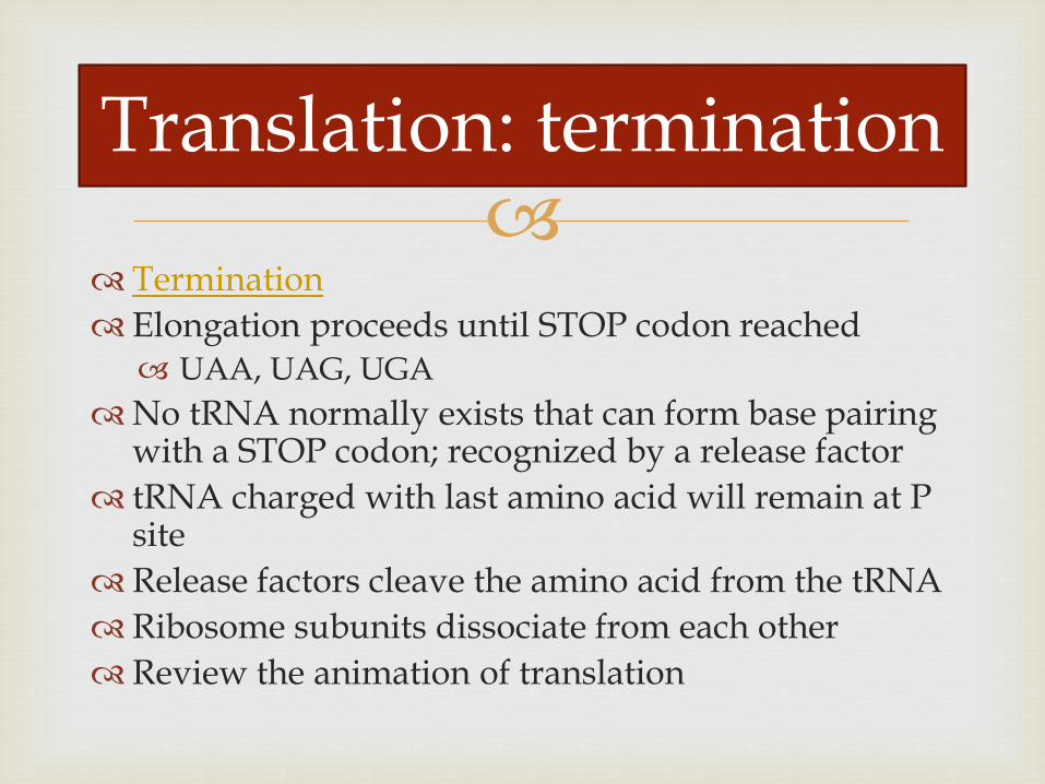
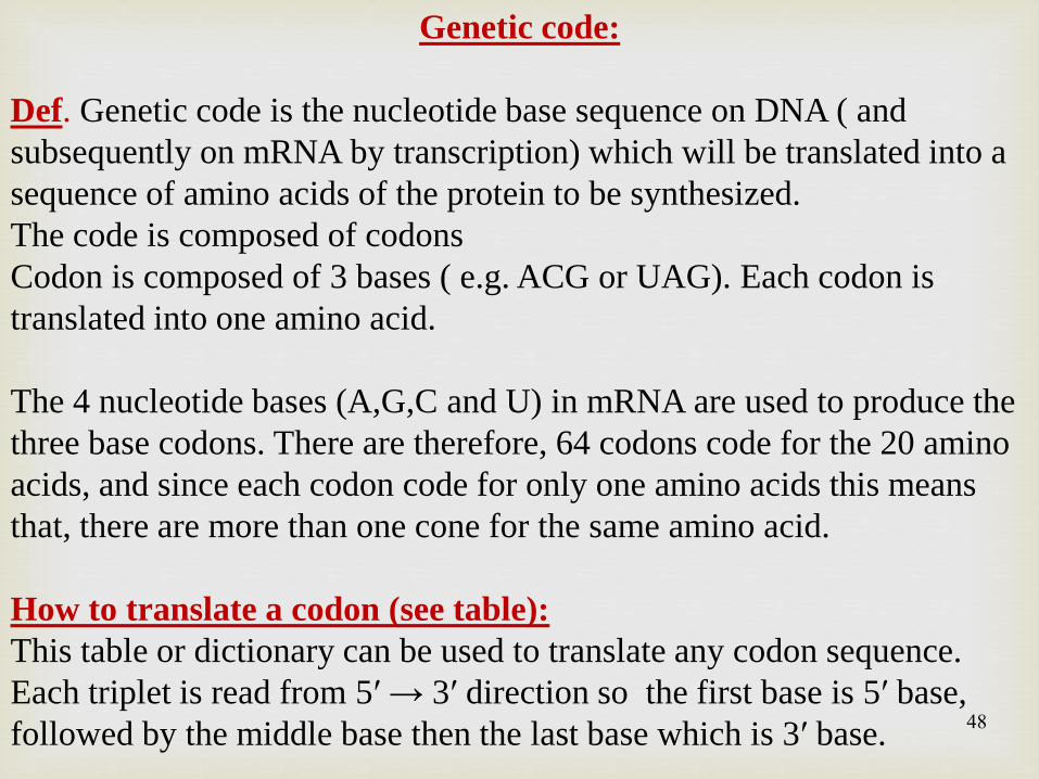
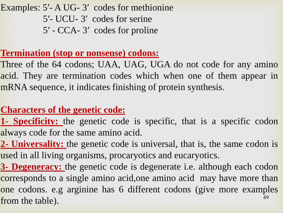
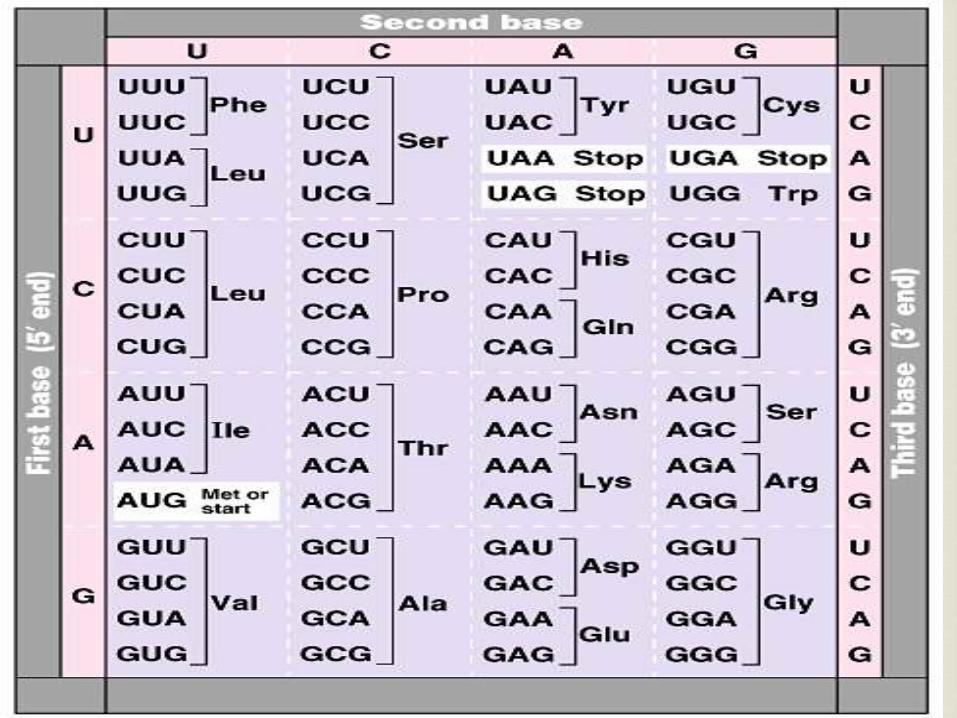
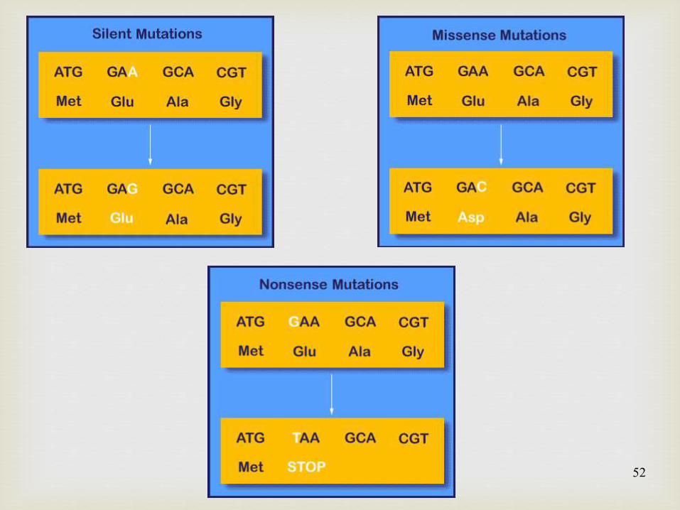
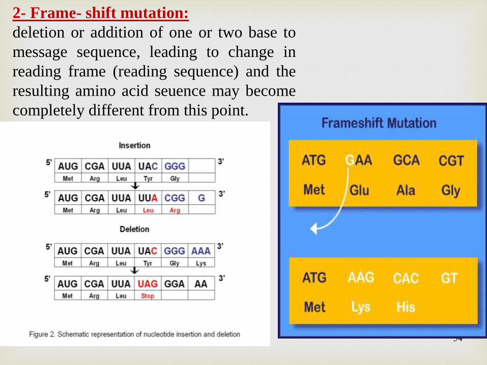