transfer of pathogens to and from patients, healthcare ... · ann arbor, michigan and 5division of...
TRANSCRIPT

Zurich Open Repository andArchiveUniversity of ZurichMain LibraryStrickhofstrasse 39CH-8057 Zurichwww.zora.uzh.ch
Year: 2018
Transfer of pathogens to and from patients, healthcare providers, andmedical devices during care activity-a systematic review and meta-analysis
Wolfensberger, Aline; Clack, Lauren; Kuster, Stefan P; Passerini, Simone; Mody, Lona; Chopra, Vineet;Msa, Jason Mann; Sax, Hugo
Abstract: OBJECTIVE The transfer of pathogens may spread antimicrobial resistance and lead tohealthcare-acquired infections. We performed a systematic literature review to generate estimates ofpathogen transfer in relation to healthcare provider (HCP) activities. METHODS For this systematicreview and meta-analysis, Medline/Ovid, EMBASE, and the Cochrane Library were searched for studiespublished before July 7, 2017. We reviewed the literature, examining transfer of pathogens associatedwith HCP activities. We included studies that (1) quantified transfer of pathogens from a defined originto a defined destination surface; (2) reported a microbiological sampling technique; and (3) describedthe associated activity leading to transfer. For studies reporting transfer frequencies, we extracted dataand calculated the estimated proportion using Freeman-Tukey double arcsine transformation and theDerSimonian-Laird random-effects model. RESULTS Of 13,121 identified articles, 32 were included.Most articles (n=27, 84%) examined transfer from patients and their environment to HCP hands, gloves,and gowns, with an estimated proportion for transfer frequency of 33% (95% confidence interval [CI],12%-57%), 30% (95% CI, 23%-38%) and 10% (95% CI, 6%-14%), respectively. Other articles addressedtransfer involving the hospital environment and medical devices. Risk factor analyses in 12 studiessuggested higher transfer frequencies after contact with moist body sites (n=7), longer duration of care(n=5), and care of patients with an invasive device (n=3). CONCLUSIONS Recognizing the heterogeneityin study designs, the available evidence suggests that pathogen transfer to HCPs occurs frequently. Moresystematic research is urgently warranted to support targeted and economic prevention policies andinterventions.
DOI: https://doi.org/10.1017/ice.2018.156
Posted at the Zurich Open Repository and Archive, University of ZurichZORA URL: https://doi.org/10.5167/uzh-153268Journal ArticlePublished Version
Originally published at:Wolfensberger, Aline; Clack, Lauren; Kuster, Stefan P; Passerini, Simone; Mody, Lona; Chopra, Vineet;Msa, Jason Mann; Sax, Hugo (2018). Transfer of pathogens to and from patients, healthcare providers,and medical devices during care activity-a systematic review and meta-analysis. Infection Control andHospital Epidemiology, 39(09):1093-1107.DOI: https://doi.org/10.1017/ice.2018.156

Infection Control & Hospital Epidemiology (2018), 0, 1–15
doi:10.1017/ice.2018.156
Review
Transfer of pathogens to and from patients, healthcare providers,and medical devices during care activity—a systematic review andmeta-analysis
Aline Wolfensberger MD1, Lauren Clack MSc1, Stefan P. Kuster MD1, Simone Passerini RN1, Lona Mody MD, MSc2,3,
Vineet Chopra MD, MSc4,5, Jason Mann MSA4 and Hugo Sax MD11Division of Infectious Diseases and Hospital Epidemiology, University Hospital Zurich, Zurich, Switzerland, 2Division of Geriatric and Palliative Medicine,Department of Internal Medicine, University of Michigan Medical School, Ann Arbor, Michigan, 3Geriatrics Research Education and Clinical Center, VeteransAffairs Ann Arbor Healthcare System, Ann Arbor, Michigan, 4Division of Hospital Medicine, Department of Medicine, University of Michigan Medical School,Ann Arbor, Michigan and 5Division of General Medicine, Department of Internal Medicine, Veterans Affairs Ann Arbor Healthcare System, Ann Arbor, Michigan
Abstract
Objective: The transfer of pathogens may spread antimicrobial resistance and lead to healthcare-acquired infections. We performed asystematic literature review to generate estimates of pathogen transfer in relation to healthcare provider (HCP) activities.Methods: For this systematic review and meta-analysis, Medline/Ovid, EMBASE, and the Cochrane Library were searched for studiespublished before July 7, 2017. We reviewed the literature, examining transfer of pathogens associated with HCP activities. We includedstudies that (1) quantified transfer of pathogens from a defined origin to a defined destination surface; (2) reported a microbiologicalsampling technique; and (3) described the associated activity leading to transfer. For studies reporting transfer frequencies, we extracteddata and calculated the estimated proportion using Freeman-Tukey double arcsine transformation and the DerSimonian-Laird random-effects model.Results: Of 13,121 identified articles, 32 were included. Most articles (n= 27, 84%) examined transfer from patients and their environmentto HCP hands, gloves, and gowns, with an estimated proportion for transfer frequency of 33% (95% confidence interval [CI], 12%–57%),30% (95% CI, 23%–38%) and 10% (95% CI, 6%–14%), respectively. Other articles addressed transfer involving the hospital environmentand medical devices. Risk factor analyses in 12 studies suggested higher transfer frequencies after contact with moist body sites (n= 7),longer duration of care (n= 5), and care of patients with an invasive device (n= 3).Conclusions: Recognizing the heterogeneity in study designs, the available evidence suggests that pathogen transfer to HCPs occursfrequently. More systematic research is urgently warranted to support targeted and economic prevention policies and interventions.
(Received 28 March 2018; accepted 8 June 2018)
Transmission of pathogens to and from patients is associated withthe spread of antimicrobial resistance and healthcare-associatedinfections. Transmission usually occurs indirectly via healthcareproviders (HCPs) or via mobile or immobile fomites.1 Hands areuniversally recognized as the most important vector,2 but equip-ment (eg, stethoscopes) may also act as a vector for pathogentransfer.3 For successful transfer, pathogens must first be trans-mitted from a surface of origin (eg, a patient) to an intermediatesurface (ie, a vector) and must then survive there long enough tobe finally transmitted to the next patient or to the next inter-mediate vector.4 The likelihood of transfer depends on multiplefactors, including number and type of pathogens present, surfacestructure,5 contact time, lag time,6 humidity,7 and pressure.8
A patient encounters many different HCPs during a stay in ahealthcare institution. Each patient is visited by a median of 3.5HCPs per hour, mostly nurses, for a median of 3 minutes pervisit. The patient environment, intact skin, and body fluids aretouched in 33%, 27%, and 18% of visits, respectively.9 If infectionprevention measures are not followed, each contact poses a risk ofpathogen transfer.
Prevention policies such as standard and isolation precautions,particularly hand hygiene and the use of gloves and gowns, aim toprevent pathogen transmission.10 Detailed understanding oftransmission dynamics during patient care, however, is pivotal totailoring preventive measures and policies. To date, no systematicreview has addressed the extent to which HCP behavior results intransfer of pathogens.
Therefore, we performed a systematic review and meta-analysis(1) to summarize and describe the current knowledge on pathogentransfer associated with care activity, including the frequency andquantity of pathogen transfer between patients, environmentalsurfaces, HCPs, their clothing, and medical devices, and (2) toidentify the factors associated with increased transmission risk.
Cite this article: Wolfensberger A, et al. (2018). Transfer of pathogens to and frompatients, healthcare providers, and medical devices during care activity—a systematicreview and meta-analysis. Infection Control & Hospital Epidemiology. 2018: 1–15. doi:10.1017/ice.2018.156
Author for correspondence: Aline Wolfensberger MD, Division of Infectious Dis-eases and Hospital Epidemiology, University Hospital Zurich, University of Zurich,Rämistrasse 100, CH-8091 Zurich, Switzerland. E-mail: [email protected]
© 2018 by The Society for Healthcare Epidemiology of America. All rights reserved.
Methods
Data sources and searches
We followed the Preferred Reporting Items for SystematicReviews and Meta-Analysis (PRISMA) recommendations whenconducting this systematic review.11 We searched Medline/Ovid,EMBASE and the Cochrane Library using a combination ofsubject headings and free text search terms (see Appendix: SearchStrategy). Studies published before July 7, 2017 in English, French,and German were included. Additional articles were identified bya reference list search of articles included in full text review.
Study selection
We retained studies that met the following criteria:
1. The study occurred in a defined healthcare setting (eg,inpatient and outpatient settings of hospitals, nursing homes,and medical practices) and included common behavior inhealthcare institutions (eg, handshakes, physical examinations,and phone calls).
2. The study reported transfer of bacteria, viruses, or fungi bycontact from a defined origin surface to a defined destinationsurface through quantification of contamination by frequency(ie, percentage of contaminated destination surfaces) or byquantity (ie, colony forming units (CFU) on destinationsurface).
3. The HCP activity associated with pathogen transfer wasdescribed.
4. Transfer to destination surface was ascertained by genomic orpulsed-field gel electrophoresis typing of the pathogen orascertaining a sterile destination surface.
5. Origin and destination surfaces were either the patient’s bodysites; surfaces in the patient’s immediate environment; critical,semicritical, or noncritical medical devices; or HCP body sites,attire, or personal protective equipment.
6. The microbiological sampling method (eg, contact plate, swab,or glove juice method12) was accurately described.
We excluded studies reporting laboratory simulations or usingartificial contamination or tracers to evaluate transfer. We alsoexcluded investigations of transfer by droplet or airborne routeand pathogen transfer during surgical procedures.
One researcher (A.W.) screened all titles and abstracts forpotentially relevant studies. To check interrater reliability, 2independent researchers (L.C. and S.P.) screened a subset of titlesand abstracts, then 2 authors (A.W. and L.C.) independentlyreviewed the full texts of all potentially eligible articles. Dis-crepancies regarding eligibility were resolved by consensus or bydecision of a third reviewer (H.S.).
Data extraction and quality assessment
Using a standardized template, the following variables wereabstracted: study setting, microorganism transferred, ‘origin’surface (surface from which pathogen was transferred), ‘destina-tion’ surface (surface to which pathogen was transferred), inter-action (activity) leading to transfer, number of interactions,frequency of transfer (percentage of positive destination surfaces),quantity of transfer (number of transferred CFU), and micro-biological sampling method. All retained articles were assessedfor quality using the Downs and Black checklist,13 which was
modified to fit the noninterventional characteristics of includedstudies (Appendix Table 1).
Data synthesis and analyses
For studies reporting raw transfer frequency data, rates werecalculated by dividing the number of positive destinations by thetotal number of exposed destination surfaces. The Freeman-Tukey double arcsine transformation for data with a binomialdistribution was applied to stabilize the variances. Transformedproportions were pooled using random-effects meta-analysis withexact confidence intervals, and results were displayed in forestplots. All meta-analyses were conducted using the metapropcommand in Stata software. Also, 2 studies that did not reportraw rates were excluded from meta-analysis but were retained inthe systematic review.14,15 An a priori subgroup analysis wasperformed for studies examining pathogenic bacteria examiningdestination surface, surface of origin, HCP behavior (standardizedvs nonstandardized), microorganism, and microbiological sam-pling method. Studies that reported transfer quantities and riskfactor analysis were not pooled, and the findings are summarizeddescriptively.
Data synthesis was done using Stata version 13.1 software (Sta-taCorp, College Station, TX). Statistical heterogeneity was initiallyinspected graphically (forest plot); the degree of heterogeneity wasquantified using the I2 statistic. We defined heterogeneity asI2> 60%. Subgroup analyses for differences between subgroupswere foreseen if the criterion for heterogeneity was notmet.P values< .05 were considered statistically significant. This review wasperformed in accordance with the Preferred Reporting Items forSystematic Reviews and Meta-Analyses (PRISMA) guidelines.16
Results
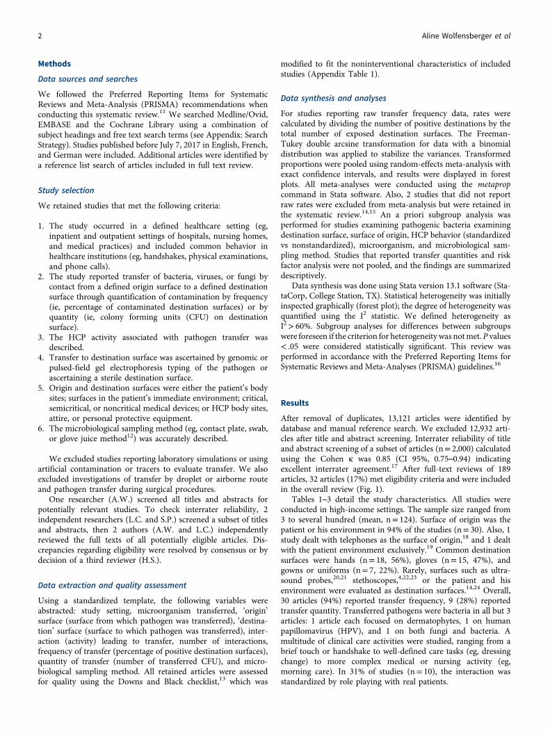
After removal of duplicates, 13,121 articles were identified bydatabase and manual reference search. We excluded 12,932 arti-cles after title and abstract screening. Interrater reliability of titleand abstract screening of a subset of articles (n= 2,000) calculatedusing the Cohen κ was 0.85 (CI 95%, 0.75–0.94) indicatingexcellent interrater agreement.17 After full-text reviews of 189articles, 32 articles (17%) met eligibility criteria and were includedin the overall review (Fig. 1).
Tables 1–3 detail the study characteristics. All studies wereconducted in high-income settings. The sample size ranged from3 to several hundred (mean, n= 124). Surface of origin was thepatient or his environment in 94% of the studies (n= 30). Also, 1study dealt with telephones as the surface of origin,18 and 1 dealtwith the patient environment exclusively.19 Common destinationsurfaces were hands (n= 18, 56%), gloves (n= 15, 47%), andgowns or uniforms (n= 7, 22%). Rarely, surfaces such as ultra-sound probes,20,21 stethoscopes,4,22,23 or the patient and hisenvironment were evaluated as destination surfaces.14,24 Overall,30 articles (94%) reported transfer frequency, 9 (28%) reportedtransfer quantity. Transferred pathogens were bacteria in all but 3articles: 1 article each focused on dermatophytes, 1 on humanpapillomavirus (HPV), and 1 on both fungi and bacteria. Amultitude of clinical care activities were studied, ranging from abrief touch or handshake to well-defined care tasks (eg, dressingchange) to more complex medical or nursing activity (eg,morning care). In 31% of studies (n= 10), the interaction wasstandardized by role playing with real patients.
2 Aline Wolfensberger et al
Various microbiological sampling techniques were used.Swabbing was applied in 12 studies (38%), contact plates in 14studies (44%), the glove juice method in 4 studies (13%),12 andwashing hands or gloves in broth in 3 studies (9%). Polymerasechain reaction (PCR) was used to detect viral transfer in 1 study.
Transfer frequency
Most studies examined transfer frequency during non-standardized tasks, that is, in real-life situations (n= 23, 76%)(Table 1). These interactions lasted up to 4 hours. In 8 studies, thetransfer frequency after standardized interactions was examined.In 6 of these studies, interactions were simple, such as a hand-shake, briefly touching a patient, or taking a phone call. In 2studies, the transfer frequency after multistep interactions, such asan outpatient consultation or a radiology procedure, was exam-ined. Hand and glove contamination ranged from 0% withvancomycin-resistant enterococci (VRE) after a standardizedmock radiology procedure25 to 100% with HPV after treatment ofurethral warts.26 Gown contamination was between 2% and 37%for VRE, methicillin-resistant Staphylococcus aureus (MRSA),multidrug-resistant (MDR) Pseudomonas aeruginosa and Acine-tobacter baumanii,22,25,27–30 and 100% for staphylococci.31
Ultrasound probes were contaminated to 100% with skin bac-teria,20,21 transfer of undifferentiated bacteria to IV stopcocks andsyringes occurred in 5%–26% of anesthesia procedures.32,33
The pooled proportions of transfer frequency of bacteria tohands and gloves were 33% (95% CI, 12%–57%; 9 studies) and30% (95% CI, 23%–38%; 21 studies) with overall heterogeneitiesof I2= 90.95% and 92.03%, respectively (Appendix Fig. 1). Theestimated proportions of transfer frequency to gowns and to‘hands after glove removal’ were 10% (95% CI, 6%–14%; 13studies) and 3% (95% CI, 1%–5%; 9 studies), respectively(Appendix Figs. 2 and 3). These results remained unchangedwhen performing a sensitivity analysis including only studies witha quality score of 100% (Appendix Figs. 4–6). Estimated transfer
frequencies stratified according to type of behavior (ie, standar-dized vs nonstandardized), surface of origin, microorganism, andmicrobiological sampling method are displayed in Appendix Figs.7–10.
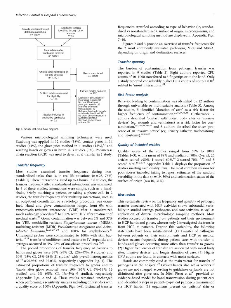
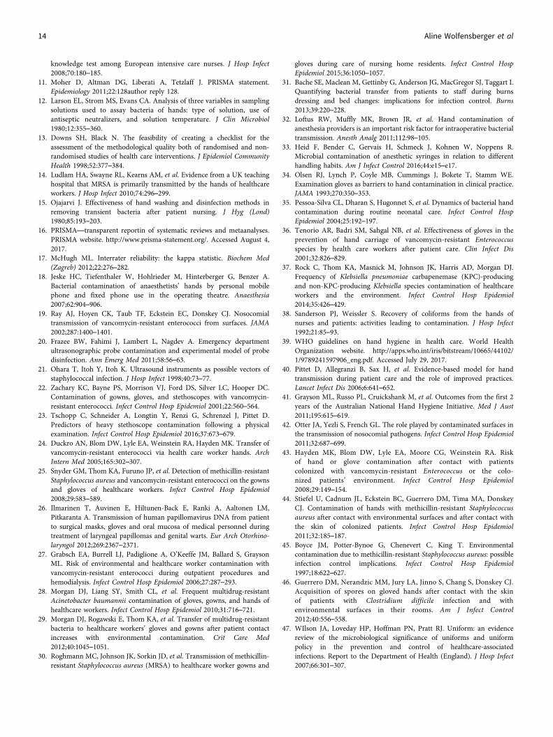
Figures 2 and 3 provide an overview of transfer frequency forthe 2 most commonly evaluated pathogens, VRE and MRSA,depending on origin and destination surfaces.
Transfer quantity
The burden of contamination from pathogen transfer wasreported in 9 studies (Table 2). Eight authors reported CFUcounts of 10–1000 transferred to 5 fingertips or to the hand. Only1 study reported considerably higher CFU counts of up to 2 × 10⁶
related to ‘moist interactions.’34
Risk factor analysis
Behavior leading to contamination was identified by 12 authorsthrough univariable or multivariable analysis (Table 3). Amongthe studies, 5 identified ‘duration of care’ as a risk factor forhigher frequency of contamination.2,28,29,35,36 Furthermore, 7authors described ‘contact with moist body sites or invasivedevices’ (eg, wounds and ventilators) as a risk factor for con-tamination,25,28–30,35–37 and 3 authors described the sheer ‘pre-sence of an invasive device’ (eg, urinary catheter, tracheostomy,and ileostomy).22,25,37
Quality of included articles
Quality scores of the studies ranged from 60% to 100%(Tables 1–3), with a mean of 88% and median of 90%. Overall, 26articles scored >80%, 1 scored 60%,14 2 scored 70%,15,34 and 3scored 80%.18,32,38 Appendix Table 1 displays the proportion ofstudies meeting each quality item. The most common reasons forpoor scores included failing to report estimates of the randomvariability in the data (n= 19, 59%) and colonization status of thesurface of origin (n= 10, 31%).
Discussion
This systematic review on the frequency and quantity of pathogentransfer associated with HCP activities shows substantial varia-bility in studied settings, pathogens, and care activities, as well asapplication of diverse microbiologic sampling methods. Moststudies focused on transfer from patients and their environmentto HCP hands and gloves, whereas only 2 investigated the transferfrom HCP to patients. Despite this variability, the followingstatements have been substantiated: (1) Transfer of pathogensbetween patients or their environments and HCP or medicaldevices occurs frequently during patient care, with transfer tohands and gloves occurring more often than transfer to gowns.(2) Higher frequencies of transfer are associated with moist bodysites, invasive devices, and longer duration of care. (3) HigherCFU counts are found in contacts with moist surfaces.
Hands are commonly cited as the main vector for transfer ofpathogens in the hospital.39 Gloved hands also act as vectors ifgloves are not changed according to guidelines or hands are notdisinfected after glove use. In 2006, Pittet et al40 provided anevidence-based model for hand transmission during patient careand identified 5 steps in patient-to-patient pathogen transmissionvia HCP hands: (1) organisms present on patients’ skin or
Fig. 1. Study inclusion flow diagram.
Infection Control & Hospital Epidemiology 3

Table 1. Summary and Quality Assessment for Studies Reporting Frequency of Contamination
Author (YearofPublication)QualityScore Microorganism Study Setting
Standardized vsNonstandardizedInteractionsa Interactions (Behavior) Origin Surfaceb Destination Surfacec Sample Size
Frequency ofContamination After
Interaction
MicrobiologicalSamplingMethod
Bache et al(2013) 31
Score:91%
Staphylococci Burns ICU(UK)
Nonstandardized Dressing change with or withoutmaking bed
Patients and theirenvironment
(colonization statusunknown)
Gowns 24 100% Contact plate
Boyce et al(1997) 45
Score:90%
MRSA Hospital(USA)
Nonstandardized 1) Morning care2) Nursing activities without patient
contact
Patients with MRSA and/ortheir environment
Gloves 1) 122) 12
1) 58%2) 42%
Swab on agarand swab inbroth
Casewellet al(1977) 50
Score: 80%
Klebsiella ICU(UK)
Nonstandardized Broad spectrum of patient activities(from touching hand toextubation)
Patients with Klebsiella andtheir environment
Hands 47 36% Glove juice
Duckro et al(2005)24
Score: 90%
VRE ICU(USA)
Nonstandardized Routine patient care Patients with VRE and theirenvironment
VRE-negativesurfaces ofpatients with VREand theirenvironment
27 11% (2 steps transfer - viahands or gloves)
Swab in broth
Frazee et al(2011)20
Score:90%
1) Undifferentiatedbacteria
2) Clinically relevantpathogens
Emergencydepartment
(USA)
Nonstandardized Performing ultrasonography ofinfection body site
Patients with skin and softtissue infection(colonization statusunknown)
Ultrasound probe 20 1) 100%2) 70% (MRSA, MSSA, or
Streptococci)
Contact plateand swab inbroth
Grabschet al(2006)27
Score: 90%
VRE Dialysis ward(Australia)
Nonstandardized Hemodialysis session (4 hours) Patients with VRE and theirenvironment
GownsGlovesHands
26 Gowns: 30%Gloves: 8%Hands: 8%
Contact plateGlove juice
Grabschet al(2006)27
Score: 90%
VRE 1) Outpatientconsultation
2) Radiologyrooms(Australia)
Standardized 1) Standardized mock outpatientconsultation (25 min)
2) Standardized mock radiologyprocedure (15 min)
Patients with VRE and theirenvironment
GownsGlovesHands
1) 252) 24
Gowns: 1) 20%, 2) 4%Gloves: 1) 16%
Hands: 1) 4%, 2) 0%
Contact plateor
Glove juice
Grunwaldet al(2015) 51
Score: 90%
Dermatophytes NA(Israel)
Standardized Brief touch Children with tinea Hands 90 80% Contact plate
Guerreroet al(2012)46
Score: 100%
C. difficile NA(USA)
Standardized Touch1) Commonly examined skin sites
(arm, abdomen, chest, and hand)2) Environment (table, phone,
bedrail, and call button)
Patients with Clostridiumdifficile or theirenvironment
Moistened gloves 30 1) 50%2) 50%
Contact plate
Hamburgeret al(1947) 52
Score: 90%
Beta-hemolyticstreptococci
NA(USA)
Standardized Handshake Sailors with beta-hemolyticstreptococci
1) Hands after noseblowing
2) Hands without blowingnose
Hands 1) 172) 9
1) 76%2) 66%
“Wash” handsin broth
Haydenet al
(2008) 43
Score: 100%
VRE ICU(USA)
Nonstandardized 1) Patient care with touching patientand environment
2) Patient care with touchingenvironment only
Patients with VRE and/ortheir environment
HandsGloves
1) 59 (4 hands,55 gloves)
2) 44 (15 hands,29 gloves)
1) Hands: 75%Gloves: 69%
2) Hands: 27%Gloves: 66%
Glove juice
4Aline
Wolfensberger
etal

Heid et al(2016) 53
Score: 82%
Undifferentiatedbacteria
Operatingroom andpostanestheticcare unit
(Germany)
Nonstandardized Preparation of syringe, andanesthetic management of singlepatient with either:
1) Syringes capped with new syringecap from a sterile wrapping
2) Syringes placed in the syringe setwithout being capped or covered
3) Syringes put back in the originalsterile wrapping
Patients and theirenvironment(colonization statusunknown)
Syringe hub, syringecontent, and firstdrop from syringe
1) 1022) 1163) 101
1) 27%2) 23%3) 14%
Swab, drop onagar, andbroth insyringe
Ilmarinenet al(2012) 26
Score: 90%
HPV Outpatientclinic (Finland)
Nonstandardized Topical application of lidocaine andprilocaine cream and CO2 lasertreatment of urethral warts
Patients with HPV Gloves 5 100% PCR
Jeskeet al(2007) 18
Score: 80%
Undifferentiatedbacteria
Operatingrooms(Austria)
Standardized 1-minute phone call Hospital environment1) Personal cell phone2) Fixed wall phone
Hands 1) 402) 40
1) 95% (10% humanpathogens)
2) 83% (10% humanpathogens)
Contact plate
Loftus et al(2011) 32
Score: 80%
Undifferentiatedbacteria
Operating room(USA)
Nonstandardized General anesthesia 1) HCP, patients, and theirenvironment
2) HCP
a) Valve and agentdial on theanesthesia machineb) IV stopcock set
164 1a) 89%1b) 10%2a) 12%2b) 5%
Swab on plate
Ludlamet al
(2010) 14
Score: 60%
MRSA Wards and ICUs(UK)
Nonstandardized Routine patient care Patients with MRSA andtheir environment
MRSA-negativesurfaces of patientswith MRSA and theirenvironment
24 auditedepisodes
In 38% of auditedepisodes at least 1
transfer occurred (2-steptransfer via hands)
Swabs, culturemediumunknown
Morganet al(2012)29
Score: 100%
MRSAVREMDR AcinetobacterbaumaniiMDR Pseudomonasaeruginosa
ICU(USA)
Nonstandardized Routine, nonemergentpatient care
Patients with MRSA, VRE,MDR A. baumanii orMDR P. aeruginosa andtheir environment
GlovesGownsHands after gloveremoval
MRSA: 152VRE: 180
MDR PA: 86MDR AB: 167
MRSA:Gloves: 11%Gown: 4%
Gloves or gown: 14%Hands after glove
removal: 3%VRE
Gloves: 10%Gown: 5%
Gloves or gown: 14%Hands after glove
removal: 2%MDR P. aeruginosa:
Gloves: 17%Gown: 2%
Gloves or gown: 17%Hands after glove
removal: 4%MDR A. baumanii
Gloves: 39%Gown: 13%
Gloves or gown: 33%Hands after glove
removal: 4%
Swab in broth
Morganet al(2010)28
Score: 100%
MDR A. baumanii MDRP.s aeruginosa
ICU(USA)
Nonstandardized Routine, Nonemergentpatient care
Patient with MDR P.aeruginosa and/or MDRA. baumanii and theirenvironment
GlovesGownsHands after gloveremoval
MDR AB: 199MDR PA and AB:
134
MDR A. baumanii:Gloves: 36%Gown: 11%
Gloves or gown: 39%Hands after glove
removal: 5%MDR P. aeruginosa:
Gloves: 7%Gown: 5%
Gloves or gown: 8%Hands after glove
removal: 1%
Swab in broth
InfectionControl
&Hospital
Epidemiology
5

Table 1. (Continued )
Author (YearofPublication)QualityScore Microorganism Study Setting
Standardized vsNonstandardizedInteractionsa Interactions (Behavior) Origin Surfaceb Destination Surfacec Sample Size
Frequency ofContamination After
Interaction
MicrobiologicalSamplingMethod
Ohara et al(1998)21
Score: 90%
Staphylococci NA(Japan)
Nonstandardized Performing ultrasonography Patients with MRSA Ultrasound probe 3 100% Swab on plate
Ojajarvi et al(1980)15
Score: 70%
a) S. aureusb) Gram-negatives
Hospital, wardtype notspecified
(Finland)
Nonstandardized 1) Making patients bed (= drycontamination)
2) Changing dressings andcompresses (=wetcontamination)
Patients with S. aureus orGram-negatives andtheir environment
Hands 1) Drycontamination:
2852) Moist
contamination:313
1a) > 90%1b) “Small percentage”
2a) >90%2b) 50%
Contact plate
Olsen et al(1993)34
Score: 70%
Gram-negative rodsorEnterococci
Hospital(ward typenotspecified)
(USA)
Nonstandardized 1) Oral and endotracheal tube carefor intensive care patients
2) Patient care involving digitalstimulation of the rectal sphincter
3) Routine dental examinations
Patients(colonization statusunknown)
GlovesHands after gloveremoval
1) 982) 213) 18
Gloves: 64% (pooled forall behaviors)
Hands after removal ofgloves: 8% (pooled for all
behaviors)
Glove juice
Pessoa-Silvaet al
(2004)35
Score: 91%
Undifferentiatedbacteria and fungiSkin floraEnterobacteriaceaeS. aureusFilamentous fungi
Neonatalnursery
(Switzerland)
Nonstandardized Neonatal patient care Neonate patients andenvironment(colonization statusunknown)
HandsHands after gloveremoval
Hands: 398Hands after
glove removal:31
Hands:Any bacteria or fungi:
91%Skin flora: 66%
Enterobacteriaceae: 14%S. aureus: 3%
Filamentous fungi: 2%Hands after glove
removal:Any bacteria or fungi:
31%Skin flora: 84%
Enterobacteriaceae: 13%S. aureus: 3%
Contact plate
Pittet et al(1999)2
Score: 91%
UndifferentiatedbacteriaS. aureusGram-negativebacilli
8 differentwards
(Switzerland)
Nonstandardized Routine patient care Patients and theirenvironment(colonization statusunknown)
Hands 417 Any bacteria: 89%S. aureus: 9%
Gram-negative bacilli:13%
Contact plate
Ray et al(2002)19
Score: 90%
VRE Nursing homeand hospital
(USA)
Standardized Touch bedrail and bedside table for5 seconds each
Environment of patientswith VRE
Gloves 13 46% “Wash” glovesin broth
Rock et al(2014)37
Score: 100%
Klebsiella KPCKlebsiella non-KPC
ICU(USA)
Nonstandardized Routine patient care Patients with Klebsiella andtheir environment
Gowns or gloves Klebsiella KPC:96
Klebsiella non-KPC: 124
Klebsiella KPC: 10%Klebsiella non-KPC: 17%
Swab in broth
Roghmannet al
(2015)30
Score: 91%
MRSA Nursing homes(USA)
Nonstandardized Routine patient care Patients with MRSA andtheir environment
GownsGloves
954 Gowns: 14%Gloves: 24%
Swabs on agarand in broth
6Aline
Wolfensberger
etal

Sandersonet al
(1992)38
Score: 80%
Coliforms OrthopedicwardSpinal injuryward
(UK)
Nonstandardized Routine patient care1) Overall2) Bed making3) Touching patients or patient’sclothing4) Handling patient’s wash cloth,towels or wash bowls5) Handling used linen6) Handling clean linen7) Sluice room, urinary catheters orbags8) Handling curtains or bedsidefurniture9) Doing drug round
Patients or theirenvironment(colonization statusunknown)
Hands 452 Orthopedic ward/spinalinjury ward1) 20%/36%2) 13%/20%3) 32%/32%4) 57%/62%5) 6%/22%6) 0%/29%7) 15%/26%8) 23%/12%9) 13%/27%
Contact plate
Snyder et al(2008)25
Score: 91%
MRSAVRE
ICU(USA)
Nonstandardized Routine, nonemergent patient care Patients with MRSA and/orVRE and theirenvironment
GownsGlovesHands after gloveremoval
137 MRSA:Gloves: 18%Gowns: 6%
Gloves or gowns: 19%Hands after glove
removal: 3VRE:
Gloves: 8%Gowns: 4%
Glove or gown: 9%Hands after glove
removal: 0%
Swab on agarand swab inbroth
Stiefel et al(2011)44
Score: 100%
MRSA Hospital, wardtype notspecified
(USA)
Standardized Imprint gloved hand on1) Patients skin (abdomen, chest,hand, and arm)
2) Patients environment (call button,bed rail, table, and phone)
Patients with MRSA or theirenvironment
Gloves 40 1) 40%2) 45%
Contact plate
Tenorio et al(2001)36
Score: 91%
VRE Hospital, wardtype notspecified
(USA)
Nonstandardized Routine patient care Patients with VRE and theirenvironment
GlovesHands after gloveremoval
44 Gloves: 39%Hands after gloveremoval: 14%
“Wash“ handsor gloves inbroth
Zacharyet al
(2001)22
Score: 91%
VRE Hospital, wardtype notspecified
(USA)
Standardized Structured physical examination(auscultation of heart and lungs,palpation of back, abdomen, andlower extremities)
Patients with VRE and theirenvironment
HandsGownsStethoscopediaphragms
49 Glove, gown, orstethoscope: 67%
(Gloves: 63%; gowns:37%; stethoscopediaphragms: 31%)
Contact plate
Note. HCP, healthcare practitioner; HPV, human papilloma virus; ICU, intensive care unit; KPC, Klebsiella pneumonia carbapenemase; NA, not applicable; MDR, multidrug resistant; MSSA, methicillin-sensitive Staphylococcus aureus; MRSA, methicillin-resistant Staphylococcus aureus; UK, United Kingdom; USA, United States of America; VRE, vancomycin-resistant Enterococci.aIn standardized interactions, the interaction was standardized by role playing; in nonstandardized interactions, the interactions were observed in real life.bSurface from which pathogen was transferred.cSurface to which pathogen was transferred.
InfectionControl
&Hospital
Epidemiology
7

Table 2. Summary and Quality Assessment for Studies Reporting Quantity of Contamination
Author(Year ofPublication)QualityScore Microorganism Study Setting
Standardized vsNonstandardizedInteractionsa Standardized Interactions Origin Surfaceb Destination Surfacec
SampleSize No. of CFU transferred
MicrobiologicalSamplingMethod
Guerreroet al(2012)46
Score:100%
C. difficile NA(USA)
Standardized Touch1) Commonly examined skin
sites (arm, abdomen, chest,and hand)
2) Environment (table, phone,bedrail, and call button)
Patients with C. difficile andtheir environment
Moistened gloves 30 CFU on handprint (mean):1) 14 (highest CFU count after
touching Abdomen: 29)2) 7 (highest CFU count after touching
bedrail: 8)
Contact plate
Hamburgeret al(1947)52
Score: 90%
Beta-hemolyticstreptococci
NA(USA)
Standardized Handshake Sailors with beta-hemolyticstreptococci
1) Hands after nose blowing2) Hands without blowing
nose
Hands 1) 172) 9
CFU on hand (mean)1) 44502) 564
“Broth wash”
Jeske et al(2007)18
Score 80%
Undifferentiatedbacteria
Operatingrooms
(Austria)
Standardized One-minute phone call “Hospital environment”1) Cell phone2) Fixed wall phone
Hands 1) 402) 40
CFU on 5 fingertips (median)1) 142) 22
Contact plate
Longtinet al(2014)4
Score: 100%
1) Aerobicbacteria
2) MRSA
Ward(Switzerland)
Standardized Standardized physicalexamination
Patients colonised orinfected with MRSA andtheir environment
Stethoscopegloves
Hands
1) 332) 50
CFU per 25cm2 (median)1)Fingertips: 467Stethoscope diaphragm: 89Thenar/hypothenar: 37Stethoscope tube: 18Dorsum hand: 82)Fingertips: 12Stethoscope diaphragm: 7Thenar: 7Hypothenar: 2Stethoscope tube: 0Dorsum hand: 0
Contact plate
Olsen et al(1993)34
Score: 70%
Gram-negativerods orEnterococci
hospital, wardtype notspecified
(USA)
Nonstandardized “Moist” interactions1) Oral and endotracheal tube
care for intensive carepatients
2) Patient care involvingdigital stimulation of therectal sphincter
3) routine dentalexaminations
Patients and theirenvironment
(colonization statusunknown)
GlovesHands after glove
removal
1) 982) 213) 18
CFU on glove or on hand after gloveremoval:
Glove: 650–2,000,000Hand after glove removal: 10–100
Glove juice
Pessoa-Silvaet al(2004)35
Score: 91%
Undifferentiatedbacteria andfungi
Neonatalnursery
(Switzerland)
Nonstandardized Neonatal patient care Neonate patients andenvironment
(colonization statusunknown)
Hands 149 Increase in CFU on 5 fingertips perminute:
Total: 20skin contact: 21Diaper change: 42Respiratory tract care: 38Contact with body secretions other than
respiratory: 20Manipulation of vascular access
Contact plate
8Aline
Wolfensberger
etal

devices: 10Contact with equipment: 9
Pittet et al(1999)2
Score: 91%
Undifferentiatedbacteria
8 differentwards
(Switzerland)
Nonstandardized Patient care Patients and theirenvironment
(colonization statusunknown)
Hands 417 CFU on 5 fingertips:100 (mean), 39 (median)Increase in CFU on 5 fingertips per
minute:Overall: 16Direct patient contact: 20Rupture in the sequence of care: 19Respiratory care: 21Handling body fluid Secretions: 16Blood sampling and intravenous
injection of care: 6Skin contact: 4
Contact plate
Stiefel et al(2011) 44
Score: 100%
MRSA Hospital, wardtype notspecified
(USA)
Standardized Imprint gloved hand on1) Patients skin (abdomen,
chest, hand, and arm)2) Patients environment (call
button, bed rail, table, andphone)
Patients with MRSA and theirenvironment
Gloves 40 CFU on hand imprint (mean)1) Touching patients skin (any): 9
(highest CFU count after touchingAbdomen: 17)
2) Touching patients environment (any):4 (highest CFU count after touchingcall button: 6)
Contact plate
Tschoppet al(2016) 23
Score: 91%
Undifferentiatedbacteria
Normal wards(Switzerland)
Standardized Standardized physicalexamination
Patient and or hisenvironment
(colonization statusunknown)
Hand (fingertips,dorsum, thenar andhypothenar)Stethoscope (tubeand diaphragm)
56 CFU per 25 cm2 (median)ngertips: 834Stethoscope diaphragm: 172Stethoscope tube: 116Thenar: 14Hypothenar: 16Dorsum hand: 2
Contact plate
Note. CFU, colony-forming unit; MRSA, methicillin-resistant Staphylococcus aureus; NA, not applicable; UK, United Kingdom; USA, United States of America.aIn standardized interactions, the interaction was standardized by role playing; in nonstandardized interactions, the interactions were observed in real life.bSurface from which pathogen was transferred.cSurface to which pathogen was transferred.
InfectionControl
&Hospital
Epidemiology
9

Table 3. Summary and Quality Assessment for Studies Reporting Risk Factor Analysis
Author(Year ofPublication)QualityScore Microorganism
StudySetting Interactions Origin Surfacea
DestinationSurfaceb Risk Factors for Transfer
Bache et al(2013)31
Score: 91%
Staphylococci Burns ICU(UK)
Dressing change with or withoutbed making
Patients and theirenvironment(colonization statusunknown)
Gown For every 9% increase in total bodysurface area of burn: CFU/plate double
Haydenet al(2008)43
Score: 100%
VRE ICU(USA)
Patient care1) Touching patient andenvironment2) Touching only environment
Patients with VRE andtheir environment
Hands orgloves
Positive predictors for contamination(univariable analysis):
- No. of contacts made (each contactresults in 10% risk of handcontamination)Independent positive predictors forcontamination:
- None
Morganet al(2012)29
Score: 100%
MRSA, VRE, MDRAcinetobacterbaumanii, MDRP. aeruginosa
ICU(USA)
Patient care Patient with MRSA, VRE,MDR. A. baumanii, orMDR P. aeruginosa andtheir environment
GlovesGownsHands after
gloveremoval
Independent positive predictors forcontamination:
- Positive environmental culture (OR, 4.15)- Time in room of more than 5 minutes- Performing a physical examination (OR,
1.74)- Contact with the ventilator (OR, 1.78)
Morganet al(2010)28
Score: 100%
MDRAcinetobacterbaumanii MDRPseudmonoasaeruginosa
ICU(USA)
Patient care Patient with MDR P.aeruginosa and/orMDR. A. baumanii andtheir environment
GlovesGownsHands after
gloveremoval
Independent positive predictors forcontamination:
- Presence of a wound dressing (OR, 25.9)- Use of endotracheal tube or
tracheostomy site (OR, 2.1)- Time in room of more than 5 minutes
(OR, 4.3)- Clinical role of physician or nurse
practitioner (OR, 7.4) or nurse (OR, 2.3)compared to clinical role of therapists
Pessoa-Silvaet al(2004)35
Score: 91%
Undifferentiatedbacteria andfungi
Neonatalnursery
(Switzerland)
Routine neonatal care Neonate patients andenvironment
HandsHands after
gloves wereremoved
Gloves
Independent positive predictors forcontamination:
- Skin contact- Diaper change- Respiratory care- Duration of care
Pittet et al(1999)2
Score: 91%
Undifferentiatedbacteria
8 differentwards
(Switzerland)
Patient care Patients and theirenvironment
Hands Independent positive predictors forcontamination:
- No hand antisepsis- Each minute spent performing patient
care (direct patient contact, rupture inthe sequence of care, respiratory care,handling body fluid secretions)
- Patient care in medical rehabilitationcompared to other wardsIndependent negative predictors forcontamination:
- Patient care in septic orthopedic surgerycompared to other wards
Rock et al(2014)37
Score: 100%
KlebsiellaKPC
KlebsiellaNonKPC
ICU(USA)
Routine patient care Patients with Klebsiellaand their environment
GownsGloves
Positive predictors for contamination(univariable analysis):
- Providing wound care- Manipulating catheter or drain- Caring for a patient with endotracheal
tube or tracheostomy- Presence of a urinary catheter- Presence of endotracheal tube or
tracheostomy
Roghmannet al(2015)30
Score: 91%
MRSA Nursinghomes
(USA)
Patient care Patients with MRSA andtheir environment
GownsGloves
Positive predictors for contamination(univariable analysis):
- Dressing the resident- Transferring the resident
Providing hygiene (brushing teeth,combing hair)
- Changing linens- Changing diapers- Patients with chronic skin breakdown
Negative predictor for contamination:- Giving medications and performing
glucose monitoring
10 Aline Wolfensberger et al

environment, (2) organism transfer to hands, (3) organism sur-vival on hands, (4) defective hand cleansing resulting in handsremaining contaminated, and (5) contaminated hands crosstransmitting organisms to the next patient. Our review examinesthis sequence and systematically summarizes the existing litera-ture. Unsurprisingly, in concordance with the prevailing percep-tion that hands are “the” vector in healthcare settings, most papersfocus on pathogen transfer to hands or gloves. Hand and glovecontamination after contact with patients and their environmentoccur in an estimated 33% and 30% of interactions, respectively.Strikingly, only 2 studies addressed the transfer to the patient(step 5 in the aforementioned concept paper), which is immedi-ately relevant for patient safety.14,24 Duckro et al24 examined thetransfer of VRE from positive to negative body sites of patientsand surfaces in their immediate environment via hands anddemonstrated a transfer frequency of 11%. Ludlam et al14 inves-tigated the transfer of MRSA in a similar study and found that in38% of audited care episodes at least 1 destination surface wascontaminated. With poor hand hygiene compliance before patientcontact,41 the question of transfer frequency to a patient is of
highest interest. We suspect that due to patient safety concerns,studies on pathogen transfer from one patient to another mightbe limited to laboratory studies not covered by this review.
However, 4 studies did address the question of contaminationfrequencies of hands after glove removal. An estimated con-tamination frequency of 3% shows that gloves do not provide fullprotection against pathogens. Contamination may occur throughglove microperforation or during doffing, which highlights theimportance of hand hygiene after glove removal.
The hospital environment and contaminated surfaces makeimportant contributions to the transmission of nosocomialpathogens,42 and patients are considered the main source forenvironmental contamination. This review identified 2 studiesinvestigating the transfer of VRE and MRSA to the patientenvironment via HCP hands.14,24 Additionally, contamination ofanesthesia machines, stopcocks, and syringes via HCP hands wasstudied.32,33 The acquisition of pathogens from the patientenvironment to HCP hands, gloves, or gowns was the subject ofmany reports: HCP behaviors entailing only environmentalcontact led to transfer frequencies >40% for VRE, MRSA, and
Table 3. (Continued )
Author(Year ofPublication)QualityScore Microorganism
StudySetting Interactions Origin Surfacea
DestinationSurfaceb Risk Factors for Transfer
Snyder et al(2008)25
Score: 91%
MRSAVRE
ICU(USA)
Routine patient care Patients with MRSA and/or VRE and theirenvironment
GownsGloves
Hands aftergloveremoval
Independent positive predictors forcontamination with MRSA and VRE:
- Percutaneous endoscopic gastrostomy/jejunostomy tube
- HCP contact with a patient’sendotracheal tube or tracheostomy
Independent positive predictors forcontamination with MRSA:
- Patient with endotracheal tube- Endotracheal tube or tracheostomy use
or care- Contact with patient’s head and/or neck,
right lower extremityIndependent positive predictors for
contamination with VRE:- Catheter/drain care or use- Contact with patient’s trunk, left lower
extremity
Tenorioet al(2001)36
Score: 91%
VRE Hospital,ward typenotspecified
(USA)
Patient care Patients with VRE andtheir environment
GlovesHands after
gloveremoval
Positive predictors for contamination withVRE on gloves (univariable analysis):
- Duration of contact- Contact with patient’s body fluid- Presence of diarrhea in a patient- Mean VRE-count on patient skin- No. of patient body sites colonized with
VRE
Tschoppet al(2016)23
Score: 91%
Undifferentiatedbacteria
Ward(Switzerland)
Standardized Physicalexamination
Patient Hand(fingertips,dorsum,thenar, andhypothenar)
Stethoscope(diaphragmand tube)
Independent predictors forcontamination:
- Stethoscope diaphragm: Higherbacterial count on patient’s skin
- Stethoscope tube: Higher bacterialcount on patient’s skin, male sex,reception of a bed bath rather than ashower or sink bath
Zacharyet al(2001)22
Score: 91%
VRE Hospital,ward typenotspecified
(USA)
Structured physical examination(auscultation of heart andlungs, palpation of back,abdomen, lower extremities)
Patients with VRE andtheir environment
HandsGown
Stethoscopediaphragm
Positive predictors for contamination withVRE on gloves (univariable analysis):
- Presence of colostomy or ileostomy- Examination of patient by first year
infectious disease fellow compared toinfection control practitioners
Note. ICU, intensive care unit; KPC, Klebsiella pneumonia carbapenemase; NA, not applicable; MDR, multidrug resistant; MRSA, methicillin-resistant Staphylococcus aureus; UK, UnitedKingdom; USA, United States of America; VRE, vancomycin-resistant Enterococci.aSurface from which pathogen was transferred.bSurface to which pathogen was transferred.
Infection Control & Hospital Epidemiology 11

Fig. 2. Transfer frequency of VRE. Abbreviations: ICU, intensive care unit; VRE, vancomycin resistant enterococci; black circle, surface of origin, patient, and the patientenvironment; grey circle, surface of origin, patient environment, or inanimate objects; dotted circle, transfer surface; dashed circle, destination surface. Percentage is thetransfer frequency or percentage of destination sites colonized or contaminated with corresponding microbe.
Fig. 3. Transfer frequency of MRSA. Abbreviations: ICU, intensive care unit; MRSA, methicillin-resistant S. aureus; black circle, surface of origin, patient, and the patientenvironment; grey circle, surface of origin, patient environment, or inanimate objects; dotted circle, transfer surface; dashed circle, destination surface. Percentage is thetransfer frequency or percentage of destination sites colonized or contaminated with corresponding microorganism.
12 Aline Wolfensberger et al

Clostridium difficile.19,43–46 In conclusion, pathogen transfer toand from patient environments and noncritical medical devicesoccurs often. These contaminated surfaces can then act as inter-mediate vectors and may play an important role in transmissionto patients.
Whether HCP attire plays a role in the transfer of pathogens isa topic of considerable debate and controversy. While we iden-tified numerous studies addressing pathogen transfer to uniformsor gowns, we did not identify any study reporting transferpathogens from gowns and uniforms to patients. In general,gowns became contaminated at a lower percentage than gloves orhands, but estimated proportions were still significant at 11%.Notably, a 2007 review was also not able to find conclusive evi-dence that contaminated uniforms act as vehicles to transferpathogens to patients.47
The quantity of contamination was reported to be ~ 10–1000CFU per 5 fingertips or hand. The single study reporting con-siderably higher CFU counts investigated moist interactions.34
This finding is in line with the finding of the risk-factor analysesshowing that contacts with moist body sites lead to higher fre-quency of transfer of pathogens.25,28–30,35–37 Sampling techniquemay impact on CFU counts when studying transmission. The 2studies reporting the highest CFU counts differed from the othersby applying glove juice or “broth wash” sampling instead of thecontact plate method.
Our review has several limitations. First, the studies in thisreview are very heterogeneous. This is partly explained by the factthat a multitude of factors influences the transfer of micro-organisms such as the type of pathogen, surface characteristics(eg, moisture), frequency and intensity of contact between sur-faces, and inoculum size. The number of organisms present onintact areas of patient skin is known to vary from 102 to 106 CFU/cm.40 Moreover, several factors influence the detection ofpathogens on the sampled destination surface such as microbialsampling, culture technique, and the size of the sampled surfacearea. A systematic review by Jullian et al48 on hand contaminationwith C. difficile also attributed the wide range of values to theheterogeneity of study designs. Second, in 10 studies the originsurface was not sampled, in others only the colonization status ofthe patient with MDR pathogens was indicated but not the exactcolonized body sites or the contamination status of the inanimatesurfaces. Such study methods could lead to an underestimatedtransfer frequency because no contact with contaminated surfacesis possible in the study scenarios. Third, during multistep pro-longed interactions, the exact origin of transferred microorgan-isms remains unknown. When the observed interaction involvedpatients and the environment, both can act as origin surfaces. TheHCP himself may even be the origin of pathogens; for example,touching the nose is a common habit and can contaminatehands.49 These 3 issues preclude a generalizable statement aboutexact transfer frequencies from the data of the 32 studies, andestimated proportions must be considered carefully. However,despite these limitations, every study provided an estimate of thetransfer frequency or the level of contamination after a definedinteraction in a real care setting. Furthermore, the studies closelymirrored clinical reality, where the colonization status usuallyremains unknown.
To the best of our knowledge, this is the first systematic reviewand meta-analysis to address the transfer of pathogens associatedwith HCP behavior. This is surprising considering that preciseknowledge about pathogen transmission impacts the nature andextent of required preventive measures. In the absence of such
knowledge, prevention policies must accept large safety marginsto safeguard the system against the spread of MDR pathogens.Beyond binding unnecessary resources, this ambiguity from weakscientific evidence jeopardizes HCP acceptance and motivation toadhere to preventive policies.
In summary, this systematic review of behavior-relatedtransmission of pathogens in healthcare settings unveiled alacunar knowledge base coming from very heterogeneous studies.Often, the exact HCP behavior leading to pathogen transferremains hidden in complex prolonged care sequences. Despitethese uncertainties, the included studies each provide uniqueinsight in the risk associated with contact between HCP andpatients, their immediate environment, and mobile objects inreal-life care settings. These commonalities allow the generalconclusion that pathogen transfer is very frequent in healthcaresettings. Risk factors for transmission are moist surfaces, themanipulation of invasive devices, and prolonged care activity.Higher CFU transfer is associated with moist surfaces. Moresystematic and well-reported research in this crucial area at thecrossroads between microbiology and HCP behavior is urgentlywarranted to support optimal design of preventive policies.
Supplementary material. To view supplementary material for this article,please visit https://doi.org/10.1017/ice.2018.156
Acknowledgments. The funder of the study had no role in study design,data collection, data analysis, data interpretation, or writing of the report.
Financial support. The study was funded by the Swiss National ScienceFoundation grant “Human Factors Analysis of Infectious Risk Moments”(grant no. 32003B_149474 to H.S.) and by the United States Centers forDisease Control and Prevention (grant no. BAA 200-2016-91954 to L.M.).
Conflicts of interest. All authors report no conflicts of interest relevant tothis article.
References
1. Glossary of terms. Center for Disease Control and Prevention website.https://www.cdc.gov/hantavirus/resources/glossary.html. Accessed July2017.
2. Pittet D, Dharan S, Touveneau S, Sauvan V, Perneger TV. Bacterialcontamination of the hands of hospital staff during routine patient care.Arch Intern Med 1999;159:821–826.
3. Schabrun S, Chipchase L. Healthcare equipment as a source of nosocomialinfection: a systematic review. J Hosp Infect 2006;63:239–245.
4. Longtin Y, Schneider A, Tschopp C, et al. Contamination of stethoscopesand physicians’ hands after a physical examination. Mayo Clin Proc2014;89:291–299.
5. Ali S, Moore G, Wilson AP. Effect of surface coating and finish upon thecleanability of bed rails and the spread of Staphylococcus aureus. J HospInfect 2012;80:192–198.
6. Bardell D. Hand-to-hand transmission of herpes simplex virus type 1.Microbios 1989;59:93–100.
7. Sattar SA, Springthorpe S, Mani S, et al. Transfer of bacteria from fabricsto hands and other fabrics: development and application of a quantitativemethod using Staphylococcus aureus as a model. J Appl Microbiol2001;90:962–970.
8. Mbithi JN, Springthorpe VS, Boulet JR, Sattar SA. Survival of hepatitis Avirus on human hands and its transfer on contact with animate andinanimate surfaces. J Clin Microbiol 1992;30:757–763.
9. Cohen B, Hyman S, Rosenberg L, Larson E. Frequency of patient contactwith health care personnel and visitors: implications for infectionprevention. Jt Comm J Qual Patient Saf 2012;38:560–565.
10. Labeau S, Vandijck D, Rello J, et al. Evidence-based guidelines forthe prevention of ventilator-associated pneumonia: results of a
Infection Control & Hospital Epidemiology 13
knowledge test among European intensive care nurses. J Hosp Infect2008;70:180–185.
11. Moher D, Altman DG, Liberati A, Tetzlaff J. PRISMA statement.Epidemiology 2011;22:128author reply 128.
12. Larson EL, Strom MS, Evans CA. Analysis of three variables in samplingsolutions used to assay bacteria of hands: type of solution, use ofantiseptic neutralizers, and solution temperature. J Clin Microbiol1980;12:355–360.
13. Downs SH, Black N. The feasibility of creating a checklist for theassessment of the methodological quality both of randomised and non-randomised studies of health care interventions. J Epidemiol CommunityHealth 1998;52:377–384.
14. Ludlam HA, Swayne RL, Kearns AM, et al. Evidence from a UK teachinghospital that MRSA is primarily transmitted by the hands of healthcareworkers. J Hosp Infect 2010;74:296–299.
15. Ojajarvi J. Effectiveness of hand washing and disinfection methods inremoving transient bacteria after patient nursing. J Hyg (Lond)1980;85:193–203.
16. PRISMA—transparent reportin of systematic reviews and metaanalyses.PRISMA website. http://www.prisma-statement.org/. Accessed August 4,2017.
17. McHugh ML. Interrater reliability: the kappa statistic. Biochem Med(Zagreb) 2012;22:276–282.
18. Jeske HC, Tiefenthaler W, Hohlrieder M, Hinterberger G, Benzer A.Bacterial contamination of anaesthetists’ hands by personal mobilephone and fixed phone use in the operating theatre. Anaesthesia2007;62:904–906.
19. Ray AJ, Hoyen CK, Taub TF, Eckstein EC, Donskey CJ. Nosocomialtransmission of vancomycin-resistant enterococci from surfaces. JAMA2002;287:1400–1401.
20. Frazee BW, Fahimi J, Lambert L, Nagdev A. Emergency departmentultrasonographic probe contamination and experimental model of probedisinfection. Ann Emerg Med 2011;58:56–63.
21. Ohara T, Itoh Y, Itoh K. Ultrasound instruments as possible vectors ofstaphylococcal infection. J Hosp Infect 1998;40:73–77.
22. Zachary KC, Bayne PS, Morrison VJ, Ford DS, Silver LC, Hooper DC.Contamination of gowns, gloves, and stethoscopes with vancomycin-resistant enterococci. Infect Control Hosp Epidemiol 2001;22:560–564.
23. Tschopp C, Schneider A, Longtin Y, Renzi G, Schrenzel J, Pittet D.Predictors of heavy stethoscope contamination following a physicalexamination. Infect Control Hosp Epidemiol 2016;37:673–679.
24. Duckro AN, Blom DW, Lyle EA, Weinstein RA, Hayden MK. Transfer ofvancomycin-resistant enterococci via health care worker hands. ArchIntern Med 2005;165:302–307.
25. Snyder GM, Thom KA, Furuno JP, et al. Detection of methicillin-resistantStaphylococcus aureus and vancomycin-resistant enterococci on the gownsand gloves of healthcare workers. Infect Control Hosp Epidemiol2008;29:583–589.
26. Ilmarinen T, Auvinen E, Hiltunen-Back E, Ranki A, Aaltonen LM,Pitkaranta A. Transmission of human papillomavirus DNA from patientto surgical masks, gloves and oral mucosa of medical personnel duringtreatment of laryngeal papillomas and genital warts. Eur Arch Otorhino-laryngol 2012;269:2367–2371.
27. Grabsch EA, Burrell LJ, Padiglione A, O’Keeffe JM, Ballard S, GraysonML. Risk of environmental and healthcare worker contamination withvancomycin-resistant enterococci during outpatient procedures andhemodialysis. Infect Control Hosp Epidemiol 2006;27:287–293.
28. Morgan DJ, Liang SY, Smith CL, et al. Frequent multidrug-resistantAcinetobacter baumannii contamination of gloves, gowns, and hands ofhealthcare workers. Infect Control Hosp Epidemiol 2010;31:716–721.
29. Morgan DJ, Rogawski E, Thom KA, et al. Transfer of multidrug-resistantbacteria to healthcare workers’ gloves and gowns after patient contactincreases with environmental contamination. Crit Care Med2012;40:1045–1051.
30. Roghmann MC, Johnson JK, Sorkin JD, et al. Transmission of methicillin-resistant Staphylococcus aureus (MRSA) to healthcare worker gowns and
gloves during care of nursing home residents. Infect Control HospEpidemiol 2015;36:1050–1057.
31. Bache SE, Maclean M, Gettinby G, Anderson JG, MacGregor SJ, Taggart I.Quantifying bacterial transfer from patients to staff during burnsdressing and bed changes: implications for infection control. Burns2013;39:220–228.
32. Loftus RW, Muffly MK, Brown JR, et al. Hand contamination ofanesthesia providers is an important risk factor for intraoperative bacterialtransmission. Anesth Analg 2011;112:98–105.
33. Heid F, Bender C, Gervais H, Schmeck J, Kohnen W, Noppens R.Microbial contamination of anesthetic syringes in relation to differenthandling habits. Am J Infect Control 2016;44:e15–e17.
34. Olsen RJ, Lynch P, Coyle MB, Cummings J, Bokete T, Stamm WE.Examination gloves as barriers to hand contamination in clinical practice.JAMA 1993;270:350–353.
35. Pessoa-Silva CL, Dharan S, Hugonnet S, et al. Dynamics of bacterial handcontamination during routine neonatal care. Infect Control HospEpidemiol 2004;25:192–197.
36. Tenorio AR, Badri SM, Sahgal NB, et al. Effectiveness of gloves in theprevention of hand carriage of vancomycin-resistant Enterococcusspecies by health care workers after patient care. Clin Infect Dis2001;32:826–829.
37. Rock C, Thom KA, Masnick M, Johnson JK, Harris AD, Morgan DJ.Frequency of Klebsiella pneumoniae carbapenemase (KPC)-producingand non-KPC-producing Klebsiella species contamination of healthcareworkers and the environment. Infect Control Hosp Epidemiol2014;35:426–429.
38. Sanderson PJ, Weissler S. Recovery of coliforms from the hands ofnurses and patients: activities leading to contamination. J Hosp Infect1992;21:85–93.
39. WHO guidelines on hand hygiene in health care. World HealthOrganization website. http://apps.who.int/iris/bitstream/10665/44102/1/9789241597906_eng.pdf. Accessed July 29, 2017.
40. Pittet D, Allegranzi B, Sax H, et al. Evidence-based model for handtransmission during patient care and the role of improved practices.Lancet Infect Dis 2006;6:641–652.
41. Grayson ML, Russo PL, Cruickshank M, et al. Outcomes from the first 2years of the Australian National Hand Hygiene Initiative. Med J Aust2011;195:615–619.
42. Otter JA, Yezli S, French GL. The role played by contaminated surfaces inthe transmission of nosocomial pathogens. Infect Control Hosp Epidemiol2011;32:687–699.
43. Hayden MK, Blom DW, Lyle EA, Moore CG, Weinstein RA. Riskof hand or glove contamination after contact with patientscolonized with vancomycin-resistant Enterococcus or the colo-nized patients’ environment. Infect Control Hosp Epidemiol2008;29:149–154.
44. Stiefel U, Cadnum JL, Eckstein BC, Guerrero DM, Tima MA, DonskeyCJ. Contamination of hands with methicillin-resistant Staphylococcusaureus after contact with environmental surfaces and after contact withthe skin of colonized patients. Infect Control Hosp Epidemiol2011;32:185–187.
45. Boyce JM, Potter-Bynoe G, Chenevert C, King T. Environmentalcontamination due to methicillin-resistant Staphylococcus aureus: possibleinfection control implications. Infect Control Hosp Epidemiol1997;18:622–627.
46. Guerrero DM, Nerandzic MM, Jury LA, Jinno S, Chang S, Donskey CJ.Acquisition of spores on gloved hands after contact with the skinof patients with Clostridium difficile infection and withenvironmental surfaces in their rooms. Am J Infect Control2012;40:556–558.
47. WIlson JA, Loveday HP, Hoffman PN, Pratt RJ. Uniform: an evidencereview of the microbiological significance of uniforms and uniformpolicy in the prevention and control of healthcare-associatedinfections. Report to the Department of Health (England). J Hosp Infect2007;66:301–307.
14 Aline Wolfensberger et al

48. Jullian-Desayes I, Landelle C, Mallaret MR, Brun-Buisson C, Barbut F.Clostridium difficile contamination of health care workers’ hands and itspotential contribution to the spread of infection: review of the literature.Am J Infect Control 2017;45:51–58.
49. Kwok YLA, Gralton J, McLaws ML. Face touching: a frequent habit thathas implications for hand hygiene. Am J Infect Control 2015;43:112–114.
50. Casewell M, Phillips I. Hands as route of transmission for Klebsiellaspecies. Br Med J 1977;2:1315–1317.
51. Grunwald MH, Amichai B, Shemer A. Fingertip contamination after abrief touch of tinea capitis lesions caused by Microsporum canis. Br JDermatol 2015;172:291–292.
52. Hamburger M Jr.. Transfer of beta hemolytic streptococci byshaking hands. Am J Med 1947;2:23–25.
53. Heid F, Bender C, Gervais H, Schmeck J, Kohnen W, Noppens R.Microbial contamination of anesthetic syringes in relation to differenthandling habits. Am J Infect Control 2016;44:e15–e17.
Infection Control & Hospital Epidemiology 15