transfer of the heart pacemaker during juvenile ... · myogenic heartbeat, (3) the developmental...
TRANSCRIPT
2393The Journal of Experimental Biology 200, 2393–2404 (1997)Printed in Great Britain © The Company of Biologists Limited 1997JEB0916
TRANSFER OF THE HEART PACEMAKER DURING JUVENILE DEVELOPMENT INTHE ISOPOD CRUSTACEAN LIGIA EXOTICA
HIROSHI YAMAGISHI* AND EUICHI HIROSE†Institute of Biological Sciences, University of Tsukuba, Tsukuba, Ibaraki 305, Japan
Accepted 11 July 1997
Developmental changes in heartbeat pacemakermechanisms were examined electrophysiologically in theisopod crustacean Ligia exotica. The heartbeat of embryosand early juveniles was myogenic. The heart muscle cellswere coupled electrically, and no localized pacemakeractivity was found in the heart. In newly hatched juveniles,the cardiac ganglion exhibited no spontaneous activity,although stimulation of the cardiac ganglion producedexcitatory junctional potentials (EJPs) in the heart muscle.The myogenic activity of the heart was reset and entrainedby the EJPs evoked by ganglionic stimulation. Duringjuvenile development, spontaneous EJPs appearedirregularly in the heart muscle. Later in development, thecardiac ganglion started rhythmic bursting, and each
muscle response followed a ganglionic burst discharge andoverlapped the EJPs evoked by ganglionic activity. At thispoint, the activity of the cardiac ganglion was suppressedby application of tetrodotoxin (TTX); however, even in oldadults, both muscle activity and the heartbeat continuedfollowing TTX application. Heartbeat frequency was lowerin TTX-containing saline than in normal saline. Theseresults show that, during juvenile development, the heartpacemaker is transferred from the heart muscle to thecardiac ganglion, which becomes the primary pacemakerand entrains the heart muscle activity to a higher frequencyvia EJPs.
Key words: Ligia exotica, Crustacea, development, heart, pacemaker.
Summary
The hearts of many crustaceans and the chelicerate Limuluspolyphemus are known to be neurogenic (reviewed byKrijgsman, 1952; Maynard, 1960; Prosser, 1973). In theseanimals, the heart muscle has no inherent automaticity and thecardiac ganglion situated in the heart acts as a pacemaker todrive the heart muscle via excitatory neuromuscular junctionalpotentials (EJPs). Therefore, the heart stops beating if thecardiac ganglion is removed (reviewed by Krijgsman, 1952;Maynard, 1960) or following application of tetrodotoxin(TTX), which suppresses neuronal spiking in the cardiacganglion (Rulon et al. 1971; Anderson and Cooke, 1971).
Carlson (1904a) first established the neurogenic nature ofthe heartbeat in adult Limulus polyphemus. Carlson and Meek(1908) reported from histological observations that theembryonic heart of Limulus polyphemus was not innervated bythe cardiac ganglion for approximately 10 days after the onsetof beating, and concluded that the embryonic heartbeat wasmyogenic. This conclusion was supported by reports ofdiffering activation energy between embryonic and adult hearts(Crozier and Stier, 1927) and of developmental changes in theeffects of acetylcholine (ACh) on embryonic hearts (Prosser,
Introduction
*e-mail: [email protected]†Present address: Department of Chemistry, Biology and Marine Scien903-01, Japan.
1942). However, Gibson and Lang (1979), using both electronmicroscopic and electrophysiological methods, could find noevidence that the embryonic Limulus polyphemus heart passesthrough a myogenic phase and concluded that the heartbeat isneurally driven throughout development.
The neurogenic nature of the crustacean heartbeat was firstreported by Alexandrowicz (1931) in adults of the isopod Ligiaoceanica as a result of sectioning the cardiac ganglion. Thecardiac ganglion of Ligia (L. oceanica and L. exotica) iscomposed of six neurones (Suzuki, 1934; Alexandrowicz,1952; Yamagishi and Ebara, 1985). In the adult, the cardiacganglion exhibits rhythmic bursting and each heartbeat followsa ganglionic burst discharge (Ai, 1966; Yamagishi and Ebara,1985). However, Ai (1966) recorded spontaneous activity ofthe heart muscle in addition to neurogenic activity andconcluded that the heart of Megaligia exotica (Ligia exotica)adults has both neurogenic and myogenic properties.
Recently, Yamagishi (1996) showed that muscle activity andbeating persist in the heart of Ligia exotica early juveniles inTTX-containing saline and that the rhythm of the muscleactivity is reset and changed in frequency by current injection
ce, College of Science, University of Ryukyus, Nishihara, Okinawa
2394 H. YAMAGISHI AND E. HIROSE
into the heart muscle. Yamagishi (1996) therefore concludedthat the heartbeat is myogenic, suggesting that the heartpacemaker of Ligia exotica is transferred from the heart muscleto the cardiac ganglion during development. The aims of thepresent study were to determine (1) the time course ofembryonic and juvenile development, (2) the mechanism of themyogenic heartbeat, (3) the developmental stage at which thecardiac ganglion becomes the heart pacemaker and (4) themechanism of the neurogenic heartbeat. The results show that,during juvenile development, the heart pacemaker istransferred from the heart muscle to the cardiac ganglion,allowing a switch from myogenic to neurogenic control. Thetype of neurogenic heartbeat is different from that knownpreviously in any other crustacean. Some of the resultspresented here have appeared previously in abstract form(Yamagishi, 1990, 1995).
Materials and methodsAnimal collection and maintenance
Over 500 specimens of various developmental stages of thelittoral isopod Ligia exotica (Roux) were used. Adult males andfemales, 20–40 mm in body length, were collected on the Pacificcoast at Shimoda and Choshi, Japan. They were kept in a plastictank containing a small amount of artificial sea water (SenjuSeiyaku) and a cement block, and were fed on dried green algaeand dried fish. The colony was maintained in the laboratoryduring the breeding period (April to September). Females thatheld eggs were kept individually in small plastic containers toobtain embryos and juveniles of known stages. Developmentalstages of embryos and juveniles were determined and expressedas the number of days before and after hatching.
Preparations and electrical recordings
The method of dissection, the composition of thephysiological saline solution and the anatomy of the adult heartwere as described previously (Yamagishi and Ebara, 1985;Yamagishi et al. 1989).
The heart (less than 1 mm in length and 0.1 mm wide in lateembryos) was kept intact in the pericardial cavity and wasisolated together with the dorsal carapace. It was pinnedventral-side up in the experimental chamber. The chamber wasperfused continuously with aerated normal physiological salinehaving the following composition (in mmol l−1); NaCl 586,KCl 14, CaCl2 25, MgCl2 16.5, MgSO4 4.5, Tris–HCl 5(pH 7.4). In some experiments, 10−6 mol l−1 tetrodotoxin (TTX)(Sigma) was added to the saline. Intracellular activity of theheart muscle cells was recorded using a conventional glassmicroelectrode filled with 3 mol l−1 KCl. For current injection,a second microelectrode was inserted into the heart muscle ata distance from the recording electrode. A glass suctionelectrode was used to record the ganglionic activity from thetrunk portion of the cardiac ganglion or for stimulating thetrunk. In these cases, the ventral heart wall was excised at thecentral region of the heart to expose the cardiac ganglion
situated on the dorsal heart wall inside the heart. In the heartof adults, ganglionic activity was also recorded from the nervebranches of the cardiac ganglion. To achieve this, the anterioror posterior nerve branch of the cardiac ganglion was exposedby partial removal of the heart wall and cut at the peripheralside. The proximal cut end of the nerve was then sucked intothe suction electrode. Contraction of the heart (mechanogramof the heartbeat) was recorded in a preparation pinned dorsal-side-up in the chamber. Part of the dorsal carapace wasremoved over the middle region of the heart. The suspensoryligament, which was left attached to the heart, was tied usingfine thread and was connected to the mechano-electrictransducer (Yamagishi et al. 1989). Data were recorded usingan FM tape recorder and a chart recorder. All experiments werecarried out at a temperature of 20–23 °C.
Light microscopy and electron microscopy
The hearts of both newly hatched juveniles and adults wereused for light microscopy and electron microscopy. Thejuvenile heart was isolated intact in the pericardial cavitytogether with the dorsal carapace. In adults, only the heart wasisolated. These hearts were prefixed using 2.5 %glutaraldehyde and postfixed using 1 % osmium tetroxide.They were then dehydrated using an ethanol series andembedded in modified Spurr’s resin (Kushida, 1980). Thicksections (1 µm) were stained with Toluidine Blue for lightmicroscopy. Thin sections (0.1 µm) were stained with uranylacetate and lead citrate for electron microscopy. For furtherdetails of the methods used, see Yamagishi and Hirose (1992).
ResultsEmbryonic and juvenile development
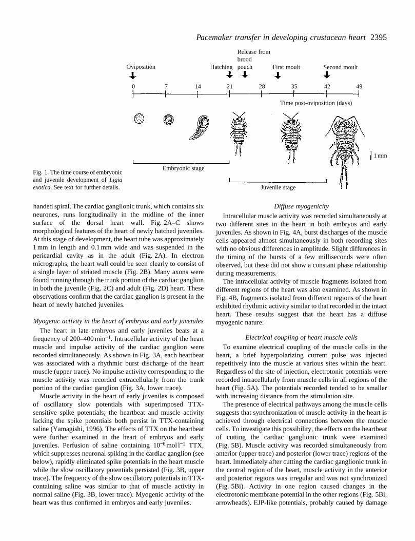
After copulation, the female moults and holds 80–120fertilized eggs in the brood pouch situated on the ventralsurface of the abdomen. Fig. 1 shows the time course ofembryonic and juvenile development of Ligia exotica. Theembryo develops in the egg for approximately 3 weeks beforehatching as a juvenile. The heartbeat starts at approximately 2weeks of embryonic development. The newly hatched juvenileis approximately 3 mm in body length and has six pairs of legs.Several days after hatching, the juveniles are released from themother’s brood pouch. The juvenile stage lasts forapproximately 3 weeks, during which time the juvenile moultstwice before becoming an immature adult. It is thenapproximately 5 mm in body length and has seven pairs of legs.The immature adult takes 4–5 months to mature(approximately 20 mm in body length) and 3–4 years to reachmaximum body length (over 40 mm).
Anatomy of newly hatched juvenile hearts
In adult Ligia exotica, the heart is tubular, is located in thedorsal side of the posterior half of the body and is suspendedin the pericardial cavity by suspensory ligaments. The heartwall consists of a single layer of muscle fibres forming a right-
2395Pacemaker transfer in developing crustacean heart
Oviposition
Embryonic stage
Juvenile stage
Release frombroodpouchHatching First moult Second moult
Time post-oviposition (days)
1 mm
0 7 14 21 28 35 42 49
Fig. 1. The time course of embryonicand juvenile development of Ligiaexotica. See text for further details.
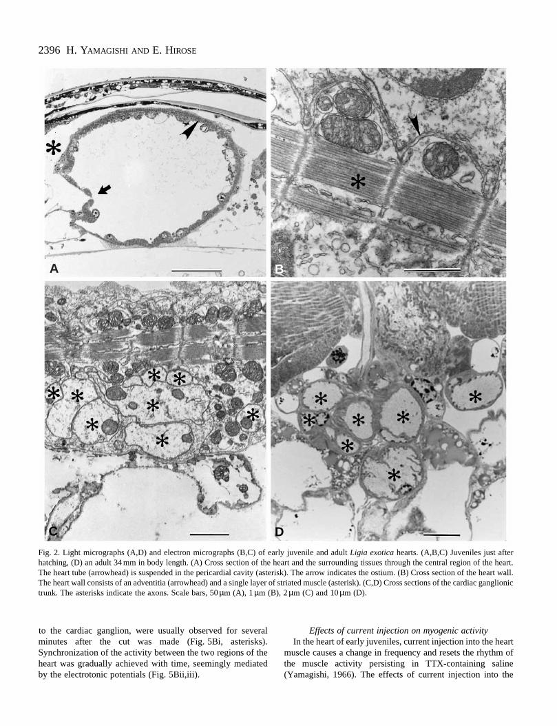
handed spiral. The cardiac ganglionic trunk, which contains sixneurones, runs longitudinally in the midline of the innersurface of the dorsal heart wall. Fig. 2A–C showsmorphological features of the heart of newly hatched juveniles.At this stage of development, the heart tube was approximately1 mm in length and 0.1 mm wide and was suspended in thepericardial cavity as in the adult (Fig. 2A). In electronmicrographs, the heart wall could be seen clearly to consist ofa single layer of striated muscle (Fig. 2B). Many axons werefound running through the trunk portion of the cardiac ganglionin both the juvenile (Fig. 2C) and adult (Fig. 2D) heart. Theseobservations confirm that the cardiac ganglion is present in theheart of newly hatched juveniles.
Myogenic activity in the heart of embryos and early juveniles
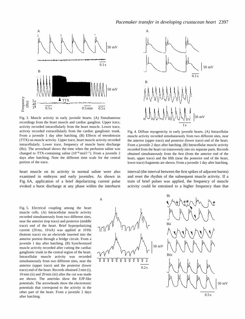
The heart in late embryos and early juveniles beats at afrequency of 200–400 min−1. Intracellular activity of the heartmuscle and impulse activity of the cardiac ganglion wererecorded simultaneously. As shown in Fig. 3A, each heartbeatwas associated with a rhythmic burst discharge of the heartmuscle (upper trace). No impulse activity corresponding to themuscle activity was recorded extracellularly from the trunkportion of the cardiac ganglion (Fig. 3A, lower trace).
Muscle activity in the heart of early juveniles is composedof oscillatory slow potentials with superimposed TTX-sensitive spike potentials; the heartbeat and muscle activitylacking the spike potentials both persist in TTX-containingsaline (Yamagishi, 1996). The effects of TTX on the heartbeatwere further examined in the heart of embryos and earlyjuveniles. Perfusion of saline containing 10−6 mol l−1 TTX,which suppresses neuronal spiking in the cardiac ganglion (seebelow), rapidly eliminated spike potentials in the heart musclewhile the slow oscillatory potentials persisted (Fig. 3B, uppertrace). The frequency of the slow oscillatory potentials in TTX-containing saline was similar to that of muscle activity innormal saline (Fig. 3B, lower trace). Myogenic activity of theheart was thus confirmed in embryos and early juveniles.
Diffuse myogenicity
Intracellular muscle activity was recorded simultaneously attwo different sites in the heart in both embryos and earlyjuveniles. As shown in Fig. 4A, burst discharges of the musclecells appeared almost simultaneously in both recording siteswith no obvious differences in amplitude. Slight differences inthe timing of the bursts of a few milliseconds were oftenobserved, but these did not show a constant phase relationshipduring measurements.
The intracellular activity of muscle fragments isolated fromdifferent regions of the heart was also examined. As shown inFig. 4B, fragments isolated from different regions of the heartexhibited rhythmic activity similar to that recorded in the intactheart. These results suggest that the heart has a diffusemyogenic nature.
Electrical coupling of heart muscle cells
To examine electrical coupling of the muscle cells in theheart, a brief hyperpolarizing current pulse was injectedrepetitively into the muscle at various sites within the heart.Regardless of the site of injection, electrotonic potentials wererecorded intracellularly from muscle cells in all regions of theheart (Fig. 5A). The potentials recorded tended to be smallerwith increasing distance from the stimulation site.
The presence of electrical pathways among the muscle cellssuggests that synchronization of muscle activity in the heart isachieved through electrical connections between the musclecells. To investigate this possibility, the effects on the heartbeatof cutting the cardiac ganglionic trunk were examined(Fig. 5B). Muscle activity was recorded simultaneously fromanterior (upper trace) and posterior (lower trace) regions of theheart. Immediately after cutting the cardiac ganglionic trunk inthe central region of the heart, muscle activity in the anteriorand posterior regions was irregular and was not synchronized(Fig. 5Bi). Activity in one region caused changes in theelectrotonic membrane potential in the other regions (Fig. 5Bi,arrowheads). EJP-like potentials, probably caused by damage
2396 H. YAMAGISHI AND E. HIROSE
A B
C DFig. 2. Light micrographs (A,D) and electron micrographs (B,C) of early juvenile and adult Ligia exotica hearts. (A,B,C) Juveniles just afterhatching, (D) an adult 34 mm in body length. (A) Cross section of the heart and the surrounding tissues through the central region of the heart.The heart tube (arrowhead) is suspended in the pericardial cavity (asterisk). The arrow indicates the ostium. (B) Cross section of the heart wall.The heart wall consists of an adventitia (arrowhead) and a single layer of striated muscle (asterisk). (C,D) Cross sections of the cardiac ganglionictrunk. The asterisks indicate the axons. Scale bars, 50 µm (A), 1 µm (B), 2 µm (C) and 10 µm (D).
to the cardiac ganglion, were usually observed for severalminutes after the cut was made (Fig. 5Bi, asterisks).Synchronization of the activity between the two regions of theheart was gradually achieved with time, seemingly mediatedby the electrotonic potentials (Fig. 5Bii,iii).
Effects of current injection on myogenic activityIn the heart of early juveniles, current injection into the heart
muscle causes a change in frequency and resets the rhythm ofthe muscle activity persisting in TTX-containing saline(Yamagishi, 1966). The effects of current injection into the
2397Pacemaker transfer in developing crustacean heartFr
eque
ncy
(Hz)
50 mV
50 mV
543
0.1 s
0.5 s0.5 s
TTX
0.5 min
A
B50 mV
50 mV
0.1 s
1 s
B
A
Fig. 3. Muscle activity in early juvenile hearts. (A) Simultaneousrecordings from the heart muscle and cardiac ganglion. Upper trace,activity recorded intracellularly from the heart muscle. Lower trace,activity recorded extracellularly from the cardiac ganglionic trunk.From a juvenile 1 day after hatching. (B) Effects of tetrodotoxin(TTX) on muscle activity. Upper trace, heart muscle activity recordedintracellularly. Lower trace, frequency of muscle burst discharge(Hz). The arrowhead shows the time when the perfusion saline waschanged to TTX-containing saline (10−6 mol l−1). From a juvenile 2days after hatching. Note the different time scale for the centralportion of the trace.
Fig. 4. Diffuse myogenicity in early juvenile hearts. (A) Intracellularmuscle activity recorded simultaneously from two different sites, nearthe anterior (upper trace) and posterior (lower trace) end of the heart.From a juvenile 2 days after hatching. (B) Intracellular muscle activityrecorded from the heart cut transversely into six separate parts. Recordsobtained simultaneously from the first (from the anterior end of theheart, upper trace) and the fifth (near the posterior end of the heart,lower trace) fragments are shown. From a juvenile 1 day after hatching.
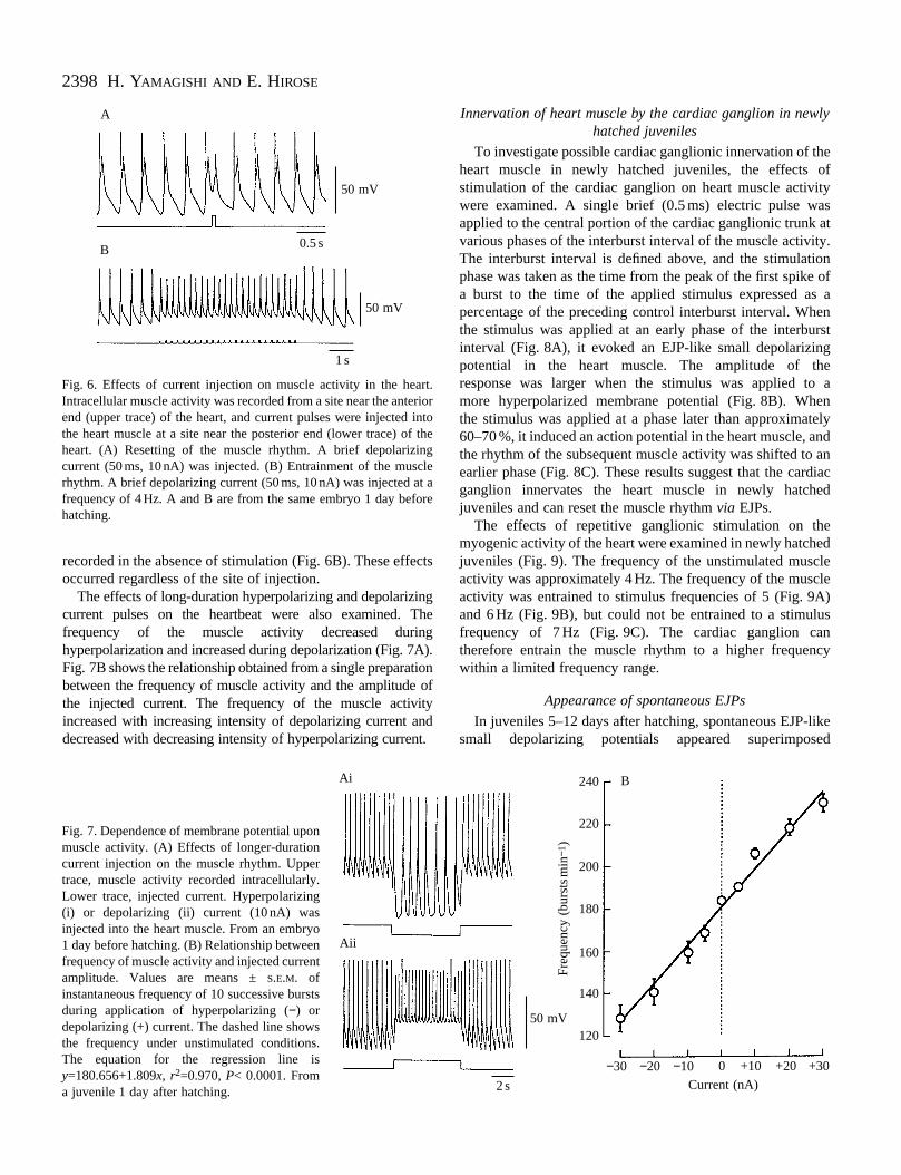
heart muscle on its activity in normal saline were alsoexamined in embryos and early juveniles. As shown inFig. 6A, application of a brief depolarizing current pulseevoked a burst discharge at any phase within the interburst
A
Fig. 5. Electrical coupling among the heartmuscle cells. (A) Intracellular muscle activityrecorded simultaneously from two different sites,near the anterior (top trace) and posterior (middletrace) end of the heart. Brief hyperpolarizingcurrent (20 ms, 10 nA) was applied at 10 Hz(bottom trace) via an electrode inserted into theanterior portion through a bridge circuit. From ajuvenile 1 day after hatching. (B) Synchronizedmuscle activity recorded after cutting the cardiacganglionic trunk in the central region of the heart.Intracellular muscle activity was recordedsimultaneously from two different sites, near theanterior (upper trace) and the posterior (lowertrace) end of the heart. Records obtained 2 min (i),10 min (ii) and 20 min (iii) after the cut was madeare shown. The asterisks show the EJP-likepotentials. The arrowheads show the electrotonicpotentials that correspond to the activity in theother part of the heart. From a juvenile 2 daysafter hatching.
interval (the interval between the first spikes of adjacent bursts)and reset the rhythm of the subsequent muscle activity. If atrain of brief pulses was applied, the frequency of muscleactivity could be entrained to a higher frequency than that
50 mV
50 mV
0.2 s
0.5 s
Bi
Bii
Biii
2398 H. YAMAGISHI AND E. HIROSE
50 mV
50 mV
0.5 s
1 s
B
A
Fig. 6. Effects of current injection on muscle activity in the heart.Intracellular muscle activity was recorded from a site near the anteriorend (upper trace) of the heart, and current pulses were injected intothe heart muscle at a site near the posterior end (lower trace) of theheart. (A) Resetting of the muscle rhythm. A brief depolarizingcurrent (50 ms, 10 nA) was injected. (B) Entrainment of the musclerhythm. A brief depolarizing current (50 ms, 10 nA) was injected at afrequency of 4 Hz. A and B are from the same embryo 1 day beforehatching.
recorded in the absence of stimulation (Fig. 6B). These effectsoccurred regardless of the site of injection.
The effects of long-duration hyperpolarizing and depolarizingcurrent pulses on the heartbeat were also examined. Thefrequency of the muscle activity decreased duringhyperpolarization and increased during depolarization (Fig. 7A).Fig. 7B shows the relationship obtained from a single preparationbetween the frequency of muscle activity and the amplitude ofthe injected current. The frequency of the muscle activityincreased with increasing intensity of depolarizing current anddecreased with decreasing intensity of hyperpolarizing current.
Ai
Aii
Fig. 7. Dependence of membrane potential uponmuscle activity. (A) Effects of longer-durationcurrent injection on the muscle rhythm. Uppertrace, muscle activity recorded intracellularly.Lower trace, injected current. Hyperpolarizing(i) or depolarizing (ii) current (10 nA) wasinjected into the heart muscle. From an embryo1 day before hatching. (B) Relationship betweenfrequency of muscle activity and injected currentamplitude. Values are means ± S.E.M. ofinstantaneous frequency of 10 successive burstsduring application of hyperpolarizing (−) ordepolarizing (+) current. The dashed line showsthe frequency under unstimulated conditions.The equation for the regression line isy=180.656+1.809x, r2=0.970, P< 0.0001. Froma juvenile 1 day after hatching.
Innervation of heart muscle by the cardiac ganglion in newlyhatched juveniles
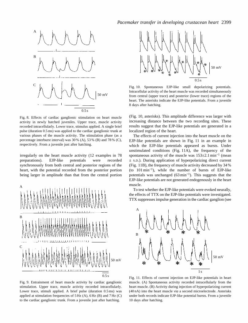
To investigate possible cardiac ganglionic innervation of theheart muscle in newly hatched juveniles, the effects ofstimulation of the cardiac ganglion on heart muscle activitywere examined. A single brief (0.5 ms) electric pulse wasapplied to the central portion of the cardiac ganglionic trunk atvarious phases of the interburst interval of the muscle activity.The interburst interval is defined above, and the stimulationphase was taken as the time from the peak of the first spike ofa burst to the time of the applied stimulus expressed as apercentage of the preceding control interburst interval. Whenthe stimulus was applied at an early phase of the interburstinterval (Fig. 8A), it evoked an EJP-like small depolarizingpotential in the heart muscle. The amplitude of theresponse was larger when the stimulus was applied to amore hyperpolarized membrane potential (Fig. 8B). Whenthe stimulus was applied at a phase later than approximately60–70 %, it induced an action potential in the heart muscle, andthe rhythm of the subsequent muscle activity was shifted to anearlier phase (Fig. 8C). These results suggest that the cardiacganglion innervates the heart muscle in newly hatchedjuveniles and can reset the muscle rhythm via EJPs.
The effects of repetitive ganglionic stimulation on themyogenic activity of the heart were examined in newly hatchedjuveniles (Fig. 9). The frequency of the unstimulated muscleactivity was approximately 4 Hz. The frequency of the muscleactivity was entrained to stimulus frequencies of 5 (Fig. 9A)and 6 Hz (Fig. 9B), but could not be entrained to a stimulusfrequency of 7 Hz (Fig. 9C). The cardiac ganglion cantherefore entrain the muscle rhythm to a higher frequencywithin a limited frequency range.
Appearance of spontaneous EJPs
In juveniles 5–12 days after hatching, spontaneous EJP-likesmall depolarizing potentials appeared superimposed
50 mV
2 s
B
180
240
220
200
160
140
120
Freq
uenc
y (b
urst
smin
− 1)
−30 −20 −10 0 +10 +20 +30
Current (nA)
2399Pacemaker transfer in developing crustacean heart
B
C
A
50 mV
0.5 s
Fig. 8. Effects of cardiac ganglionic stimulation on heart muscleactivity in newly hatched juveniles. Upper trace, muscle activityrecorded intracellularly. Lower trace, stimulus applied. A single briefpulse (duration 0.5 ms) was applied to the cardiac ganglionic trunk atvarious phases of the muscle activity. The stimulation phase (as apercentage interburst interval) was 30 % (A), 53 % (B) and 78 % (C),respectively. From a juvenile just after hatching.
50 mV
0.5 s
Fig. 10. Spontaneous EJP-like small depolarizing potentials.Intracellular activity of the heart muscle was recorded simultaneouslyfrom central (upper trace) and posterior (lower trace) regions of theheart. The asterisks indicate the EJP-like potentials. From a juvenile8 days after hatching.
irregularly on the heart muscle activity (12 examples in 78preparations). EJP-like potentials were recordedsynchronously from both central and posterior regions of theheart, with the potential recorded from the posterior portionbeing larger in amplitude than that from the central portion
50 mV
0.5 s
B
C
A
Fig. 9. Entrainment of heart muscle activity by cardiac ganglionicstimulation. Upper trace, muscle activity recorded intracellularly.Lower trace, stimuli applied. A brief pulse (duration 0.5 ms) wasapplied at stimulation frequencies of 5 Hz (A), 6 Hz (B) and 7 Hz (C)to the cardiac ganglionic trunk. From a juvenile just after hatching.
(Fig. 10, asterisks). This amplitude difference was larger withincreasing distance between the two recording sites. Theseresults suggest that the EJP-like potentials are generated in alocalized region of the heart.
The effects of current injection into the heart muscle on theEJP-like potentials are shown in Fig. 11 in an example inwhich the EJP-like potentials appeared as bursts. Underunstimulated conditions (Fig. 11A), the frequency of thespontaneous activity of the muscle was 153±2.1 min−1 (mean± S.D.). During application of hyperpolarizing direct current(Fig. 11B), the frequency of muscle activity decreased by 34 %(to 101 min−1), while the number of bursts of EJP-likepotentials was unchanged (63 min−1). This suggests that theEJP-like potentials are not generated endogenously in the heartmuscle.
To test whether the EJP-like potentials were evoked neurally,the effects of TTX on the EJP-like potentials were investigated.TTX suppresses impulse generation in the cardiac ganglion (see
50 mV
1 s
B
A
Fig. 11. Effects of current injection on EJP-like potentials in heartmuscle. (A) Spontaneous activity recorded intracellularly from theheart muscle. (B) Activity during injection of hyperpolarizing current(40 nA) into the heart muscle via a second microelectrode. Asterisksunder both records indicate EJP-like potential bursts. From a juvenile10 days after hatching.

2400 H. YAMAGISHI AND E. HIROSE
50 mV
0.5 s
B
A
50 mV
20 mV
0.5 s
0.5 s
Bi
Bii
A
Fig. 13. Periodic burst discharges of the cardiac ganglion andEJPs underlying the muscle activity in late juvenile hearts.(A) Simultaneous recording of intracellular activity of the heart muscle(upper trace) and impulse activity of the cardiac ganglionic trunk(lower trace). From a juvenile 17 days after hatching. (B) Intracellularactivity recorded from the heart muscle (upper trace). The heart wallwas pinched, causing deterioration in muscle activity. The musclemembrane was then hyperpolarized by current injection (30 nA) intothe heart muscle (lower trace). From a juvenile 16 days after hatching.
Fig. 12. Effects of tetrodotoxin (TTX) on EJP-like potentials.(A) Intracellular activity recorded from the heart muscle. (B) Activityrecorded 1 min after changing the perfusion medium to 10−6 mol l−1
TTX-containing saline. EJP-like potentials were abolished followingTTX application. From a juvenile 12 days after hatching.
below). The EJP-like potentials, which were superimposedirregularly on the muscle spontaneous activity (Fig. 12A), werecompletely eliminated by application of 10−6 mol l−1 TTX(Fig. 12B). The effect was reversible, and the EJP-likepotentials reappeared after washout of TTX (results not shown).These results suggest that the EJP-like potentials are EJPsevoked by the spontaneous activity of the cardiac ganglion.
Spontaneous periodic burst discharges of the cardiacganglion
The cardiac ganglion began to show spontaneous periodicbursting during juvenile development. The earliest periodicburst discharges were recorded extracellularly from the cardiacganglionic trunk of a juvenile 9 days after hatching. As shownin Fig. 13A, each muscle response followed a burst dischargein the cardiac ganglion. EJPs were not usually detectable in therising phase of muscle activity.
To detect the EJP underlying the muscle activity, the heartwall was pinched using fine tweezers to cause a deteriorationin muscle activity, but taking care to keep the cardiac ganglionintact. In some cases, ‘notches’ representing the EJPcomponent, were observed clearly in the rising phase of themuscle activity (Fig. 13Bi). During subsequent injection ofhyperpolarizing direct current into the muscle, the musclepotentials were largely eliminated and only periodic EJP burstswere recorded (Fig. 13Bii). The frequency of EJP bursts wasunchanged or decreased slightly during hyperpolarization.These results suggest that the muscle activity was entrained bythe activity of the cardiac ganglion via EJPs.
Effects of TTX on adult heartbeats
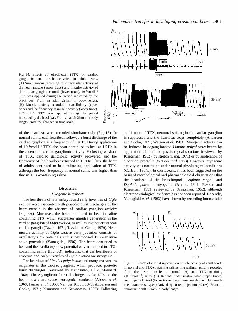
After the cardiac ganglion began periodic bursting duringjuvenile development, heartbeats were associated withganglionic burst discharges. The effects of TTX on the cardiacganglionic and muscle activities were then examined in adulthearts. The intracellular activity of the heart muscle and impulseactivity of the cardiac ganglion were recorded simultaneously(Fig. 14A). Each muscle response followed a burst discharge ofthe cardiac ganglion. During application of 10−6 mol l−1 TTX,
the ganglionic burst discharges were eliminated rapidly, whilethe heart muscle exhibited oscillatory slow potentials. Thefrequency of the oscillatory slow potential reached a constantlevel during perfusion of TTX-containing saline that was lowerthan that recorded in normal saline (Fig. 14B). Transientchanges in the frequency of the muscle activity were usuallyobserved immediately following application and washout ofTTX. These results suggest that the heart muscle of adults alsopossessed endogenous oscillatory properties.
To confirm the presence of myogenicity in adult hearts, theeffects of current injection into the heart muscle on the muscleactivity were examined in normal and TTX-containing salines(Fig. 15). The frequency of muscle activity in normal salinewas 3.7 Hz (Fig. 15Ai), and it was not affected byhyperpolarizing current injection into the heart muscle(Fig. 15Aii). In contrast, in saline containing 10−6 mol l−1 TTX,the frequency of the muscle slow oscillatory potential was3.7 Hz, and it decreased to 3.1 Hz (16 % reduction) followinginjection of hyperpolarizing current (Fig. 15B). These resultsstrongly support the suggestion that the muscle activityrecorded in TTX-containing saline is endogenous.
To investigate the effects of TTX on the adult heartbeat, theimpulse activity of the cardiac ganglion and a mechanogram
2401Pacemaker transfer in developing crustacean heart
Freq
uenc
y(H
z)
50 mV
50 mV
3
2
1
0.5 s1 min
1 s
TTX
TTX 2 min
A
B
Fig. 14. Effects of tetrodotoxin (TTX) on cardiacganglionic and muscle activities in adult hearts.(A) Simultaneous recording of intracellular activity ofthe heart muscle (upper trace) and impulse activity ofthe cardiac ganglionic trunk (lower trace). 10−6 mol l−1
TTX was applied during the period indicated by theblack bar. From an adult 22 mm in body length.(B) Muscle activity recorded intracellularly (uppertrace) and the frequency of muscle activity (lower trace).10−6 mol l−1 TTX was applied during the periodindicated by the black bar. From an adult 26 mm in bodylength. Note the changes in time scale.
Fig. 15. Effects of current injection on muscle activity of adult heartsin normal and TTX-containing salines. Intracellular activity recordedfrom the heart muscle in normal (A) and TTX-containing(10−6 mol l−1) saline (B). Records under unstimulated (upper traces)and hyperpolarized (lower traces) conditions are shown. The musclemembrane was hyperpolarized by current injection (40 nA). From animmature adult 12 mm in body length.
50 mV
0.5 s
Bi
BiiAii
Ai
of the heartbeat were recorded simultaneously (Fig. 16). Innormal saline, each heartbeat followed a burst discharge of thecardiac ganglion at a frequency of 1.9 Hz. During applicationof 10−6 mol l−1 TTX, the heart continued to beat at 1.5 Hz inthe absence of cardiac ganglionic activity. Following washoutof TTX, cardiac ganglionic activity recovered and thefrequency of the heartbeat returned to 1.9 Hz. Thus, the heartof adults continued to beat following application of TTX,although the beat frequency in normal saline was higher thanthat in TTX-containing saline.
DiscussionMyogenic heartbeats
The heartbeats of late embryos and early juveniles of Ligiaexotica were associated with periodic burst discharges of theheart muscle in the absence of cardiac ganglion activity(Fig. 3A). Moreover, the heart continued to beat in salinecontaining TTX, which suppresses impulse generation in thecardiac ganglion of Ligia exotica, as well as in other crustaceancardiac ganglia (Tazaki, 1971; Tazaki and Cooke, 1979). Heartmuscle activity of Ligia exotica early juveniles consists ofoscillatory slow potentials with superimposed TTX-sensitivespike potentials (Yamagishi, 1996). The heart continued tobeat and the oscillatory slow potential was maintained in TTX-containing saline (Fig. 3B), indicating that the heartbeats ofembryos and early juveniles of Ligia exotica are myogenic.
The heartbeat of Limulus polyphemus and many crustaceansoriginates in the cardiac ganglion, which produces periodicburst discharges (reviewed by Krijgsman, 1952; Maynard,1960). These ganglionic burst discharges evoke EJPs on theheart muscle and cause neurogenic heartbeats (Abbott et al.1969; Parnas et al. 1969; Van der Kloot, 1970; Anderson andCooke, 1971; Kuramoto and Kuwasawa, 1980). Following
application of TTX, neuronal spiking in the cardiac ganglionis suppressed and the heartbeat stops completely (Andersonand Cooke, 1971; Watson et al. 1983). Myogenic activity canbe induced in deganglionated Limulus polyphemus hearts byapplication of modified physiological solutions (reviewed byKrijgsman, 1952), by stretch (Lang, 1971) or by application ofa peptide, proctolin (Watson et al. 1983). However, myogenicactivity was not found under normal physiological conditions(Carlson, 1904b). In crustaceans, it has been suggested on thebasis of morphological and pharmacological observations thatthe heartbeat of the branchiopods Daphnia magna andDaphnia pulex is myogenic (Baylor, 1942; Bekker andKrijgsman, 1951, reviewed by Krijgsman, 1952), althoughelectrophysiological evidence has not been reported. Recently,Yamagishi et al. (1993) have shown by recording intracellular

2402 H. YAMAGISHI AND E. HIROSE
Freq
uenc
y(H
z)
100 mg
2
1
1 min 1 sTTX
Fig. 16. Effects of TTX on adult heartbeats. Theimpulse activity recorded from the cardiacganglionic trunk (top trace), the mechanical recordof the heartbeat (middle trace) and the frequency ofthe heartbeat (bottom trace) are shown. 10−6 mol l−1
TTX was applied during the period indicated by theblack bar. From an adult 32 mm in body length.
activity of the heart muscle that the heartbeat of thebranchiopod Triops longicaudatus is myogenic.
Characteristics of the heart as a single muscle oscillator
In late embryos and early juveniles, muscle fragmentsisolated from all regions of the heart exhibited spontaneousbursting, indicating that there is no localized pacemaker regionof the heart (Fig. 4). The muscle cells were coupled electricallyand discharged almost simultaneously (Fig. 5A). These resultssuggest that the heart muscle cells individually possessoscillatory properties and that electrical pathways exist tocoordinate their synchronous discharge. This was confirmed bycutting the cardiac ganglionic trunk. In the neurogenic heart ofadult Ligia oceanica and Ligia exotica, a transverse cut of thecardiac ganglionic trunk at a central region of the heart resultedin the generation of independent burst activity in the twoseparated ganglia (Yamagishi and Ebara, 1985) andasynchronous beating between the anterior and posteriorhalves of the heart (Alexandrowictz, 1931). In contrast, in theheart of embryos and early juveniles, synchronous beating wasrecorded between the anterior and posterior halves of the heartafter cutting the cardiac ganglionic trunk (Fig. 5B). Theseresults indicate that the heart of late embryos and earlyjuveniles of Ligia exotica has a diffuse myogenic nature andthe characteristics of a functional syncytium. These features ofthe heart are very similar to those of molluscan myogenichearts (reviewed by Hill and Welsh, 1966; Irisawa, 1978;Jones, 1983), especially the oyster ventricle (Irisawa et al.1973; Ebara and Kuwasawa, 1975).
The heart of late embryos and early juveniles of Ligiaexotica is a unitary muscle, as was confirmed by currentinjection into the heart muscle. Regardless of the injection site,the rhythm of the muscle activity was reset by a single briefdepolarizing current and entrained to a higher frequency byrepeated stimuli (Fig. 6). The frequency of the muscle activitychanged depending on the membrane potential level at whichthe current was injected (Fig. 7). These responses to theapplied stimuli are characteristic of endogenous oscillators ingeneral (Pavlidis, 1973) and indicate that the heart of lateembryos and early juveniles of Ligia exotica can be regardedas an endogenous muscle oscillator.
Cardiac ganglionic innervation in early juvenile hearts
In the myogenic heart of newly hatched juveniles, the heart
muscle produced EJP-like small depolarizing potentialsfollowing stimulation of the cardiac ganglionic trunk(Fig. 8A). The rhythm of the myogenic activity in newlyhatched juvenile hearts could be reset by a single stimulusapplied to the cardiac ganglion (Fig. 8C) and entrained to ahigher frequency by repeated stimuli (Fig. 9). In newly hatchedjuveniles, many axons were identified in the cardiac ganglionictrunk (Fig. 2). In the heart of early juveniles, a pair of extrinsiccardioregulatory nerves, each containing one inhibitory andtwo acceleratory axons, innervates the heart muscle and canregulate the myogenic heartbeat (Yamagishi and Hirose,1992). These cardioregulatory axons enter the heart and runthrough the cardiac ganglionic trunk (Yamagishi et al. 1989).Therefore, stimulation of the cardiac ganglionic trunk mightproduce compound junctional potentials on the heart muscle.However, stimulation of only the extrinsic cardioregulatorynerves at a site outside the heart failed to reset or entrain themuscle activity to a higher frequency (Yamagishi and Hirose,1992). These results suggest that, in the heart of early juveniles,the cardiac ganglion cells have already innervated the heartmuscle and have the ability to reset and entrain its myogenicactivity.
Development of cardiac ganglion activity
In juveniles approximately 1 week after hatching,spontaneous EJP-like potentials appeared irregularly in theheart muscle (Fig. 10). These responses were not changed infrequency by hyperpolarizing the muscle membrane potential,in contrast to the myogenic activity (Fig. 11). Moreover, theEJP-like responses were eliminated by application of TTX(Fig. 12), which suppresses impulse generation in the cardiacganglion (Fig. 14). These results suggest that the EJP-likeresponses are neurally evoked EJPs and represent thebeginning of spontaneous activity of the cardiac ganglion.However, the EJPs appeared locally in the heart and no impulseactivity corresponding to the EJPs was recorded from thecardiac ganglionic trunk (results not shown). This may indicatethat coordinated activity is not established among the cardiacganglion cells at this stage.
In juveniles more than 10 days after hatching, periodicbursts of impulses appeared in the cardiac ganglionic trunk(Fig. 13A). In adult hearts, six cardiac ganglion cells dischargesynchronously via electrical connections and send periodicbursts of impulses peripherally (Yamagishi and Ebara, 1985).

2403Pacemaker transfer in developing crustacean heart
The cardiac ganglion cells start spontaneous coordinatedbursting during juvenile development.
Pacemaker mechanisms in late juvenile and adult hearts
Entrainment of spontaneous muscle activity by synapticinputs from the ganglionic motor neurones has been reportedin the crustacean stomatogastric system (Meyrand andMoulins, 1986; Tazaki and Miyazaki, 1991). Myogenicactivity in the heart of early juveniles could also be entrainedby EJPs evoked by stimulation of the cardiac ganglion (Fig. 9).When the cardiac ganglion begins spontaneous periodicbursting during juvenile development, muscle activity occursin response to ganglionic burst discharges (Fig. 13A). Theheart muscle produces periodic bursts of EJPs correspondingto the ganglionic burst discharges (Fig. 13B). These resultsstrongly suggest that the cardiac ganglion entrains themyogenic activity to a higher frequency via EJPs.
This was confirmed in adult hearts by the results of TTXapplication. TTX suppressed impulse generation in the cardiacganglion (Fig. 14), as reported in other crustaceans (Andersonand Cooke, 1971; Tazaki, 1971). In TTX-containing saline, theheart muscle of adults continued to exhibit endogenousoscillatory activity (Figs 14, 15). Therefore, the heartbeat ofadults was changed reversibly from neurogenic to myogenicby the application of TTX, with the neurogenic heartbeat beinghigher in frequency than the myogenic one (Fig. 16). Althoughthe frequency of the heartbeat decreases during growth, theheartbeat of the same preparation was generally higher infrequency in normal saline than in TTX-containing saline(Yamagishi and Mori, 1993).
In the heart of all crustaceans investigated so far, the heartmuscle has no myogenicity and the cardiac ganglion acts as adominant pacemaker (reviewed by Prosser, 1973). In the leechHirudo medicinalis, Maranto and Calabrese (1984) haveshown that the heart excitor motor neurones located in thecentral nervous system reset and entrain the heart myogenicrhythm to a higher frequency via EJPs. In the heart of adultLigia exotica, two pacemaker sites, the cardiac ganglion andthe heart muscle itself, are present, and the cardiac ganglionacts as a primary pacemaker to entrain the muscle activity toa higher frequency via EJPs. This type of neurogenic heartbeatis different from that known in any other crustacean, but issimilar to that in the leech.
Transfer of the heart pacemaker during development
The results of the present study suggest that the pacemakerfunction of the heart of Ligia exotica is transferred from theheart muscle to the cardiac ganglion during juveniledevelopment. As a result, the initiation of the heartbeatchanged from myogenic to neurogenic but of a type differentfrom that reported for other crustaceans and Limuluspolyphemus.
Recently, we have shown that the heartbeat of adults of theprimitive crustacean Triops longicaudatus (Branchiopoda) ismyogenic (Yamagishi et al. 1993). This suggests that there issome diversity in the heartbeat pacemaker mechanism in
Crustacea. The present findings show that this diversity ispresent not only at the phylogenetic but also at the ontogeneticlevel.
We thank Dr K. Oami for helpful discussions and Dr DarrylMacer for his reading of this manuscript. This work wassupported in part by a Grant-in-Aid (no. 08640857) from theMinistry of Education, Science, Sports and Culture of Japan toH.Y. Contribution no. 608 from the Shimoda Marine ResearchCenter, University of Tsukuba.
ReferencesABBOTT, B. C., LANG, F. AND PARNAS, I. (1969). Physiological
properties of the heart and cardiac ganglion of Limulus polyphemus.Comp. Biochem. Physiol. 28, 149–158.
AI, N. (1966). Electrophysiological investigation of the automaticityin the cardiac muscle of the Ligia, Megaligia exotica. Sci. Rep.Tokyo Kyoiku Daigaku B 12, 131–149.
ALEXANDROWICZ, J. S. (1931). Quelques expériences sur lefonctionnement du système nerveux du cœur des Crustacésisopodes. C. r. soc. Biol. Paris 108, 1270–1272.
ALEXANDROWICZ, J. S. (1952). Innervation of the heart of Ligiaoceanica. J. mar. biol. Ass. U.K. 31, 85–97.
ANDERSON, M. AND COOKE, I. M. (1971). Neural activation of theheart of the lobster Homarus americanus. J. exp. Biol. 55, 449–468.
BAYLOR, E. R. (1942). Cardiac pharmacology of the cradoceran,Daphnia. Biol. Bull. mar. biol. Lab., Woods Hole 83, 165–172.
BEKKER, J. M. AND KRIJGSMAN, B. J. (1951). Physiologicalinvestigations into the heart function of Daphnia. J. Physiol., Lond.115, 249–257.
CARLSON, A. J. (1904a). The nervous origin of the heartbeat inLimulus and the nervous nature of coordination in the heart. Am. J.Physiol. 12, 67–74.
CARLSON, A. J. (1904b). The nature of the action of drugs on the heart.Science 20, 684–689.
CARLSON, A. J. AND MEEK, W. J. (1908). On the mechanism of theembryonic heart rhythm in Limulus. Am. J. Physiol. 21, 1–10.
CROZIER, W. J. AND STIER, T. J. B. (1927). Temperature and frequencyof cardiac contraction in embryos of Limulus. J. gen. Physiol. 10,501–518.
EBARA, A. AND KUWASAWA, K. (1975). The initiating site forspontaneous electrical activity in the oyster ventricle. Annot. zool.Jap. 48, 219–226.
GIBSON, D. AND LANG, F. (1979). Is embryonic Limulus heart reallymyogenic? Am. Zool. 19, 39–51.
HILL, R. B. AND WELSH, J. H. (1966). Heart, circulation and bloodcells. In Physiology of Mollusca (ed. K. M. Wilbur and C. M.Yonge), pp. 125–174. New York: Academic Press.
IRISAWA, H. (1978). Comparative physiology of the cardiacpacemaker mechanism. Physiol. Rev. 58, 461–498.
IRISAWA, H., IRISAWA, A. AND SHIGETO, N. (1973). Physiological andmorphological correlation of the functional syncytium in thebivalve myocardium. Comp. Biochem. Physiol. 44A, 207–219.
JONES, H. D. (1983). The circulatory system of gastropods andbivalves. In The Mollusca (ed. A. S. M. Saleuddin and C. M.Yonge), pp. 189–235. New York: Academic Press.
KRIJGSMAN, B. J. (1952). Contractile and pacemaker mechanisms ofthe heart of arthropods. Biol. Rev. 27, 320–346.
2404 H. YAMAGISHI AND E. HIROSE
KURAMOTO, T. AND KUWASAWA, K. (1980). Ganglionic activation ofthe myocardium of the lobster, Panulirus japonicus. J. comp.Physiol. A 139, 67–76.
KUSHIDA, H. (1980). An improved embedding method using ERL4206 and Quetol 653. J. Electron Microsc. 29, 193–194.
LANG, F. (1971). Induced myogenic activity in the neurogenic heartof Limulus polyphemus. Biol. Bull. mar. biol. Lab., Woods Hole141, 269–277.
MARANTO, R. M. AND CALABRESE, R. L. (1984). Neural control of thehearts in the leech, Hirudo medicinalis. II. Myogenic activity andits control by heart motor neurons. J. comp. Physiol. A 154,381–391.
MAYNARD, D. M. (1960). Circulation and heart function. In ThePhysiology of Crustacea 1 (ed. T. H. Watermann), pp. 161–226.New York: Academic Press.
MEYRAND, P. AND MOULINS, M. (1986). Myogenic oscillatory activityin the pyloric rhythmic motor system of Crustacea. J. comp.Physiol. A 158, 489–503.
PARNAS, I., ABBOTT, B. C. AND LANG, F. (1969). Electrophysiologicalproperties of Limulus heart and effect of drugs. Am. J. Physiol. 217,1814–1822.
PAVLIDIS, T. (1973). Biological Oscillators: Their MathematicalAnalysis. New York: Academic Press.
PROSSER, C. L. (1942). An analysis of the action of acetylcholine onhearts, particularly in arthropods. Biol. Bull. mar. biol. Lab., WoodsHole 83, 145–164.
PROSSER, C. L. (1973). Circulation of body fluids. In ComparativeAnimal Physiology (ed. C. L. Prosser), pp. 822–856. Washington,DC: W. B. Saunders Co.
RULON, R., HERMSMEYER, K. AND SPERELAKIS, N. (1971).Regenerative action potentials induced in the neurogenic heart ofLimulus polyphemus. Comp. Biochem. Physiol. 39A, 333–355.
SUZUKI, S. (1934). Ganglion cells in the heart of Ligia exotica (Roux).Sci. Rep. Tohoku Imp. Univ. 9, 213–217.
TAZAKI, K. (1971). The effects of tetrodotoxin on the slow potentialand spikes in the cardiac ganglion of a crab, Eriocheir japonicus.Jap. J. Physiol. 21, 529–536.
TAZAKI, K. AND COOKE, I. M. (1979). Isolation and characterization
of slow, depolarizing responses of cardiac ganglion neurons in thecrab, Portunus sanguinolentus. J. Neurophysiol. 42, 1000–1021.
TAZAKI, K. AND MIYAZAKI, T. (1991). Neural control of the posteriorcardiac plate and pyloric regions of the mantis shrimp Squillaoratoria: neurogenic and myogenic activities of muscles. J. comp.Physiol. A 168, 265–279.
VAN DER KLOOT, W. G. (1970). The electrophysiology of muscle fibersin the hearts of decapod crustaceans. J. exp. Zool. 174, 369–380.
WATSON, W. H., AUGUSTINE, G. J., BENSON, A. AND SULLIVAN, R. E.(1983). Proctolin and endogenous proctolin-like peptide enhancethe contractility of the Limulus heart. J. exp. Biol. 103, 55–73.
YAMAGISHI, H. (1990). Physiological change of the heart beat injuvenile stage of the isopod crustacean Ligia exotica. (Proc. zool.Soc. Jap.) Zool. Sci. 7, 1037.
YAMAGISHI, H. (1995). Transfer of heart pacemaker during juveniledevelopment in the isopod crustacean, Ligia exotica. (Proc. 4th Int.Congr. Comp. Physiol. Biochem). Physiol. Zool. 68, 68.
YAMAGISHI, H. (1996). Endogenous oscillatory activity and TTX-sensitive spikes of the heart muscle in early juveniles of the isopodcrustacean Ligia exotica. Experientia 52, 583–586.
YAMAGISHI, H., ANDO, H. AND MAKIOKA, T. (1993). The heart beat ofTriopus longicaudatus (Crustacea, Branchiopoda) is myogenic.(Proc. zool. Soc. Jap.) Zool. Sci. 10 (Suppl.), 95.
YAMAGISHI, H. AND EBARA, A. (1985). Spontaneous activity andpacemaker property of neurons in the cardiac ganglion of an isopod,Ligia exotica. Comp. Biochem. Physiol. 81A, 55–62.
YAMAGISHI, H. AND HIROSE, E. (1992). Nervous regulation of themyogenic heart in early juveniles of the isopod crustacean, Ligiaexotica. In Comparative Physiology, vol. 11, Phylogenic Models inFunctional Coupling of the CNS and the Cardiovascular System(ed. R. B. Hill and K. Kuwasawa), pp. 141–148. Basel: Karger.
YAMAGISHI, H. AND MORI, A. (1993). Developmental changes inpacemaker mechanisms in the heart of the isopod crustacean Ligiaexotica. Jap. comp. Physiol. Biochem. 10, 208.
YAMAGISHI, H., UESAKA, H. AND EBARA, A. (1989). Inter- and motor-neuronal functions of the cardio-inhibitory nerve in the heart of theisopod crustacean, Ligia exotica. Comp. Biochem. Physiol. 94A,471–476.