transient monocular blindness shirley h. wray, m.d., ph.d., professor of neurology, harvard medical...
TRANSCRIPT
Transient Monocular Blindness
Shirley H. Wray, M.D., Ph.D., Professor of Neurology, Harvard Medical School
Director, Unit for Neurovisual DisordersMassachusetts General Hospital
HARVARD MEDICAL SCHOOLDEPARTMENT OF NEUROLOGY
MASSACHUSETTS GENERAL HOSPITAL
HARVARD MEDICAL SCHOOLDEPARTMENT OF NEUROLOGY
MASSACHUSETTS GENERAL HOSPITAL
HARVARD MEDICAL SCHOOLDEPARTMENT OF NEUROLOGY
MASSACHUSETTS GENERAL HOSPITAL
Transient Monocular Blindness
a
Herald Symptom of Stroke
to the
Brain and / or Eye

937-2

Arterial supply of optic nerve and retina
Four types of monocular TMB
TMB Type I due to transient retinal ischemia
TMB Type II due to retinal vascular insufficiency
TMB Type III due to transient angiospasm
TMB Type IV - idiopathic
Identify Risk Factors
Cardiac / MI, Atrial Fib
Stroke – TIA
Hypertension
Hyperlipidemia
Diabetes Mellitus
Smoking
Obesity
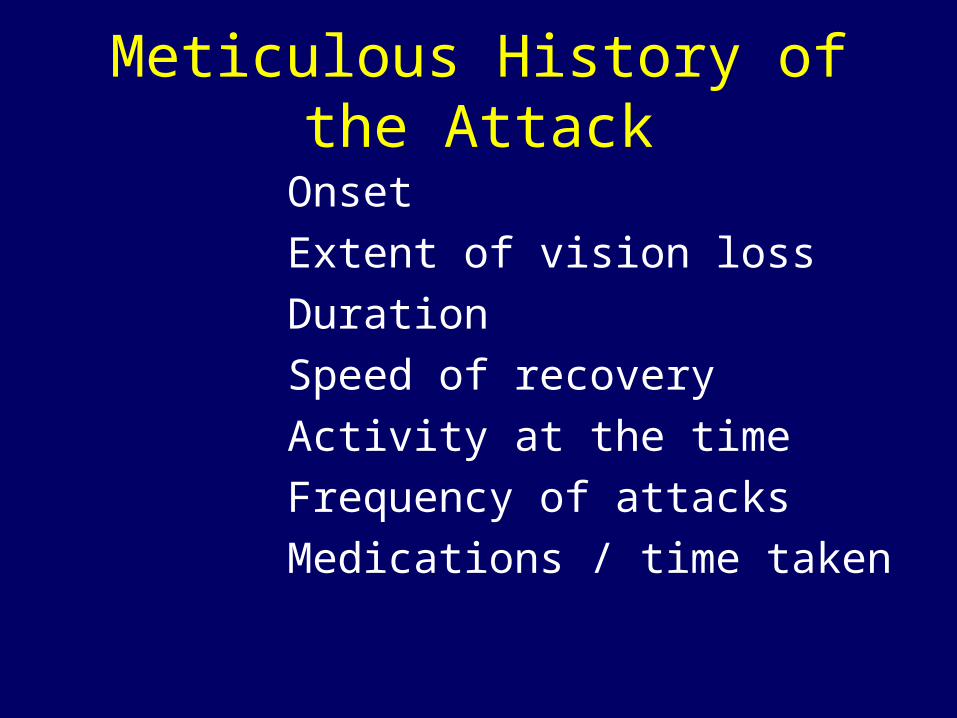
Meticulous History of the Attack
Onset
Extent of vision loss
Duration
Speed of recovery
Activity at the time
Frequency of attacks
Medications / time taken
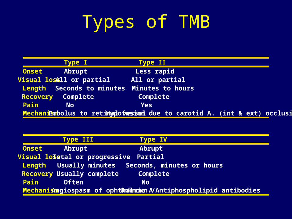
Types of TMB
Type I Type II
Onset Abrupt Less rapidVisual loss All or partial All or partialLength Seconds to minutes Minutes to hoursRecovery Complete CompletePain No YesMechanism Embolus to retinal vessel Hypofusion due to carotid A. (int & ext) occlusion
Type III Type IV
Onset Abrupt AbruptVisual loss Total or progressive PartialLength Usually minutes Seconds, minutes or hoursRecovery Usually complete CompletePain Often NoMechanism Angiospasm of ophthalmic AUnknown /Antiphospholipid antibodies
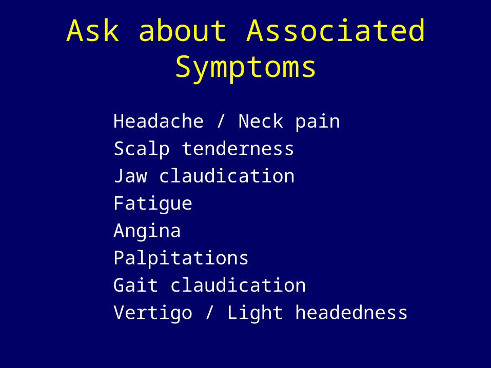
Ask about Associated Symptoms
Headache / Neck pain
Scalp tenderness
Jaw claudication
Fatigue
Angina
Palpitations
Gait claudication
Vertigo / Light headedness

List Medications
Anticoagulants (warfarin, heparin)
Antiplatelets (ASA, Plavix, ASA/Dipyridamal)
BP meds – time taken
Sildenafil (Viagra (EDD))
Cardiac meds
Statins (Lipitor etc)
Others
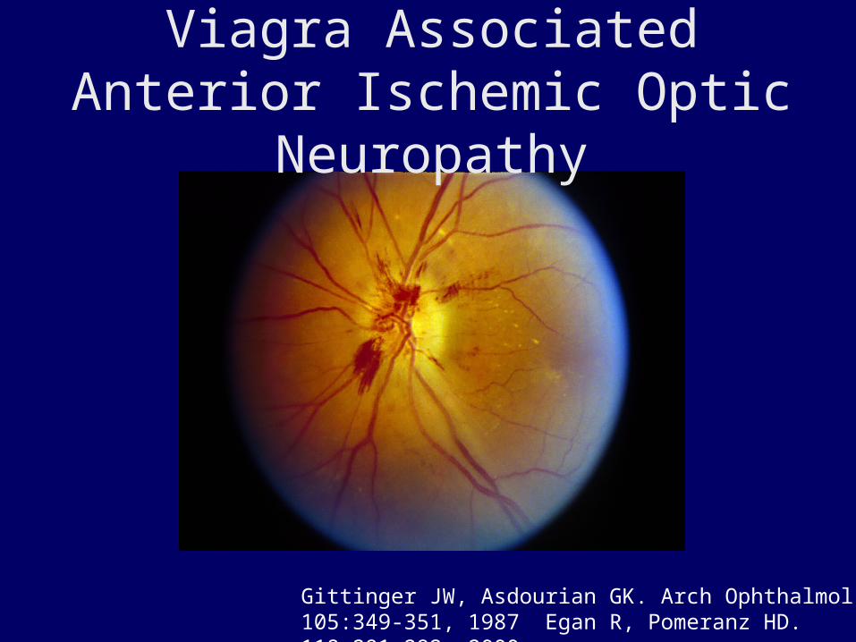
Viagra Associated Anterior Ischemic Optic Neuropathy
Gittinger JW, Asdourian GK. Arch Ophthalmol 105:349-351, 1987 Egan R, Pomeranz HD. 118:291-292, 2000
TMB Type I is one variety of an internal carotid artery distribution transient ischemic attack (TIA)
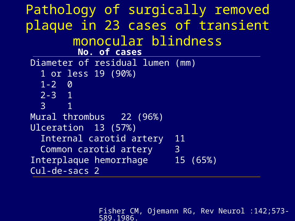
Pathology of surgically removed plaque in 23 cases of transient monocular blindness
No. of casesDiameter of residual lumen (mm)
1 or less 19 (90%)1-2 02-3 13 1
Mural thrombus 22 (96%)Ulceration 13 (57%)
Internal carotid artery 11Common carotid artery 3
Interplaque hemorrhage 15 (65%)Cul-de-sacs 2
Fisher CM, Ojemann RG, Rev Neurol :142;573-589.1986.
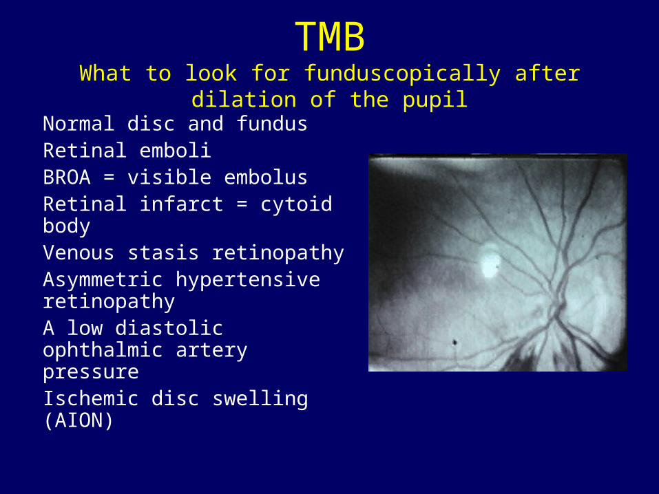
TMBWhat to look for funduscopically after dilation of the pupil
Normal disc and fundusRetinal emboliBROA = visible embolusRetinal infarct = cytoid bodyVenous stasis retinopathyAsymmetric hypertensive retinopathyA low diastolic ophthalmic artery pressureIschemic disc swelling (AION)


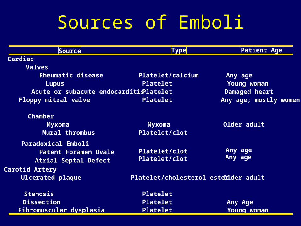
Sources of EmboliType Patient Age
CardiacValves
Rheumatic disease Platelet/calcium Any ageLupus Platelet Young womanAcute or subacute endocarditis Platelet Damaged heart
Floppy mitral valve Platelet Any age; mostly women
ChamberMyxoma Myxoma Older adultMural thrombus Platelet/clot
Carotid ArteryUlcerated plaque Platelet/cholesterol ester Older adult
Stenosis PlateletDissection Platelet Any AgeFibromuscular dysplasia Platelet Young woman
Source
Paradoxical EmboliPatent Foramen OvaleAtrial Septal Defect
Platelet/clotPlatelet/clot
Any ageAny age

Internal Carotid Stenosis
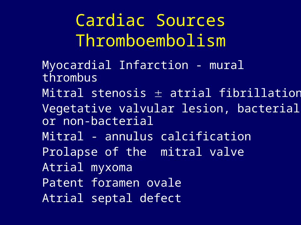
Cardiac Sources Thromboembolism
Myocardial Infarction - mural thrombusMitral stenosis atrial fibrillationVegetative valvular lesion, bacterial or non-bacterialMitral - annulus calcificationProlapse of the mitral valveAtrial myxomaPatent foramen ovaleAtrial septal defect
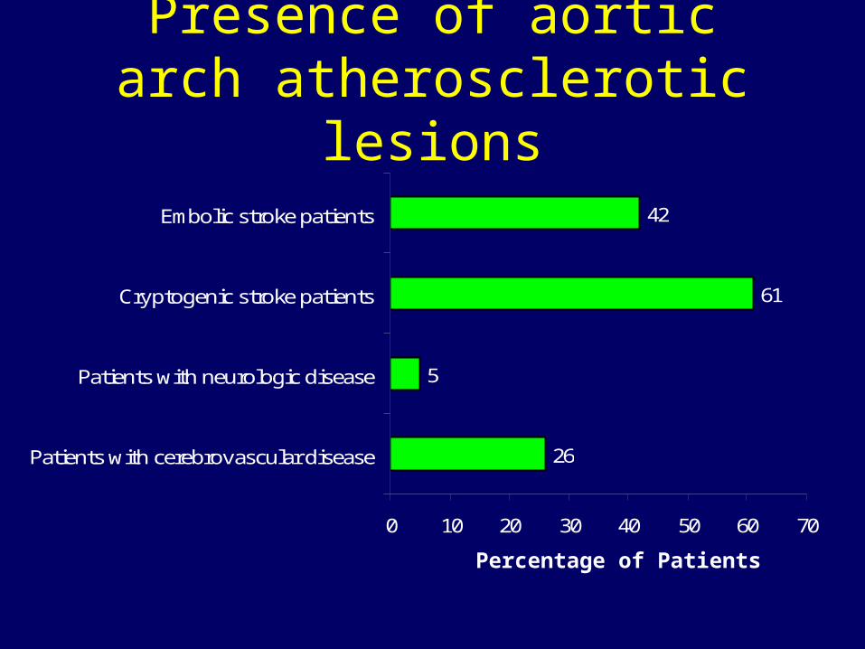
26
5
61
42
0 10 20 30 40 50 60 70
Patients with cerebrovascular disease
Patients with neurologic disease
Cryptogenic stroke patients
Embolic stroke patients
Presence of aortic arch atherosclerotic lesions
Percentage of Patients
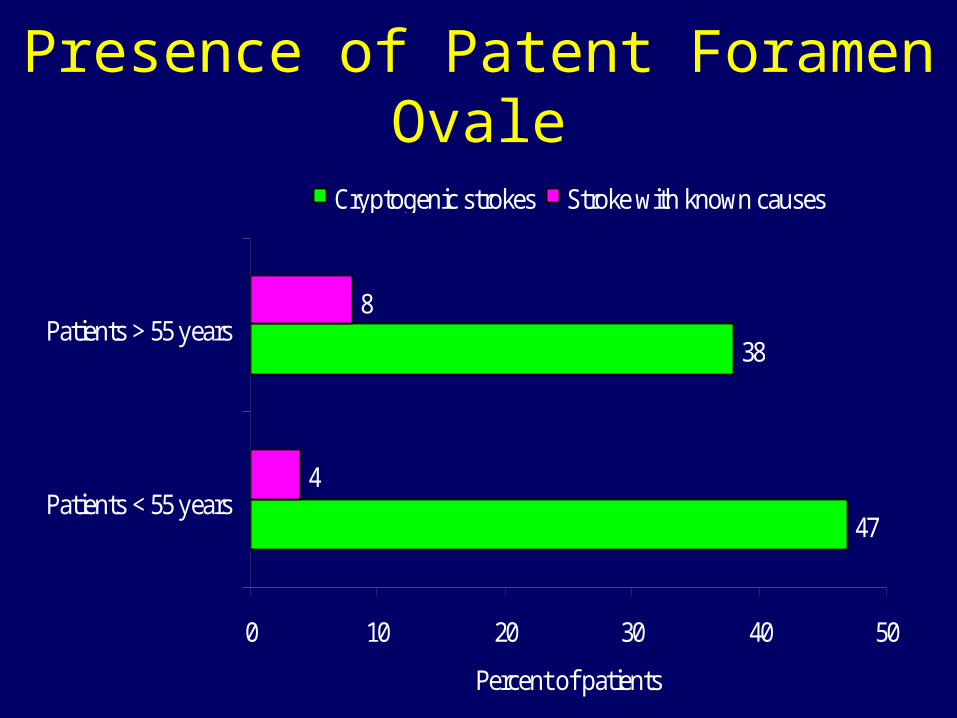
Presence of Patent Foramen Ovale
47
38
4
8
0 10 20 30 40 50
Patients < 55 years
Patients > 55 years
Percent of patients
Cryptogenic strokes Stroke with known causes

TEE w/ contrast of a PFO
A. Diastole
B. Opacification of the right atrium immediately post injection of agitated saline
C. Contrast passing from RA to LA through PFO (red arrow)
D. Large amount of contrast in LA (red arrow)
(Courtesy of Thomas Binder, MD Univ Vienna)


Bilateral Carotid Dissection
TMB Type II due to retinal vascular insufficiency

TMB Type II
A sudden attack of temporary monocular visual loss, less rapid in onset and longer in duration (minutes to up to two hours) in comparison with Type I. Recovery also takes place gradually.
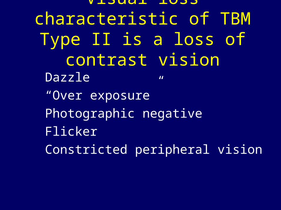
Visual loss characteristic of TBM Type II is a loss of contrast vision
Dazzle
“Over exposure”
Photographic negative
Flicker
Constricted peripheral vision
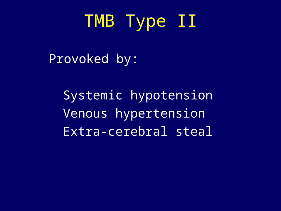
TMB Type II
Provoked by:
Systemic hypotension
Venous hypertension
Extra-cerebral steal
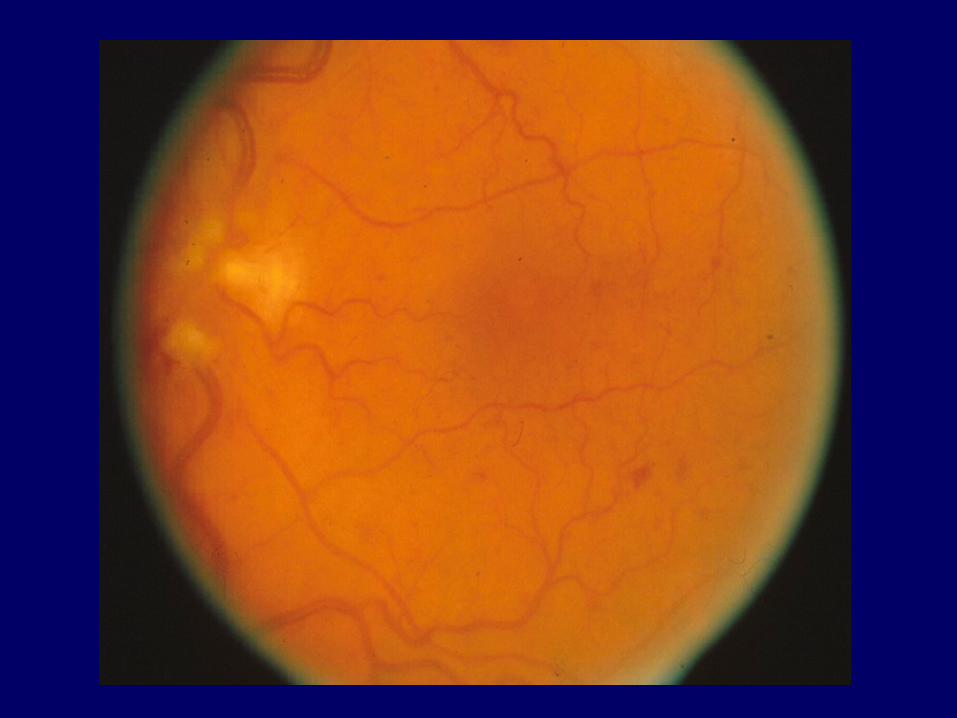
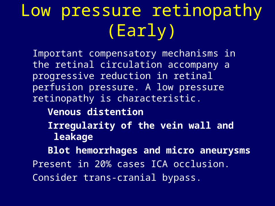
Low pressure retinopathy (Early)
Important compensatory mechanisms in the retinal circulation accompany a progressive reduction in retinal perfusion pressure. A low pressure retinopathy is characteristic.
Venous distention
Irregularity of the vein wall and leakage
Blot hemorrhages and micro aneurysms
Present in 20% cases ICA occlusion.
Consider trans-cranial bypass.

Low pressure retinopathy (Late)
Signs of anterior segment ischemia usually co-exist:
• Rubeosis of the iris• Neovascular changes in the anterior chamber• Secondary glaucoma and cataract formation
Trans-cranial bypass too late
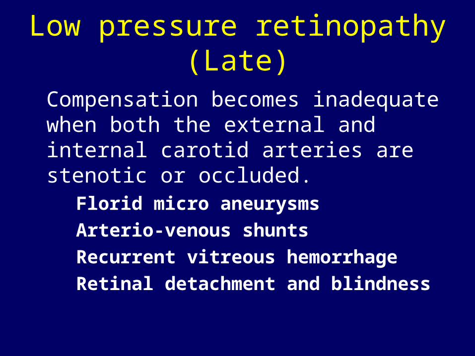
Low pressure retinopathy (Late)
Compensation becomes inadequate when both the external and internal carotid arteries are stenotic or occluded.
Florid micro aneurysms
Arterio-venous shunts
Recurrent vitreous hemorrhage
Retinal detachment and blindness

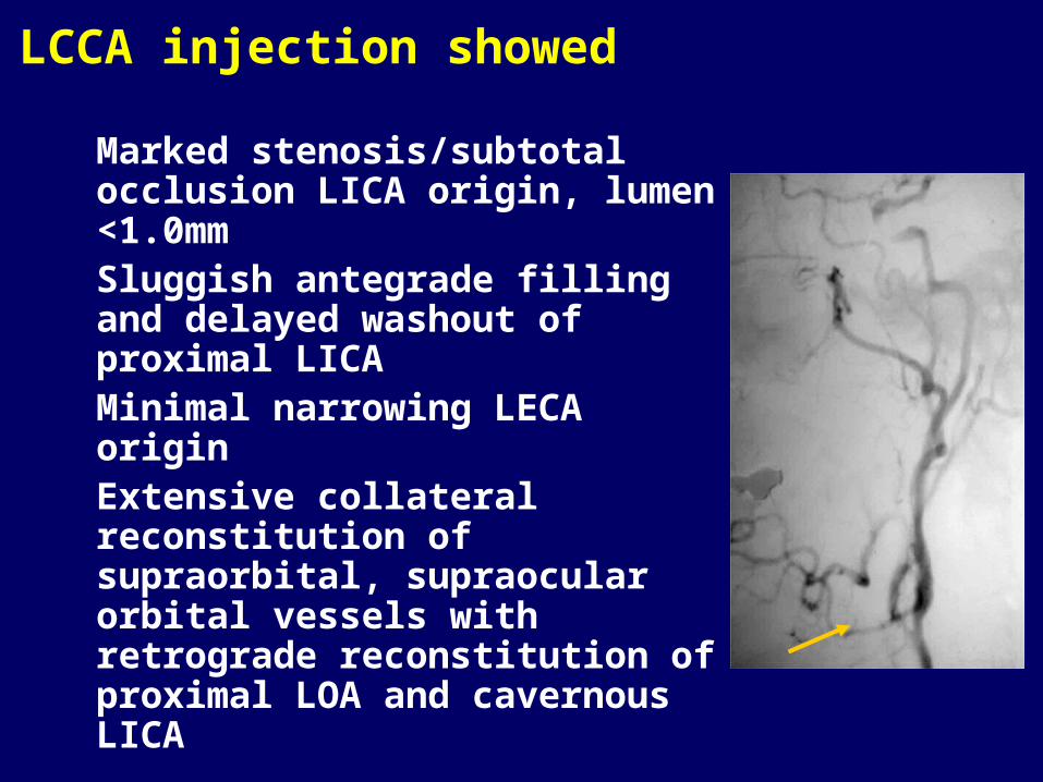
LCCA injection showed
Marked stenosis/subtotal occlusion LICA origin, lumen <1.0mmSluggish antegrade filling and delayed washout of proximal LICAMinimal narrowing LECA originExtensive collateral reconstitution of supraorbital, supraocular orbital vessels with retrograde reconstitution of proximal LOA and cavernous LICA

In TMB Type I and Type II the etiology may be giant cell arteritis where there is a similar state of impaired retinal perfusion
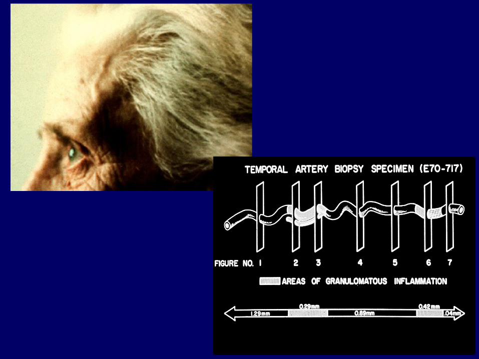
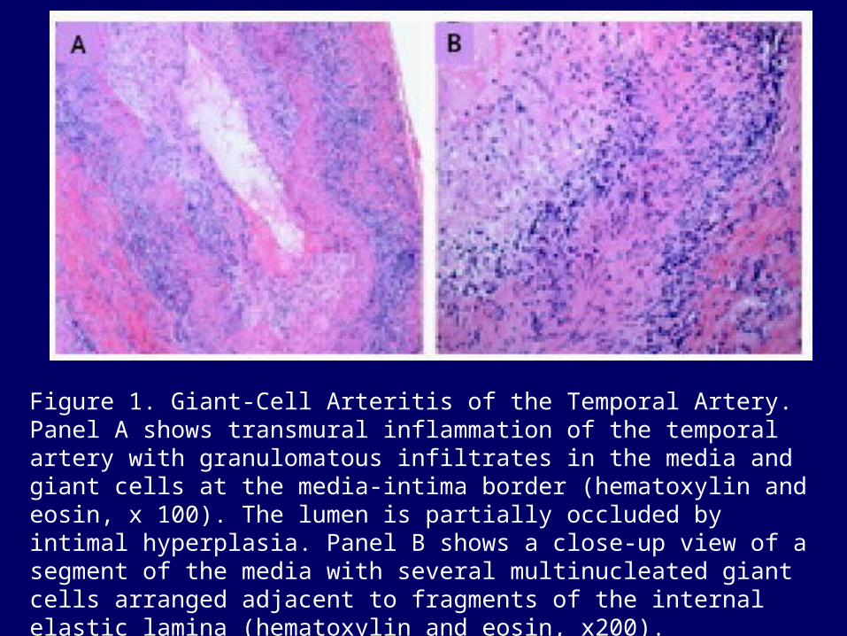
Figure 1. Giant-Cell Arteritis of the Temporal Artery. Panel A shows transmural inflammation of the temporal artery with granulomatous infiltrates in the media and giant cells at the media-intima border (hematoxylin and eosin, x 100). The lumen is partially occluded by intimal hyperplasia. Panel B shows a close-up view of a segment of the media with several multinucleated giant cells arranged adjacent to fragments of the internal elastic lamina (hematoxylin and eosin, x200).
Weyand: N Engl J Med, Volume 349(2).July 10, 2003.160-169
GCA is a T-cell dependent disease
CD4+ T cells orchestrate the vasculitic process
T-cell activation requires the activation of specialized antigen-presenting cells, the dendritic cells
Antigens recognized by CD4+ T cells are unknown
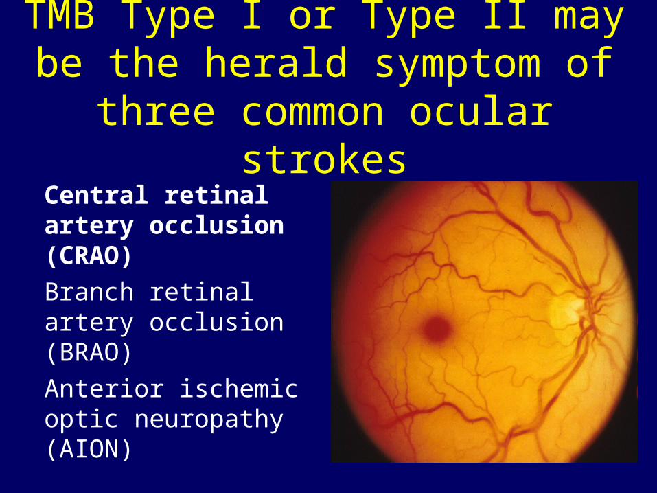
TMB Type I or Type II may be the herald symptom of three common
ocular strokes
Central retinal artery occlusion (CRAO)
Branch retinal artery occlusion (BRAO)
Anterior ischemic optic neuropathy (AION)
TMB Type III
Type III resembles Type II with less rapid onset and longer duration compared with Type I.
The mechanism is transient angiospasm.
In rare cases the retinal arteries appear narrow on funduscopy micro infarcts.
Berger, S.K. et al. TMB caused by vasospasm. NEJM, 1991, 325, 870-3
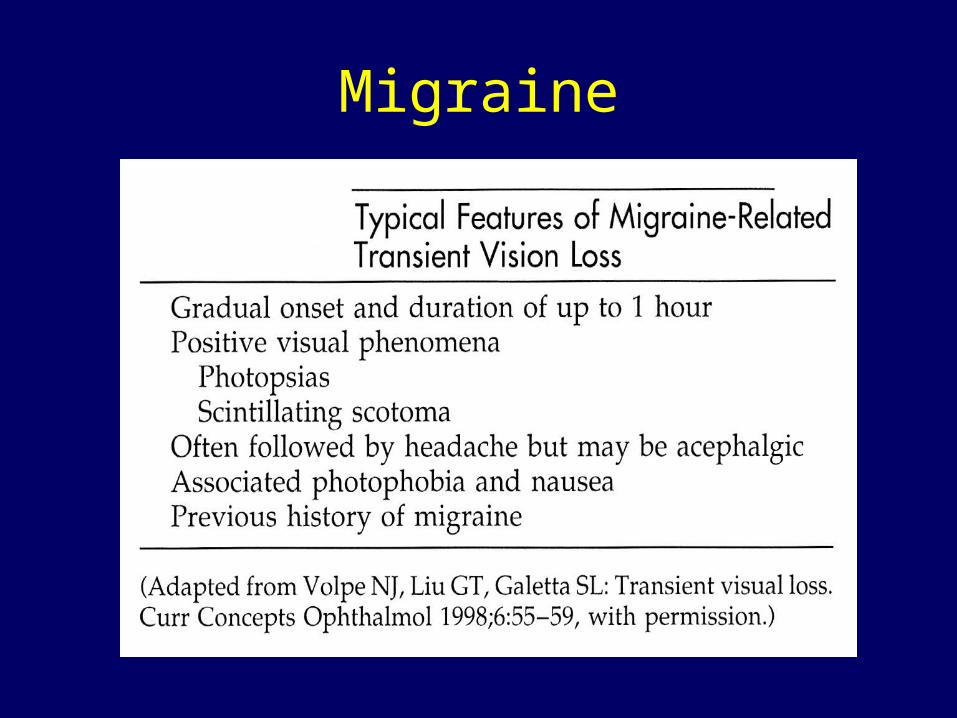
Migraine
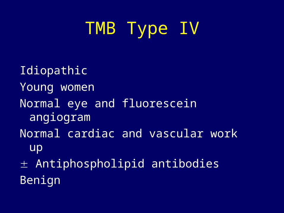
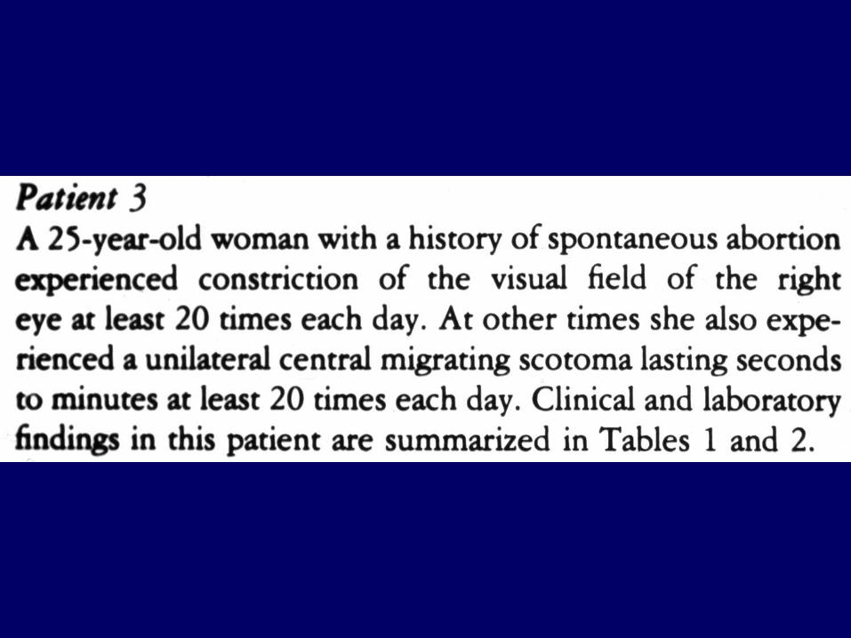
TMB Type IV
Idiopathic
Young women
Normal eye and fluorescein angiogram
Normal cardiac and vascular work up
Antiphospholipid antibodies
Benign


1985 1986 1987TMB n=33 38 43 38
Type I 24 31 34 42 (121) 80%Emboli 1 8 12 3 (24) 19%T. Arteritis 4 2 3 3 (12) 9%ICA sten. 4 8 2 4 (13) 10%Post. Endart. 1 - 1 - (2) 1%Fibromu. D. 1 - - - (1) -Heart D. 2 1 2 4 (9) 7%
Type II 3 4 2 1 (10) 6%Type III 2 1 2 2 (7) 4%Type IV 4 2 5 3 (14) 9%
1984 Total151

TMB EvaluationMeticulous historyOphthalmological exam / dilated
funduscopyCardiac and carotid Bruits
BP both arms rest and standingHeart rate / Holter / TEE
Sed rate / C-reactive protein / lipid panel /homocysteine / fasting glucose / HgA1c
Temporal artery biopsyHypercoagulable workupCarotid non-Invasive

Neuroimaging of TMB
Brain MRI (DWI/ADC)MRA of the head and neck (fat saturation)
CT/CTA head and neck if TMB + signs of infarction (reformatted 3-d reconstruction)
* If cardiac embolic source or PFO suspected, may consider cardiac CTA as well (research)

http://www.library.med.utah.edu/NOVEL