transient versus persistent functional and structural changes
TRANSCRIPT

The Journal of Neuroscience, November 1995, 15(11): 7517-7527
Transient versus Persistent Functional and Structural Changes Associated with Facilitation of Aplysia Sensorimotor Synapses Are Second Messenger Dependent
Fang Wu, Leah Friedman, and Samuel Schacher
Center for Neurobiology and Behavior, Columbia University College of Physicians and Surgeons, and New York State Psychiatric Institute, New York, New York 10032
Increases in activity of both protein kinase A (PKA) and protein kinase C (PKC) contribute to shot-&-term facilitation of Aplysia sensorimotor synapses evoked by serotonin (5- HT). We report here that increasing levels of CAMP in sen- sory neurons evokes increases in both synaptic efficacy and in the number of sensory neuron varicosities contact- ing the major axons of motor cell L7 at intermediate times (3 hr) that persist for 24 hr. Treatment with phorbol esters results in a large transient increase in synaptic efficacy that is accompanied by a large transient increase in the number of sensory neuron varicosities with the newest var- icosities most susceptible to elimination. The reversal of the synaptic facilitation and the structural changes does not appear to be the result of long-term inhibitory actions of persistent PKC activation by phorbol esters, since changes in synaptic efficacy can be evoked by additional applications of either phorbol esters or 5-HT. The short- lived changes in structure evoked by phorbol esters occur in preexisting sensory neurites and not by new growth, since increases in PKC activity with phorbol esters lead to reductions in neurite extension and to retractions by sen- sory neuron growth cones. The action of phorbol esters on growth cone extension is reversible with washout. The re- sults suggest that increases in PKA and PKC activities by 5-HT contribute to short (minutes) and intermediate (hours) forms of facilitation of sensorimotor synapses while in- creases in PKA activity also mediate long-term (days) maintenance of synaptic facilitation.
[Key words: protein kinase A, protein kinase C, synaptic plasticity, intermediate term, long term, sensorimotor syn- apse, Aplysia]
Changes in the activity of protein kinases play important roles in mediating short- and long-term changes in the properties of mature neurons and their connections. Changes in kinase activ- ities influence ionic conductances and excitability (Kandel and Schwartz, 1982; Acousta-Urquidi et al., 1984; Farly and Auer-
Received May 24. IYYS; rrvised July 14, 1995; accepted July lY, 1995.
We thank Robert Woolley for assistance in preparing the figures, and Drs. J. Koester. I. Kupfermann, and J. H. Schwartz for helpful comments on earlier drafts of the manuscript. This research was supported by NIH Grants GM
32099 and NS 27541. Correspondence should be addressed to Samuel Schacher, Center for Neu-
robiology and Behavior, Columbia University College of Physicians and Sur-
geons. New York State Psychiatric Instituie. 722 We\t 168th Street. New York, NY 10032.
Copyright 0 I995 Souety for Neuroscience 0270.6474/Y5/1575 17. I I $0.5.00/O
bath, 1986; Miller et al., 1992; McGlade-McCulloh et al., 1993; Raymond et al., 1993; Wang et al., 1994), the efficacy of syn- aptic transmission (Kandel and Schwartz, 1982; Malinow et al., 1988; Dixon and Atwood; 1989; Schuman and Clark, 1994). or the number and structural properties of transmitter release sites (Wojtowitz et al., 1989; Corfas and Dudai, 1991; Nazif et al., 1991; Zhong and Wu, 1991; Schacher et al., 1993). In some forms of hippocampal LTP, changes in the activities of several kinases, such as certain forms of Ca/calmodulin kinase (Malenka et al., 1989; Malinow et al., 1989; Silva et al., 1992). tyrosine kinase (O’dell et al., 1991; Grant et al., 1992), protein kinase C (Akers et al., 1986; Hu et al., 1987; Malinow et al., 1989; Abe- liovich et al., 1993), and protein kinase A (Frey et al., 1993; Bourtchuladze et al., 1994; Huang et al., 1994; Weisskopf et al., 1994), contribute in varying degrees to the induction or main- tenance of the plasticity. The order of activation of these kinases,
the critical substrates and sites of their activities in the presyn- aptic versus postsynaptic cells, and the time course of their ac- tivation and/or inactivation in mediating the change in synaptic
function are not well understood. Sensitization of defensive reflexes in Aplwitr is mediated in
part by the neurotransmitter 5-HT (Glanzman et al., 1989b) which evokes short- or long-term changes in the properties of the neurons in the circuit controlling the behaviors by affecting the activities of protein kinases. Various properties of mechano- sensory cells are affected by 5-HT: potassium and calcium con- ductances are altered which influence excitability and spike du- ration (Klein et al., 1982, 1986; Boyle et al., 1984; Ocorr and Byrne, 1985; Walsh and Byrne, 1989; Baxter and Byrne, 1990; Braha et al., 1993), evoked and spontaneous transmitter release from preexisting sensory terminals are increased (Brunelli et al., 1976; Hochner et al., 1986; Rayport and Schacher, 1986; Dale et al., 1988; Braha et al., 1990; Dale and Kandel, 1990; Klein, 1993, 1994), and the growth of new presynaptic branches and varicosities contacting target neurons are evoked with long-term facilitation (Glanzman et al., 1990; Bailey et al., 1992). Bio- chemical, physiological and pharmacological evidence indicate that many of the short-term changes in the sensory cells evoked
by 5-HT or sensitizing stimuli are mediated by increases in the activity of PKA and PKC (Brunelli et al., 1976; Castellucci et al., 1980, 1982; Bernier et al., 1982; Siegelbaum et al., 1982; Ocorr and Byrne, 1985; Klein et al., 1986; Braha et al., 1990; Sacktor and Schwartz, 1990; Mercer et al., 1991; Critz and Byrne, 1992; Sossin and Schwartz, 1992; Ghirardi et al., 1992; Sugita et al., 1992, 1994). Persistent activation of the PKA path- way (Greenberg et al., 1987; Sweatt and Kandel, 1989; Schacher
7518 Wu et al. * Duration of Facilitation Evoked by PKA versus PKC
et al., 1991; Backsai et al., 1993) can produce the long-term changes lasting at least 24 hr that are evoked by S-HT or sen- sitization training: increases in synaptic efficacy (Schacher et al., 1988, 1993; Alberini et al., 1994), increases in the number of presynaptic varicosities and branches (Nazif et al., 199 I ; Schacher et al., I993), and changes in potassium conductance (Scholz and Byrne, 1987, 1988). The contribution of 5-HT-induced increase in PKC activity to long-term presynaptic facilitation is not clear, since an increase in PKC activity with phorbol esters fails to evoke long-term facilitation (Schacher et al., 1988; Sossin et al., 1994) while pharmacological blockade primarily of the increase in PKC activity did not interfere with the expression of long term facilitation evoked with 5-HT (Emptage and Carew, 1993). However, recent evidence suggests that PKC activation may contribute to an intermediate phase of facilitation that lasts sev- eral hours. Treatments with 5-HT that evoke long-term facilita- tion first elicit a transient, protein synthesis-dependent phase of facilitation whose magnitude is greater than either short-term facilitation lasting minutes or long-term facilitation measured 24 hr after treatment (Ghirardi et al., 199.5). Treatment with 5-HT or repeated sensitizing stimuli also evoke a persistent activation of some PKC isoforms in the sensory cells that lasts several hours after the completion of the facilitating stimulus (Sossin et al.. 1994).
What ire the contributions of increasing the activity of each of these second messenger pathways in evoking functional and structural changes in the sensorimotor connection during inter- mediate phases of facilitation (hours) and to long-term facilita- tion (days)‘? Taking advantage of the simple in vitro preparation (Rayport and Schacher, l986), we examined the functional and structural changes in sensorimotor connections over time follow- ing prolonged activation of each second messenger pathway. Increasing CAMP levels in the sensory neurons evoked a func- tional change at intermediate times (three hours), while the struc- tural change is expressed partially at the intermediate time point and is further developed after 24 hr. By contrast, treatment with an activator of PKC evoked a large increase both in the efficacy of the connection and in the number of sensory varicosities at intermediate times, but these changes were no longer expressed by the cells 24 hr after treatment. The decline in varicosity num- ber after the intermediate time point was correlated with the elimination of the newest varicosities. Our results are consistent with the hypothesis that persistent activation of the PKC and PKA pathways by 5-HT can act in parallel in producing short- and intermediate-term synaptic facilitation of this connection. Persistent changes in synaptic efficacy and sensory neuron struc- ture require the critical contribution of increases in PKA activity that stabilizes both local changes in preexisting terminals and neurites as well as other changes that are essential for the growth and maintenance of new presynaptic structures.
Materials and Methods
Crll c-u/fur-r. Mechanosensory neurons of Apiysicc were isolated from the pleural ganglion dissected from adult animals (70-100 gm) and were cultured either alone or cocultured with identified motor cell L7 isolated from the abdominal ganglion of juvenile animals (l-3 gm; University of Miami Mariculture Facility) and maintained for up to 5 d as described previously (Schacher and Proshansky, 1983; Schacher, 1985; Rayport and Schacher, 1986). Individual cells were isolated with a segment (100-600 km) of their original axons intact (Schacher and Proshansky, 1983). Each culture contained either a single sensory cell cocultured with a single L7 or S-IO sensory cells. Cocultures were allowed to grow processes for 4 d to permit the establishment of stable synaptic contacts and neuritic arbors (Montarolo et al., 1986; Dale et al., 1988;
Glanzman et al., 1990; Schacher and Montarolo, 1991). Cultures with sensory cells alone were examined after 3 d to determine the effects of PKC activation during the period of rapid neurite growth (Peter et al., 1994).
Electrophysiology. The stimulation and recording techniques for mea- suring changes in the efficacy of the SN-L7 connection have been de- scribed (Montarolo et al., 1986). For monitoring changes in connectiv- ity, the motor cell was impaled with a microelectrode (15-20 M(2) containing 2.0 M KCI and held at a potential of -30 mV below the resting level (-49 to -65 mV) to permit accurate measurements of the amplitude of the EPSI? For each coculture, synaptic potentials were evoked in L7 before treatments and at 3 and 24 hr after the onset of the control or experimental treatments by stimulating the sensory neu- ron with a single brief (SO msec) depolarization using the dye-filled intracellular electrode after the iontophoretic injection of the dye. Dur- ing the recording and dye-filling, cultures were superfused at I ml/min with medium consisting of artificial sea water (Instant Ocean) and mod- ified Ll5 with salt concentrations added to levels consistent with sea water (Schacher et al., 1990).
Drug applications. After recording the initial amplitudes of the EPSPs and imaging sensory neurites interacting with the major axon of L7 (see below), cocultures were divided into four groups. Cultures were incubated for 2 hr with 50 nM phorbol 12, I3-dibutyrate (Sigma; PDBU or phorbol) or 100 nM 4cY-phorbol (Sigma; inactive form referred in the text as c-u-phorbol) dissolved in perfusion medium. Alternatively, CAMP or S’AMP (Sigma) was injected via iontophoresis for IO min (Scholz and Byrne, 1988; Schacher et al., 1993) with intracellular microelec- trodes filled with 200 mM CAMP or S’AMI? Beginning IO min before and ending IO min after the injection, cultures were perfused with 100 FM IBMX (Sigma) in perfusion medium (Schacher et al., 1993). All cultures were rinsed 4X with perfusion medium, and reexamined three hours after starting drug incubation or intracellular injection. Cultures were rinsed with culture medium, placed back into the incubator and reexamined for a third time after 24 hr. Another set of cocultures were used to examine the long-term consequences of PDBU treatment. EPSPs were recorded before and 24 hr after a 2 hr treatment with PDBU or cy-phorbol as described above. At the second recording, either S-HT (2 pM) was applied to the cultures after the sensory neuron was given IO stimuli at 30 set intervals to produce homosynaptic depression, or PDBU was applied for I hr after a single test stimulus to determine whether the synapse can undergo synaptic facilitation. To examine ef- fects of PDBU on sensory neuron growth, cultures with sensory neurons only that had IO or more sensory growth cones were first rinsed with perfusion medium and every growth cone was photographed before the 2 hr incubation with PDBU or a-phorbol. The cultures were rinsed with perfusion medium and neurites and growth cones re-photographed start- ing 30 min or 4 hr after washout. Cultures treated with PDBU that had at least 10 growth cones that had stopped or retracted with treatment were reexamined at 4 hr after washout. The percent for each growth cone behavior was measured in each culture and averaged over all the cultures (Peter et al., 1994).
Dye injrctim und imuging structurd charrgrs. The fluorescent dye 5(6)-carboxyfluorescein (Molecular Probes; 6% in 0.44 M KOH; pH = 7.0) was injected into the sensory cell in the cocultures with 0.3-0.5 nA hyperpolarizing current pulses (500 msec at I Hz) for 5-6 min (Glanzman et al., 1989a; Schacher and Montarolo, 1991) at each time point. Nomarski contrast and fluorescent images of the same view areas along the major axons of the motor cell were taken both before and after treatments with a Nikon Diaphot microscope and a SIT (Dage 66) video camera. The images were processed by a Dell 3 IO computer with a PC Vision Plus frame grabber, and subsequently stored on a Panasonic optical disk drive. Alignment of the live view area at the 3 hr and 24 hr time points with the initial recorded image was aided by the computer with line adjustments made with the stage controls and by manual ro- tation of the culture dish. Illumination used for obtaining fluorescent images was kept as low as possible to prevent photodamage. To mini- mize differences in imaged structures that might arise as a result of differences in the extent of dye filling, light intensities used at the 3 hr and 24 hr points were adjusted to match the intensity of the stored images taken before treatment. Phase contrast Micrographs of the sen- sory growth cones before and after treatments were used to measure changes in growth cone behavior. Micrographs of the images were made with a Panasonic or Sony video printer.
Qumr(ficurion qf .structurul change. Counts of varicosity number were obtained from fluorescent images of sensory cell neurites con-
The Journal of Neuroscience, November 1995, 75(11) 7519
tatting the axon hillock and major processes of L7. Previous studies had indicated that this region of L7 is the most favorable for the growth of sensory neurites, and contains most of the varicosities and transmitter release sites (Glanzman et al., 1989a, 1990; Schacher et al., 1991). Since the major axons of L7 are relatively thick structures, it often required as many as four different focal planes to image all of the labeled neu- rites and varicosities in a given view area. To minimize slight differ- ences in focus which could obscure differences in varicosity number, we used computer-assisted superimposition of the various focal planes onto a single two-dimensional image. The matched fluorescent images of each focal plane along with the superimpositions for the three time points were compared and the total number of varicosities counted. Net change in varicosity number was measured for all treatments. We also examined the effect of treatment on the maintenance of the new vari- cosities. A varicosity (swelling along a sensory cell process greater than 2 pm in diameter) was considered new if the structure was not located within a 2 pm radius in the same region of the motor axon on an earlier image of the area. The varicosity was considered to be eliminated if the structure was absent within a 2 pm radius in the same region of the motor axon. Structures that were slightly elongated spheres greater or equal to 2 pm connected by narrow neuritic necks were counted as varicosities (Bailey and Chen, 1983, 1988). Counts of varicosities were performed blind; the individual did not know the amplitude of the EPSPs before or after treatment or the nature of the treatment. Growth cone behaviors in sensory neuron cultures were scored for extension or retraction behavior; growth of 10 pm or more over the approximately 3 hr time period was scored as extension; retraction of 10 pm or more over the same period was scored as a retraction, and intermediate be- havior was scored as a stop).
Analysis of data. All data are represented as the mean t SEM. Stu- dent i tests (two-tailed) or ANOVA’s (one- or two-factor) followed by corrected multicomparison r tests (Dunnett’s) were used to measure sig- nificance of the change in EPSP amplitude, and the number of sensory varicosities with treatment over time, and the effect of treatment on sensory neuron growth cone behavior.
Results Activation of PkVA pathway evokes an early change in both the function and structure of sensorimotor connections that persists for 24 hr During the first 4 d in culture, the regenerating neurons reesta- blish stable functional connections (Rayport and Schacher, 1986; Glanzman et al., 1989a; Zhu et al., 1994). The stability of the connection is strongly correlated with the stability of the sensory neuron arbor both in the overall number of presynaptic varicos- ities interacting with the motor axon as well as in the mainte- nance of existing varicosities (Bank and Schacher, 1992; Zhu et al., 1995). Applications of 5-HT to 4 or 5 d old cultures evoke a functional change in the connection lasting at least 24 hr that is accompanied by the formation of new sensory cell varicosities and transmitter release sites in contact with the major axonal processes of motor cell L7 (Glanzman et al., 1989a, 1990; Schacher et al., 1991). Recent studies indicated that the efficacy of sensorimotor connections show as much as a 200% enhance- ment at intermediate times (0.5-3 hr) after repeated applications of 5-HT compared to the long-term change of about 80% de- tected 24 hr after treatment (Ghirardi et al., 1995). Raising in- tracellular CAMP in the sensory neurons (Schacher et al., 1993) evokes increases in the efficacy of the connection at both the short-term (80% at 10 min) and long-term (60% at 24 hr) time points, and the long-term change with CAMP is accompanied by the formation of new sensory varicosities and branches (Schach- er et al., 1993). We first examined whether raising CAMP levels in sensory cells with intracellular injections of CAMP evoke large functional and structural changes at an intermediate time point (3 hr). We also compared the changes at 3 hr to the changes observed for the same preparations at 24 hr. After al- lowing the cells to regenerate and form stable connections after
“AMp’O Hr
B 4c
c 50
s 40 i
0 Hr
J 3 Hr
r’ 3 Hr
3 3 ‘;; 30-
s ‘5 20-
3
z lo- ul
.r I
0 Hr 3 Hr
-......._. b -I
24 Hr
n VAMP 0 q lC.4MP
i 24 Hr
24 Hr
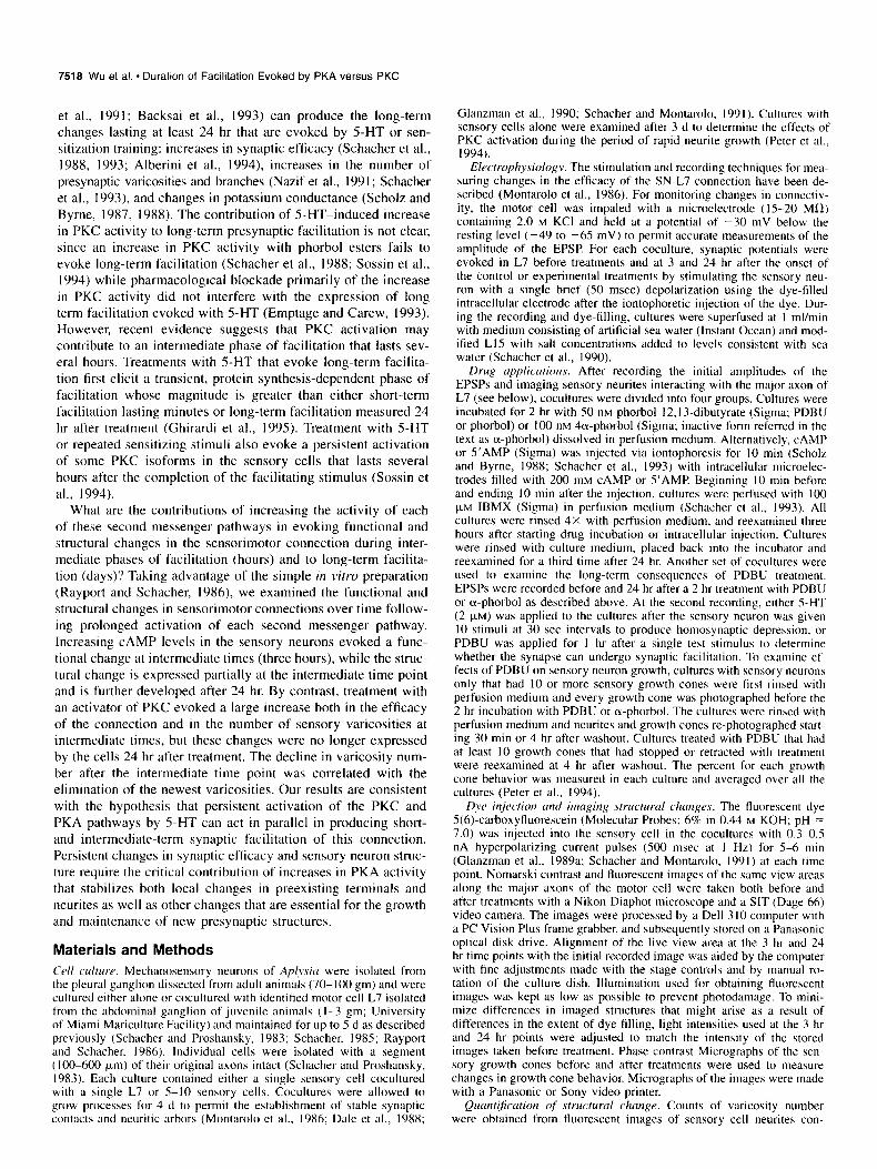
Figure 1. CAMP evoked persistent functional and structural changes in the SN-L7 connections. A, The efficacy of the connection is enhanced by injection with CAMP, but not with S’AMP EPSPs were recorded before (0 hr) and twice after treatment (3 hr and 24 hr). Whereas the amplitude of the EPSP declines with S’AMP (top), CAMP increases the EPSP amplitude both at 3 hr and 24 hr. The vertical bar equals 5 mV, and the horizontal bar equals 10 msec. B, Summary of the changes in EPSP amplitude evoked by S’AMP (solid bars) and CAMP (white bars). The height of each bar is the average ampltitude t SEM of the EPSP at each time point. A two-factor ANOVA indicated a significant effect of treatment over time (df = 2,28, F = 53.778, p < 0.001). One-factor ANOVA and t tests indicated that CAMP evoked a significant change in the EPSP amplitude at both the 3 hr and 24 hr time points. C, Sum- mary of the changes in the number of sensory neuron varicosities evoked by S’AMP and CAMP The height of each bar is the average number of sensory varicosities t SEM at each time point. A two-factor ANOVA indicated a significant effect of treatment over time (df = 2,28, F = 42.813, p < 0.001). One-factor ANOVA and r tests indicated that CAMP evoked a significant change in varicosity number at both the 3 hr and 24 hr time points. Note that the difference between CAMP and AMP at 24 hr is greater than the difference at 3 hr.
4 d, the amplitude of the EPSP evoked in motor cell L7 was measured before and twice after (at 3 hr and 24 hr) intracellular injection (10 min) of CAMP or S’AMP (Scholz et al., 1988; Nazif et al., 1991; Schacher et al., 1993). At the same time points, the structure of the sensory cell net&es interacting with major axonal processes of L7 were imaged and photographed following intracellular injection of the fluorescent dye 5(6) car- boxyfluorescein (Glanzman et al., 1989a, 1990).
7520 Wu et al. * Duration of Facilitation Evoked by PKA versus PKC
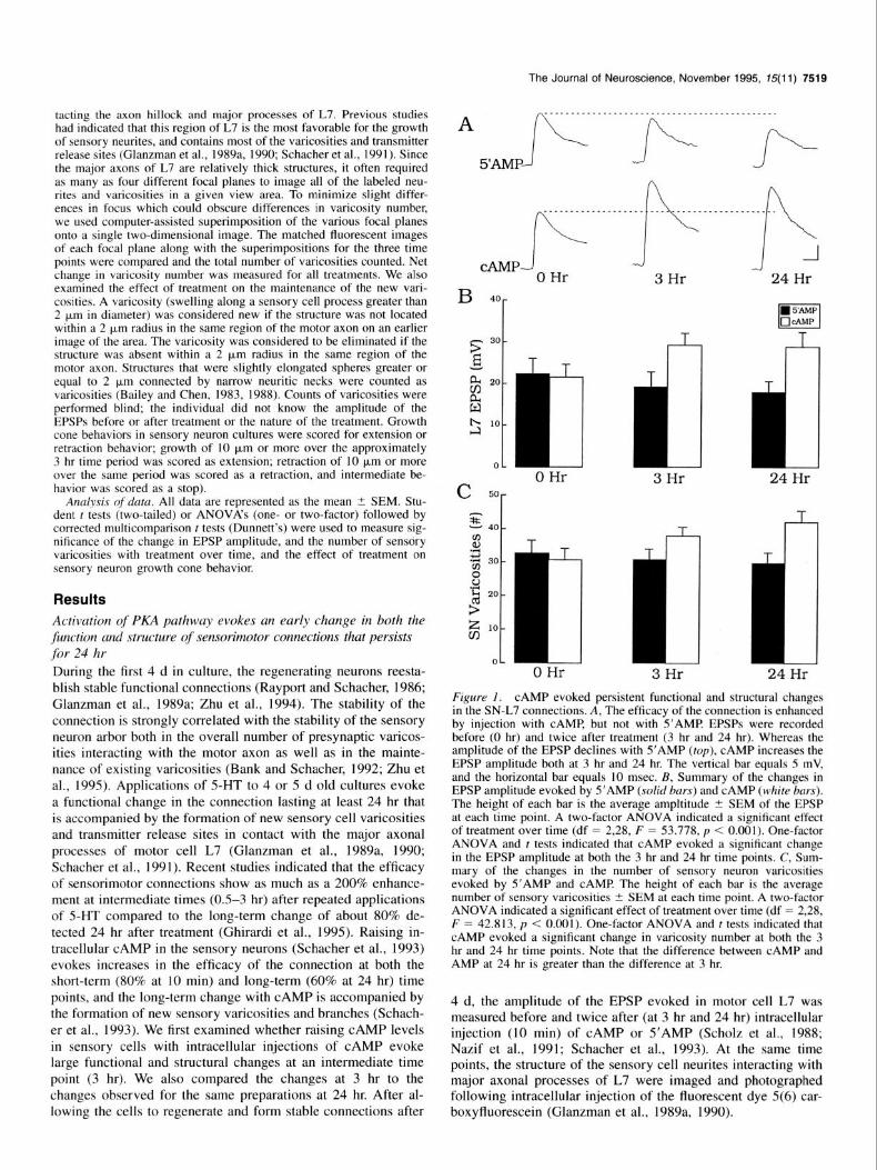
Figure 2. CAMP evoked a persistent change in the structure of the sensory neurons. A, Epifluorescent view of a portion of the sensory cell neurites and varicosities in contact with the major motor axon (middle half of micrograph in the horizontal plane) before injection with CAMP Image is a superimposition of three focal planes photographed in this view area. B, View of the same area in A 3 hr after injection of CAMP Three new varicosities (arrows) have formed in this region. Overall, this sen- sory neuron gained six varicosities. C, View of the same area at 24 hr. Note the new branches and varicosities (ar- rowhead) near the center of the field. There was a net gain of six varicosities in this region. Two of the three new varicosities (arrows) established at the 3 hr time point are still present. Overall there was a net gain of 13 varicosities for this sensory neuron compared to the 0 hr time point. Scale bar, 15 pm.
The Journal of Neuroscience, November 1995, 75(11) 7521
Injection of CAMP into the sensory neurons, but not S’AMP evoked an increase in the amplitude of the EPSP at both the 3 hr and 24 hr time points (Fig. IA,@. The change in EPSP am- plitude evoked by cAMP at 3 hr was not different than the change measured at 24 hr. Starting from the same level of syn- aptic efficacy before experimental or control treatments (2 1.7 mV _+ 2.8 vs 22.5 mV +- 3.5, p > 0.8; 0 hr), injection with CAMP (N = 8) evoked significant increases of 7.6 mV (to 29.3 mV t 2.7; p < 0.04) by 3 hr and 7.2 mV (28.9 mV t 3.4; p < 0.02) at 24 hr, while injection with S’AMP (N = 8) resulted in small declines of -3.2 mV after 3 hr (19.3 mV + 3.4) and -4.5 mV (18.0 mV Ifr 2.5) after 24 hr.
The increases in EPSP amplitude evoked by CAMP were ac- companied by structural changes in the sensory cell neurites that increased in magnitude over time (Figs. lC, 2). After 3 hr, there was a net average increase of 7.0 varicosities in the number of sensory neuron varicosities contacting the major axonal pro- cesses of L7 (30.8 -+ 3.2 to 37.8 t 2.7) compared to a slight decline of - 1.9 varicosities following injections with S’AMP (32.9 t 3.5 to 31.0 t 2.8; p < 0.01). The new varicosities with CAMP injections formed primarily as swellings along preexist- ing sensory cell neurites (Fig. 2A,B). With reexamination of the same cultures 21 hr later, we observed that additional varicosi- ties were formed on both preexisting and newly formed neurites (Fig. 2C) raising the net average increase to 11.3 varicosities (to 42.1 ? 3.2; p < 0.01). Approximately 2/3 (67.5%) of the new varicosities formed by 3 hr after injection were still present at 24 hr. At 24 hr, injection with S’AMP resulted in a small decline of -3.1 varicosities (to 29.8 + 3.1). Thus, CAMP elevation leads to structural changes in the sensory neuron that continue over a prolonged period and may serve to consolidate the change in synaptic efficacy expressed during short-term (Schacher et al., 1988, 1993) and intermediate-term time points.
Activation of the PKC pathway evokes a large transient increase both in the efficacy of sensorimotor connections and in the number of sensory varicosities
In previous studies, increases in PKC activity in sensory neurons significantly facilitated transmitter release for short durations (Braha et al., 1990), while persistent activation for at least 2 hr failed to enhance the efficacy of the connection after 24 hr (Schacher et al., 1988). Recent evidence indicates that treatments with 5-HT that evoke long-term facilitation lead to a persistent activation of some PKC isoforms for about 2-3 hr (Sossin et al., 1994). To test whether persistent activation of the PKC pathway can produce facilitation at intermediate time points, we exam- ined both the change in synaptic efficacy and in sensory cell structure as described above both 1 hr and 24 hr after a 2 hr incubation with phorbol ester.
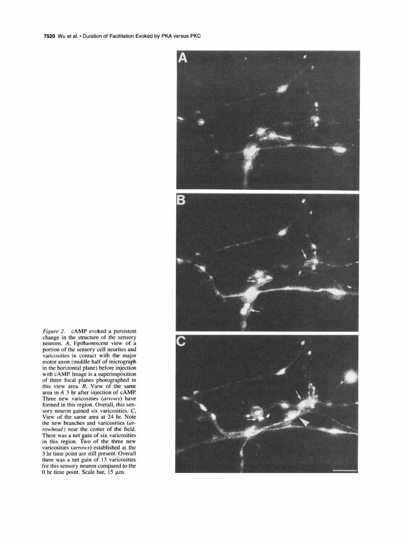
Incubation with phorbol, but) not the inactive a-phorbol, evoked a significant increase in the amplitude of the EPSP at the intermediate time point (3 hr), but not after 24 hr (Fig. 3A,B). Starting from the same level of synaptic efficacy before exper- imental or control incubations (29.7 mV -C 2.5 vs 26.5 mV t 5.2, p > 0.6; 0 hr), phorbol evoked a significant increase of at least 27.3 mV (to 57.0 mV rfr 3.0; p < 0.01; N = 6) by 3 hr compared to a small decline of - 1.7 mV (to 24.8 mV ? 4.5) following incubation with a-phorbol (N = 6). The large change in EPSP amplitude evoked with phorbol at 3 l-u is an underes- timate of the actual level of facilitation, since in four of six cultures the stimulus to the sensory neuron evoked an action potential in the motor cell (measured as a 60 mV EPSP) despite
A A---
. . . . . . . . . .._........................
a-Phor -Ii-
Phor
Ofi B 60 -
- 50 - > a40 -
P OHr L z50
- L
3Hr
rrl 3Hr
i 3Hr
24 Hr
24 Hr
24Hr
Figure 3. PDBU evoked transient functional and structural changes in the SN-L7 connections. A, The efficacy of the connection is enhanced by PDBU at 3 hr, but not at 24 hr. EPSPs were recorded before (0 hr) and twice after treatment (3 hr and 24 hr). Whereas the amplitude of the EPSP was unchanged with a-phorbol (top, o-Phor), PDBU (Phor) increased significantly the EPSP amplitude at 3 hr. By 24 hr the EPSP amplitude returned to baseline. The vertical bar equals 10 mV, and the horizontal bar equals 10 msec. B, Summary of the changes in EPSP amplitude evoked by a-phorbol (Lu-Phor, solid bars) and PDBU (Phor, open bars). The height of each bar is the average amplitude of the EPSP at each time point. A two-factor ANOVA indicated a significant effect of treatment over time (df = 2,20, F = 74.423, p < 0.001). One-factor ANOVA and t tests indicated that PDBU evoked a significant change in the EPSP amplitude at the 3 hr time point, but not at the 24 hr time point. C, Summary of the changes in the number of sensory neurons varicosities evoked by a-phorbol and PDBU. The height of each bar is the average number of sensory varicosities at each time point. A two- factor ANOVA indicated a significant effect of treatment over time (df = 2,20, F = 32.654, p < 0.001). One-factor ANOVA and t tests in- dicated that PDBU evoked a significant change in varicosity number at the 3 hr time point, but not at the 24 hr time point.
the fact that the motor cell was hyperpolarized by 30 mV (from -80 to -95 mV) below their resting potential. Similar large short-term facilitation can be evoked in some sensorimotor cul- tures with application of 2 PM 5-HT. By 24 hr however, the amplitudes of the EPSP in cultures treated with phorbol had returned to approximately pretreatment levels (to 3 1.8 mV 2 2.8) and were no longer significantly different than those treated with inactive a-phorbol (27.5 mV t: 4.4; p > 0.4).
The return to baseline was not accompanied by long-term
7522 Wu et al. * Duration of Facilitation Evoked by PKA versus PKC
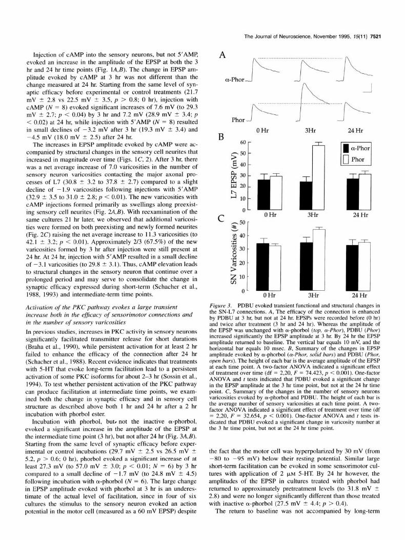
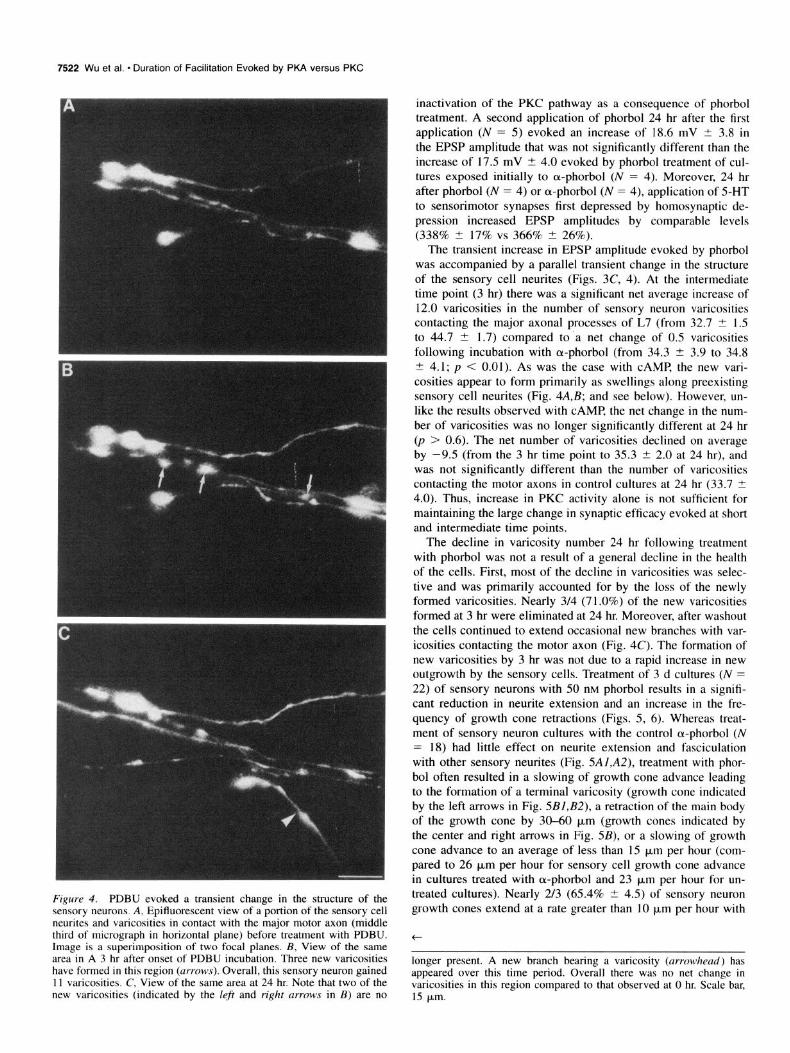
Figure 4. PDBU evoked a transient change in the structure of the sensory neurons. A, Epifluorescent view of a portion of the sensory cell neurites and varicosities in contact with the major motor axon (middle third of micrograph in horizontal plane) before treatment with PDBU. Image is a superimposition of two focal planes. B, View of the same area in A 3 hr after onset of PDBU incubation. Three new varicosities have formed in this region (arrows). Overall, this sensory neuron gained 11 varicosities. C, View of the same area at 24 hr. Note that two of the new varicosities (indicated by the left and right arruws in B) are no
inactivation of the PKC pathway as a consequence of phorbol treatment. A second application of phorbol 24 hr after the first application (N = 5) evoked an increase of 18.6 mV t 3.8 in the EPSP amplitude that was not significantly different than the increase of 17.5 mV t 4.0 evoked by phorbol treatment of cul- tures exposed initially to ol-phorbol (N = 4). Moreover, 24 hr after phorbol (N = 4) or ol-phorbol (N = 4). application of 5-HT to sensorimotor synapses first depressed by homosynaptic de- pression increased EPSP amplitudes by comparable levels (338% 2 17% vs 366% t 26%).
The transient increase in EPSP amplitude evoked by phorbol was accompanied by a parallel transient change in the structure of the sensory cell neurites (Figs. 3C, 4). At the intermediate time point (3 hr) there was a significant net average increase of 12.0 varicosities in the number of sensory neuron varicosities contacting the major axonal processes of L7 (from 32.7 ? 1.5 to 44.7 t 1.7) compared to a net change of 0.5 varicosities following incubation with cu-phorbol (from 34.3 2 3.9 to 34.8 I? 4.1; p < 0.01). As was the case with CAMP, the new vari- cosities appear to form primarily as swellings along preexisting sensory cell neurites (Fig. 4A,B; and see below). However, un- like the results observed with CAMP, the net change in the num- ber of varicosities was no longer significantly different at 24 hr (p > 0.6). The net number of varicosities declined on average by -9.5 (from the 3 hr time point to 35.3 t 2.0 at 24 hr), and was not significantly different than the number of varicosities contacting the motor axons in control cultures at 24 hr (33.7 -C 4.0). Thus, increase in PKC activity alone is not sufficient for maintaining the large change in synaptic efficacy evoked at short and intermediate time points.
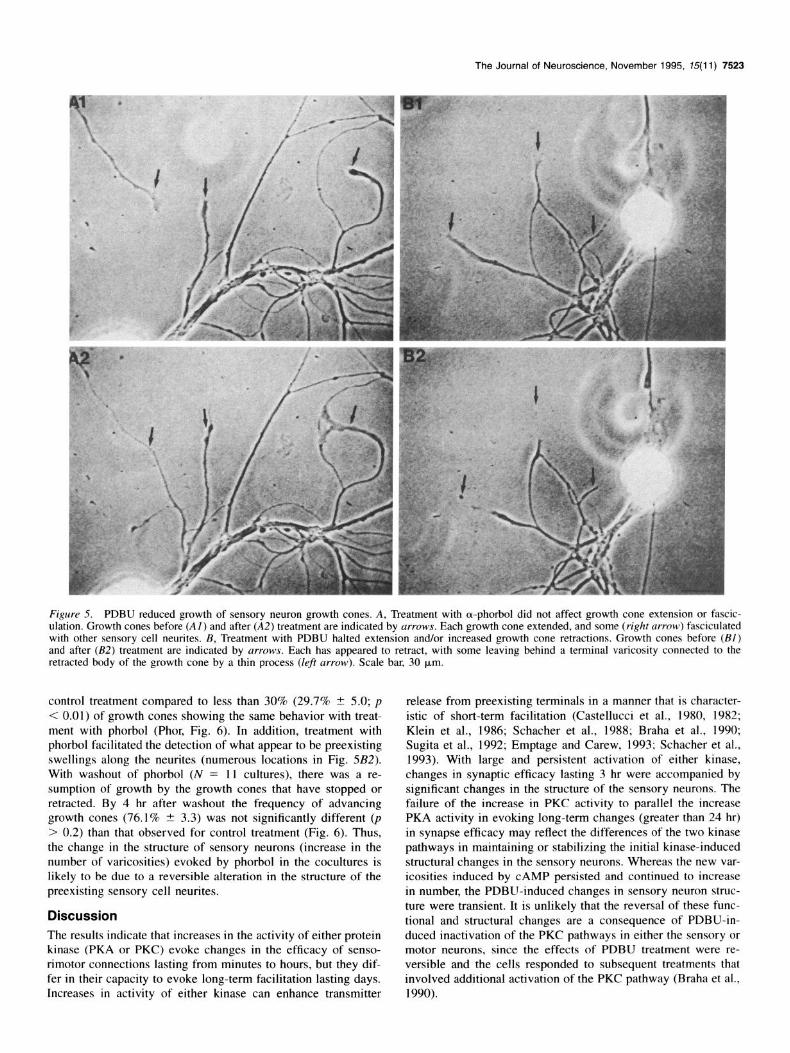
The decline in varicosity number 24 hr following treatment with phorbol was not a result of a general decline in the health of the cells. First, most of the decline in varicosities was selec- tive and was primarily accounted for by the loss of the newly formed varicosities. Nearly 3/4 (71.0%) of the new varicosities formed at 3 hr were eliminated at 24 hr. Moreover, after washout the cells continued to extend occasional new branches with var- icosities contacting the motor axon (Fig. 4C). The formation of new varicosities by 3 hr was not due to a rapid increase in new outgrowth by the sensory cells. Treatment of 3 d cultures (N = 22) of sensory neurons with 50 nM phorbol results in a signifi- cant reduction in neurite extension and an increase in the fre- quency of growth cone retractions (Figs. 5, 6). Whereas treat- ment of sensory neuron cultures with the control a-phorbol (N = 18) had little effect on neurite extension and fasciculation with other sensory neurites (Fig. 5Al,A2), treatment with phor- bol often resulted in a slowing of growth cone advance leading to the formation of a terminal varicosity (growth cone indicated by the left arrows in Fig. 5BI,B2), a retraction of the main body of the growth cone by 30-60 p,rn (growth cones indicated by the center and right arrows in Fig. 5B), or a slowing of growth cone advance to an average of less than 15 pm per hour (com- pared to 26 pm per hour for sensory cell growth cone advance in cultures treated with a-phorbol and 23 p,rn per hour for un- treated cultures). Nearly 2/3 (65.4% + 4.5) of sensory neuron growth cones extend at a rate greater than 10 p,rn per hour with
t
longer present. A new branch bearing a varicosity (arrowhead) has appeared over this time period. Overall there was no net change in varicosities in this region compared to that observed at 0 hr. Scale bar, 15 bm.
The Journal of Neuroscience, November 1995, 15(11) 7523
Figure 5. PDBU reduced growth of sensory neuron growth cones. A, Treatment with a-phorbol did not affect growth cone extension or fascic- ulation. Growth cones before (Al) and after (AZ) treatment are indicated by arrows. Each growth cone extended, and some (right m-row) fasciculated with other sensory cell neurites. B, Treatment with PDBU halted extension and/or increased growth cone retractions. Growth cones before (Bl) and after (B2) treatment are indicated by arrows. Each has appeared to retract, with some leaving behind a terminal varicosity connected to the retracted body of the growth cone by a thin process (lef arrow). Scale bar, 30 km.
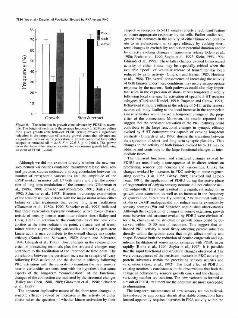
control treatment compared to less than 30% (29.7% t 5.0; p < 0.01) of growth cones showing the same behavior with treat- ment with phorbol (Phor, Fig. 6). In addition, treatment with phorbol facilitated the detection of what appear to be preexisting swellings along the neurites (numerous locations in Fig. 5B2). With washout of phorbol (N = 11 cultures), there was a re- sumption of growth by the growth cones that have stopped or retracted. By 4 hr after washout the frequency of advancing growth cones (76.1% + 3.3) was not significantly different (p > 0.2) than that observed for control treatment (Fig. 6). Thus, the change in the structure of sensory neurons (increase in the number of varicosities) evoked by phorbol in the cocultures is likely to be due to a reversible alteration in the structure of the preexisting sensory cell neurites.
Discussion The results indicate that increases in the activity of either protein kinase (PKA or PKC) evoke changes in the efficacy of senso- rimotor connections lasting from minutes to hours, but they dif- fer in their capacity to evoke long-term facilitation lasting days. Increases in activity of either kinase can enhance transmitter
release from preexisting terminals in a manner that is character- istic of short-term facilitation (Castellucci et al., 1980, 1982; Klein et al., 1986; Schacher et al., 1988; Braha et al., 1990; Sugita et al., 1992; Emptage and Grew, 1993; Schacher et al., 1993). With large and persistent activation of either kinase, changes in synaptic efficacy lasting 3 hr were accompanied by significant changes in the structure of the sensory neurons. The failure of the increase in PKC activity to parallel the increase PKA activity in evoking long-term changes (greater than 24 hr) in synapse efficacy may reflect the differences of the two kinase pathways in maintaining or stabilizing the initial kinase-induced structural changes in the sensory neurons. Whereas the new var- icosities induced by CAMP persisted and continued to increase in number, the PDBLJ-induced changes in sensory neuron struc- ture were transient. It is unlikely that the reversal of these func- tional and structural changes are a consequence of PDBU-in- duced inactivation of the PKC pathways in either the sensory or motor neurons, since the effects of PDBU treatment were re- versible and the cells responded to subsequent treatments that involved additional activation of the PKC pathway (Braha et al., 1990).
7524 Wu et al. l Duration of Facilitation Evoked by PKA versus PKC
80
60 -
e/ ? 50- 31
2 40-
s g 30-
lz 20 -
lo-
O-
- llphor 1
’ Growth Stop & retract
Figure 6. The reduction in growth cone advance by PDBU is revers- ible. The height of each bar is the average frequency + SEM per culture for a given growth cone behavior. PDBU (Phor) evoked a significant reduction in the proportion of sensory growth cones that advance and a significant increase in the proportion of growth cones that have either stopped or retracted (df = 2,48, F = 27.615, p < 0.001). The growth cones that have either stopped or retracted can resume growth following washout of PDBU (wash).
Although we did not examine directly whether the new sen- sory neuron varicosities contained transmitter release sites, sev- eral previous studies indicated a strong correlation between the number of presynaptic varicosities and the amplitude of the EPSP evoked in motor cell L7 both before and after the induc- tion of long-term modulation of the connections (Glanzman et al., 1989a, 1990; Schacher and Montarolo, 1991; Bailey et al., 1992; Schacher et al., 1993). Electron microscope examination of the sensory neuron contacts with the major motor axons either before or after treatments that evoke long term facilitation (Glanzman et al., 1989a, 1990; Schacher et al., 1991) indicated that these varicosities typically contained active zones charac- teristic of sensory neuron transmitter release sites (Bailey and Chen, 1983). In addition to the contributions of the new vari- cosities at the intermediate time point, enhancement of trans- mitter release at pre-existing varicosities induced by persistent kinase activity may contribute to the overall change in synaptic efficacy (Kandel and Schwartz, 1982; Sossin and Schwartz, 1994; Ghirardi et al., 1995). Thus, changes in the release prop- erties of preexisting terminals plus the structural changes may contribute to the facilitation at the intermediate time point. The correlations between the persistent increase in synaptic efficacy following PKA activation and the decline in efficacy following PKC activation with the capacity to maintain the new sensory neuron varicosities are consistent with the hypothesis that some aspects of the long-term “consolidation” of the functional changes of the connection are mediated by the structural changes (Bailey and Chen, 1988, 1989; Glanzman et al., 1990; Schacher et al., 1993).
The transient functional and structural changes evoked by PDBU are most likely a consequence of its direct actions on preexisting sensory cell neurites and varicosities. Unlike the changes evoked by increases in PKC activity in some regener- ating systems (Hsu, 1985; Bixby, 1989; Lankford and Letour- neau, 1991), the application of PDBU during the active phase of regeneration of Aplysia sensory neurons did not enhance neu- rite outgrowth. Treatment resulted in a significant reduction in growth cone extension, as well as an increase in the frequency of growth cone retractions. By contrast, 2 hr treatment with for- skolin or CAMP analogues did not reduce neurite extension by sensory neurons (Wu and Schacher, unpublished observations). Although the expression of changes in neurite extension, growth cone behavior and structure evoked by PDBU were obvious af- ter 2 hr, changes in the structure of growth cones could be ob- served within 15-30 min of treatment. This suggest that en- hanced PKC activity is most likely affecting protein substrates directly within the growth cone that might affect motility and shape. Because both the reduction of neurite outgrowth and sig- nificant facilitation of sensorimotor synapses with PDBU occur rapidly (Braha et al., 1990; Sugita et al., 1992), it is possible that the rapid functional and structural changes observed at 3 hr were consequences of the persistent increase in PKC activity on protein substrates within the preexisting sensory neurites and varicosities (Knox et al., 1992). The local effect of PDBU on existing net&es is consistent with the observations that both the change in behavior by sensory growth cones and the change in varicosity number are transient. The new varicosities formed as a result of PDBU treatment are the ones that are most susceptible to elimination.
The apparent duplicative nature of the short-term changes in The long-term maintenance of new sensory neuron varicosi- synaptic efficacy evoked by increases in the activity of either ties induced by appropriate stimuli after stable connections have kinase raises the question of whether kinase activation by their formed apparently requires increases in PKA activity within the
respective receptors to 5-HT simply reflects a redundant feature to insure appropriate responses by the cells. Earlier studies sug- gested that increases in the activity of either kinase can contrib- ute to an enhancement in synapse efficacy by evoking short- term changes in excitability and action potential duration and/or by directly evoking changes in transmitter release (Klein et al., 1986; Braha et al., 1990; Sugita et al., 1992; Klein, 1993, 1994; Ghirardi et al., 1995). These latter changes evoked by increased activity of either kinase may be especially critical when the available “pool” of vesicular release of transmitter has been reduced by prior activity (Gingrich and Byrne, 1985; Hochner et al., 1986). The overall consequence of increasing the activity of both kinases under these conditions may insure an appropriate response by the neurons. Both pathways could also play impor- tant roles in the expression of short- versus long-term plasticity following local site-specific activation of specific 5-HT receptor subtypes (Clark and Kandel, 1993; Emptage and Carew, 1993). Behavioral stimuli resulting in the release of 5-HT at the sensory neuron cell body leading to the local increase in the appropriate kinase activities would evoke a long-term change in the prop- erties of the connections. Moreover, the results reported here suggest that the persistent activation of the PKC pathway could contribute to the large functional changes in synaptic efficacy evoked by 5-HT concentrations capable of evoking long-term plasticity (Ghirardi et al., 1995) during the transition between the expression of short- and long-term synaptic plasticity. Thus changes in the activity of both kinases evoked by 5-HT may be additive and contribute to the large functional changes at inter- mediate times.
The Journal of Neuroscience, November 1995, 15(11) 7525
sensory neurons. This would trigger appropriate changes in the identified target neuron in vitro: structural remodeling visualized over
nucleus of the sensory neuron for regulating expression of syn- time. J Neurosci 12:2960-2972.
apse-specific macromolecules (Schacher et al., 1988; Nazif et Baxter DA, Byrne JH (1990) Differential effects of cAMP and sero-
al., 1991; Backsai et al., 1993; Hu et al., 1993; Alherini et al., tonin on membrane current, action potential duration, and excitability in somata of pleura1 sensory neurons of Ap/vsia. J Neurophysiol 64:
1994). The changes in the sensory neuron soma would include 978-990. PKA-induced changes in the regulatory machinery for transcrip- Bergold PJ, Sweatt JD, Winicov I, Weiss KR, Kandel ER, Schwartz JH
tion (Dash et al., 1990; Kaang et al., 1993; Alberini et al., 1994) (1990) Protein synthesis during acquisition of long-term facilitation
and the expression, synthesis, and transport of macromolecules is needed for the persistent loss of regulatory subunits of the Ap/ysiu
needed for both the formation of new terminals as well as their cAMP-dependent protein kinase. Proc Natl Acad Sci USA 87:3788- 3791.
maintenance (Forscher et al., 1987: Schacher et al., 1988, 1993;
Bailey et al., 1992; Funte and Haydon, 1993; Hu et al.. 1993).
In addition, increases in PKA activity in the sensory neuron may
be required to trigger appropriate long-term changes in the post-
synaptic motor cell such as the formation of new postsynaptic
receptive zones for the new presynaptic structures (Trudeau and
Castellucci, 1995). Stabilization of new varicosities may also
require CAMP-induced alterations in the relative expression of
adhesion molecules on the surface of the interacting cells (May-
ford et al., 1992; Hu et al.. 199.3; Wu and Schacher, 1994; Zhu
et al., 199.5). Changes in the level of expression of adhesion
molecules and their ligands on the surface of the cells could
signal changes in other intracellular cascades affecting kinase
activities (Atashi et al., 1992; Doherty and Walsh, 1992). In
addition to changes in the activities of PKA and PKC, S-HT
may also affect the activities of CaM kinase (Saitoh and
Schwartz, 1983, 1985) or others (Homayouni et al., 1995). The
nature of this reciprocal sequence of intercellular and intracel-
lular signals initiated by the binding of 5HT to multiple receptor
subtypes and the role of various protein kinases and their inter-
actions in regulating both pre- and postsynaptic cells in the for-
mation and stabilization of new synaptic connections require fur-
ther study.
References
Abeliovich A. Chen C. Goda Y, Silva AJ, Stevens CF. Tonegawa S (1993) Modified hippocampal long-term potentiation in PKC gam- ma-mutant mice. Cell 75: 1253-l 262.
Acosta-Urquidi J, Alkon DL, Neary JT ( 1984) Calcium-dependent pro- tein kinase injection in a photoreceptor mimics biophysical effects of associative learning. Science 224: 1254-I 257.
Akers RF, Lovinger DM, Colley PA, Linden DJ. Routtenberg A (1986) Translocation of protein kinase C activity may mediate hippocampal long-term potentiation. Science 23 I :S87-589.
Alberini CM, Ghirardi M, Met7 R. Kandel ER (1994) ClEBP is an immediate early gene required for the consolidation of long-term fz cilitation in Ap/y.sitr. Cell 76: 1099--l I 14.
Atashi JR, Kline SG, lngraham CA, Matten WT, Schachner M, Maness PF (1992) Neural cell adhesion molecules modulate tyrosine phos- phorylation of tuhulin in nerve growth cone membranes. Neuron 8:83 l-842.
Backsai BJ, Hochner B. Mahaut-Smith M. Adams SR, Kaang B, Kandel ER. Tsien RY (1993) Spatially resolved dynamics of CAMP and protein kinase A subunits in Ap/y.sitr sensory neurons. Science 260: 222-226.
Bailey CH, Chen M ( 1983) Morphological basis of long-term habitu- ation and sensitization. Science 220:9 I-93.
Bailey CH, Chen M (1988) Long-term memory in Ap/y.sitr modulates the total number of varicosities of single identified sensory neurons. Proc Natl Acad Sci USA 85:2373-2377.
Bailey CH, Chen M ( 1989) Time course of structural changes at iden- tified sensory neuron synapses during long-term sensitization in Ap/w sin. J Neurosci 9: 1774-l 780.
Bailey CH, Montarolo PG. Chen M. Kandel ER. Schacher S (1992) Inhibitors of protein and RNA synthesis block the structural changes that accompany long-term heterosynaptic plasticity in the sensory neurons of Aplysitr. Neuron 9:749-7.58.
Bank M, Schacher S (1992) Segregation of presynaptic inputs on an
Bernier L, Castellucci VE Kandel ER, Schwartz JH (1982) Facilitatory transmitter causes a selective and prolonged increase in cyclic AMP in sensory neurons mediating the gill and siphon withdrawal reflex in Ap/vsin. J Neurosci 2: 1682-1691.
Bixby JL (1989) Protein kinase C is involved in laminin stimulation of neurite outgrowth. Neuron 3:287-297.
Bourtchulad7e R, Frenguelli B, Blendy J, Cioffi D, Schutz G, Silva AJ ( 1994) Deficient long-term memory in mice with a targeted mutation of the CAMP-responsive element-binding protein. Cell 79:59-68.
Boyle MB, Klein M, Smith SJ, Kandel ER (1984) Serotonin increases intracellular Ca’+ transients in voltage clamped sensory neurons of Ap/ysirr ccr/(fiwnicn. Proc Natl Acad Sci USA 8 I :7642-7646.
Braha 0, Dale N, Hochner B, Klein M, Abrams TW, Kandel ER (1990) Second messengers involved in the two processes of presynaptic fa- cilitation that contribute to sensitization and dishabituation in Aplysia
sensory neurons. Proc Nat1 Acad Sci USA 87:2040-2044. Braha 0. Edmonds B, Saktor T, Kandel ER, Klein M (1993) The con-
tributions of protein kinase A and protein kinase C to the actions of S-HT on L-type Ca+’ channels of the sensory neurons in Ap/ysio. J Neurosci 13:1839-1851.
Brunelli M, Castellucci VF, Kandel ER (1976) Synaptic facilitation and behavioral sensitization in Ap[ysiu: possible role of serotonin and cyclic AMP Science 194:1178-l 181.
Castellucci VF, Kandel ER, Schwartz JH, Wilson AC, Nairn A, Green- gard P (I 980) Intracellular injection of the catalytic subunit of cyclic AMP-dependent protein kinase stimulates facilitation of transmitter release underlying behavioral sensitization in Ap/ysirr. Proc Natl Acad Sci USA 7717492-7496.
Castellucci VE Nairn AC, Greengard P, Schwartz JH, Kandel ER (I 982) Inhibitor of CAMP-dependent protein kinase blocks presyn- aptic facilitation in Apl.ysia. J Neurosci 2: 1673-168 I
Clark GA. Kandel ER (1993) Induction of long-term facilitation in Aplwin sensory neurons by local application of serotonin to remote synapses. Proc Natl Acad Sci USA 90:11411-I 1415.
Corfas G, Dudai Y (1991) The morphology of a sensory neuron in Drosnphilrr is abnormal in memory mutants and changes during ag- ing. Pioc Natl Acad Sci USA 88:?252-7256. - - -
Critz SD. Bvrne JH (1992) Modulation of I,, bv nhorbol ester-me- diated activation 0; PKC in pleural senso;y‘“ne;r&s of Apiysiu. J Neurophysiol 68: 1079-l 086.
Dale N. Kandel ER (1990) Facilitatory and inhibitory transmitters modulate spontaneous transmitter release at cultured Ap/ysiu senso- rimotor synapses. J Physiol (Lond) 42 1:203-222.
Dale N, Schacher S, Kandel ER (1988) Long-term facilitation in A/~/y- sin involves increase in transmitter release. Science 239:282-285.
Dash PK. Hochner B, Kandel ER (1990) Injection of the cyclic AMP responsive element into the nucleus of Ap!,~icr sensory neurons blocks long-term facilitation. Nature 345:718-72 1,
Dixon D. Atwood HL (1989) Adenylate cyclase system is essential for long-term facilitation at the crayfish neuromuscular junction. J Neu- rosci 9i4246-4252.
Doherty P, Walsh FS (1992) Cell adhesion molecules, second messen- gers and axonal growth. Curr Opin Neurobiol 2:595-601.
Emptage NJ, Carew TJ (1993) Long-term synaptic facilitation in the absence of short-term facilitation in Ap/ysia neurons. Science 262: 253-256.
Farley J. Auerbach S (1986) Protein kinase C activation induces con- ductance changes in Hermissmdu photoreceptors like those seen in associative learning. Nature 319:22&223.
Forscher P, Kaczmarek LK, Buchanan J Smith SJ (1987) Cyclic AMP induces changes in distribution and transport of organelles within growth cones of Aplysia bag cell neurons. J Neurosci 7:3600-3611.
Frey U. Huang Y-Y, Kandel ER (1993) Effects of CAMP simulate a
7526 Wu et al.. Durahon of Facilitation Evoked by PKA versus PKC
late stage of LTP in hippocampal CAI neurons. Science 260:1661- 1664.
Funte LR, Haydon PC (1993) Synaptic target contact enhances pre- synaptic calcium influx by activating CAMP-dependent protein kinase during synaptogenesis. Neuron IO: IO69-1078.
Ghirardi M, Braha 0, Hochner B, Montarolo PG. Kandel ER Dale N (I 992) Roles of PKA and PKC in facilitation of evoked and spon- taneous transmitter release at depressed and nondepressed synapses in Ap/yic~ sensory neurons. Neuron 9: I-20.
Ghirardi M, Montarolo PC, Kandel ER (1995) A novel intermediate stage in the transition between short- and long-term facilitation in the sensory to motor neuron synapse of Ap/ysiu. Neuron 14:4 13420.
Gingrich KJ, Byrne JH (1985) Simulation of synaptic depression, post- tetanic potentiation and presynaptic facilitation of synaptic potentials from sensory neurons mediating the gill withdrawal reflex in Ap/.ysitr. J Neurophysiol 53:652-669.
Glanzman DL. Kandel ER, Schacher S (1989a) Identified target motor neuron regulates neurite outgrowth and synapse formation of Aplysirr sensory neurons in vitro. Neuron 3:441-450.
Glanzman DL. Kandel ER, Schacher S (I 990) Target-dependent struc- tural changes accompanying long-term synaptic facilitation in Ap/ysicl neurons. Science 249:799-802.
Glanzman DL, Mackey SL, Hawkins RD, Dyke AM, Lloyd PE. Kandel ER ( I989b) Depletion of serotonin in the nervous system of Ap/ysitr reduces the behavioral enhancement of gill withdrawal as well as the heterosynaptic facilitation produced by tail shock. J Neurosci 9:4200- 4213.
Grant SGN. O’dell TJ. Karl KA, Stein PL, Soriano P, Kandel ER (1992) Impaired long-term potentiation, spatial learning, and hippocampal development in fyn mutant mice. Science 258:1903-1910.
Greenberg SM. Castellucci VF, Bayley H, Schwartz JH (1987) A mo- lecular mechanism for long-term sensitization in Ap/wia. Nature 329: 62-65.
Hochner B, Klein M, Schacher S, Kandel ER (1986) Additional com- ponents in the cellular mechanism of presynaptic facilitation contrib- utes to behavioral dishabituation in Ap/y.sicr. Proc Nat] Acad Sci USA 83:8794-8798.
Homayouni R, Byrne JH, Eskin A (1995) Dynamics of protein phos- phorylation in sensory neurons of Ap/y.ritr. J Neurosci 15:429438.
Hsu L (1985) Neurite-promoting effects of TPA on chick embryo neu- rons. Neurosci Lett 62:2X3-289.
Hu GY, Hvalby 0, Walaas SI, Albert KA, Skjeflo P, Andersen P, Green- gard P (I 987) Protein kinase C injection into hippocampus pyra- midal cells elicits features of long-term potentiation. Nature 328:426- 429.
Hu Y, Barzilai A, Chen M, Bailey CH. Kandel ER (1993) S-HT and CAMP induce the formation of coated pits and vesicles and increase the expression of clathrin light chain in sensory neurons of Ap/ysitr. Neuron IO:92 l-929.
Huang Y-Y, Li X-C, Kandel ER (1994) CAMP contributes to mossy fiber LTP by initiating both ;I covalently mediated early phase and macromolecular synthesis-dependent late phase. Cell 79:69-79.
Kaang B-K. Kandel ER, Grant SGN (1993) Stimuli that produce long- term facilitation in Ap/vsirr sensory neurons activate the transcription of CAMP-responsive genes. Neuron 10:427435.
Kandel ER, Schwartz JH (1982) Molecular biology of learning: mod- ulation of transmitter release. Science 2 18:433-%43.
Klein M (1993) Differential cyclic AMP dependence of facilitation at Ap/ysitr sensorimotor synapses as a function of prior stimulation: aug- mentation versus restoration of transmitter release. J Neurosci 13: 3793-3801.
Klein M ( 1994) Synaptic augmentation by S-HT at rested Ap!\‘sicl sen- sorimotor synapses: independence of action potential prolongation. Neuron 13:159-166.
Klein M, Camardo J. Kandel ER ( 1982) Serotonin modulates a specific potassium current in the sensory neurons that show presynaptic fa- cilitation in Aplysio. Proc Nat] Acad Sci USA 79:57 13-57 17.
Klein M, Hochner B, Kandel ER (1986) Facilitatory transmitters and CAMP can modulate accommodation as well as transmitter release in Ap/ysirr sensory neurons: evidence for parallel processing in a single cell. Proc Nat] Acad Sci USA 83:7994-7998.
Knox RJ, Quattrocki EA. Connor JA, Kaczmarek LK (1992) Recruit- ment of calcium channels by protein kinase C during rapid formation of putative neuropeptide release sites in isolated Ap/ysitr neurons. Neuron 8:883-889.
Lankford KL, Letourneau PC (1991) Roles of actin filaments and three second-messenger systems in short-term regulation of chick dorsal root ganglion neurite outgrowth. Cell Motil Cytoskel 20:7-29.
Malenka RC, Kauer JA, Perkel DJ, Mark MD. Kelly PT. Nicholl RA. Waxham NN (1989) An essential role for postsynaptic calmodulin and protein kinase activity in long-term potentiation. Nature 340: 554-ss7.
Malinow R, Madison DV, Tsien RW (1988) Persistent protein kinase activity underlying long-term potentiation. Nature 335.820-824.
Malinow R. Shulman H. Tsien RW (1989) Inhibition of oostsvnantic PKC or CAMKII blocks induction but not expression of LTP &i;nce 245:X62-866.
Mayford M, Barzilai A, Keller F, Schacher S, Kandel ER ( 1992) Mod- ulation of an NCAM-related adhesion molecule with long-term syn- aptic plasticity in Ap1y.k. Science 256:63X-644.
McGlade-McCulloh E. Yamamoto H, Tan SE, Brickey DA. Soderling TR ( 1993) Phosphorylation and regulation of glutamate receptors by calcium/calmodulin dependent protein kinase II. Nature 362:640- 642.
Mercer AR, Emptage NJ, Carew TJ (1991) Pharmacological dissoci- ation of modulatory effects of serotonin in Ap/y.sitr sensory neurons. Science 254:181 I-1813.
Miller B, Sarantis M, Traynelis SF, Attwell D (1992) Potentiation of NMDA receptor currenis by arachidonic acid. Nature 355:722-725.
Montarolo PG. Goelet I? Castellucci VE Morean J. Kandel ER. Schach- er S (1986) A critical time window for macromolecular synthesis in long-term heterosynaptic facilitation in A/dysicr. Science 234: l249- 1254.
Narif FA, Byrne JH, Cleary LJ (1991) Cyclic AMP induces long-term morphological changes in sensory neurons of A/dysitr. Brain Res 539: 324-327.
Occor KA, Byrne JH (198.5) Membrane responses and changes in cy- clic AMP levels in Ap/ysiu neurons by serotonin. tryptamine. FMRFamide, and small cardioactive peptide (SCPb). Neurosci Lett 55:113-l 18.
O’dell TJ, Kandel ER, Grant SGN ( 199 I ) Long-term potentiation in the hippocampus is blocked by tyrosine kinase inhibitors. Nature 353: 558-560.
Peter N, Aronoff B, Wu F, Schacher S ( 1994) Decrease in growth cone- neurite fasciculation by sensory or motor cells iu vitro accompanies modulation of Ap/y,sitr cell adhesion molecules by neurotransmitters. J Neurosci 14:1413-1421.
Raymond LA, Blackstone CD, Huganir RL (1993) Phosphorylation and modulation of recombinant GluR6 glutamate receptors by cAMP- dependent protein kinase. Nature 361:637-641,
Rayport SG, Schacher S (1986) Synaptic plasticity in \,itro: cell culture of identified Ap!\‘sio neurons mediating short-term habituation and sensitization. J Neurosci 6:759-763.
Saitoh T, Schwartz JH (1983) Serotonin alters the subcellular distri- bution of a Ca+‘/calmodulin binding protein in neurons of Ap!\“irr. Proc Nat1 Acad Sci USA 80:6708-67 12.
Saitoh T, Schwartz JH (1985) Phosphorylation-dependent subcellular translocation of a Ca+‘/calmodulin-dependent protein kinase produces an autonomous enzyme in Aplysicr neurons. J Cell Biol 100:835-842.
Saktor TC, Schwartz JH (I 990) Sensitizing stimuli cause translocation of protein kinase C in Aplysirr sensory neurons. Proc Nat] Acad Sci USA 87:203&2039.
Schacher S ( 1985) Differential synapse formation and neuriric ou- growth at two branches of the metacerebral cell of Ap/x.\itr in disso- ciated cell culture. J Neurosci S:2028-2034.
Schacher S, Montarolo PG (1991) Target-dependent structural changes in sensory neurons of Aplysia accompany long-term heterosynaptic inhibition. Neuron 6:679-690.
Schacher S, Proshansky E (1983) Neurite regeneration by Aplysia neu- rons in dissociated cell culture: modulation by Ap/ysicl hemolymph and the presence of the initial axon segment. J Neurosci 3:2403- 2413.
Schacher S, Castellucci VE Kandel ER (1988) Cyclic AMP evokes long-term facilitation in Aplysirr sensory neurons that requires new protein synthesis. Science 240:1667-1669.
Schacher S, Montarolo PG. Kandel ER (1990) Selective shol-t- and long-term effects of serotonin, small cardioactive peptide. and tetanic stimulation on sensorimotor synapses of Ap/y.sicr in culture. J Neu- rosci 10:328&3294.
Schacher S, Glanzman DL, Barzilai A, Dash P, Grant SGN, Keller E
The Journal of Neuroscience, November 1995, 15(11) 7527
Mayford M, Kandel ER (1991) Long-term facilitation in Ap/ysia: persistent phosphorylation and structural changes. Cold Spring Harb Symp Quant Biol 55: 187-202.
Schacher S, Kandel ER, Montarolo PG (1993) cAMP and arachidonic acid simulate long-term structural and functional changes produced by neurotransmitters in A/dysiu sensory neurons. Neuron IO: IO79- 1088.
Scholz KP Byrne JH (I 987) Long-term sensitization in Aplysio: bio- physical correlates in tail sensory neurons. Science 235:685-687.
Scholz KP Byrne JH (1988) Intracellular injection of cyclic AMP in- duces a long-term reduction of neuronal potassium currents. Science 240: 166441667.
Schuman EM, Clark GA ( 1994) Synaptic facilitation at connections of Hermissmd~~ type B photoreceptors. J Neurosci 14: I6 13- 1622.
Siegelbaum SA, Camardo JS, Kandel ER (I 982) Serotonin and CAMP close single K’ channels in Ap/ysia sensory neurons. Nature 299: 413417.
Silva AJ, Stevens CF, Tonegawa S, Wang Y (1992) Deficient hippo- campal long-term potentiation in calcium-calmodulin kinase II mu- tant mice. Science 257:201-206.
Sossin WS, Schwartz JH (1992) Selective activation of calcium-acti- vated PKCs in Ap/ysiu neurons by 5.HT. J Neurosci 9:3218-3229.
Sossin WS, Saktor TC, Schwartz JH (1994) Persistent activation of protein kinase C during the development of long-term facilitation in Ap/ysiu. Learning Memory 1: 189-202.
Sugita S. Goldsmith JR, Baxter DA, Byrne JH (1992) Involvement of protein kinase C in serotonin-induced spike broadening and synaptic facilitation of sensorimotor connections in Apiysici. J Neurophysiol 68:643-65 I.
Sugita S, Baxter DA, Byrne JH (1994) Activators of protein kinase C mimic serotonin-induced modulation of a voltage-dependent potas-
sium current in pleural sensory neurons of Ap/.vsicr. J Neurophysiol 72:1240-1249.
Sweatt JD, Kandel ER (1989) Persistent and transcriptionally depen- dent increase in protein phosphorylation in long-term facilitation of Aplysiu sensory neurons. Nature 339:5 l-54.
Trudeau L-E, Castellucci VF (1995) Postsynaptic modifications in long-term facilitation in Aplysia: upregulation of excitatory amino acid receptors. J Neurosci 15: 1275- 1284.
Walsh JP, Byrne JH (I 989) Modulation of a steady state Ca+‘-activated K’ current in tail sensory neurons of Aplvsin: role of serotonin and CAMP J Neurophysiol 61:3244.
Wang LY, Dudek EM, Browning MD, MacDonald JF (1994) Modu- lation of AMPA/kainate receptors in cultured murine hippocampal neurons by protein kinase C. J Physiol (Lond) 475:431437.
Weisskopf MG, Castillo PE, Zalutsky RA, Nicholl RA ( 1994) Medi- ation of hippocampal mossy fiber long-term potentiation by cyclic AMP Science 265: 1878-l 882.
Wojtowicz JM, Marin L, Atwood HL (1989) Synaptic restructuring during long-term facilitation at the crayfish neuromuscular junction. Can J Physiol Pharmacol 67: I67- I7 I.
Wu F, Schacher S (1994) Pre- and postsynaptic changes mediated by two second messengers contribute to expression of Ap!\‘sitr long-term heterosynaptic inhibition. Neuron 12:407-42 I.
Zhong Y, Wu C-F (1991) Altered synaptic plasticity in Dr-o.rophi/a memory mutant with altered CAMP cascade. Science-25 I :1562-j 564.
Zhu H. Wu E Schacher S (1994) Anlvsitr cell adhesion molecules and serotonin regulate sensory ceil-motor cell interactions during early stages of synapse formation i~r virro. J Neurosci 14:6886-6900.
Zhu H, Wu E Schacher S (1995) Changes in the expression and dis- tribution of Ap!\sicl cell adhesion molecules can influence synapse formation and elimination ilr vitro, J Neurosci I5:4 I734 183.