translating dosage compensation to trisomy 21
DESCRIPTION
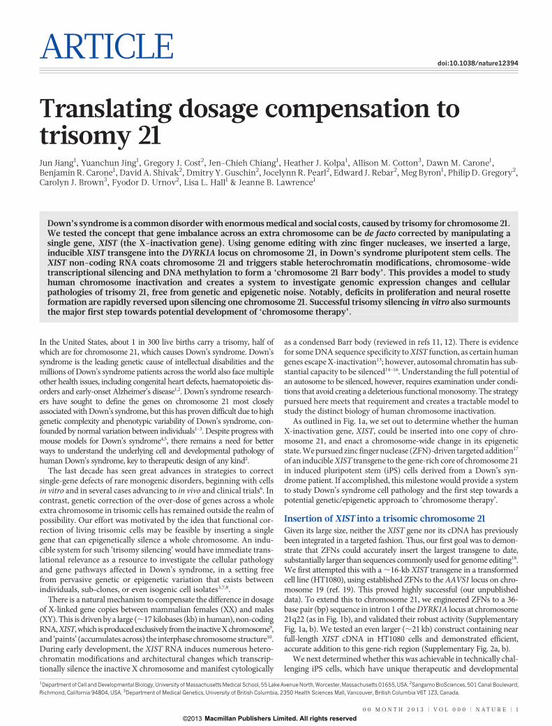
Down’s syndrome is a common disorder with enormous medical and social costs, caused by trisomy for chromosome 21.We tested the concept that gene imbalance across an extra chromosome can be de facto corrected by manipulating asingle gene, XIST (the X-inactivation gene). Using genome editing with zinc finger nucleases, we inserted a large,inducible XIST transgene into the DYRK1A locus on chromosome 21, in Down’s syndrome pluripotent stem cells. TheXIST non-coding RNA coats chromosome 21 and triggers stable heterochromatin modifications, chromosome-widetranscriptional silencing and DNA methylation to form a ‘chromosome 21 Barr body’. This provides a model to studyhuman chromosome inactivation and creates a system to investigate genomic expression changes and cellularpathologies of trisomy 21, free from genetic and epigenetic noise. Notably, deficits in proliferation and neural rosetteformation are rapidly reversed upon silencing one chromosome 21. Successful trisomy silencing in vitro also surmountsthe major first step towards potential development of ‘chromosome therapy’.TRANSCRIPT
ARTICLEdoi:10.1038/nature12394
Translating dosage compensation totrisomy 21Jun Jiang1, Yuanchun Jing1, Gregory J. Cost2, Jen-Chieh Chiang1, Heather J. Kolpa1, Allison M. Cotton3, Dawn M. Carone1,Benjamin R. Carone1, David A. Shivak2, Dmitry Y. Guschin2, Jocelynn R. Pearl2, Edward J. Rebar2, Meg Byron1, Philip D. Gregory2,Carolyn J. Brown3, Fyodor D. Urnov2, Lisa L. Hall1 & Jeanne B. Lawrence1
Down’s syndrome is a common disorder with enormous medical and social costs, caused by trisomy for chromosome 21.We tested the concept that gene imbalance across an extra chromosome can be de facto corrected by manipulating asingle gene, XIST (the X-inactivation gene). Using genome editing with zinc finger nucleases, we inserted a large,inducible XIST transgene into the DYRK1A locus on chromosome 21, in Down’s syndrome pluripotent stem cells. TheXIST non-coding RNA coats chromosome 21 and triggers stable heterochromatin modifications, chromosome-widetranscriptional silencing and DNA methylation to form a ‘chromosome 21 Barr body’. This provides a model to studyhuman chromosome inactivation and creates a system to investigate genomic expression changes and cellularpathologies of trisomy 21, free from genetic and epigenetic noise. Notably, deficits in proliferation and neural rosetteformation are rapidly reversed upon silencing one chromosome 21. Successful trisomy silencing in vitro also surmountsthe major first step towards potential development of ‘chromosome therapy’.
In the United States, about 1 in 300 live births carry a trisomy, half ofwhich are for chromosome 21, which causes Down’s syndrome. Down’ssyndrome is the leading genetic cause of intellectual disabilities and themillions of Down’s syndrome patients across the world also face multipleother health issues, including congenital heart defects, haematopoietic dis-orders and early-onset Alzheimer’s disease1,2. Down’s syndrome research-ers have sought to define the genes on chromosome 21 most closelyassociated with Down’s syndrome, but this has proven difficult due to highgenetic complexity and phenotypic variability of Down’s syndrome, con-founded by normal variation between individuals1–3. Despite progress withmouse models for Down’s syndrome4,5, there remains a need for betterways to understand the underlying cell and developmental pathology ofhuman Down’s syndrome, key to therapeutic design of any kind2.
The last decade has seen great advances in strategies to correctsingle-gene defects of rare monogenic disorders, beginning with cellsin vitro and in several cases advancing to in vivo and clinical trials6. Incontrast, genetic correction of the over-dose of genes across a wholeextra chromosome in trisomic cells has remained outside the realm ofpossibility. Our effort was motivated by the idea that functional cor-rection of living trisomic cells may be feasible by inserting a singlegene that can epigenetically silence a whole chromosome. An indu-cible system for such ‘trisomy silencing’ would have immediate trans-lational relevance as a resource to investigate the cellular pathologyand gene pathways affected in Down’s syndrome, in a setting freefrom pervasive genetic or epigenetic variation that exists betweenindividuals, sub-clones, or even isogenic cell isolates3,7,8.
There is a natural mechanism to compensate the difference in dosageof X-linked gene copies between mammalian females (XX) and males(XY). This is driven by a large (,17 kilobases (kb) in human), non-codingRNA, XIST, which is produced exclusively from the inactive X chromosome9,and ‘paints’ (accumulates across) the interphase chromosome structure10.During early development, the XIST RNA induces numerous hetero-chromatin modifications and architectural changes which transcrip-tionally silence the inactive X chromosome and manifest cytologically
as a condensed Barr body (reviewed in refs 11, 12). There is evidencefor some DNA sequence specificity to XIST function, as certain humangenes escape X-inactivation13; however, autosomal chromatin has sub-stantial capacity to be silenced14–16. Understanding the full potential ofan autosome to be silenced, however, requires examination under condi-tions that avoid creating a deleterious functional monosomy. The strategypursued here meets that requirement and creates a tractable model tostudy the distinct biology of human chromosome inactivation.
As outlined in Fig. 1a, we set out to determine whether the humanX-inactivation gene, XIST, could be inserted into one copy of chro-mosome 21, and enact a chromosome-wide change in its epigeneticstate. We pursued zinc finger nuclease (ZFN)-driven targeted addition17
of an inducible XIST transgene to the gene-rich core of chromosome 21in induced pluripotent stem (iPS) cells derived from a Down’s syn-drome patient. If accomplished, this milestone would provide a systemto study Down’s syndrome cell pathology and the first step towards apotential genetic/epigenetic approach to ‘chromosome therapy’.
Insertion of XIST into a trisomic chromosome 21Given its large size, neither the XIST gene nor its cDNA has previouslybeen integrated in a targeted fashion. Thus, our first goal was to demon-strate that ZFNs could accurately insert the largest transgene to date,substantially larger than sequences commonly used for genome editing18.We first attempted this with a ,16-kb XIST transgene in a transformedcell line (HT1080), using established ZFNs to the AAVS1 locus on chro-mosome 19 (ref. 19). This proved highly successful (our unpublisheddata). To extend this to chromosome 21, we engineered ZFNs to a 36-base pair (bp) sequence in intron 1 of the DYRK1A locus at chromosome21q22 (as in Fig. 1b), and validated their robust activity (SupplementaryFig. 1a, b). We tested an even larger (,21 kb) construct containing nearfull-length XIST cDNA in HT1080 cells and demonstrated efficient,accurate addition to this gene-rich region (Supplementary Fig. 2a, b).
We next determined whether this was achievable in technically chal-lenging iPS cells, which have unique therapeutic and developmental
1Department of Cell and Developmental Biology, University of Massachusetts Medical School, 55 Lake Avenue North, Worcester, Massachusetts 01655, USA. 2Sangamo BioSciences, 501 Canal Boulevard,Richmond, California 94804, USA. 3Department of Medical Genetics, University of British Columbia, 2350 Health Sciences Mall, Vancouver, British Columbia V6T 1Z3, Canada.
0 0 M O N T H 2 0 1 3 | V O L 0 0 0 | N A T U R E | 1
Macmillan Publishers Limited. All rights reserved©2013
potential to form various cell types, and thus would be important forany future ex vivo cellular therapy efforts. We used a male Down’ssyndrome iPS cell line20 which we confirmed maintains pluripotencymarkers and trisomy 21. Although a constitutively transcribed trans-gene could be used, we engineered an inducible system to maximizeutility for investigating Down’s syndrome biology. In one step, weintegrated both the doxycycline-controlled XIST transgene into chro-mosome 21 (Fig. 1b and Supplementary Fig. 3a) and a transgene car-rying the doxycycline control component (rtTA) into the AAVS1chromosome 19 safe harbour, disruption of which creates no knownadverse effects19 (Supplementary Fig. 3b).
We analysed 245 colonies from the pooled transformants by inter-phase RNA/DNA fluorescence in situ hybridization (FISH) (Fig. 1c)to determine whether XIST was present and overlapped one of threeDYRK1A alleles. Notably, 98.5% of XIST RNA-positive colonies car-ried XIST at this location on chromosome 21, and also contained thertTA/selection transgene (Supplementary Table 1). Efficiency wassufficiently high that, through modifications to editing conditions,we obtained a few sub-clones with XIST integrated into two or even
all three alleles of DYRK1A (see Methods and Supplementary Fig. 3cand Supplementary Table 1). Six independent sub-clones were chosenfor further study based on: an XIST transgene on one of three chro-mosome 21 copies, pluripotent colony morphology, OCT4 (also calledPOU5F1) staining (Fig. 1d and Supplementary Fig. 4a), and formationof embryoid bodies. FISH to metaphase chromosomes (Fig. 1e and Sup-plementary Fig. 3d) and Southern blotting (Supplementary Fig. 1c–e)confirmed the gene addition accuracy, with 47 chromosomes, for all sixclones. High-resolution cytogenetic banding and/or array comparativegenomic hybridization (CGH) on selected clones showed no significantabnormalities other than full chromosome 21 trisomy (SupplementaryFig. 4c–e).
XIST RNA induces a chromosome 21 Barr bodyIn the panel of six independent genome-edited clones, we inducedtransgene expression and detected XIST RNA by FISH 3 days later. Alocalized XIST RNA ‘territory’ over one chromosome 21 (Fig. 1c) wasseen in over 85% of cells in all six clones (Fig. 1d and SupplementaryFig. 4a, b). This mirrored the unique behaviour of endogenous XISTRNA which ‘paints’ the inactive X chromosome nuclear territory10.
The natural inactivated X chromosome forms a condensed Barr bodywhich carries repressive histone marks11. Similarly, 5 days after XISTinduction, the edited chromosome 21 became markedly enriched in allheterochromatin marks examined, including H3K27me3, UbH2A andH4K20me in 90–100% of cells and, later, with macroH2A (Fig. 2a, b andSupplementary Fig. 5a). Supplementary Fig. 5b illustrates that H3K27mespreads across the whole metaphase chromosome 21. Moreover, chro-mosome 21 DNA in many nuclei became notably condensed, furtherevidence that we successfully generated a heterochromatic chromosome21 Barr body (Fig. 2c).
Allele-specific silencing across chromosome 21To measure overall transcription across the XIST-targeted chro-mosome 21, we used an approach that we developed to broadly assayheterogeneous nuclear RNA (hnRNA) expression and to distinguishinactive from active X chromosome16, on the basis of in situ hybrid-ization to CoT-1 repeat RNA. This showed that the chromosome 21XIST RNA territory was depleted for hnRNA detected by CoT-1(Supplementary Fig. 5c), similar to the inactive X chromosome16.
We next used multi-colour RNA FISH to determine the presence oftranscription foci at each allele for six specific chromosome 21 genes, anestablished approach that we earlier showed10,15 discriminates activeversus silenced genes on inactive X chromosome. Without XIST expres-sion, there are three bright transcription foci from each DYRK1A allele(Fig. 1c, top), but after XIST expression, the targeted allele becomes
FibroblastFibrobla
XIST
ZFN
Down’ssyndrome
patient
+Dox
Chr 21 Barr body
XIST
T
Trisomy 21iPS cell
XISTDYRK1A BAC
Single targetXIST paint
XISTDYRK1A BAC
Single targetNo XIST paint
c
b
a
*
Single targetXIST
DYRK1A BAC
e
Colony OCT 4 XIST
d Clone 1
14 kb XIST cDNA pTRE3G promoter 3′ armpA5′ arm
C C A G C C A C C C C T T T G C C A G T T T A C A C G G G T G A T G A G C A G G C T G T T T G GG G T C G G T G G G G A A A C G G T C A A A T G T G C C C A C T A C T C G T C C G A C A A A C C
XIST genomic DNAFull-length XIST cDNA
Donor: pTRE3G-XIST
Chromosome 21DYRK1A locus
3′ E6 E5 E4 E3 E2 E1 5′
2 kb
5 kb E6E7E8 E9 E10 E115′ E5E4E3E2E1
ZFN target site
Fok1
Fok1
3′
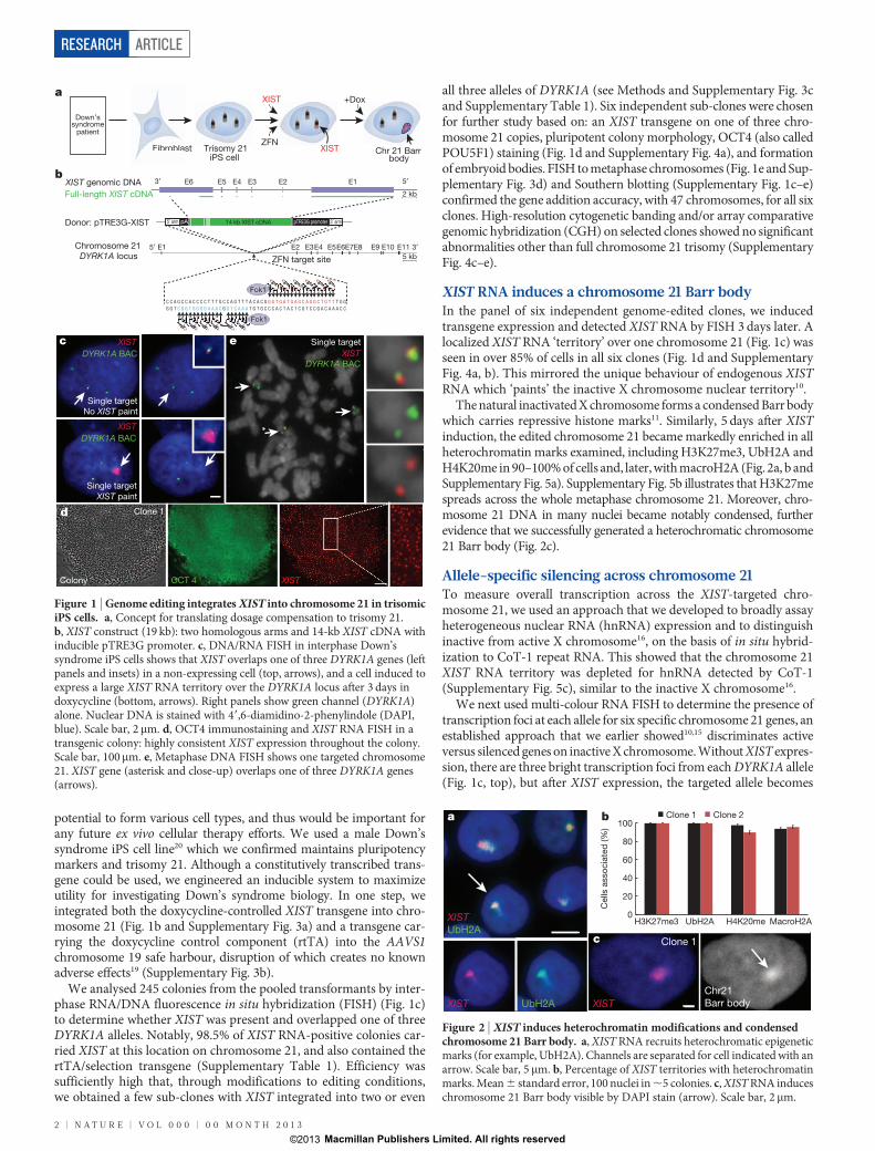
Figure 1 | Genome editing integrates XIST into chromosome 21 in trisomiciPS cells. a, Concept for translating dosage compensation to trisomy 21.b, XIST construct (19 kb): two homologous arms and 14-kb XIST cDNA withinducible pTRE3G promoter. c, DNA/RNA FISH in interphase Down’ssyndrome iPS cells shows that XIST overlaps one of three DYRK1A genes (leftpanels and insets) in a non-expressing cell (top, arrows), and a cell induced toexpress a large XIST RNA territory over the DYRK1A locus after 3 days indoxycycline (bottom, arrows). Right panels show green channel (DYRK1A)alone. Nuclear DNA is stained with 49,6-diamidino-2-phenylindole (DAPI,blue). Scale bar, 2mm. d, OCT4 immunostaining and XIST RNA FISH in atransgenic colony: highly consistent XIST expression throughout the colony.Scale bar, 100mm. e, Metaphase DNA FISH shows one targeted chromosome21. XIST gene (asterisk and close-up) overlaps one of three DYRK1A genes(arrows).
XISTUbH2A
UbH2AXIST
a
%ce
llsas
socia
ted
H3K27me3 UbH2A H4K20me
Cel
ls a
ssoc
iate
d (%
)
b
XIST
Clone 1
Chr21Barr body
c
Clone 1 Clone 2
0
20
40
60
80
100
MacroH2A
Figure 2 | XIST induces heterochromatin modifications and condensedchromosome 21 Barr body. a, XIST RNA recruits heterochromatic epigeneticmarks (for example, UbH2A). Channels are separated for cell indicated with anarrow. Scale bar, 5mm. b, Percentage of XIST territories with heterochromatinmarks. Mean 6 standard error, 100 nuclei in ,5 colonies. c, XIST RNA induceschromosome 21 Barr body visible by DAPI stain (arrow). Scale bar, 2mm.
RESEARCH ARTICLE
2 | N A T U R E | V O L 0 0 0 | 0 0 M O N T H 2 0 1 3
Macmillan Publishers Limited. All rights reserved©2013
weaker or undetectable, indicating repression of DYRK1A (Fig. 1c,bottom).
The APP gene on chromosome 21 encodes b-amyloid precursorprotein; mutations in APP which cause accumulation of b-amyloidlead to early-onset familial Alzheimer’s disease, and APP overexpres-sion is linked to the Alzheimer’s disease characteristic of Down’ssyndrome1. Initially, three bright RNA transcription foci are apparent(Fig. 3a, top). Short-term XIST expression resulted in incompleterepression of the targeted allele (Fig. 3a, middle), which after 20 dayswas completely silenced, as shown in two independent clones (Fig. 3a,bottom, and Fig. 3b).
We examined four more loci, 3–21 megabases (Mb) from XIST:ITSN1, USP25, CXADR and COL18A1. Complete silencing of each alleleon the edited chromosome 21 was seen in ,100% of cells accumulatingXIST RNA (Fig. 3c, d and Supplementary Fig. 6a). Allele-specific silen-cing was further validated using single nucleotide polymorphism (SNP)analysis. PCR with reverse transcription (RT–PCR) products for eightknown polymorphic sites (in four genes) were sequenced (ADAMTS1,ETS2, TIAM1 and HSPA13) (Supplementary Fig. 6b, c). Interestingly,clones 2 and 3 showed an identical pattern of eight SNP alleles repressed,whereas clone 1 showed an alternative pattern of SNPs repressed. Assummarized in Supplementary Fig. 6c, this chromosome-wide patternallows extrapolation of the haplotype for each of the three chromosome21 homologues, and indirectly identifies for each clone which chro-mosome 21 was silenced by an XIST transgene.
We also examined clones carrying XIST on two or all three copies ofchromosome 21 and found that after 20 days in doxycycline, most orall cells lost XIST localization or expression, and the targeted chromo-somes did not silence the APP gene (Supplementary Fig. 7a, b). Thus,there is in vitro selection and epigenetic adaptation to circumventcreating a functional monosomy or nullisomy, consistent with obser-vations that monosomic cells do not persist in mosaic patients.
Genome-wide silencing and methylationHaving demonstrated allele-specific repression for the ten genes exam-ined above, we extended this to genome-wide expression profiling. Wetreated three transgenic clones and the parental line with doxycyclinefor 3 weeks, and compared their transcriptomes to parallel cultureswithout XIST transcription, all in triplicate. Only on chromosome 21is there overwhelming change, in all three clones (Fig. 4a), with ,95%of significantly expressed genes becoming repressed (SupplementaryTable 2).
Dosage compensation corrects chromosome 21 expression to nearnormal disomic levels, based on the change in total output of expressedgenes per chromosome after XIST is induced. Because evidence suggeststhat many chromosome 21 genes are not increased the theoretical1.5-fold in trisomy21,22, we also directly compared trisomic to disomiccells. This provides a baseline for evaluating the degree to which chro-mosome 21 overexpression is corrected by XIST. After XIST induction,overall chromosome 21 expression is reduced by 20%, 15% and 19% forclones 1, 2 and 3, respectively; this mirrors very well the 22% reductionfor disomic iPS cells that lack the third chromosome 21 altogether(Fig. 4a). This disomic line is representative, as a similar difference(21%) was seen for an isogenic disomic sub-clone that we isolated fromthe trisomic parental iPS cells (not shown). Individual genes repressedby XIST are distributed throughout chromosome 21, as do genes over-expressed in trisomic versus disomic cells (Fig. 4b). In addition, qRT–PCRconfirmed repression for individually examined genes (SupplementaryFig. 7c). Clearly, XIST induces robust dosage compensation of most chro-mosome 21 genes overexpressed in trisomy.
Trisomy 21 may have an impact on genome-wide expression path-ways, but differences attributable to trisomy 21 are confounded bygenetic and epigenetic variability21. This inducible trisomy silencing systemprovides a new foothold into this important question. For example,even the three isogenic transgenic sub-clones show many expressiondifferences (.1,000), but upon XIST induction, ,200 genes through-out the genome change in all three clones (but not the doxycycline-treated parental), most probably directly due to chromosome 21 trisomy.Therefore, ‘trisomy correction in a dish’ has promise as a means toidentify genome-wide pathways perturbed by trisomy 21.
In addition to transcriptional silencing, X-inactivation is stabilizedby hypermethylation of promoter CpG islands23,24, which occurs late
a
Day 0
Day 5
Day 20
XISTAPP APP
USP25 CXADR APP ITSN1DYRK1Ad
% c
ells
that
sile
nce
APP
Cel
ls th
at s
ilenc
e A
PP
(%)
Silenced Partially silenced Not silenced
R 1
no DOX
Clone 1 Clone 2
US
P25
CXA
DR
ITS
N1
CO
L18A
1
No dox Dox
Cel
ls th
at s
ilenc
e XI
ST-
asso
ciat
ed a
llele
(%)c
b
Day 20
Clone 1
COL18A1
100806040200
Clone 2
Day 5
Clone 1 Clone 2
Day 20
100806040200
US
P25
CXA
DR
ITS
N1
CO
L18A
1
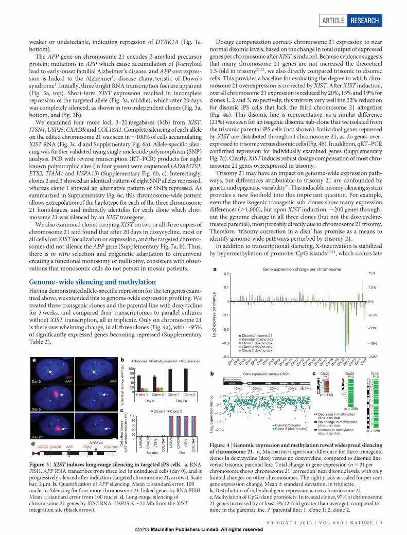
Figure 3 | XIST induces long-range silencing in targeted iPS cells. a, RNAFISH. APP RNA transcribes from three loci in uninduced cells (day 0), and isprogressively silenced after induction (targeted chromosome 21, arrows). Scalebar, 2 mm. b, Quantification of APP silencing. Mean 6 standard error, 100nuclei. c, Silencing for four more chromosome-21-linked genes by RNA FISH.Mean 6 standard error from 100 nuclei. d, Long-range silencing ofchromosome 21 genes by XIST RNA. USP25 is ,21 Mb from the XISTintegration site (black arrow).
0%
Clone 3 (dox/no dox)Disomic/trisomic
a
Chr1
Chr2
Chr3
Chr4
Chr5
Chr6
Chr7
Chr9
Chr8
Chr10
Chr11
Chr12
Chr13
Chr14
Chr15
Chr16
Chr17
Chr18
Chr19
Chr20
Chr21
Chr22
ChrX
ChrY
Disomic/trisomic 21Parental dox/no doxClone 1 dox/no doxClone 2 dox/no doxClone 3 dox/no dox
Gene expression change per chromosome
10Mb 30Mb 40Mb20Mb 48.1Mb
Log 2
expr
essi
on c
hang
e
–1.0
–0.5
0
–1.5
–2.0
b
Log2
exp
ress
ion
chan
ge
Gene repression across Chr21 c
Decrease in methylation(dox < no dox)
Increase in methylation(dox > no dox)
No change in methylation(dox = no dox)
ChrX
(n = 529)
Chr22
(n = 336)
Chr21
(n = 143)
P12No probesets
–6.5%
–13%
–19%
–24%
7.5%
15%0.2
0.1
0
–0.1
–0.2
–0.3
–0.4
P12 P12
Figure 4 | Genomic expression and methylation reveal widespread silencingof chromosome 21. a, Microarray: expression difference for three transgenicclones in doxycycline (dox) versus no doxycycline, compared to disomic lineversus trisomic parental line. Total change in gene expression (n 5 3) perchromosome shows chromosome 21 ‘correction’ near disomic levels, with onlylimited changes on other chromosomes. The right y axis is scaled for per centgene expression change. Mean 6 standard deviation, in triplicate.b, Distribution of individual gene repression across chromosome 21.c, Methylation of CpG island promoters. In treated clones, 97% of chromosome21 genes increased by at least 5% (2-fold greater than average), compared tonone in the parental line. P, parental line; 1, clone 1; 2, clone 2.
ARTICLE RESEARCH
0 0 M O N T H 2 0 1 3 | V O L 0 0 0 | N A T U R E | 3
Macmillan Publishers Limited. All rights reserved©2013
in the silencing process. Therefore, we also examined the promotermethylome in two genome-edited clones 3 weeks after XIST inductionand found it largely unaltered, with one striking exception, genes onchromosome 21 (P value ,2.2 3 10216) (Fig. 4c). Here, 97% of CpG-island-containing genes exhibited a robust increase in promoter DNAmethylation, within the range of that seen for the inactive X chro-mosome24 (adjusted for active/inactive chromosomes; see Methods).This change swept the entire chromosome, with the interesting excep-tion of a few genes that ‘escape’ methylation in both clones.
In summary, data from eight different approaches demonstrateimpressive competence of most chromosome 21 genes to undergo epi-genetic modification and silencing in response to an RNA that evolvedto silence the X chromosome.
Phenotypic correction in vitroDosage compensation of chromosome imbalance presents a newparadigm, with opportunities to advance Down’s syndrome researchin multiple directions, including a new means to investigate humanDown’s syndrome cellular pathologies, which are largely unknown.Inducing trisomy silencing in parallel cultures of otherwise identicalcells may reveal cellular pathologies due to trisomy 21, which could beobscured by differences between cell isolates. We examined cell pro-liferation and neural rosette formation to look for an impact on cellphenotype.
There is some evidence of proliferative impairment in Down’ssyndrome brains4,25; however, we observed that this varied in vitrobetween our Down’s syndrome fibroblast samples, and this would behighly sensitive to culture history. A clear answer emerged from com-paring identical cell cultures, grown with or without doxycycline for1 week. XIST induction in six independent transgenic sub-clonesrapidly and consistently resulted in larger, more numerous and tightlypacked colonies in just 7 days (Fig. 5a and Supplementary Fig. 8a),with 18–34% more cells (Fig. 5b). Doxycycline did not enhancegrowth of the parental Down’s syndrome cells or sub-clone (Fig. 5band Supplementary Fig. 8a). Thus, a proliferative impairment linkedto chromosome 21 overexpression can be rapidly ameliorated bydosage compensation.
We next examined differentiation of targeted Down’s syndromeiPS cells into neural progenitor cells. In 11–12 days after neural induc-tion of already confluent cultures, all three XIST-expressing culturesbegan to form neural rosettes, and in 1–2 days were replete with
neural rosettes (Fig. 5c), a signature of neural progenitors (confirmedby PAX6 and SOX1 staining) (Supplementary Fig. 8b). Notably, evenat day 14, parallel uninduced cultures remained devoid of rosettes(Fig. 5c). Uncorrected cultures required 4–5 more days in neural-induction media to fill with neural rosettes of similar size and number,which they did on day 17 (Fig. 5d and Supplementary Fig. 8d). Therewas no effect of doxycycline on neurogenesis in the parental line(Fig. 5c, d and Supplementary Fig. 8d). This marked delay in neuraldifferentiation seems to be primarily independent of cell proliferation(Methods). Variability in the kinetics of neural differentiationbetween various iPS cell lines can obscure differences due to trisomy21 (ref. 26). We circumvented this using parallel cultures and on-demand chromosome 21 silencing, which made clear these importantphenotypic differences. This highlights the potential of this newexperimental model to illuminate cellular pathologies directly attri-butable to chromosome 21 overexpression in iPS cells and their dif-ferentiated progeny.
Towards future applicationsThe Supplementary Information summarizes two significant pointsrelevant to potential applications and therapeutic strategies. First, weshow that heterochromatic silencing is stably maintained, even uponremoval of doxycycline and XIST expression (Supplementary Fig. 9a, b),consistent with previous studies23. Second, although not investigatedextensively, we targeted XIST in non-immortalized fibroblasts from afemale Down’s syndrome patient, which generated many cells carryingXIST (and some heterochromatin marks) on chromosome 21 (Sup-plementary Fig. 9c, d). Finally, we note that our XIST transgene lacksX-chromosome ‘counting’ sequences, and thus is compatible with nat-ural female X inactivation.
DiscussionWe set out to bridge the basic biology of X-chromosome dosagecompensation with the pathology of chromosomal dosage disorders,particularly Down’s syndrome. In so doing, the present work yieldsadvances that have an impact on three important areas: one basic andtwo translational.
Although not our primary focus here, a significant impact of thiswork is that we have created a tractable, inducible system to studyhuman chromosome silencing. Importantly, unlike random integrationinto a diploid cell, silencing a trisomic autosome avoids selection againstfull autosomal silencing, and this demonstrated remarkably robustcompetence of chromosome 21 to be silenced. Thus, XIST RNA evolvedfor the X chromosome uses epigenome-wide mechanisms12. The abilityto insert a single XIST transgene in any locus provides a more powerfultool to study XIST function, and our effort also almost triples the size oftransgenes that can be thus targeted for a host of other applications.
From a translational perspective, trisomy silencing has immediateimpact as a new means to define the poorly understood cellular path-ways deregulated in Down’s syndrome, and creates the opportunity toderive and study various patient-compatible cell types potentiallyrelevant to Down’s syndrome therapeutics. Inducible trisomy silen-cing in vitro compares otherwise identical cultures, allowing greaterdiscrimination of differences directly due to chromosome 21 over-expression distinct from genetic and epigenetic variation betweentransgenic sub-clones, or potentially even rare disomic sub-clonesisolated from a trisomic population (refs 27, 28 and this study). XISTexpression triggers not only chromosome 21 repression, but a definedeffect on the genomic expression profile, and reverses deficits in cellproliferation and neural progenitors, which has implications for hypo-cellularity in the Down’s syndrome brain4,25. This new approach canilluminate the cohort of genes and cognate pathways most consistentlyimpacted in Down’s syndrome, to inform the search for drugs that mayrebalance those pathways and cell pathologies. This general strategycould be extended to study other chromosomal disorders, such astrisomy 13 and 18, often fatal in the first 1–2 years.
b
Parent dox Clone 3 dox
c
Parent no dox Clone 3 no dox
Day 14
No dox Dox
Clone 1
No dox Dox
Day 14 Day 14
Day 14 Day 14
a
Day 17
35
25
15
5
–5
27
18
9
0
Incr
ease
ingr
owth
rate
(%)
Num
ber o
f neu
ral r
oset
tes
dPL PL-s C1 C2 C3 C4 C5 C6
P C1 C3 P C1 C3
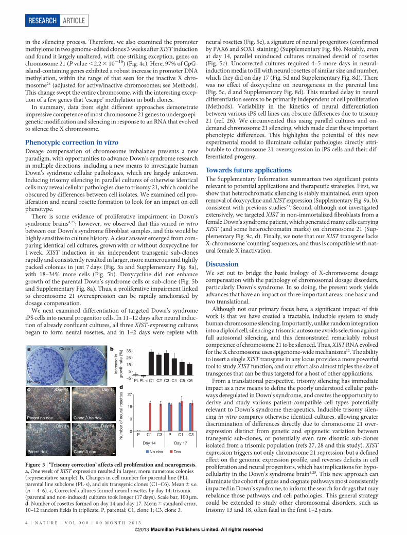
Figure 5 | ‘Trisomy correction’ affects cell proliferation and neurogenesis.a, One week of XIST expression resulted in larger, more numerous colonies(representative sample). b, Changes in cell number for parental line (PL),parental line subclone (PL-s), and six transgenic clones (C1–C6). Mean 6 s.e.(n 5 4–6). c, Corrected cultures formed neural rosettes by day 14; trisomic(parental and non-induced) cultures took longer (17 days). Scale bar, 100mm.d, Number of rosettes formed on day 14 and day 17. Mean 6 standard error,10–12 random fields in triplicate. P, parental; C1, clone 1; C3, clone 3.
RESEARCH ARTICLE
4 | N A T U R E | V O L 0 0 0 | 0 0 M O N T H 2 0 1 3
Macmillan Publishers Limited. All rights reserved©2013
Finally, the more forward-looking implication of this work is tobring Down’s syndrome into the realm of consideration for futuregene therapy research. Although development of any clinical genetherapy is a multi-step process, any prospect requires that the firststep, functional correction of the underlying genetic defect in livingcells, is achievable. We have demonstrated that this step is no longerinsurmountable for chromosomal imbalance in Down’s syndrome.Our hope is that for individuals and families living with Down’ssyndrome, the proof-of-principle demonstrated here initiates mul-tiple new avenues of translational relevance for the 50 years ofadvances in basic X-chromosome biology.
METHODS SUMMARYZFNs against the DYRK1A locus on chromosome 21 were designed and validatedby established procedures18. These and previously identified ZFNs to chro-mosome 19 AAVS1 (ref. 19) were used to deliver the XIST gene and rtTA/puroto chromosome 21 and chromosome 19, respectively. All constructs were simul-taneously electroporated into a Down’s syndrome iPS cell line (DS1-iPS4) (G. Q.Daley, Children’s Hospital Boston)20. Over 100 clones were isolated and 6 chosenfor more analysis. Silencing of the targeted chromosome 21 was demonstrated byeight different approaches as detailed in Methods (RNA microarray, DNA methy-lation array, RNA FISH to chromosome 21 genes, heterochromatin hallmarks,qRT–PCR, gene SNP analysis, Barr body formation, RNA FISH to hnRNA).Down’s syndrome iPS cells were assessed for phenotypic differences (proliferationand neural rosette formation) before and after trisomy correction, as detailed inMethods.
Full Methods and any associated references are available in the online version ofthe paper.
Received 21 May 2012; accepted 18 June 2013.Published online 17 July 2013.
1. Megarbane, A. et al. The 50th anniversary of the discovery of trisomy 21: the past,present, and future of research and treatment of Down syndrome. Genet. Med. 11,611–616 (2009).
2. Gardiner, K. J. Molecular basis of pharmacotherapies for cognition in Downsyndrome. Trends Pharmacol. Sci. 31, 66–73 (2010).
3. Prandini, P. et al. Natural gene-expression variation in Down syndrome modulatesthe outcome of gene-dosage imbalance. Am. J. Hum. Genet. 81, 252–263 (2007).
4. Haydar, T. F. & Reeves, R. H. Trisomy 21 and early brain development. TrendsNeurosci. 35, 81–91 (2012).
5. O’Doherty, A. et al. An aneuploid mouse strain carrying human chromosome 21with Down syndrome phenotypes. Science 309, 2033–2037 (2005).
6. Lee, B. & Davidson, B. L. Gene therapy grows into young adulthood: special reviewissue. Hum. Mol. Genet. 20, R1 (2011).
7. Hall, L. L. et al. X-inactivation reveals epigenetic anomalies in most hESC butidentifies sublines that initiate as expected. J. Cell. Physiol. 216, 445–452 (2008).
8. Nazor, K. L. et al. Recurrent variations in DNA methylation in human pluripotentstem cells and their differentiated derivatives. Cell Stem Cell 10, 620–634 (2012).
9. Brown, C. J. et al. The human XIST gene: analysis of a 17 kb inactive X-specific RNAthat contains conserved repeats and is highly localized within the nucleus. Cell 71,527–542 (1992).
10. Clemson, C. M., McNeil, J. A., Willard, H. F. & Lawrence, J. B. XIST RNA paints theinactive X chromosome at interphase: evidence for a novel RNA involved innuclear/chromosome structure. J. Cell Biol. 132, 259–275 (1996).
11. Heard, E. Delving into the diversity of facultative heterochromatin: the epigeneticsof the inactive X chromosome. Curr. Opin. Genet. Dev. 15, 482–489 (2005).
12. Hall, L. L. & Lawrence, J. B. XIST RNA and architecture of the inactive Xchromosome: implications for the repeat genome. Cold Spring Harb. Symp. Quant.Biol. 75, 345–356 (2010).
13. Carrel, L. & Willard, H. F. X-inactivation profile reveals extensive variability inX-linked gene expression in females. Nature 434, 400–404 (2005).
14. Lee, J. T., Strauss, W. M., Dausman, J. A. & Jaenisch, R. A 450 kb transgene displaysproperties of the mammalian X-inactivation center. Cell 86, 83–94 (1996).
15. Hall, L. L., Clemson, C. M., Byron, M., Wydner, K. & Lawrence, J. B. UnbalancedX;autosome translocations provide evidence for sequence specificity in theassociation of XIST RNA with chromatin. Hum. Mol. Genet. 11, 3157–3165 (2002).
16. Hall, L. L.et al.AnectopichumanXISTgenecan inducechromosome inactivation inpostdifferentiation human HT-1080 cells. Proc. Natl Acad. Sci. USA 99,8677–8682 (2002).
17. Moehle, E. A. et al. Targeted gene addition into a specified location in the humangenome using designed zinc finger nucleases. Proc. Natl Acad. Sci. USA 104,3055–3060 (2007).
18. Urnov, F.D., Rebar, E. J., Holmes,M.C., Zhang, H. S. & Gregory,P.D.Genome editingwith engineered zinc finger nucleases. Nature Rev. Genet. 11, 636–646 (2010).
19. DeKelver, R. C. et al. Functional genomics, proteomics, and regulatory DNAanalysis in isogenic settings using zinc finger nuclease-driven transgenesis into asafe harbor locus in the human genome. Genome Res. 20, 1133–1142 (2010).
20. Park, I. H. et al. Disease-specific induced pluripotent stem cells. Cell 134, 877–886(2008).
21. Aıt Yahya-Graison, E. et al. Classification of human chromosome 21 gene-expression variations in Down syndrome: impact on disease phenotypes. Am.J. Hum. Genet. 81, 475–491 (2007).
22. Biancotti, J. C. et al. Human embryonic stem cells as models for aneuploidchromosomal syndromes. Stem Cells 28, 1530–1540 (2010).
23. Csankovszki, G., Nagy, A. & Jaenisch, R. Synergism of Xist RNA, DNA methylation,and histone hypoacetylation in maintaining X chromosome inactivation. J. CellBiol. 153, 773–784 (2001).
24. Cotton, A. M. et al. Chromosome-wide DNA methylation analysis predicts humantissue-specific X inactivation. Hum. Genet. 130, 187–201 (2011).
25. Guidi, S., Ciani, E., Bonasoni, P., Santini, D. & Bartesaghi, R. Widespreadproliferation impairment and hypocellularity in the cerebellum of fetuses withdown syndrome. Brain Pathol. 21, 361–373 (2011).
26. Shi, Y. et al. A human stem cell model of early Alzheimer’s disease pathology inDown syndrome. Sci. Transl. Med. 4, 124ra29 (2012).
27. Lavon, N. et al. Derivation of euploid human embryonic stem cells from aneuploidembryos. Stem Cells 26, 1874–1882 (2008).
28. Li, L. B. et al. Trisomy correction in down syndrome induced pluripotent stem cells.Cell Stem Cell 11, 615–619 (2012).
Supplementary Information is available in the online version of the paper.
Acknowledgements We appreciate recent initiatives by administrators of NIGMS andNIH to support more high-risk, high-impact research. Research began with supportfrom GM053234 to J.B.L. for basic X chromosome research, and was made fullypossible by GM085548 and GM096400 RC4 to J.B.L. C.J.B. and A.M.C. were supportedby CIHR (MOP-13680) to C.J.B. We thank T. Flotte for encouragement and adviceregarding genome editing strategies, and similarly appreciate the support of S. Jonesand P. Newburger. We thank T. Collingwood for initial discussions regarding thisproject, and the George Daley laboratory (Harvard) for the Down’s syndrome iPS cellline. L. Lizotte, Z. Matijasevic, K. Smith and E. Swanson provided various assistance.M. S. Kobor and L. Lam (Kobor laboratory) assisted with methylation analysis. D.M.C. issupported by an NIH fellowship 1F32CA154086 and B.R.C. (O. Rando laboratory) issupported by NIH training grant 2T32HD007439 (G. Witman, PI).
Author Contributions J.J., with the assistance of Y.J., designed and produced allconstructs, edited all cell lines, and designed and performed most experiments. J.B.L.,J.J. and L.L.H. were the main contributors to designing experiments and interpretingresults. J.B.L., J.J., L.L.H. and F.D.U. wrote the manuscript. F.D.U., P.D.G. and G.J.C.engineered and validated ZFNs. J.R.P. performed Cel1 and Southern analysis. J.-C.C.performed SNP analysis, characterized three sub-clones, and helped with proliferationexperiments. J.J. and Y.J., with help from J.-C.C., M.B., H.J.K. and L.L.H., carried out initialscreening of targeted iPS cell sub-clones. H.J.K. edited and characterized primaryDown’s syndrome fibroblast line. A.M.C. and C.J.B. carried out DNA methylationanalysis andprovidedXISTcDNA. J.J. andF.D.U.prepared themicroarray library.D.M.C.and B.R.C. analysed microarray data with help from D.A.S., D.Y.G. and E.J.R.
Author Information Microarray data for 27 samples is deposited in GEO underaccession number GSE47014. Reprints and permissions information is available atwww.nature.com/reprints. The authors declare competing financial interests: detailsare available in the online version of the paper. Readers are welcome to comment onthe online version of the paper. Correspondence and requests for materials should beaddressed to J.B.L. ([email protected]) or F.D.U.([email protected], requests for ZFNs).
ARTICLE RESEARCH
0 0 M O N T H 2 0 1 3 | V O L 0 0 0 | N A T U R E | 5
Macmillan Publishers Limited. All rights reserved©2013
METHODSCell culture. HT1080 TetR cells (Invitrogen) and female Down’s syndromehuman primary fibroblast line (Coriell) (AG13902) were cultured as recom-mended by the supplier. Down’s syndrome iPS cell parental line (DS1-iPS4) wasprovided by G. Q. Daley (Children’s Hospital Boston)20 and maintained on irra-diated mouse embryonic fibroblasts (iMEFs) (R&D Systems, PSC001) in hiPSCmedium containing DMEM/F12 supplemented with 20% knockout serum replace-ment (Invitrogen), 1 mM glutamine (Invitrogen), 100mM non-essential aminoacids (Invitrogen), 100mM b-mercaptoethanol (Sigma) and 10 ng ml21 FGF-b(Invitrogen, PHG0024). Cultures were passaged every 5–7 days with 1 mg ml21
of collagenase type IV (Invitrogen).ZFN design. ZFNs against the human AAVS1 locus on chromosome 19 havebeen previously described19. ZFNs against the DYRK1A locus were designed usingan archive of pre-validated zinc finger modules18,29, and validated for genomeediting activity by transfection into K562 cells and Surveyor endonuclease-basedmeasurement of endogenous locus disruption (‘Cel1’30,31) exactly as described29.Southern blotting for targeted gene addition was performed exactly as described17,32
on SphI-digested genomic DNA probed with a fragment corresponding to positionsChr21:38825803138826056 (hg19).XIST and rtTA/puro plasmid construction. Fourteen-kilobase human XISTcDNA, a splicing isoform of full-length XIST cDNA, was subcloned into pTRE3G(Clontech, catalogue no. 631167). Two homologous arms (left arm, 690 bp; rightarm, 508 bp) of DYRK1A gene on chromosome 21 were amplified by PCR fromprimary Down’s syndrome fibroblasts (AG13902) (Coriell) and cloned into thepTRE3G vector (human chromosome 21 DYRK1A left arm primers: forward 59-GCCGTATACCATTAACTCTTTACTGTTC-39, reverse 59-TCTGTATACGTAAACTGGCAAAGGGGTGG-39; human chromosome 21 DYRK1A right arm pri-mers: forward 59-ATTTCGCGAACGGGTGATGAGCAGGCTGT-39, reverse 59-CCGTCGCGAAAACCAGAAAGTATTCTCAG-39). The pEF1a-3G rtTA-pAcassette from pEF1a-Tet3G vector (Clontech) was subcloned into a plasmid fortargeted gene addition to the PPP1R12C/AAVS1 locus19, which contains a uniqueHindIII site flanked by two 800-bp stretches of homology to the ZFN-specifiedposition in the genome.Dual-targeted addition of human Down’s syndrome iPS cells and generationof stable targeted clones. The Down’s syndrome iPS cell line was cultured in10 mM of Rho-associated protein kinases (ROCK) inhibitor (Calbiochem;Y27632) 24 h before electroporation. Single cells (1 3 107) were collected usingTryPLE select (Invitrogen), re-suspended in 13 PBS and electroporated with atotal of 55 mg DNA including five plasmids (XIST, DYRK1A ZFN1, DYRK1AZFN2, rtTA/puro and AAVS1 ZFN) with both 3:1 and 5:1 ratios of XIST:rtTA/puro. The electroporation conditions were 220 V and 750mF (BioRad GenePulser II System). Cells were subsequently plated on puromycin-resistant DR4MEF feeders (Open Biosystems, catalogue no. MES3948) in hiPSC medium sup-plemented with ROCK inhibitor for the first 24 h. Over 300 colonies remainedafter 12 days of 0.4mg ml21 puromycin selection and 245 randomly chosen indi-vidual colonies across 36 pooled wells were examined by interphase DNA/RNAFISH for the presence and expression of XIST, correct targeting and retention oftrisomy (because some subclones lacked XIST or showed just two DYRK1A DNAsignals). Over 100 individual clones were isolated and characterized, and those ofinterest, containing targeted XIST on one of three DYRK1A loci, were frozen. Sixsingle target clones with good pluripotent morphology, OCT4 positive staining,correct targeting to one trisomic chromosome, and good XIST RNA paint wereexpanded for further characterization. One double and one triple target line, twonon-target clones, and one disomic clone were also isolated and frozen. Targetingand correct chromosome number (47) was confirmed by interphase and meta-phase FISH and genome integrity was confirmed by high-resolution G-bandkaryotype and CGH array.Chromosome preparation. iPS cells were treated with 100 ng ml21 KaryoMAXcolcemid (Invitrogen) for 2–4 h at 37 uC in a 5% CO2 incubator. Cells weretrypsinized, treated with hypotonic solution, and fixed with methanol:acetic acid(3:1). Metaphases were spread on microscope slides, and at least 20 analysed perclone. Karyotype analysis was done on pro-metaphase chromosomes usingStandard Giemsa-trypsin G band methods.CGH array. CGH was performed in the Cytogenetics Laboratory at University ofMassachusetts Medical School. Genomic Microarray analysis using University ofMassachusetts Genomic Microarray platform (Human Genome Build hg19) wasperformed with 1 mg of DNA. The array contains approximately 180,000 oligo-nucleotides (60-mers) that represent coding and non-coding human sequencesand high-density coverage for clinically relevant deletion/duplication syndromesand the telomeric and pericentromeric regions of the genome. Data were analysedby BlueFuse Multi, v3.1 (BlueGnome, Ltd).DNA/RNA FISH and immunostaining. DNA and RNA FISH were carried outas previously described10,15,16,33. The XIST probe is a cloned 14-kb XIST cDNA
(the same sequence as XIST transgene in Fig. 1b) in pGEM-7Zf(1) (Promega).Six chromosome 21 gene probes are BACs from BACPAC Resources (DYRK1A,Rp11-105O24; APP, RP11-910G8; USP25, RP11-840D8; CXADR, RP11-1150I14;ITSN1, RP11-1033C16; COL18A1, RP11-867O18). DNA probes were labelled bynick translation with either biotin-11-dUTP or digoxigenin-16-dUTP (Roche). Insimultaneous DNA/RNA FISH (interphase targeting assay), cellular DNA wasdenatured and hybridization performed without eliminating RNA and alsotreated with 2 U ml21 of RNasin Plus RNase inhibitor (Promega). For immuno-staining with RNA FISH, cells were immunostained first with RNasin Plus andfixed in 4% paraformaldehyde before RNA FISH. Antibodies were as follows:H3K27me3 (Millipore, 07-449), UbH2A (Cell Signaling, 8240), H4K20me(Abcam, ab9051), macroH2A (Millipore, 07-219), OCT4 (Santa Cruz, sc-9081),PAX6 (Stemgent, 09-0075), SOX1 (R&D Systems, AF3369).Allele-specific SNP analysis. Primers were designed to amplify 39 untranslatedregions of chromosome 21 genes reported to contain SNPs (Supplementary Table3). Total cDNA was used from three transgenic clones with and without XISTinduction for 22 days. RT–PCR products were sequenced by GENEWIZ. Of ,10genes examined, four were heterozygous and informative in the patient Down’ssyndrome iPS cell line used here.Microarray analysis. Three independently targeted subclones plus the parentalchromosome 21 trisomic (non-targeted) iPS cell line were grown with or withoutdoxycycline (2 mg ml21) for 22 d. Normal male iPS cell and disomic isogenic lineswere also cultured for 22 d and total RNA was extracted with a High Pure RNAextraction kit (Roche) in triplicate for each, processed with a Gene Chip 39 IVTexpress kit (Affymetrix), and hybridized to Affymetrix human gene expressionPrimeView arrays. Array normalization was performed with AffymetrixExpression Console Software with Robust Multichip Analysis (RMA)34. Probesets with the top 60% of signal values were considered present and ‘expressed’ andwere used for all further analysis. Data in Fig. 4 has no other threshold applied.When designated, a gene expression change significance threshold was appliedusing a two-tailed t-test comparing samples with or without doxycycline in trip-licate (n 5 3) (Supplementary Table 2, P , 0.01). For the ,200 genes found tosignificantly change in all three clones (in text), a t-test with P , 0.001 wasapplied.Microarray data interpretation. Using extraction-based methods, changes onjust one of three alleles (from the XIST-bearing chromosome) will be diluted bythe other two. If all three chromosomes are fully expressed, this would predict a33% reduction in chromosome 21 expression levels per cell when one chro-mosome 21 is fully silenced. However, 33% would apply only if chromosome21 genes are fully overexpressed to start, and previous evidence and results in thisstudy show this is not the case for many genes. Previous microarray studies haveanalysed expression levels of chromosome 21 in Down’s syndrome patient cells,although such analyses are hampered by the extensive genetic and epigeneticdifferences between any two individuals3. The fraction of chromosome 21 genesdetected as overexpressed varies with the study and tissue, but generally is in the19–36% range3,22, with individual gene increases often in the ,1.2–1.4 range (lessthan the theoretical 1.5). For example, one study of Down’s syndrome embryoidbodies showed that only 6–15% of genes appeared significantly upregulated, butthis was comparing non-isogenic samples of different ES cell isolates22.
Our trisomy correction system allows direct comparison of the same cellsgrown in identical parallel cultures, with and without XIST-mediated chro-mosome silencing. Our data show a ,20% reduction in chromosome 21 express-ion overall; importantly, this level of reduction is seen either when the thirdchromosome is silenced in trisomic cells, or when disomic and trisomic cellsare compared. This 20% reduction represents an average per cell for all threechromosomes, but corresponds to a 60% reduction in expression for just onechromosome 21 (the one silenced by XIST RNA, as shown here).
Apart from our goal here of trisomy dosage compensation, these results addsignificantly to understanding the extent of chromosome 21 overexpression inDown’s syndrome, by providing a more comprehensive analysis that shows thatexpression of most genes is increased, but less than the theoretical 1.5 fold.qRT–PCR. qRT–PCR was performed for eight downregulated chromosome 21genes determined by microarray on a Bio-Rad MyiQ real-time PCR detectionsystem in triplicate for clone 3 with/without doxycycline treatment for 22 d. Theb-actin gene was used as an internal standard for calculation of expression levels.Primers for eight chromosome 21 genes and b-actin were described inSupplementary Table 4.DNA methylation analysis. The parental line and two independent targeted lineswere grown with and without doxycycline for 22 d, in duplicate cultures. GenomicDNA was extracted using PureLink Genomic DNA mini kit (Invitrogen) and750 ng bisulphite modified with the Alternative Incubation Conditions from theEZ DNA methylation kit (Zymo Research). 160 ng of bisulphite DNA was amp-lified, fragmented and hybridized to Illumina Infinium HumanMethylation450
RESEARCH ARTICLE
Macmillan Publishers Limited. All rights reserved©2013
array following the standard protocol as outlined in the user guide. CpG islandswere defined as high and intermediate CpG densities using the CpG densityclassifications based on those used previously35. The program CpGIE was usedto locate HC and IC islands on the X chromosome and chromosomes 21 and 22.When multiple probes in CpG islands were associated with the same TSS, anaverage genic methylation value was calculated. These average genic values werecompared before and after doxycycline induction using the Mann–WhitneyU-test. Analysis was based on CpG islands within promoters of 143 chromosome21 genes (Fig. 4c).
The average methylation value was 6% on chromosome 21 before XIST induc-tion, and increased to 20–21% in both subclones after induction. Because anymethylation increase on the transgenic chromosome would be diluted by thepresence of three chromosome 21 copies, this suggests the range of 60% methyla-tion on the one XIST-coated chromosome, which is within the range seen for theinactive X chromosome24.Cell proliferation analysis. Eight different iPS cell lines (parental line, one non-targeted subclone, and six independent targeted subclones) were passaged onto6-well plates at equal cell densities per well of each line and grown with or withoutdoxycycline for 7 d. At least four replicates of each line were analysed in twoindependent experiments. Rigorous measures were taken to minimize and con-trol for any minor variations in seeding densities of iPS cells, which cannot beplated as single cell suspensions. First, the analysis was done twice for six differenttransgenic clones, in each case comparing triplicate plates of corrected versus notcorrected (doxycycline versus no doxycycline). To avoid differences in platingefficiencies of doxycycline and no doxycycline cells, we performed the experi-ments over a time course that did not require passage. For each of the six trans-genic clones, the parental line and one negative control (non-targeted) subclone, asingle well of Down’s syndrome iPS cells (without doxycycline) was used togenerate a cell suspension (cells and small disaggregated clumps). Next, equalaliquots of the cell suspension were plated into each of six wells four times (notrelying on one measurement but the average of four for seeding each well). Afterplating, doxycycline was added to three of the six wells, and the cultures weremaintained for 7 d. For images, plates were fixed, stained with 1 mg ml–1 crystalviolet (Sigma) in 70% ethanol for 30 min and scanned to generate TIFF images.For cell counts, single cells were collected by TryPLE select and counted usingBeckman Coulter Z1 Particle Counter.Differentiation of neural progenitors and irreversibility in cortical neurons.For differentiation, independent XIST-transgenic iPS cell clones and the parentalDown’s syndrome iPS cell line were dissociated with Accutase (Innovative CellTechnologies) and 4 3 105 single cells were plated on Matrigel-coated 6-wellplates in mTeSR1 medium (Stemcell technologies). Once the cell culture reached90–100% confluence, neural induction was initiated by changing the culturemedium to neural induction medium, a 1:1 mixture of N2- and B27-containingmedia supplemented with 500 ng ml–1 noggin (R&D Systems), 10 mM SB431542(Tocris Bioscience), and 1mM retinoic acid (Sigma, catalogue no. R2625), with/without treatment of doxycycline for the specified times. The neural rosettes werecounted and their diameter measured for at least 300 rosettes (sampled in randomareas from triplicate dishes). At day 14, the doxycycline-induced culture had anaverage rosette diameter of 142mm 6 0.55mm in clone 1 and 141mm 6 3.49 mmin clone 3. Rosettes could not be measured at the same time point in the uncor-rected culture, as they had not formed. At day 17, the uncorrected culture had
neural rosettes of similar number and size for both clones 1 (140mm 6 0.87 mm)and 3 (140 mm 6 1.09 mm). The corrected culture could not be accurately com-pared for day 17 because the rosettes had become so mature and often hadmerged. After 17 d, neural rosettes were collected by dissociation with dispaseand replated on poly-ornithine and laminin-coated plastic dishes in N2- and B27-containing media including 20 ng ml21 FGF2. After a further 2 d, FGF2 waswithdrawn to promote differentiation of cortical neurons. To test for the irre-versibility of silencing, two independent clones were differentiated to corticalneurons in the presence of doxycycline for 70 days to initiate silencing. They werethen split into parallel cultures grown with and without doxycycline for another30 days, and XIST and APP expression analysed by RNA FISH.Targeted addition to primary fibroblasts. We used non-immortalized primaryhuman female Down’s syndrome fibroblasts, which like all primary fibroblastshave a limited lifespan in culture (potentially more limited for Down’s syndromefibroblasts). We reasoned that the robustness of ZFN-driven editing, combinedwith reduction to disomy for the DRYK1A gene, may make it possible to observesome edited cells before they senesce. We used a transgene carrying near full-length (,14 kb) XIST cDNA under a TetO2 inducible promoter, and a selectablemarker on the same construct, with ,600-bp homology arms to the DYRK1Agene (vector is ,21 kb, with a ,17-kb insert) (data not shown). When introducedwithout the Tet-repressor construct, the TetO2 CMV promoter is constitutivelyactive. Two ZFN-containing vectors and the 21-kb XIST transgene were trans-fected into primary DS fibroblasts (Coriell AG13902) using Stemfect polymer(Stemgent) (10:1 ratio of XIST to ZFN, and 13mg DNA to 1.3ml Stemfect per wellof 6-well plate). The frequency of stable integrants was such that a sparse mono-layer of transgenic fibroblasts emerged, rather than a few individual coloniesfollowing selection with hygromycin (75mg ml21). The pooled population of selectedcells was analysed by FISH and immunostaining for targeting, XIST expression andheterochromatin marks. XIST RNA was observed over the DYRK1A locus in,74% of cells, indicating accurate transgene targeting, which was also verifiedby metaphase FISH (Supplementary Fig. 9c). In many cells there was notableenrichment of H3K27me, UbH2A and H4K20me heterochromatic marks(Supplementary Fig. 9d). Owing to the limited lifespan of primary cells and theprogressive silencing of the CMV promoter used in this construct, these cells werenot more fully characterized.
29. Doyon, J. B. et al. Rapid and efficient clathrin-mediated endocytosisrevealed in genome-edited mammalian cells. Nature Cell Biol. 13, 331–337(2011).
30. Miller, J. C. et al. An improved zinc-fingernuclease architecture for highly specificgenome editing. Nature Biotechnol. 25, 778–785 (2007).
31. Guschin, D. Y. et al. A rapid and general assay for monitoring endogenous genemodification. Methods Mol. Biol. 649, 247–256 (2010).
32. Urnov, F. D. et al. Highly efficient endogenous human gene correction usingdesigned zinc-finger nucleases. Nature 435, 646–651 (2005).
33. Byron, M., Hall, L. L. & Lawrence, J. B. A multifaceted FISH approach to studyendogenous RNAs and DNAs in native nuclear and cell structures. Curr. Protoc.Hum. Gen. Chapter 4, Unit 4 15 (2013).
34. Irizarry, R. A. et al. Summaries of Affymetrix GeneChip probe level data. NucleicAcids Res. 31, e15 (2003).
35. Weber, M. et al. Distribution, silencing potential and evolutionary impact ofpromoter DNA methylation in the human genome. Nature Genet. 39, 457–466(2007).
ARTICLE RESEARCH
Macmillan Publishers Limited. All rights reserved©2013