treatment of osteonecrosis of the femoral head with ... · of the femoral head with implantation of...
TRANSCRIPT

COPYRIGHT © 2004 BY THE JOURNAL OF BONE AND JOINT SURGERY, INCORPORATED
Treatment of Osteonecrosis of the Femoral Head with
Implantation of Autologous Bone-Marrow Cells
A PILOT STUDY
BY VALÉRIE GANGJI, MD, JEAN-PHILIPPE HAUZEUR, MD, PHD, CELSO MATOS, MD, VIVIANE DE MAERTELAER, PHD, MICHEL TOUNGOUZ, MD, PHD, AND MICHELINE LAMBERMONT, PHARMD
Investigation performed at the Department of Rheumatology and Physical Medicine, the Department of Radiology, and the Cellular and Molecular Therapy Unit, Erasme University Hospital, Brussels, Belgium
Background: Aseptic nontraumatic osteonecrosis of the femoral head is a disorder that can lead to femoral headcollapse and the need for total hip replacement. Since osteonecrosis may be a disease of mesenchymal cells orbone cells, the possibility has been raised that bone marrow containing osteogenic precursors implanted into a ne-crotic lesion of the femoral head may be of benefit in the treatment of this condition. For this reason, we studied theimplantation of autologous bone-marrow mononuclear cells in a necrotic lesion of the femoral head to determine theeffect on the clinical symptoms and the stage and volume of osteonecrosis.
Methods: We studied thirteen patients (eighteen hips) with stage-I or II osteonecrosis of the femoral head, accordingto the system of the Association Research Circulation Osseous. The hips were allocated to a program of either coredecompression (the control group) or core decompression and implantation of autologous bone-marrow mononuclearcells (the bone-marrow-graft group). Both patients and assessors were blind with respect to treatment-group assign-ment. The primary outcomes studied were safety, clinical symptoms, and disease progression.
Results: After twenty-four months, there was a significant reduction in pain (p = 0.021) and in joint symptoms mea-sured with the Lequesne index (p = 0.001) and the WOMAC index (p = 0.013) within the bone-marrow-graft group. Attwenty-four months, five of the eight hips in the control group had deteriorated to stage III, whereas only one of theten hips in the bone-marrow-graft group had progressed to this stage. Survival analysis showed a significant differ-ence in the time to collapse between the two groups (p = 0.016). Implantation of bone-marrow mononuclear cellswas associated with only minor side effects.
Conclusions: Implantation of autologous bone-marrow mononuclear cells appears to be a safe and effective treat-ment for early stages of osteonecrosis of the femoral head. Although the findings of this study are promising, their in-terpretation is limited because of the small number of patients and the short duration of follow-up. Further study isneeded to confirm the results.
Level of Evidence: Therapeutic study, Level II-1 (prospective cohort study). See Instructions to Authors for a com-plete description of levels of evidence.
septic nontraumatic osteonecrosis is a painful disorderof the hip that often leads, in its final stage, to femoralhead collapse, osteoarthritis, and the need for total hip
replacement. Glucocorticoid use and alcohol abuse are amongthe most widely recognized risk factors for osteonecrosis inwhite individuals1. Core decompression of the hip is the mostwidespread procedure used to treat early stages of osteonecro-sis of the femoral head2. Notwithstanding the fact that thisprocedure has been used for more than three decades, its effi-cacy remains controversial3,4. Accordingly, a more pathophysi-
AA commentary is available with the electronic versions of this article,on our web site (www.jbjs.org) and on our quarterly CD-ROM (call oursubscription department, at 781-449-9780, to order the CD-ROM).

TH E JO U R NA L OF BONE & JOINT SURGER Y · JBJS .ORG
VO LU M E 86-A · NU M B E R 6 · JU N E 2004TRE A T M EN T OF OSTE ON E CROSIS OF T H E FEMOR AL HEA D W ITH IM P L A N T A T I O N OF AUTO L O GO U S BONE-MA R ROW CE L L S
ological approach to the treatment of osteonecrosis may bemore appropriate. Different pathophysiological mechanismshave been postulated for this disease, including fat emboli5,microvascular tamponade of the blood vessels of the femoralhead by marrow fat6, retrograde embolization of the marrowfat7, and microfracture of trabecular bone8. The levels of ac-tivity and the number of mesenchymal stem cells in both thehematopoietic and the stromal compartments of the bonemarrow have been shown to be depressed in patients with os-teonecrosis of the femoral head9, suggesting that this might bea disease of bone cells and/or mesenchymal cells. The capacityof osteoblastic cells to replicate is decreased in the proximalpart of the femur of patients with osteonecrosis of the femoralhead10. This finding raised the possibility that bone marrowcontaining stromal cells, which have many of the characteris-tics of the stem cells for mesenchymal tissues including bone,could be implanted into a necrotic lesion of the femoral head.Moreover, Hernigou et al.11 reported the case of a patient whohad been treated successfully with autologous bone-marrowimplantation for osteonecrosis of the humeral head, secondaryto sickle-cell disease. On the basis of this experience, we begana two-year prospective, controlled, double-blind pilot study onthe effect of implantation of autologous bone-marrow mono-nuclear cells into the necrotic lesion in femoral heads withearly osteonecrosis.
Materials and MethodsPatients
atients were considered eligible for the study if they hadstage-I or II osteonecrosis of the femoral head according
to the system of the Association Research Circulation Osseous(ARCO)12. According to this system, normal radiographicfindings in the femoral head were classified as ARCO stage-I
osteonecrosis; trabecular bone remodeling within the femoralhead, as stage II; and subchondral collapse of the femoral head,as stage III. We excluded patients with evidence of a malignantdisorder during the past five years. The study was approved bythe ethical committee of the hospital, and informed consentwas obtained from the patients. Osteonecrosis was diagnosedwith magnetic resonance imaging13. The primary outcomesthat were assessed were safety, clinical symptoms, and diseaseprogression. Clinical symptoms included pain and other jointsymptoms. Disease progression included an analysis of thestage of osteonecrosis and the volume of the osteonecrotic le-sion at the time of the final follow-up. Since there were nopublished data on this particular procedure, as far as we know,we could not predetermine the number of hips that wouldbe needed for adequate power in this study. Accordingly, wecarried out this pilot study. Seventeen patients were recruited.Four patients were excluded, and thirteen patients (six womenand seven men) were able to complete the study. There wereno missing data. Demographic data are listed in Table I. Atbaseline, the stages of osteonecrosis, the age of the patients,the clinical symptoms, and the volume of osteonecrosis for thetwo groups were not found to be significantly different, withthe numbers available.
Five patients had bilateral involvement. Two hips hadstage-I osteonecrosis, and sixteen hips had stage-II osteo-necrosis. Ten patients (fourteen hips) had osteonecrosis as aresult of corticosteroid therapy, and one patient (two hips)had alcohol-induced osteonecrosis. For one patient (two hips),no etiological factor was determined. All patients were symp-tomatic. The eighteen hips were allocated to treatment witheither a core decompression (the control group) or to coredecompression and implantation of autologous bone-marrowmononuclear cells (the bone-marrow-graft group). All patients
P
TABLE I Demographic Data and Baseline Characteristics of the Hips
Characteristics* Control Group Bone-Marrow-Graft Group
No. of hips 8 10
Age† (yr) 48.8 ± 11.2 40.9 ± 9.8
Time to diagnosis† (mo) 4.6 ± 0.6 5.2 ± 0.9
Etiology of osteonecrosis
Corticosteroid use 6 8
Alcohol abuse 1 1
Idiopathic 1 1
Visual analog pain scale† (mm) 34.6 ± 10.1 37.8 ± 8.4
Lequesne index† 5.4 ± 1.5 7.7 ± 1.5
WOMAC score† 21 ± 5 30 ± 5
ARCO stage I 1 1
ARCO stage II 7 9
Volume of osteonecrosis/volume of femoral head† (%) 16.7 ± 4.6 15.6 ± 1.5
*WOMAC = Western Ontario and McMaster Universities Osteoarthritis Index, and ARCO = Association Research Circulation Osseous. †Thevalues are given as the mean and the standard error of the mean.

TH E JO U R NA L OF BONE & JOINT SURGER Y · JBJS .ORG
VO LU M E 86-A · NU M B E R 6 · JU N E 2004TRE A T M EN T OF OSTE ON E CROSIS OF T H E FEMOR AL HEA D W ITH IM P L A N T A T I O N OF AUTO L O GO U S BONE-MA R ROW CE L L S
were blind to the treatment assignment. The hips were notrandomized. The surgeon performed both procedures (bone-marrow implantation or a core decompression) on an alter-
nating basis. For patients with bilateral involvement, the surgeonchose which hip to treat first. The first hip to treat was alter-nately the right and then the left hip. To control for bias, thesurgeon never examined the patients before or after the proce-dure and was the only person to know the group assignment.For ethical reasons, the patient was informed specifically ofthis procedure and knew that the surgeon could be called incase of complications or side effects. Investigators who as-sessed the outcomes were blind to group assignment. Recruit-ment began in January 1999, and all patients were followed fortwenty-four months.
Core DecompressionWith the patient under general anesthesia, a 5-mm incisionwas made through the skin and the fascia at the level of thegreater trochanter. A 3-mm trephine (inner diameter) (Collin,Paris, France) was used as described by Hauzeur et al.14,15. Thetrephine was introduced manually under fluoroscopic controlthrough the greater trochanter into the necrotic lesion. Thedirection of the trephine was adjusted in the intertrochantericregion so that it was pointing toward the necrotic area. The tipof the trephine was placed at a distance of 2 to 3 mm from thearticular cartilage (Fig. 1). No other areas of the necrotic le-sion were penetrated with the trephine.
Bone-Marrow Grafting The bone-marrow harvesting was performed during the sameoperative session as the core decompression. About 400 mLof marrow was obtained from the anterior iliac crest (seeAppendix). Mononuclear cells were sorted on a Spectra cellseparator (777006-300; Cobe, Lakewood, Colorado) and con-centrated to a mean final volume (and standard error of themean) of 51 ± 1.8 mL, which was injected through the tre-
Fig. 2
Fig. 1
Profile radiograph of the hip made at the time of the operation.
The 3-mm trephine was introduced by hand under fluoroscopy
through the greater trochanter into the anterosuperior region of
the femoral head. The trephine was placed into the osteonecrotic
zone within 2 to 3 mm of the subchondral bone. The mononuclear
cells were injected through the trephine into the necrotic zone.
A comparison of the bone-marrow-graft group (solid line) and
the control group (dashed line) with respect to the evolution of
the scores on the visual analog scale (VAS), the Lequesne in-
dex, and the WOMAC index over time. The results are shown
as the mean and the standard error of the mean. One asterisk
(p < 0.05) and two asterisks (p < 0.01) indicate a significant
difference compared with baseline.
TH E JO U R NA L OF BONE & JOINT SURGER Y · JBJS .ORG
VO LU M E 86-A · NU M B E R 6 · JU N E 2004TRE A T M EN T OF OSTE ON E CROSIS OF T H E FEMOR AL HEA D W ITH IM P L A N T A T I O N OF AUTO L O GO U S BONE-MA R ROW CE L L S
phine that was placed into the necrotic zone16. The meannumber of leukocytes injected was 2.0 ± 0.3 × 10⁹, including1.0% ± 0.2% of CD34+ cells, which are precursors of he-matopoietic cells. Fibroblast colony-forming units were usedas an indicator of stromal cell activity9. The mean number offibroblast colony-forming units was 92 ± 9/107 cells. Thesorted bone-marrow mononuclear cells contained lymphocy-toid cells (mean, 29% ± 2.2%), monocytoid cells (4% ±1%), and myeloid cells (6% ± 1.3%).
Clinical EvaluationPatients were assessed preoperatively and at three, six, twelve,and twenty-four months postoperatively. Pain was measuredwith use of a visual analog scale17. The severity of hip diseasewas gauged with use of the algofunctional index of Lequesneet al.18. Symptoms of osteonecrosis were assessed with theWestern Ontario and McMaster Universities Osteoarthritis In-dex (WOMAC)19.
At each visit, patients were assessed for possible side ef-fects of treatment.
Radiographic EvaluationAnteroposterior and frog-leg lateral radiographs and mag-netic resonance images of the affected hip were made at thetime of each clinical assessment. Radiographic progression ofthe osteonecrosis was measured according to the ARCO stag-ing system12. All radiographs were analyzed by a single readerwho was unaware of treatment assignments. The measure-ments of the magnetic resonance images were prepared on 3-mmcoronal T1-weighted scans with use of a separate computerworkstation (Easy Vision; Philips, Best, The Netherlands). Thecontours of the necrotic lesion and the femoral head weredrawn on each slice. The volume of the femoral head and theosteonecrotic zone was then calculated by the computer work-station. The relative volume of the necrotic lesion was calcu-lated as a percentage of the entire femoral head20. To evaluate
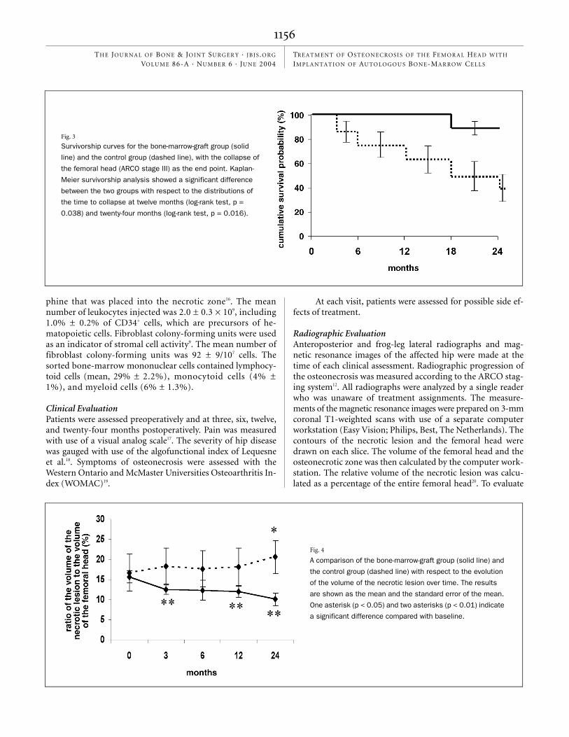
Fig. 4
A comparison of the bone-marrow-graft group (solid line) and
the control group (dashed line) with respect to the evolution
of the volume of the necrotic lesion over time. The results
are shown as the mean and the standard error of the mean.
One asterisk (p < 0.05) and two asterisks (p < 0.01) indicate
a significant difference compared with baseline.
Fig. 3
Survivorship curves for the bone-marrow-graft group (solid
line) and the control group (dashed line), with the collapse of
the femoral head (ARCO stage III) as the end point. Kaplan-
Meier survivorship analysis showed a significant difference
between the two groups with respect to the distributions of
the time to collapse at twelve months (log-rank test, p =
0.038) and twenty-four months (log-rank test, p = 0.016).
TH E JO U R NA L OF BONE & JOINT SURGER Y · JBJS .ORG
VO LU M E 86-A · NU M B E R 6 · JU N E 2004TRE A T M EN T OF OSTE ON E CROSIS OF T H E FEMOR AL HEA D W ITH IM P L A N T A T I O N OF AUTO L O GO U S BONE-MA R ROW CE L L S
the reliability of this method, the magnetic resonance imageswere analyzed by two different observers, each unaware oftreatment assignments. The first observer took two sets of mea-surements from each magnetic resonance image of the hip,and the second observer took one. This process resulted in 270separate determinations.
Statistical AnalysisContinuous variables are described as the mean and the stan-dard error of the mean.
We assessed the change over time with respect to the vis-ual analog scale, the Lequesne index, the WOMAC score, andthe volume of the necrotic lesion in both groups. Nonpara-metric Friedman tests were used. These tests were followed bythe use of Wilcoxon paired samples in order to compare the
baseline data with the values obtained at three, six, twelve, andtwenty-four months. The reliability of the magnetic resonanceimaging measurements was calculated with use of correlationcoefficients of reliability21. Intraobserver reliability was esti-mated with use of the readings of the first observer. Interob-server reliability was estimated by taking the first reading ofthe first observer and the unique reading of the second one. AKaplan-Meier survivorship analysis was used to compare theprogression to subchondral fracture (stage III). The rates ofsurvival of the femoral head for the two treatment groups,that is, the duration between the time of enrollment in thestudy and the end point (collapse of the femoral head), werecompared with the log-rank test. The rates of survival withinthe subgroups of patients who had bilateral osteonecrosis werealso compared. A Cox regression model was used with the rate
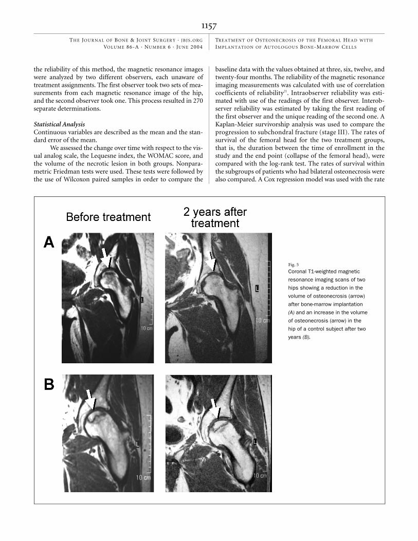
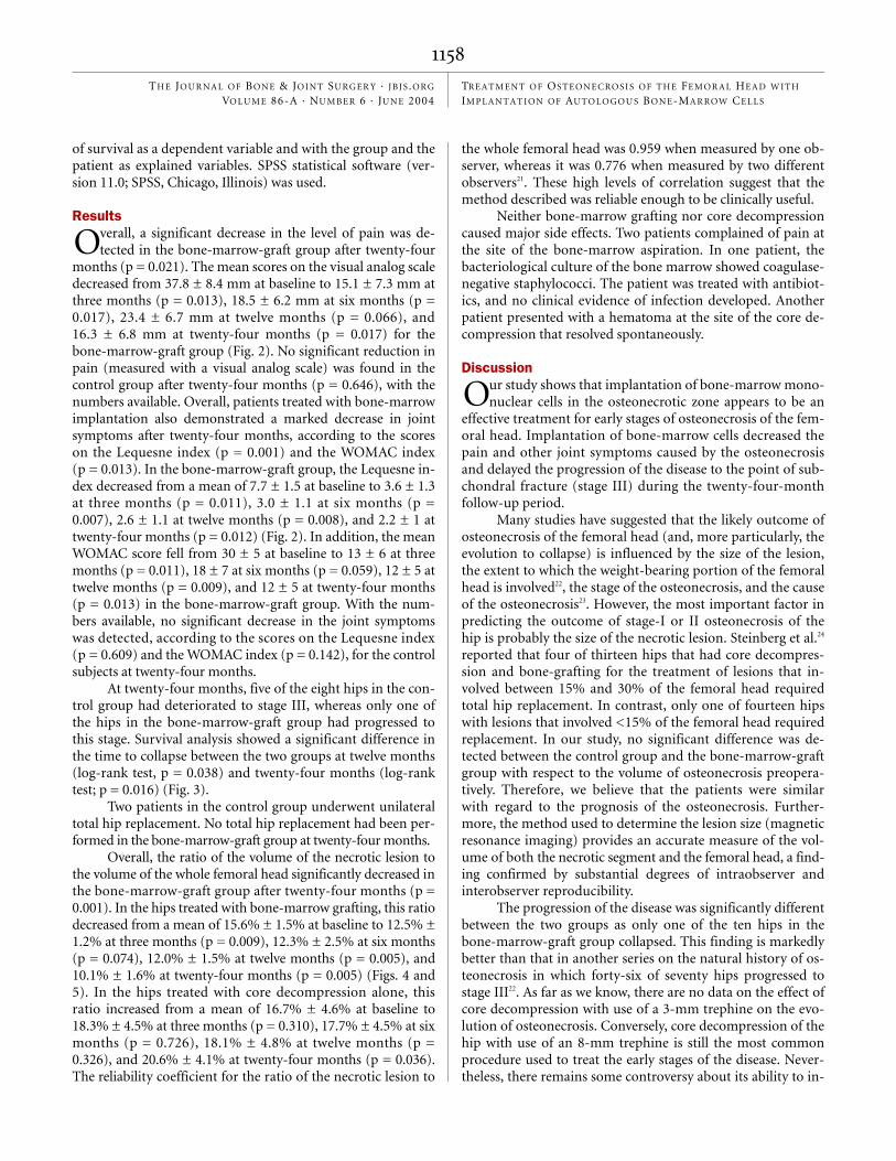
Fig. 5
Coronal T1-weighted magnetic
resonance imaging scans of two
hips showing a reduction in the
volume of osteonecrosis (arrow)
after bone-marrow implantation
(A) and an increase in the volume
of osteonecrosis (arrow) in the
hip of a control subject after two
years (B).
TH E JO U R NA L OF BONE & JOINT SURGER Y · JBJS .ORG
VO LU M E 86-A · NU M B E R 6 · JU N E 2004TRE A T M EN T OF OSTE ON E CROSIS OF T H E FEMOR AL HEA D W ITH IM P L A N T A T I O N OF AUTO L O GO U S BONE-MA R ROW CE L L S
of survival as a dependent variable and with the group and thepatient as explained variables. SPSS statistical software (ver-sion 11.0; SPSS, Chicago, Illinois) was used.
Resultsverall, a significant decrease in the level of pain was de-tected in the bone-marrow-graft group after twenty-four
months (p = 0.021). The mean scores on the visual analog scaledecreased from 37.8 ± 8.4 mm at baseline to 15.1 ± 7.3 mm atthree months (p = 0.013), 18.5 ± 6.2 mm at six months (p =0.017), 23.4 ± 6.7 mm at twelve months (p = 0.066), and16.3 ± 6.8 mm at twenty-four months (p = 0.017) for thebone-marrow-graft group (Fig. 2). No significant reduction inpain (measured with a visual analog scale) was found in thecontrol group after twenty-four months (p = 0.646), with thenumbers available. Overall, patients treated with bone-marrowimplantation also demonstrated a marked decrease in jointsymptoms after twenty-four months, according to the scoreson the Lequesne index (p = 0.001) and the WOMAC index(p = 0.013). In the bone-marrow-graft group, the Lequesne in-dex decreased from a mean of 7.7 ± 1.5 at baseline to 3.6 ± 1.3at three months (p = 0.011), 3.0 ± 1.1 at six months (p =0.007), 2.6 ± 1.1 at twelve months (p = 0.008), and 2.2 ± 1 attwenty-four months (p = 0.012) (Fig. 2). In addition, the meanWOMAC score fell from 30 ± 5 at baseline to 13 ± 6 at threemonths (p = 0.011), 18 ± 7 at six months (p = 0.059), 12 ± 5 attwelve months (p = 0.009), and 12 ± 5 at twenty-four months(p = 0.013) in the bone-marrow-graft group. With the num-bers available, no significant decrease in the joint symptomswas detected, according to the scores on the Lequesne index(p = 0.609) and the WOMAC index (p = 0.142), for the controlsubjects at twenty-four months.
At twenty-four months, five of the eight hips in the con-trol group had deteriorated to stage III, whereas only one ofthe hips in the bone-marrow-graft group had progressed tothis stage. Survival analysis showed a significant difference inthe time to collapse between the two groups at twelve months(log-rank test, p = 0.038) and twenty-four months (log-ranktest; p = 0.016) (Fig. 3).
Two patients in the control group underwent unilateraltotal hip replacement. No total hip replacement had been per-formed in the bone-marrow-graft group at twenty-four months.
Overall, the ratio of the volume of the necrotic lesion tothe volume of the whole femoral head significantly decreased inthe bone-marrow-graft group after twenty-four months (p =0.001). In the hips treated with bone-marrow grafting, this ratiodecreased from a mean of 15.6% ± 1.5% at baseline to 12.5% ±1.2% at three months (p = 0.009), 12.3% ± 2.5% at six months(p = 0.074), 12.0% ± 1.5% at twelve months (p = 0.005), and10.1% ± 1.6% at twenty-four months (p = 0.005) (Figs. 4 and5). In the hips treated with core decompression alone, thisratio increased from a mean of 16.7% ± 4.6% at baseline to18.3% ± 4.5% at three months (p = 0.310), 17.7% ± 4.5% at sixmonths (p = 0.726), 18.1% ± 4.8% at twelve months (p =0.326), and 20.6% ± 4.1% at twenty-four months (p = 0.036).The reliability coefficient for the ratio of the necrotic lesion to
the whole femoral head was 0.959 when measured by one ob-server, whereas it was 0.776 when measured by two differentobservers21. These high levels of correlation suggest that themethod described was reliable enough to be clinically useful.
Neither bone-marrow grafting nor core decompressioncaused major side effects. Two patients complained of pain atthe site of the bone-marrow aspiration. In one patient, thebacteriological culture of the bone marrow showed coagulase-negative staphylococci. The patient was treated with antibiot-ics, and no clinical evidence of infection developed. Anotherpatient presented with a hematoma at the site of the core de-compression that resolved spontaneously.
Discussionur study shows that implantation of bone-marrow mono-nuclear cells in the osteonecrotic zone appears to be an
effective treatment for early stages of osteonecrosis of the fem-oral head. Implantation of bone-marrow cells decreased thepain and other joint symptoms caused by the osteonecrosisand delayed the progression of the disease to the point of sub-chondral fracture (stage III) during the twenty-four-monthfollow-up period.
Many studies have suggested that the likely outcome ofosteonecrosis of the femoral head (and, more particularly, theevolution to collapse) is influenced by the size of the lesion,the extent to which the weight-bearing portion of the femoralhead is involved22, the stage of the osteonecrosis, and the causeof the osteonecrosis23. However, the most important factor inpredicting the outcome of stage-I or II osteonecrosis of thehip is probably the size of the necrotic lesion. Steinberg et al.24
reported that four of thirteen hips that had core decompres-sion and bone-grafting for the treatment of lesions that in-volved between 15% and 30% of the femoral head requiredtotal hip replacement. In contrast, only one of fourteen hipswith lesions that involved <15% of the femoral head requiredreplacement. In our study, no significant difference was de-tected between the control group and the bone-marrow-graftgroup with respect to the volume of osteonecrosis preopera-tively. Therefore, we believe that the patients were similarwith regard to the prognosis of the osteonecrosis. Further-more, the method used to determine the lesion size (magneticresonance imaging) provides an accurate measure of the vol-ume of both the necrotic segment and the femoral head, a find-ing confirmed by substantial degrees of intraobserver andinterobserver reproducibility.
The progression of the disease was significantly differentbetween the two groups as only one of the ten hips in thebone-marrow-graft group collapsed. This finding is markedlybetter than that in another series on the natural history of os-teonecrosis in which forty-six of seventy hips progressed tostage III22. As far as we know, there are no data on the effect ofcore decompression with use of a 3-mm trephine on the evo-lution of osteonecrosis. Conversely, core decompression of thehip with use of an 8-mm trephine is still the most commonprocedure used to treat the early stages of the disease. Never-theless, there remains some controversy about its ability to in-
O
O

TH E JO U R NA L OF BONE & JOINT SURGER Y · JBJS .ORG
VO LU M E 86-A · NU M B E R 6 · JU N E 2004TRE A T M EN T OF OSTE ON E CROSIS OF T H E FEMOR AL HEA D W ITH IM P L A N T A T I O N OF AUTO L O GO U S BONE-MA R ROW CE L L S
fluence the outcome of osteonecrosis. Mont et al. assessedforty-two studies in which a total of 1206 hips were treatedwith core decompression and 819 had various nonoperativeinterventions25. Three hundred and forty-five of the 466 hipstreated prior to collapse resolved satisfactorily following coredecompression, and 182 of the 819 hips that had nonoperativeinterventions had a satisfactory result. Since we used a 3-mmtrephine, our results need to be compared with the results ob-tained for core decompression with a larger trephine.
A minimum follow-up period of twenty-four monthswas chosen because collapse of the femoral head generally oc-curs over this span of time. It also occurs with greater fre-quency in the first twelve months following the initialdiagnosis22. Although the findings of this study are promising,their interpretation is limited by the constraints imposed bythe relatively small number of patients and the short durationof follow-up.
The bone marrow was injected through the trephinethat was placed into the necrotic zone. Although some of thebone-marrow cells might have leaked through the trephine orinto the circulation of the proximal aspect of the femur, thegreatest part of the bone marrow remained in the area of os-teonecrosis or in the femoral head as shown by radionuclidelabeling, which was performed in two patients. So far we havebeen unable to define, using imaging techniques, the exact lo-cation of the bone marrow after the injection. Larger trials andthe use of other techniques are needed to confirm and to fullyunderstand our results.
Recent advances in our understanding of the patho-physiology of osteonecrosis suggest that a decrease in the mes-enchymal stem-cell pool of the proximal aspect of the femurmight not provide enough osteoblasts to meet the needs ofbone-remodeling in the early stage of the disease9. An insuffi-ciency of osteogenic cells could explain the inadequate repairmechanism that, it is postulated, leads to femoral head col-lapse. The effectiveness of bone-marrow mononuclear cellsmay be related to the availability of stem cells endowed withosteogenic properties, arising from an increase in the supplyof such cells to the femoral head through bone-marrow im-plantation. Indeed, in the very early stages of osteonecrosis,providing sufficient repair capacity through the implantationof osteogenic cells could make these lesions reversible26,27. Inour study, the ratio of the volume of the necrotic lesion de-creased by a mean of 35% in the bone-marrow-graft group,whereas it increased by a mean of 23% in the control group.This finding suggests that necrotic lesions might be reversibleto some extent. Another possible explanation for the thera-peutic effect of bone-marrow implantation is that injected
marrow stromal cells secrete angiogenic cytokines resulting inincreased angiogenesis and subsequent improvement in os-teogenesis. One study has suggested that the efficacy of suchimplantation was due to a supply of endothelial progenitorcells included in the CD34+ fraction as well as to multiple an-giogenic factors (vascular endothelial growth factors, basic fi-broblast growth factor, and angiopoietin-1) released from theCD34+ fractions28.
In summary, we have shown the efficacy and apparentsafety of the implantation of bone-marrow mononuclear cellsin the early stages of osteonecrosis of the femoral head in asmall number of patients. This promising new approach forthe treatment of osteonecrosis could benefit from the recentadvances made in the field of stem-cell biology, including theuse of subpopulations of progenitors with greater therapeuticpotential.
AppendixA description of the details of the bone-marrow harvestand grafting procedure is available with the electronic
versions of this article, on our web site at www.jbjs.org (go tothe article citation and click on “Supplementary Material”)and on our quarterly CD-ROM (call our subscription depart-ment, at 781-449-9780, to order the CD-ROM). �
NOTE: The authors thank Professor Appelboom and Dominique Egrise for revising the manu-script, Dr. Azadeh Sattari and Dr. Bernard Stallenberg for reading the radiographs, and Dr. Oliv-ier Pradier for marrow-cell counting.
References
1. Ficat P, Arlet J. Ischémie et nécrose osseuse. L’exploration fonctionnelle de la circulation intraosseuse et ses applications. Paris: Masson; 1977. Necro-ses de la tete femorale; p 67-87.
2. Steinberg ME, Larcom PG, Stafford BB, Hosick WB, Corces A, Bands RE, Hartmann KM. Treatment of osteonecrosis of the femoral head by core de-compression, bone grafting, and electrical stimulation. In: Urbaniak JR, Jones JP, editors. Osteonecrosis: etiology, diagnosis, and treatment. Rose-
mont, IL: American Academy of Orthopaedic Surgeons; 1997. p 293-9.
3. Stulberg BN, Davis AW, Bauer TW, Levine M, Easley K. Osteonecrosis of the femoral head. A prospective randomized treatment protocol. Clin Orthop. 1991;268:140-51.
4. Koo KH, Kim R, Ko GH, Song HR, Jeong ST, Cho SH. Preventing collapse in early osteonecrosis of the femoral head. A randomised clinical trial of core
Valérie Gangji, MDJean-Philippe Hauzeur, MD, PhDCelso Matos, MDMichel Toungouz, MD, PhDMicheline Lambermont, PharmDDepartment of Rheumatology and Physical Medicine (V.G. and J.-P.H.), Department of Radiology (C.M.), and the Cellular and Molecular Therapy Unit (M.T. and M.L.), Erasme University Hospital, 808 Route de Lennik, 1070 Brussels, Belgium. E-mail address for V. Gangji: [email protected]
Viviane De Maertelaer, PhDDepartment of Biostatistics, Institut de Recherche Interdisciplinaire en Biologie Humaine et Moleculaire, School of Medicine, Université Libre de Bruxelles, 808 Route de Lennik, 1070 Brussels, Belgium
The authors did not receive grants or outside funding in support of their research or preparation of this manuscript. They did not receive pay-ments or other benefits or a commitment or agreement to provide such benefits from a commercial entity. No commercial entity paid or directed, or agreed to pay or direct, any benefits to any research fund, foundation, educational institution, or other charitable or nonprofit organization with which the authors are affiliated or associated.

TH E JO U R NA L OF BONE & JOINT SURGER Y · JBJS .ORG
VO LU M E 86-A · NU M B E R 6 · JU N E 2004TRE A T M EN T OF OSTE ON E CROSIS OF T H E FEMOR AL HEA D W ITH IM P L A N T A T I O N OF AUTO L O GO U S BONE-MA R ROW CE L L S
decompression. J Bone Joint Surg Br. 1995;77:870-4.
5. Jones JP Jr. Fat embolism and osteonecrosis. Orthop Clin North Am. 1985;16:595-633.
6. Wang GJ, Sweet DE, Reger SI, Thompson RC. Fat-cell changes as a mecha-nism of avascular necrosis of the femoral head in cortisone-treated rabbits. J Bone Joint Surg Am. 1977;59:729-35.
7. Simkin PA, Downey DJ. Hypothesis: retrograde embolization of marrow fat may cause osteonecrosis. J Rheumatol. 1987;14:870-2.
8. Laurent J, Meunier P, Courpron P, Edouard C, Bernard J, Vignon G. [Re-search on the pathogenesis of aseptic necrosis of the femoral head. Evalua-tion of the constitutional bony factors in 35 cases of iliac crest biopsy]. Nouv Presse Med. 1973;2:1755-60. French.
9. Hernigou P, Beaujean F, Lambotte JC. Decrease in the mesenchymal stem-cell pool in the proximal femur in corticosteroid-induced osteonecrosis. J Bone Joint Surg Br. 1999;81:349-55.
10. Gangji V, Hauzeur JP, Schoutens A, Hinsenkamp M, Appelboom T, Egrise D. Abnormalities in the replicative capacity of osteoblastic cells in the proximal femur of patients with osteonecrosis of the femoral head. J Rheumatol. 2003;30:348-51.
11. Hernigou P, Bernaudin F, Reinert P, Kuentz M, Vernant JP. Bone-marrow transplantation in sickle-cell disease. Effect on osteonecrosis: a case report with a four-year follow-up. J Bone Joint Surg Am. 1997;79:1726-30.
12. ARCO (Association Research Circulation Osseous): Committee on Terminol-ogy and Classification. ARCO News. 1992;4:41-6.
13. Hauzeur JP, Pasteels JL, Schoutens A, Hinsenkamp M, Appelboom T, Cho-chrad I, Perlmutter N. The diagnostic value of magnetic resonance imaging in non-traumatic osteonecrosis of the femoral head. J Bone Joint Surg Am. 1989;71:641-9.
14. Hauzeur JP, Orloff S, Taverne-Verbanck J, Pasteels JL. Diagnosis of aseptic osteonecrosis of the femoral head by percutaneous transtrochanterian nee-dle biopsy. Clin Rheumatol. 1986;5:346-58.
15. Hauzeur JP, Sintzoff S Jr, Appelboom T, De Maertelaer V, Bentin J, Pasteels JL. Relationship between magnetic resonance imaging and histologic findings by bone biopsy in nontraumatic osteonecrosis of the femoral head. J Rheu-matol. 1992;19:385-92.
16. Hernigou P, Beaujean F. Treatment of osteonecrosis with autologous bone marrow grafting. Clin Orthop. 2002;405:14-23.
17. Frank AJ, Moll JM, Hort JF. A comparison of three ways of measuring pain.
Rheumatol Rehabil. 1982;21:211-7.
18. Lequesne MG, Mery C, Samson M, Gerard P. Indexes of severity for osteoar-thritis of the hip and knee. Validation—value in comparison with other as-sessment tests. Scand J Rheumatol Suppl. 1987;65:85-9. Erratum in: Scand J Rheumatol. 1988;17:following 241. Scand J Rheumatol Suppl. 1988;73:1.
19. Bellamy N, Buchanan WW, Goldsmith CH, Campbell J, Stitt LW. Validation study of WOMAC: a health status instrument for measuring clinically impor-tant patient relevant outcomes to antirheumatic drug therapy in patients with osteoarthritis of the hip or knee. J Rheumatol. 1988;15:1833-40.
20. Steinberg ME, Hayken GD, Steinberg DR. A quantitative system for staging avascular necrosis. J Bone Joint Surg Br. 1995;77:34-41.
21. Fleiss JL. The design and analysis of clinical experiments. New York: Wiley; 1986.
22. Ohzono K, Saito M, Takaoka K, Ono K, Saito S, Nishina T, Kadowski T. Natu-ral history of nontraumatic avascular necrosis of the femoral head. J Bone Joint Surg Br. 1991;73:68-72.
23. Sugano N, Ohzono K, Masuhara K, Takaoka K, Ono K. Prognostication of os-teonecrosis of the femoral head in patients with systemic lupus erythemato-sus by magnetic resonance imaging. Clin Orthop. 1994;305:190-9.
24. Steinberg ME, Bands RE, Parry S, Hoffman E, Chan T, Hartman KM. Does lesion size affect the outcome in avascular necrosis? Clin Orthop. 1999;367:262-71.
25. Mont MA, Carbone JJ, Fairbank AC. Core decompression versus nonop-erative management for osteonecrosis of the hip. Clin Orthop. 1996;324:169-78.
26. Inoue A, Ono K. A histological study of idiopathic avascular necrosis of the head of the femur. J Bone Joint Surg Br. 1979;61:138-43.
27. Hauzeur JP, Pasteels JL. Pathology of bone marrow distant from the seques-trum in nontraumatic aseptic necrosis of the femoral head. In: Arlet J, Mazières B, editors. Bone circulation and bone necrosis. Berlin: Springer; 1990. p 73-6.
28. Tateishi-Yuyama E, Matsubara H, Murohara T, Ikeda U, Shintani S, Masaki H, Amano K, Kishimoto Y, Yoshimoto K, Akashi H, Shimada K, Iwasaka T, Imaizumi T; Therapeutic Angiogenesis using Cell Transplantation (TACT) Study Investigators. Therapeutic angiogenesis for patients with limb is-chaemia by autologous transplantation of bone-marrow cells: a pilot study and a randomised controlled trial. Lancet. 2002;360:427-35.