treatment of pediatric diaphyseal femur ... i. summary of recommendations the original guideline on...
TRANSCRIPT

TREATMENT OF PEDIATRIC DIAPHYSEAL FEMUR FRACTURES
EVIDENCE-BASED CLINICAL PRACTICE GUIDELINE
Adopted by the American Academy of Orthopaedic Surgeons Board of Directors
June 12, 2015

AAOS v 1.0 061909
2015 REPORT FOR THE REISSUE OF THE 2009 CLINICAL
PRACTICE GUIDELINE ON THE TREATMENT OF PEDIATRIC
DIAPHYSEAL FEMUR FRACTURES “This guideline is greater than 5 years old and is reviewed every five years. New studies have
been published since this guideline was developed, however the AAOS has determined that these
studies are not sufficient to warrant changing the guideline at this time. The information
contained in this guideline provides the user with the best evidence available at the time this
guideline was published.”
OVERVIEW OF 2015 UPDATES TO THE 2009 ORIGINAL
GUIDELINE 1) Addition of the Shemshaki, et al, 2011 study findings to Elastic Intramedullary
Nails.
2) Updated strength of recommendation language to match current AAOS guideline
language (see Grading the Recommendations).
3) Removed “inconclusive” recommendations due to lack of evidence (see Appendix
XI)
For a user-friendly version of this clinical practice guideline,
please visit the AAOS
OrthoGuidelines Web-Based App at:
http://www.orthoguidelines.org

ii
Disclaimer
This Clinical Practice Guideline was developed by an AAOS physician volunteer Work
Group based on a systematic review of the current scientific and clinical information and
accepted approaches to treatment and/or diagnosis. This Clinical Practice Guideline is not
intended to be a fixed protocol, as some patients may require more or less treatment or
different means of diagnosis. Clinical patients may not necessarily be the same as those
found in a clinical trial. Patient care and treatment should always be based on a
clinician’s independent medical judgment, given the individual patient’s clinical
circumstances.
Disclosure Requirement
In accordance with AAOS policy, all individuals whose names appear as authors or
contributors to Clinical Practice Guideline filed a disclosure statement as part of the
submission process. All panel members provided full disclosure of potential conflicts of
interest prior to voting on the recommendations contained within this Clinical Practice
Guidelines.
Funding Source
This Clinical Practice Guideline was funded exclusively by the American Academy of
Orthopaedic Surgeons who received no funding from outside commercial sources to
support the development of this document.
FDA Clearance
Some drugs or medical devices referenced or described in this Clinical Practice Guideline
may not have been cleared by the Food and Drug Administration (FDA) or may have
been cleared for a specific use only. The FDA has stated that it is the responsibility of the
physician to determine the FDA clearance status of each drug or device he or she wishes
to use in clinical practice.
Copyright
All rights reserved. No part of this Clinical Practice Guideline may be reproduced, stored
in a retrieval system, or transmitted, in any form, or by any means, electronic,
mechanical, photocopying, recording, or otherwise, without prior written permission
from the AAOS.
Originally Published in 2009 by the American Academy of Orthopaedic Surgeons
Reissued in 2015
9400 West Higgins Road
Rosemont, IL 60018
Copyright 2015 by the American Academy of Orthopaedic Surgeons

iii
I. SUMMARY OF RECOMMENDATIONS
The original guideline on the Treatment of Pediatric Diaphyseal Femur Fractures (PDFF)
was the third guideline developed by the AAOS in-house. It had fourteen
recommendations of varying strengths. However, per current AAOS policy, all
recommendations in the original guideline identified as “inconclusive” were removed
from this 2015 reissue (see Appendix XI for a full list of the inconclusive
recommendations that were removed). Based on the current procedure for updating
AAOS guidelines, the Medical Librarian ran a preliminary search to identify literature
that could address and possibly change the original recommendations. The AAOS
Evidence-Based Medicine Unit then used the inclusion criteria from the original
guideline to determine if any articles published after the final literature search date of the
original guideline were relevant to the original recommendations.
The following is a summary of the recommendations in the AAOS’ clinical practice
guideline on the Treatment of Pediatric Diaphyseal Femur Fractures (PDFF). This
summary does not contain rationales that explain how and why these recommendations
were developed nor does it contain the evidence supporting these recommendations. All
readers of this summary are strongly urged to consult the full guideline and evidence
report for this information. We are confident that those who read the full guideline and
evidence report will also see that the recommendations were developed using systematic
evidence-based processes designed to combat bias, enhance transparency, and promote
reproducibility. This summary of recommendations is not intended to stand alone.

iv
Strength of Recommendation Descriptions
Strength
Overall
Strength of
Evidence Description of Evidence Strength Strength Visual
Strong Strong
Evidence from two or more “High”
strength studies with consistent findings
for recommending for or against the
intervention.
Moderate Moderate
Evidence from two or more “Moderate”
strength studies with consistent findings,
or evidence from a single “High” quality
study for recommending for or against
the intervention.
Limited
Low Strength
Evidence or
Conflicting
Evidence
Evidence from two or more “Low”
strength studies with consistent findings
or evidence from a single study for
recommending for or against the
intervention or diagnostic test or the
evidence is insufficient or conflicting
and does not allow a recommendation
for or against the intervention.
Consensus* No Evidence
There is no supporting evidence. In the
absence of reliable evidence, the work
group is making a recommendation
based on their clinical opinion.
Consensus recommendations can only be
created when not establishing a
recommendation could have catastrophic
consequences.
v
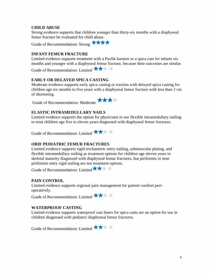
CHILD ABUSE
Strong evidence supports that children younger than thirty-six months with a diaphyseal
femur fracture be evaluated for child abuse.
Grade of Recommendation: Strong
INFANT FEMUR FRACTURE
Limited evidence supports treatment with a Pavlik harness or a spica cast for infants six
months and younger with a diaphyseal femur fracture, because their outcomes are similar.
Grade of Recommendation: Limited
EARLY OR DELAYED SPICA CASTING
Moderate evidence supports early spica casting or traction with delayed spica casting for
children age six months to five years with a diaphyseal femur fracture with less than 2 cm
of shortening.
Grade of Recommendation: Moderate
ELASTIC INTRAMEDULLARY NAILS
Limited evidence supports the option for physicians to use flexible intramedullary nailing
to treat children age five to eleven years diagnosed with diaphyseal femur fractures.
Grade of Recommendation: Limited
ORIF PEDIATRIC FEMUR FRACTURES
Limited evidence supports rigid trochanteric entry nailing, submuscular plating, and
flexible intramedullary nailing as treatment options for children age eleven years to
skeletal maturity diagnosed with diaphyseal femur fractures, but piriformis or near
piriformis entry rigid nailing are not treatment options.
Grade of Recommendation: Limited
PAIN CONTROL
Limited evidence supports regional pain management for patient comfort peri-
operatively.
Grade of Recommendation: Limited
WATERPROOF CASTING
Limited evidence supports waterproof cast liners for spica casts are an option for use in
children diagnosed with pediatric diaphyseal femur fractures.
Grade of Recommendation: Limited

vi
2015 GUIDELINE REISSUE WORK GROUP
Chair, AAOS Evidence-Based Quality and Value Committee:
David S. Jevsevar, MD, MBA
Vice Chair, Orthopaedics, Assistant Professor of Orthopaedic Surgery
Dartmouth-Hitchcock Medical Center
1 Medical Center Drive
Lebanon, NH 03756
Phone 630-650-5133
Guidelines Oversight Leader, AAOS Evidence-Based Quality and Value Committee:
Kevin Shea, MD
Intermountain Orthopaedics
600 N. Robbins Rd Ste 400
Boise, ID 83702
AAOS Staff (2015 Guideline Reissue)
William Shaffer, MD
AAOS Medical Director
Deborah Cummins, PhD
Director, Department of Research and Scientific Affairs
Jayson Murray, MA
Manager, Evidence-Based Medicine Unit
Ben Brenton, MPH
Research Analyst, Evidence-Based Medicine Unit
Anne Woznica
AAOS Medical Librarian
Kaitlyn Sevarino
Evidence-Based Quality and Value (EBQV) Coordinator
Erica Linskey
Administrative Assistant, Evidence-Based Medicine Unit

vii
2007 GUIDELINE WORK GROUP
Mininder S. Kocher, MD, MPH,
Chair
Children's Hospital
300 Longwood Ave
Orthopaedic Dept
Boston, MA 02115
Ernest L. Sink, MD, Co-Chair
The Children's Hospital
13123 East 16th Ave B060
Aurora, CO 80045
R Dale Blasier, MD
Dept of Orthopaedic Surgery
800 Marshall St Sturgis 363
Little Rock, AR 72202
Scott J. Luhmann, MD
St Louis Children's Hospital
One Children's Pl Ste 4S 20
Saint Louis, MO 63110
Charles T. Mehlman, DO, MPH
Children's Hospital Medical Center
3333 Burnet Avenue, MLC 2017
Cincinnati, Ohio 45229-3039
David M. Scher, MD
Hospital for Special Surgery
535 E 70th St 5th Fl
New York, NY 10021
Travis Matheney, MD
Children's Hospital Boston
Orthopedic Surgery
47 Joy Street
Boston, MA 02115
James O Sanders, MD
Department of Orthopaedics
Rehabilitation University of Rochester
601 Elmwood Avenue
Rochester NY 14642
Guidelines Oversight Chair:
William C. Watters, III MD
6624 Fannin #2600
Houston, TX 77030
Guidelines Oversight Vice-Chair:
Michael J. Goldberg MD
Children’s Hospital and Regional
Medical Ctr.
4800 Sand Point Way NE #w7706
PO Box 5371
Seattle WA 98105-5371
Evidence-Based Practice Committee
Chair:
Michael Warren Keith, MD
2500 Metro Health Drive
Cleveland, OH 44109-1900

viii
Former AAOS Staff (2007 Guideline)
Robert H. Haralson III, MD, MBA
Charles M. Turkelson, PhD
Janet L. Wies, MPH
Patrick Sluka, MPH
Kristin Hitchcock

ix
Table of Contents
2015 Report for the ReIssue of the 2009 Clinical Practice Guideline on the Treatment of Pediatric
Diaphyseal Femur Fractures ........................................................................................................................ i
Overview of 2015 updates to the 2009 original guideline ........................................................................... i
I. SUMMARY OF RECOMMENDATIONS ....................................................... III
Child Abuse ................................................................................................................................................... v
Infant Femur Fracture ................................................................................................................................. v
Early or Delayed Spica Casting ................................................................................................................... v
Elastic Intramedullary Nails........................................................................................................................ v
ORIF Pediatric Femur Fractures ............................................................................................................... v
Pain Control .................................................................................................................................................. v
Waterproof Casting ...................................................................................................................................... v
2015 Guideline Reissue Work Group ........................................................................................................ vi
2007 Guideline Work Group ..................................................................................................................... vii
TABLE OF CONTENTS ..................................................................................... IX
LIST OF TABLES .............................................................................................. XII
LIST OF FIGURES ............................................................................................ XII
II. INTRODUCTION ........................................................................................... 1
Overview........................................................................................................................................................ 1
Goals and Rationale ..................................................................................................................................... 1
Intended Users .............................................................................................................................................. 1
Patient Population ........................................................................................................................................ 2
Incidence ....................................................................................................................................................... 2
Prevalence ..................................................................................................................................................... 2
Burden of Disease ......................................................................................................................................... 2
Etiology .......................................................................................................................................................... 2

x
Risk Factors .................................................................................................................................................. 2
Emotional and Physical Impact of Pediatric Diaphyseal Femur Fractures ............................................ 3
Potential Benefits, Harms, And Contraindications ................................................................................... 3
III. METHODS ................................................................................................. 4
Guideline Reissue ......................................................................................................................................... 4
Preliminary Recommendations ................................................................................................................... 4
Study Selection Criteria ............................................................................................................................... 5 Types of Studies ........................................................................................................................................ 5
Original and Updated Literature Searches ................................................................................................ 6 Search for RCTs and other study designs .................................................................................................. 7
Data Extraction ............................................................................................................................................. 7
Grading the Recommendations ................................................................................................................... 8
Statistical Methods ....................................................................................................................................... 9
Peer Review ................................................................................................................................................... 9
Public Commentary...................................................................................................................................... 9
The AAOS Guideline Approval Process ....................................................................................................10
Revision Plans ..............................................................................................................................................10
Guideline Dissemination Plans ...................................................................................................................10
IV. RECOMMENDATIONS AND SUPPORTING DATA ............................... 11
Child Abuse ..................................................................................................................................................11 Rationale ...................................................................................................................................................11 Supporting Evidence .................................................................................................................................12
Infant Femur Fracture ................................................................................................................................13 Rationale ...................................................................................................................................................13 Supporting Evidence .................................................................................................................................13
Early or Delayed Spica Casting ..................................................................................................................15 Rationale ...................................................................................................................................................15 Supporting Evidence .................................................................................................................................15 Previously Published Systematic Reviews ...............................................................................................19
Elastic Intramedullary Nails.......................................................................................................................20 Rationale ...................................................................................................................................................20 Supporting Evidence .................................................................................................................................21 Previously Published Systematic Reviews ...............................................................................................45

xi
ORIF Pediatric Femur Fractures ..............................................................................................................46 Rationale ...................................................................................................................................................46 Supporting Evidence .................................................................................................................................46 Previously Published Systematic Reviews ...............................................................................................51
Pain Control .................................................................................................................................................52 Rationale ...................................................................................................................................................52 Supporting Evidence .................................................................................................................................52 Previously Published Systematic Reviews ...............................................................................................55
Waterproof Casting .....................................................................................................................................56 Rationale ...................................................................................................................................................56 Supporting Evidence .................................................................................................................................56
Future Research ..........................................................................................................................................57
V. APPENDIXES .............................................................................................. 59
Appendix I ....................................................................................................................................................60 AAOS Bodies That Approved the 2015 Guideline Reissue .....................................................................60 Appendix II ...............................................................................................................................................61 Literature Searches ...................................................................................................................................61
General Search .....................................................................................................................................61 Waterproof Cast Liner Search ..............................................................................................................62
Appendix III .................................................................................................................................................63 Study Attrition Flowcharts .......................................................................................................................63
Original 2008 Literature Search Flowchart ..........................................................................................63 Updated 2013 Literature Search Flowchart .........................................................................................63 Waterproof Cast Liner Search Flowchart ............................................................................................64
Appendix IV .................................................................................................................................................65 Data Extraction Elements .........................................................................................................................65
Appendix V ..................................................................................................................................................66 Form for Assigning Grade of Recommendation (Interventions) ..............................................................66
Appendix VI .................................................................................................................................................68 Peer Review Panel for the Original 2009 Guideline .................................................................................68 Public Commentary for Original 2009 Guideline .....................................................................................69
Appendix VII ...............................................................................................................................................70 Structured Peer Review Form ...................................................................................................................70
Appendix VIII ..............................................................................................................................................73 Interpreting the Forest Plots
49 ...................................................................................................................73
Description of Symbols Used in Figures and Tables ................................................................................74
Appendix IX .................................................................................................................................................75 Conflict of Interest ....................................................................................................................................75 AAOS Disclosure Program Information ...................................................................................................75
Appendix X ..................................................................................................................................................77 References ................................................................................................................................................77 Included Articles .......................................................................................................................................81
xii
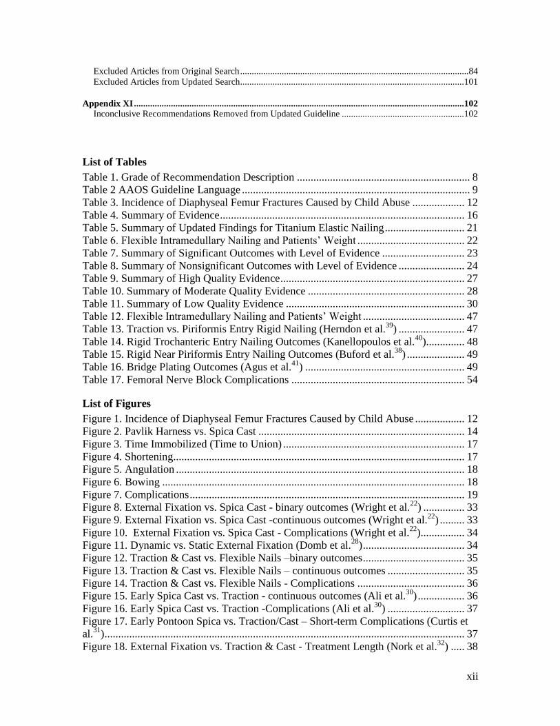
Excluded Articles from Original Search ...................................................................................................84 Excluded Articles from Updated Search .................................................................................................101
Appendix XI ...............................................................................................................................................102 Inconclusive Recommendations Removed from Updated Guideline .....................................................102
List of Tables
Table 1. Grade of Recommendation Description ............................................................... 8 Table 2 AAOS Guideline Language ................................................................................... 9
Table 3. Incidence of Diaphyseal Femur Fractures Caused by Child Abuse ................... 12 Table 4. Summary of Evidence ......................................................................................... 16 Table 5. Summary of Updated Findings for Titanium Elastic Nailing ............................. 21
Table 6. Flexible Intramedullary Nailing and Patients’ Weight ....................................... 22 Table 7. Summary of Significant Outcomes with Level of Evidence .............................. 23 Table 8. Summary of Nonsignificant Outcomes with Level of Evidence ........................ 24 Table 9. Summary of High Quality Evidence ................................................................... 27
Table 10. Summary of Moderate Quality Evidence ......................................................... 28 Table 11. Summary of Low Quality Evidence ................................................................. 30
Table 12. Flexible Intramedullary Nailing and Patients’ Weight ..................................... 47 Table 13. Traction vs. Piriformis Entry Rigid Nailing (Herndon et al.
39) ........................ 47
Table 14. Rigid Trochanteric Entry Nailing Outcomes (Kanellopoulos et al.40
) .............. 48
Table 15. Rigid Near Piriformis Entry Nailing Outcomes (Buford et al.38
) ..................... 49 Table 16. Bridge Plating Outcomes (Agus et al.
41) .......................................................... 49
Table 17. Femoral Nerve Block Complications ............................................................... 54
List of Figures
Figure 1. Incidence of Diaphyseal Femur Fractures Caused by Child Abuse .................. 12
Figure 2. Pavlik Harness vs. Spica Cast ........................................................................... 14
Figure 3. Time Immobilized (Time to Union) .................................................................. 17
Figure 4. Shortening.......................................................................................................... 17
Figure 5. Angulation ......................................................................................................... 18
Figure 6. Bowing .............................................................................................................. 18
Figure 7. Complications .................................................................................................... 19
Figure 8. External Fixation vs. Spica Cast - binary outcomes (Wright et al.22
) ............... 33
Figure 9. External Fixation vs. Spica Cast -continuous outcomes (Wright et al.22
) ......... 33
Figure 10. External Fixation vs. Spica Cast - Complications (Wright et al.22
) ................ 34
Figure 11. Dynamic vs. Static External Fixation (Domb et al.28
) ..................................... 34
Figure 12. Traction & Cast vs. Flexible Nails –binary outcomes ..................................... 35
Figure 13. Traction & Cast vs. Flexible Nails – continuous outcomes ............................ 35
Figure 14. Traction & Cast vs. Flexible Nails - Complications ....................................... 36
Figure 15. Early Spica Cast vs. Traction - continuous outcomes (Ali et al.30
) ................. 36
Figure 16. Early Spica Cast vs. Traction -Complications (Ali et al.30
) ............................ 37
Figure 17. Early Pontoon Spica vs. Traction/Cast – Short-term Complications (Curtis et
al.31
) ................................................................................................................................... 37
Figure 18. External Fixation vs. Traction & Cast - Treatment Length (Nork et al.32
) ..... 38

xiii
Figure 19. External Fixation vs. Traction & Cast – Complications (Nork et al.32
) .......... 38
Figure 20. External Fixation vs. Traction - Treatment Length (Hedin et al.34
) ................ 39
Figure 21. External Fixation vs. Traction - Patient Satisfaction (Hedin et al.34
) .............. 39
Figure 22. Flexible Nails vs. Traction & Cast - binary outcomes .................................... 40
Figure 23. Flexible Nails vs. Traction & Cast -Leg Length Discrepancy (Song et al.33
) . 40
Figure 24. Flexible Nails vs. Traction & Cast - Major Complications ............................. 41
Figure 25. Flexible Nails vs. Traction & Cast - Minor Complications............................. 41
Figure 26. Titanium vs. Stainless Steel Flexible Nails – Complications (Wall et al.37
) ... 42
Figure 27. Immediate vs. Delayed Spica Cast - Complications (Rasit et al.35
) ................ 42
Figure 28. Early Intervention vs. Traction (Sturdee et al.36
) ............................................ 43
Figure 29. Flexible Nailing vs. External Fixation - Continuous Outcomes (Barlas et al.
200626
)............................................................................................................................... 43
Figure 30. Flexible Nailing vs. External Fixation -Binary Outcomes (Barlas et al. 200626
)
........................................................................................................................................... 44
Figure 31. Flexible Nailing vs. External Fixation - Complications(Barlas et al. 200626
) . 44
Figure 32. Titanium Elastic Nailing Outcomes Among Age 11+ (Moroz et al.27
) ........... 48
Figure 33. Bridge Plating - Percentage of Patients with Frontal Plane Angulation (Agus et
al.41
) ................................................................................................................................... 50
Figure 34. Bridge Plating -Percentage of Patients with Sagittal Plane Angulation (Agus et
al.41
) ................................................................................................................................... 50
Figure 35. Hematoma Block vs. Control - Time until First Post-Operative Narcotic Dose
........................................................................................................................................... 53
Figure 36. Hematoma Block vs. Control - Post-Operative Narcotic Requirement .......... 53
Figure 37. Hematoma Block vs. Control - Binary Outcomes ........................................... 54
Figure 38. Femoral Nerve Block – Pain Relief ................................................................. 54
Figure 39. Waterproof Liner vs. No Waterproof Liner .................................................... 57

1
II. INTRODUCTION
OVERVIEW This clinical practice guideline presents the results of a systematic review of published
studies on the treatment of isolated diaphyseal femur fractures in children, where children
are defined as those not having reached skeletal maturity. In addition to providing
practice recommendations, this guideline also highlights gaps in the literature and areas
that require future research.
This guideline is intended to be used by all appropriately trained surgeons and all
qualified physicians considering treatment of isolated diaphyseal femur fractures in
children. It is also intended to serve as an information resource for decision makers and
developers of practice guidelines and recommendations.
GOALS AND RATIONALE The purpose of this clinical practice guideline is to help improve treatment based on the
current best evidence. Current evidence-based practice (EBP) standards demand that
physicians use the best available evidence in their clinical decision making. To assist in
this decision making, this clinical practice guideline consists of a systematic review of the
available literature on the treatment of isolated diaphyseal femur fractures in children.
The systematic review detailed herein includes evidence published from 1966 through
October 1, 2008 and demonstrates where there is good evidence, where evidence is
lacking, and what topics future research must target in order to improve the treatment of
children with isolated diaphyseal femur fractures. AAOS staff and the Pediatric
Diaphyseal Femur Fractures physician work group systematically reviewed the available
literature and subsequently wrote the following recommendations based on a rigorous,
standardized process.
Musculoskeletal care is provided in many different settings by many different providers.
We created this guideline as an educational tool to guide qualified physicians through a
series of treatment decisions in an effort to improve the quality and efficiency of care.
This guideline should not be construed as including all proper methods of care or
excluding methods of care reasonably directed to obtaining the same results. The ultimate
judgment regarding any specific procedure or treatment must be made in light of all
circumstances presented by the patient and the needs and resources particular to the
locality or institution.
INTENDED USERS This guideline is intended to be used by orthopaedic surgeons and all qualified physicians
managing pediatric patients. Typically, orthopaedic surgeons will have completed
medical training, a qualified residency in orthopaedic surgery, and some may have
completed additional sub-specialty training. Insurance payers, governmental bodies, and
health-policy decision-makers may also find this guideline useful as an evolving standard
of evidence regarding treatment of diaphyseal femur fractures in pediatric patients.
Treatment of pediatric diaphyseal femur fractures is based on the assumption that
decisions are predicated on guardian and physician mutual communication with
2
discussion of available treatments and procedures applicable to the individual patient.
Once the patient’s guardian has been informed of available therapies and has discussed
these options with his/her child’s physician, an informed decision can be made. Clinician
input based on experience with both conservative management and surgical skills
increases the probability of identifying patients who will benefit from specific treatment
options.
PATIENT POPULATION This document addresses the treatment of isolated diaphyseal femur fractures in children
who have not yet reached skeletal maturity. The guideline provides information on
pediatric patient management after diagnosis of a diaphyseal femur fracture. This
guideline is not intended for use in pediatric patients who present with additional
coexisting injuries that require formal surgical intervention or other life-threatening
conditions that take precedence over the treatment of the diaphyseal femur fracture.
INCIDENCE The annual rate of children who present with femoral shaft fracture has been estimated at
19 per 100,000.1 Boys have a higher risk of fracture than girls and this is consistent with
participation of boys in sporting activites.1,2
PREVALENCE Diaphyseal femur fractures account for 1.4%
3 to 1.7%
4 of all pediatric fractures.
BURDEN OF DISEASE There are many components to consider when calculating the overall cost of treatment for
pediatric femoral fracture.5 The main considerations for patients and third party payers
are the relative cost and effectiveness of each treatment option. But hidden costs for
pediatric patients must also be considered. These costs include the additional home care
required for a patient, the costs of rehabilitation and of missed school for the patient,
child care costs if both parents work, and time off of work required by one or both
parents to care for the pediatric patient.6
ETIOLOGY The primary cause of diaphyseal femur fracture in children varies by age groups but
includes falls, motor-vehicle accidents, and sports injuries.1 In addition, the Cincinnati
Children’s Hospital Medical Center states, “In children less than one year of age, child
abuse is the leading cause of femoral fractures and abuse remains a significant concern in
toddlers up to about five years of age.”7
RISK FACTORS Occurrences of pediatric diaphyseal femur fractures are higher in boys than in girls in all
age groups.1,2
This literature also suggests that the primary mechanism of fracture is age-
related, including falls and child abuse for younger children, falls, motor vehicle-
pedestrian, bicycle, and motor-vehicle collisions for school age children and motor-
vehicle or sports related accidents in teenagers.

3
One study suggests increased risk of fracture for blacks over whites1 and one study
suggests no difference by race/ethnicity.2 Both studies suggest that lower socioeconomic
conditions also increase fracture risk.
EMOTIONAL AND PHYSICAL IMPACT OF PEDIATRIC
DIAPHYSEAL FEMUR FRACTURES The prolonged loss of mobility and absence from school often associated with the
treatment of pediatric diaphyseal femur fractures can lead to adverse physical, social, and
emotional consequences for the child as well as the child’s family. Treatments that
minimize the child’s length of immobilization and time out of school are therefore
desirable.
POTENTIAL BENEFITS, HARMS, AND CONTRAINDICATIONS Invasive and operative treatments are associated with known risks. Contraindications
vary widely based on the treatment administered. Therefore, discussion of available
treatments and procedures applicable to the individual patient rely on mutual
communication between the patient’s guardian and physician, weighing the potential
risks and benefits for that patient.
Further, the age groups referred to in the specific recommendations are general guides.
Obviously, additional factors may affect the physician’s choice of treatment including but
not limited to associated injuries the patient may present with as well as the individual’s
comorbidities, skeletal maturity, and/or specific patient characteristics including obesity.
The individual patient’s family dynamic will also influence treatment decisions;
therefore, treatment decisions made for children who border any age group should be
made on the basis of the individual. Decisions will always need to be predicated on
guardian and physician communication with discussion of available treatments and
procedures applicable to the individual patient. Once the patient’s guardian has been
informed of available therapies and has discussed these options with his/her child’s
physician, an informed decision can be made. Clinician input based on experience
increases the probability of identifying patients who will benefit from specific treatment
options.

4
III. METHODS
This clinical practice guideline and the systematic review upon which it is based evaluate
the effectiveness of treatments for isolated pediatric diaphyseal femur fractures. This
section describes the methods used to prepare this guideline and systematic review,
including search strategies used to identify literature, criteria for selecting eligible
articles, grading the evidence, data extraction, methods of statistical analysis, and the
review and approval of the guideline. The methods used to perform this systematic
review were employed to minimize bias in the selection and summary of the available
evidence.8,9
These processes are vital to the development of reliable, transparent, and
accurate clinical recommendations for treating isolated diaphyseal femur fractures in
children.
To develop the original guideline, the work group initially met in an introductory meeting
on April 5, 2008, to establish the scope of the guideline and systematic review. Upon
completion of the systematic review the work group participated in a two-day
recommendation meeting on November 8 and 9, 2008, at which the final
recommendations were written and voted on. The resulting draft guidelines were then
peer-reviewed, subsequently sent for public commentary, and then sequentially approved
by the AAOS Evidence Based Practice Committee, AAOS Guidelines and Technology
Oversight Committee, AAOS Council on Research, Quality Assessment, and
Technology, and the AAOS Board of Directors.
GUIDELINE REISSUE The original guideline and systematic review were prepared by the AAOS Pediatric
Diaphyseal Femur Fractures physician work group with the assistance of the AAOS
Clinical Practice Guidelines Unit. Based on the current procedure for updating AAOS
guidelines, the Medical Librarian ran an updated search to identify literature published
after the original search for the 2007 guideline that could address and possibly change the
original recommendations. The AAOS Evidence-Based Medicine Unit then used the
inclusion criteria from the original guideline to determine if any articles published after
the final literature search date of the original guideline were relevant to the
recommendations. The resulting reissue draft was then sequentially approved by the
AAOS Committee on Evidence Based Quality and Value, the AAOS Council on
Research and Quality and the AAOS Board of Directors (see Appendix I).
PRELIMINARY RECOMMENDATIONS The original work group began work on this guideline by constructing a set of
preliminary recommendations. These recommendations specify [what] should be done in
[whom], [when], [where], and [how often or how long]. They function as questions for
the systematic review, not as final recommendations or conclusions. Simulated
recommendations are almost always modified on the basis of the results of the systematic
review. These recommendations also form the guideline’s scope and guide the searches
for literature. These a priori simulated recommendations are inviolate in that, once
specified, they cannot be modified, they must all be addressed by the systematic review,
and the relevant review results must be presented in the final guideline. The a priori and
inviolate nature of the preliminary recommendations combats bias.

5
STUDY SELECTION CRITERIA TYPES OF STUDIES
We developed a priori article selection criteria for our review. Specifically, to be
included in our systematic reviews an article had to be a report of a study that:
Evaluated a treatment for isolated pediatric diaphyseal femur fracture.
Was a full article published in the peer reviewed literature.
Was an English language article published after 1965.
Was not a cadaveric, animal, or in vitro study.
Was not a retrospective case series, medical records review, meeting abstract,
unpublished study report, case report, historical article, editorial, letter, or
commentary.
Was the most recent report of a study or the report with the largest number of
enrolled patients in a study with multiple publications.
Enrolled ≥ 10 patients in each of its study groups.
Enrolled a patient population of at least 80% of patients with a diaphyseal
femur fracture and were not skeletally mature (closure of proximal and distal
femoral growth plates).
Reported quantified results.
Enrolled patients without the following conditions
subtrochanteric fractures, supracondylar femur fractures, physeal
fractures, open fractures, compound fractures, pathologic fractures, or
multiple lower extremity fractures.
co-existing abdominal or neurological injuries requiring surgical
intervention (the physician work group chair and co-chair determined
whether an article met inclusion criteria in cases when studies reported
insufficient detail to determine whether co-existing injuries required
surgical intervention).
osteogenesis imperfecta, cerebral palsy, myelodysplasia (spina bifida),
metabolic bone diseases, or skeletal dysplasia.
When examining primary studies, we analyzed the best available evidence regardless of
study design. We first considered the randomized controlled trials identified by the search
strategy. In the absence of two or more RCTs, we sequentially searched for prospective
controlled trials, prospective comparative studies, retrospective comparative studies, and
prospective case-series studies. Only studies of the highest level of available evidence
were included, assuming that there were 2 or more studies of that higher level. For
example, if there were two high quality studies that addressed the recommendation,
moderate, low, and very low quality studies were not included.
For the recommendation on waterproof cast liners only, we considered for inclusion
studies that included patients with conditions other than diaphyseal femur fractures
6
because the complications potentially avoided by using waterproof liners are not specific
to diaphyseal femur fractures.
The Pediatric Diaphyseal Femur Fracture physician work group requested that the AAOS
guidelines unit capture surrogate outcome measures if the study inclusion criteria were
met. For this patient population, children, surrogate outcomes are often used because
patients’ communication skills are limited or not yet developed. Surrogate outcome
measures are laboratory measurements or another physical sign that are used as
substitutes for clinically meaningful end points that measure directly how a patient feels,
functions, or survives.10
In order for a surrogate measure to be valid, it must be in the
causal pathway between the intervention and the outcome and it must demonstrate a
large, consistently measurable association with the outcome.10
The main surrogate measures we considered were radiographic measures, such as those
indicating a malunion of the fracture. It should be noted that generally accepted
definitions of malunion have not necessarily been correlated to function and risk of
developing further problems.
We only considered an outcome if ≥ 50% (80% for case series) of the patients were
followed for that outcome (for example, some studies reported short-term outcomes data
on nearly all enrolled patients, and reported longer-term data on only a few patients. In
such cases, we did not include the longer-term data). We also excluded outcomes for
study groups that did not have at least 10 patients.
When distinguishing between stable and unstable fractures, we defined transverse and
short oblique fractures as stable. We defined comminuted and long oblique fractures as
unstable.
When the age range of patients in a study overlapped the target age range of two or more
recommendations, we included the study in the evidence base of the recommendation
whose age range included the study’s median patient age.
ORIGINAL AND UPDATED LITERATURE SEARCHES The updated guideline searched for articles published up to November 27, 2013. The
original guideline searched for articles published up to October 1, 2008. Search strategies
were reviewed by the original work group prior to conducting the searches. All literature
searches were supplemented with manual screening of bibliographies of all publications
retrieved. We also searched the bibliographies of recent systematic reviews and other
review articles for potentially relevant citations. A list of potentially relevant studies, not
identified by the literature search, was also provided by the work group members. Three
such studies met the inclusion criteria. We conducted one recommendation-specific
search for primary articles on waterproof cast liners. For the entire guideline, thirty-two
primary studies were included and two hundred forty-three studies were excluded.
7
SEARCH FOR RCTS AND OTHER STUDY DESIGNS
To identify primary studies for this guideline, we searched four electronic databases;
PubMed, EMBASE, CINAHL, and The Cochrane Central Register of Controlled Trials.
The search strategies we used are provided in Appendix II.
We used a previously published search strategy 11
to identify relevant randomized
controlled trials. In the absence of relevant RCTs, we modified the search strategy to
identify studies of other designs.
The study attrition diagram in Appendix I provides details about the inclusion and
exclusion of these studies.
DATA EXTRACTION Data elements extracted from studies were defined in consultation with the physician
work group. Three reviewers completed data extraction independently for all studies.
Disagreements were resolved by consensus and by consulting the work group. Evidence
tables were constructed to summarize the best evidence pertaining to each preliminary
recommendation. The elements extracted are shown in Appendix IV.

8
GRADING THE RECOMMENDATIONS Following data extraction and analyses, each guideline recommendation was assigned a
preliminary grade that was based on the total body of evidence available using the
following system:
Table 1. Grade of Recommendation Description
Strength
Overall
Strength of
Evidence Description of Evidence Strength Strength Visual
Strong Strong
Evidence from two or more “High”
strength studies with consistent findings
for recommending for or against the
intervention.
Moderate Moderate
Evidence from two or more “Moderate”
strength studies with consistent findings,
or evidence from a single “High” quality
study for recommending for or against
the intervention.
Limited
Low Strength
Evidence or
Conflicting
Evidence
Evidence from two or more “Low”
strength studies with consistent findings
or evidence from a single study for
recommending for or against the
intervention or diagnostic test or the
evidence is insufficient or conflicting
and does not allow a recommendation
for or against the intervention.
Consensus* No Evidence
There is no supporting evidence. In the
absence of reliable evidence, the work
group is making a recommendation
based on their clinical opinion.
Consensus recommendations can only be
created when not establishing a
recommendation could have catastrophic
consequences.

9
Each recommendation was constructed using the following language which took into
account the final grade of recommendation.
Table 2 AAOS Guideline Language
Guideline Language
Strength of
Recommendation
Strong evidence supports that the practitioner
should/should not do X, because… Strong
Moderate evidence supports that the
practitioner could/could not do X, because… Moderate
Limited evidence supports that the practitioner
might/might not do X, because… Limited
In the absence of reliable evidence, it is the
opinion of this work group that…* Consensus*
STATISTICAL METHODS We calculated, where applicable, odds ratios (OR) for dichotomous data and mean
differences for continuous data.
When published studies only reported the median, range and size of the trial, we
estimated their means and variances according to a published method.13
We used StatXact for the calculation of exact odds ratios confidence intervals for
dichotomous data. All other calculations were performed using STATA 10.0 (StataCorp
LP, College Station, Texas). We used the Wilson score method to calculate confidence
intervals for proportions.14
For ordinal data, we used ordinal logistic regression to
calculate odds ratios. Ordinal logistic regression produces proportional odds ratios, which
assumes that the odds ratio is the same between each pair of outcome groups (lowest
category vs. all higher categories, lowest two categories vs. all higher categories, etc.).
PEER REVIEW The original draft of the guideline and evidence report were peer reviewed by an expert
outside advisory panel that was nominated by the physician work group prior to the
development of the guideline (Appendix VI). In addition, the physician members of the
AAOS Guidelines and Technology Oversight Committee and the Evidence Based
Practice Committee provided peer review of the draft document. Peer review was
accomplished using a structured peer review form. (Appendix VII) We forwarded the
draft guideline to a total of thirty-three reviewers and eleven returned reviews. The
disposition of all non-editorial peer review comments was documented and accompanied
this guideline through the public commentary and the following approval process.
PUBLIC COMMENTARY After modifying the draft in response to peer review, the original guideline was subjected
to a thirty day period of “Public Commentary.” Commentators consist of members of the
AAOS Board of Directors (BOD), members of the Council on Research, Quality
Assessment, and Technology (CORQAT), members of the Board of Councilors (BOC),
10
and members of the Board of Specialty Societies (BOS). Based on these bodies, up to
185 commentators had the opportunity to provide input into the development of this
guideline. Of these, 12 returned public comments.
THE AAOS GUIDELINE APPROVAL PROCESS Following peer review, the 2009 CPG was approved by the AAOS Guidelines and
Technology Oversight Committee, the AAOS Evidence Based Practice Committee, the
AAOS Council on Research, Quality Assessment and Technology, and the AAOS Board
of Directors.
The 2015 Guideline Reissue was approved by the AAOS Committee on Evidence Based
Quality and Value, the AAOS Council on Research and Quality and the AAOS Board of
Directors. Descriptions of these bodies are provided in Appendix I.
REVISION PLANS This guideline represents a cross-sectional view of current treatment and will become
outdated when more sophisticated tests, more objective assessments, and more rigorous
differential diagnoses are possible. Linkage to other disorders, genetic diagnosis, and
occupational and human factors literature will contribute to our understanding of
pediatric diaphyseal femur fractures.
Because of the pediatric population, changing medical reimbursement practices by all
payors, and the high level of interest in this topic, the guideline will be revised in
accordance with changing practice, rapidly emerging treatment options, new technology,
and new evidence. This guideline will be revised or withdrawn in five years in
accordance with the standards set forth by the National Guidelines Clearinghouse.
GUIDELINE DISSEMINATION PLANS Dissemination of the guideline is coordinated by the vice-chair of physician work group
and the AAOS Evidence Based Quality and Value Coordinator. Dissemination efforts
vary by guideline. Publication of most guidelines is announced by an Academy press
release and corresponding articles authored by the vice chair and published in the Journal
of the American Academy of Orthopaedic Surgeons and AAOS Now.
For selected guidelines, dissemination also includes developing a webinar, developing an
Online Module for the Orthopaedic Knowledge Online website, producing a Radio Media
Tour and producing Media Briefings. The guideline is also distributed at the AAOS
Annual Meeting in various venues such as Academy Row and Committee Scientific
Exhibits. It will also be distributed at applicable Continuing Medical Education (CME)
courses and the AAOS Resource Center.
Other dissemination efforts outside the Academy will include submission of the guideline
to the National Guideline Clearinghouse and distribution at other medical specialty
societies’ meetings.

11
IV. RECOMMENDATIONS AND SUPPORTING DATA
CHILD ABUSE
Strong evidence supports that children younger than thirty-six months with a
diaphyseal femur fracture be evaluated for child abuse.
Grade of Recommendation: Strong
Figures relevant to this recommendation are: Figure 1
Tables relevant to this recommendation are: Table 3
RATIONALE
Our systematic review identified three high quality population-based studies that
identified femur fractures in children caused by child abuse from three different
registries. Two of these studies1,2
reported 14% and 12% of the fractures were the result
of abuse in children zero to one year old and zero to three years old, respectively. The
third study reported that only two (2%) of the fractures were caused by abuse among
children zero to 15 years old, which would correspond to 13% if both of these fractures
occurred in children zero to one year old.
The work group recognizes that the most important elements in evaluating a child for
abuse are a complete history and physical exam with attention to the signs and symptoms
of child abuse. The work group defines “evaluating” a child for abuse however, as not
only these routine elements, but also including direct communication with the patient’s
pediatrician or family doctor, consultation with the child abuse team at institutions where
this may be available, and selective ordering of a skeletal survey by the orthopaedist
when considered appropriate by the treating physician. In cases of possible child abuse,
these professionals can add valuable input, based on experience, which increases the
probability of identifying patients who may be at increased risk.15
In addition, the work group emphasizes that children who are not yet walking and sustain
a femur fracture are at particular risk for abuse7, so one must make every attempt to
identify these patients. One of the studies2 reports 48 of 49 child abuse-related femur
fractures occurred in the less than three year old age group. This author found that in 332
femur fractures in children 0-3 years of age forty-eight of them were due to abuse.
Accordingly, there were 451 children, four to twelve years of age, who had femur
fractures and only one child in this age group was confirmed as abused. There were no
cases of child abuse identified in the thirteen to seventeen year old age group. The work
group acknowledges that this study is not exclusively reporting data on shaft fractures
and has isolated the data specific to shaft fracture in the following data tables. However,
the study does illustrate the need to focus on the patients who are less than three years
old.
Estimates of child abuse suggest that the incidence is underreported and the consequences
of missing it result in serious complications including death.2
12
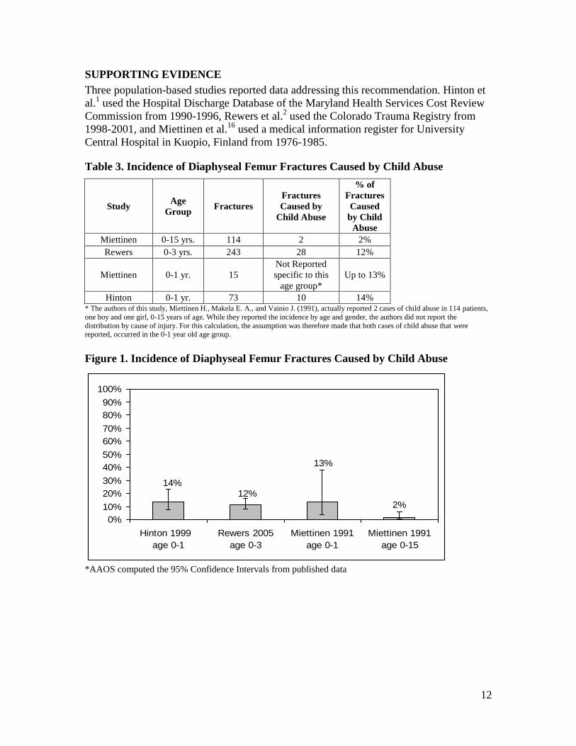
SUPPORTING EVIDENCE
Three population-based studies reported data addressing this recommendation. Hinton et
al.1 used the Hospital Discharge Database of the Maryland Health Services Cost Review
Commission from 1990-1996, Rewers et al.2 used the Colorado Trauma Registry from
1998-2001, and Miettinen et al.16
used a medical information register for University
Central Hospital in Kuopio, Finland from 1976-1985.
Table 3. Incidence of Diaphyseal Femur Fractures Caused by Child Abuse
Study Age
Group Fractures
Fractures
Caused by
Child Abuse
% of
Fractures
Caused
by Child
Abuse
Miettinen 0-15 yrs. 114 2 2%
Rewers 0-3 yrs. 243 28 12%
Miettinen 0-1 yr. 15
Not Reported
specific to this
age group*
Up to 13%
Hinton 0-1 yr. 73 10 14% * The authors of this study, Miettinen H., Makela E. A., and Vainio J. (1991), actually reported 2 cases of child abuse in 114 patients,
one boy and one girl, 0-15 years of age. While they reported the incidence by age and gender, the authors did not report the
distribution by cause of injury. For this calculation, the assumption was therefore made that both cases of child abuse that were reported, occurred in the 0-1 year old age group.
Figure 1. Incidence of Diaphyseal Femur Fractures Caused by Child Abuse
*AAOS computed the 95% Confidence Intervals from published data
2%
13%
12%14%
0%
10%
20%
30%
40%
50%
60%
70%
80%
90%
100%
Hinton 1999
age 0-1
Rewers 2005
age 0-3
Miettinen 1991
age 0-1
Miettinen 1991
age 0-15

13
INFANT FEMUR FRACTURE
Limited evidence supports treatment with a Pavlik harness or a spica cast for
infants six months and younger with a diaphyseal femur fracture, because
their outcomes are similar.
Grade of Recommendation: Limited
Figures relevant to this recommendation are: Figure 2
RATIONALE
The first 6 months of a child’s life is a time of most rapid growth. Because of this, rapid
healing of diaphyseal femur fractures and post-fracture skeletal remodeling is maximal.
Hence spontaneous, complete correction after fracture healing is expected. Due to the
rapid union and complete remodeling, treatment of diaphyseal femur fractures centers on
assuring ease of patient care and minimizing treatment complications. Both Pavlik
harnesses and spica casts result in good outcomes with minimal complications. In the
studies we reviewed, the only identifiable difference between these two treatments was
more frequent skin complications in the spica cast group. Because this is a minor and
correctable issue that does not cause long-term problems or disability, either type of
treatment is an option.
SUPPORTING EVIDENCE
Two studies addressed this recommendation. One retrospective comparative study17
compared the Pavlik harness to a spica cast, and one case series examined Pavlik
harnesses.18
The case series reported that all 16 patients achieved stable union by 5 weeks
in a Pavlik harness. In the comparative study, the spica cast group had significantly more
skin complications (p<.01) than the Pavlik harness group, but there were no other
statistically significant differences between groups. The Pavlik harness group was
significantly younger (p=.028), with an average age of 3.6 months versus an average age
of 6.5 months in the spica cast group.

14
Figure 2. Pavlik Harness vs. Spica Cast
Note on figures: Appendix contains information on how to interpret forest plots such as
the one above as well as explanations of symbols used in this guideline’s figures and
tables.
Loss of reduction
Cast revision due to reduction loss
Shortening during treatment
Skin breakdown
Superficial reactive dermatitis
Total skin complications
Outcome
4 weeks
4 weeks
4 weeks
4 weeks
4 weeks
4 weeks
Duration
0.00 (0.00, 1.53)
0.00 (0.00, 26.00)
0.56 (0.11, 2.74)
0.00 (0.00, 1.53)
0.00 (0.00, 1.53)
0.00 (0.00, 0.45)
Ratio (95% CI)
Odds
0.00 (0.00, 1.53)
0.00 (0.00, 26.00)
0.56 (0.11, 2.74)
0.00 (0.00, 1.53)
0.00 (0.00, 1.53)
0.00 (0.00, 0.45)
Ratio (95% CI)
Odds
Pavlik Harness Spica Cast 11
15
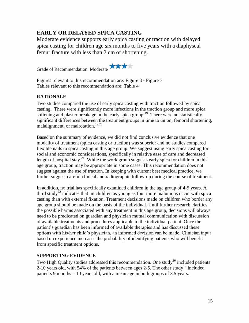
EARLY OR DELAYED SPICA CASTING
Moderate evidence supports early spica casting or traction with delayed
spica casting for children age six months to five years with a diaphyseal
femur fracture with less than 2 cm of shortening.
Grade of Recommendation: Moderate
Figures relevant to this recommendation are: Figure 3 - Figure 7
Tables relevant to this recommendation are: Table 4
RATIONALE
Two studies compared the use of early spica casting with traction followed by spica
casting. There were significantly more infections in the traction group and more spica
softening and plaster breakage in the early spica group.19
There were no statistically
significant differences between the treatment groups in time to union, femoral shortening,
malalignment, or malrotation.19,20
Based on the summary of evidence, we did not find conclusive evidence that one
modality of treatment (spica casting or traction) was superior and no studies compared
flexible nails to spica casting in this age group. We suggest using early spica casting for
social and economic considerations, specifically in relative ease of care and decreased
length of hospital stay.21
While the work group suggests early spica for children in this
age group, traction may be appropriate in some cases. This recommendation does not
suggest against the use of traction. In keeping with current best medical practice, we
further suggest careful clinical and radiographic follow-up during the course of treatment.
In addition, no trial has specifically examined children in the age group of 4-5 years. A
third study22
indicates that in children as young as four more malunions occur with spica
casting than with external fixation. Treatment decisions made on children who border any
age group should be made on the basis of the individual. Until further research clarifies
the possible harms associated with any treatment in this age group, decisions will always
need to be predicated on guardian and physician mutual communication with discussion
of available treatments and procedures applicable to the individual patient. Once the
patient’s guardian has been informed of available therapies and has discussed these
options with his/her child’s physician, an informed decision can be made. Clinician input
based on experience increases the probability of identifying patients who will benefit
from specific treatment options.
SUPPORTING EVIDENCE
Two High Quality studies addressed this recommendation. One study20
included patients
2-10 years old, with 54% of the patients between ages 2-5. The other study19
included
patients 9 months – 10 years old, with a mean age in both groups of 3.5 years.

16
One High quality study,22
with a mean patient age of 6 years old, but that addressed
harms in children as young as 4 was also included to address this recommendation. (See
Recommendation 8)
Summary of Evidence
Table 4. Summary of Evidence
Author Level of
Evidence n Comparison Outcome (follow-up duration) Result
Burton High 183
Spica Cast vs.
Traction
Time to Union (n/a) ○ Burton High 183 Shortening (at Union) ○ Rasool High 170 Shortening at (6 wk) ○ Burton High 183 Varus angulation (at Union) ○ Rasool High 170 Varus angulation (6 wk) ○ Burton High 183 Valgus angulation (at Union) ○ Rasool High 170 Valgus angulation (6 wk) ○ Burton High 183 Anterior Bowing (at Union) ○ Rasool High 170 Anterior Bowing (6 wk) ○ Burton High 183 Posterior Bowing (at Union) ○ Rasool High 170 Posterior Bowing (6 wk) ○ Rasool High 170 Infectious disease contraction (6 wk) ● sc
Rasool High 170 Pressure from ring of splint (6 wk) ○ Rasool High 170 Blisters (6 wk) ○ Rasool High 170 Spica softening (6 wk) ● t Rasool High 170 Plaster breakage (6 wk) ● t Rasool High 170 Soilage (6 wk) ○
● = result is statistically significant n/a = not applicable sc = spica cast
○ = result is not statistically significant nr = not reported t = traction

17
Figure 3. Time Immobilized (Time to Union)
*Odds Ratio from ordered logistic regression (AAOS calculation)
Figure 4. Shortening
*Odds Ratios from ordered logistic regression (AAOS calculation)
Time to Union
Outcome
Burton
Study
0.86 (0.48, 1.56)
OR (95% CI)
0.86 (0.48, 1.56)
OR (95% CI)
Spica Cast Traction 11
Burton
Rasool
Study
Union
6 weeks
Duration
1.18 (0.69, 2.02)
0.79 (0.45, 1.39)
OR (95% CI)
1.18 (0.69, 2.02)
0.79 (0.45, 1.39)
OR (95% CI)
Spica Cast Traction 11

18
Figure 5. Angulation
*Odds Ratios from ordered logistic regression (AAOS calculation)
Figure 6. Bowing
*Anterior Bowing Odds Ratios from ordered logistic regression (AAOS calculation)
Varus Angulation
Valgus Angulation
Outcome
Burton
Rasool
Burton
Rasool
Study
Union
6 weeks
Union
6 weeks
Duration
1.02 (0.56, 1.85)
0.70 (0.38, 1.29)
0.94 (0.42, 2.15)
0.82 (0.19, 3.39)
OR (95% CI)
1.02 (0.56, 1.85)
0.70 (0.38, 1.29)
0.94 (0.42, 2.15)
0.82 (0.19, 3.39)
OR (95% CI)
Spica Cast Traction 11
Anterior Bowing
Posterior Bowing
Outcome
Burton
Rasool
Burton
Rasool
Study
Union
6 weeks
Union
6 weeks
Duration
1.53 (0.87, 2.67)
1.16 (0.64, 2.10)
0.74 (0.25, 2.15)
0.00 (0.00, 1.49)
OR (95% CI)
1.53 (0.87, 2.67)
1.16 (0.64, 2.10)
0.74 (0.25, 2.15)
0.00 (0.00, 1.49)
OR (95% CI)
Spica Cast Traction 1

19
Figure 7. Complications
PREVIOUSLY PUBLISHED SYSTEMATIC REVIEWS
Two previous systematic reviews21,23
concluded that early spica casting was associated
with shorter inpatient hospital stays and fewer adverse events than traction. Both of these
reviews, however, were not specific to the population of interest for this
recommendation, so we did not include them in our systematic review.
Infectious disease contraction
Blisters
Pressure from ring of splint
Spica softening
Plaster breakage
Soilage
Outcome
Rasool
Rasool
Rasool
Rasool
Rasool
Rasool
Study
6 weeks
6 weeks
6 weeks
6 weeks
6 weeks
6 weeks
Duration
0.12 (0.01, 0.57)
0.00 (0.00, 1.49)
0.00 (0.00, 2.40)
1000.00 (2.42, 1000.00)
1000.00 (1.22, 1000.00)
1000.00 (0.67, 1000.00)
OR (95% CI)
0.12 (0.01, 0.57)
0.00 (0.00, 1.49)
0.00 (0.00, 2.40)
1000.00 (2.42, 1000.00)
1000.00 (1.22, 1000.00)
1000.00 (0.67, 1000.00)
OR (95% CI)
Spica Cast Traction 1

20
ELASTIC INTRAMEDULLARY NAILS
Limited evidence supports the option for physicians to use flexible
intramedullary nailing to treat children age five to eleven years diagnosed
with diaphyseal femur fractures.
Grade of Recommendation: Limited
Figures relevant to this recommendation are: Figure 8 - Figure 31
Tables relevant to this recommendation are: Table 6 - Table 11
RATIONALE
There are few statistically significant differences between treatments in healing of the
fracture. The evidence reviewed included ten studies that examined one hundred varying
outcomes. Of these one hundred outcomes twenty-one were significant. There were no
studies that directly compared flexible nails to spica casting. When flexible nails were
compared to external fixation and traction plus casting, nine outcomes were significant
favoring flexible nails, one significant outcome favored external fixation and one
significant outcome favored traction plus casting. (Please refer to Tables 6 and 7 below.)
The high quality study22
found to address this recommendation compared external
fixation to spica casting. External fixation was favored over spica casting for malunions,
including anterior/posterior angulation. Twelve other outcomes for this comparison had
non-significant results.
In summary, the overall body of evidence considered for this recommendation indicates
that there are few significant outcomes when all comparisons are considered. Further,
important comparisons have not been investigated (spica casting and flexible nails).
Two moderate quality studies24, 50
shows more rapid return to walking and school with
flexible intramedullary nailing and one low quality study25
illustrates less associated
hospital costs when compared to traction and casting. The ability to mobilize the patient,
return them to school rapidly, and suggested decrease in hospital costs leads the work
group to suggest flexible intramedullary nailing over traction followed by casting. There
is evidence that flexible intramedullary nailing has less adverse events and more rapid
return to school than external fixation in both stable and unstable fractures.26
In making this recommendation, the work group acknowledges that they are including
their expert opinion and they have therefore, downgraded the Grade of this
Recommendation to a “limited” recommendation. Based on the advantages suggested,
less adverse events and more rapid return to school, flexible intramedullary nailing is a
treatment option for children five to eleven years diagnosed with diaphyseal femur
fractures.
There is currently insufficient literature in specially designed pediatric rigid
intramedullary nails and bridge plating for inclusion in the current guideline.

21
Patients over age 11 or with weight over 49 kg are at increased risk of a poor outcome27
with flexible intramedullary nailing. The mean weight between patients with a poor
outcome and those with an excellent or satisfactory outcome was significant, but weight
was not independent of age and had a sensitivity of only 59% in predicting poor
outcomes.
SUPPORTING EVIDENCE
One High quality study,22
six moderate quality studies24,28-31, 50
and eight low quality
studies25,26,32-37
addressed this recommendation. Low quality studies are included in this
recommendation because they examine treatments not compared in the high and
moderate quality studies. The average age of the patients enrolled in these studies was 4-
11 years but ten studies included patients outside of this range.
One very low quality study27
addressed the issue of patient weight as a criterion for the
use of flexible nailing in this age group by comparing the weight of patients with an
excellent or satisfactory outcome to the weight of patients with a poor outcome. Sixty
percent (60%) of the patients in this study were less than 11 years old. The 15 kg
difference in mean weight between patients with a poor outcome and those with an
excellent or satisfactory outcome was statistically significant according to the author’s
calculations (p=.003). Moreover, using a cut-off point of 49 kg, heavier patients were
about five times more likely than lighter patients to have poor outcomes. However, the
investigators found that weight did not independently predict a poor outcome when age
was also included in a logistic regression model. The investigators also found that the
weight cut-off point had 78.5% specificity and 59% sensitivity for detecting a poor
outcome.
Table 5. Summary of Updated Findings for Titanium Elastic Nailing
Study Quality N Comparison Outcome Author
p-value
Result
Shemshaki,H.R.,
2011
Moderate
46
TEN vs.
Traction + spica
casting
Hopital Stay (days) <0.001 TEN
Time to start
walking with
aids(days)
<0.001 TEN
Time to start
walking
independently
(days)
<0.001 TEN
Time to return to
school (days)
<0.001 TEN
Knee range of
motion (degrees)
0.078 NS
Malunion 0.117 NS
Infection 0.117 NS
Parent satisfaction-
excellent
0.003 TEN
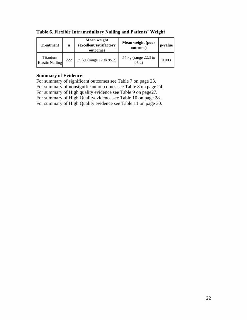
22
Table 6. Flexible Intramedullary Nailing and Patients’ Weight
Treatment n
Mean weight
(excellent/satisfactory
outcome)
Mean weight (poor
outcome)p-value
Titanium
Elastic Nailing222 39 kg (range 17 to 95.2)
54 kg (range 22.3 to
95.2)0.003
Summary of Evidence:
For summary of significant outcomes see Table 7 on page 23.
For summary of nonsignificant outcomes see Table 8 on page 24.
For summary of High quality evidence see Table 9 on page27.
For summary of High Qualityevidence see Table 10 on page 28.
For summary of High Quality evidence see Table 11 on page 30.

23
Table 7. Summary of Significant Outcomes with Level of Evidence
TREATMENT FAVORED
Flexible Nails External Fixation Spica Cast Traction (+
Casting)
Flexible
Nails N/A
Loss of movement at
knee (post-op)/III
Irritation at nail
entry site/II
External
Fixation
Time to full knee
movement/III
Return to school/III
N/A Duration of treatment/I
Pin-tract infections/I
Duration of
treatment/III
Duration of
treatment/III
Spica Cast
Malunion/I
Anterior/Posterior
Angulation/I
N/A
Traction (+
Casting)
Walking with
aids/II
Walking
independently/II
Walking
independently/II
Return to school/II
Angular
deformity/III
Flynn’s outcome/III
Acceptable scar/III
Patient satisfaction/III
Blisters/superficial
ulcers/II
Knee flexion
contraction/II
Overall short-term
complications/II
N/A
*Number of outcomes examined: 100
**Number of significant outcomes: 21
***Number of studies:10
****Number of nonsignificant outcomes:
Flexible nails: 49
External fixation: 26
Spica cast: 27
Traction and casting: 56
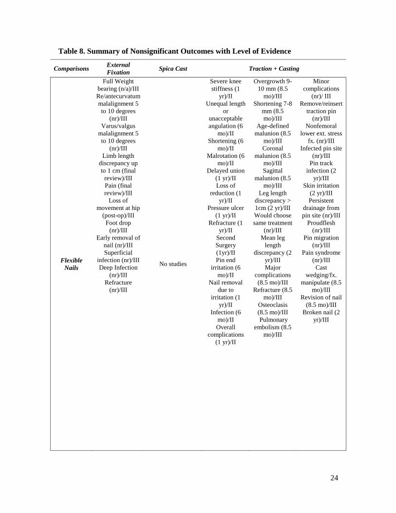
24
Table 8. Summary of Nonsignificant Outcomes with Level of Evidence
Comparisons External
Fixation Spica Cast Traction + Casting
Flexible
Nails
Full Weight
bearing (n/a)/III
Re/antecurvatum
malalignment 5
to 10 degrees
(nr)/III
Varus/valgus
malalignment 5
to 10 degrees
(nr)/III
Limb length
discrepancy up
to 1 cm (final
review)/III
Pain (final
review)/III
Loss of
movement at hip
(post-op)/III
Foot drop
(nr)/III
Early removal of
nail (nr)/III
Superficial
infection (nr)/III
Deep Infection
(nr)/III
Refracture
(nr)/III
No studies
Severe knee
stiffness (1
yr)/II
Unequal length
or
unacceptable
angulation (6
mo)/II
Shortening (6
mo)/II
Malrotation (6
mo)/II
Delayed union
(1 yr)/II
Loss of
reduction (1
yr)/II
Pressure ulcer
(1 yr)/II
Refracture (1
yr)/II
Second
Surgery
(1yr)/II
Pin end
irritation (6
mo)/II
Nail removal
due to
irritation (1
yr)/II
Infection (6
mo)/II
Overall
complications
(1 yr)/II
Overgrowth 9-
10 mm (8.5
mo)/III
Shortening 7-8
mm (8.5
mo)/III
Age-defined
malunion (8.5
mo)/III
Coronal
malunion (8.5
mo)/III
Sagittal
malunion (8.5
mo)/III
Leg length
discrepancy >
1cm (2 yr)/III
Would choose
same treatment
(nr)/III
Mean leg
length
discrepancy (2
yr)/III
Major
complications
(8.5 mo)/III
Refracture (8.5
mo)/III
Osteoclasis
(8.5 mo)/III
Pulmonary
embolism (8.5
mo)/III
Minor
complications
(nr)/ III
Remove/reinsert
traction pin
(nr)/III
Nonfemoral
lower ext. stress
fx. (nr)/III
Infected pin site
(nr)/III
Pin track
infection (2
yr)/III
Skin irritation
(2 yr)/III
Persistent
drainage from
pin site (nr)/III
Proudflesh
(nr)/III
Pin migration
(nr)/III
Pain syndrome
(nr)/III
Cast
wedging/fx.
manipulate (8.5
mo)/III
Revision of nail
(8.5 mo)/III
Broken nail (2
yr)/III

25
Table 8. Summary of Nonsignificant Outcomes with Level of Evidence (continued)
Comparisons External
Fixation Spica Cast Traction + Casting
External
Fixation N/A
Malunion inc.
rotation (2 yr)/I
Leg length
discrepancy ≥ 2
cm (2 yr)/I
Varus/valgus
angulation ≥ 10° (2
yr)/I
Rotational
Malunion (2 yr)/I
Treatment
alteration (2 yr)/I
Unacceptable loss
of reduction (2
yr)/I
RAND overall (2
yr)/I
Duration of
treatment (n/a)/I
Behavioural
Questionnaire
(post-hosp)/I
Parent Satisfaction
(2 yr)/I
Child Satisfaction
(2 yr)/I
Refracture (2 yr)/I
Total treatment days until union/III
Reoperation (until union)/III
Patient Satisfaction/III

26
Table 8. Summary of Nonsignificant Outcomes with Level of Evidence (continued)
Comparisons External
Fixation Spica Cast Traction + Casting
Spica Cast N/A N/A
Shortening (1 yr)/II
Union time (n/a)/II
Coronal angulation (1 yr) /II
Sagittal angulation (1 yr)/II
Excessive override (3 mo)/II
Varus angulation > 10° (3 mo/II)
Valgus angulation > 10° (3 mo)/II
Posterior angulation > 10° (3 mo)/II
Delayed union (3 mo)/II
Readmission for manipulation (3 mo)/II
Pin-tract infection (3 mo)/II
Post-cast fracture of ipsi. limb (3 mo)/II
Spinal muscular atrophy(3 mo)/II
Fever of unknown origin (3 mo)/II
Cast sore (3 mo)/II
*Number of outcomes examined: 100
**Number of significant outcomes: 21 (See Table 6)
***Number of studies:10
****Number of nonsignificant outcomes:
Flexible nails: 49
External fixation: 26
Spica cast: 27
Traction and casting: 56
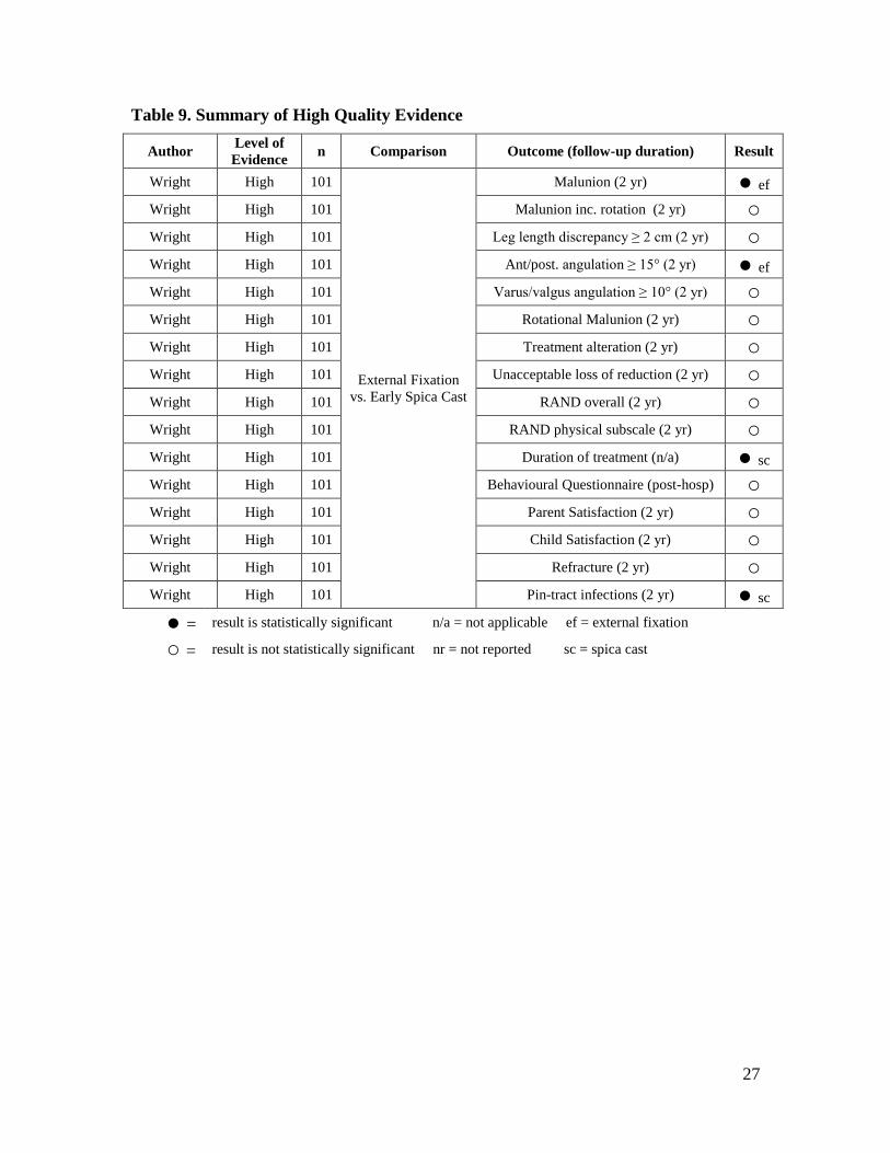
27
Table 9. Summary of High Quality Evidence
Author Level of
Evidence n Comparison Outcome (follow-up duration) Result
Wright High 101
External Fixation
vs. Early Spica Cast
Malunion (2 yr) ● ef Wright High 101 Malunion inc. rotation (2 yr) ○
Wright High 101 Leg length discrepancy ≥ 2 cm (2 yr) ○
Wright High 101 Ant/post. angulation ≥ 15° (2 yr) ● ef Wright High 101 Varus/valgus angulation ≥ 10° (2 yr) ○
Wright High 101 Rotational Malunion (2 yr) ○
Wright High 101 Treatment alteration (2 yr) ○
Wright High 101 Unacceptable loss of reduction (2 yr) ○
Wright High 101 RAND overall (2 yr) ○
Wright High 101 RAND physical subscale (2 yr) ○
Wright High 101 Duration of treatment (n/a) ● sc Wright High 101 Behavioural Questionnaire (post-hosp) ○
Wright High 101 Parent Satisfaction (2 yr) ○
Wright High 101 Child Satisfaction (2 yr) ○
Wright High 101 Refracture (2 yr) ○
Wright High 101 Pin-tract infections (2 yr) ● sc
● = result is statistically significant n/a = not applicable ef = external fixation
○ = result is not statistically significant nr = not reported sc = spica cast
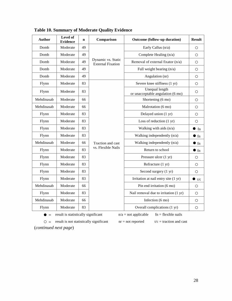
28
Table 10. Summary of Moderate Quality Evidence
Author Level of
Evidence n Comparison Outcome (follow-up duration) Result
Domb Moderate 49
Dynamic vs. Static
External Fixation
Early Callus (n/a) ○
Domb Moderate 49 Complete Healing (n/a) ○
Domb Moderate 49 Removal of external fixator (n/a) ○
Domb Moderate 49 Full weight bearing (n/a) ○
Domb Moderate 49 Angulation (nr) ○
Flynn Moderate 83
Traction and cast
vs. Flexible Nails
Severe knee stiffness (1 yr) ○
Flynn Moderate 83 Unequal length
or unacceptable angulation (6 mo) ○
Mehdinasab Moderate 66 Shortening (6 mo) ○
Mehdinasab Moderate 66 Malrotation (6 mo) ○
Flynn Moderate 83 Delayed union (1 yr) ○
Flynn Moderate 83 Loss of reduction (1 yr) ○ Flynn Moderate 83 Walking with aids (n/a) ● fn
Flynn Moderate 83 Walking independently (n/a) ● fn
Mehdinasab Moderate 66 Walking independently (n/a) ● fn
Flynn Moderate 83 Return to school ● fn
Flynn Moderate 83 Pressure ulcer (1 yr) ○
Flynn Moderate 83 Refracture (1 yr) ○
Flynn Moderate 83 Second surgery (1 yr) ○
Flynn Moderate 83 Irritation at nail entry site (1 yr) ● t/c Mehdinasab Moderate 66 Pin end irritation (6 mo) ○
Flynn Moderate 83 Nail removal due to irritation (1 yr) ○
Mehdinasab Moderate 66 Infection (6 mo) ○
Flynn Moderate 83 Overall complications (1 yr) ○
● = result is statistically significant n/a = not applicable fn = flexible nails
○ = result is not statistically significant nr = not reported t/c = traction and cast
(continued next page)

29
Table 10. Summary of Moderate Quality Evidence (continued)
Author Level of
Evidence n Comparison Outcome (follow-up duration) Result
Ali Moderate 66
Early spica cast vs.
traction
Shortening (1 yr) ○
Ali Moderate 100 Union time (n/a) ○
Ali Moderate 66 Coronal angulation (1 yr) ○
Ali Moderate 66 Sagittal angulation (1 yr) ○ Ali Moderate 100 Blisters/superficial ulcers (until union) ● sc
Curtis Moderate 91
Early pontoon spica
cast vs. traction,
then spica cast
Excessive override (3 mo) ○
Curtis Moderate 91 Varus angulation > 10° (3 mo) ○
Curtis Moderate 91 Valgus angulation > 10° (3 mo) ○
Curtis Moderate 91 Posterior angulation > 10° (3 mo) ○
Curtis Moderate 91 Delayed union (3 mo) ○
Curtis Moderate 91 Knee flexion contraction > 20° (3 mo) ● psc
Curtis Moderate 91 Readmission for manipulation (3 mo) ○
Curtis Moderate 91 Pin-tract infection (3 mo) ○
Curtis Moderate 91 Post-cast fracture of ipsi. limb (3 mo) ○
Curtis Moderate 91 Spinal muscular atrophy(3 mo) ○
Curtis Moderate 91 Fever of unknown origin (3 mo) ○
Curtis Moderate 91 Cast sore (3 mo) ○
Curtis Moderate 91 Overall short-term complication (3 mo) ● psc
● = result is statistically significant n/a = not applicable sc = spica cast
○ = result is not statistically significant nr = not reported psc = (pontoon) spica cast

30
Table 11. Summary of Low Quality Evidence
Author Level of
Evidence n Comparison Outcome (follow-up duration) Result
Nork Low 29 External fixation vs.
traction and cast
Total treatment days (until union) ○
Nork Low 29 Reoperation (until union) ○
Hedin 2004 Low 59 External fixation vs.
home traction
Duration of treatment (n/a) ● t Hedin 2004 Low 66 Patient satisfaction (nr) ○ Hedin 2004 Low 59 External fixation vs.
hospital traction
Duration of treatment (n/a) ● t Hedin 2004 Low 66 Patient satisfaction (nr) ● ef
Buechsensc. Low 71
Flexible nails vs.
traction and cast
Overgrowth 9-10 mm (8.5 mo) ○
Buechsensc. Low 71 Shortening 7-8 mm (8.5 mo) ○
Buechsensc. Low 71 Age-defined malunion (8.5 mo) ○
Buechsensc. Low 71 Coronal malunion (8.5 mo) ○
Buechsensc. Low 71 Sagittal malunion (8.5 mo) ○
Song Low 51 Angular deformity (2 yr) ● fn
Song Low 51 Leg length discrepancy > 1cm (2 yr) ○
Song Low 51 Flynn’s outcome (2 yr) ● fn
Buechsensc. Low 43 Would choose same treatment (nr) ○
Buechsensc. Low 43 Acceptable scar (nr) ● fn
Song Low 51 Mean leg length discrepancy (2 yr) ○
Buechsensc. Low 71 Major complications (8.5 mo) ○
Buechsensc. Low 71 Refracture (8.5 mo) ○
Buechsensc. Low 71 Osteoclasis (8.5 mo) ○
Buechsensc. Low 71 Cast wedging/fx. manipulate (8.5 mo) ○
Buechsensc. Low 71 Revision of nail (8.5 mo) ○
Song Low 51 Broken nail (2 yr) ○
Buechsensc. Low 68 Pulmonary embolism (8.5 mo) ○
● = result is statistically significant n/a = not applicable t = traction ef = external fixation
○ = result is not statistically significant nr = not reported fn = flexible nails
(continued next page)

31
Table 11. Summary of Low Quality Evidence (continued)
Author Level of
Evidence n Comparison Outcome (follow-up duration) Result
Buechsensc. Low 71
Flexible nails vs.
traction and cast
Minor complications (nr) ○
Buechsensc. Low 71 Remove/reinsert traction pin (nr) ○
Buechsensc. Low 71 Nonfemoral lower ext. stress fx. (nr) ○
Buechsensc. Low 71 Infected pin site (nr) ○
Song Low 51 Pin track infection (2 yr) ○
Song Low 51 Skin irritation (2 yr) ○
Buechsensc. Low 71 Persistent drainage from pin site (nr) ○
Buechsensc. Low 71 Proudflesh (nr) ○
Buechsensc. Low 71 Pin migration (nr) ○
Buechsensc. Low 71 Pain syndrome (nr) ○
Wall Low 104
Titanium vs.
stainless steel
flexible nail
Malunion (1 yr) ● ss
Wall Low 104 Osteotomy (1 yr) ○
Wall Low 104 Infection (1 yr) ○
Wall Low 104 Implant revision (1 yr) ○
Wall Low 104 Nail irritation (1 yr) ○
Wall Low 104 Nail breakage (1 yr) ○
Wall Low 104 Delayed union (3 mo) ○
Wall Low 104 Minor complications (1 yr) ○
Rasit Low 40 Immediate vs.
delayed spica cast
2nd surgery/loss of reduction(2 wk) ○
Rasit Low 40 Angulation > 20° (2 wk) ○
Sturdee Low 56
Early intervention
vs. traction
Pain (3 mo) ○
Sturdee Low 56 Malunion (3 mo) ○
Sturdee Low 56 Pin-site infections (until union) ○
Sturdee Low 56 Broken external fixation pin
(until union) ○
Sturdee Low 56 Refracture (until union) ○
Sturdee Low 56 Reapplication of traction pin
(until union) ○
Sturdee Low 56 Pressure sores (until union) ○
● = result is statistically significant n/a = not applicable ss = stainless steel
○ = result is not statistically significant nr = not reported
(continued next page)
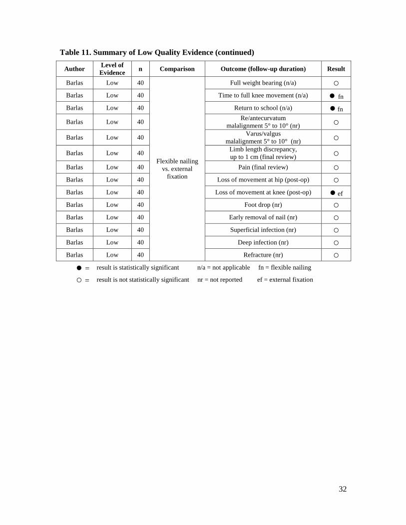
32
Table 11. Summary of Low Quality Evidence (continued)
Author Level of
Evidence n Comparison Outcome (follow-up duration) Result
Barlas Low 40
Flexible nailing
vs. external
fixation
Full weight bearing (n/a) ○ Barlas Low 40 Time to full knee movement (n/a) ● fn
Barlas Low 40 Return to school (n/a) ● fn
Barlas Low 40 Re/antecurvatum
malalignment 5° to 10° (nr) ○
Barlas Low 40 Varus/valgus
malalignment 5° to 10° (nr) ○
Barlas Low 40 Limb length discrepancy,
up to 1 cm (final review) ○
Barlas Low 40 Pain (final review) ○
Barlas Low 40 Loss of movement at hip (post-op) ○
Barlas Low 40 Loss of movement at knee (post-op) ● ef
Barlas Low 40 Foot drop (nr) ○
Barlas Low 40 Early removal of nail (nr) ○
Barlas Low 40 Superficial infection (nr) ○
Barlas Low 40 Deep infection (nr) ○
Barlas Low 40 Refracture (nr) ○
● = result is statistically significant n/a = not applicable fn = flexible nailing
○ = result is not statistically significant nr = not reported ef = external fixation

33
Figure 8. External Fixation vs. Spica Cast - binary outcomes (Wright et al.22
)
Figure 9. External Fixation vs. Spica Cast -continuous outcomes (Wright et al.22
)
Malunion
Malunion inc. rotation
Leg length discrepancy >=2cm
Anterior/posterior angulation >= 15 deg
Varus/valgus angulation >=10 deg
Rotational malunion
Treatment alteration
Unacceptable loss of reduction
Outcome
0.23 (0.07, 0.64)
0.50 (0.21, 1.19)
0.50 (0.08, 2.38)
0.24 (0.07, 0.77)
0.40 (0.01, 5.25)
0.71 (0.27, 1.82)
1.11 (0.31, 3.84)
0.00 (0.00, 1.01)
Ratio (95% CI)
Odds
0.23 (0.07, 0.64)
0.50 (0.21, 1.19)
0.50 (0.08, 2.38)
0.24 (0.07, 0.77)
0.40 (0.01, 5.25)
0.71 (0.27, 1.82)
1.11 (0.31, 3.84)
0.00 (0.00, 1.01)
Ratio (95% CI)
Odds
External Fixation Spica Cast 11
RAND overall
RAND physical subscale
Duration of treatment (weeks)
Post-hospitalizaiton behavioural questionnaire
Parent satisfaction
Child satisfaction
Outcome
1.00 (-4.20, 2.10)
0.10 (-0.30, 0.60)
2.71 (1.57, 4.00)
-0.50 (-5.90, 4.90)
-0.10 (-0.60, 0.30)
0.80 (-0.50, 2.20)
Difference (95% CI)
Mean
1.00 (-4.20, 2.10)
0.10 (-0.30, 0.60)
2.71 (1.57, 4.00)
-0.50 (-5.90, 4.90)
-0.10 (-0.60, 0.30)
0.80 (-0.50, 2.20)
Difference (95% CI)
Mean
External Fixation Spica Cast 00
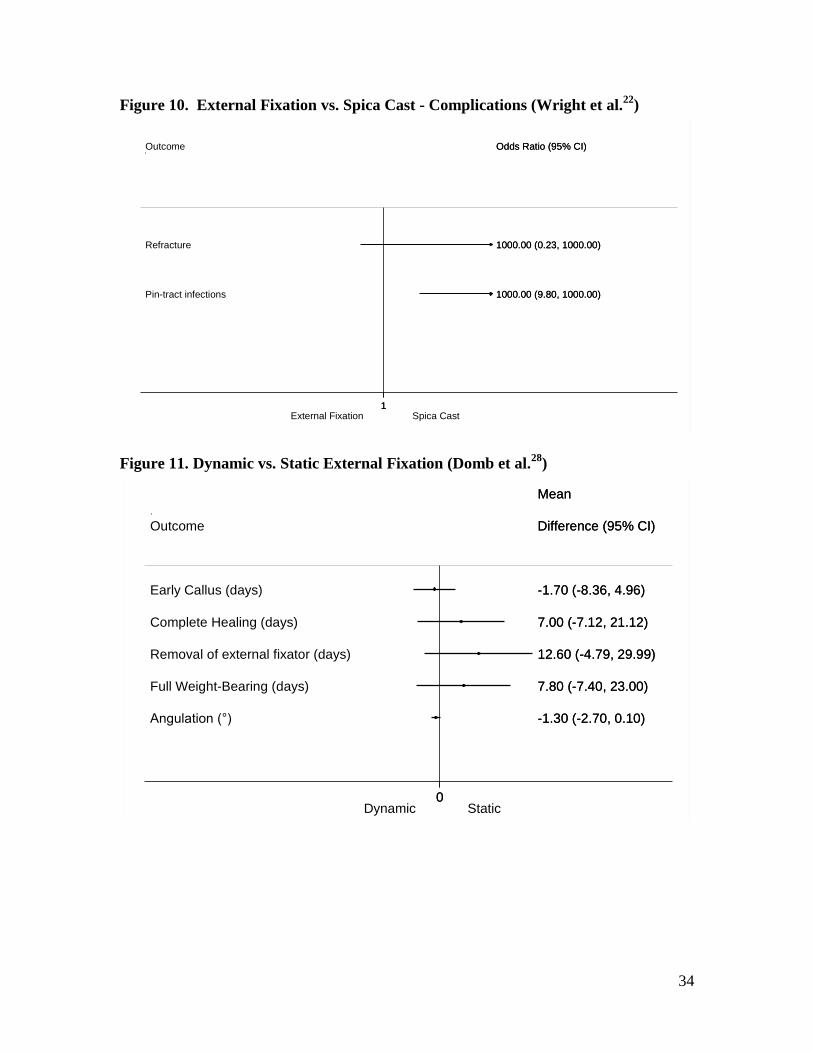
34
Figure 10. External Fixation vs. Spica Cast - Complications (Wright et al.22
)
Figure 11. Dynamic vs. Static External Fixation (Domb et al.28
)
Refracture
Pin-tract infections
Outcome
1000.00 (0.23, 1000.00)
1000.00 (9.80, 1000.00)
Odds Ratio (95% CI)
1000.00 (0.23, 1000.00)
1000.00 (9.80, 1000.00)
Odds Ratio (95% CI)
External Fixation Spica Cast 11
Early Callus (days)
Complete Healing (days)
Removal of external fixator (days)
Full Weight-Bearing (days)
Angulation (°)
Outcome
-1.70 (-8.36, 4.96)
7.00 (-7.12, 21.12)
12.60 (-4.79, 29.99)
7.80 (-7.40, 23.00)
-1.30 (-2.70, 0.10)
Difference (95% CI)
Mean
-1.70 (-8.36, 4.96)
7.00 (-7.12, 21.12)
12.60 (-4.79, 29.99)
7.80 (-7.40, 23.00)
-1.30 (-2.70, 0.10)
Difference (95% CI)
Mean
Dynamic Static 00

35
Figure 12. Traction & Cast vs. Flexible Nails –binary outcomes
Figure 13. Traction & Cast vs. Flexible Nails – continuous outcomes
Severe knee stiffness
Unequal length or
unacceptable angulation
Shortening
Malrotation
Delayed union
Loss of reduction
Outcome
Flynn
Flynn
Mehdinasab
Mehdinasab
Flynn
Flynn
Study
1000.00 (0.04, 1000.00)
1000.00 (0.58, 1000.00)
5.38 (0.48, 273.00)
1000.00 (0.51, 1000.00)
0.00 (0.00, 53.50)
1000.00 (0.26, 1000.00)
Odds Ratio (95% CI)
1000.00 (0.04, 1000.00)
1000.00 (0.58, 1000.00)
5.38 (0.48, 273.00)
1000.00 (0.51, 1000.00)
0.00 (0.00, 53.50)
1000.00 (0.26, 1000.00)
Odds Ratio (95% CI)
Traction/Cast Flexible Nails 11
Walking with aids
Walking independently
Total treatment course
(until independent walking)
Return to school
Outcome
Flynn
Flynn
Mehdinasab
Flynn
Study
56.00 (29.00, 83.00)
39.00 (20.00, 58.00)
14.10 (13.10, 15.10)
55.00 (28.00, 82.00)
(days) (95% CI)
Mean Difference
56.00 (29.00, 83.00)
39.00 (20.00, 58.00)
14.10 (13.10, 15.10)
55.00 (28.00, 82.00)
(days) (95% CI)
Mean Difference
Traction/Cast Flexible Nails 0
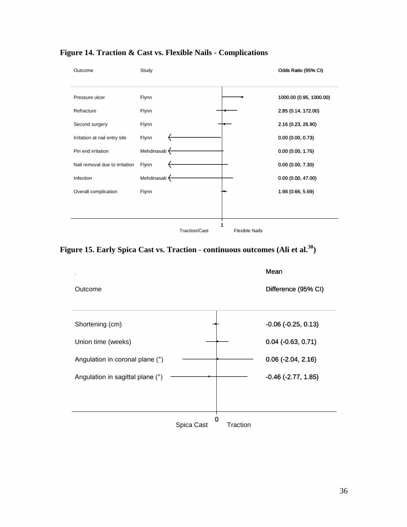
36
Figure 14. Traction & Cast vs. Flexible Nails - Complications
Figure 15. Early Spica Cast vs. Traction - continuous outcomes (Ali et al.30
)
Pressure ulcer
Refracture
Second surgery
Irritation at nail entry site
Pin end irritation
Nail removal due to irritation
Infection
Overall complication
Outcome
Flynn
Flynn
Flynn
Flynn
Mehdinasab
Flynn
Mehdinasab
Flynn
Study
1000.00 (0.95, 1000.00)
2.85 (0.14, 172.00)
2.16 (0.23, 26.90)
0.00 (0.00, 0.73)
0.00 (0.00, 1.76)
0.00 (0.00, 7.30)
0.00 (0.00, 47.00)
1.98 (0.66, 5.99)
Odds Ratio (95% CI)
1000.00 (0.95, 1000.00)
2.85 (0.14, 172.00)
2.16 (0.23, 26.90)
0.00 (0.00, 0.73)
0.00 (0.00, 1.76)
0.00 (0.00, 7.30)
0.00 (0.00, 47.00)
1.98 (0.66, 5.99)
Odds Ratio (95% CI)
Traction/Cast Flexible Nails 11
Shortening (cm)
Union time (weeks)
Angulation in coronal plane (°)
Angulation in sagittal plane (°)
Outcome
-0.06 (-0.25, 0.13)
0.04 (-0.63, 0.71)
0.06 (-2.04, 2.16)
-0.46 (-2.77, 1.85)
Difference (95% CI)
Mean
-0.06 (-0.25, 0.13)
0.04 (-0.63, 0.71)
0.06 (-2.04, 2.16)
-0.46 (-2.77, 1.85)
Difference (95% CI)
Mean
Spica Cast Traction 00
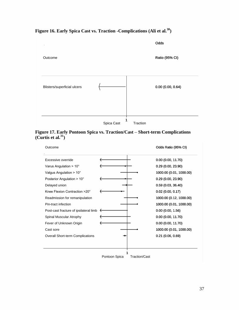
37
Figure 16. Early Spica Cast vs. Traction -Complications (Ali et al.30
)
Figure 17. Early Pontoon Spica vs. Traction/Cast – Short-term Complications
(Curtis et al.31
)
Blisters/superficial ulcers
Outcome
0.00 (0.00, 0.64)
Ratio (95% CI)
Odds
0.00 (0.00, 0.64)
Ratio (95% CI)
Odds
Spica Cast Traction 11
Excessive override
Varus Angulation > 10°
Valgus Angulation > 10°
Posterior Angulation > 10°
Delayed union
Knee Flexion Contraction >20°
Readmission for remanipulation
Pin-tract infection
Post-cast fracture of ipsilateral limb
Spinal Muscular Atrophy
Fever of Unknown Origin
Cast sore
Overall Short-term Complications
Outcome
0.00 (0.00, 11.70)
0.29 (0.00, 23.90)
1000.00 (0.01, 1000.00)
0.29 (0.00, 23.90)
0.59 (0.03, 36.40)
0.02 (0.00, 0.17)
1000.00 (0.12, 1000.00)
1000.00 (0.01, 1000.00)
0.00 (0.00, 1.56)
0.00 (0.00, 11.70)
0.00 (0.00, 11.70)
1000.00 (0.01, 1000.00)
0.21 (0.06, 0.69)
Odds Ratio (95% CI)
0.00 (0.00, 11.70)
0.29 (0.00, 23.90)
1000.00 (0.01, 1000.00)
0.29 (0.00, 23.90)
0.59 (0.03, 36.40)
0.02 (0.00, 0.17)
1000.00 (0.12, 1000.00)
1000.00 (0.01, 1000.00)
0.00 (0.00, 1.56)
0.00 (0.00, 11.70)
0.00 (0.00, 11.70)
1000.00 (0.01, 1000.00)
0.21 (0.06, 0.69)
Odds Ratio (95% CI)
Pontoon Spica Traction/Cast 11

38
Figure 18. External Fixation vs. Traction & Cast - Treatment Length (Nork et al.32
)
Figure 19. External Fixation vs. Traction & Cast – Complications (Nork et al.
32)
Total treatment
days (fracture union)
Outcome
10.10 (-2.40, 22.60)
Difference (95% CI)
Mean
10.10 (-2.40, 22.60)
Difference (95% CI)
Mean
External Fixation Traction/Cast 00
Reoperation
Outcome
Until Union
Duration
1.71 (0.08, 109.40)
Odds Ratio (95% CI)
1.71 (0.08, 109.40)
Odds Ratio (95% CI)
External Fixation Traction/Cast 11

39
Figure 20. External Fixation vs. Traction - Treatment Length (Hedin et al.34
)
Figure 21. External Fixation vs. Traction - Patient Satisfaction (Hedin et al.34
)
Duration of treatment (days)
Duration of treatment (days)
Outcome
Home Traction
Hospital Traction
Type of Traction
12.00 (9.40, 14.60)
17.00 (14.80, 19.20)
Difference (95% CI)
Mean
12.00 (9.40, 14.60)
17.00 (14.80, 19.20)
Difference (95% CI)
Mean
External Fixation Traction 00
Patients' satisfaction
Patients' satisfaction
Outcome
Home Traction
Hospital Traction
Type of Traction
4.44 (0.63, 33.70)
14.50 (3.03, 89.70)
Odds Ratio (95% CI)
4.44 (0.63, 33.70)
14.50 (3.03, 89.70)
Odds Ratio (95% CI)
External Fixation Traction 11
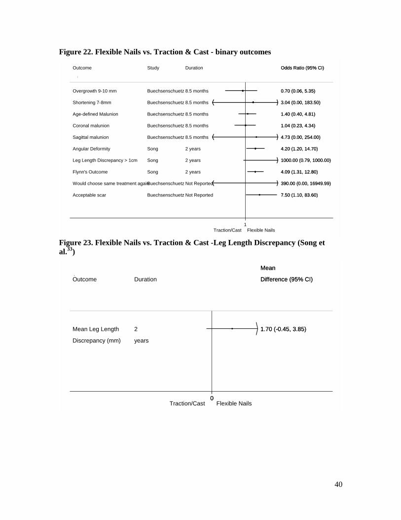
40
Figure 22. Flexible Nails vs. Traction & Cast - binary outcomes
Figure 23. Flexible Nails vs. Traction & Cast -Leg Length Discrepancy (Song et
al.33
)
Overgrowth 9-10 mm
Shortening 7-8mm
Age-defined Malunion
Coronal malunion
Sagittal malunion
Angular Deformity
Leg Length Discrepancy > 1cm
Flynn's Outcome
Would choose same treatment again
Acceptable scar
Outcome
Buechsenschuetz
Buechsenschuetz
Buechsenschuetz
Buechsenschuetz
Buechsenschuetz
Song
Song
Song
Buechsenschuetz
Buechsenschuetz
Study
8.5 months
8.5 months
8.5 months
8.5 months
8.5 months
2 years
2 years
2 years
Not Reported
Not Reported
Duration
0.70 (0.06, 5.35)
3.04 (0.00, 183.50)
1.40 (0.40, 4.81)
1.04 (0.23, 4.34)
4.73 (0.00, 254.00)
4.20 (1.20, 14.70)
1000.00 (0.79, 1000.00)
4.09 (1.31, 12.80)
390.00 (0.00, 16949.99)
7.50 (1.10, 83.60)
Odds Ratio (95% CI)
0.70 (0.06, 5.35)
3.04 (0.00, 183.50)
1.40 (0.40, 4.81)
1.04 (0.23, 4.34)
4.73 (0.00, 254.00)
4.20 (1.20, 14.70)
1000.00 (0.79, 1000.00)
4.09 (1.31, 12.80)
390.00 (0.00, 16949.99)
7.50 (1.10, 83.60)
Odds Ratio (95% CI)
Traction/Cast Flexible Nails
1
Mean Leg Length
Discrepancy (mm)
Outcome
2
years
Duration
1.70 (-0.45, 3.85)
Difference (95% CI)
Mean
1.70 (-0.45, 3.85)
Difference (95% CI)
Mean
Traction/Cast Flexible Nails 00

41
Figure 24. Flexible Nails vs. Traction & Cast - Major Complications
Figure 25. Flexible Nails vs. Traction & Cast - Minor Complications
Major complications
Refracture
Osteoclasis
Cast wedging/fracture manipulation
Revison of nail
Broken nail
Pulmonary Emoblism
Outcome
Buechsenschuetz
Buechsenschuetz
Buechsenschuetz
Buechsenschuetz
Buechsenschuetz
Song
Buechsenschuetz
Study
2.31 (0.24, 29.00)
1000.00 (0.04, 1000.00)
1000.00 (0.04, 1000.00)
1000.00 (0.04, 1000.00)
0.00 (0.00, 56.50)
0.00 (0.00, 44.00)
0.00 (0.00, 63.00)
Odds Ratio (95% CI)
2.31 (0.24, 29.00)
1000.00 (0.04, 1000.00)
1000.00 (0.04, 1000.00)
1000.00 (0.04, 1000.00)
0.00 (0.00, 56.50)
0.00 (0.00, 44.00)
0.00 (0.00, 63.00)
Odds Ratio (95% CI)
Traction/Cast Flexible Nails 1
Minor Complications
Removal & reinsertion of traction pin
Nonfemoral lower extremity stress fx
Infected pin site
Pin track infection
Skin irritation
Persistent drainage from pin site
Proudflesh
Pin migration
Pain syndrome
Outcome
Buechsenschuetz
Buechsenschuetz
Buechsenschuetz
Buechsenschuetz
Song
Song
Buechsenschuetz
Buechsenschuetz
Buechsenschuetz
Buechsenschuetz
Study
2.63 (0.75, 9.48)
1000.00 (0.62, 1000.00)
1000.00 (0.62, 1000.00)
1000.00 (0.27, 1000.00)
1000.00 (0.21, 1000.00)
0.00 (0.00, 2.67)
1000.00 (0.04, 1000.00)
1000.00 (0.04, 1000.00)
0.00 (0.00, 1.61)
0.00 (0.00, 56.50)
Odds Ratio (95% CI)
2.63 (0.75, 9.48)
1000.00 (0.62, 1000.00)
1000.00 (0.62, 1000.00)
1000.00 (0.27, 1000.00)
1000.00 (0.21, 1000.00)
0.00 (0.00, 2.67)
1000.00 (0.04, 1000.00)
1000.00 (0.04, 1000.00)
0.00 (0.00, 1.61)
0.00 (0.00, 56.50)
Odds Ratio (95% CI)
Traction/Cast Flexible Nails 1

42
Figure 26. Titanium vs. Stainless Steel Flexible Nails – Complications (Wall et al.37
)
Figure 27. Immediate vs. Delayed Spica Cast - Complications (Rasit et al.
35)
Malunion
Osteotomy
Infection
Implant Revision
Nail Irritation
Nail Breakage
Delayed union
Minor Complications
Outcome
1 year
1 year
1 year
1 year
1 year
1 year
3 months
1 year
Duration
4.53 (1.12, 26.20)
1000.00 (0.00, 1000.00)
1000.00 (0.36, 1000.00)
0.85 (0.11, 6.67)
0.85 (0.11, 6.67)
0.85 (0.01, 68.50)
0.00 (0.00, 33.00)
1.77 (0.24, 20.30)
Odds Ratio (95% CI)
4.53 (1.12, 26.20)
1000.00 (0.00, 1000.00)
1000.00 (0.36, 1000.00)
0.85 (0.11, 6.67)
0.85 (0.11, 6.67)
0.85 (0.01, 68.50)
0.00 (0.00, 33.00)
1.77 (0.24, 20.30)
Odds Ratio (95% CI)
Titanium Stainless Steel 11
Second surgery due to loss of reduction
Loss of reduction
Angulation of > 20°
Outcome
2 weeks
2 weeks
2 years
Duration
1000.00 (0.12, 1000.00)
1000.00 (0.12, 1000.00)
0.00 (0.00, 1.23)
Odds Ratio (95% CI)
1000.00 (0.12, 1000.00)
1000.00 (0.12, 1000.00)
0.00 (0.00, 1.23)
Odds Ratio (95% CI)
Immediate Spica Delayed Spica 11

43
Figure 28. Early Intervention vs. Traction (Sturdee et al.36
)
Figure 29. Flexible Nailing vs. External Fixation - Continuous Outcomes (Barlas et
al. 200626
)
*Standard deviations estimated from range
Pain
Malunion
Pin-site Infections
Broken external fixation pin
Refracture
Reapplication of traction pin
Pressure Sores
Outcome
3 months
3 months
Until Union
Until Union
Until Union
Until Union
Until Union
Duration
1000.00 (0.03, 1000.00)
1.10 (0.30, 3.93)
2.70 (0.13, 164.10)
1000.00 (0.03, 1000.00)
1000.00 (0.03, 1000.00)
0.00 (0.00, 52.00)
0.00 (0.00, 7.10)
Odds Ratio (95% CI)
1000.00 (0.03, 1000.00)
1.10 (0.30, 3.93)
2.70 (0.13, 164.10)
1000.00 (0.03, 1000.00)
1000.00 (0.03, 1000.00)
0.00 (0.00, 52.00)
0.00 (0.00, 7.10)
Odds Ratio (95% CI)
Early Intervention Traction 11
Full Weight-bearing (weeks)
Time to full knee movement (weeks)
Return to school (weeks)
Outcome
-1.00 (-2.01, 0.01)
-7.00 (-8.22, -5.78)
-7.00 (-7.96, -6.04)
Difference (95% CI)
Mean
-1.00 (-2.01, 0.01)
-7.00 (-8.22, -5.78)
-7.00 (-7.96, -6.04)
Difference (95% CI)
Mean
Flexible Nailing External Fixation 00

44
Figure 30. Flexible Nailing vs. External Fixation -Binary Outcomes (Barlas et al.
200626
)
Figure 31. Flexible Nailing vs. External Fixation - Complications(Barlas et al.
200626
)
Recurvatum/ Antecurvatum (Malalignment 5 to 10°)
Varus/Valgus (Malalignment 5 to 10°)
Limb length discrepancy at final review (up to 1 cm)
Pain on final review
Post-op loss of movement at hip
Post-op loss of movement at knee
Outcome
0.00 (0.00, 39.00)
0.00 (0.00, 2.34)
0.00 (0.00, 5.28)
0.00 (0.00, 2.34)
0.00 (0.00, 39.00)
0.00 (0.00, 0.98)
OR (95% CI)
0.00 (0.00, 39.00)
0.00 (0.00, 2.34)
0.00 (0.00, 5.28)
0.00 (0.00, 2.34)
0.00 (0.00, 39.00)
0.00 (0.00, 0.98)
OR (95% CI)
Flexible Nailing External Fixation 11
Foot drop
Early removal of nail
Superficial infection
Deep infection
Refracture
Outcome
1000.00 (0.03, 1000.00)
1000.00 (0.03, 1000.00)
0.41 (0.06, 2.41)
0.00 (0.00, 39.00)
0.00 (0.00, 5.28)
OR (95% CI)
1000.00 (0.03, 1000.00)
1000.00 (0.03, 1000.00)
0.41 (0.06, 2.41)
0.00 (0.00, 39.00)
0.00 (0.00, 5.28)
OR (95% CI)
Flexible Nailing External Fixation 11
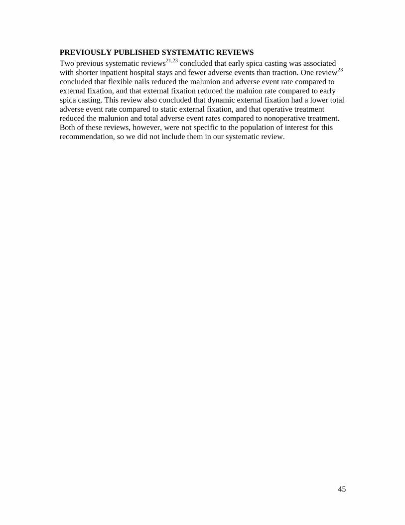
45
PREVIOUSLY PUBLISHED SYSTEMATIC REVIEWS
Two previous systematic reviews21,23
concluded that early spica casting was associated
with shorter inpatient hospital stays and fewer adverse events than traction. One review23
concluded that flexible nails reduced the malunion and adverse event rate compared to
external fixation, and that external fixation reduced the maluion rate compared to early
spica casting. This review also concluded that dynamic external fixation had a lower total
adverse event rate compared to static external fixation, and that operative treatment
reduced the malunion and total adverse event rates compared to nonoperative treatment.
Both of these reviews, however, were not specific to the population of interest for this
recommendation, so we did not include them in our systematic review.

46
ORIF PEDIATRIC FEMUR FRACTURES
Limited evidence supports rigid trochanteric entry nailing, submuscular
plating, and flexible intramedullary nailing as treatment options for children
age eleven years to skeletal maturity diagnosed with diaphyseal femur
fractures, but piriformis or near piriformis entry rigid nailing are not
treatment options.
Grade of Recommendation: Limited
Figures relevant to this recommendation are: Figure 32 - Figure 34
Tables relevant to this recommendation are: Table 12- Table 16
RATIONALE
Skeletally immature patients are at increased risk for avascular necrosis of the femoral
head when piriformis or near piriformis fossa entry nails are used. The rate of this
potentially devastating complication is at least 4%.38
Every effort should be made to
decrease the risk of avascular necrosis.
Fracture patterns that compromise post-reduction stability (i.e. axial and / or angular
stability) as well as heavier patients may stimulate the surgeon to choose rigid
trochanteric entry nailing or submuscular plating over flexible intramedullary nailing.
One Low quality study demonstrated a five times higher risk of poor outcomes for
flexible nailing in patients whose weight met or exceeded 49 kg (108 lbs).27
In the expert
opinion of the work group, external fixation is another option in the older patient with an
unstable fracture pattern, but its significantly higher complication rates, as demonstrated
in other age groups,23,26
make it less desirable than rigid trochanteric entry nailing or
submuscular plating.
SUPPORTING EVIDENCE
One High Quality and four Low quality studies addressed this recommendation. The
High Quality study39
compared nonoperative treatment, mainly traction and cast bracing,
to closed intramedullary nailing. Of the 20 patients (21 fractures) in the operative group,
16 were treated with piriformis entry rigid nailing. There was a statistically significant
difference in favor of intramedullary nailing for two outcomes, time to healing and
malunion. There were no other statistically significant differences between the two
groups.
Of the four Low quality studies, one investigated flexible nailing,27
one investigated rigid
trochanteric entry nailing, 38
one investigated near piriformis entry rigid nailing,38,40
and
one investigated submuscular plating of comminuted fractures.41
The study of flexible nailing27
also compared the weight of patients with an excellent or
satisfactory outcome to the weight of patients with a poor outcome. Forty percent (40%)
of the patients in this study were at least 11 years old. The 15 kg difference in mean
weight between patients with a poor outcome and those with an excellent or satisfactory

47
outcome was statistically significant according to the author’s calculations (p=.003).
Moreover, using a cut-off point of 49 kg, heavier patients were about five times more
likely than lighter patients to have poor outcomes. However, the investigators found that
weight did not independently predict a poor outcome when age was also included in a
logistic regression model. The investigators also found that the weight cut-off point had
78.5% specificity and 59% sensitivity for detecting a poor outcome.
Table 12. Flexible Intramedullary Nailing and Patients’ Weight
Treatment n
Mean weight
(excellent/satisfactory
outcome)
Mean weight (poor
outcome)p-value
Titanium
Elastic Nailing222 39 kg (range 17 to 95.2)
54 kg (range 22.3 to
95.2)0.003
Table 13. Traction vs. Piriformis Entry Rigid Nailing (Herndon et al.39
)
Outcome Duration n
Mean
Difference
(95% CI)
%
(Traction)
%
(Rigid
Nails)
Favors
Healing
(weeks)1.5 (0.5, 2.5) n/a n/a IM Nailing
Malunion 29.0% 0.0% IM Nailing
Shortening
>2cm20.8% 0.0% N/S
Varus >10° 12.5% 0.0% N/S
Valgus >10° 4.2% 0.0% N/S
Anterior angle
>20°8.3% 0.0% N/S
Pressure sore 4.2% 0.0% N/S
Pin track
infection4.2% 0.0% N/S
Limp 8.3% 0.0% N/S
Second
Surgery8.3% 0.0% N/S
Growth plate
arrest0.0% 0.0% N/S
(<1 to 7 yrs.
follow up)n/a
44
*N/S = no significant difference

48
Figure 32. Titanium Elastic Nailing Outcomes Among Age 11+ (Moroz et al.27
)
* AAOS computed the 95% confidence intervals from published data
Table 14. Rigid Trochanteric Entry Nailing Outcomes (Kanellopoulos et al.40
)
Outcome Duration n Mean %Secondary Healing n/a 20 9 weeks (8-13) n/a
Weight Bearing (full) 6 weeks 20 n/a 80%
Full Range of
Motion6 weeks 20 n/a 100%
Return to Preinjury
Activity29 months 20 n/a 100%
Limp 29 months 20 n/a 0%
Delayed or
Nonunion13 weeks 20 n/a 0%
Deep infections 29 months 20 n/a 0%
Hip Osteonecrosis 29 months 20 n/a 0%
16%
29%
55%
0%
20%
40%
60%
80%
100%
Excellent Satisfactory Poor
Outcome
% o
f P
ati
en
ts
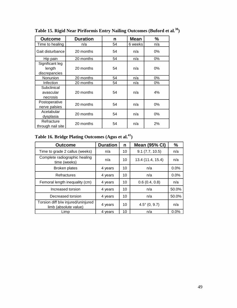
49
Table 15. Rigid Near Piriformis Entry Nailing Outcomes (Buford et al.38
)
Outcome Duration n Mean %Time to healing n/a 54 6 weeks n/a
Gait disturbance 20 months 54 n/a 0%
Hip pain 20 months 54 n/a 0%
Significant leg
length
discrepancies
20 months 54 n/a 0%
Nonunion 20 months 54 n/a 0%
Infection 20 months 54 n/a 0%
Subclinical
avascular
necrosis
20 months 54 n/a 4%
Postoperative
nerve palsies20 months 54 n/a 0%
Acetabular
dysplasia20 months 54 n/a 0%
Refracture
through nail site20 months 54 n/a 2%
Table 16. Bridge Plating Outcomes (Agus et al.41
)
Outcome Duration n Mean (95% CI) %
Time to grade 2 callus (weeks) n/a 10 9.1 (7.7, 10.5) n/a
Complete radiographic healing
time (weeks)n/a 10 13.4 (11.4, 15.4) n/a
Broken plates 4 years 10 n/a 0.0%
Refractures 4 years 10 n/a 0.0%
Femoral length inequality (cm) 4 years 10 0.6 (0.4, 0.8) n/a
Increased torsion 4 years 10 n/a 50.0%
Decreased torsion 4 years 10 n/a 50.0%
Torsion diff b/w injured/uninjured
limb (absolute value)4 years 10 4.5° (0, 9.7) n/a
Limp 4 years 10 n/a 0.0%
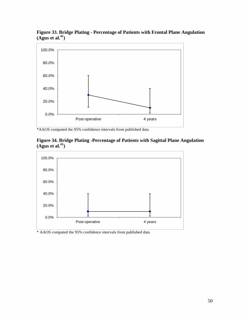
50
Figure 33. Bridge Plating - Percentage of Patients with Frontal Plane Angulation
(Agus et al.41
)
*AAOS computed the 95% confidence intervals from published data
Figure 34. Bridge Plating -Percentage of Patients with Sagittal Plane Angulation
(Agus et al.41
)
* AAOS computed the 95% confidence intervals from published data
0.0%
20.0%
40.0%
60.0%
80.0%
100.0%
Post-operative 4 years
0.0%
20.0%
40.0%
60.0%
80.0%
100.0%
Post-operative 4 years
51
PREVIOUSLY PUBLISHED SYSTEMATIC REVIEWS
A previous systematic review23
concluded that intramedullary nailing resulted in fewer
malunions and adverse events than traction or subsequent casting. This review, however,
was not specific to the population of interest for this recommendation, so we did not
include it in our systematic review.
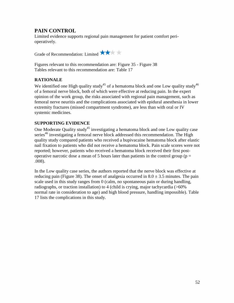
52
PAIN CONTROL Limited evidence supports regional pain management for patient comfort peri-
operatively.
Grade of Recommendation: Limited
Figures relevant to this recommendation are: Figure 35 - Figure 38
Tables relevant to this recommendation are: Table 17
RATIONALE
We identified one High quality study45
of a hematoma block and one Low quality study46
of a femoral nerve block, both of which were effective at reducing pain. In the expert
opinion of the work group, the risks associated with regional pain management, such as
femoral nerve neuritis and the complications associated with epidural anesthesia in lower
extremity fractures (missed compartment syndrome), are less than with oral or IV
systemic medicines.
SUPPORTING EVIDENCE
One Moderate Quality study45
investigating a hematoma block and one Low quality case
series46
investigating a femoral nerve block addressed this recommendation. The High
quality study compared patients who received a bupivacaine hematoma block after elastic
nail fixation to patients who did not receive a hematoma block. Pain scale scores were not
reported; however, patients who received a hematoma block received their first post-
operative narcotic dose a mean of 5 hours later than patients in the control group (p =
.008).
In the Low quality case series, the authors reported that the nerve block was effective at
reducing pain (Figure 38). The onset of analgesia occurred in 8.0 ± 3.5 minutes. The pain
scale used in this study ranges from 0 (calm, no spontaneous pain or during handling,
radiographs, or traction installation) to 4 (child is crying, major tachycardia (>60%
normal rate in consideration to age) and high blood pressure, handling impossible). Table
17 lists the complications in this study.
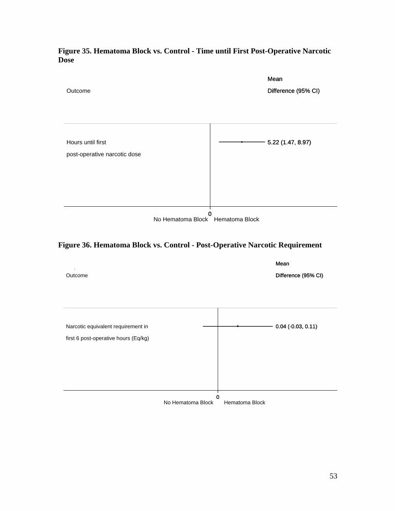
53
Figure 35. Hematoma Block vs. Control - Time until First Post-Operative Narcotic
Dose
Figure 36. Hematoma Block vs. Control - Post-Operative Narcotic Requirement
Hours until first
post-operative narcotic dose
Outcome
5.22 (1.47, 8.97)
Difference (95% CI)
Mean
5.22 (1.47, 8.97)
Difference (95% CI)
Mean
No Hematoma Block Hematoma Block 00
Narcotic equivalent requirement in
first 6 post-operative hours (Eq/kg)
Outcome
0.04 (-0.03, 0.11)
Difference (95% CI)
Mean
0.04 (-0.03, 0.11)
Difference (95% CI)
Mean
No Hematoma Block Hematoma Block 00
54
Figure 37. Hematoma Block vs. Control - Binary Outcomes
Figure 38. Femoral Nerve Block – Pain Relief
*p<.001 (AAOS calculation); AAOS computed the 95% confidence intervals from published data
Table 17. Femoral Nerve Block Complications
Outcome n %Failed block 7%
Femoral artery puncture 7%
ECG changes 0%
Seizure 0%
Respiratory Rate Abnormality 0%
Adverse Sequelae 0%
Neurologic Abnormality (at discharge) 0%
14
3
4
Before
Pain Grade
Received narcotic pain medication
within first 2 post-operative hours
Mild oxygen desaturation in
first 12 post-operative hours
Urinary retention
Outcome
0.15 (0.00, 1.43)
0.00 (0.00, 66.00)
1000.00 (0.04, 1000.00)
Odds Ratio (95% CI)
0.15 (0.00, 1.43)
0.00 (0.00, 66.00)
1000.00 (0.04, 1000.00)
Odds Ratio (95% CI)
Hematoma Block No Hematoma Block 11
0
1
2
3
4
Before After
Pa
in G
rad
e

55
PREVIOUSLY PUBLISHED SYSTEMATIC REVIEWS
A previous systematic review 47
concluded that femoral nerve block effectively reduces
pain in children with femoral shaft fractures. Although the stated subject of this
systematic review was children, two of the three included studies included adults.
Therefore, we did not include it in our systematic review.

56
WATERPROOF CASTING Limited evidence supports waterproof cast liners for spica casts are an option for use in
children diagnosed with pediatric diaphyseal femur fractures.
Grade of Recommendation: Limited
Figures relevant to this recommendation are: Figure 39
RATIONALE
Waterproof cast liners are often used when applying a spica cast for the management of
femur fractures in children in order to improve ease of care.
We identified one High Quality study48
that addressed the use of waterproof liners in
spica casts. Use of a waterproof liner was associated with significantly fewer skin
problems and unexpected cast changes. However, in this study spica casts were used for
the management of developmental dysplasia of the hip, not specifically for diaphyseal
femur fractures. In addition, other outcomes such as impact on family and financial
considerations were not studied. Waterproof liners may make cast care easier for the
family, thus decreasing the overall impact of treatment on family functioning. Cast liners
add increased cost to overall management. Nevertheless, the patient ages were similar to
the patient ages for spica cast management of diaphyseal femur fractures and the findings
should be able to be extrapolated. The overall benefit in terms of skin problems,
unplanned cast changes, and ease of care for the family likely obviates the increased costs
from the use of waterproof cast liners in the expert opinion of the physician work group.
SUPPORTING EVIDENCE
One High Quality study48
addressed this recommendation. In this study, however, hip
spica casts were applied to treat conditions other than diaphyseal femur fractures, such as
developmental dysplasia. The study compared the use of hip spica casts with and without
a waterproof liner. The use of a waterproof liner was associated with significantly fewer
occurrences of skin excoriation and unplanned cast changes.

57
Figure 39. Waterproof Liner vs. No Waterproof Liner
Future Research The quality of scientific data regarding the management of femur fractures in children is
clearly lacking. Controversy exists regarding the optimal management of pediatric femur
fractures. A multitude of treatment options exist including Pavlik harness, spica casting,
traction, external fixation, flexible intramedullary nailing, rigid intramedullary nailing,
and bridge plating. Properly designed randomized clinical trials comparing treatment
options are necessary to determine optimal treatment. These trials would benefit from
being multicenter trials in terms of accrual of patients and external validity.
Specific trials which would be helpful include:
1. Delayed spica casting versus immediate spica casting for femur fractures in
children 6 months – 6 years old.
2. Flexible intramedullary nailing versus immediate spica casting for femur fractures
in children 5 and 6 years old, and even children younger than 5-6 years of age.
3. External fixation versus bridge plating versus elastic nails versus rigid
trochanteric nails for length unstable femur fractures in children 6 years old –
skeletal maturity.
4. Flexible intramedullary nailing versus rigid intramedullary nailing versus bridge
plating for femur fractures in children 6 years old – skeletal maturity.
Intermediate outcome measures are often used in studies regarding pediatric femur
fractures such as radiographic parameters. Functional outcome measures and later
development of osteoarthritis are difficult to measure and have a long time course.
However, the relationship between commonly accepted radiographic measures of
malunion and functional outcome or later development of problems is not clear. Further
research to validate accepted radiographic standards of malunion would be extremely
valuable. Also the inclusion of family function outcomes may improve recommendations
Skin Excoriation
Unplanned Cast Changes (per cast)
Outcome
0.06 (0.01, 0.54)
0.20 (0.04, 0.93)
OR (95% CI)
1/36
2/70
Liner
Events,
11/36
10/77
Liner
Events, No
0.06 (0.01, 0.54)
0.20 (0.04, 0.93)
OR (95% CI)
1/36
2/70
Liner
Events,
Waterproof Liner No Liner 11
58
for those younger patients that may either get intramedullary nailing versus immediate
spica casting.

59
V. APPENDIXES

60
APPENDIX I AAOS BODIES THAT APPROVED THE 2015 GUIDELINE REISSUE
This final guideline draft must be approved by the AAOS Committee on Evidence Based
Quality and Value, the AAOS Council on Research and Quality, and the AAOS Board of
Directors. These decision-making bodies are invited to provide comments during the
review process for consideration by the work group but are not designated to modify the
contents of the guideline. Their charge is to approve or reject its publication by majority
vote.
Committee on Evidence Based Quality and Value - April 18, 2015
The committee on Evidence Based Quality and Value (EBQV) consists of twenty AAOS
members who implement evidence-based quality initiatives such as clinical practice
guidelines (CPGs) and appropriate use criteria (AUCs). They also oversee the
dissemination of related educational materials and promote the utilization of orthopaedic
value products by the Academy’s leadership and its members.
Council on Research and Quality – May 8, 2015
The Council on Research and Quality promotes ethically and scientifically sound clinical
and translational research to sustain patient care in musculoskeletal disorders. The
Council also serves as the primary resource for educating its members, the public, and
public policy makers regarding evidenced-based medical practice, orthopaedic devices
and biologics, regulatory pathways and standards development, patient safety,
occupational health, technology assessment, and other related important errors.
The Council is comprised of the chairs of the committees on Biological Implants,
Biomedical Engineering, Occupational Health and Workers’ Compensation, Patient
Safety, Research Development, U.S. Bone and Joint Decade, and chair and Appropriate
Use Criteria and Clinical Practice Guideline section leaders of the Evidence Based
Quality and Value committee. Also on the Council are the second vice-president, three
members at large, and representatives of the Diversity Advisory Board, Women's Health
Issues Advisory Board, Board of Specialty Societies (BOS), Board of Councilors (BOC),
Communications Cabinet, Orthopaedic Research Society (ORS), Orthopedic Research
and Education Foundation (OREF).
Board of Directors – June 5, 2015
The 17 member Board of Directors manage the affairs of the AAOS, set policy, and
oversee the Strategic Plan.
The 2009 CPG was approved by the AAOS Guidelines and Technology Oversight Committee, the
AAOS Evidence Based Practice Committee, the AAOS Council on Research, Quality Assessment
and Technology, and the AAOS Board of Directors

61
APPENDIX II
LITERATURE SEARCHES
The search for eligible literature began with a search of the following databases on May
8, 2008, and updated on October 6, 2008, and November 27, 2013:
PubMed (from 1966 through November 27, 2013)
EMBASE (from 1966 through November 27, 2013)
CINAHL (from 1982 through November 27, 2013)
The Cochrane Central Register of Controlled Trials (through November 27,
2013)
The original search (after removal of duplicates) yielded 1181 articles, of which 274 were
retrieved and evaluated. The full search strategies are listed below. The updated search
conducted in November 2013 yielded an additional 384 articles published after the
original search.
All literature searches were supplemented with manual screening of bibliographies in
publications accepted for inclusion into the evidence base. In addition, the bibliographies
of recent systematic reviews and other review articles were searched for potentially
relevant citations.
GENERAL SEARCH
PubMed was searched using the following strategy:
(diaphyseal OR diaphysis OR shaft OR diaphysial) AND fracture AND (femur OR
femoral OR thigh) NOT "comment"[Publication Type] NOT "editorial"[Publication
Type] NOT "letter"[Publication Type] NOT "Addresses"[Publication Type] NOT
"News"[Publication Type] NOT "Newspaper Article"[Publication Type] AND
(("1966/1/1"[EDat]:"2008/10/01"[EDat]) AND (Humans[Mesh]) AND (English[lang])
AND ((infant[MeSH] OR child[MeSH] OR adolescent[MeSH])) )
EMBASE was searched using the following strategy:
(diaphyseal OR ('diaphysis'/exp OR 'diaphysis') OR shaft OR diaphysial) AND
('fracture'/exp OR 'fracture') AND (('femur'/exp OR 'femur') OR femoral OR ('thigh'/exp
OR 'thigh')) AND ([article]/lim OR [review]/lim) AND [english]/lim AND [humans]/lim
AND ([infant]/lim OR [child]/lim OR [adolescent]/lim) AND [embase]/lim AND [1966-
2008]/py
CINAHL was searched using the following strategy:
(diaphyseal OR diaphysis OR shaft OR diaphysial) AND fracture AND (femur OR
femoral OR thigh)
Cochrane Central Register of Controlled Trials was searched using the following
strategy:
(diaphyseal OR diaphysis OR shaft OR diaphysial) AND fracture AND (femur OR
femoral OR thigh)
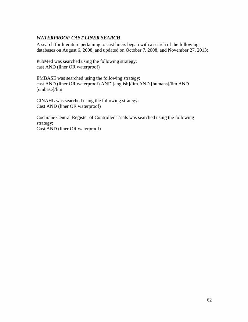
62
WATERPROOF CAST LINER SEARCH
A search for literature pertaining to cast liners began with a search of the following
databases on August 6, 2008, and updated on October 7, 2008, and November 27, 2013:
PubMed was searched using the following strategy:
cast AND (liner OR waterproof)
EMBASE was searched using the following strategy:
cast AND (liner OR waterproof) AND [english]/lim AND [humans]/lim AND
[embase]/lim
CINAHL was searched using the following strategy:
Cast AND (liner OR waterproof)
Cochrane Central Register of Controlled Trials was searched using the following
strategy:
Cast AND (liner OR waterproof)
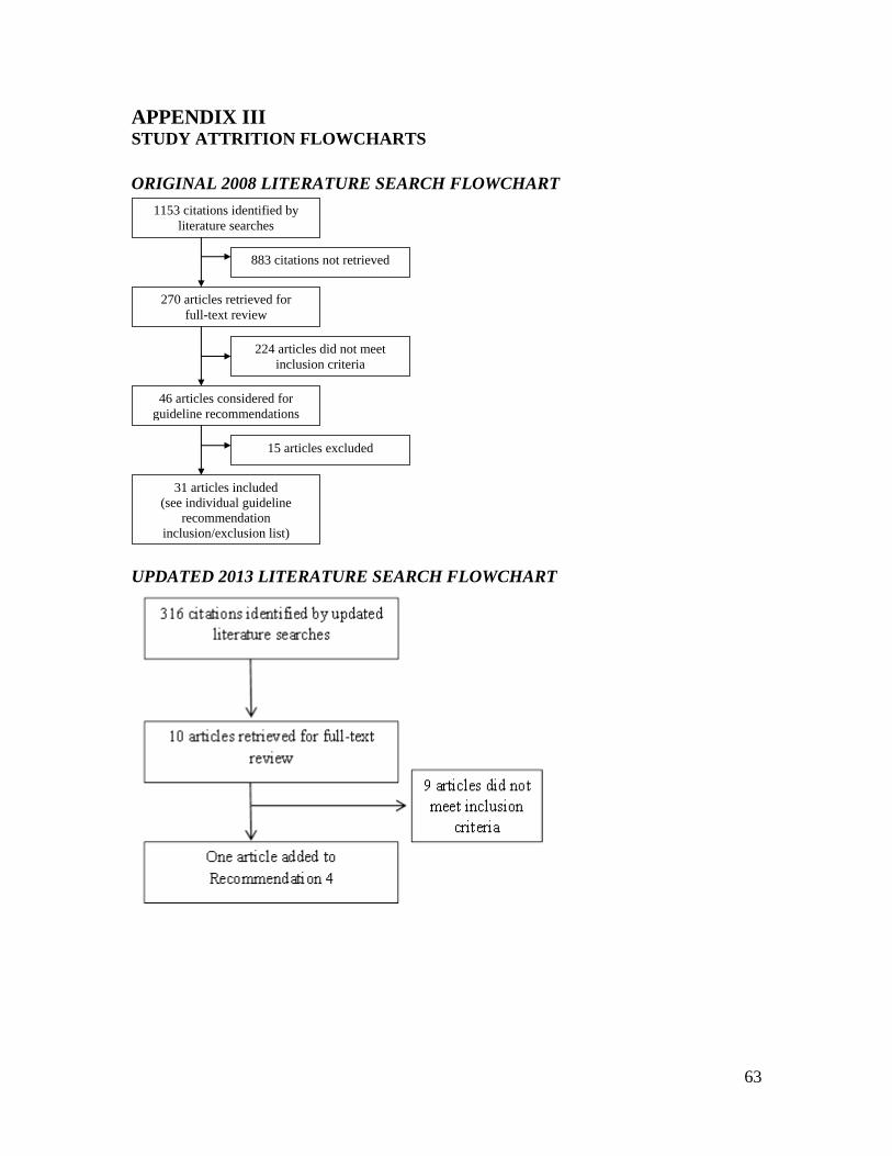
63
APPENDIX III STUDY ATTRITION FLOWCHARTS
ORIGINAL 2008 LITERATURE SEARCH FLOWCHART
UPDATED 2013 LITERATURE SEARCH FLOWCHART
1153 citations identified by
literature searches
270 articles retrieved for
full-text review
883 citations not retrieved
224 articles did not meet
inclusion criteria
46 articles considered for
guideline recommendations
31 articles included
(see individual guideline
recommendation
inclusion/exclusion list)
15 articles excluded

64
WATERPROOF CAST LINER SEARCH FLOWCHART
48 citations identified by
literature searches
5 articles retrieved for
full-text review
43 citations not retrieved
4 articles did not meet
inclusion criteria
1 article included

65
APPENDIX IV DATA EXTRACTION ELEMENTS
The data elements below were extracted into electronic forms in Microsoft® Excel from
published studies. The extracted information includes:
Study Characteristics (for all relevant outcomes in a study)
methods of randomization and allocation
use of blinding (patient, caregiver, evaluator)
funding source/conflict of interest
duration of the study
number of subjects and follow-up percentage
experimental and control groups
a priori power analysis
Patient Characteristics (for all treatment groups in a study)
patient inclusion/exclusion criteria
age
weight
surgical complications
adverse events
Results (for all relevant outcomes in a study)
duration at which outcome measure was evaluated
mean value of statistic reported (for dichotomous results)
mean value of measure and value of dispersion (for continuous results)
statistical test p-value
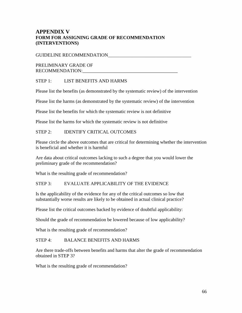
66
APPENDIX V FORM FOR ASSIGNING GRADE OF RECOMMENDATION
(INTERVENTIONS)
GUIDELINE RECOMMENDATION___________________________________
PRELIMINARY GRADE OF
RECOMMENDATION:________________________________________
STEP 1: LIST BENEFITS AND HARMS
Please list the benefits (as demonstrated by the systematic review) of the intervention
Please list the harms (as demonstrated by the systematic review) of the intervention
Please list the benefits for which the systematic review is not definitive
Please list the harms for which the systematic review is not definitive
STEP 2: IDENTIFY CRITICAL OUTCOMES
Please circle the above outcomes that are critical for determining whether the intervention
is beneficial and whether it is harmful
Are data about critical outcomes lacking to such a degree that you would lower the
preliminary grade of the recommendation?
What is the resulting grade of recommendation?
STEP 3: EVALUATE APPLICABILITY OF THE EVIDENCE
Is the applicability of the evidence for any of the critical outcomes so low that
substantially worse results are likely to be obtained in actual clinical practice?
Please list the critical outcomes backed by evidence of doubtful applicability:
Should the grade of recommendation be lowered because of low applicability?
What is the resulting grade of recommendation?
STEP 4: BALANCE BENEFITS AND HARMS
Are there trade-offs between benefits and harms that alter the grade of recommendation
obtained in STEP 3?
What is the resulting grade of recommendation?

67
STEP 5 CONSIDER STRENGTH OF EVIDENCE
Does the strength of the existing evidence alter the grade of recommendation obtained in
STEP 4?
What is the resulting grade of recommendation?
NOTE: Because we are not performing a formal cost analyses, you should only consider
costs if their impact is substantial.

68
APPENDIX VI PEER REVIEW PANEL FOR THE ORIGINAL 2009 GUIDELINE
Participation in the AAOS peer review process does not constitute an endorsement
of this guideline by the participating organization.
Peer review of the draft guideline is completed by an outside Peer Review Panel. Outside
peer reviewers are solicited for each AAOS guideline and consist of experts in the
guideline’s topic area. These experts represent professional societies other than AAOS
and are nominated by the guideline Work Group prior to beginning work on the
guideline. For this guideline, five outside peer review organizations were invited to
review the draft guideline and all supporting documentation. All five societies
participated in the review of the Treatment of Pediatric Diaphyseal Femur Fractures
guideline draft and four consented to be listed as a peer review organization in this
appendix. One organization did not give explicit consent that the organization name
could be listed in this publication. The organizations that reviewed the document and
consented to publication are listed below:
American Academy of Pediatrics, Section on Orthopaedics
European Paediatric Orthopaedic Society
Orthopaedic Trauma Association
American Osteopathic Academy of Orthopedics
Individuals who participated in the peer review of this document and gave their consent
to be listed as reviewers of this document are:
James Breivis, MD, San Francisco, CA
Blair C. Filler MD, Los Angeles, CA
J. Eric Gordon MD, St. Louis MS
Michael Heggeness MD, Houston, TX
Harvey Insler MD, Erie, PA
John Kirkpatrick MD, Jacksonville, FL
Pierre Lascombes MD, Nancy France
David A. Podenswa MD, Dallas, TX
Charles A Reitman MD, Houston, TX
Debra K. Spatz, D.O, Prince Frederick, MD
Again, participation in the AAOS guideline peer review process does not constitute
an endorsement of the guideline by the participating organizations or the
individuals listed above.

69
PUBLIC COMMENTARY FOR ORIGINAL 2009 GUIDELINE
A period of public commentary follows the peer review of the draft guideline. If
significant non-editorial changes are made to the document as a result of public
commentary, these changes are also documented and forwarded to the AAOS bodies that
approve the final guideline. Public commentators who gave explicit consent to be listed
in this document include the following:
Participation in the AAOS guideline public commentary review process does not
constitute an endorsement of the guideline by the participating organizations or the
individual listed nor does it is any way imply the reviewer supports this document.
Jeffrey Anglen MD, Indianapolis, IN
Howard R. Epps MD, Houston TX
M. Bradford Henley MD MBA, Seattle WA
William C McMaster MD, Orange, CA
Jack R. Steel MD, Huntington WV
J. Mark Melhorn MD, Wichita, KS on behalf of:
The American Academy of Disability Evaluating Physicians

70
APPENDIX VII STRUCTURED PEER REVIEW FORM
Reviewer Information:
Name of Reviewer_________________________________________
Address_________________________________________________
City___________________ State_________________ Zip Code___________
Phone _____________________Fax ________________________
E-mail_______________________
Specialty Area/Discipline: _______________________________________
Work setting: _________________________________________________
Credentials: _________________________________________________
May we list you as a Peer Reviewer in the final Guidelines? Yes No
Are you reviewing this guideline as Yes No
a representative of a professional society?
If yes, may we list your society as a reviewer Yes No
of this guideline?
Reviewer Instructions
Please read and review this Draft Clinical Practice Guideline and its associated Technical
Report with particular focus on your area of expertise. Your responses are confidential
and will be used only to assess the validity, clarity, and accuracy of the interpretation of
the evidence. If applicable, please specify the draft page and line numbers in your
comments. Please feel free to also comment on the overall structure and content of the
guideline and Technical Report.
If you need more space than is provided, please attach additional pages.
Please complete and return this form electronically to [email protected] or fax the form
back to Jan Weis at (847) 823-9769.
Thank you in advance for your time in completing this form and giving us your feedback.
We value your input and greatly appreciate your efforts. Please send the completed form
and comments by Month, Day, Year

71

72
COMMENTS
Please provide a brief explanation of both your positive and negative answers in the
preceding section. If applicable, please specify the draft page and line numbers in your
comments. Please feel free to also comment on the overall structure and content of the
guideline and Technical Report
OVERALL ASSESSMENT
Would you recommend these guidelines for use in practice? (check one)
Strongly recommend _______
Recommend (with provisions or alterations) _______
Would not recommend _______
Unsure _______
COMMENTS:
Please provide the reason(s) for your recommendation.

73
APPENDIX VIII INTERPRETING THE FOREST PLOTS
49
Throughout the guideline we use descriptive diagrams or forest plots to present data from
studies comparing the differences in outcomes between two treatment groups. In this
guideline there are no meta-analyses (combining results of multiple studies into a single
estimate of overall effect), so each point and corresponding horizontal line on a sample
plot should be viewed independently. In the example below, the odds ratio is the effect
measure used to depict differences in outcomes between the two treatment groups of a
study. In other forest plots, the point can refer to other summary measures (such as the
mean difference or relative risk). The horizontal line running through each point
represents the 95% confidence interval for that point. In this graph, the solid vertical line
represents “no effect” where the Odds Ratio, OR, is equal to one. When mean differences
are portrayed, the vertical line of no effect is at zero.
For example, in the figure below the odds of a patient experiencing Outcome 1 are 5.9
times greater for patients who received Treatment B than for patients who received
Treatment A.. This result is statistically significant because the 95% Confidence Interval
does not cross the “no effect” line. In general, the plots are arranged such that results to
the left of the “no effect” line favor Treatment A while results to the right favor
Treatment B. In the example below, the odds ratio for Outcome 1 favors Treatment B,
the odds ratio for Outcome 3 favors Treatment A, and the odds ratio for Outcome 2 does
not favor either treatment because the 95% CI crosses the “no effect” line (i.e. the
difference is not statistically significant).
Sample Plot
Outcome 1
Outcome 2
Outcome 3
Outcome
5.90 (3.38, 10.29)
0.72 (0.43, 1.19)
0.11 (0.06, 0.20)
Odds Ratio (95% CI)
5.90 (3.38, 10.29)
0.72 (0.43, 1.19)
0.11 (0.06, 0.20)
Group A Group B 1 1
Outcome 1
Outcome 2
Outcome 3
Outcome
5.90 (3.38, 10.29)
0.72 (0.43, 1.19)
0.11 (0.06, 0.20)
Odds Ratio (95% CI)
5.90 (3.38, 10.29)
0.72 (0.43, 1.19)
0.11 (0.06, 0.20)
Treatment A Treatment B 1 1

74
DESCRIPTION OF SYMBOLS USED IN FIGURES AND TABLES
Symbol Description
OR
Odds Ratio = The odds in Group B divided by the odds in Group A, where the odds is
the probability of the outcome occurring divided by the probability of the outcome not
occurring.
95% CI
95% Confidence Interval = A measure of uncertainty of the point estimate: if the trial
were repeated an infinite number of times, then the 95% CI calculated for each trial
would contain the true effect 95% of the time.
An arrow in a forest plot indicates that the 95% confidence interval continues beyond
the range of the graph.
○ An open circle in a Summary of Evidence Table indicates that the result is not
statistically significant.
● fn
A filled-in circle in a Summary of Evidence Table indicates that the result is
statistically significant in favor of the listed treatment (in this example, in favor of fn
= flexible nails)
75
APPENDIX IX CONFLICT OF INTEREST
All members of the AAOS work group disclosed their conflicts of interest prior to the
development of the recommendations for this guideline. Conflicts of interest are
disclosed in writing with the American Academy of Orthopaedic Surgeons via a private
on-line reporting database and also verbally at the recommendation approval meeting.
Members of all AAOS Work Groups are required to disclose their conflicts of interest at
the same level and depth of detail as the AAOS Board of Directors.
AAOS DISCLOSURE PROGRAM INFORMATION
R Dale Blasier, MD (Little Rock, AR): 4 (Synthes); 7 (Synthes). Submitted on:
04/23/2008.
Michael J Goldberg, MD: 2 (Journal of Pediatric Orthopedics; Journal of Children's
Orthopaedics). Submitted on: 12/11/2007.
Mininder S Kocher, MD (Boston, MA): 1 (Pediatric Orthopaedic Society of North
America); 5A (Biomet; Regen Biologics; Smith & Nephew); 7 (CONMED Linvatec); 9
(Saunders/Mosby-Elsevier). Submitted on: 05/27/2008.
Scott J Luhmann, MD (Saint Louis, MO): 4 (Medtronic Sofamor Danek; Stryker); 5A
(Medtronic Sofamor Danek); 7 (Medtronic Sofamor Danek). Submitted on: 08/05/2008.
Travis Matheney, MD (Boston, MA): (n). Submitted on: 10/29/2008
Charles T Mehlman, DO (Cincinnati, OH): 2 (Journal of Bone and Joint Surgery -
American; Journal of Orthopaedics and Traumatology; Journal of Pediatric Orthopedics;
Saunders/Mosby-Elsevier; Wolters Kluwer Health - Lippincott Williams & Wilkins;
eMedicine (Orthopaedic Surgery); Journal Children's Orthopaedics (EPOS); The
Orthopod (JAOAO); The Spine Journal (NASS)); 5B (Stryker); 7 (DePuy, A Johnson &
Johnson Company; Medtronic Sofamor Danek; National Institutes of Health (NIAMS &
NICHD); Synthes; University Cincinnati); 8 (Eli Lilly; Zimmer). Submitted on:
04/08/2008.
James O Sanders, MD (Rochester, NY): 7 (Medtronic Sofamor Danek; K2M); 8
(Abbott; Biomedical Enterprises). Submitted on: 07/30/2008.
David M Scher, MD (New York, NY): 1 (American Academy for Cerebral Palsy and
Developmental Medicine; Pediatric Orthopaedic Club of New York); 2 (Hospital for
Special Surgery Journal). Submitted on: 03/28/2008.
Ernest L Sink, MD (Aurora, CO): 1 (Pediatric Orthopaedic Society of North America);
2 (Orthopedics); 4 (Biomet); 5B (Biomet). Submitted on: 04/15/2008.
76
William Charles Watters III, MD: 1 (North American Spine Society; Work Loss Data
Institute); 2 (The Spine Journal); 5A (Stryker; Intrinsic Therapeutics; MeKessen Health
Care Solutions). Submitted on: 10/09/2007 at 08:09 PM and last confirmed as accurate on
04/23/2008.
Disclosure Items: (n) = Respondent answered 'No' to all items indicating no
conflicts. 1=Board member/owner/officer/committee appointments; 2=
Medical/Orthopaedic Publications; 3= Royalties; 4= Speakers bureau/paid
presentations;5A= Paid consultant; 5B= Unpaid consultant; 6= Research or institutional
support from a publisher; 7= Research or institutional support from a company or
supplier; 8= Stock or Stock Options; 9= Other financial/material support from a
publisher; 10= Other financial/material support from a company or supplier.

77
APPENDIX X REFERENCES
(1) Hinton RY, Lincoln A, Crockett MM, Sponseller P, Smith G. Fractures of the
femoral shaft in children. Incidence, mechanisms, and sociodemographic risk
factors. J Bone Joint Surg Am. 1999;81:500-509.
(2) Rewers A, Hedegaard H, Lezotte D et al. Childhood femur fractures, associated
injuries, and sociodemographic risk factors: a population-based study. Pediatrics.
2005;115:e543-e552.
(3) Sahlin Y. Occurrence of fractures in a defined population: a 1-year study. Injury.
1990;21:158-160.
(4) McCartney D, Hinton A, Heinrich SD. Operative stabilization of pediatric femur
fractures. Orthop Clin North Am. 1994;25:635-650.
(5) Coyte PC, Bronskill SE, Hirji ZZ, Daigle-Takacs G, Trerise BS, Wright JG.
Economic evaluation of 2 treatments for pediatric femoral shaft fractures. Clin
Orthop Relat Res. 1997;205-215.
(6) Morton L, Bridgman S, Dwyer J, Theis J. Interventions for treating femoral shaft
fractures in children and adolescents (Protocol). Cochrane Database of Systematic
Reviews. 2002;1-8.
(7) Femoral Shaft Fractures Guideline Team. Evidence-based care guideline for
medical management of Femoral Shaft Fractures. Guideline 22 , 1-19. 2006.
Cincinnati Children's Hospital Medical Center.
Ref Type: Electronic Citation
(8) Cook D.J., Mulrow CD, Haynes RB. Systematic Reviews:synthesis of best
evidence for clinical decisions. Ann Intern Med. 1997;126:376-380.
(9) Mulrow C.D., Cook D.J., Davidoff F. Systematic Reviews:critical links in the
great chain of evidence. Ann Intern Med. 1997;126:389-391.
(10) Bucher H.C., Guyatt G.H., Cook D.J., Holbrook A., McAlister F.A. Users' Guides
to the Medical Literature. JAMA. 1999;282.
(11) Robinson KA, Dickerson K. Development of a highly sensitive search strategy for
the retrieval of reports of controlled trials using PubMed. Int Journal of
Epidemiology. 2002;31:150-153.
(12) GRADE Working Group. Grading quality of evidence and strength of
recommendations. BMJ. 2004;328.
(13) Hozo SP, Djulbegovic B, Hozo I. Estimating the mean and variance from the
median, range, and the size of a sample. BMC Med Res Methodol. 2005;5:13.

78
(14) Wilson E. Probable inference, the law of succession, and statistical inference.
Journal of the American Statistical Association. 1927;22:209-212.
(15) Akbarnia BM, Torg JSM, Kirkpatrick JM, Sussman SM. Manifestations of the
Battered-Child Syndrome. The Journal of Bone and Joint Surgery. 1974;56-
A:1159-1166.
(16) Miettinen H, Makela EA, Vainio J. The incidence and causative factors
responsible for femoral shaft fractures in children. Ann Chir Gynaecol.
1991;80:392-395.
(17) Podeszwa DA, Mooney JF, III, Cramer KE, Mendelow MJ. Comparison of Pavlik
harness application and immediate spica casting for femur fractures in infants. J
Pediatr Orthop. 2004;24:460-462.
(18) Stannard JP, Christensen KP, Wilkins KE. Femur fractures in infants: a new
therapeutic approach. J Pediatr Orthop. 1995;15:461-466.
(19) Rasool MN, Govender S, Naidoo KS. Treatment of femoral shaft fractures in
children by early spica casting. S Afr Med J. 1989;76:96-99.
(20) Burton VW, Fordyce AJ. Immobilization of femoral shaft fractures in children
aged 2-10 years. Injury. 1972;4:47-53.
(21) Wright JG. The treatment of femoral shaft fractures in children: a systematic
overview and critical appraisal of the literature. Can J Surg. 2000;43:180-189.
(22) Wright JG, Wang EE, Owen JL et al. Treatments for paediatric femoral fractures:
a randomised trial. Lancet. 2005;365:1153-1158.
(23) Poolman RW, Kocher MS, Bhandari M. Pediatric femoral fractures: a systematic
review of 2422 cases. J Orthop Trauma. 2006;20:648-654.
(24) Flynn JM, Luedtke LM, Ganley TJ et al. Comparison of titanium elastic nails with
traction and a spica cast to treat femoral fractures in children. J Bone Joint Surg
Am. 2004;86-A:770-777.
(25) Buechsenschuetz KE, Mehlman CT, Shaw KJ, Crawford AH, Immerman EB.
Femoral shaft fractures in children: traction and casting versus elastic stable
intramedullary nailing. J Trauma. 2002;53:914-921.
(26) Barlas K, Beg H. Flexible intramedullary nailing versus external fixation of
paediatric femoral fractures. Acta Orthop Belg. 2006;72:159-163.
(27) Moroz LA, Launay F, Kocher MS et al. Titanium elastic nailing of fractures of
the femur in children. Predictors of complications and poor outcome. J Bone Joint
Surg Br. 2006;88:1361-1366.
79
(28) Domb BG, Sponseller PD, Ain M, Miller NH. Comparison of dynamic versus
static external fixation for pediatric femur fractures. J Pediatr Orthop.
2002;22:428-430.
(29) Mehdinasab SA, Nejad SAM, Sarrafan N. Short term outcome of treatment of
femoral shaft fractures in children by two methods: Traction plus casting, versus
intramedullary pin fixation - A comparative study. Pak J Med Sci. 2008;24:147-
151.
(30) Ali M, Raza A. Union and complications after Thomas splint and early hip spica
for femoral shaft fractures in children. J Coll Physicians Surg Pak. 2005;15:799-
801.
(31) Curtis JF, Killian JT, Alonso JE. Improved treatment of femoral shaft fractures in
children utilizing the pontoon spica cast: a long-term follow-up. J Pediatr Orthop.
1995;15:36-40.
(32) Nork SE, Hoffinger SA. Skeletal traction versus external fixation for pediatric
femoral shaft fractures: a comparison of hospital costs and charges. J Orthop
Trauma. 1998;12:563-568.
(33) Song HR, Oh CW, Shin HD et al. Treatment of femoral shaft fractures in young
children: comparison between conservative treatment and retrograde flexible
nailing. J Pediatr Orthop B. 2004;13:275-280.
(34) Hedin H, Borgquist L, Larsson S. A cost analysis of three methods of treating
femoral shaft fractures in children: a comparison of traction in hospital, traction in
hospital/home and external fixation. Acta Orthop Scand. 2004;75:241-248.
(35) Rasit AH, Mohammad AW, Pan KL. The pattern of femoral diaphyseal fractures
in children admitted in Sarawak General Hospital. Med J Malaysia. 2006;61
Suppl A:79-82.
(36) Sturdee SW, Templeton PA, Dahabreh Z, Cullen E, Giannoudis PV. Femoral
fractures in children, is early interventional treatment beneficial? Injury.
2007;38:937-944.
(37) Wall EJ, Jain V, Vora V, Mehlman CT, Crawford AH. Complications of titanium
and stainless steel elastic nail fixation of pediatric femoral fractures. J Bone Joint
Surg Am. 2008;90:1305-1313.
(38) Buford D, Jr., Christensen K, Weatherall P. Intramedullary nailing of femoral
fractures in adolescents. Clin Orthop Relat Res. 1998;85-89.
(39) Herndon WA, Mahnken RF, Yngve DA, Sullivan JA. Management of femoral
shaft fractures in the adolescent. J Pediatr Orthop. 1989;9:29-32.

80
(40) Kanellopoulos AD, Yiannakopoulos CK, Soucacos PN. Closed, locked
intramedullary nailing of pediatric femoral shaft fractures through the tip of the
greater trochanter. J Trauma. 2006;60:217-222.
(41) Agus H, Kalenderer O, Eryanilmaz G, Omeroglu H. Biological internal fixation
of comminuted femur shaft fractures by bridge plating in children. J Pediatr
Orthop. 2003;23:184-189.
(42) Mazda K, Khairouni A, Pennecot GF, Bensahel H. Closed flexible intramedullary
nailing of the femoral shaft fractures in children. J Pediatr Orthop B. 1997;6:198-
202.
(43) Cramer KE, Tornetta P, III, Spero CR, Alter S, Miraliakbar H, Teefey J. Ender
rod fixation of femoral shaft fractures in children. Clin Orthop Relat Res.
2000;119-123.
(44) Hedin H, Hjorth K, Rehnberg L, Larsson S. External fixation of displaced femoral
shaft fractures in children: a consecutive study of 98 fractures. J Orthop Trauma.
2003;17:250-256.
(45) Herrera JA, Wall EJ, Foad SL. Hematoma Block Reduces Narcotic Pain
Medication after Femoral Elastic Nailing in Children. J Pediatr Orthop.
2004;24:254-256.
(46) Ronchi L, Rosenbaum D, Athouel A et al. Femoral nerve blockade in children
using bupivacaine. Anesthesiology. 1989;70:622-624.
(47) Williams R, Wallman P. Towards evidence based emergency medicine: best
BETs from the Manchester Royal Infirmary. Regional anaesthesia for femoral
shaft fractures in children. J Accid Emerg Med. 2000;17:283-284.
(48) Wolff CR, James P. The prevention of skin excoriation under children's hip spica
casts using the goretex pantaloon. J Pediatr Orthop. 1995;15:386-388.
(49) Oleckno W.A. Epidemiology Concepts and Methods. Long Grove, IL.: Waveland
Press, Inc.; 2008.
(50) Shemshaki,H.R., Mousavi,H., Salehi,G., Eshaghi,M.A. Titanium elastic nailing
versus hip spica cast in treatment of femoral-shaft fractures in children. J Orthop
Traumatol. 2011/3; 1: 45-48

81
INCLUDED ARTICLES
Agus H, Kalenderer O, Eryanilmaz G, Omeroglu H. Biological internal fixation of
comminuted femur shaft fractures by bridge plating in children. J Pediatr Orthop 2003
March;23(2):184-9.
Ali M, Raza A. Union and complications after Thomas splint and early hip spica for
femoral shaft fractures in children. J Coll Physicians Surg Pak 2005
December;15(12):799-801.
Barlas K, Beg H. Flexible intramedullary nailing versus external fixation of paediatric
femoral fractures. Acta Orthop Belg 2006 April;72(2):159-63.
Buechsenschuetz KE, Mehlman CT, Shaw KJ, Crawford AH, Immerman EB. Femoral
shaft fractures in children: traction and casting versus elastic stable intramedullary
nailing. J Trauma 2002 November;53(5):914-21.
Buford D, Jr., Christensen K, Weatherall P. Intramedullary nailing of femoral fractures in
adolescents. Clin Orthop Relat Res 1998 May;(350):85-9.
Burton VW, Fordyce AJ. Immobilization of femoral shaft fractures in children aged 2-10
years. Injury 1972 August;4(1):47-53.
Cramer KE, Tornetta P, III, Spero CR, Alter S, Miraliakbar H, Teefey J. Ender rod
fixation of femoral shaft fractures in children. Clin Orthop Relat Res 2000
July;(376):119-23.
Curtis JF, Killian JT, Alonso JE. Improved treatment of femoral shaft fractures in
children utilizing the pontoon spica cast: a long-term follow-up. J Pediatr Orthop 1995
January;15(1):36-40.
Domb BG, Sponseller PD, Ain M, Miller NH. Comparison of dynamic versus static
external fixation for pediatric femur fractures. J Pediatr Orthop 2002 July;22(4):428-30.
Flynn JM, Luedtke LM, Ganley TJ et al. Comparison of titanium elastic nails with
traction and a spica cast to treat femoral fractures in children. J Bone Joint Surg Am 2004
April;86-A(4):770-7.
Hedin H, Hjorth K, Rehnberg L, Larsson S. External fixation of displaced femoral shaft
fractures in children: a consecutive study of 98 fractures. J Orthop Trauma 2003
April;17(4):250-6.
Hedin H, Borgquist L, Larsson S. A cost analysis of three methods of treating femoral
shaft fractures in children: a comparison of traction in hospital, traction in hospital/home
and external fixation. Acta Orthop Scand 2004 June;75(3):241-8.
Herndon WA, Mahnken RF, Yngve DA, Sullivan JA. Management of femoral shaft
fractures in the adolescent. J Pediatr Orthop 1989 January;9(1):29-32.

82
Herrera JA, Wall EJ, Foad SL. Hematoma Block Reduces Narcotic Pain Medication after
Femoral Elastic Nailing in Children. J Pediatr Orthop 2004;24(3):254-6.
Hinton RY, Lincoln A, Crockett MM, Sponseller P, Smith G. Fractures of the femoral
shaft in children. Incidence, mechanisms, and sociodemographic risk factors. J Bone
Joint Surg Am 1999 April;81(4):500-9.
Kanellopoulos AD, Yiannakopoulos CK, Soucacos PN. Closed, locked intramedullary
nailing of pediatric femoral shaft fractures through the tip of the greater trochanter. J
Trauma 2006 January;60(1):217-22.
Mazda K, Khairouni A, Pennecot GF, Bensahel H. Closed flexible intramedullary nailing
of the femoral shaft fractures in children. J Pediatr Orthop B 1997 July;6(3):198-202.
Mehdinasab SA, Nejad SAM, Sarrafan N. Short term outcome of treatment of femoral
shaft fractures in children by two methods: Traction plus casting, versus intramedullary
pin fixation - A comparative study. Pak J Med Sci 2008;24(1):147-51.
Miettinen H, Makela EA, Vainio J. The incidence and causative factors responsible for
femoral shaft fractures in children. Ann Chir Gynaecol 1991;80(4):392-5.
Moroz LA, Launay F, Kocher MS et al. Titanium elastic nailing of fractures of the femur
in children. Predictors of complications and poor outcome. J Bone Joint Surg Br 2006
October;88(10):1361-6.
Nork SE, Hoffinger SA. Skeletal traction versus external fixation for pediatric femoral
shaft fractures: a comparison of hospital costs and charges. J Orthop Trauma 1998
November;12(8):563-8.
Podeszwa DA, Mooney JF, III, Cramer KE, Mendelow MJ. Comparison of Pavlik
harness application and immediate spica casting for femur fractures in infants. J Pediatr
Orthop 2004 September;24(5):460-2.
Rasit AH, Mohammad AW, Pan KL. The pattern of femoral diaphyseal fractures in
children admitted in Sarawak General Hospital. Med J Malaysia 2006 February;61 Suppl
A:79-82.
Rasool MN, Govender S, Naidoo KS. Treatment of femoral shaft fractures in children by
early spica casting. S Afr Med J 1989 August 5;76(3):96-9.
Rewers A, Hedegaard H, Lezotte D et al. Childhood femur fractures, associated injuries,
and sociodemographic risk factors: a population-based study. Pediatrics 2005
May;115(5):e543-e552.
Ronchi L, Rosenbaum D, Athouel A et al. Femoral nerve blockade in children using
bupivacaine. Anesthesiology 1989 April;70(4):622-4.

83
Song HR, Oh CW, Shin HD et al. Treatment of femoral shaft fractures in young children:
comparison between conservative treatment and retrograde flexible nailing. J Pediatr
Orthop B 2004 July;13(4):275-80.
Stannard JP, Christensen KP, Wilkins KE. Femur fractures in infants: a new therapeutic
approach. J Pediatr Orthop 1995 July;15(4):461-6.
Sturdee SW, Templeton PA, Dahabreh Z, Cullen E, Giannoudis PV. Femoral fractures in
children, is early interventional treatment beneficial? Injury 2007 August;38(8):937-44.
Wall EJ, Jain V, Vora V, Mehlman CT, Crawford AH. Complications of titanium and
stainless steel elastic nail fixation of pediatric femoral fractures. J Bone Joint Surg Am
2008 June;90(6):1305-13.
Wolff CR, James P. The prevention of skin excoriation under children's hip spica casts
using the goretex pantaloon. J Pediatr Orthop 1995 May;15(3):386-8.
Wright JG, Wang EE, Owen JL et al. Treatments for paediatric femoral fractures: a
randomised trial. Lancet 2005 March 26;365(9465):1153-8

84
EXCLUDED ARTICLES FROM ORIGINAL SEARCH
Abbas D, Faisal M, Butt MS. Unreamed femoral nailing. Injury 2000
November;31(9):711-7.
Allen BL, Jr., Kant AP, Emery FE. Displaced fractures of the femoral diaphysis in
children. Definitive treatment in a double spica cast. J Trauma 1977 January;17(1):8-19.
Allen BL, Jr., Schoch EP, III, Emery FE. Immediate spica cast system for femoral shaft
fractures in infants and children. South Med J 1978 January;71(1):18-22.
Anglen JO, Choi L. Treatment options in pediatric femoral shaft fractures. J Orthop
Trauma 2005 November;19(10):724-33.
Anwar IA, Battistella FD, Neiman R, Olson SA, Chapman MW, Moehring HD. Femur
fractures and lung complications: a prospective randomized study of reaming. Clin
Orthop Relat Res 2004;71-6.
Arneson TJ, Melton LJ, III, Lewallen DG, O'Fallon WM. Epidemiology of diaphyseal
and distal femoral fractures in Rochester, Minnesota, 1965-1984. Clin Orthop Relat Res
1988 September;(234):188-94.
Aronson DD, Singer RM, Higgins RF. Skeletal traction for fractures of the femoral shaft
in children. A long-term study. J Bone Joint Surg Am 1987 December;69(9):1435-9.
Bain GI, Zacest AC, Paterson DC, Middleton J, Pohl AP. Abduction strength following
intramedullary nailing of the femur. J Orthop Trauma 1997;11:93-7.
Bar-On E, Sagiv S, Porat S. External fixation or flexible intramedullary nailing for
femoral shaft fractures in children. A prospective, randomised study. J Bone Joint Surg
Br 1997 November;79(6):975-8.
Barfod B, Christensen J. Fractures of the femoral shaft in children with special reference
to subsequent overgrowth. Acta Chir Scand 1959 February 28;116(3):235-50.
Basumallick MN, Bandopadhyay A. Effect of dynamization in open interlocking nailing
of femoral fractures. A prospective randomized comparative study of 50 cases with a 2-
year follow-up. Acta Orthop Belg 2002 February;68(1):42-8.
Beals RK, Tufts E. Fractured femur in infancy: the role of child abuse. J Pediatr Orthop
1983 November;3(5):583-6.
Beaty JH, Austin SM, Warner WC, Canale ST, Nichols L. Interlocking intramedullary
nailing of femoral-shaft fractures in adolescents: preliminary results and complications. J
Pediatr Orthop 1994 March;14(2):178-83.
Berger P, De Graaf JS, Leemans R. The use of elastic intramedullary nailing in the
stabilisation of paediatric fractures. Injury 2005 October;36(10):1217-20.
85
Blakemore LC, Loder RT, Hensinger RN. Role of intentional abuse in children 1 to 5
years old with isolated femoral shaft fractures. J Pediatr Orthop 1996
September;16(5):585-8.
Blasier RD, Aronson J, Tursky EA. External fixation of pediatric femur fractures. J
Pediatr Orthop 1997 May;17(3):342-6.
Blichert-Toft M, Hammer A. Treatment of fractures of the femoral shaft. Acta Orthop
Scand 1970;41(3):341-53.
Blumberg KD, Foster WC, Blumberg JF et al. A comparison of the Brooker-Wills and
Russell-Taylor nails for treatment of patients who have fractures of the femoral shaft. The
Journal of bone and joint surgery American volume 1990;72:1019-24.
Boman A, Gardell C, Janarv PM. Home traction of femoral shaft fractures in younger
children. J Pediatr Orthop 1998 July;18(4):478-80.
Bourdelat D. Fracture of the femoral shaft in children: advantages of the descending
medullary nailing. J Pediatr Orthop B 1996;5(2):110-4.
Bridgman S, Wilson R. Epidemiology of femoral fractures in children in the West
Midlands region of England 1991 to 2001. J Bone Joint Surg Br 2004
November;86(8):1152-7.
Brouwer KJ, Molenaar JC, van LB. Rotational deformities after femoral shaft fractures in
childhood. A retrospective study 27-32 years after the accident. Acta Orthop Scand 1981
February;52(1):81-9.
Brumback RJ, Wells JD, Lakatos R, Poka A, Bathon GH, Burgess AR. Heterotopic
ossification about the hip after intramedullary nailing for fractures of the femur. J Bone
Joint Surg Am 1990 August;72(7):1067-73.
Brumback RJ, Ellison TS, Poka A, Bathon GH, Burgess AR. Intramedullary nailing of
femoral shaft fractures. Part III: Long-term effects of static interlocking fixation. J Bone
Joint Surg Am 1992 January;74(1):106-12.
Bucholz RW, Jones A. Fractures of the shaft of the femur. J Bone Joint Surg Am 1991
December;73(10):1561-6.
Buckley SL. Current trends in the treatment of femoral shaft fractures in children and
adolescents. Clin Orthop Relat Res 1997 May;(338):60-73.
Buehler KC, Thompson JD, Sponseller PD, Black BE, Buckley SL, Griffin PP. A
prospective study of early spica casting outcomes in the treatment of femoral shaft
fractures in children. J Pediatr Orthop 1995 January;15(1):30-5.

86
Bulut S, Bulut O, Tas F, Egilmez H. The measurement of the rotational deformities with
computed tomography in femoral shaft fractures of the children treated with early spica
cast. Eur J Radiol 2003 July;47(1):38-42.
Burwell HN. Fractures of the femoral shaft in children. Postgrad Med J 1969
September;45(527):617-21.
Burwell HN. Internal fixation in the treatment of fractures of the femoral shaft. Injury
1971 January;2(3):235-44.
Caglar O, Aksoy MC, Yazici M, Surat A. Comparison of compression plate and flexible
intramedullary nail fixation in pediatric femoral shaft fractures. J Pediatr Orthop B 2006
May;15(3):210-4.
Caird MS, Mueller KA, Puryear A, Farley FA. Compression plating of pediatric femoral
shaft fractures. J Pediatr Orthop 2003 July;23(4):448-52.
Cameron CD, Meek RN, Blachut PA, O'Brien PJ, Pate GC. Intramedullary nailing of the
femoral shaft: a prospective, randomized study. J Orthop Trauma 1992;6(4):448-51.
Canadian Orthopaedic Trauma Society. Nonunion following intramedullary nailing of the
femur with and without reaming. Results of a multicenter randomized clinical trial. The
Journal of bone and joint surgery American volume 2003;85-A:2093-6.
Canadian Orthopaedic Trauma Society. Reamed versus unreamed intramedullary nailing
of the femur: comparison of the rate of ARDS in multiple injured patients. J Orthop
Trauma 2006;20:384-7.
Carey TP, Galpin RD. Flexible intramedullary nail fixation of pediatric femoral fractures.
Clin Orthop Relat Res 1996 November;(332):110-8.
Casas J, Gonzalez-Moran G, Albinana J. Femoral fractures in children from 4 years to 10
years: conservative treatment. J Pediatr Orthop B 2001 January;10(1):56-62.
Celiker O, Cetin I, Sahlan S, Pestilci F, Altug M. Femoral shaft fractures in children:
technique of immediate treatment with supracondylar Kirschner wires and one-and-a-half
spica cast. J Pediatr Orthop 1988 September;8(5):580-4.
Chan KM, Tse PY, Chow YY, Leung PC. Closed medullary nailing for fractured shaft of
the femur--a comparison between the Kuntscher and the AO techniques. Injury 1984
May;15(6):381-7.
Charash WE, Fabian TC, Croce MA. Delayed surgical fixation of femur fractures is a
risk factor for pulmonary failure independent of thoracic trauma. J Trauma 1994
October;37(4):667-72.
Cheng JC, Cheung SS. Modified functional bracing in the ambulatory treatment of
femoral shaft fractures in children. J Pediatr Orthop 1989 July;9(4):457-62.
87
Chitgopkar SD. Internal fixation of femoral shaft fractures in children by intramedullary
Kirschner wires (a prospective study): its significance for developing countries. BMC
Surg 2005;5:6.
Chu RS, Browne GJ, Cheng NG, Lam LT. Femoral nerve block for femoral shaft
fractures in a paediatric Emergency Department: can it be done better? Eur J Emerg Med
2003 December;10(4):258-63.
Cigala F, Rega AN, Lotito FM. Growth disturbances following fractures of the femur and
tibia in children. Ital J Orthop Traumatol 1985 March;11(1):121-5.
Clement DA, Colton CL. Overgrowth of the femur after fracture in childhood. An
increased effect in boys. J Bone Joint Surg Br 1986 August;68(4):534-6.
Clinkscales CM, Peterson HA. Isolated closed diaphyseal fractures of the femur in
children: comparison of effectiveness and cost of several treatment methods. Orthopedics
1997 December;20(12):1131-6.
Coyte PC, Bronskill SE, Hirji ZZ, igle-Takacs G, Trerise BS, Wright JG. Economic
evaluation of 2 treatments for pediatric femoral shaft fractures. Clin Orthop Relat Res
1997 March;(336):205-15.
Czertak DJ, Hennrikus WL. The treatment of pediatric femur fractures with early 90-90
spica casting. J Pediatr Orthop 1999 March;19(2):229-32.
Damholt V, Zdravkovic D. Quadriceps function following fractures of the femoral shaft.
Acta Orthop Scand 1972;43(2):148-56.
Damholt V, Zdravkovic D. Quadriceps function following fractures of the femoral shaft
in children. Acta Orthop Scand 1974;45(5):756-63.
Danckwardt-Lilliestrom G. Intramedullary nailing of femoral shaft fractures after
reaming of the medullary cavity. Report on a sex-year material. Acta Chir Scand
1973;139(2):155-66.
Davids JR. Rotational deformity and remodeling after fracture of the femur in children.
Clin Orthop Relat Res 1994 May;(302):27-35.
Davis TJ, Topping RE, Blanco JS. External fixation of pediatric femoral fractures. Clin
Orthop Relat Res 1995 September;(318):191-8.
Denton JS, Manning MP. Femoral nerve block for femoral shaft fractures in children:
brief report. J Bone Joint Surg Br 1988 January;70(1):84.
Di Gennaro GL, Valdiserri L. Femoral shaft fractures during childhood. Chir Organi
Mov 1999 October;84(4):299-307.

88
Dwyer AJ, Mam MK, John B, Gosselin RA. Femoral shaft fractures in children--a
comparison of treatment. Int Orthop 2003;27(3):141-4.
Epps HR, Molenaar E, O'connor DP. Immediate single-leg spica cast for pediatric
femoral diaphysis fractures. J Pediatr Orthop 2006 July;26(4):491-6.
Eriksson E, Wallin C. Immediate or delayed Kuntscher-rodding of femoral shaft
fractures. Orthopedics 1986 February;9(2):201-4.
Esenyel CZ, Ozturk K, Adanir O, Aksoy B, Esenyel M, Kara AN. Skin traction in hip
spica casting for femoral fractures in children. J Orthop Sci 2007 July;12(4):327-33.
Ferguson J, Nicol RO. Early spica treatment of pediatric femoral shaft fractures. J
Pediatr Orthop 2000 March;20(2):189-92.
Finsen V, Svenningsen S, Harnes OB, Nesse O, Benum P. Osteopenia after plated and
nailed femoral shaft fractures. J Orthop Trauma 1988;2(1):13-7.
Finsen V, Harnes OB, Nesse O, Benum P. Muscle function after plated and nailed
femoral shaft fractures. Injury 1993 September;24(8):531-4.
Flynn JM, Hresko T, Reynolds RA, Blasier RD, Davidson R, Kasser J. Titanium elastic
nails for pediatric femur fractures: a multicenter study of early results with analysis of
complications. J Pediatr Orthop 2001 January;21(1):4-8.
Flynn JM, Luedtke L, Ganley TJ, Pill SG. Titanium elastic nails for pediatric femur
fractures: lessons from the learning curve. Am J Orthop 2002 February;31(2):71-4.
Ford DJ, Hubbard MJ. Fractures of the femoral shaft. The advantages of internal fixation
and problems after skeletal traction. J R Coll Surg Edinb 1987 December;32(6):368-70.
Fry K, Hoffer MM, Brink J. Femoral shaft fractures in brain-injured children. J Trauma
1976 May;16(5):371-3.
Fyodorov I, Sturm PF, Robertson WW, Jr. Compression-plate fixation of femoral shaft
fractures in children aged 8 to 12 years. J Pediatr Orthop 1999 September;19(5):578-81.
Geist RW, Laros GS. Symposium. Rigid internal fixation of fractures. Femoral shaft
fractures: editorial comment and comparative results. Clin Orthop Relat Res 1979
January;(138):5-9.
Giannoudis PV, Furlong AJ, MacDonald DA, Smith RM. Reamed against unreamed
nailing of the femoral diaphysis: a retrospective study of healing time. Injury 1997
January;28(1):15-8.
Giannoudis PV, MacDonald DA, Matthews SJ, Smith RM, Furlong AJ, De BP.
Nonunion of the femoral diaphysis. The influence of reaming and non-steroidal anti-
inflammatory drugs. J Bone Joint Surg Br 2000 July;82(5):655-8.
89
Gogi N, Khan SA, Varshney MK. Limb length discrepancy following titanium elastic
nailing in paediatric femoral shaft fractures. Acta Orthop Belg 2006 April;72(2):154-8.
Gogus A, Ozturk C, Tezer M, Camurdan K, Hamzaoglu A. "Sandwich technique" in the
surgical treatment of primary complex fractures of the femur and humerus. Int Orthop
2007 February;31(1):87-92.
Gracilla RV, Diaz HM, Penaranda NR et al. Traction spica cast for femoral-shaft
fractures in children. Int Orthop 2003;27(3):145-8.
Gregory P, Sullivan JA, Herndon WA. Adolescent femoral shaft fractures: rigid versus
flexible nails. Orthopedics 1995 July;18(7):645-9.
Greisberg J, Bliss MJ, Eberson CP, Solga P, d'Amato C. Social and economic benefits of
flexible intramedullary nails in the treatment of pediatric femoral shaft fractures...
including discussion. Orthopedics 2002 October;25(10):1067-70.
Griffin PP, Green WT. Fractures of the shaft of the femur in children: treatment and
results. Orthop Clin North Am 1972 March;3(1):213-24.
Gross RH, Davidson R, Sullivan JA, Peeples RE, Hufft R. Cast brace management of the
femoral shaft fracture in children and young adults. J Pediatr Orthop 1983
November;3(5):572-82.
Gross RH, Stranger M. Causative factors responsible for femoral fractures in infants and
young children. J Pediatr Orthop 1983 July;3(3):341-3.
Grossbard GD, Love BR. Femoral nerve block: a simple and safe method of instant
analgesia for femoral shaft fractures in children. Aust N Z J Surg 1979
October;49(5):592-4.
Grover J, Wiss DA. A prospective study of fractures of the femoral shaft treated with a
static, intramedullary, interlocking nail comparing one versus two distal screws.
ORTHOP CLIN NORTH AM 1995;26(1):139-46.
Gupta RC, Varma AN, Mittal KK. Treatment of femoral diaphyseal fractures in children
by strapping. Injury 1980 November;12(3):234-8.
Hagglund G, Hansson LI, Norman O. Correction by growth of rotational deformity after
femoral fracture in children. Acta Orthop Scand 1983 December;54(6):858-61.
Haley CA, DeJong ES, Ward JA, Kragh JF, Jr. Waterproof versus cotton cast liners: a
randomized, prospective comparison. Am J Orthop 2006 March;35(3):137-40.
Hansen TB. Fractures of the femoral shaft in children treated with an AO-compression
plate. Report of 12 cases followed until adulthood. Acta Orthop Scand 1992
February;63(1):50-2.

90
Harper MC. Fractures of the femur treated by open and closed intramedullary nailing
using the fluted rod. J Bone Joint Surg Am 1985 June;67(5):699-708.
Hedin H, Hjorth K, Larsson S, Nilsson S. Radiological outcome after external fixation of
97 femoral shaft fractures in children. Injury 2003 May;34(4):287-92.
Hedlund R, Lindgren U. The incidence of femoral shaft fractures in children and
adolescents. J Pediatr Orthop 1986 January;6(1):47-50.
Heinrich SD, Drvaric DM, Darr K, MacEwen GD. The operative stabilization of pediatric
diaphyseal femur fractures with flexible intramedullary nails: a prospective analysis. J
Pediatr Orthop 1994 July;14(4):501-7.
Herscovici D, Jr., Scott DM, Behrens F, Nelson B, Benton J. The use of Ender nails in
femoral shaft fractures: what are the remaining indications? J Orthop Trauma
1992;6(3):314-7.
Heyworth BE, Galano GJ, Vitale MA, Vitale MG. Management of closed femoral shaft
fractures in children, ages 6 to 10: national practice patterns and emerging trends. J
Pediatr Orthop 2004 September;24(5):455-9.
Holmes SJ, Sedgwick DM, Scobie WG. Domiciliary gallows traction for femoral shaft
fractures in young children. Feasibility, safety and advantages. J Bone Joint Surg Br 1983
May;65(3):288-90.
Holst-Nielsen F. Dynamic intramedullary osteosynthesis in fractures of the femoral shaft.
Acta Orthop Scand 1972;43(5):411-20.
Houshian S, Gothgen CB, Pedersen NW, Harving S. Femoral shaft fractures in children:
elastic stable intramedullary nailing in 31 cases. Acta Orthop Scand 2004 June;75(3):249-
51.
Hull JB, Sanderson PL, Rickman M, Bell MJ, Saleh M. External fixation of children's
fractures: use of the Orthofix Dynamic Axial Fixator. J Pediatr Orthop B 1997
July;6(3):203-6.
Humberger FW, Eyring EJ. Proximal tibial 90-90 traction in treatment of children with
femoral-shaft fractures. J Bone Joint Surg Am 1969 April;51(3):499-504.
Infante AF, Jr., Albert MC, Jennings WB, Lehner JT. Immediate hip spica casting for
femur fractures in pediatric patients. A review of 175 patients. Clin Orthop Relat Res
2000 July;(376):106-12.
Iqbal QM. Preliminary survey of aetiological factors in femoral shaft fractures. Med J
Malaya 1970 September;25(1):25-8.
Iqbal QM. An appraisal of the treatment of femoral shaft fracture: open versus closed.
Med J Aust 1976 March 20;1(12):392-5.
91
Irani RN, Nicholson JT, Chung SM. Long-term results in the treatment of femoral-shaft
fractures in young children by immediate spica immobilization. J Bone Joint Surg Am
1976 October;58(7):945-51.
Jamaludin M. Femoral shaft fracture in children treated by early hip spica cast: early
result of a prospective study. Med J Malaysia 1995 March;50(1):72-5.
Jeerathanyasakun Y, Hiranyavanitch P, Bhummichitra D, Sukswai P, Kovitvanitcha D,
Thumkunanon V. Causes of femoral shaft fracture in children under five years of age. J
Med Assoc Thai 2003 August;86 Suppl 3:S661-S666.
Johnson KD, Johnston DW, Parker B. Comminuted femoral-shaft fractures: treatment by
roller traction, cerclage wires and an intramedullary nail, or an interlocking
intramedullary nail. J Bone Joint Surg Am 1984 October;66(8):1222-35.
Kamal MH, Razak M, Ibrahim S, Lim A. Proximal third femoral shaft fractures in
children: prevention of angular deformities using bilateral Thomas splints. Med J
Malaysia 2000 September;55 Suppl C:68-73.
Kamdar BA, Arden GP. Intramedullary nailing for fractures of the femoral shaft. Injury
1974 August;6(1):7-12.
Kanlic EM, Anglen JO, Smith DG, Morgan SJ, Pesantez RF. Advantages of submuscular
bridge plating for complex pediatric femur fractures. Clin Orthop Relat Res 2004
September;(426):244-51.
Kapukaya A, Subasi M, Necmioglu S, Arslan H, Kesemenli C, Yildirim K. Treatment of
closed femoral diaphyseal fractures with external fixators in children. Arch Orthop
Trauma Surg 1998;117(6-7):387-9.
Karaoglu S, Baktir A, Tuncel M, Karakas ES, Sakir TM. Closed Ender nailing of
adolescent femoral shaft fractures. Injury 1994 October;25(8):501-6.
Karpos PA, McFerran MA, Johnson KD. Intramedullary nailing of acute femoral shaft
fractures using manual traction without a fracture table. J Orthop Trauma 1995
February;9(1):57-62.
Kaufer H. Nonoperative ambulatory treatment for fracture of the shaft of the femur. Clin
Orthop Relat Res 1972 September;87:192-9.
Kesemenli CC, Subasi M, Arslan H, Tuzuner T, Necmioglu S, Kapukaya A. Is External
Fixation in Pediatric Femoral Fractures a Risk Factor for Refracture? J Pediatr Orthop
2004;24(1):17-20.
Kevau I, Watters DA. Conservative management of femoral shaft fractures. P N G Med J
1996 June;39(2):143-51.

92
Khan AQ, Sherwani MK, Agarwal S. Percutaneous K-wire fixation for femoral shaft
fractures in children. Acta Orthop Belg 2006 April;72(2):164-9.
King J, Diefendorf D, Apthorp J, Negrete VF, Carlson M. Analysis of 429 fractures in
189 battered children. J Pediatr Orthop 1988 September;8(5):585-9.
Kirby RM, Winquist RA, Hansen ST, Jr. Femoral shaft fractures in adolescents: a
comparison between traction plus cast treatment and closed intramedullary nailing. J
Pediatr Orthop 1981;1(2):193-7.
Kissel EU, Miller ME. Closed Ender nailing of femur fractures in older children. J
Trauma 1989 November;29(11):1585-8.
Kostuik JP, Harrington IJ. Treatment of infected un-united femoral shaft fractures. Clin
Orthop Relat Res 1975 May;(108):90-4.
Kraus R, Schiefer U, Schafer C, Meyer C, Schnettler R. Elastic stable intramedullary
nailing in pediatric femur and lower leg shaft fractures: intraoperative radiation load. J
Pediatr Orthop 2008 January;28(1):14-6.
Kruse RW, Fracchia M, Boos M, Guille JT, Bowen JR. Goretex fabric as a cast
underliner in children. J Pediatr Orthop 1991 November;11(6):786-7.
Lee YH, Lim KB, Gao GX et al. Traction and spica casting for closed femoral shaft
fractures in children. J Orthop Surg (Hong Kong) 2007 April;15(1):37-40.
Lee ZL, Chang CH, Yang WE, Hung SS. Rush pin fixation versus traction and casting
for femoral fracture in children older than seven years. Chang Gung Med J 2005
January;28(1):9-15.
Leet AI, Pichard CP, Ain MC. Surgical treatment of femoral fractures in obese children:
does excessive body weight increase the rate of complications? J Bone Joint Surg Am
2005 December;87(12):2609-13.
Lepore L, Lepore S, Maffulli N. Intramedullary nailing of the femur with an inflatable
self-locking nail: comparison with locked nailing. J Orthop Sci 2003;8(6):796-801.
Letts M, Jarvis J, Lawton L, Davidson D. Complications of rigid intramedullary rodding
of femoral shaft fractures in children. J Trauma 2002 March;52(3):504-16.
Ligier JN, Metaizeau JP, Prevot J, Lascombes P. Elastic stable intramedullary nailing of
femoral shaft fractures in children. J Bone Joint Surg Br 1988 January;70(1):74-7.
Lorenzi GL, Rossi P, Quaglia F, Parenti G, De GG, Pelilli E. Growth disturbances
following fractures of the femur and tibia in children. Ital J Orthop Traumatol 1985
March;11(1):133-7.
93
Luhmann SJ, Schootman M, Schoenecker PL, Dobbs MB, Gordon JE. Complications of
titanium elastic nails for pediatric femoral shaft fractures. J Pediatr Orthop 2003
July;23(4):443-7.
Malkawi H, Shannak A, Hadidi S. Remodeling after femoral shaft fractures in children
treated by the modified blount method. J Pediatr Orthop 1986 July;6(4):421-9.
Mariotti JR, Mann RJ, Sarmiento A. Two-plate fixation of fractures of the femoral shaft.
South Med J 1969 November;62(11):1353-8.
Maruenda-Paulino JI, Sanchis-Alfonso V, Gomar-Sancho F, rder-Prats A, Gasco-Gomez
de MJ. Kuntscher nailing of femoral shaft fractures in children and adolescents. Int
Orthop 1993;17(3):158-61.
Masse A, Bruno A, Bosetti M, Biasibetti A, Cannas M, Gallinaro P. Prevention of pin
track infection in external fixation with silver coated pins: clinical and microbiological
results. J Biomed Mater Res 2000 September;53(5):600-4.
Matsubara H, Yasutake H, Matsuda E, Uehara K, Niwada M, Tanzawa Y. Treatment of
femoral shaft fractures in children using intramedullary Kirschner wire pinning. J Orthop
Sci 2005;10(2):187-91.
McLaren AC, Roth JH, Wright C. Intramedullary rod fixation of femoral shaft fractures:
comparison of open and closed insertion techniques. Can J Surg 1990 August;33(4):286-
90.
Mehlman CT, Bishai SK. Tibial nails for femoral shaft fractures in large adolescents with
open femoral physes. J Trauma 2007 August;63(2):424-8.
Merickel J, Indeck W. Femoral shaft fractures in children. Minn Med 1973
May;56(5):377-81.
Miller ME, Bramlett KW, Kissell EU, Niemann KM. Improved treatment of femoral
shaft fractures in children. The "pontoon" 90-90 spica cast. Clin Orthop Relat Res 1987
June;(219):140-6.
Miller R, Renwick SE, DeCoster TA, Shonnard P, Jabczenski F. Removal of
intramedullary rods after femoral shaft fracture. J Orthop Trauma 1992;6(4):460-3.
Miner T, Carroll KL. Outcomes of external fixation of pediatric femoral shaft fractures. J
Pediatr Orthop 2000 May;20(3):405-10.
Mohan K. The Kuentscher rod in the treatment of femoral shaft fractures. Int Surg 1971
June;55(6):431-4.
Mohan K. Fracture of the shaft of the femur in children. Int Surg 1975 May;60(5):282-4.

94
Momberger N, Stevens P, Smith J, Santora S, Scott S, Anderson J. Intramedullary nailing
of femoral fractures in adolescents. J Pediatr Orthop 2000 July;20(4):482-4.
Morita S, Oda H. Surgical treatment of femur-shaft fractures in children. Nippon Geka
Hokan 1967 September 1;36(5):627-36.
Moses T, Pan KL, Razak M. Conservative management of femoral shaft fractures in
children. Med J Malaysia 1998 September;53 Suppl A:22-6.
Muckle DS, Siddiqi S. Ender's nails in femoral shaft fractures. Injury 1982
January;13(4):287-91.
Mutty CE, Jensen EJ, Manka MA, Jr., Anders MJ, Bone LB. Femoral nerve block for
diaphyseal and distal femoral fractures in the emergency department. J Bone Joint Surg
Am 2007 December;89(12):2599-603.
Nafei A, Teichert G, Mikkelsen SS, Hvid I. Femoral shaft fractures in children: an
epidemiological study in a Danish urban population, 1977-86. J Pediatr Orthop 1992
July;12(4):499-502.
Narayanan UG, Hyman JE, Wainwright AM, Rang M, Alman BA. Complications of
elastic stable intramedullary nail fixation of pediatric femoral fractures, and how to avoid
them. J Pediatr Orthop 2004 July;24(4):363-9.
Neer CS, Cadman E. Treatment of fractures of the femoral shaft in children. J Am Med
Assoc 1957 February 23;163(8):634-7.
Norman D, Peskin B, Ehrenraich A, Rosenberg N, Bar-Joseph G, Bialik V. The use of
external fixators in the immobilization of pediatric fractures. Arch Orthop Trauma Surg
2002 September;122(7):379-82.
O'Donnell TM, Murphy DP, Mullett H, Moore DP, Fogarty EE, Dowling FE. Treatment
of femoral shaft fractures in children using the "Tobruk" method. Am J Orthop 2006
August;35(8):356-61.
Ogunlade SO, Omololu AB, Alonge TO, Salawu SA. Domiciliary treatment of femoral
shaft fracture in children. West Afr J Med 2003 January;22(1):67-71.
Oh CW, Park BC, Kim PT, Kyung HS, Kim SJ, Ihn JC. Retrograde flexible
intramedullary nailing in children's femoral fractures. Int Orthop 2002;26(1):52-5.
Ostrum RF, Agarwal A, Lakatos R, Poka A. Prospective comparison of retrograde and
antegrade femoral intramedullary nailing. J Orthop Trauma 2000;14:496-501.
Pahud B, Vasey H. Delayed internal fixation of femoral shaft fractures--is there an
advantage? A review of 320 fractures. J Bone Joint Surg Br 1987 May;69(3):391-4.

95
Pandy S. Nailing and immediate weight bearing in femoral shaft fractures. Int Surg 1973
October;58(10):712-5.
Parrini L, Paleari M, Biggi F. Growth disturbances following fractures of the femur and
tibia in children. Ital J Orthop Traumatol 1985 March;11(1):139-45.
Parvinen T, Viljanto J, Paananen M, Vilkki P. Torsion deformity after femoral fracture in
children. A follow-up study. Ann Chir Gynaecol Fenn 1973;62(1):25-9.
Powell HD. Domiciliary gallows traction for femoral shaft fractures in young children. Br
Med J 1972 July 8;3(5818):108.
Puttaswamaiah R, Chandran P, Sen R, Kataria S, Gill SS. Deformities in conservatively
treated closed fractures of the shaft of the femur in children. Acta Orthop Belg 2006
April;72(2):147-53.
Rascher JJ, Nahigian SH, Macys JR, Brown JE. Closed nailing of femoral-shaft fractures.
J Bone Joint Surg Am 1972 April;54(3):534-44.
Rathjen KE, Riccio AI, De La GD. Stainless steel flexible intramedullary fixation of
unstable femoral shaft fractures in children. J Pediatr Orthop 2007 June;27(4):432-41.
Reeves RB, Ballard RI, Hughes JL. Internal fixation versus traction and casting of
adolescent femoral shaft fractures. J Pediatr Orthop 1990 September;10(5):592-5.
Reis ND, Aginsky J. A double compression medullary nail: preliminary report of an early
clinical trial. Injury 1980 February;11(3):197-202.
Ricci WM, Devinney S, Haidukewych G, Herscovici D, Sanders R. Trochanteric nail
insertion for the treatment of femoral shaft fractures. J Orthop Trauma 2005
September;19(8):511-7.
Ricci WM, Schwappach J, Tucker M et al. Trochanteric versus piriformis entry portal for
the treatment of femoral shaft fractures. J Orthop Trauma 2006 November;20(10):663-7.
Robertson CR. Fractured shaft of femur. Cent Afr J Med 1968 May;14(5):97-103.
Rokkanen P, Slatis P, Vanka E. Closed or open intramedullary nailing of femoral shaft
fractures? A comparison with conservatively treated cases. J Bone Joint Surg Br 1969
May;51(2):313-23.
Roser LA. Initial spica cast for femoral shaft fractures in children. Northwest Med 1969
November;68(11):1012-5.
Rush J. The Kuntscher rod in the treatment of femoral shaft fractures: the question of
early versus delayed operation. Aust N Z J Surg 1970 August;40(1):44-52.
96
Ryan JR. 90-90 skeletal femoral traction for femoral shaft fractures in children. J Trauma
1981 January;21(1):46-8.
Sahin V, Baktir A, Turk CY, Karakas ES, Aktas S. Femoral shaft fractures in children
treated by closed reduction and early spica cast with incorporated supracondylar
Kirschner wires: a long-term follow-up results. Injury 1999 March;30(2):121-8.
Salem KH, Lindemann I, Keppler P. Flexible intramedullary nailing in pediatric lower
limb fractures. J Pediatr Orthop 2006 July;26(4):505-9.
Sarmiento A. Functional bracing of tibial and femoral shaft fractures. Clin Orthop Relat
Res 1972 January;82:2-13.
Savastano AA, Cadena E. A comparative study of the treatment of fractures of the shaft
of the femur by traction and open fixation. Traction indicated for patients under sixteen,
with intramedullary nailing reserved for patients over twenty-two. R I Med J 1971
March;54(3):144-6.
Scheerder FJM, Schnater JM, Sleeboom C, Aronson DC. Bryant traction in paediatric
femoral shaft fractures, home traction versus hospitalisation. Injury 2008;39(4):456-62.
Schonk JW. Comparative follow-up study of conservative and surgical treatment of
femoral shaft fractures in children. Arch Chir Neerl 1978;30(4):231-8.
Schranz PJ, Gultekin C, Colton CL. External fixation of fractures in children. Injury
1992;23(2):80-2.
Scott J, Wardlaw D, McLauchlan J. Cast bracing of femoral shaft fractures in children: a
preliminary report. J Pediatr Orthop 1981;1(2):199-201.
Scully TJ. Ambulant, nonoperative management of femoral shaft fractures. I. Clin Orthop
Relat Res 1974 May;(100):195-203.
Selesnick H, Griffiths G. A waterproof cast liner earns high marks. PHYS SPORTSMED
1997;25(9):67-74.
Shannon EG, DiFazio R, Kasser J, Karlin L, Gerbino P. Waterproof casts for
immobilization of children's fractures and sprains. J Pediatr Orthop 2005
January;25(1):56-9.
Shepherd LE, Shean CJ, Gelalis ID, Lee J, Carter VS. Prospective randomized study of
reamed versus unreamed femoral intramedullary nailing: an assessment of procedures. J
Orthop Trauma 2001 January;15(1):28-32.
Shih HN, Chen LM, Lee ZL, Shih CH. Treatment of femoral shaft fractures with the
Hoffmann external fixator in prepuberty. J Trauma 1989 April;29(4):498-501.

97
Shih JS, Chen WT. Closed vs open nailing in the treatment of femoral shaft fractures.
Taiwan Yi Xue Hui Za Zhi 1981 July;80(7):732-9.
Sia S, Pelusio F, Barbagli R, Rivituso C. Analgesia before performing a spinal block in
the sitting position in patients with femoral shaft fracture: a comparison between femoral
nerve block and intravenous fentanyl. Anesth Analg 2004;99:1221-4, table.
Siegmeth A, Wruhs O, Vecsei V. External fixation of lower limb fractures in children.
Eur J Pediatr Surg 1998 February;8(1):35-41.
Sink EL, Gralla J, Repine M. Complications of pediatric femur fractures treated with
titanium elastic nails: a comparison of fracture types. J Pediatr Orthop 2005
September;25(5):577-80.
Sink EL, Hedequist D, Morgan SJ, Hresko T. Results and technique of unstable pediatric
femoral fractures treated with submuscular bridge plating. J Pediatr Orthop 2006
March;26(2):177-81.
Skak SV, Jensen TT. Femoral shaft fracture in 265 children. Log-normal correlation with
age of speed of healing. Acta Orthop Scand 1988 December;59(6):704-7.
Skak SV, Overgaard S, Nielsen JD, Andersen A, Nielsen ST. Internal fixation of femoral
shaft fractures in children and adolescents: a ten- to twenty-one-year follow-up of 52
fractures. J Pediatr Orthop B 1996;5(3):195-9.
Sletten R, Gustilo RB, Miller D, Hamel A, Kleven L. 148 femoral shaft fractures in 144
patients. An analysis. Minn Med 1971 April;54(4):261-4.
Smason JB. Fractures of the femoral shaft. Med Trial Tech Q 1967 March;13(3):1-9.
Snowdowne RB. Fractures of the femoral shaft--an ambulatory method of treatment. S
Afr J Surg 1973 December;11(4):281-5.
Sola J, Schoenecker PL, Gordon JE. External fixation of femoral shaft fractures in
children: enhanced stability with the use of an auxiliary pin. J Pediatr Orthop 1999
September;19(5):587-91.
Spinner M, Freundlich BD, Miller IJ. Double-spica technic for primary treatment of
fractures of the shaft of the femur in children and adolescents. Clin Orthop Relat Res
1967 July;53:109-14.
Staheli LT. Femoral and tibial growth following femoral shaft fracture in childhood. Clin
Orthop Relat Res 1967 November;55:159-63.
Staheli LT, Sheridan GW. Early spica cast management of femoral shaft fractures in
young children. A technique utilizing bilateral fixed skin traction. Clin Orthop Relat Res
1977 July;(126):162-6.

98
Stans AA, Morrissy RT, Renwick SE. Femoral shaft fracture treatment in patients age 6
to 16 years. J Pediatr Orthop 1999 March;19(2):222-8.
Stella J, Ellis R, Sprivulis P. Nerve stimulator-assisted femoral nerve block in the
emergency department. Emerg Med 2000;12(4):322-5.
Stilli S, Magnani M, Lampasi M, Antonioli D, Bettuzzi C, Donzelli O. Remodelling and
overgrowth after conservative treatment for femoral and tibial shaft fractures in children.
Chir Organi Mov 2008 January;91(1):13-9.
Sudmann E. Rotational displacement after percutaneous, intramedullary osteosynthesis of
femur shaft fractures. Acta Orthop Scand 1973;44(2):242-8.
Sugi M, Cole WG. Early plaster treatment for fractures of the femoral shaft in childhood.
J Bone Joint Surg Br 1987 November;69(5):743-5.
Suiter RD, Bianco AJ, Jr. Fractures of the femoral shaft. J Trauma 1971
March;11(3):238-47.
Taitz J, Moran K, O'Meara M. Long bone fractures in children under 3 years of age: is
abuse being missed in Emergency Department presentations? J Paediatr Child Health
2004 April;40(4):170-4.
ten Duis HJ, Nijsten MW, Klasen HJ, Binnendijk B. Fat embolism in patients with an
isolated fracture of the femoral shaft. J Trauma 1988 March;28(3):383-90.
Thompson JD, Buehler KC, Sponseller PD et al. Shortening in femoral shaft fractures in
children treated with spica cast. Clin Orthop Relat Res 1997 May;(338):74-8.
Tigani D, Fravisini M, Stagni C, Pascarella R, Boriani S. Interlocking nail for femoral
shaft fractures: is dynamization always necessary? Int Orthop 2005 April;29(2):101-4.
Till H, Huttl B, Knorr P, Dietz HG. Elastic stable intramedullary nailing (ESIN) provides
good long-term results in pediatric long-bone fractures. Eur J Pediatr Surg 2000
October;10(5):319-22.
Timmerman LA, Rab GT. Intramedullary nailing of femoral shaft fractures in
adolescents. J Orthop Trauma 1993;7(4):331-7.
Tondare AS, Nadkarni AV. Femoral nerve block for fractured shaft of femur. Can
Anaesth Soc J 1982 May;29(3):270-1.
Tornetta IP, Tiburzi D. Antegrade or retrograde reamed femoral nailing. Journal of Bone
& Joint Surgery British Volume 2000;82:652-4.
Tornetta P, Tiburzi D. The treatment of femoral shaft fractures using intramedullary
interlocked nails with and without intramedullary reaming: a preliminary report. J Orthop
Trauma 1997;11:89-92.
99
Tortolani PJ, Ain MC, Miller NH, Brumback RJ, Sponseller PD. Tibial nails for femoral
shaft fractures in adolescents: "off-label" usage. Orthopedics 2001 June;24(6):553-7.
Townsend DR, Hoffinger S. Intramedullary nailing of femoral shaft fractures in children
via the trochanter tip. Clin Orthop Relat Res 2000 July;(376):113-8.
Tucker MC, Schwappach JR, Leighton RK, Coupe K, Ricci WM. Results of femoral
intramedullary nailing in patients who are obese versus those who are not obese: a
prospective multicenter comparison study. J Orthop Trauma 2007 September;21(8):523-
9.
van Niekerk JL, ten Duis HJ, Binnendijk B, Schoots FJ, Goris RJ. Duration of fracture
healing after early versus delayed internal fixation of fractures of the femoral shaft. Injury
1987 March;18(2):120-2.
van Niekerk JL, Schoots FJ. Femoral shaft fractures treated with plate fixation and
interlocked nailing: a comparative retrospective study. Injury 1992;23(4):219-22.
van Tets WF, Van der WC. External fixation for diaphyseal femoral fractures: a benefit
to the young child? Injury 1992;23(3):162-4.
Verbeek HO. Does rotation deformity, following femur shaft fracture, correct during
growth? Reconstr Surg Traumatol 1979;17:75-81.
Vijaya S. Treatment of fracture of the shaft of the femur in the newborn. J Singapore
Paediatr Soc 1966 October;8(2):66-70.
Viljanto J, Paananen M. Return to work after femoral shaft fracture. Ann Chir Gynaecol
Fenn 1973;62(1):30-5.
Viljanto J, Linna MI, Kiviluoto H, Paananen M. Indications and results of operative
treatment of femoral shaft fractures in children. Acta Chir Scand 1975;141(5):366-9.
Walsh MG. Limb lengths following femoral shaft fracture in children. J Ir Med Assoc
1973 August 25;66(16):447-53.
Ward WT, Levy J, Kaye A. Compression plating for child and adolescent femur
fractures. J Pediatr Orthop 1992 September;12(5):626-32.
Wathen JE, Gao D, Merritt G, Georgopoulos G, Battan FK. A randomized controlled trial
comparing a fascia iliaca compartment nerve block to a traditional systemic analgesic for
femur fractures in a pediatric emergency department. Ann Emerg Med 2007
Aug;50(2):162-71.
Webb LX, Gristina AG, Fowler HL. Unstable femoral shaft fractures: a comparison of
interlocking nailing versus traction and casting methods. J Orthop Trauma 1988;2(1):10-
2.

100
Weinberg AM, Hasler CC, Leitner A, Lampert C, Von Laer L. External fixation of
pediatric femoral shaft fractures. Treatment and results of 121 fractures. Eur J Trauma
2000;26(1):25-32.
Wilber MC, Evans EB. Fractures of the femoral shaft treated surgically. Comparative
results of early and delayed operative stabilization. J Bone Joint Surg Am 1978
June;60(4):489-91.
Wilson NC, Stott NS. Paediatric femoral fractures: factors influencing length of stay and
readmission rate. Injury 2007 August;38(8):931-6.
Wong J, Boyd R, Keenan NW et al. Gait patterns after fracture of the femoral shaft in
children, managed by external fixation or early hip spica cast. J Pediatr Orthop 2004
September;24(5):463-71.
Wong PC. An epidemiological appraisal of femoral shaft fractures in a mixed Asian
population--Singapore. Singapore Med J 1967 December;7(4):236-9.
Wu C. Exchange nailing for aseptic nonunion of femoral shaft: a retrospective cohort
study for effect of reaming size. J TRAUMA 2007 October;63(4):859-65.
Wu CC, Shih CH, Ueng WN, Chen YJ. Treatment of segmental femoral shaft fractures.
Clin Orthop Relat Res 1993 February;(287):224-30.
Wu CC, Shih CH, Chen WJ, Tai CL. Treatment of ununited femoral shaft fractures
associated with locked nail breakage: comparison between closed and open revision
techniques. J Orthop Trauma 1999 September;13(7):494-500.
Yamaji T, Ando K, Nakamura T, Washimi O, Terada N, Yamada H. Femoral shaft
fracture callus formation after intramedullary nailing: a comparison of interlocking and
Ender nailing. J Orthop Sci 2002;7(4):472-6.
Yandow SM, Archibeck MJ, Stevens PM, Shultz R. Femoral-shaft fractures in children: a
comparison of immediate casting and traction. J Pediatr Orthop 1999 January;19(1):55-
9.
Yu CK, Singh VA, Mariapan S, Chong STB. Antegrade versus retrograde locked
intramedullary nailing for femoral fractures: which is better? European Journal of
Trauma and Emergency Surgery 2007 April;33(2):135-40.
Ziv I, Blackburn N, Rang M. Femoral intramedullary nailing in the growing child. J
Trauma 1984 May;24(5):432-4.
Zlowodzki M, Bhandari M, Marek DJ, Cole PA, Kregor PJ. Operative treatment of acute
distal femur fractures: systematic review of 2 comparative studies and 45 case series
(1989 to 2005). J Orthop Trauma 2006 May;20(5):366-71.

101
Zlowodzki M, Vogt D, Cole PA, Kregor PJ. Plating of femoral shaft fractures: Open
reduction and internal fixation versus submuscular fixation. J Trauma Inj Infect Crit Care
2007;63(5):1061-5.
EXCLUDED ARTICLES FROM UPDATED SEARCH
Leu,D.; Sargent,M.C.; Ain,M.C.; Leet,A.I.; Tis,J.E.; Sponseller,P.D. Spica casting for
pediatric femoral fractures: a prospective, randomized controlled study of single-leg
versus double-leg spica casts. Journal of Bone and Joint Surgery, 2012; 94(14): 1259-
1264
Kumar,S.; Roy,S.K.; Jha,A.K.; Chatterjee,D.; Banerjee,D.; Garg,A.K. An evaluation
of flexible intramedullary nail fixation in femoral shaft fractures in paediatric age group.
Journal of the Indian Medical Association, 2011; 109 (6): 416-7
Vanlaningham,C.J.; Schaller,T.M.; Wise,C. Skeletal versus skin traction before
definitive management of pediatric femur fractures: a comparison of patient narcotic
requirements. Journal of Pediatric Orthopaedics 2009; 29 (6): 609-611
Park,K.C.; Oh,C.W.; Byun,Y.S.; Oh,J.K.; Lee,H.J.; Park,K.H.; Kyung,H.S.;
Park,B.C. Intramedullary nailing versus submuscular plating in adolescent femoral
fracture. Journal of Injury 2012; 43 (6): 870-875
Flynn,J.M.; Garner,M.R.; Jones,K.J.; D'Italia,J.; Davidson,R.S.; Ganley,T.J.;
Horn,B.D.; Spiegel,D.; Wells,L. The treatment of low-energy femoral shaft fractures: a
prospective study comparing the walking spica with the traditional spica cast. Journal of
Bone and Joint Science, 2011; 93 (23): 2196-2202
Altay,M.A.; Erturk,C.; Cece,H.; Isikan,U.E. Mini-open versus closed reduction in
titanium elastic nailing of paediatric femoral shaft fractures: a comparative study. Acta
Orthopaedics Belgica, 2011; 77 (2), 211-217
Garner,M.R.; Bhat,S.B.; Khujanazarov,I.; Flynn,J.M.; Spiegel,D. Fixation of length-
stable femoral shaft fractures in heavier children: flexible nails vs rigid locked nails.
Journal of Pediatric Orthopaedics, 2011; 31 (1), 11-16
Assaghir,Y. The safety of titanium elastic nailing in preschool femur fractures: A
retrospective comparative study with spica cast. Journal of Pediatric Orthopaedics, 2013.
22 (4), 289-295
Nascimento,F.P.; Santili,C.; Akkari,M.; Waisberg,G.; Braga,S.R.; Fucs,P.M.M.B.
Flexible intramedullary nails with traction versus plaster cast for treating femoral shaft
fractures in children: Comparative retrospective study. Sao Paulo Medical Journal, 2013;
131 (1), 5-13

102
APPENDIX XI
INCONCLUSIVE RECOMMENDATIONS REMOVED FROM UPDATED
GUIDELINE
The recommendations listed below were published in the original 2009 guideline, but
removed from the 2015 reissue due to a lack of evidence.
We are unable to recommend for or against early spica casting for children age six
months to five years with a diaphyseal femur fracture with greater than 2 cm of
shortening.
We are unable to recommend for or against patient weight as a criterion for the
use of spica casting in children age six months to five years with a diaphyseal
femur fracture.
We are unable to recommend for or against using any specific degree of
angulation or rotation as a criterion for altering the treatment plan when using the
spica cast in children six months to five years of age.
We are unable to recommend for or against removal of surgical implants from
asymptomatic patients after treatment of diaphyseal femur fractures.
We are unable to recommend for or against outpatient physical therapy to
improve function after treatment pediatric diaphyseal femur fractures.
We are unable to recommend for or against the use of locked versus non-locked
plates for fixation of pediatric femur fractures.