trigeminal brochure
TRANSCRIPT
7/27/2019 Trigeminal Brochure
http://slidepdf.com/reader/full/trigeminal-brochure 1/7
TRIGEMINAL NEURALGIA TREATMENT CENTER OF VIRGINIAA Patient’s Guide to Understanding of TN
A GUIDE TO USING THIS BOOKLET
This booklet is designed to give you the patient a brief education about trigeminal neuralgia and the treatments offered through Dr.K. Singh Sahni. We have tried to explain difficult terms and concepts in lay terms. For instance, you will find unfamiliar medical
terms in bold italics explained within the text and in the glossary that follows. Of course, this information is by no means exhaustive
or all-inclusive, and you are encouraged to discuss any specific concerns or questions with your surgeon.
TRIGEMINAL NEURALGIATrigeminal Neuralgia (TN), also known as tic douloureux, has been described as one of the most painful human afflictions. It has
been compared to severe toothache or even labor pain. The pain is usually sharp and shooting and is very episodic and intermittent.It involves the trigeminal nerve which provides sensation to the face, teeth, mouth and nose. In most but not all patients, symptoms
can be triggered by touching the face, brushing the teeth, feeling a breeze of air, putting on makeup, shaving, or merely touching
certain parts of the face.
There is no specific test that can identify trigeminal neuralgia, so the best method of diagnosis is to obtain a good history from thepatient. About two percent of trigeminal neuralgia patients harbor a tumor or some other sort of mass compressing the trigeminal
nerve. Some of the most common causes are believed to be:
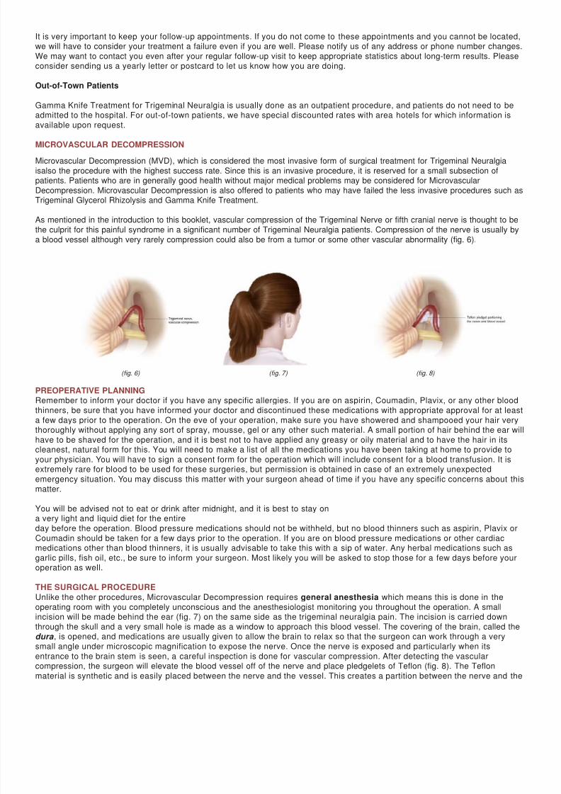
Vascular compression in which a blood vessel causes compression on the trigeminal nerve in the region very close to the brain
stem called the root entry zone. (fig. 6)
• Multiple sclerosis (MS), While trigeminal neuralgia is usually found in patients over the age of 50, multiple sclerosis patients are
usually younger than the average TN patient and may have bilateral (two sided) facial pain. Every young person with trigeminal
neuralgia does not necessarily have MS.
A physician experienced in the management of trigeminal neuralgia can often make the appropriate diagnosis in a single interview.
The trigeminal nerve, also known as the fifth cranial nerve, has three branches:
1. Ophthalmic (around the eye);2. Maxillary (around the upper jaw);
3. Mandibular (around the lower jaw).
The pain may be limited to one or more of these branches. In fact, many categorize the pain as atypical if it spreads beyond the
distribution of the trigeminal nerve. For example, pain going down to the neck, shoulder, on top of or behind the head would not becoming from the trigeminal nerve. Trigeminal neuralgia involves attacks of severe pain on the affected side of the face and is rarely
bilateral. The sharp attack may last just for a few seconds, but in advanced cases these attacks could last much longer. As
mentioned above, the exception to this rule is the multiple sclerosis patient who may suffer from bilateral facial pain.
TREATMENT OPTIONSMedications
In general, the first line of treatment for trigeminal neuralgia is medical. Anticonvulsants are frequently used for medical treatment oftrigeminal neuralgia. For the majority of patients, these medications are quite effective.
Medical treatment with anticonvulsants must be tailored to the patient’s medical condition, age and general medical status including
kidney and liver function since many of these medications may have effects with long-term use on kidney and liver function if they
are not closely monitored. Usually the prescribing physician will closely monitor the dosage and obtain kidney and liver functions testsas well as blood counts for as long as these medications are taken.
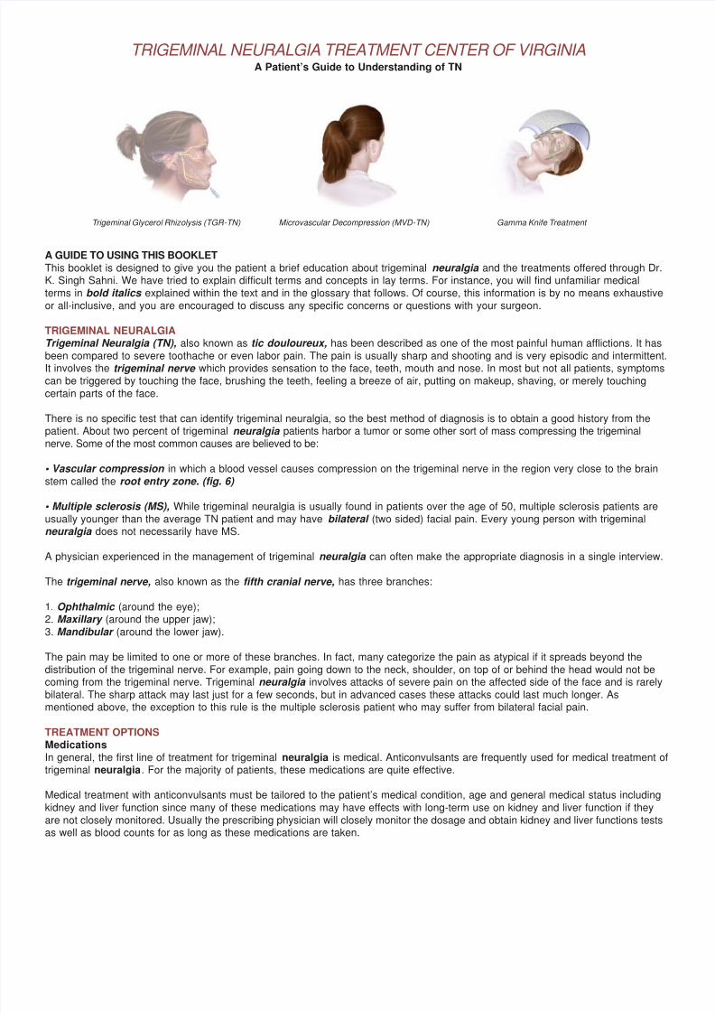
Trigeminal Glycerol Rhizolysis (TGR-TN) Microvascular Decompression (MVD-TN) Gamma Knife Treatment

7/27/2019 Trigeminal Brochure
http://slidepdf.com/reader/full/trigeminal-brochure 2/7
SurgeryPatients unable to tolerate the medications or whose pain has become refractory (resistant to treatment) are then referred to a
neurosurgeon with special interest and experience with trigeminal neuralgia. The three primary surgical options for the treatment oftrigeminal neuralgia are:
• Trigeminal Glycerol Rhizolysis (TGR)
• Microvascular Decompression (MVD)
• Gamma Knife(GK)
TRIGEMINAL GLYCEROL RHIZOLYSIS (TGR)
A procedure done in the hospital operat-ing room under monitored anesthesia. The anesthesiologist will utilize appropriate
medications to decrease anxiety and help with sedation. General anesthesia (being put to sleep) is usually not necessary;however, intravenous medications are given so that you will be very relaxed and sleepy. In the majority of cases, you will not
even remember the procedure. This is called IV sedation.
After being properly sedated, you will be placed on the operating table and a special x-ray device will be used to help the
neurosurgeon insert the needle. The needle is advanced until it reaches a small pocket of fluid surrounding the trigeminalnerve (fig. 1). A special contrast (dye) is then injected which can be easily seen on the x-ray, and this determines the volume of
fluid around the trigeminal nerve (fig. 2). In this way, the surgeon can calculate the exact amount of glycerol that needs to begiven. This is an important part of the procedure and we find it an extremely important part of the process to avoid giving an
overdose of the glycerol. Once the glycerol is measured and injected into this pocket, the needle is removed.
During this part of the procedure, you will usually be sitting up in the operating room after which you are transferred to a
stretcher in the same position, sitting with the chin flexed and the head turned to the side of the procedure. The nurse in therecovery room will help you remain in that position for the next two hours during which time you will be closely monitored for
comfort, your pulse, blood pressure and breathing will be closely watched.
IMPORTANT REMINDERS ABOUT TRIGEMINAL GLYCEROL RHIZOLYSIS
Generally you will not be “put to sleep”, but you must have nothing to eat or drink after midnight on the night before the
procedure. You may resume your usual diet after surgery.
The area around the needle will be numbed before insertion of the needle; therefore, you should let your doctor know if youhave any pain.
There will be no incision made. A band-aid will be used to cover the area where the needle was inserted. The band-aid may beremoved the day after the procedure.
If you are taking any blood thinners such as aspirin, Plavix, Coumadin or any other such medications, please inform your
physician so that they can be withheld with appropriate approval from your primary care physicians prior to the procedure.
Patients can usually go home the same day or the day after the procedure with a follow-up appointment to be seen back in the
office in three to four weeks.
(fig. 1)
(fig. 2)
7/27/2019 Trigeminal Brochure
http://slidepdf.com/reader/full/trigeminal-brochure 3/7
If you are taking any anticonvulsants, you will probably stay on the medication as advised by your surgeon. The medication willprobably be decreased on your first follow-up appointment. Generally about a month later you will gradually be taken off the
medications.
Notify your surgeon immediately if you have severe headache, nausea, vomiting, neck stiffness or fever.
RESULTS
Some patients are completely pain-free within twenty-four hours of the procedure while others can take as long as three to sixweeks to gain complete relief.
TGR is the preferred surgical approach for elderly patients with some medical issues who are in such extreme distress thatthey need urgent and immediate relief.
Patients are forewarned that there is a small risk of potential complications including some loss of feeling or decreased
sensation of the face. If there is any numbness along the cornea or eye, you must be extremely careful and see an eye doctor.Your surgeon will go over the details of other potential risks and benefits of each procedure.
Some patients may develop fever blisters after the injection which usually go away quickly and need only to be taken care of withgood hygiene and appropriate medications if needed.
GAMMA KNIFE
Gamma Knife treatment is done under the leadership of a neurosurgeon, a team of physicists - doctors specializing in radiation
- and nurses. Gamma Knife was pioneered in Stockholm, Sweden by Professor Lars Leksell and has been in use for more thanthirty-five years. It is actually not a knife at all but is a term used for 201 individual beams of focused radiation that all
converge on a selected target. While each individual beam is not very effective, the combination of all the beams causes abiological and structural change in the targeted tissue. Trigeminal neuralgia is just one of the indications for Gamma Knife,which is also used for brain tumors.
Gamma Knife for trigeminal neuralgia is performed for patients who are not ideal candidates for open surgery, such as patients on
blood thinners or who have other known cardiovascular problems and patients unable or unwilling to take anticonvulsants. Othersprefer this treatment due to its relative less invasiveness. Gamma Knife treatment is a simple,
relatively painless and quite straight- forward process that consists of four steps:
1. Attaching the head frame;
2. Imaging with MRI or CT scan
(for patients with pacemakers)
3. Treatment planning; and
4. Treatment
Prior to treatment, your surgeon will review the entire procedure with you and risks and benefits will be discussed in great
depth. Ample opportunity will be given for any questions you may have. given for any questions you may have. Usually, no
incisions, stitches or shaving of the head is necessary. You will be offered to tour the Gamma Knife facility, watch aninstructional DVD and meet the nurses.
PROCEDURE
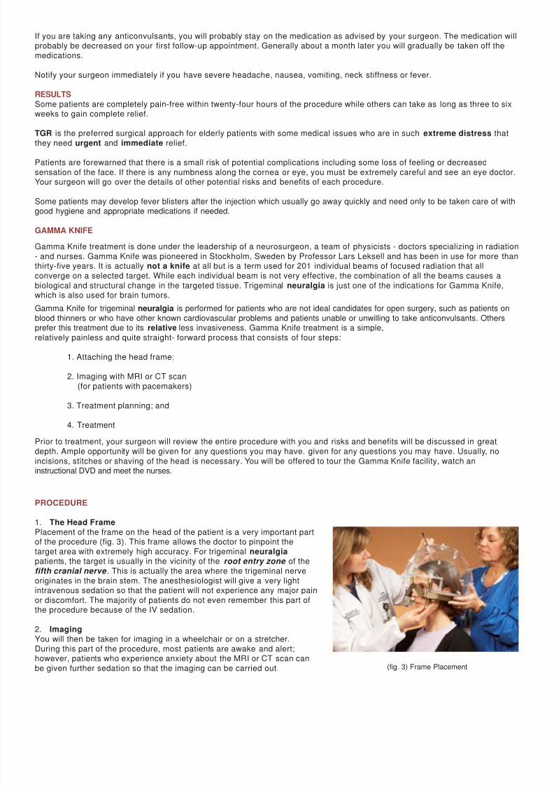
1. The Head Frame
Placement of the frame on the head of the patient is a very important partof the procedure (fig. 3). This frame allows the doctor to pinpoint the
target area with extremely high accuracy. For trigeminal neuralgiapatients, the target is usually in the vicinity of the root entry zone of the
fifth cranial nerve . This is actually the area where the trigeminal nerve
originates in the brain stem. The anesthesiologist will give a very lightintravenous sedation so that the patient will not experience any major pain
or discomfort. The majority of patients do not even remember this part ofthe procedure because of the IV sedation.
2. Imaging
You will then be taken for imaging in a wheelchair or on a stretcher.
During this part of the procedure, most patients are awake and alert;however, patients who experience anxiety about the MRI or CT scan can
be given further sedation so that the imaging can be carried out. (fig. 3) Frame Placement
7/27/2019 Trigeminal Brochure
http://slidepdf.com/reader/full/trigeminal-brochure 4/7
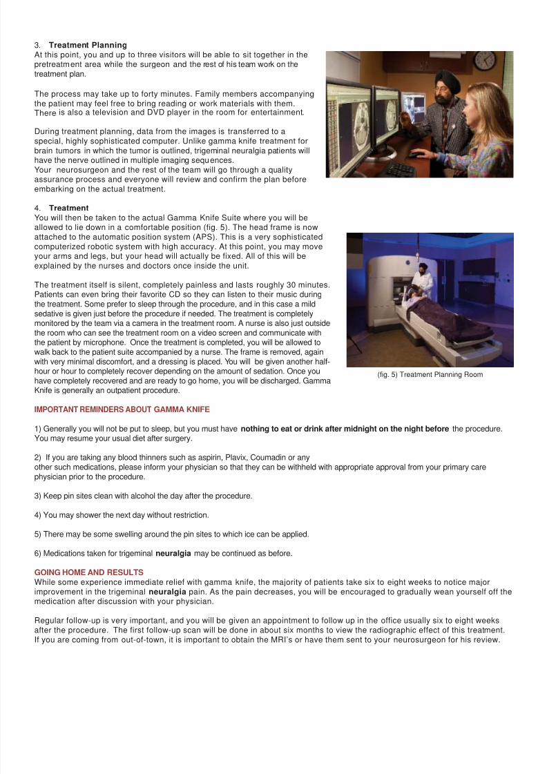
3. Treatment Planning
At this point, you and up to three visitors will be able to sit together in thepretreatment area while the surgeon and the rest of his team work on the
treatment plan.
The process may take up to forty minutes. Family members accompanying
the patient may feel free to bring reading or work materials with them.There is also a television and DVD player in the room for entertainment.
During treatment planning, data from the images is transferred to aspecial, highly sophisticated computer. Unlike gamma knife treatment for
brain tumors in which the tumor is outlined, trigeminal neuralgia patients willhave the nerve outlined in multiple imaging sequences.
Your neurosurgeon and the rest of the team will go through a qualityassurance process and everyone will review and confirm the plan before
embarking on the actual treatment.
4. Treatment
You will then be taken to the actual Gamma Knife Suite where you will beallowed to lie down in a comfortable position (fig. 5). The head frame is now
attached to the automatic position system (APS). This is a very sophisticated
computerized robotic system with high accuracy. At this point, you may moveyour arms and legs, but your head will actually be fixed. All of this will be
explained by the nurses and doctors once inside the unit.
The treatment itself is silent, completely painless and lasts roughly 30 minutes.Patients can even bring their favorite CD so they can listen to their music during
the treatment. Some prefer to sleep through the procedure, and in this case a mild
sedative is given just before the procedure if needed. The treatment is completelymonitored by the team via a camera in the treatment room. A nurse is also just outside
the room who can see the treatment room on a video screen and communicate withthe patient by microphone. Once the treatment is completed, you will be allowed to
walk back to the patient suite accompanied by a nurse. The frame is removed, again
with very minimal discomfort, and a dressing is placed. You will be given another half-hour or hour to completely recover depending on the amount of sedation. Once you
have completely recovered and are ready to go home, you will be discharged. GammaKnife is generally an outpatient procedure.
IMPORTANT REMINDERS ABOUT GAMMA KNIFE
1) Generally you will not be put to sleep, but you must have nothing to eat or drink after midnight on the night before the procedure.You may resume your usual diet after surgery.
2) If you are taking any blood thinners such as aspirin, Plavix, Coumadin or any
other such medications, please inform your physician so that they can be withheld with appropriate approval from your primary care
physician prior to the procedure.
3) Keep pin sites clean with alcohol the day after the procedure.
4) You may shower the next day without restriction.
5) There may be some swelling around the pin sites to which ice can be applied.
6) Medications taken for trigeminal neuralgia may be continued as before.
GOING HOME AND RESULTS
While some experience immediate relief with gamma knife, the majority of patients take six to eight weeks to notice major
improvement in the trigeminal neuralgia pain. As the pain decreases, you will be encouraged to gradually wean yourself off themedication after discussion with your physician.
Regular follow-up is very important, and you will be given an appointment to follow up in the office usually six to eight weeks
after the procedure. The first follow-up scan will be done in about six months to view the radiographic effect of this treatment.
If you are coming from out-of-town, it is important to obtain the MRI’s or have them sent to your neurosurgeon for his review.
(fig. 5) Treatment Planning Room
7/27/2019 Trigeminal Brochure
http://slidepdf.com/reader/full/trigeminal-brochure 5/7
7/27/2019 Trigeminal Brochure
http://slidepdf.com/reader/full/trigeminal-brochure 6/7
vessel so that the vessel no longer pulsates on the brain stem. Closure is done by suturing the dura and placing a syntheticmaterial to cover the hole made in the bone. Upon completion, there is no defect in the bone and the incision will heal very
nicely.
Once the surgery is complete, you will be transferred to the recovery room. The surgeon will then explain the results to your
family in the waiting room and you will be transferred to the Neuroscience Intensive Care Unit (NICU).
Usually you will spend one night in the Neuroscience Intensive Care Unit with mobilization the following day out of ICU to aregular private room. On the first day after surgery, you will usually be allowed to get out of bed and have breakfast . The dressing
will not be changed for three days. On the second or third day, the patient is discharged after dressing change.
RESULTS
The results of Microvascular Decompression are usually very good, and there is usually a much lower recurrence rate thanwith other procedures. However, this is a major surgical intervention, and the potential for complications is also much higher
compared to less invasive procedures of Trigeminal Glycerol Rhizolysis and Gamma Knife Treatment. Since this is an openoperation, it does carry the potential complications that come with general anesthesia. At the same time, other potential risks
of open procedure such as hemorrhage, stroke, loss of function of certain cranial nerves - specifically loss of hearing in a very
small percentage of patients - must be given consideration.
In experienced hands, this surgery has very good results, but even in the best of hands major complications can occur, anddetails should be thoroughly discussed with your surgeon. Whenever Microvascular Decompression is offered, your surgeon
will sit down with
you and your family members and go over the entire protocol of the operation with thorough discussion of the risks andbenefits. Ample opportunity will be provided for you and your family to ask and receive satisfactory answers to questions.
DISCHARGE PLANNING
Once you are discharged, you will be advised to keep your head elevated on two or three pillows for at least two to threeweeks. You will usually be instructed not to get your incision wet until return to the surgeon’s office for removal of the stitches
and dressing change. During the first seven to ten days, you must refrain from any strenuous activities such as lifting, bending
or for that matter even driving. After ten days and having seen the surgeon in the office for the first follow-up visit, you areusually allowed to return to normal activities.
We usually instruct patients to stay very active after the operation and encourage you to start moving as soon as you are
stable and regain complete balance. It is very important to keep in mind that while the patient is in the bed and not ambulating,
the blood can pool in the leg and lead to blood clots in the legs,which can be very dangerous. To prevent this during the operation and your stay in the intensive care unit, special stockings
are used which cause intermittent massage and compression of the legs. This helps to prevent the formation of any blood clotsin the leg. Once you are transferred to a regular room and after you are discharged home, you should continue to wear TED
hose stockings until you are very active with ambulation and no longer need the elastic hose. It is very important for smokers
to quit smoking several days before the operation. Deep breathing and gentle coughing exercises after surgery are also veryimportant to keep your lungs moving and prevent pneumonia. Usually a nurse and the respiratory therapist will help to educate
you on specific exercises during your hospital stay. Elderly patients and smokers are especially encouraged tocontinue these exercises at home.
If you notice any leakage of fluid through the nose or back of the throat, you should immediately alert your surgeon, as this
could be a sign of spinal fluid leakage from the surgery. If you experience fever, headache, nausea, vomiting, neck stiffness or
any such problems, the surgeon should also be promptly informed. Most patients are taken off of anticonvulsants rather quicklyafter this operation unless you have been on the medication for a very long time in which case you will be gradually tapered
off.
FINAL DECISION
Dr. K. Singh Sahni spends a lot of time discussing each individual procedure with his patients and family members.
The final decision to select the surgical procedure is made by the patient while Dr. Sahni helps them with this process.
You should feel comfortable asking any questions during your consultation with Dr. Sahni. He has a lot of patience and
compassion for his TN patients.
7/27/2019 Trigeminal Brochure
http://slidepdf.com/reader/full/trigeminal-brochure 7/7
ABOUT THE SURGEONK. Singh Sahni, MD, FACS is a distinguished neurosurgeon with a special interest in the treatment of trigeminal neuralgia . Over the
last twenty years, he has treated close to 2,000 patients who suffer with facial pain syndromes. While there are a number ofphysicians who may offer one or the other preferred methods of treatment for trigeminal neuralgia , as one who subspecializes in
this disease Dr. Sahni has expertise in all methods of treatment, surgical and nonsurgical. This enables him to tailor each patient’s
treatment based on the patient’s age, general medical condition and overall manifestation of pain. All of this affords the best-casescenario for the trigeminal neuralgia patient of having the opportunity to work with a neurosurgeon who is able to offer all treatment
options at the same facility with a single physician without bias toward a specific procedure. Dr. Sahni is a board certifiedNeurosurgeon. He is the Medical director of Gamma Knife Unit. He obtained his GK training in Stockholm, Sweden. Dr. Sahni has
been performing TGR (Glycerol injections) and MVD (Microvasular Decompression) for over twenty years.
GLOSSARY OF UNFAMILIAR TERMS Aplastic anemia: Low white blood cell count caused by decreased bone marrow function.Bilateral: Two-sided.Dura: The covering of the brain.Fifth cranial nerve: Cranial nerve that serves the face, teeth, mouth and nose. Same as trigeminal nerve.GK: Gamma Knife Intractable: Resistant to cure, relief or control.Mandibular: Related to the lower jaw.Maxillary: Related to the upper jaw.MVD: Microvascular Decompression Ophthalmic: Related to the eye.Refractory: Resistant to treatment.Root entry zone: The region very close to the brain stem, especially where the trigeminal nerve originates.
TGR: Trigeminal Glyerol Rhizolysis Tic douloureus: Painful condition characterized by severe, excruciating, episodic face pain. Same as trigeminal neuralgia.
Trigeminal nerve: Cranial nerve that serves the face, teeth, mouth and nose.
K. Singh Sahni, M.D., F.A.C.S.Neurosurgical Associates, P.C.
10710 Midlothian Turnpike, Suite 138Richmond, Virginia 23231
804-330-4990
804-272-04701-800-868-8204