trigeminal neuralgia, glossopharyngeal neuralgia, and...
TRANSCRIPT

Review ArticleTrigeminal Neuralgia, Glossopharyngeal Neuralgia, andMyofascial Pain Dysfunction Syndrome: An Update
Mohammad Khan,1 Shamima Easmin Nishi,2 Siti Nazihahasma Hassan,3
Md. Asiful Islam,4 and Siew Hua Gan4
1Community Medicine, School of Dental Sciences, Universiti Sains Malaysia, 16150 Kubang Kerian, Kelantan, Malaysia2Orthodontic Unit, School of Dental Sciences, Universiti Sains Malaysia, 16150 Kubang Kerian, Kelantan, Malaysia3Hematology, School of Dental Sciences, Universiti Sains Malaysia, 16150 Kubang Kerian, Kelantan, Malaysia4Human Genome Centre, School of Medical Sciences, Universiti Sains Malaysia, 16150 Kubang Kerian, Kelantan, Malaysia
Correspondence should be addressed to Mohammad Khan; [email protected] Siew Hua Gan; [email protected]
Received 5 February 2017; Revised 12 April 2017; Accepted 24 April 2017; Published 30 July 2017
Academic Editor: Francesca A. Bianchi
Copyright © 2017 Mohammad Khan et al. This is an open access article distributed under the Creative Commons AttributionLicense, which permits unrestricted use, distribution, and reproduction in any medium, provided the original work is properlycited.
Neuropathic pain is a common phenomenon that affects millions of people worldwide. Maxillofacial structures consist of varioustissues that receive frequent stimulation during food digestion. The unique functions (masticatory process and facial expression)of the maxillofacial structure require the exquisite organization of both the peripheral and central nervous systems. Neuralgia ispainful paroxysmal disorder of the head-neck region characterized by some commonly shared features such as the unilateral pain,transience and recurrence of attacks, and superficial and shock-like pain at a trigger point. These types of pain can be experiencedafter nerve injury or as a part of diseases that affect peripheral and central nerve function, or they can be psychological. Sincethe trigeminal and glossopharyngeal nerves innervate the oral structure, trigeminal and glossopharyngeal neuralgia are the mostcommon syndromes following myofascial pain dysfunction syndrome. Nevertheless, misdiagnoses are common. The aim of thisreview is to discuss the currently available diagnostic procedures and treatment options for trigeminal neuralgia, glossopharyngealneuralgia, and myofascial pain dysfunction syndrome.
1. Introduction
Neuralgia is known as pain that occurs in the nerve pathways.Usually, neuralgia is not a sickness but a symptom of aninjury or a disorder. Pain in the maxillofacial region exhibitsmedical, dental, social, and psychological burdens. Maxillo-facial pain originates from many sole target tissues such asthe meninges, cornea, tooth pulp, oral/nasal mucosa, andtemporomandibular joint, thus showing numerous uniquephysiological features associated with the spinal nociceptivesystem [1].Maxillofacial pain disorders cover amajor remark-able and extensive part of neurological disorders and collec-tively have a high occurrence rate and an often overwhelminginfluence on the quality of life [2].
Although there are several common features of paintransduction and processing between the trigeminal and
spinal nerve systems, numerous characteristic features in theperipheral and central components of the trigeminal painsystem exist. Trigeminal neuralgia (TN) is the incidence ofuncontrollable and electrical stun-like pain with a triggerzone, while glossopharyngeal neuralgia (GPN) is consideredas pain in the oropharyngeal area throughout themandibularactions, mainly deglutition [3, 4]. Myofascial pain dysfunc-tion syndrome (MPDS) is categorized by extensive pain,decreased pain relief, sleep disruption, exhaustion, psychoso-matic distress, and chronic headache. Patients withMPDS areidentified based on the presence of numerous tender points[5]. Consequently, current basic and clinical studies that focuson acute and chronic maxillofacial pain need to realize theunique features of the pain system and to advance and assessbetter treatments for orofacial pain.
HindawiPain Research and ManagementVolume 2017, Article ID 7438326, 18 pageshttps://doi.org/10.1155/2017/7438326

2 Pain Research and Management
Themost important obstruction of enhanced patient careand translational research is the absence of approved diagnos-tic criteria [6].The intricate innervation and function of facialregion assemblies make the diagnosis of facial pain and itstreatment very problematic and frustrating [7]. Patients withlong-lasting facial pain should be cautiously reevaluated andclinically examined even after getting multiple treatments.The situation is compounded when neuralgia, ear nose andthroat (ENT) diseases, dental pain, myofascial pain syn-dromes, tumors, temporomandibular disorders (TMD), neu-rovascular pain, or psychiatric diseases commonly presentwith covering signs and symptoms. In addition, referred,severe, and acute pain frequently makes the diagnosis morechallenging [7, 8]. However, most neuralgia conditionsdemonstrate similar clinical features. Therefore, sequentialclinical examinations with additional laboratory diagnosesare essential for the proper treatment and management ofsuch conditions [9].
An effective evidence-based management of neuralgicpain requires a thorough appreciation of the underlyingmechanism of pain. In this review, we describe the updatedknowledge on the pathophysiology, clinical features, diagnos-tic criteria, and management of TN, GPN, and MPDS.
2. Trigeminal Neuralgia
Trigeminal neuralgia is a unilateral disorder highlighted byelectric stun-like neuropathic pain near the distribution ofthe trigeminal nerves with sudden onset and termination. Itis defined as a syndrome that is characterized by paroxysmalfacial pain. According to Loh et al. (1998), the affliction siteratio was observed as 1.4 (right) : 1 (left) of the facial area [10].The characteristics of TN include sudden, severe, periodic,stabbing, lancinating, lightning-like, and shock-like painattacks that are usually one-sided in the 2nd and/or 3rdtrigeminal branch [11–13]. TN is one of the most commonneurological pains involving the orofacial region, whichgenerally has the most intensive type of pain [4, 14, 15]. Ittypically affects the elderly (1 in 25,000 of the population),with the most frequently reported cause being neurovascularcompression [16–18]. From an etiological aspect, TN is clas-sified as classic or primary or idiopathic and symptomatic orsecondary. Classic or primaryTNappearswith no clear cause.Symptomatic or secondary TN occurs with the presence ofintracranial lesions such as a tumor, infarction, and multiplesclerosis (MS). From a symptomatic aspect, TN is classified as“typical” in the presence of paroxysmal pain alone and “atyp-ical” when the paroxysmal pain is associated with constantpain [19].
Pain in 95% of the TN cases is almost unilateral, com-monly affecting the mandibular and maxillary divisions [13].According to Loh et al. (1998), there was a greater contri-bution of the mandibular branch of the trigeminal nervethan the maxillary branch in TN patients (𝑛 = 44) [10]. Anattack of TN is typically originated by a slightmucocutaneousincentive in the region of the affected trigeminal nerve alsoknown as a trigger point [18, 19].The diagnosis of TN is basedon the typical case history of episodic electric pain in thesupply of the trigeminal nerve [20]. However, the diagnosis
and management of TN require a group of experts consistingof neurologists, anesthesiologists, neurosurgeons, neuroradi-ologists, oral andmaxillofacial surgeons, and dentists [21, 22].
2.1. Epidemiology. The prevalence of TN has been estimatedat 107.5 males and 200.2 females per 1 million populations[23]. The incidence rate of TN was 4.3 per 100,000 in theUS population, with the age-adjusted rate for females beingsignificantly higher than that for males [24]. The predictablefrequency of TN is approximately 4–12.5/100,000 peopleannually, with increasing occurrence based on stage [25].Because age is a primary risk factor, symptom manifestationis more likely after the age of 50 years [26]. The topmostinception of TN occurs between the ages of 50 and 70 years,but it can happen even at a younger age. A study reported theusual age of sign onset at 19.6±3.4 years [27].Themost widelyrecognized age group was 51 to 60 years (36.90%), followedby 61 to 70 years (23.68%) and 41 to 50 years (17.35%) [28]. Itis unusual in individuals older than 30 years, with only 1% ofcases in thosemore youthful than 20 years of age [29, 30].Theannual occurrence in females is approximately 5.9/100,000cases per females, while it is approximately 3.4/100,000 casesper males [25, 31]. The female-to-male proportion of TN was3 : 2 [32]. TN is more common among females (62%) thanamongmales (38%), with a female-to-male ratio of 1.6 : 1 [33].Thus, females have a higher risk of having TN thanmales, andthe risk again increases with age [30].
In addition, a retrospective study of TN patients in Sin-gapore and Malaysia revealed that the age of onset fluctuatesfrom 24 to 89 years with a mean age of 54.9 years. Theultimate occurrence was in the 6th (29.5%) and 7th (27.3%)decades of life, followed by the 5th decade (13.6%).The fourthand eighth decades had similar incidences (11.4%). This wasfollowed by the third (4.5%) and ninth (2.3%) decades. Withrespect to sexual orientation, females account for 63.7% ofthe patients, with a ratio of 1.75 : 1.00. The distribution of TNin multiethnic populations was 68.2% among the Chinese,followed by 13.6% and 11.4% in Malays and Indians, respec-tively. The remaining distribution was solitary Japanese andtwo Eurasians [10].
Other than the age and gender, multiple sclerosis (MS) isa well-known risk factor to TN [10, 27].MS has been reportedin 2 to 4% of patients with TN where demyelination is alsopresent [24]. The reported prevalence of TN in the MSpopulation was between 1.0% and 6.3% [34, 35]. In addition,approximately 4% of MS patients have a lifetime risk to haveTN, with no significant difference observed among the dif-ferent forms of MS [36]. Approximately 5% of idiopathic TNcases have been reported to have a family history [33]. Hence,older females, MS, and family history are important riskfactors for TN.
2.2. Pathophysiology. TN occurs due to the specific abnor-malities of the trigeminal nerve in the trigeminal root organglion. The pathophysiological characteristics of classic oridiopathic TN are identified with the pressure of the trigem-inal nerve root by a vein at or nearby the root passage zone[17, 37, 38]. An artery crossing the nerve can provoke furtherdisplacement [39], which can lead to damage and injury of

Pain Research and Management 3
the trigeminal nerve.The damage tends to be localized and isspecifically related to the vascular contact [40, 41]. The dam-aged nerves cause pain via several mechanisms, includingthe hyperexcitability of the demyelinated nerve fibers, ectopicimpulse discharge, spontaneous and triggered after discharge,cross excitation between sensory channels, deafferentation,impaired segmental inhibition, and emphatic transmission[38, 40–43]. The vascular pressure of the trigeminal nerveroot is associated with the focal loss of myelin and the closejuxtaposition of the demyelinated axons with few mediatingastrocytic processes [40].
Symptomatic or secondary TN occurs due to intracra-nial lesions such as a tumor, infarction, and MS [19, 44].Intracranial tumors in the presence of aneurysms, angiomas,or vascular malformation are among other causes of TN[45, 46] that may occur by either direct tumoral compressionor the wrapping of the trigeminal nerve root [47, 48]. Tumoroccurrence has been reported to bemore common amongTNpatients aged less than 39 years than among those older than40 years [49]. Approximately 2 to 4%ofMS has been reportedto occur in TN patients [24] as well.
2.3. Clinical Manifestation. The hallmark of TN includesrecurrent attacks of lancinating pain in the trigeminal nerve[50]. The attacks typically last just seconds, yet they may berepeated over and again inside a brief time frame. Approx-imately 79% of the pain experienced is intermittent, while21% of pain is continuous [13].The nature of pain is sporadic,sudden, and often like an electric shock, enduring from a fewmoments to a fewminutes [27]. In fact, TN has been reportedto be a relatively common pain condition, occurring morecommonly on the right side (72.63%) than on the left side(27.37%) with a ratio of 2.6 : 1.0 [30, 51] and mainly affectingthe mandibular division [10, 30]. The communal outlyingnerve involved is infraorbital with various patients presentedwith sensible pain [30].
The brief paroxysms pain in TN is restricted to the facialconveyance of the trigeminal nerve and can be triggered bystimuli to sensory endings in the trigeminal receptive area[52]. The triggering stimuli include simple actions such astalking, swallowing, laughing, washing, wind-blowing, shav-ing, mouth opening, touching, and chewing. Nevertheless,the presence of these triggering stimulimay cause the patientsto circumvent any stimulus on the face or mouth. Mostpatients respond to different strengths of the aggravatingstimulus and react to more than a single triggering stimulus[53].
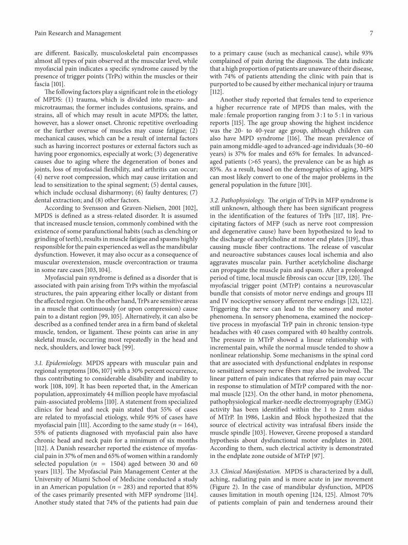
2.4. Diagnostic Criteria. The diagnosis of TN basically relieson a patient’s description of pathognomonic pain occurrences[54]. The primary diagnostic tools are MRI (magnetic reso-nance imaging) and CT (computed tomography), since thereis no specific laboratory test available (Table 4). Therefore,patients’ signs and symptoms are key important factors inmaking the diagnosis (Figure 1).
TN is generally idiopathic [55]; however, it might like-wise emerge optional to different conditions, including theintracranial space involving lesions andmultiple sclerosis [35,56, 57]. The International Association for the Study of Pain
(IASP) and International Headache Society (IHS) has rec-ommended their own indicative criteria for TN [58, 59]. TheIASP has defined the TN as a sudden, normally one-sided,extremely brief cutting intermittent pain in the disseminationof at least one branch of the fifth cranial nerve. On theother hand, the IHS defines TN as an excruciating one-sideddistress of the face, described by brief electric stun-like painrestricted to the circulation of at least one division of thetrigeminal nerve. Pain is usually induced by insignificantstimuli, which include smoking, sneezing, and brushing theteeth, all of which often regularly occur impulsively. Littlezones in the nasolabial fold and chin might be prone to theprecipitation of pain that may remit for variable periods [58].
According to Eller et al. (2005), TN is defined and clas-sified into seven types: (i) TN1: idiopathic, sharp, shooting,electrical shock-like, and episodic pain; (ii) TN2: idiopathic,aching, throbbing, burning, and more than 50% constantpain; (iii) TN3: trigeminal neuropathic pain and accidentalharm to the trigeminal nerve from injury or surgery (facialtrauma; oral operation; ear, nose, and throat operation; skullbase operation; posterior fossa operation; or stroke); (iv)TN4: trigeminal deafferentation pain and intentional injuryto the trigeminal nerve (gangliolysis, nucleotomy, neurec-tomy, tractotomy, rhizotomy, or other denervating proce-dures); (v) TN5: symptomatic, associated with MS; (vi) TN6:postherpetic because of a flare-up of facial herpes zoster;and (vii) TN7: atypical facial pain and facial pain auxiliaryto a somatoform pain issue, requiring mental testing forindicative diagnostic affirmation [32].
2.5. Treatment and Management. Table 1 presents variouspharmacological agents and available treatment optionsbased on surgical procedures. Bothmethods are effective andwidely used [60–62]. Usually, TN patients are first treatedwith pharmacological agents. The pain can be readily man-aged with medication in approximately 80% of patients [63].However, if the pain is not effectively relieved by medicationsor there is medication intolerance due to toxicity or allergicreactions, surgical treatment becomes an option.
In standard practice, the first-line treatment is carba-mazepine, which can relieve most of the observed symptoms[22, 64–66]. Other drugs, including oxcarbazepine [67], phe-nytoin [68, 69], baclofen [70, 71], lamotrigine [72, 73], gaba-pentin [74, 75], and sodium valproate [76, 77], are also effi-cient in reducing the signs-symptoms of TN inmost patients.Sometimes, some of the drugs are taken with carbamazepineas an adjuvant for the synergistic effects in relieving theTN symptoms [78, 79]. The decreasing relief provided bycarbamazepine or other drugs with continual use as well asunacceptable side effect profiles may necessitate the discon-tinuation of therapy. In fact, it has been reported that approx-imately 50% of patients eventually require an operation torelieve pain [25]. Research has indicated that there is nosignificant influence of age, sex, ethnicity, or the side of theface on the decision of themedication regimen and the lengthof treatment for pain control [79].
When drugs no longer offer pain relief, surgical interven-tion is preferred. The objective of surgery procedures is toprevent the blood vessel from squeezing the trigeminal nerve

4 Pain Research and ManagementTa
ble1:Ch
aracteris
ticsa
ndmanagem
ento
ftrig
eminalneuralgiainthem
axillofacialregion.
Dise
ases
Clinicalfeatures
Pharmacological
treatments
Side
effects
Surgical/lo
caltreatments
Limitatio
n
Trigem
inal
neuralgia
Pain,electric
shocklik
ePain
duratio
n,second
sIntensity,severe
Localization,
good
Characteris
tics,triggerz
one,diurnal
Triggerp
oints,no
nnoxious
stim
ulus
Carbam
azepine
(i)Develo
pmento
fresistance
and
intolerance
(ii)D
izziness
(iii)Nausea
(iv)A
taxia
(v)V
omiting
(vi)Xe
rosto
mia
Percutaneous
trigem
inal
rhizotom
y
(i)Re
currence
ofpain
(ii)D
ysesthesia(tr
oublesom
enu
mbn
ess)
(iii)Lo
ssof
cornealreflex
Oxcarbazepine
(i)Dizziness
(ii)F
atigue
(iii)Nausea
(iv)V
omiting
(v)H
eadache
(vi)Ac
ne(vii)
Dry
mou
th(viii)C
onstipatio
n
Ablativep
eripheralprocedu
res
(neurectom
y)(i)
Lessmorbiditybu
twith
chance
ofrecurrence
Gabapentin
(i)Dizziness
(ii)F
atigue
(iii)weightg
ain
(iv)D
rowsin
ess
(v)p
eripheraledema
Microvascular
decompressio
n(i)
Lowrecurrence
ofpain
(ii)C
hanceo
fcausin
gnerved
amage
(iii)Haringloss,dou
blev
ision
,facial
numbn
ess,or
paralysis
Pregabalin
(i)Dizziness
(ii)B
lurred
visio
n(iii)Diplopia
(iv)Increased
appetitea
ndsubsequent
weightg
ain
(v)E
upho
ria,con
fusio
n,andvivid
dreams
(vi)Ch
angesinlib
ido(in
crease
ordecrease)
(vii)
Mem
oryim
pairm
ent
(viii)T
remors
(ix)D
rymou
th(x)E
rectile
dysfu
nctio
n(xi)Perip
heraledema
(xii)
Nasop
haryng
itis
(xiii)Increased
creatin
inek
inaselevel
Topiramate
(i)Paresthesia
(ii)N
ausea
(iii)Lack
ofconcentration
(iv)D
iplopia
(v)N
ervousnessanddizziness
(vi)Mem
oryim
pairm
ent
(vii)
Speech
distu
rbance
(viii)D
isturbance
(ix)D
iarrhea
Gam
mak
niferadiosurgery
(i)Tend
ernessdevelops
where
the
screwso
rpinsw
erep
laced
(ii)H
airlossw
here
ther
adiatio
nwas
directed
(iii)Dam
agetosurrou
ndingtissues
intheb
rain,causedby
swellin
g

Pain Research and Management 5
Table1:Con
tinued.
Dise
ases
Clinicalfeatures
Pharmacological
treatments
Side
effects
Surgical/lo
caltreatments
Limitatio
n
Trigem
inal
neuralgia
Pain,electric
shocklik
ePain
duratio
n,second
sIntensity,severe
Localization,
good
Characteris
tics,triggerz
one,diurnal
Triggerp
oints,no
nnoxious
stim
ulus
Valproates
(i)Skin
rash
(ii)H
airloss
(iii)Weightg
ain
Myotherapy
(i)Re
gularm
onito
ringisno
tpossib
le
Tricyclic
antid
epressants
(TCA
)
(i)Sedatio
n(ii)D
rymou
th(iii)Blurredvisio
n(iv
)Breastenlargementinmales
(v)C
onstipation,
mania
(vi)Weightg
ain
(vii)
Highbloo
dglucoselevel
Injections
(i)Asitisa
painfulprocedu
re,the
patie
ntfeelsu
ncom
fortabledu
ring
injection
Sedativ
es(oncond
ition
)
(i)Ap
nea
(ii)P
hysic
aldepend
ence
(iii)Decreased
meanarteria
lpressure
(iv)T
achycardia
(v)P
aradoxicalexcitement
Acup
uncture
(i)Painfulprocedu
re
Therapeutic
ultrasou
nd(i)
Askilled
person
isneeded,and
the
patie
ntisstillun
dero
bservatio
n

6 Pain Research and Management
Unilateral orofacial pain/paroxysmal facial
TN
IHS
onset onsetFifth
shock-sided
trigeminal nerve
Figure 1: The basic diagnostic criteria of trigeminal neuralgia (TN).
or injuring the trigeminal nerve. Several surgical approachesused to relieve the pain due to TN include neurectomy oftrigeminal nerve branches outside the skull, percutaneousradiofrequency thermal rhizotomy, percutaneous ablationthat creates trigeminal nerve or trigeminal ganglion lesionswith heat [80–82], percutaneous retrogasserian glycerol rhi-zotomy, injection of glycerol into the trigeminal cistern[62, 83, 84], physical compression, trigeminal ganglion bal-loon microcompression [85], alcohol injections [57, 86],botulinum toxin injection [87, 88], cryotherapy [89, 90], andgamma-knife radiosurgery (GKRS) [91]. These proceduresare intended to alleviate the symptoms of TN by relievingnerve compression at some point along its course. Hereafter,the management of TN is dependent on a fast, correct diag-nosis, prompt, and effective treatment because the symptomscan be very severe.
2.6. Complications. Many drugs used in the treatment andmanagement of TN are associated with several side-effects.Nevertheless, most of the effects can be tolerated and imme-diate drug discontinuation is conducted when the commonpain tolerance level is exceeded. In addition, some of thesurgical procedures may contribute to some complicationssuch as microvascular decompression (MVD) which is ableto relieve an abnormal compression of an artery to thetrigeminal nerve. Although it is generally successful, achiev-ing 70–90% pain control and less than 1% of the mortalityrisk [92], the procedure may lead to hearing loss whichis associated with a refutation injury of cranial nerve VIII[93]. Nevertheless, the complications were less common
following an intraoperative monitoring of the brainstemevoked response.
The rate of ipsilateral hearing loss was 3% (previous1980) and 1% thereafter (𝑝 = 0.008). Radiosurgery, whichcan increase the pain control rate up to 83%, is effectivein treating TN [91]. Its complication rate is only 6% forfacial paresthesia and 4% for hypoesthesia [94]. An imme-diate complete loss of vision in one eye after trigeminalradiofrequency rhizotomy was reported in three patients dueto acute traumatic optic neuropathy [95]. Moreover, therewere patients who have masseter weakness and paralysis,keratitis and transient paralysis of cranial nerves III andVI, diminished corneal reflex, dysesthesia, and anesthesiadolorosa [96]. Nevertheless, MVD remains the best choiceand a useful alternative to carbamazepine in the treatment ofTN (Table 1).
3. Myofascial Pain Dysfunction Syndrome
Myofascial pain dysfunction syndrome (MPDS) is knownas a psychophysiological disease, which is associated withmuscular structure—in particular, the muscle of mastication[97]. MPDS is a commonly observed phenomenon in themedical, dental, and psychological sectors. MPDS is alsotermed myalgia, myofasciitis, myogelosis, myofascial pain,fibromyalgia, myofibrositis, and myofascial pain syndrome(MPS). When it is mainly associated with temporomandibu-lar join dysfunction, it is termed as myofascial pain dysfunc-tion syndrome [98–100]. Although the terms myofascial andmusculoskeletal pain are used frequently and alternately, they
Pain Research and Management 7
are different. Basically, musculoskeletal pain encompassesalmost all types of pain observed at the muscular level, whilemyofascial pain indicates a specific syndrome caused by thepresence of trigger points (TrPs) within the muscles or theirfascia [101].
The following factors play a significant role in the etiologyof MPDS: (1) trauma, which is divided into macro- andmicrotraumas; the former includes contusions, sprains, andstrains, all of which may result in acute MPDS; the latter,however, has a slower onset. Chronic repetitive overloadingor the further overuse of muscles may cause fatigue; (2)mechanical causes, which can be a result of internal factorssuch as having incorrect postures or external factors such ashaving poor ergonomics, especially at work; (3) degenerativecauses due to aging where the degeneration of bones andjoints, loss of myofascial flexibility, and arthritis can occur;(4) nerve root compression, which may cause irritation andlead to sensitization to the spinal segment; (5) dental causes,which include occlusal disharmony; (6) faulty dentures; (7)dental extraction; and (8) other factors.
According to Svensson and Graven-Nielsen, 2001 [102],MPDS is defined as a stress-related disorder. It is assumedthat increased muscle tension, commonly combined with theexistence of some parafunctional habits (such as clenching orgrinding of teeth), results inmuscle fatigue and spasms highlyresponsible for the pain experienced aswell as themandibulardysfunction. However, it may also occur as a consequence ofmuscular overextension, muscle overcontraction or traumain some rare cases [103, 104].
Myofascial pain syndrome is defined as a disorder that isassociated with pain arising from TrPs within the myofascialstructures, the pain appearing either locally or distant fromthe affected region.On the other hand, TrPs are sensitive areasin a muscle that continuously (or upon compression) causepain to a distant region [99, 105]. Alternatively, it can also bedescribed as a confined tender area in a firm band of skeletalmuscle, tendon, or ligament. These points can arise in anyskeletal muscle, occurring most repeatedly in the head andneck, shoulders, and lower back [99].
3.1. Epidemiology. MPDS appears with muscular pain andregional symptoms [106, 107] with a 30% percent occurrence,thus contributing to considerable disability and inability towork [108, 109]. It has been reported that, in the Americanpopulation, approximately 44million people have myofascialpain-associated problems [110]. A statement from specializedclinics for head and neck pain stated that 55% of casesare related to myofascial etiology, while 95% of cases havemyofascial pain [111]. According to the same study (𝑛 = 164),55% of patients diagnosed with myofascial pain also havechronic head and neck pain for a minimum of six months[112]. A Danish researcher reported the existence of myofas-cial pain in 37%ofmen and 65%ofwomenwithin a randomlyselected population (𝑛 = 1504) aged between 30 and 60years [113]. The Myofascial Pain Management Center at theUniversity of Miami School of Medicine conducted a studyin an American population (𝑛 = 283) and reported that 85%of the cases primarily presented with MFP syndrome [114].Another study stated that 74% of the patients had pain due
to a primary cause (such as mechanical cause), while 93%complained of pain during the diagnosis. The data indicatethat a high proportion of patients are unaware of their disease,with 74% of patients attending the clinic with pain that ispurported to be caused by eithermechanical injury or trauma[112].
Another study reported that females tend to experiencea higher recurrence rate of MPDS than males, with themale : female proportion ranging from 3 : 1 to 5 : 1 in variousreports [115]. The age group showing the highest incidencewas the 20- to 40-year age group, although children canalso have MPD syndrome [116]. The mean prevalence ofpain amongmiddle-aged to advanced-age individuals (30–60years) is 37% for males and 65% for females. In advanced-aged patients (>65 years), the prevalence can be as high as85%. As a result, based on the demographics of aging, MPScan most likely convert to one of the major problems in thegeneral population in the future [101].
3.2. Pathophysiology. The origin of TrPs in MFP syndrome isstill unknown, although there has been significant progressin the identification of the features of TrPs [117, 118]. Pre-cipitating factors of MFP (such as nerve root compressionand degenerative cause) have been hypothesized to lead tothe discharge of acetylcholine at motor end plates [119], thuscausing muscle fiber contractions. The release of vascularand neuroactive substances causes local ischemia and alsoaggravates muscular pain. Further acetylcholine dischargecan propagate the muscle pain and spasm. After a prolongedperiod of time, local muscle fibrosis can occur [119, 120]. Themyofascial trigger point (MTrP) contains a neurovascularbundle that consists of motor nerve endings and groups IIIand IV nociceptive sensory afferent nerve endings [121, 122].Triggering the nerve can lead to the sensory and motorphenomena. In sensory phenomena, examined the nocicep-tive process in myofascial TrP pain in chronic tension-typeheadaches with 40 cases compared with 40 healthy controls.The pressure in MTrP showed a linear relationship withincremental pain, while the normal muscle tended to show anonlinear relationship. Some mechanisms in the spinal cordthat are associated with dysfunctional endplates in responseto sensitized sensory nerve fibers may also be involved. Thelinear pattern of pain indicates that referred pain may occurin response to stimulation of MTrP compared with the nor-mal muscle [123]. On the other hand, in motor phenomena,pathophysiological marker-needle electromyography (EMG)activity has been identified within the 1 to 2mm nidusof MTrP. In 1986, Laskin and Block hypothesized that thesource of electrical activity was intrafusal fibers inside themuscle spindle [103]. However, Greene proposed a standardhypothesis about dysfunctional motor endplates in 2001.According to them, such electrical activity is demonstratedin the endplate zone outside of MTrP [97].
3.3. Clinical Manifestation. MPDS is characterized by a dull,aching, radiating pain and is more acute in jaw movement(Figure 2). In the case of mandibular dysfunction, MPDScauses limitation in mouth opening [124, 125]. Almost 70%of patients complain of pain and tenderness around their

8 Pain Research and Management
MPDS
Clinical features
Dull and aching type pain
Unilateral and radiating
Diminished hearing
Radiological investigations(i) CT scan(ii) MRI(iii) Scintigraphy
Tenderness around TMJ
Clicking sound in TMJ
Trigger point present
Diagnostic tools
Laboratory investigations(i) FBC(ii) Serum calcium(iii) Alkaline phosphatase(iv) Serum creatinine (v) ESR
Figure 2: Commonly used diagnostic tools and features of myofascial pain dysfunction syndrome (MPDS).
temporomandibular joints. Some patients also reported thepresence of a clicking sound or cracking noises in the tem-poromandibular joint (TMJ) [124]. MPDS usually involvesonly the face unilaterally. Frequent headache is also a com-mon complaint (23% of cases). Only tension headaches maybe directly or indirectly associated with this syndrome. Inaddition, a patient may also complain of diminished hearing,tinnitus, burning tongue, and neuralgic pains. MPDS alsoleads to some functional disorders and may cause organicvariations in the temporomandibular joint as well as inmasticatory muscles [103, 125]. Usually, the patient showspain in the trigger area when approximately 3 kg of pressureis applied. The fibers show the local twitch response (LTR)during plucking palpation inside the band of muscle [107].
3.4. Diagnostic Criteria. Myofascial pain syndrome is asso-ciated with MTrPs and can be reproduced by firm palpationover the trigger zone. The diagnostic features for myofascialtrigger points include the following:
(i) Upon palpation of the muscle, a focal point of tender-ness may be found.
(ii) Trigger point palpation leads to a reproduction of thepain.
(iii) There is a “taut band” in adjacent muscle.(iv) There is restriction of muscle movement.
(v) It may appear with pseudo-weakness of the musclefiber.
(vi) Paresthesia occurs over the muscle area.The diagnosis of MFP syndrome may be a challenge becauseit shows some complex clinical characteristics. Most patientstend to show some tenderness (67%) of the elevator musclesduring palpation. Nearly 40% of the patients’ complain ofpain on chewing. Thus, 30% have significant myalgia withbruxism [126]. Sometimes, the patients may also complain ofsleep disturbance because the sleeping position may act as anaggravating factor to the pain caused by MTrP [107].
Arthrography is a useful procedure for the diagnosis ofMPDS when the TMJ is involved. Radiographic interpreta-tions of the head and neck region, CT scans and scintigraphy,can also help to establish the concluding diagnosis. In thiscase, MRI is indicated to allow better view of the condyle anddisc position of TMJ specifically. Sometimes laboratory testssuch as a complete blood count (in the case of suspected infec-tion), serum calcium, phosphorus, and alkaline phosphatase(for bone disorder) need to be investigated. Serum uric acid(for gout), serum creatinine, and phosphokinase levels helpas useful indicators of muscular disorders. If rheumatoidarthritis is suspected, the electrolyte sedimentation rate (ESR)and rheumatoid factors should be investigated. Additionally,electromyography can be used to assess muscle function andfatigue. However, psychologic evaluation and psychometrictesting may also help in the diagnosis [127].

Pain Research and Management 9
3.5. Treatment and Management. Patient counseling regard-ing the disease is very important. Usually, it is often difficultfor patients to accept a psychophysiologic explanation fortheir diseases. Connecting the symptoms to specific masti-catory muscles can facilitate the determination of the actualcause of the type and location of pain. In cases of the involve-ment of the temporal muscle, patients may suffer whereheadache and jaw ache are common in the masseter andpterygoid muscles, showing discomfort during swallowingand earache associated with the lateral pterygoid muscle[128].
Normally, injection is the first treatment approach inthe treatment of MPDS. An injection using dry needlingand lidocaine anesthetic solution injection may be helpfulin MPDS treatment. In 2010, Ay et al. [129] reported thatusing dry needling and lidocaine injection with stretchingexercises may show a momentous role in the treatment ofMFP syndrome. Besides, botulinum toxin injection has beenreported to produce significant effects such as sudden painreduction, especially when a saline injection is not effective[130–132].Moreover,Gazi et al. (2010) compared the analgesicresult of acupuncture (which is purported to help restoreblood circulation) to TrPs injections (containing 0.25% bupi-vacaine administered twice weekly) with cyclobenzaprinechlorhydrate and sodium dipyrone on trigger points toprevent the recurrence ofMFP symptoms.The results showedcomparable pain liberation and progress in the quality of lifeat 4 weeks [133]. Additionally, therapeutic ultrasound is a newtreatment strategy for MFP syndrome. Because it is a non-invasive procedure, it is valuable in the treatment of deepermuscle. Manual therapeutic techniques are not sufficient forthe assessment of deep muscle [134]. Other approaches havebeen used in the treatment and management of MPDSincluding psychotherapy [135, 136], home therapies [137],medications [138, 139], and dental management [140, 141].Sequential treatment is needed to manage such pain syn-drome (Table 2).
3.6. Complications. In the case of a misdiagnosis, chronicpain syndrome and complex behavioral problemsmay result,leading to psychosocial problems. Most of the time, chronicpainmay cause sleep disturbance where patients have troublefinding a comfortable sleeping position. Sometimes, posturechanges during sleepmay affect the disease, thus affecting thepatients’ sleep throughout the night [108].
4. Glossopharyngeal Neuralgia
Glossopharyngeal neuralgia (GPN) is a very sporadic condi-tion related to hyperactivity of the glossopharyngeal nerve[142]. GPN is rare compared with TN. The pain affects thesensory areas corresponding to the glossopharyngeal neural-gia with a branch of sensory vagus nerves. GPN consists ofspasmodic, momentary, and severe sharp pain in the poste-rior area of the throat, tonsillar fossa, base of the tongue, earcanal, and areas inferior to the angle of the mandible [143].Generally, the pain persists for seconds to minutes and isoften triggered by chewing, coughing, yawning, talking, andswallowing [144]. The prevalence of GPN is estimated to be
approximately 0.8 per 100,000 populations in a year, indicat-ing that it is less common than TN (4.7 cases per 100,00) [26].GPN is usually represented by a painful condition on the leftside of the body in females, whereas TN is more commonlyobserved on the right side [145].
GPN is a mixture of cranial nerves that have somaticsensory fibers from the oropharynx, mastoid, middle ear,and Eustachian tube, and posterior third of the tongue. Themiddle ear and mastoid have a sensory supply of glossopha-ryngeal nerve along with the tympanic branch or Jacobson’snerve [146]. It also receives special sensory fibers for taste aswell as chemoreceptor and baroreceptor afferent input fromthe carotid body and carotid sinus. Stylopharyngeus musclesare supplied by the motor component, and the parotid glandis supplied by the parasympathetic secretomotor supply. Thenerve of Hering is an important branch of the carotid sinusbranch, which conveys chemoreceptor and baroreceptorinformation centrally for circulatory reflux function andmaybe accountable for the arrhythmogenicity of GPN [146, 147].
GPN is classified according to HIS, based on the involvedstructural areas.The proposed classification of IHS is (1) clas-sical GPN (occasional or episodic pain) and (2) symptomaticGPN (constant pain) [58]. The classification is based on theareas involved in otitis, that is, pain in (or around) the auricleor earlobe. On the other hand, for the oropharyngeal type,the pain is in and around the neck as well as the maxillofacialregions. Nevertheless, these areas significantly overlap withother cranial nerve-supplied areas [148].
GPN may be idiopathic with the absence of any obviouslesion. Most cases are mainly recognized as glossopharyngealnerve compression triggered by a vessel at the root entry zoneof the brainstem [148, 149]. Idiopathic causes may be vasculardecompression and/or central pontine dysfunction. The sec-ondary cause is a noticeable lesion that includes trauma (skullbase fracture, penetrating injury), postradiation, neoplasm(skull base, cerebellopontine, brainstem, pharynx, tongue,tonsil, metastatic head, and neck tumors), infection (tonsil-litis, pharyngitis, petrositis, arachnoiditis, para pharyngealabscess, and tuberculosis), surgery (posttonsillectomy, post-neck dissection, and postcraniotomy), vascular malforma-tions (arteriovenous malformation, fusiform aneurisms, per-sistent hypoglossal artery, and dissection of the vertebralartery), demyelination (MS), and Eagle’s syndrome as well asothers which include direct carotid puncture, choroid plexusovergrowth, and hyperactive dysfunction syndrome. Thistype of GPN is usually accompanied with numbness or painaround the affected area [150].
4.1. Clinical Manifestation. There are paroxysmal attacks offacial pain, which last for seconds to minutes (Table 3). Thecharacteristics of pain are unilateral sharp, stabbing, andsevere shooting pain. The distribution of pain is persistentwithin the posterior part of the tongue, pharynx to innerangle of the lower jaw, and tonsillar fossa [151]. Pain occurswithin a certain interval with most occurring after a longtime, although some patients may experience it within a day[152, 153].
The common trigger factors are swallowing, chewing,talking, coughing, yawning, lateral movement of the jaw,

10 Pain Research and Management
Table2:Ch
aracteris
ticsa
ndmanagem
ento
fmyofascialp
aindysfu
nctio
nsynd
romeinthem
axillofacialregion.
Dise
ases
Clinicalfeatures
Pharmacologicaltre
atments
Side
effects
Surgical/lo
cal
treatments
Limitatio
n
Myofascialp
ain
dysfu
nctio
nsynd
rome
Pain,dulland
aching
type
Pain
duratio
n,constant
Intensity,m
oderate
Localization,
diffu
seCh
aracteris
tics,generalized,
spon
taneou
sTriggerp
oint,palpatio
n,functio
n
Non
steroidal
anti-inflammatorydrug
(i)Indigestion
(ii)S
tomachup
set
(iii)Stom
achbleeding
(iv)P
eptic
ulcer
(v)F
luid
retention
Psycho
therapy
(i)Failu
reof
episo
dicc
ounseling
and
patie
nt’signo
rance
Tricyclic
antid
epressants
(TCA
)(A
sstatedin
Table1)
Myotherapy
(i)Re
gularm
onito
ringisno
tpossib
le
Injections
(i)Asitisa
painfulprocedu
re,the
patie
ntfeelsu
ncom
fortabledu
ringinjection
Sedativ
es(oncond
ition
)
(i)Ap
nea
(ii)D
ecreased
meanarteria
lpressure
(iii)Tachycardia
(iv)P
aradoxicalexcitement
Acup
uncture
(i)Painfulprocedu
re
Therapeutic
ultrasou
nd(i)
Askilled
person
isneeded,and
the
patie
ntisstillun
dero
bservatio
n

Pain Research and Management 11
Table3:Ch
aracteris
ticsa
ndmanagem
ento
fglossop
haryngealn
euralgiain
them
axillofacialregion.
Dise
ases
Clinicalfeatures
Pharmacological
treatments
Surgical/lo
caltreatments
Limitatio
n
Glossop
haryngeal
neuralgia
Pain,dulltype
Pain
duratio
n,shortd
uration
Intensity,m
ildto
mod
erate
Localization,
diffu
seCh
aracteris
tics,usually
pain
inthe
throat/m
outh
floor
Triggerp
oint,swallowing
Carbam
azepine
GNnerveb
lock
(i)Ch
ance
oftraumatotheinternaljugular
vein
andcarotid
artery
(ii)H
ematom
aformation
Gabapentin
pregabalin
Myotherapy
Percutaneous
radiofrequ
ency
thermal
rhizotom
y
(i)Re
gularm
onito
ringisno
tpossib
le(ii)R
ecurrence
(iii)Hoarsenesso
fvoice
(iv)V
ocalcord
paralysis
,and
dysphagia
(diffi
culty
insw
allowing)
Injections
(i)Asitisa
painfulprocedu
re,the
patie
ntfeelsu
ncom
fortabledu
ringinjection
Dire
ctsectionof
then
erve
inthe
cerebello
pontinea
ngle
(i)Highmorbiditywith
neurologicandlife
threateningcond
ition
(ii)Th
rombo
embo
liccomplication
(iii)Meningitis
(iv)C
erebrospinalflu
idleak,
(v)C
utaneous
flapdiste
nsion
(vi)Facialnerved
ysfunctio
n(vii)
Oculard
ysfunctio
n(viii)T
innitus
Sedativ
es(oncond
ition
)Microvascular
decompressio
n
(i)Lo
wrecurrence
ofpain
(ii)C
hanceo
fnerve
damager
esult
(iii)Hoarseness
(iv)D
ifficulty
swallowing(dysph
agia)
(v)U
nsteadygait

12 Pain Research and Management
Table 4: Radiological criteria in the diagnosis of TN, MPDS, and GPN.
Diseases Imaging (CT/MRI)
TNVascular compression of the trigeminal nerve (root entry zone) (demyelination and remyelination)MS plaque (dorsal root entry zone)Trigeminal ganglion (degenerative hypermyelination and microneuromata)
MPDS Condyle and disc position of TMJGPN Vascular compression of the glossopharyngeal nerve (root entry zone)
cleaning of the throat, tinnitus, sudden movement of thehead, touching the periodontium, and touching the externalsurface of the ear. GPN is not attributed to other disordersbut may be related to vascular malformation, oropharyngealtumor, cerebellopontine angle masses, Chiari type I disorder,multiple sclerosis, or TN [144, 154–158]. GPN may be notfound during an early screening or examination [159]. Infact, GPN is often confused with Jacobson’s neuralgia duringclinical examination [160].
4.2. Diagnostic Criteria. GPN should be diagnosed by clinicalexaminations. Patients usually experience unilateral stabbingpain in the throat. The characteristics of pain and triggerfactors are helpful to confirm GPN. Neuralgic pain is severe,episodic, and radiating for intervals compared with inflam-matory pain. Inflammatory pain is persistent in nature,lasting for minutes. The distribution of pain should be deter-mined to ascertain the involvement of the glossopharyngealnerve or its association with other cranial nerves.
Classical GPN is mainly tympanic or posttonsillar painwith a history of surgery. Distribution of the trigger pointis within the auricular, oropharyngeal area or can be trig-gered by swallowing, talking or hearing. Lignocaine (2%) orbupivacaine (0.5%) injection into the area close to the triggerpoint may be helpful for the identification of otologic pain.A complete history including trauma, radiotherapy, post-surgery, inflammation or pathology related to the oral andmaxillofacial areas is useful to elucidate the cause of sec-ondary GPN [161, 162].
Laboratory test including erythrocyte sedimentation rate(ESR), serum chemistry, complete blood count, and anti-nuclear antibody is helpful to determine if infection, inflam-mation, and neoplastic malignancy occur. To determine ifvascular compression, any malignancy, or hard tissue changehas occurred, magnetic resonance angiography (MRA) and3D CT-angiography, magnetic resonance imaging (MRI) canbe useful [163, 164]. These imaging techniques may help elu-cidate vascular compression by identifying the origin of theposterior inferior cerebellar artery since the posterior inferiorcerebellar artery makes an upward loop and compresses thesupraolivary fossette [165, 166]. If the offending vessel is theanterior inferior cerebellar artery, the diagnosis of GPN is achallenge without surgery [167]. GPN secondary to Eagle’ssyndrome is identified by a panoramic radiograph [168].
4.3. Treatment and Management. Nonsurgical pharmaco-therapy is the treatment of choice for most of the cases(Table 3). Failure of pharmacotherapy treatment, where the
nerve is compressed, can prompt the physician to opt forsurgery.
4.3.1. Nonsurgical Pharmacotherapy Approach. Carbamaze-pine, gabapentin, and pregabalin are first-line pharmaco-logical treatments for GPN. Vitamin B12 and a low-doseselective serotonin reuptake inhibitor such as paroxetine(20–50mg/day) or sertraline (50–200mg/day) are also help-ful. A long-term nonsteroidal anti-inflammatory drug is notrecommended as long as the activity resulting in the inflam-matory process can be reduced for treating GPN [169].Opioids can be used, although they have been reported toprovide no additional benefit [170]. If pain relief cannot beachieved, different medications such as baclofen and dex-tromethorphan can be used.The combination of two or mul-tiple agents is effective with physical therapy or psychologicaltreatment [171].
(1) Nerve Block. A glossopharyngeal nerve block is an excel-lent adjunct to the pharmacological treatment of GPN forrapid pain. A nerve block can be performed using a localanesthetic agent such as lignocaine (2%) and bupivacaine(0.5%) with or without steroids, ketamine, phenol, glycerol,and alcohol. A nerve block can be performed either asan intra- or extraoral approach. The extraoral approach ispreferred, as it is simpler andmore comfortable to the patient[172, 173]. Difficulties in deglutition and huskiness of voicecan be an unwanted consequence of a glossopharyngeal nerveblock. In addition, a bilateral nerve block can cause theparalysis of the vocal cord [172, 174].
4.3.2. Surgical Approach. There are several surgical proce-dures offered such as direct surgical neurotomies or per-cutaneous radiofrequency thermal rhizotomy [173], directsectioning of the nerve in the cerebellopontine angle [175], oropen trigeminal tractotomy-nucleotomy or nucleus caudalisoperations. The microvascular decompression (MVD) ofvascular roots and the rhizotomy of nerve roots are thebest options for surgical treatment. Vascular compression iswell recovered by MVD. Normally, intracranial root sectionis widely considered when MVD is not possible [151, 176].Through the improvement of microsurgical and anesthesi-ology techniques, MVD has been established as an effectiveand safe treatment option for drug-resistant GPN [175]. Over76% of the cases showed improvement with MVD [162].Extracranial neurotomy and percutaneous radiofrequencyrhizotomy are not commonly performed, and their applica-tion is limited to drug-resistant GPN. Stylectomy is usually
Pain Research and Management 13
conducted for Eagle’s syndrome after ruling out the primarycause of GPN [177, 178]. Pulsed radiofrequency neurolysis,gamma-knife surgery, and stereotactic radiosurgery have alsoshown beneficial effects in both idiopathic and secondaryGPN [148, 179].
5. Conclusion and Future Perspective
This review has outlined the current knowledge of the patho-physiology, diagnostic criteria, and treatment of three com-mon neuralgic maxillofacial pain syndromes. TN, GPN, andMPDS can present with common signs. Hence, the appro-priate use of definite diagnostic measures is significant tosupport the differential diagnosis. Severe and diffuse pain canobscure the diagnosis, possibly leading to an inappropriatefinding or an incorrect diagnosis. Precise diagnostic criteriawill facilitate clear diagnoses and the framing of good treat-ment plans for a proper therapeutic schedule. Various phar-macological and surgical treatment options have been usedwith varying success rates andmorbidities.There is a need forfurther research into the management of neuralgic maxillo-facial pain with appropriately designed treatment strategies.A multidisciplinary team is frequently essential for the diag-nosis and management of many maxillofacial painful condi-tions.
Abbreviations
TN: Trigeminal neuralgiaMPDS: Myofascial pain dysfunction syndromeGPN: Glossopharyngeal neuralgiaMS: Multiple sclerosisTrPs: Trigger pointsTMJ: Temporomandibular jointTMD: Temporomandibular disordersMRI: Magnetic resonance imagingCT scan: Computed tomography scanningIASP: International Association for the Study of PainIHS: International Headache SocietyMVD: Microvascular decompression.
Conflicts of Interest
The authors confirm that there are no conflicts of interest todeclare.
Authors’ Contributions
Mohammad Khan, Shamima Easmin Nishi, and Siti Naziha-hasma Hassan are equal contributors.
Acknowledgments
The authors would like to acknowledge the Universiti SainsMalaysia (USM) Fellowships awarded to Mohammad Khan,Shamima Easmin Nishi, and Siti Nazihahasma Hassan. Theauthors would also like to acknowledge the USM Vice
Chancellor Award (2015/2016) for financially supportingMd.Asiful Islam to pursue his Ph.D. degree.
References
[1] D. Bereiter, K. Hargreaves, and J. Hu, “Trigeminal mechanismsof nociception: peripheral and brainstem organization,” Scienceof pain, vol. 5, pp. 235–460, 2008.
[2] K.M.Hargreaves, “Orofacial pain,” Pain, vol. 152, no. 3, pp. S25–S32, 2011.
[3] J. T. T. de Siqueira and M. J. Teixeira, Dor orofacial: diagnostico,terapeutica e qualidade de vida, Editora Maio, 2001.
[4] P. Cascone, F. M. G. Fatone, F. Paparo, P. Arangio, and G.Iannetti, “Trigeminal impingement syndrome:The relationshipbetween atypical trigeminal symptoms and antero-medial diskdisplacement,” Cranio - Journal of Craniomandibular Practice,vol. 28, no. 3, pp. 177–180, 2010.
[5] D. L. Goldenberg, C. Burckhardt, and L. Crofford, “Manage-ment of fibromyalgia syndrome,” Journal of the AmericanMedi-cal Association, vol. 292, no. 19, pp. 2388–2395, 2004.
[6] R. Benoliel, N. Birman, E. Eliav, and Y. Sharav, “The Interna-tional Classification of Headache Disorders: Accurate diagnosisof orofacial pain?” Cephalalgia, vol. 28, no. 7, pp. 752–762, 2008.
[7] J. T. Tesseroli De Siqueira, L. H. Ching, C. Nasri et al., “Clinicalstudy of patients with persistent orofacial pain,” Arquivos deNeuro-Psiquiatria, vol. 62, no. 4, pp. 988–996, 2004.
[8] A. Garven, S. Brady, S. Wood et al., “The impact of enrollmentin a specialized interdisciplinary neuropathic pain clinic,” PainResearch and Management, vol. 16, no. 3, pp. 159–168, 2011.
[9] I. Gilron, S. L. Booher, J. S. Rowan, B. Smoller, andM. B.Max, “Arandomized, controlled trial of high-dose dextromethorphan infacial neuralgias,” Neurology, vol. 55, no. 7, pp. 964–971, 2000.
[10] H. S. Loh, S. Y. Ling, P. Shanmuhasuntharam, R. Zain, J. F. Yeo,and S. P. Khoo, “Trigeminal neuralgia. A retrospective surveyof a sample of patients in Singapore and Malaysia,” AustralianDental Journal, vol. 43, no. 3, pp. 188–191, 1998.
[11] P. Rasmussen, “Facial pain II. A prospective survey of 1052patients with a view of: Character of the attacks, onset, course,and character of pain,” Acta Neurochirurgica, vol. 107, no. 3-4,pp. 121–128, 1990.
[12] R. G. D. Sonucları, “Long-term outcomes of percutaneous ret-rogasserian glycerol rhizotomy in 3370 patients with trigeminalneuralgia,” Turkish Neurosurgery, vol. 21, no. 1, pp. 48–52, 2011.
[13] M. C. Ichida, L. Alvarenga Da Silva, M. J. Teixeira, J. T. T. DeSiqueira, and S. R. D. T. De Siqueira, “Functional and sensoryevaluation of patients with idiopathic trigeminal neuralgia:Comparison with controls,” Clinical Neurology and Neuro-surgery, vol. 130, pp. 114–121, 2015.
[14] M. Penarrocha, E. Mora, J.-V. Bagan, B. Garcıa, and M.Penarrocha, “Idiopathic Trigeminal Neuropathies: A Presenta-tion of 15 Cases,” Journal of Oral and Maxillofacial Surgery, vol.67, no. 11, pp. 2364–2368, 2009.
[15] A. M. Hegarty and J. M. Zakrzewska, “Differential diagnosis fororofacial pain, including sinusitis, TMD, trigeminal neuralgia,”Dental update, vol. 38, no. 6, pp. 396–400, 2011.
[16] P. J. Jannetta, “Arterial compression of the trigeminal nerve atthe pons in patients with trigeminal neuralgia. 1967.,” Journal ofneurosurgery, vol. 107, no. 1, pp. 216–219, 2007.
[17] P. J. Hamlyn and T. T. King, “Neurovascular compression intrigeminal neuralgia: A clinical and anatomical study,” Journalof Neurosurgery, vol. 76, no. 6, pp. 948–954, 1992.

14 Pain Research and Management
[18] D. A. Crooks and J. B. Miles, “Trigeminal neuralgia due tovascular compression in multiple sclerosis - Post-mortem find-ings,” British Journal of Neurosurgery, vol. 10, no. 1, pp. 85–88,1996.
[19] K. Toda, “Etiology of Trigeminal Neuralgia,” Oral Science Inter-national, vol. 4, no. 1, pp. 10–18, 2007.
[20] S. Love and H. B. Coakham, “Trigeminal neuralgia: Pathologyand pathogenesis,” Brain, vol. 124, no. 12, pp. 2347–2360, 2001.
[21] Y. Fan, “Management of trigeminal neuralgia,”Medical Bulletin,vol. 11, no. 12, pp. 3-4, 2006.
[22] N. M. H. McLeod, K. M. Tekeli, and J. Cheriyan, “Trigeminalneuralgia: assessment and management by oral and maxillofa-cial surgeons in the United Kingdom,” British Journal of Oraland Maxillofacial Surgery, vol. 47, no. 1, pp. 42–45, 2009.
[23] F. Roncallo, I. Turtulici, G. Macchia, E. Arena, N. Bisio, and A.Bartolini, “Trigeminal neuropathy: A pictorial essay,” Rivista diNeuroradiologia, vol. 12, no. 5, pp. 659–677, 1999.
[24] T. J. Nurmikko and P. R. Eldridge, “Trigeminal neuralgia -Pathophysiology, diagnosis and current treatment,”British Jour-nal of Anaesthesia, vol. 87, no. 1, pp. 117–132, 2001.
[25] S. Katusic, C. M. Beard, E. Bergstralh, and L. T. Kurland, “Inci-dence and clinical features of trigeminal neuralgia, Rochester,Minnesota, 1945-1984,” Annals of Neurology, vol. 27, no. 1, pp.89–95, 1990.
[26] J. S. H. A. Koopman, J. P. Dieleman, F. J. Huygen, M. de Mos,C. G. M. Martin, and M. C. J. M. Sturkenboom, “Incidence offacial pain in the general population,” Pain, vol. 147, no. 1-3, pp.122–127, 2009.
[27] A. Rehman, I. Abbas, S. A. Khan, E. Ahmed, F. Fatima, and S.A. Anwar, “Spectrum of trigeminal neuralgia,” Journal of AyubMedical College Abbottabad, vol. 25, no. 1-2, pp. 168–171, 2013.
[28] A. Fisher, J. M. Zakrzewska, and P. N. Patsalos, “Trigeminalneuralgia: Current treatments and future developments,”ExpertOpinion on Emerging Drugs, vol. 8, no. 1, pp. 123–143, 2003.
[29] D. Bahgat, D. K. Ray, A. M. Raslan, S. McCartney, and K. J.Burchiel, “Trigeminal neuralgia in young adults,” Journal ofNeurosurgery, vol. 114, no. 5, pp. 1306–1311, 2011.
[30] Khan, S. Khan, andU. Khitab, “Occurrence and clinical charais-tics of trigeminal neuralgia: a study,”Cell, vol. 300, no. 1, pp. 6–9,2014.
[31] L. LeResche, L. A.Mancl,M. T.Drangsholt, G.Huang, andM.V.Korff, “Predictors of onset of facial pain and temporomandibu-lar disorders in early adolescence,” Pain, vol. 129, no. 3, pp. 269–278, 2007.
[32] J. L. Eller, A. M. Raslan, and K. J. Burchiel, “Trigeminal neural-gia: definition and classification,”Neurosurgical focus, vol. 18, no.5, pp. 1–3, 2005.
[33] L. Bennetto, N. K. Patel, and G. Fuller, “Trigeminal neuralgiaand its management,” BritishMedical Journal, vol. 334, no. 7586,pp. 201–205, 2007.
[34] A. B. O’Connor, S. R. Schwid, D. N. Herrmann, J. D. Markman,and R. H. Dworkin, “Pain associated with multiple sclerosis:systematic review and proposed classification,”Pain, vol. 137, no.1, pp. 96–111, 2008.
[35] N. Putzki, A. Pfriem, V. Limmroth et al., “Prevalence of mig-raine, tension-type headache and trigeminal neuralgia in mul-tiple sclerosis,” European Journal of Neurology, vol. 16, no. 2, pp.262–267, 2009.
[36] F. M. Boneschi, B. Colombo, P. Annovazzi et al., “Lifetime andactual prevalence of pain and headache in multiple sclerosis,”Multiple Sclerosis, vol. 14, no. 4, pp. 514–521, 2008.
[37] W. Hong, X. Zheng, Z. Wu et al., “Clinical features and surgicaltreatment of trigeminal neuralgia caused solely by venouscompression,” Acta Neurochirurgica, vol. 153, no. 5, pp. 1037–1042, 2011.
[38] G. H. Fromm and B. J. Sessle, “Pathophysiology of trigeminalneuralgia,” in Trigeminal neuralgia: current concepts regardingpathogenesis and treatment, Butterworth-Heinemann, Boston,Mass, USA, 1991.
[39] T. C. Brightbill, R. S. Goodwin, and R. G. Ford, “Magneticresonance imaging of intracranial hypotension syndrome withpathophysiological correlation,” Headache, vol. 40, no. 4, pp.292–299, 2000.
[40] D. A. Hilton, S. Love, T. Gradidge, and H. B. Coakham, “Patho-logical findings associated with trigeminal neuralgia caused byvascular compression,”Neurosurgery, vol. 35, no. 2, pp. 299–303,1994.
[41] M. Devor, R. Govrin-Lippmann, and Z. H. Rappaport, “Mecha-nism of trigeminal neuralgia: An ultrastructural analysis of tri-geminal root specimens obtained duringmicrovascular decom-pression surgery,” Journal of Neurosurgery, vol. 96, no. 3, pp.532–543, 2002.
[42] K. J. Burchiel, “Abnormal impulse generation in focally demyeli-nated trigeminal roots.,” Journal of Neurosurgery, vol. 53, no. 5,pp. 674–683, 1980.
[43] Z. H. Rappaport andM. Devor, “Trigeminal neuralgia:The roleof self-sustaining discharge in the trigeminal ganglion,” Pain,vol. 56, no. 2, pp. 127–138, 1994.
[44] A. Delitala, A. Brunori, and F. Chiappetta, “Trigeminal neu-ralgia resulting from infarction of the root entry zone of thetrigeminal nerve: Case report,” Neurosurgery, vol. 45, no. 1, pp.199–203, 1999.
[45] T. Nomura, K. Ikezaki, T. Matsushima, andM. Fukui, “Trigemi-nal neuralgia: Differentiation between intracranial mass lesionsand ordinary vascular compression as causative lesions,”Neuro-surgical Review, vol. 17, no. 1, pp. 51–57, 1994.
[46] M. P. Sindou, M. Chiha, and P. Mertens, “Anatomical findingsobserved duringmicrosurgical approaches of the cerebellopon-tine angle for vascular decompression in trigeminal neuralgia(350 cases),” Stereotactic and Functional Neurosurgery, vol. 63,no. 1-4, pp. 203–207, 1994.
[47] T. Hori, H. Numata, Y. Hokama, K. Muraoka, M. Takami, andY. Saito, “Trigeminal pain caused by a parapontine epidermalcyst,” Surgical Neurology, vol. 19, no. 6, pp. 517–519, 1983.
[48] K. Y. Ogleznev, Y. A. Grigoryan, and K. V. Slavin, “Parapon-tine epidermoid tumours presenting as trigeminal neuralgias:Anatomical findings and operative results,” Acta Neurochirur-gica, vol. 110, no. 3-4, pp. 116–119, 1991.
[49] J. Yang, T. M. Simonson, A. Ruprecht, D. Meng, S. D. Vincent,and W. T. C. Yuh, “Magnetic resonance imaging used to assesspatients with trigeminal neuralgia,”Oral Surgery, OralMedicine,Oral Pathology, Oral Radiology, and Endodontics, vol. 81, no. 3,pp. 343–350, 1996.
[50] C. Denny E, J. Priya K, and R. Ongole, “Trigeminal neuralgia:current concepts in the medical management,”World Journal ofDentistry, vol. 1, no. 1, pp. 43–46, 2010.
[51] G. Cruccu, G. Gronseth, J. Alksne et al., “AAN-EFNS guidelineson trigeminal neuralgia management,” European Journal ofNeurology, vol. 15, no. 10, pp. 1013–1028, 2008.
[52] R. M. Krafft, “Trigeminal neuralgia,” American Family Physi-cian, vol. 77, no. 9, pp. 1291–1296, 2008.

Pain Research and Management 15
[53] A. Jainkittivong, V. Aneksuk, and R. P. Langlais, “Trigeminalneuralgia: A retrospective study of 188 Thai cases,” Gerodontol-ogy, vol. 29, no. 2, pp. e611–e617, 2012.
[54] J. M. Zakrzewska and R. McMillan, “Trigeminal neuralgia: Thediagnosis and management of this excruciating and poorlyunderstood facial pain,” Postgraduate Medical Journal, vol. 87,no. 1028, pp. 410–416, 2011.
[55] S. Katusic, D. B. Willaims, C. M. Beard, E. J. Bergstralh, and L.T. Kurland, “Epidemiology and clinical features of idiopathictrigeminal neuralgia and glossopharyngeal neuralgia: Similar-ities and differences, rochester, minnesota, 1945-19841,” Neu-roepidemiology, vol. 10, no. 5-6, pp. 276–281, 1991.
[56] A. Joffroy, M. Levivier, and N. Massager, “Trigeminal neuralgiapathophysiology and treatment,” Acta Neurologica Belgica, vol.101, no. 1, pp. 20–25, 2001.
[57] N. M. H. McLeod and D. W. Patton, “Peripheral alcohol injec-tions in the management of trigeminal neuralgia,”Oral Surgery,Oral Medicine, Oral Pathology, Oral Radiology and Endodontol-ogy, vol. 104, no. 1, pp. 12–17, 2007.
[58] Headache classification committee of the international head-ache society (IHS), “The international classification of headachedisorders,” Cephalalgia, vol. 24, no. 9, 2004.
[59] S. E. Nishi, M. Khan, S. J. Yusufzai, and N. B. Jamayet, “Toothloss and need for replacement of teeth among adult populationattending out patient department of two dental colleges inuttara, Dhaka: a cross-sectional study,” International Journal ofPreventive and Public Health Sciences, vol. 1, no. 1, p. 5, 2015.
[60] J. M. Zakrzewska, “Differential diagnosis of facial pain andguidelines for management,” British Journal of Anaesthesia, vol.111, no. 1, pp. 95–104, 2013.
[61] R.H.Dworkin, A. B.O’Connor, J. Audette et al., “Recommenda-tions for the pharmacologicalmanagement of neuropathic pain:an overview and literature update,”MayoClinic Proceedings, vol.85, no. 3, supplement, pp. S3–S14, 2010.
[62] M. Kodeeswaran, V. G. Ramesh, N. Saravanan, and R. Udesh,“Percutaneous retrogasserian glycerol rhizotomy for trigeminalneuralgia: A simple, safe, cost-effective procedure,” NeurologyIndia, vol. 63, no. 6, pp. 889–894, 2015.
[63] J. M. Zakrzewska, Trigeminal Neuralgia, WB Saunders Com-pany, 1995.
[64] J. S. H. A. Koopman, L.M. DeVries, J. P. Dieleman, F. J. Huygen,B. H. C. Stricker, and M. C. J. M. Sturkenboom, “A nationwidestudy of three invasive treatments for trigeminal neuralgia,”Pain, vol. 152, no. 3, pp. 507–513, 2011.
[65] J. C. Taylor, S. Brauer, andM. L. E. Espir, “Long-term treatmentof trigeminal neuralgia with carbiamazepine,” PostgraduateMedical Journal, vol. 57, no. 663, pp. 16–18, 1981.
[66] N. Finnerup,N.Attal, S.Haroutounian et al., “Pharmacotherapyfor neuropathic pain in adults: a systematic review and meta-analysis,”The Lancet Neurology, vol. 14, no. 2, pp. 162–173, 2015.
[67] S. H. Sindrup and T. S. Jensen, “Efficacy of pharmacologicaltreatments of neuropathic pain: an update and effect related tomechanism of drug action,” Pain, vol. 83, no. 3, pp. 389–400,1999.
[68] R. Dubner, Y. Sharav, R. H. Gracely, and D. D. Price, “Idiopathictrigeminal neuralgia: sensory features and pain mechanisms,”Pain, vol. 31, no. 1, pp. 23–33, 1987.
[69] R. Tate, L. M. Rubin, and K. C. Krajewski, “Treatment of refrac-tory trigeminal neuralgia with intravenous phenytoin,” Ameri-can Journal ofHealth-SystemPharmacy, vol. 68, no. 21, pp. 2059–2061, 2011.
[70] K. A. Baker, J. W. Taylor, and G. E. Lilly, “Treatment of trigemi-nal neuralgia: Use of baclofen in combination with carbamaze-pine,” Clinical Pharmacy, vol. 4, no. 1, pp. 93–96, 1985.
[71] P. J. Wiffen, S. Collins, H. McQuay, D. Carroll, A. Jadad, andA. Moore, “Anticonvulsant drugs for acute and chronic pain,”Cochrane Database of Systematic Reviews, no. 3, Article IDCD001133, 2005.
[72] G. Lunardi, M. Leandri, C. Albano et al., “Clinical effectivenessof lamotrigine and plasma levels in essential and symptomatictrigeminal neuralgia,” Neurology, vol. 48, no. 6, pp. 1714–1717,1997.
[73] S. Shaikh, H. B. Yaacob, and R. B. Abd Rahman, “Lamotriginefor trigeminal neuralgia: Efficacy and safety in comparison withcarbamazepine,” Journal of the Chinese Medical Association, vol.74, no. 6, pp. 243–249, 2011.
[74] T. Sist, V. Filadora, M. Miner, and M. Lema, “Gabapentin foridiopathic trigeminal neuralgia: Report of two cases,” Neurol-ogy, vol. 48, no. 5, p. 1467, 1997.
[75] C. K. Pandey, N. Singh, and P. K. Singh, “Gabapentin for refrac-tory idiopathic trigeminal neuralgia,” Journal of the IndianMed-ical Association, vol. 106, no. 2, pp. 124-125, 2008.
[76] J. M. Zakrzewska and P. N. Patsalos, “Drugs used in the man-agement of trigeminal neuralgia,” Oral Surgery, Oral Medicine,Oral Pathology, vol. 74, no. 4, pp. 439–450, 1992.
[77] E. Spina and G. Perugi, “Antiepileptic drugs: Indications otherthan epilepsy,” Epileptic Disorders, vol. 6, no. 2, pp. 57–75, 2004.
[78] T. P. Jorns and J. M. Zakrzewska, “Evidence-based approachto the medical management of trigeminal neuralgia,” BritishJournal of Neurosurgery, vol. 21, no. 3, pp. 253–261, 2007.
[79] A. Ariyawardana, R. Pallegama, M. Sitheeque, and A. Ranas-inghe, “Use of single-and multi-drug regimens in the man-agement of classic (idiopathic) trigeminal neuralgia: an 11-yearexperience at a single Sri Lankan institution,” Journal of inves-tigative and clinical dentistry, vol. 3, no. 2, pp. 98–102, 2012.
[80] F. G. Barker II, P. J. Jannetta, D. J. Bissonette, M. V. Larkins, andH. D. Jho, “The long-term outcome of microvascular decom-pression for trigeminal neuralgia,” New England Journal ofMedicine, vol. 334, no. 17, pp. 1077–1083, 1996.
[81] J.M. Taha and J. Tew J.M., “Treatment of trigeminal neuralgia bypercutaneous radiofrequency rhizotomy,” Neurosurgery Clinicsof North America, vol. 8, no. 1, pp. 31–39, 1997.
[82] D. A. Sun, L. Martin, and C. R. Honey, “Percutaneous radiofre-quency trigeminal rhizotomy in a patient with an implantedcardiac pacemaker,” Anesthesia and Analgesia, vol. 99, no. 6, pp.1585-1586, 2004.
[83] S. S. Saini, “Reterogasserian anhydrous glycerol injection ther-apy in trigeminal neuralgia: observations in 552 patients,”Journal of Neurology, Neurosurgery and Psychiatry, vol. 50, no.11, pp. 1536–1538, 1987.
[84] D. Kondziolka and L. D. Lunsford, “Percutaneous retrogasse-rian glycerol rhizotomy for trigeminal neuralgia: technique andexpectations,” Neurosurgical Focus, vol. 18, no. 5, pp. 1–4, 2005.
[85] M. Mizuno, K. Saito, M. Takayasu, and J. Yoshida, “Percuta-neous microcompression of the trigeminal ganglion for elderlypatients with trigeminal neuralgia and patients with atypicaltrigeminal neuralgia,” Neurologia Medico-Chirurgica, vol. 40,no. 7, pp. 347–351, 2000.
[86] M. J. Fardy, J.M. Zakrzewska, andD.W. Patton, “Peripheral sur-gical techniques for the management of trigeminal neuralgia-Alcohol and glycerol injections,” Acta Neurochirurgica, vol. 129,no. 3-4, pp. 181–184, 1994.

16 Pain Research and Management
[87] W. C. Ngeow and R. Nair, “Injection of botulinum toxin type A(BOTOX) into trigger zone of trigeminal neuralgia as a meansto control pain,” Oral Surgery, Oral Medicine, Oral Pathology,Oral Radiology and Endodontology, vol. 109, no. 3, pp. e47–e50,2010.
[88] H. M. B. Lunde, Ø. Torkildsen, L. Bø, and A. K. Bertelsen,“Botulinum Toxin as Monotherapy in Symptomatic TrigeminalNeuralgia,” Headache, vol. 56, no. 6, pp. 1035–1039, 2016.
[89] J. M. Zakrzewska and F. F. Nally, “The role of cryotherapy(cryoanalgesia) in the management of paroxysmal trigeminalneuralgia: A six year experience,” British Journal of Oral andMaxillofacial Surgery, vol. 26, no. 1, pp. 18–25, 1988.
[90] G. Gronseth, G. Cruccu, J. Alksne et al., “Practice Parameter:Thediagnostic evaluation and treatment of trigeminal neuralgia(an evidence-based review): Report of the Quality StandardsSubcommittee of the American Academy of Neurology and theEuropean Federation of Neurological Societies,”Neurology, vol.71, no. 15, pp. 1183–1190, 2008.
[91] N. E. Martınez Moreno, J. Gutierrez-Sarraga, G. Rey-Portoles,and R. M. Alvarez, “Long-Term Outcomes in the Treatment ofClassical Trigeminal Neuralgia by Gamma Knife Radiosurgery:A Retrospective Study in Patients With Minimum 2-YearFollow-up,” Neurosurgery, 2016.
[92] W. Pradel, M. Hlawitschka, U. Eckelt, R. Herzog, and K. Koch,“Cryosurgical treatment of genuine trigeminal neuralgia,”British Journal of Oral and Maxillofacial Surgery, vol. 40, no. 3,pp. 244–247, 2002.
[93] Z. Sarsam,M. Garcia-Fiana, T. J. Nurmikko, T. R. K. Varma, andP. Eldridge, “The long-term outcome of microvascular decom-pression for trigeminal neuralgia,” British Journal of Neuro-surgery, vol. 24, no. 1, pp. 18–25, 2010.
[94] J. Regis, P. Metellus, M. Hayashi, P. Roussel, A. Donnet, and F.Bille-Turc, “Prospective controlled trial of gamma knife surgeryfor essential trigeminal neuralgia,” Journal of Neurosurgery, vol.104, no. 6, pp. 913–924, 2006.
[95] R. A. Egan, M. Pless, and W. T. Shults, “Monocular blindnessas a complication of trigeminal radiofrequency rhizotomy,”American Journal of Ophthalmology, vol. 131, no. 2, pp. 237–240,2001.
[96] Y. Kanpolat, A. Savas, A. Bekar, andC. Berk, “Percutaneous con-trolled radiofrequency trigeminal rhizotomy for the treatmentof idiopathic trigeminal neuralgia: 25-year experiencewith 1600patients,” Neurosurgery, vol. 48, no. 3, pp. 524–534, 2001.
[97] C. S. Greene, “The etiology of temporomandibular disorders:implications for treatment,” Journal of Oral & Facial Pain andHeadache, vol. 15, no. 2, pp. 93–105, 2001.
[98] D. G. Simons, “Review of enigmatic MTrPs as a common causeof enigmatic musculoskeletal pain and dysfunction,” Journalof Electromyography and Kinesiology, vol. 14, no. 1, pp. 95–107,2004.
[99] J. G. Travell andD.G. Simons,Myofascial PainAndDysfunction:The Trigger Point Manual, LippincottWilliams &Wilkins, 1992.
[100] R. A. Moss, J. Garrett, and J. F. Chiodo, “Temporomandibularjoint dysfunction and myofascial pain dysfunction syndromes:Parameters, etiology, and treatment,” Psychological Bulletin, vol.92, no. 2, pp. 331–346, 1982.
[101] J. Z. Srbely, “New trends in the treatment and management ofmyofascial pain syndrome,”Current Pain andHeadacheReports,vol. 14, no. 5, pp. 346–352, 2010.
[102] P. Svensson and T. Graven-Nielsen, “Craniofacial Muscle Pain:Review of Mechanisms and Clinical Manifestations,” Journal ofOrofacial Pain, vol. 15, no. 2, pp. 117–145, 2001.
[103] D. M. Laskin and S. Block, “Diagnosis and treatment of myo-facial pain-dysfunction (MPD) syndrome,”The Journal of Pros-thetic Dentistry, vol. 56, no. 1, pp. 75–84, 1986.
[104] C. H. Wilkes, “Internal derangements of the temporomandib-ular joint: pathological variations,”Archives of Otolaryngology—Head and Neck Surgery, vol. 115, no. 4, pp. 469–477, 1989.
[105] J. G. Travell and D. G. Simons,Myofascial pain and dysfunction:the trigger pointmanual, vol. 2 ofThe lower extremities,Williams&Wilkins, 1999.
[106] P. Marchettini, “Muscle pain: Animal and human experimentaland clinical studies,” Muscle & Nerve, vol. 16, no. 10, pp. 1033–1039, 1993.
[107] M. Cummings and P. Baldry, “Regional myofascial pain: diag-nosis and management,” Best Practice and Research: ClinicalRheumatology, vol. 21, no. 2, pp. 367–387, 2007.
[108] J. Borg-Stein and D. G. Simons, “Focused review: myofascialpain,” Archives of Physical Medicine and Rehabilitation, vol. 83,supplement 1, no. 3, pp. S40–S47, 2002.
[109] K. G. Henriksson, E. Backman, C. Henriksson, and J. H. DeLaval, “Chronic regional muscular pain in women with precisemanipulation work. A study of pain characteristics, musclefunction, and impact on daily activities,” Scandinavian Journalof Rheumatology, vol. 25, no. 4, pp. 213–223, 1996.
[110] A. H.Wheeler, “Myofascial Pain Disorders:Theory toTherapy,”Drugs, vol. 64, no. 1, pp. 45–62, 2004.
[111] J. R. Fricton, R. Kroening, D. Haley, and R. Siegert, “Myofascialpain syndrome of the head and neck: a review of clinicalcharacteristics of 164 patients,” Oral Surgery Oral Medicine andOral Pathology, vol. 60, no. 6, pp. 615–623, 1985.
[112] R. Gerwin, “A study of 96 subjects examined both for fibromyal-gia and myofascial pain,” Journal Musculoskeletal Pain, vol. 3,supplement 1, p. 121, 1995.
[113] Drewes and P. Jennum, “Epidemiology of myofascial pain, lowback pain, morning stiffness and sleep-related complaints inthe general population,” Journal Musculoskeletal Pain, vol. 3,supplement 1, p. 68, 1995.
[114] D. A. Fishbain, M. Goldberg, B. Robert Meagher, R. Steele, andH. Rosomoff, “Male and female chronic pain patients catego-rized by DSM-III psychiatric diagnostic criteria,” Pain, vol. 26,no. 2, pp. 181–197, 1986.
[115] G. Carlsson, T. Magnusson, and A. Wedel, “Survey of patientsseen at a department of Stomatognathic Physiology,” SwedishDental Journal, vol. 69, p. 115, 1976.
[116] M. A. Giamberardino, G. Affaitati, A. Fabrizio, and R. Costan-tini, “Myofascial pain syndromes and their evaluation,” BestPractice and Research: Clinical Rheumatology, vol. 25, no. 2, pp.185–198, 2011.
[117] C. Couppe, A. Midttun, J. Hilden, U. Jørgensen, P. Oxholm, andA. Fuglsang-Frederiksen, “Spontaneous needle electromyo-graphic activity in myofascial trigger points in the infraspinatusmuscle: a blinded assessment,” Journal of Musculoskeletal Pain,vol. 9, no. 3, pp. 7–16, 2001.
[118] Pongratz and M. Spath, “Morphologic aspects of muscle painsyndromes: a critical review,” Physical Medicine and Rehabilita-tion Clinics of North America, vol. 8, pp. 55–68, 1997.
[119] P. Baldry, M. B. Yunus, and F. Inanici, “Myofascial pain andfibromyalgia syndromes: a clinical guide to diagnosis andmanagement,” Elsevier Health Sciences, 2001.
[120] S. Mense, D. Simons, and I. Russell,Muscle pain. Understandingits nature, diagnosis, and treatment, Lippincott Williams &Wilkins, Philadelphia, Pa, USA, 2001.

Pain Research and Management 17
[121] L. Bendtsen, R. Jensen, and J. Olesen, “Qualitatively alterednociception in chronic myofascial pain,” Pain, vol. 65, no. 2-3,pp. 259–264, 1996.
[122] D. R. Hubbard and G. M. Berkoff, “Myofascial trigger pointsshow spontaneous needle EMG activity,” Spine, vol. 18, no. 13,pp. 1803–1807, 1993.
[123] C.-Z. Hong and D. G. Simons, “Pathophysiologic and electro-physiologic mechanisms of myofascial trigger points,” Archivesof Physical Medicine and Rehabilitation, vol. 79, no. 7, pp. 863–872, 1998.
[124] J. P. Okeson,Management of Temporomandibular Disorders andOcclusion, Elsevier Health Sciences, 2014.
[125] C. J. Manheim, The myofascial release manual, Slack Incorpo-rated, 2008.
[126] E. Winocur, A. Gavish, A. Emodi-Perlman, M. Halachmi, andI. Eli, “Hypnorelaxation as treatment for myofascial pain dis-order: A comparative study,” Oral Surgery, Oral Medicine, OralPathology, Oral Radiology, and Endodontics, vol. 93, no. 4, pp.429–434, 2002.
[127] S. Ingawale and T. Goswami, “Temporomandibular joint: Dis-orders, treatments, and biomechanics,” Annals of BiomedicalEngineering, vol. 37, no. 5, pp. 976–996, 2009.
[128] A. Kamanli, A. Kaya, O. Ardicoglu, S. Ozgocmen, F. O. Zengin,and Y. Bayik, “Comparison of lidocaine injection, botulinumtoxin injection, and dry needling to trigger points in myofascialpain syndrome,” Rheumatology International, vol. 25, no. 8, pp.604–611, 2005.
[129] S. Ay, D. Evcik, and B. S. Tur, “Comparison of injectionmethodsin myofascial pain syndrome: a randomized controlled trial,”Clinical Rheumatology, vol. 29, no. 1, pp. 19–23, 2010.
[130] K.-Y.Ho andK.-H. Tan, “Botulinum toxinA formyofascial trig-ger point injection: A qualitative systematic review,” EuropeanJournal of Pain, vol. 11, no. 5, pp. 519–527, 2007.
[131] L. Kalichman and S. Vulfsons, “Dry needling in the manage-ment of musculoskeletal pain,” Journal of the American Boardof Family Medicine, vol. 23, no. 5, pp. 640–646, 2010.
[132] N. A. Scott, B. Guo, P. M. Barton, and R. D. Gerwin, “Triggerpoint injections for chronic non-malignant musculoskeletalpain: a systematic review,” Pain Medicine, vol. 10, no. 1, pp. 54–69, 2009.
[133] M. C. B. Gazi, A. M. Issy, I. P. Avila, and R. K. Sakata, “Compar-ison of Acupuncture to Injection for Myofascial Trigger PointPain,” Pain Practice, vol. 11, no. 2, pp. 132–138, 2011.
[134] J. Z. Srbely, J. P. Dickey, M. Lowerison, A. M. Edwards, P. S.Nolet, and L. L.Wong, “Stimulation ofmyofascial trigger pointswith ultrasound induces segmental antinociceptive effects: Arandomized controlled study,” Pain, vol. 139, no. 2, pp. 260–266,2008.
[135] T. Dao and G. J. Lavigne, “Oral splints: the crutches for tempo-romandibular disorders and bruxism,” Critical Reviews in OralBiology and Medicine, vol. 9, no. 3, pp. 345–361, 1998.
[136] A. Michelotti, G. Iodice, S. Vollaro, M. H. Steenks, and M.Farella, “Evaluation of the short-term effectiveness of educationversus an occlusal splint for the treatment of myofascial pain ofthe jaw muscles,” Journal of the American Dental Association,vol. 143, no. 1, pp. 47–53, 2012.
[137] R. S. S. Silva, P. C. Conti, J. R. Lauris, R. O. da Silva, and L. F.Pegoraro, “Pressure pain threshold in the detection of masti-catory myofascial pain: an algometer-based study,” Journal ofOrofacial Pain, vol. 19, no. 4, pp. 318–324, 2005.
[138] T. T. T. Dao, G. J. Lavigne, A. Charbonneau, J. S. Feine, and J. P.Lund, “The efficacy of oral splints in the treatment ofmyofascialpain of the jawmuscles: a controlled clinical trial,” Pain, vol. 56,no. 1, pp. 85–94, 1994.
[139] D. J. Alvarez and P. G. Rockwell, “Trigger points: diagnosis andmanagement,” American Family Physician, vol. 65, no. 4, pp.653–660, 2002.
[140] M. S. Medlicott and S. R. Harris, “A systematic review of theeffectiveness of exercise, manual therapy, electrotherapy, relax-ation training, and biofeedback in themanagement of temporo-mandibular disorder,” Physical Therapy, vol. 86, no. 7, pp. 955–973, 2006.
[141] E. Yap, “Myofascial pain—An overview,” Annals Academy ofMedicine Singapore, vol. 36, no. 1, pp. 43–48, 2007.
[142] K. J. Burchiel, “Editorial: Glossopharyngeal neuralgia,” Journalof Neurosurgery, vol. 115, no. 5, pp. 934-935, 2011.
[143] D. M. Patel, E. Besson, P. Schmalz, J. H. Miller, and W. S. F. Iii,“Chapter 5 - glossopharyngeal neuralgia treatment a2 - tubbs, R.Shane,” in Nerves and Nerve Injuries, E. Rizk, M. M. Shoja, M.Loukas, N. Barbaro, and R. J. Spinner, Eds., pp. 71–80, AcademicPress, San Diego, CA, USA.
[144] A. Minagar andW. A. Sheremata, “Glossopharyngeal neuralgiaand MS,” Neurology, vol. 54, no. 6, pp. 1368–1370, 2000.
[145] R. Rey-Dios and A. A. Cohen-Gadol, “Current neurosurgi-cal management of glossopharyngeal neuralgia and technicalnuances for microvascular decompression surgery,” Neurosur-gical Focus, vol. 34, no. 3, article no. E8, 2013.
[146] A. R. Stogicza and A. M. Trescot, “Glossopharyngeal nerveentrapment,” in Peripheral Nerve Entrapments, pp. 241–250,Springer, 2016.
[147] P. M. Singh, M. Kaur, and A. Trikha, “An uncommonly com-mon: Glossopharyngeal neuralgia,” Annals of Indian Academyof Neurology, vol. 16, no. 1, pp. 1–8, 2013.
[148] M. J. Teixeira, S. R. D. T. De Siqueira, and E. Bor-Seng-Shu,“Glossopharyngeal neuralgia: Neurosurgical treatment and dif-ferential diagnosis,” Acta Neurochirurgica, vol. 150, no. 5, pp.471–475, 2008.
[149] O. Hamada, T. Ogata, H. Abe et al., “A patient with vertebralartery dissection who initially suffered from pharyngeal pain,”Neurological Surgery, vol. 41, no. 12, pp. 1081–1085, 2013.
[150] M. Sindou and J. Chen, “Treatment of vagoglossopharyngealneuralgia with MVD and other neurosurgical procedures,” inMicrovascular Decompression Surgery, pp. 137–150, Springer,2016.
[151] P. Ferroli, A. Fioravanti, M. Schiariti et al., “Microvasculardecompression for glossopharyngeal neuralgia: A long-termretrospectic review of the Milan-Bologna experience in 31consecutive cases,” Acta Neurochirurgica, vol. 151, no. 10, pp.1245–1250, 2009.
[152] C. Webb, Z. Makura, and M. McCormick, “Glossopharyngealneuralgia following foreign body impaction in the neck,” Jour-nal of Laryngology and Otology, vol. 114, no. 1, pp. 70–72, 2000.
[153] R. Benoliel and E. Eliav, “Neuropathic Orofacial Pain,”Oral andMaxillofacial Surgery Clinics of North America, vol. 20, no. 2, pp.237–254, 2008.
[154] T. J. Nurmikko, “Chapter 38 Trigeminal neuralgia and otherfacial neuralgias,” Handbook of Clinical Neurology, vol. 81, pp.573–596, 2006.
[155] H. Kobata, A. Kondo, K. Iwasaki, and T. Nishioka, “Combinedhyperactive dysfunction syndrome of the cranial nerves: Tri-geminal neuralgia, hemifacial spasm, and glossopharyngeal

18 Pain Research and Management
neuralgia: 11- Year experience and review,”Neurosurgery, vol. 43,no. 6, pp. 1351–1362, 1998.
[156] M. Romero-Reyes and J. M. Uyanik, “Orofacial pain manage-ment: current perspectives,” Journal of Pain Research, vol. 21, no.7, pp. 99–115, 2014.
[157] M. Ozenci, R. Karaoguz, C. Conkbayir, T. Altin, and Y. Kanpo-lat, “Glossopharyngeal neuralgia with cardiac syncope treatedby glossopharyngeal rhizotomy and microscular decompres-sion,” Europace, vol. 5, no. 2, pp. 149–152, 2003.
[158] S. G. Kapnadak, I. Mikolaenko, K. Enfield, D. R. Gress, and B.R. Nathan, “Ondine’s Curse with accompanying trigeminal andglossopharyngeal neuralgia secondary to medullary telangiec-tasia,” Neurocritical Care, vol. 12, no. 3, pp. 395–399, 2010.
[159] A.-M.Childs, J. F.Meaney, C.D. Ferrie, and P. C.Holland, “Neu-rovascular compression of the trigeminal and glossopharyngealnerve:Three case reports,”Archives of Disease in Childhood, vol.82, no. 4, pp. 311–315, 2000.
[160] J. M. S. Pearce, “Glossopharyngeal neuralgia,” European Neurol-ogy, vol. 55, no. 1, pp. 49–52, 2006.
[161] M. Sindou, Y. Keravel, and B. Laurent, “Aspects cliniques ettherapeutiques des nevralgies essentielles du trijumeau et duglossopharyngien,” EMC—Neurologie, vol. 11, no. 2, pp. 1–21,2014.
[162] A. Patel, A. Kassam, M. Horowitz, and Y.-F. Chang, “Microvas-cular decompression in the management of glossopharyngealneuralgia: analysis of 217 cases,”Neurosurgery, vol. 50, no. 4, pp.705–711, 2002.
[163] H. Karibe, R. Shirane, and T. Yoshimoto, “Preoperative visual-ization of microvascular compression of cranial nerve IX usingconstructive interference in steady state magnetic resonanceimaging in glossopharyngeal neuralgia,” Journal of ClinicalNeuroscience, vol. 11, no. 6, pp. 679–681, 2004.
[164] F. Fischbach, T. N. Lehmann, J. Ricke, and H. Bruhn, “Vascularcompression in glossopharyngeal neuralgia: Demonstration byhigh-resolution MRI at 3 tesla,” Neuroradiology, vol. 45, no. 11,pp. 810-811, 2003.
[165] C. Gaul, P. Hastreiter, A. Duncker, and R. Naraghi, “Diagnosisand neurosurgical treatment of glossopharyngeal neuralgia:Clinical findings and 3-D visualization of neurovascular com-pression in 19 consecutive patients,” Journal of Headache andPain, vol. 12, no. 5, pp. 527–534, 2011.
[166] M. Kawashima, T.Matsushima, T. Inoue, T.Mineta, J. Masuoka,and N. Hirakawa, “Microvascular decompression for glos-sopharyngeal neuralgia through the transcondylar fossa (supra-condylar transjugular tubercle) approach,” Operative Neuro-surgery, vol. 66, no. 6, pp. 275–280, 2010.
[167] A. Hiwatashi, T. Matsushima, T. Yoshiura et al., “MRI of glos-sopharyngeal neuralgia caused by neurovascular compression,”American Journal of Roentgenology, vol. 191, no. 2, pp. 578–581,2008.
[168] Foczpanska-Setta, M. Split, and W. Split, “Radiological evalua-tion of elongated styloid process,” Polski Merkuriusz Lekarski:organ Polskiego Towarzystwa Lekarskiego, vol. 29, no. 171, pp.194–197, 2010.
[169] N. B. Finnerup, S. H. Sindrup, and T. S. Jensen, “The evidencefor pharmacological treatment of neuropathic pain,” Pain, vol.150, no. 3, pp. 573–581, 2010.
[170] M. Yang,M. Zhou, L. He, N. Chen, and J.M. Zakrzewska, “Non-antiepileptic drugs for trigeminal neuralgia,” Cochrane Library,2011.
[171] R. H. Dworkin, M. Backonja, M. C. Rowbotham et al., “Advan-ces in neuropathic pain: diagnosis, mechanisms, and treatmentrecommendations,” Archives of Neurology, vol. 60, no. 11, pp.1524–1534, 2003.
[172] M. A. Cosgrove, D. K. Towns, G. J. Fanciullo, and A. D. Kaye,“Interventional pain management,” Essentials of Pain Manage-ment, pp. 237–299, 2011.
[173] P. M. Singh, M. Dehran, V. K. Mohan, A. Trikha, and M. Kaur,“Analgesic efficacy and safety of medical therapy alone vs com-bined medical therapy and extraoral glossopharyngeal nerveblock in glossopharyngeal neuralgia,” PainMedicine, vol. 14, no.1, pp. 93–102, 2013.
[174] M. Romanoff, “Somatic nerve blocks of the head and neck,” inPractical Management of Pain, 596, p. 579, St Louis, Mo: Mosby,3rd edition, 2000.
[175] S. R. Kandan, S. Khan,D. S. Jeyaretna, S. Lhatoo, N. K. Patel, andH. B. Coakham, “Neuralgia of the glossopharyngeal and vagalnerves: Long-term outcome following surgical treatment andliterature review,” British Journal of Neurosurgery, vol. 24, no. 4,pp. 441–446, 2010.
[176] M. Stevens and B. Onofrio, “Glossopharyngeal rhizotomy forglossopharyngeal neuralgia,” in Neurosurgical Operative Atlas:Functional neurosurgery Stuttgart, pp. 127–130, Thieme MedicalPublishers, Germany, 2008.
[177] S. B. Graff-Radford, A. Newman, and A. Ananda, “Treatmentoptions for glossopharyngeal neuralgia,” Therapy, vol. 2, no. 5,pp. 733–737, 2005.
[178] M. N. Piagkou, S. Anagnostopoulou, K. Kouladouros, and G.Piagkos, “Eagle’s syndrome: A review of the literature,” ClinicalAnatomy, vol. 22, no. 5, pp. 545–558, 2009.
[179] R. V. Shah andG. B. Racz, “Pulsedmode radiofrequency lesion-ing to treat chronic post-tonsillectomy pain (secondary glosso-pharyngeal neuralgia),” Pain Practice, vol. 3, no. 3, pp. 232–237,2003.

Submit your manuscripts athttps://www.hindawi.com
Stem CellsInternational
Hindawi Publishing Corporationhttp://www.hindawi.com Volume 2014
Hindawi Publishing Corporationhttp://www.hindawi.com Volume 2014
MEDIATORSINFLAMMATION
of
Hindawi Publishing Corporationhttp://www.hindawi.com Volume 2014
Behavioural Neurology
EndocrinologyInternational Journal of
Hindawi Publishing Corporationhttp://www.hindawi.com Volume 2014
Hindawi Publishing Corporationhttp://www.hindawi.com Volume 2014
Disease Markers
Hindawi Publishing Corporationhttp://www.hindawi.com Volume 2014
BioMed Research International
OncologyJournal of
Hindawi Publishing Corporationhttp://www.hindawi.com Volume 2014
Hindawi Publishing Corporationhttp://www.hindawi.com Volume 2014
Oxidative Medicine and Cellular Longevity
Hindawi Publishing Corporationhttp://www.hindawi.com Volume 2014
PPAR Research
The Scientific World JournalHindawi Publishing Corporation http://www.hindawi.com Volume 2014
Immunology ResearchHindawi Publishing Corporationhttp://www.hindawi.com Volume 2014
Journal of
ObesityJournal of
Hindawi Publishing Corporationhttp://www.hindawi.com Volume 2014
Hindawi Publishing Corporationhttp://www.hindawi.com Volume 2014
Computational and Mathematical Methods in Medicine
OphthalmologyJournal of
Hindawi Publishing Corporationhttp://www.hindawi.com Volume 2014
Diabetes ResearchJournal of
Hindawi Publishing Corporationhttp://www.hindawi.com Volume 2014
Hindawi Publishing Corporationhttp://www.hindawi.com Volume 2014
Research and TreatmentAIDS
Hindawi Publishing Corporationhttp://www.hindawi.com Volume 2014
Gastroenterology Research and Practice
Hindawi Publishing Corporationhttp://www.hindawi.com Volume 2014
Parkinson’s Disease
Evidence-Based Complementary and Alternative Medicine
Volume 2014Hindawi Publishing Corporationhttp://www.hindawi.com