trillions of cells make up the human body. they · pdf filetrillions of cells make up the...
TRANSCRIPT


Trillions of cells make up the human body. They vary in size and shape.

cytoplasm – is the gel-like substance that is found inside of cells
plasma membrane – membrane around each cell that encloses the cytoplasm

interstitial fluid – salt water fluid that is outside each cell in the body
organelles – specialized structures inside the cells
nucleus – small circular body inside the cell

Plasma membrane:
Two layers of phosphate-containing fat molecules form the framework of the plasma membrane. Cholesterol is another molecule that helps stabilize the membrane.
The plasma membrane is very strong and also acts as the gateway to the inside of the cell. There are special proteins that act as receptors for certain molecules.

Cytoplasm is the specialized living material of cells that lies between the membrane and the nucleus.
The small structures that are in the cytoplasm are called organelles.

1). ribosomes – tiny particles make up of rRNA. They are temporally attached to the endoplasmic reticulum. There function is to make enzymes and other proteins.

2). endoplasmic reticulum (ER) – membranous network of interconnected canals and sacs that stretch from the nucleus to the cell membrane.
They carry proteins from one part of the cell to another.
There are two types:
Rough ER – have ribosomes attached to them
Smooth ER – have no ribosomesattched to them

3). Golgi apparatus – consists of tiny, flattened sacs stacked on one another near the nucleus.
Little sacs break off the smooth ER and carry proteins and other things to the Golgi apparatus. These little sacs that are floating are called vesicles.
The Golgi apparatus then process the proteins from the smooth ER and puts it into a vesicle and sends it to the membrane where the components are released to the outside.

4). mitochondria – are two membranous sacs, one within the other, that performs energy-releasing chemical reactions.
The mitochondria supply most of the energy required by the cell.

5). lysosomes - small sacs that have small particles inside of them. They contain enzymes that are able to digest foods.
6). centrioles – paired rod-shaped organelles that are important in cell division

7). cilia – hair-like extensions on the outside of the cell They are used for movement.
8). flagella – a single projection from the surface of a some cells This is used for movement and is sometimes referred to as a tail.
The only human example is the male sperm.

9). nucleus – round central portion of the cell
The nucleus is enclosed by the nuclear envelope. The material inside of the nuclear membrane is called nucleoplasm which contains the nucleolus, chromatin, and granules.

nucleolus – center part of the nucleus that is involved in protein formation
chromatin and chromosomes – thread-like structures made of DNA and proteins This is where all of the genetic information is held.
DNA stands for deoxyribonucleic acid.

Molecules of water, foods, gases, and wastes all move in and out of the cell.
There are two types of transport:
1). passive transport – no cellular energy is required to move substances from a high concentration to low concentrations
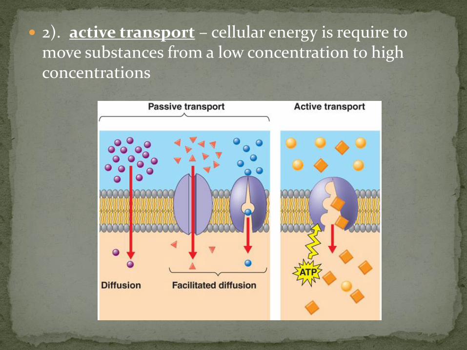
2). active transport – cellular energy is require to move substances from a low concentration to high concentrations
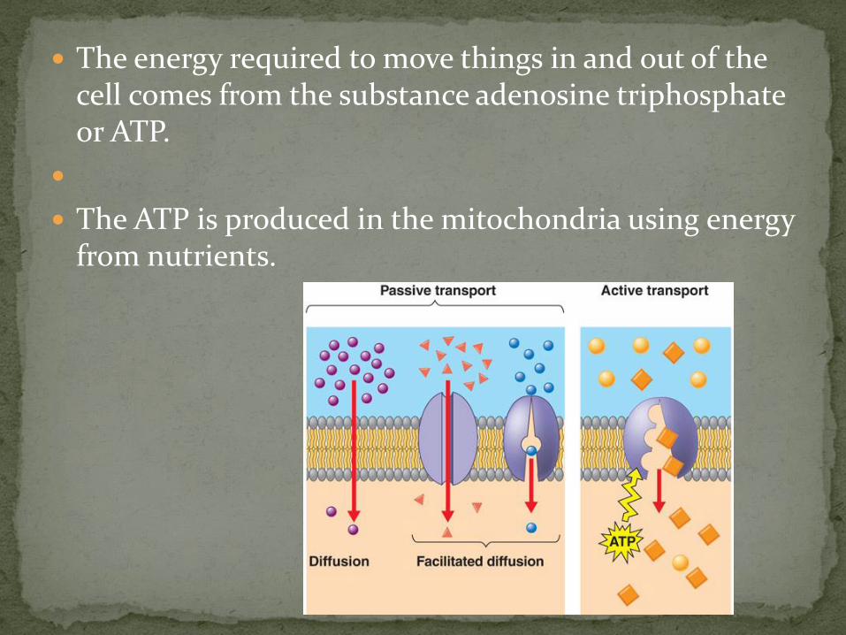
The energy required to move things in and out of the cell comes from the substance adenosine triphosphate or ATP.
The ATP is produced in the mitochondria using energy from nutrients.

Passive transport uses diffusion and filtration to transport molecules in and out of the cell.
Active transport uses ion pumps to pump molecules across the membrane.

All human cells reproduce by a process called mitosis.
Cell reproduction transfers heritable traits that are tied to the production of proteins.
Two nucleic acids: ribonucleic acid (RNA) and deoxyribonucleic acid (DNA) play crucial roles in protein synthesis.

Chromosomes which are made up of DNA make the passing of genetic information possible. The shape of DNA is described a double helix.
DNA bases are adenine, thymine, guanine, and cytosine. The base pairing rules are Adenine with thymine and guanine with cytosine. This is called complementary base pairing.

gene – is a specific segment of base pairs in a chromosome.
The order of the base pairs in each gene determines heredity.
RNA is specialized for transferring genetic information from the nucleus to the cytoplasm.

transcription – the double-stranded DNA unwinds and it makes messenger RNA (mRNA)
translation – is the synthesis of proteins by the ribosomes. The amino acid sequence that makes each protein is determined by the mRNA.


cell division – involves the division of the nucleus and the cytoplasm. The splitting of the nucleus is called mitosis.
This results in two daughter cells that have the exact same genetic information.

Interphase – this is said to be the resting stage, but the very important step of the DNA replicating itself occurs in this stage
As DNA replicates the double helix relaxed and starts to pull apart. A new strand starts to form on each side from materials in the nucleoplasm.

Steps in Mitosis:
1. Prophase – the chromosomes condense. Chromosomes are made up of two strands called chromatids. The chromatids are held together by a beadlike structure called a centromere.
Also during this step the centrioles start to move to opposite ends of the cell. While doing this a network of tubules called spindle fibers stays attached to each centriole. These will help pull the chromosomes apart.

2. Metaphase – the chromosomes align across the center of the cell. By this time the nuclear envelope has disappeared. The spindle fibers have attached to the centriole of each chromosome.

3. Anaphase – the chromosomes are pulled apart. As the centriole breaks apart the spindle fibers pull each chromatid to opposite ends of the cell.
Also a cleavage furrow – which is an indentation in the cell membrane starts to split the cell in two.
4. Telophase – the cell completes division. Two nuclei appear and the chromosomes unravel and become less distinct. Two new nuclear envelopes appear and the cleavage furrow completes the splitting of the cytoplasm.

The splitting of the cytoplasm is often called cytokinesis.
Now you have two identical daughter cells that are in the interphase stage.
During periods of body growth, mitosis allows groups of similar cells to differentiate – or become different from each other and form different body tissues.

If mitosis is out of control and can develop a mass of cells called a neoplasm or cancer. Some of these masses of cells are benign tumors which are harmless or malignant tumors which are very dangerous.

The four main tissues of the human body are
1) Epithelial tissue
2) Connective tissue
3) Muscle tissue
4) Nervous tissue
Tissues in the body are very different from each other in ways such as size, shape and function.

Epithelial tissue - covers and lines many parts of the body. The reason it can do this is because the cells of epithelial tissues are so closely packed together. They are so close together that there are no blood vessels between the cells.

The epithelial cells can be classified by their shape and arrangement.
Shapes:
1. squamous – flat and scale-like
2. cuboidal – cube shaped
3. columnar – higher than they are wide, like columns
4. transitional – shapes that can be stretched
Arrangement:
1. simple – a single layer
2. stratified – many layers

Tissue Location Function
1.) Simple Squamous 1A. Alveoli of Lungs 1.B Lining of blood and lymphatic vessels
1A. Diffusion of oxygen and carbon dioxide 1B. Absorption by diffusion and osmosis
2.) Stratified Squamous 2A. Lining of mouth and esophagus 2B. Surface of skin
2A. Protection 2B. Protection
3.) Simple columnar 3. Lining of stomach and intestines
3. Protection, secretion, and absorption
4.) Stratified Transitional 4. Urinary Bladder 4. Protection
5.) Pseudostratified 5. Surface of lining of trachea
5. Protection
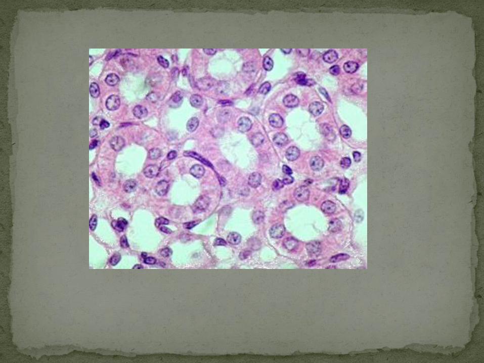
6.) Simple cuboidal 6. Glands; kidney tubules 6. Secretion, absorption
Epithelial Tissue


Connective Tissue
connective tissue – is the most abundant and widely distributed tissue in the body. It is found everywhere in the body.
It is very flexible, very rigid, and even in the form of liquid.
Connective tissue has many different types of cells suspended in what we call a matrix.


There are many types of connective tissue:
1) areolar – this gives shape to internal organs. It is like glue.
2) adipose – is the fat tissue. This has a lot of spaces to store lipids.

3) fibrous connective tissue – bundles of fibers which are called collagen. These make up all the tendons in the body. They have the ability to stretch.
4) bone – hard and calcified tissue. They are made up of building blocks called osteons.

5) cartilage – more gel-like compared to bone. Cartilage cells are called chondrocytes and they have many spaces.
6) blood – it is liquid and has transportational and protective functions. There are red and white blood cells in blood.

7) hemopoietic – connective tissue found in bone marrow. This is where the blood cells are made.

Tissue Location Function
1.) Areolar 1. Between tissues and organs
1. Connection
2.) Adipose 2. Under skin 2. Protection, insulation
3.) Dense fibrous 3. Tendons; ligaments; scar tissue
3. Flexible but strong connection
4.) Bond 4. Skeleton 4. Support, protection
5.) Cartilage 5. Nose, larynx, trachea, ear
5. Firm but flexible support
6.) Blood 6. Blood vessels 6. Transportation
7.) Hemopoietic Tissue 7. Red bone marrow 7. Blood cell formation
ConnectiveTissue
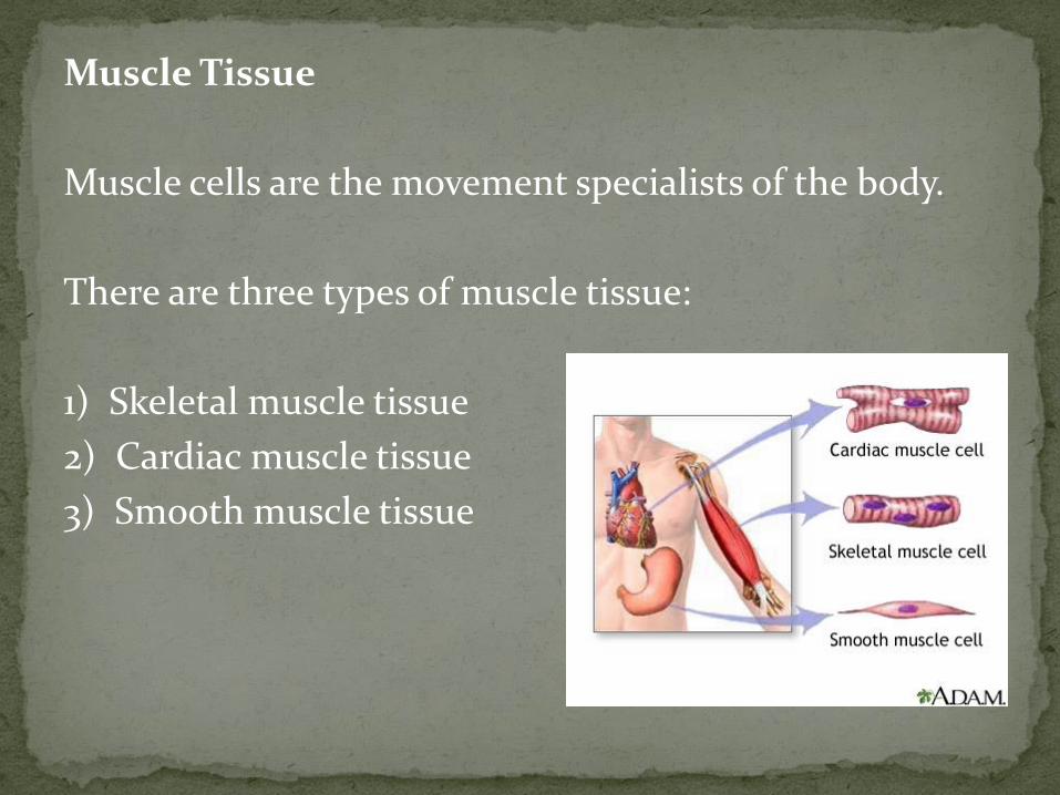
Muscle Tissue
Muscle cells are the movement specialists of the body.
There are three types of muscle tissue:
1) Skeletal muscle tissue
2) Cardiac muscle tissue
3) Smooth muscle tissue

Skeletal muscle tissue – is often called voluntary. The reason for this is these muscles can be controlled.
These are the muscles attached to the skeleton.
Skeletal muscle is characterized by many nuclei per cell and also many striations. Under the microscope striations look like dark bands.

Cardiac muscle tissue – is found in the heart. These muscles are involuntary. We cannot control our heartbeat.
The cardiac muscle fibers branch to form an interlocking mass of tissue.

Smooth muscle tissue – is also an involuntary muscle. They only have one nucleus per fiber and do not have striations so they appear smooth.
Smooth muscle forms the walls of blood vessels and the walls of the intestines. The muscles contract to move things through the organs such as blood and food.

Tissue Location Function
1.) Skeletal 1. Muscle that attaches to bone
1. Movement of bones
2.) Cardiac 2. Wall of heart 2. Contraction of heart
3.) Smooth 3. Walls of digestive and respiratory tract. Blood vessels
3. Movement of substances
Muscle Tissue

Nervous tissue
nervous tissue – its function is to communicate between body structures and the brain.
This needs to happen very rapidly to avoid damaging the body.
neurons – are nerve cells

Neurons consist of three things:
1) cell body – the main part of the cell which contains the nucleus
2) axon – carries impulses away from the cell body
3) dendrites – carries impulses toward the cell body

Tissue Location Function
1.) Nervous 1. Brain and spinal cord, nerves
1. Irritability, conduction
Nervous Tissue