trim29 regulates the p63-mediated pathway in …...1 trim29 regulates the p63-mediated pathway in...
TRANSCRIPT

Instructions for use
Title TRIM29 regulates the p63-mediated pathway in cervical cancer cells
Author(s) Masuda, Yasushi; Takahashi, Hidehisa; Hatakeyama, Shigetsugu
Citation Biochimica et Biophysica Acta. Molecular Cell Research, 1853(10A), 2296-2305https://doi.org/10.1016/j.bbamcr.2015.05.035
Issue Date 2015-10
Doc URL http://hdl.handle.net/2115/62917
Rights © 2015. This manuscript version is made available under the CC-BY-NC-ND 4.0 licensehttp://creativecommons.org/licenses/by-nc-nd/4.0/
Rights(URL) https://creativecommons.org/licenses/by-nc-nd/4.0/
Type article (author version)
File Information BBA_Hatakeyama.pdf
Hokkaido University Collection of Scholarly and Academic Papers : HUSCAP

1
TRIM29 regulates the p63-mediated pathway in cervical cancer cells
Yasushi Masuda a, Hidehisa Takahashi a, Shigetsugu Hatakeyama a,*
aDepartment of Biochemistry, Hokkaido University Graduate School of Medicine, Kita
15, Nishi 7, Kita-ku, Sapporo, Hokkaido 060-8638, Japan
*Corresponding author. Dr. Shigetsugu Hatakeyama, Department of Biochemistry,
Hokkaido University Graduate School of Medicine, Kita 15 Nishi 7, Kita-ku, Sapporo,
Hokkaido 060-8638, Japan
Tel: +81-11-706-5899; Fax: +81-11-706-5169
E-mail: [email protected]
Keywords: TRIM29, cell adhesion, cervical cancer, p63, integrin
2
Abstract
Cell invasion and adhesion play an important role in cancer metastasis and are
orchestrated by a complicated network of transcription factors including p63. Here, we
show that a member of the tripartite motif protein family, TRIM29, is required for
regulation of the p63-mediated pathway in cervical cancer cells. TRIM29 knockdown
alters the adhesion and invasion activities of cervical cancer cells. TRIM29 knockdown
and overexpression cause a significant decrease and increase of TAp63α expression,
respectively. TRIM29 knockdown alters the expression pattern of integrins and
increases ZEB1 expression. TRIM29 is required for suppression of an increase in the
adhesion activity of cells by TAp63α. These findings suggest that TRIM29 regulates the
p63-mediated pathway and the behavior of cervical cancer cells.

3
Highlights
l TRIM29 regulates cell adhesion and invasion.
l TRIM29 regulates TAp63α and integrin expression
l TRIM29 interacts with TA p63α in cervical cancer cells

4
1. Introduction
Invasion and migration are significant hallmarks of metastatic cancer cells.
Epithelial to mesenchymal transition (EMT) allows cancer cells to acquire metastatic
ability [1]. During the EMT process, interaction of integrins with the extracellular
matrix (ECM) exerts a prominent role by regulating the function of matrix
metalloproteinases (MMPs) [1]. The degradation of ECM components by MMPs
contributes to cancer metastasis by changing the tumor microenvironment [1]. These
biological processes are orchestrated by a network of transcription factors including
SNAIL1/2, TWIST1/2 and ZEB1/2 [1]. In addition to these transcription factors, one of
the members of the p53 family, p63, is involved in cancer metastasis by regulating the
expression of integrins [2, 3].
Uterine cervical cancer is one of the common health problems for women
worldwide [4]. Human papillomavirus (HPV) is a significant risk factor associated with
invasive cervical cancer [5]. Among the various HPV types, HPV-18 is strongly related
to adenocarcinoma of the uterine cervix, whereas HPV-16 is detected more frequently in
squamous cell carcinoma than in adenocarcinoma [6, 7]. HPV infection enhances the
invasion activity of cervical cancer cells by upregulation of MMPs [8]. In cervical
cancer cells, epidermal growth factor promotes the transition of epithelial cells to
mesenchymal cells, being dependent on SNAIL1 and α5β1 integrin [9]. Interaction of
α5β1 integrin with fibronectin contributes to the migration with up-regulation of
MMP-9 in cervical cancer cells [10]. Although EMT-related genes contribute to
metastasis of cervical cancer, how cervical cancer cells gain invasive ability remains to
elucidated.

5
TRIM29 has been identified as an ataxia telangiectasia group D-complementing
gene in a functional complementation study with an AT cell line by transfection with a
human cosmid library [11]. TRIM29 is a member of the tripartite motif (TRIM) protein
family and is specifically expressed in squamous cells [12, 13]. There is increasing
evidence that TRIM29 enhances or suppresses cell invasion depending on cell type. In
breast cancer cells, TRIM29 suppresses invasiveness by down-regulating the expression
of TWIST1 [14]. TRIM29 promotes cell invasion by regulating MMP-9 in lung cancer
[15]. TRIM29 also regulates the invasion of pancreatic cancer cells by inducing EMT
[16]. Furthermore, it has been reported that overexpression of TRIM29 is associated
with malignancy of various cancers including bladder, colorectal, gastric lung and
pancreatic cancers [12]. To date, it has been controversial whether TRIM29 functions as
an oncogene or tumor suppressor. Although expression of TRIM29 is closely related to
the characteristics and malignancy of cancer, the relationship between TRIM29 and
cervical cancers remains incompletely understood.
In this study, we identified TRIM29 as a regulator of the p63-mediated pathway in
cervical cancer cells. TRIM29 was required for the suppression of ZEB1 and integrin β1
(ITGB1). Depletion of TRIM29 suppressed an elevation of the adhesion activity of cells
by TAp63α. We propose that TRIM29 regulates the p63-mediated pathway and the
behavior of cervical cancer cells.
6
2. Materials and Methods
2.1. Plasmid
Full-length human TRIM29 (NM_012101.3) was amplified using total RNA from
HeLa cells. TAp63α-FLAG was a gift from David Sidransky (Addgene plasmid #
27008).
2.2. Generation of stable cell lines
cDNAs encoding full-length TRIM29 and TAp63α were introduced by retroviral
transduction into a HeLa S3 cell line stably expressing the mouse ectopic retrovirus
receptor (mCAT-1) [17]. Retroviruses were produced with Plat-E packaging cells [18].
2.3. Cell culture
HeLa, HeLa S3 and SiHa cell lines were obtained from American Type Culture
Collection (Manassas, VA), which authenticates human cell lines in their collection, and
were cultured in DMEM (Sigma-Aldrich Corp., St Louis, MO) supplemented with 10%
fetal bovine serum (FBS) under an atmosphere of 5% CO2 at 37°C. Unless otherwise
indicated, cells were cultured in a 100-mm plastic dish (Corning 430167; Corning Co.,
NY, USA). For large-scale cultures, parental and FLAG-tagged TRIM29-expressing
HeLa S3 cells were cultured in Joklik’s medium (Sigma) supplemented with 5% calf
7
serum (Invitrogen) using spinner culture.
2.4. Antibodies
Mouse anti-FLAG M2 was purchased from Sigma (Sigma). Mouse anti-TRIM29
(A-5: sc-166718), rabbit anti-p63 (H-137: sc-8343), mouse anti-p53 (DO-1: sc-126)
rabbit anti-Pol II (N-20: sc-899) and mouse anti-actin (C-2: sc-8432) were purchased
from Santa Cruz Biotechnology (Santa Cruz, CA.). Rabbit anti-Integrin β4 (4707) was
purchased from Cell Signaling Technology (Beverly, MA). Rabbit anti-Integrin β1
(ab52971) was purchased from Abcam (Cambridge, UK). Mouse anti-GAPDH
(AM4300) was purchased from Ambion (Austin, TX). Rabbit anti-MED12 was
purchased from Bethyl Laboratories (Bethyl Laboratories, Montbomery, TX).
2.5. siRNA transfections
HeLa S3, HeLa and SiHa cells in 10-cm dishes (~2 × 10⁶ cells/dish) were
transfected with 25 nM siRNA ON-TARGETplus human TRIM29 siRNA SMART pool
(Dharmacon, Pittsburgh, PA) or 25 nM siGENOME NON-TARGETING siRNA Pool
#2 (Dharmacon) using Lipofectamine RNAi MAX (Invitrogen). After 48-h culture, cells
were subjected to each analysis.
2.6.Quantitative real-time PCR
Total RNA was extracted using ISOGEN II (Nippon Gene, Tokyo, Japan). cDNA

8
was prepared by using oligonucleotide (dT) and a ReverTra-Plus kit (TOYOBO, Japan)
for quantitative real-time PCR (qRT-PCR). The threshold cycle (Ct) values were
determined by qRT-PCR using Express SYBRGreenER qPCR Supermix Universal
(Invitrogen) on an ABI Prism 7900HT Sequence Detection System (Applied Biosystems,
Foster City, CA). ΔCt was obtained by subtracting the Ct value of the GAPDH gene
from the Ct value of the respective gene. The relative expression level of mRNA was
determined by 2-ΔCt. All PCR primers were as follows: TRIM29,
5’-AAAGGCTATCCCTCCCTCAT-3’ (forward) and
5’-TAGAATGGCCGGTAGTGAGA-3’ (reverse); p63, 5’-GCAGCTTTGCAAACC
CATTA-3’ (forward) and 5’-GCTCGGAAGTCCTAGGTTTATT-3’ (reverse); TAp63,
5’-TGTATCCGCATGCAGGACT-3’ (forward) and
5’-CTGTGTTATAGGGACTGGTGGAC-3’ (reverse); ΔNp63,
5’-GAAAACAATGCCCAGACTCAA-3’ (forward) and
5’-TGCGCGTGGTCTGTGTTA-3’ (reverse); p63α,
5’-ATTGATGCTGTGCGATTCACC-3’ (forward) and
5’-TGTTGCTTATTGCGGCGAG-3’ (reverse); p63β,
5’-AACGCCCTCACTCCTACAAC-3’ (forward) and
5’-GACTTGCCAGATCCTGACAATG-3’ (reverse); p63γ,
5’-GCAGCACCAGCACTTACTTCA-3’ (forward) and
5’-TGTTTTGGAGTTTCTCTCCGG-3’ (reverse); SNAIL2,
5’-TTTCTGGGCTGGCCAAACATAAGC-3’ (forward) and
5’-ACACAAGGTAATGTGTGGGTCCGA-3’ (reverse); ZEB1,
5’-CAGGAAAGGAAGGGCAAGAA-3’ (forward) and
5’-TCTGCATCTGACTCGCATTC-3’ (reverse); p53,

9
5’-TTTCGACATAGTGTGGTGGTG-3’ (forward) and
5’-GCCCATGCAGGAACTGTTA-3’ (reverse); ITGB1,
5’-CCTGAAAGTCCCAAGTGTCAT-3’ (forward) and
5’-ACCAACACGCCCTTCATT-3’ (reverse); ITGB4,
5’-GAGCTGCATCGGCTCAA-3’ (forward) and 5’-AACACGTAGGAGTGGTTGG-3’
(reverse); ITGA3, 5’-AAACTGTGGAGGATGTAGGAAG-3’ (forward) and
5’-CACTCCAGACCTAGGACCA-3’ (reverse); ITGA5,
5’-GGGACCTCAGATCCTGAAATG-3’ (forward) and
5’-CTGCAGACTTTGGCTCTCTT-3’ (reverse); ITGA6,
5’-AGACTCTTAACTGTAGCGTGAAC-3’ (forward) and
5’-ATAAGAGACGCCTTGCTGTC-3’ (reverse); FN1,
5’-AGGAATATCTCGGTGCCATTT-3’ (forward) and
5’-CTTCGGGACTGGGTTCAC-3’ (reverse); VIM,
5’-ATCCAAGTTTGCTGACCTCTC-3’ (forward) and
5’-CTCAGTGGACTCCTGCTTTG-3’ (reverse); SOX2,
5’-TACAGCATGATGCAGGACCA-3’ (forward) and
5’-TACTGCAGGGCGCTCAC-3’ (reverse); RB1,
5’-GGATGGAGTATTGGGAGGTTAT-3’ (forward) and
5’-AGTGAACGACATCTCATCTAGG-3’ (reverse); FOXM1,
5’-AAGATGAGTTCTGATGGACTGG-3’ (forward) and
5’-AAGGCTCCTCAACCTTAACC-3’ (reverse); E2F1,
5’-GGAGAAGTCACGCTATGAGAC-3’ (forward) and
5’-GTCGACGACACCGTCAG-3’ (reverse); E2F2, 5’-GTCCCTGAGTTCCCAACC-3’
(forward) and 5’-GAAGTGTCATACCGAGTCTTCT-3’ (reverse); E2F3,
10
5’-AGGCTGGAGCTAGGAGAAA-3’ (forward) and
5’-CGTAGTGCAGCTCTTCCTTT-3’ (reverse); E2F4,
5’-CCGGGAGATTGCTGACAAA-3’ (forward) and
5’-CCTTGTGCTGGTCTAGTTCTT-3’ (reverse); E2F5,
5’-ACCAATGTCTTAGAGGGAATTGA-3’ (forward) and
5’-TTTAGTATTACAGCCAGCACCT-3’ (reverse) ; E2F6,
5’-TCCAAGAACCATATTAGATGGATAGG-3’ (forward) and
5’-ATTAACTCATCCAAAGCATCTTCC-3’ (reverse); HOXB5,
5’-TCCGCAAATATTCCCCTGGA-3’ (forward) and
5’-CGGGTCAGGTAGCGGTTGAA-3’ (reverse); HOXB7,
5’-GACTTGGCGGCGGAGAGTAA-3’ (forward) and
5’-CAGGGTCTGGTAGCGGGTGT-3’ (reverse); HOXB9,
5’-CCACTGGCTGCTGGACTGTT-3’ (forward) and
5’-TATGACTGGGCCAGGTTCTTTG-3’ (reverse); BRCA1,
5’-GCATGCTGAAACTTCTCAACC-3’ (forward) and
5’-CGTACTTTCTTGTAGGCTCCTT-3’ (reverse); GAPDH,
5’-GTCAACGGATTTGGTCGTATTG-3’ (forward) and
5’-TGTAGTTGAGGTCAATGAAGGG-3’ (reverse).
2.7. Cell adhesion assay
Cell adhesion assay to fibronectin, collagen I, collagen IV, laminin I and vimentin
was performed using a CytoSelect 48-well cell adhesion assay extracellular matrix
(ECM) array (Cell Biolabs Inc., San Diego, CA) according to the manufacturer’s

11
instructions. Cells were incubated on the ECM array for 1 h under an atmosphere of 5%
CO2 at 37°C in a serum-free medium. Each well was gently washed to remove
non-adherent cells. Adherent cells were stained by Cell Stain Solution. The stains were
extracted by Extract Solution and quantified by measuring absorbance at 595 nm.
2.8. Cell invasion assay
Cell invasion assay was performed using a CytoSelect 24-well cell invasion assay
kit (Cell Biolabs Inc.) according to the manufacturer’s instructions. Cells were incubated
on the upper chamber on a polycarbonate membrane coated with basement membrane
matrix in a serum-free medium. Cells were incubated for 1 h under an atmosphere of 5%
CO2 at 37°C. Non-invasive cells were gently removed from the interior of the chamber.
Cells that had invaded to the bottom of the membrane were stained by Cell Stain
Solution. The stains were extracted by Extract Solution and quantified by measuring
absorbance at 595 nm.
2.9. Immunostaining
Cells were cultured on a cover glass in DMEM supplemented with 10% FBS under
an atmosphere of 5% CO2 at 37°C. The cells were fixed by 4% paraformaldehyde in
PBS for 15 min at room temperature, washed with PBS three times, and then incubated
with blocking buffer (5% normal goat serum and 0.1% Triton X-100 in PBS) for 1 h.
After blocking, the cells were incubated with primary antibodies in blocking buffer
overnight at 4°C and incubated with goat Alexa Fluor 488-labeled anti-mouse IgG or

12
goat Alexa Fluor 555-labeled anti-rabbit IgG antibody (Invitrogen Life Technologies,
Carlsbad, CA) for 1 h at room temperature. DNA was counterstained with 0.4 µg/ml
DAPI in PBS for 30 min at room temperature. Confocal images were taken using an
upright microscope (FV1000, Olympus, Tokyo, Japan).
2.10. Analysis of focal adhesion area
Cells were transfected with siControl or siTRIM29 and incubated on
fibronectin-coated coverslips in serum-free medium. After 48 h, cells were fixed and
stained with anti-actin and anti-ITGB1 antibodies. DNA was counterstained with DAPI
(0.4 µg/ml). Cell adhesion area was calculated as the ratio of ITGB1-stained area to
DAPI-stained area. ITGB1- and DAPI-stained areas were quantified using NIH ImageJ
software (n = 20).
2.11. FLAG immunoprecipitation
Nuclear extracts of HeLa S3 cells expressing FLAG-tagged TRIM29 were prepared
according to Dignam’s method [19]. Anti-FLAG affinity purifications were performed
as described previously [20].

13
3. Results
3.1. TRIM29 regulates cell adhesion and invasion
To reveal the function of TRIM29 in cervical cancer cells, we knocked down
TRIM29 in HeLa S3 (HPV-18-positive), HeLa (HPV-18-positive) and SiHa
(HPV-16-positive) cells by siRNA transfection and then performed an in vitro cell
adhesion assay using plates coated with fibronectin, fibrinogen, collagen I, collagen IV
or laminin I. TRIM29 knockdown in HeLa S3 cells increased the adhesion activity of
cells to fibronectin (4.4-fold), fibrinogen (2.6-fold), collagen IV (1.7-fold) and collagen
I (1.5-fold) (Fig. 1A), and TRIM29 knockdown in HeLa cells increased in adhesion
activity to fibronectin (2.2-fold), collagen IV (2.7-fold) and collagen I (3.8-fold) (Fig.
1B). TRIM29 depletion in SiHa cells did not have a significant effect on adhesion
activity (Fig. 1C). It has been reported that TRIM29 regulates invasion of lung and
breast cancer cells [14-16]. To examine whether TRIM29 regulates cell invasion, we
performed an in vitro cell invasion assay using polycarbonate membranes coated with
basement membrane matrix. TRIM29 knockdown reduced the invasion activity of HeLa
S3 and SiHa cells (Fig. 1D). In addition, we confirmed a reciprocal relationship between
cell morphology and TRIM29 knockdown in each cell line. Knockdown of TRIM29
caused a significant change in morphology of HeLa S3 cells but not morphology in
HeLa and SiHa cells (Fig. 1E). These findings suggest that TRIM29 regulates adhesion
and invasion depending on the type of cervical cancer cells.
3.2. Effect of TRIM29 knockdown on focal adhesion
14
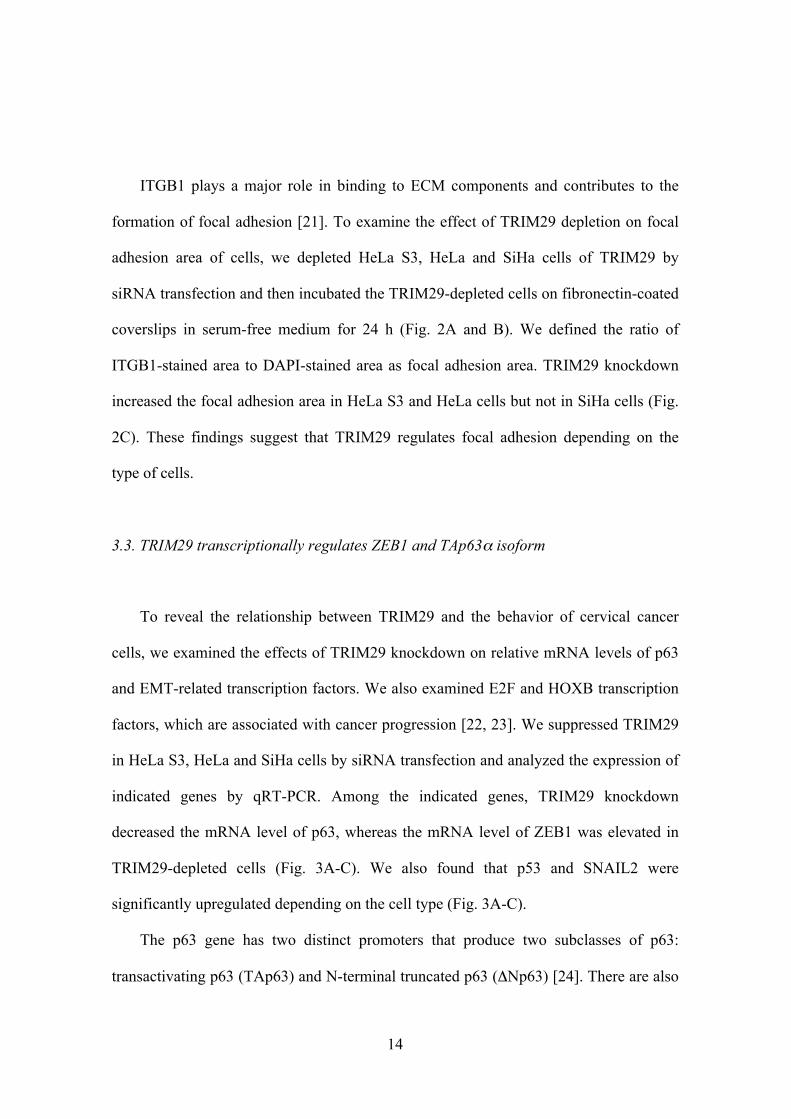
ITGB1 plays a major role in binding to ECM components and contributes to the
formation of focal adhesion [21]. To examine the effect of TRIM29 depletion on focal
adhesion area of cells, we depleted HeLa S3, HeLa and SiHa cells of TRIM29 by
siRNA transfection and then incubated the TRIM29-depleted cells on fibronectin-coated
coverslips in serum-free medium for 24 h (Fig. 2A and B). We defined the ratio of
ITGB1-stained area to DAPI-stained area as focal adhesion area. TRIM29 knockdown
increased the focal adhesion area in HeLa S3 and HeLa cells but not in SiHa cells (Fig.
2C). These findings suggest that TRIM29 regulates focal adhesion depending on the
type of cells.
3.3. TRIM29 transcriptionally regulates ZEB1 and TAp63α isoform
To reveal the relationship between TRIM29 and the behavior of cervical cancer
cells, we examined the effects of TRIM29 knockdown on relative mRNA levels of p63
and EMT-related transcription factors. We also examined E2F and HOXB transcription
factors, which are associated with cancer progression [22, 23]. We suppressed TRIM29
in HeLa S3, HeLa and SiHa cells by siRNA transfection and analyzed the expression of
indicated genes by qRT-PCR. Among the indicated genes, TRIM29 knockdown
decreased the mRNA level of p63, whereas the mRNA level of ZEB1 was elevated in
TRIM29-depleted cells (Fig. 3A-C). We also found that p53 and SNAIL2 were
significantly upregulated depending on the cell type (Fig. 3A-C).
The p63 gene has two distinct promoters that produce two subclasses of p63:
transactivating p63 (TAp63) and N-terminal truncated p63 (ΔNp63) [24]. There are also

15
C-terminal splicing variants known as p63α, p63β and p63γ. To determine which
isoforms of p63 are regulated by TRIM29, we performed qRT-PCR using previously
reported primers that are specific for the isoforms of p63 (TAp63, ΔNp63, p63α, p63β
and p63γ) [3]. While TAp63 isoforms were highly expressed, mRNA level of the
ΔNp63 isoform was much lower than that of the TAp63 isoform (Fig. 3D-F). In addition,
mRNA level of the p63α isoform was much higher than the levels of p63β and p63γ
isoforms (Fig. 3D-F). TRIM29 knockdown in each cell line suppressed the expression
of TAp63, p63α and p63γ isoforms (Fig. 3D-F). Taken together, these findings indicate
that TRIM29 is a transcriptional regulator of TAp63α and ZEB1 in cervical cancer cells.
3.4. TRIM29 regulates the expression of cell adhesion genes
p63 regulates the expression of cell adhesion genes including β1-integrin (ITGB1),
β4-integrin (ITGB4), α3-integrin (ITGA3), α5-integrin (ITGA5), α6-integrin (ITGA6),
fibronectin (FN1) and vimentin (VIM) [2]. To clarify the relationship between TRIM29
and p63, we analyzed the effect of TRIM29 depletion on the expression of these genes.
Knockdown of TRIM29 in HeLa S3 and HeLa cells had similar effects on the
expression of ITGB1, ITGB4, ITGA5 and FN1 (Fig. 4A and B). TRIM29 knockdown in
SiHa cells caused an increase in the expression of ITGB4 and a decrease in the
expression of FN1 (Fig. 4C). In addition, loss of TRIM29 in each cell line increased the
mRNA level of VIM, which is a marker of mesenchymal cells (Fig. 4). These findings
suggest that TRIM29 regulates the expression of cell adhesion molecules in cervical
cancer cells. However, the transcriptional regulation of cell adhesion genes by TRIM29
is dependent on the type of cells.
16
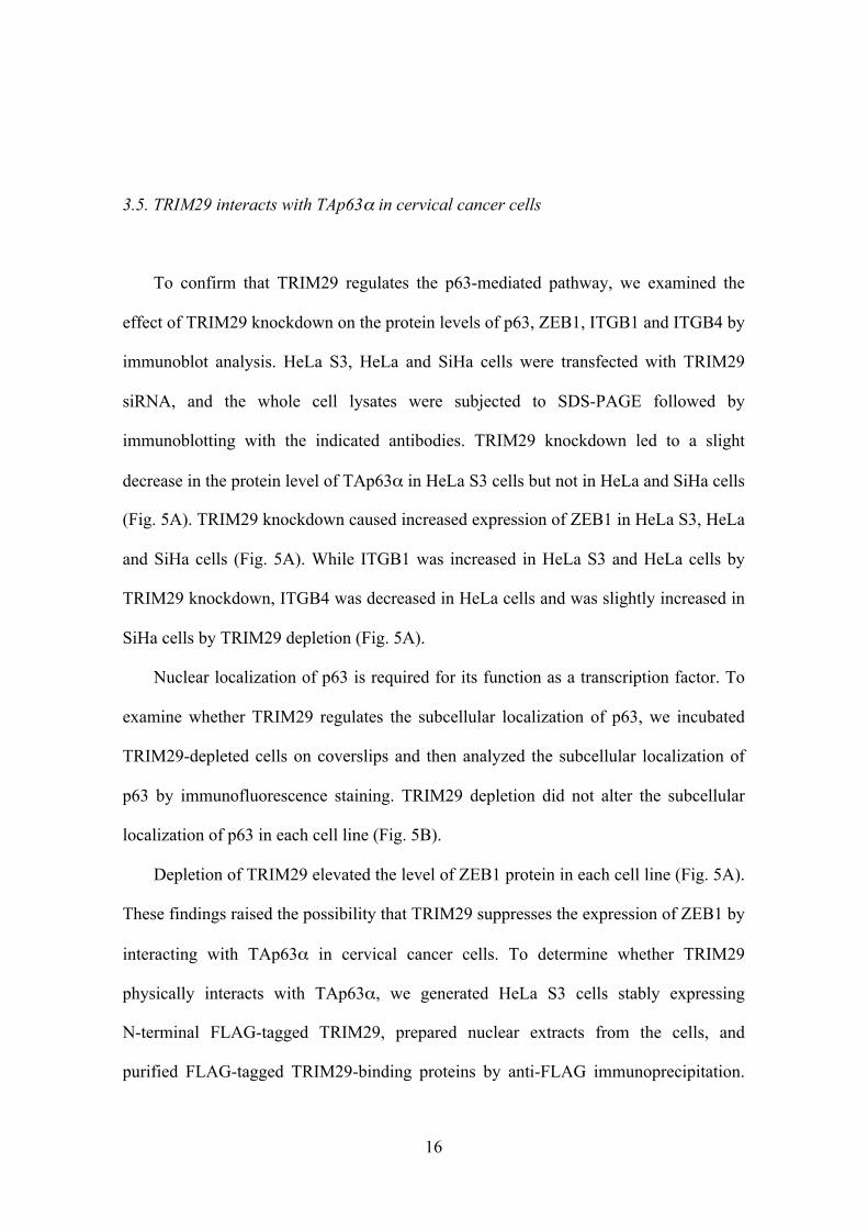
3.5. TRIM29 interacts with TAp63α in cervical cancer cells
To confirm that TRIM29 regulates the p63-mediated pathway, we examined the
effect of TRIM29 knockdown on the protein levels of p63, ZEB1, ITGB1 and ITGB4 by
immunoblot analysis. HeLa S3, HeLa and SiHa cells were transfected with TRIM29
siRNA, and the whole cell lysates were subjected to SDS-PAGE followed by
immunoblotting with the indicated antibodies. TRIM29 knockdown led to a slight
decrease in the protein level of TAp63α in HeLa S3 cells but not in HeLa and SiHa cells
(Fig. 5A). TRIM29 knockdown caused increased expression of ZEB1 in HeLa S3, HeLa
and SiHa cells (Fig. 5A). While ITGB1 was increased in HeLa S3 and HeLa cells by
TRIM29 knockdown, ITGB4 was decreased in HeLa cells and was slightly increased in
SiHa cells by TRIM29 depletion (Fig. 5A).
Nuclear localization of p63 is required for its function as a transcription factor. To
examine whether TRIM29 regulates the subcellular localization of p63, we incubated
TRIM29-depleted cells on coverslips and then analyzed the subcellular localization of
p63 by immunofluorescence staining. TRIM29 depletion did not alter the subcellular
localization of p63 in each cell line (Fig. 5B).
Depletion of TRIM29 elevated the level of ZEB1 protein in each cell line (Fig. 5A).
These findings raised the possibility that TRIM29 suppresses the expression of ZEB1 by
interacting with TAp63α in cervical cancer cells. To determine whether TRIM29
physically interacts with TAp63α, we generated HeLa S3 cells stably expressing
N-terminal FLAG-tagged TRIM29, prepared nuclear extracts from the cells, and
purified FLAG-tagged TRIM29-binding proteins by anti-FLAG immunoprecipitation.

17
As expected, we found that TRIM29 interacted with TAp63α (Fig. 5C). A recent study
showed that knockdown of MED12, which is a key subunit of the Mediator complex,
upregulates the expression of EMT-related genes [25]. To further determine the role of
TRIM29 in the transcription of EMT-related genes, we confirmed whether TRIM29
interacts with MED12 and RNA polymerase II (Pol II). Western blot analysis showed
that TRIM29 binds to Pol II and MED12 (Fig. 5C). These findings suggest that TRIM29
regulates ZEB1 suppression and integrin expression through TAp63α, Pol II and
MED12.
3.6. Forced expression of TRIM29 upregulates ITGB4
To address the reciprocal relationship between TRIM29 and TAp63α, we analyzed
the effect of TRIM29 overexpression on the expression of p63 isoforms and integrins by
qRT-PCR. We confirmed that the mRNA level of TRIM29 was greatly increased in
FLAG-TRIM29-expressing HeLa S3 cells (Fig. 6A). TRIM29 overexpression caused an
increase in the expression levels of TAp63, p63α, p63β and p63γ but not in that of
ΔNp63 (Fig. 6B). Overexpression of TRIM29 also resulted in increases in ITGB4,
ITGA3 and ITGA6 expression levels (Fig. 6C). Although cells expressing
FLAG-TRIM29 showed an increase in ITGB1 expression, TRIM29 overexpression had
less of an impact on ITGB1 expression than did TRIM29 knockdown (Fig. 4A and 6C).
We also confirmed the effect of TRIM29 overexpression on protein levels of TAp63α,
ITGB1 and ITGB4 by Western blotting. TRIM29 overexpression resulted in an
increased level of ITGB4 but not in increased levels of TAp63α and ITGB1 (Fig. 6D).
To confirm the effect of TRIM29 overexpression on cell invasion and adhesion, we

18
performed in vitro cell invasion and adhesion assays. TRIM29 overexpression caused a
1.9-fold increase in the invasion activity of HeLa S3 cells (Fig. 6E). Although TRIM29
overexpression decreased the adhesion activity of cells to collagen IV, there was no
significant difference in the adhesion activity of cells to other ECM components (Fig.
6F). HeLa S3 cells expressing FLAG-TRIM29 also exhibited a marked change with
tight cobblestone-like structures in cell morphology (Fig. 6G). These findings suggest
that TRIM29 overexpression enhances cell invasion and cell-cell interactions in HeLa
S3 cells.
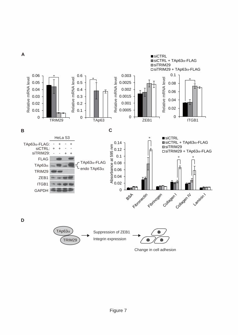
3.7. TRIM29 is required for regulation of the p63-mediated pathway
To clarify the functional relationship between TRIM29 and TAp63α, we generated
HeLa S3 cells stably expressing FLAG-tagged TAp63α and knocked down TRIM29 in
the cells by siRNA transfection. We confirmed that FLAG-tagged TAp63α was
expressed at a higher level than that of endogenous TAp63α (Fig. 7A and B). Although
TRIM29 depletion increased the levels of ZEB1 and ITGB1, overexpression of TAp63α
did not suppress the elevation of ZEB1 and ITGB1 levels (Fig. 7A and B). These
findings suggest that TRIM29 regulates the expression of ZEB1 and ITGB1
independently of TAp63α.
We showed that TRIM29 depletion increases the adhesion activity of HeLa S3 cells
to ECM components (Fig. 1A). To examine the effect of TAp63α overexpression on
cell adhesion to the ECM, we performed in vitro cell adhesion assays. In the presence of
endogenous TRIM29, overexpression of TAp63α had no effect on the adhesion activity
of cells to ECM components (Fig. 7C). Conversely, overexpression of TAp63α

19
increased the adhesion activity of TRIM29-depleted cells to fibronectin, collagen I and
collagen IV (Fig. 7C). These findings suggest that TRIM29 negatively regulates an
increase in the adhesion activity of HeLa S3 cells by TAp63α.

20
4. Discussion
In this study, we showed that TRIM29 is an important regulator of the
p63-mediated pathway in cervical cancer cells (Fig. 7D). p63 has a critical role in cell
adhesion through the regulation of integrin expression [2], and it inhibits the expression
of ZEB1 in pancreatic and bladder cancer cells [30, 31]. TRIM29 knockdown increased
ZEB1 and ITGB1 levels in cervical cancer cells. However, TAp63α overexpression did
not suppress the elevation of ZEB1 and ITGB1 levels in TRIM29-depleted cells,
indicating that TRIM29 suppresses the expression of ZEB1 and ITGB1 independently of
TAp63α. We also identified MED12, which is a key component of the Mediator
complex [28, 29], as a binding partner of TRIM29. Given that loss of MED12
upregulates EMT-related genes in lung cancer cells [25], our observations raise the
possibility that TRIM29 suppresses the expression of ZEB1 and ITGB1 through MED12.
Furthermore, knockdown and overexpression experiments showed that TRIM29
regulates the mRNA level of TAp63α, suggesting that mRNA levels of TAp63α are
regulated through the interaction with TRIM29 and MED12. This is important for
understanding the detailed mechanism by which TRIM29 regulates TAp63α via
MED12.
In cervical cancer cells, depletion and overexpression of TRIM29 affected the
expression pattern of ITGB1 and ITGB4 and adhesion activity to the ECM. ITGB1 is
essential for interaction of basal epithelial cells with the ECM [32]. ITGB4 expression
disrupts adhesive activity and enhances invasive activity of epithelial cells [33].
However, loss of TRIM29 exerted different effects on integrin expression and cell
adhesion in HPV-18-positive cells (HeLa and HeLa S3) and HPV-16-positive cells
21
(SiHa). In cervical cancer cells, HPV genomes are integrated into the host genome,
followed by chromosomal rearrangement and instability [34]. The integration of HPV
genomes into the host genome disrupts several genes [35, 36]. Previous studies showed
that HPV-18 and HPV-16 genomes are integrated into chromosomes 8 and 13,
respectively [35, 36]. Thus, the difference in HPV integration sites may cause an
alteration in the expression of cell adhesion genes. Further detailed studies on the
mechanism by which TRIM29 regulates the expression of EMT-related genes in
HPV-18 and HPV-16-positive cells might be useful for understanding the phenotypes of
cervical cancers.
In this study, we found that TRIM29 enhances invasion of cervical cancer cells.
While TRIM29 inhibits invasion of breast cancer cells [14], it enhances invasion of
pancreatic and lung cancer cells [15, 16]. Degradation of the ECM by MMPs has an
important role in cell invasion and metastasis [37]. It has been reported that HPV
infection increases the expression of various MMPs in cervical cancer cells [8]. Thus,
the invasive activity of cervical cancer cells may be regulated by the interplay between
the function of TRIM29 and the effect of HPV infection. Further studies will be required
for determination of the mechanism by which TRIM29 regulates the invasive activity of
cervical cancer cells.
We found that TAp63α is maintained at a constant protein level independently of
the mRNA level of TAp63α. There are several reports that ubquitination of TAp63α by
E3 ubiquitin ligase plays an important role in its stability. Ectopically expressed
TAp63α is ubiquitinated and degraded in an Itch-dependent manner [26]. In contrast, it
has been reported that βTrCP1 ubiquitinates and stabilizes TAp63α [27]. These findings
suggest that TAp63α is maintained at a constant level in a ubiquitination-dependent

22
manner specific for cervical cancer cells.
Our results demonstrate that TRIM29 regulates the p63-mediated pathway and the
behavior of cervical cancer cells. Further studies on the mechanism by which TRIM29
regulates the characteristics of cervical cancer cells may contribute to the establishment
of new therapies for cervical cancers.

23
Acknowledgements
We would like to thank Drs. Toshio Kitamura (Tokyo University) and Kentaro
Hanada (National Institute of Infectious Diseases) for the plasmids. We are grateful to
Ms. Miho Uchiumi for help in preparing the manuscript and Ms. Misumi Matsuo for
technical assistance.
This work was supported in part by KAKENHI (24112006, 24390065) from the
Ministry of Education, Culture, Sports, Science and Technology in Japan and by The
Naito Foundation and the Uehara Memorial Foundation (to S. Hatakeyama).
24
References
[1] S. Lamouille, J. Xu, R. Derynck, Molecular mechanisms of epithelial-mesenchymal
transition, Nat Rev Mol Cell Biol, 15 (2014) 178-196.
[2] D.K. Carroll, J.S. Carroll, C.O. Leong, F. Cheng, M. Brown, A.A. Mills, J.S. Brugge,
L.W. Ellisen, p63 regulates an adhesion programme and cell survival in epithelial cells,
Nat Cell Biol, 8 (2006) 551-561.
[3] X. Su, D. Chakravarti, E.R. Flores, p63 steps into the limelight: crucial roles in the
suppression of tumorigenesis and metastasis, Nat Rev Cancer, 13 (2013) 136-143.
[4] M.H. Forouzanfar, K.J. Foreman, A.M. Delossantos, R. Lozano, A.D. Lopez, C.J.
Murray, M. Naghavi, Breast and cervical cancer in 187 countries between 1980 and
2010: a systematic analysis, Lancet, 378 (2011) 1461-1484.
[5] J.M. Walboomers, M.V. Jacobs, M.M. Manos, F.X. Bosch, J.A. Kummer, K.V. Shah,
P.J. Snijders, J. Peto, C.J. Meijer, N. Munoz, Human papillomavirus is a necessary
cause of invasive cervical cancer worldwide, J Pathol, 189 (1999) 12-19.
[6] G.M. Clifford, J.S. Smith, M. Plummer, N. Munoz, S. Franceschi, Human
papillomavirus types in invasive cervical cancer worldwide: a meta-analysis, Br J
Cancer, 88 (2003) 63-73.
[7] N. Munoz, F.X. Bosch, S. de Sanjose, R. Herrero, X. Castellsague, K.V. Shah, P.J.
Snijders, C.J. Meijer, G. International Agency for Research on Cancer Multicenter
Cervical Cancer Study, Epidemiologic classification of human papillomavirus types
associated with cervical cancer, N Engl J Med, 348 (2003) 518-527.
[8] J. Kaewprag, W. Umnajvijit, J. Ngamkham, M. Ponglikitmongkol, HPV16
oncoproteins promote cervical cancer invasiveness by upregulating specific matrix

25
metalloproteinases, PLoS One, 8 (2013) e71611.
[9] M.Y. Lee, C.Y. Chou, M.J. Tang, M.R. Shen, Epithelial-mesenchymal transition in
cervical cancer: correlation with tumor progression, epidermal growth factor receptor
overexpression, and snail up-regulation, Clin Cancer Res, 14 (2008) 4743-4750.
[10] G. Maity, S. Fahreen, A. Banerji, P. Roy Choudhury, T. Sen, A. Dutta, A.
Chatterjee, Fibronectin-integrin mediated signaling in human cervical cancer cells
(SiHa), Mol Cell Biochem, 336 (2010) 65-74.
[11] L.N. Kapp, R.B. Painter, L.C. Yu, N. van Loon, C.W. Richard, 3rd, M.R. James,
D.R. Cox, J.P. Murnane, Cloning of a candidate gene for ataxia-telangiectasia group D,
Am J Hum Genet, 51 (1992) 45-54.
[12] S. Hatakeyama, TRIM proteins and cancer, Nat Rev Cancer, 11 (2011) 792-804.
[13] Y. Kanno, M. Watanabe, T. Kimura, K. Nonomura, S. Tanaka, S. Hatakeyama,
TRIM29 as a novel prostate basal cell marker for diagnosis of prostate cancer, Acta
Histochem, 116 (2014) 708-712.
[14] L. Ai, W.J. Kim, M. Alpay, M. Tang, C.E. Pardo, S. Hatakeyama, W.S. May, M.P.
Kladde, C.D. Heldermon, E.M. Siegel, K.D. Brown, TRIM29 Suppresses TWIST1 and
Invasive Breast Cancer Behavior, Cancer Res, 74 (2014) 4875-4887.
[15] Z.P. Tang, Q.Z. Cui, Q.Z. Dong, K. Xu, E.H. Wang, Ataxia-telangiectasia group D
complementing gene (ATDC) upregulates matrix metalloproteinase 9 (MMP-9) to
promote lung cancer cell invasion by activating ERK and JNK pathways, Tumour Biol,
34 (2013) 2835-2842.
[16] L. Wang, H. Yang, E.V. Abel, G.M. Ney, P.L. Palmbos, F. Bednar, Y. Zhang, J.
Leflein, M. Waghray, S. Owens, J.E. Wilkinson, J. Prasad, M. Ljungman, A.D. Rhim,
M. Pasca di Magliano, D.M. Simeone, ATDC induces an invasive switch in

26
KRAS-induced pancreatic tumorigenesis, Genes Dev, 29 (2015) 171-183.
[17] L.M. Albritton, L. Tseng, D. Scadden, J.M. Cunningham, A putative murine
ecotropic retrovirus receptor gene encodes a multiple membrane-spanning protein and
confers susceptibility to virus infection, Cell, 57 (1989) 659-666.
[18] S. Morita, T. Kojima, T. Kitamura, Plat-E: an efficient and stable system for
transient packaging of retroviruses, Gene Ther, 7 (2000) 1063-1066.
[19] J.D. Dignam, R.M. Lebovitz, R.G. Roeder, Accurate transcription initiation by
RNA polymerase II in a soluble extract from isolated mammalian nuclei, Nucleic Acids
Res, 11 (1983) 1475-1489.
[20] H. Takahashi, S. Martin-Brown, M.P. Washburn, L. Florens, J.W. Conaway, R.C.
Conaway, Proteomics reveals a physical and functional link between hepatocyte nuclear
factor 4alpha and transcription factor IID, J Biol Chem, 284 (2009) 32405-32412.
[21] A. Huttenlocher, A.R. Horwitz, Integrins in cell migration, Cold Spring Harb
Perspect Biol, 3 (2011) a005074.
[22] H.Z. Chen, S.Y. Tsai, G. Leone, Emerging roles of E2Fs in cancer: an exit from
cell cycle control, Nat Rev Cancer, 9 (2009) 785-797.
[23] N. Shah, S. Sukumar, The Hox genes and their roles in oncogenesis, Nat Rev
Cancer, 10 (2010) 361-371.
[24] A. Yang, M. Kaghad, Y. Wang, E. Gillett, M.D. Fleming, V. Dotsch, N.C.
Andrews, D. Caput, F. McKeon, p63, a p53 homolog at 3q27-29, encodes multiple
products with transactivating, death-inducing, and dominant-negative activities, Mol
Cell, 2 (1998) 305-316.
[25] S. Huang, M. Holzel, T. Knijnenburg, A. Schlicker, P. Roepman, U. McDermott, M.
Garnett, W. Grernrum, C. Sun, A. Prahallad, F.H. Groenendijk, L. Mittempergher, W.

27
Nijkamp, J. Neefjes, R. Salazar, P. Ten Dijke, H. Uramoto, F. Tanaka, R.L.
Beijersbergen, L.F. Wessels, R. Bernards, MED12 controls the response to multiple
cancer drugs through regulation of TGF-beta receptor signaling, Cell, 151 (2012)
937-950.
[26] M. Rossi, R.I. Aqeilan, M. Neale, E. Candi, P. Salomoni, R.A. Knight, C.M. Croce,
G. Melino, The E3 ubiquitin ligase Itch controls the protein stability of p63, Proc Natl
Acad Sci U S A, 103 (2006) 12753-12758.
[27] J.R. Gallegos, J. Litersky, H. Lee, Y. Sun, K. Nakayama, K. Nakayama, H. Lu,
SCF TrCP1 activates and ubiquitylates TAp63gamma, J Biol Chem, 283 (2008) 66-75.
[28] R.C. Conaway, J.W. Conaway, The Mediator complex and transcription elongation,
Biochim Biophys Acta, 1829 (2013) 69-75.
[29] D.J. Taatjes, The human Mediator complex: a versatile, genome-wide regulator of
transcription, Trends Biochem Sci, 35 (2010) 315-322.
[30] M.N. Tran, W. Choi, M.F. Wszolek, N. Navai, I.L. Lee, G. Nitti, S. Wen, E.R.
Flores, A. Siefker-Radtke, B. Czerniak, C. Dinney, M. Barton, D.J. McConkey, The p63
protein isoform DeltaNp63alpha inhibits epithelial-mesenchymal transition in human
bladder cancer cells: role of MIR-205, J Biol Chem, 288 (2013) 3275-3288.
[31] P. Tucci, M. Agostini, F. Grespi, E.K. Markert, A. Terrinoni, K.H. Vousden, P.A.
Muller, V. Dotsch, S. Kehrloesser, B.S. Sayan, G. Giaccone, S.W. Lowe, N. Takahashi,
P. Vandenabeele, R.A. Knight, A.J. Levine, G. Melino, Loss of p63 and its
microRNA-205 target results in enhanced cell migration and metastasis in prostate
cancer, Proc Natl Acad Sci U S A, 109 (2012) 15312-15317.
[32] I. Taddei, M.A. Deugnier, M.M. Faraldo, V. Petit, D. Bouvard, D. Medina, R.
Fassler, J.P. Thiery, M.A. Glukhova, Beta1 integrin deletion from the basal

28
compartment of the mammary epithelium affects stem cells, Nat Cell Biol, 10 (2008)
716-722.
[33] W. Guo, Y. Pylayeva, A. Pepe, T. Yoshioka, W.J. Muller, G. Inghirami, F.G.
Giancotti, Beta 4 integrin amplifies ErbB2 signaling to promote mammary
tumorigenesis, Cell, 126 (2006) 489-502.
[34] C.A. Moody, L.A. Laimins, Human papillomavirus oncoproteins: pathways to
transformation, Nat Rev Cancer, 10 (2010) 550-560.
[35] A. Adey, J.N. Burton, J.O. Kitzman, J.B. Hiatt, A.P. Lewis, B.K. Martin, R. Qiu, C.
Lee, J. Shendure, The haplotype-resolved genome and epigenome of the aneuploid
HeLa cancer cell line, Nature, 500 (2013) 207-211.
[36] K. Akagi, J. Li, T.R. Broutian, H. Padilla-Nash, W. Xiao, B. Jiang, J.W. Rocco,
T.N. Teknos, B. Kumar, D. Wangsa, D. He, T. Ried, D.E. Symer, M.L. Gillison,
Genome-wide analysis of HPV integration in human cancers reveals recurrent, focal
genomic instability, Genome Res, 24 (2014) 185-199.
[37] K. Kessenbrock, V. Plaks, Z. Werb, Matrix metalloproteinases: regulators of the
tumor microenvironment, Cell, 141 (2010) 52-67.

29
Figure legends
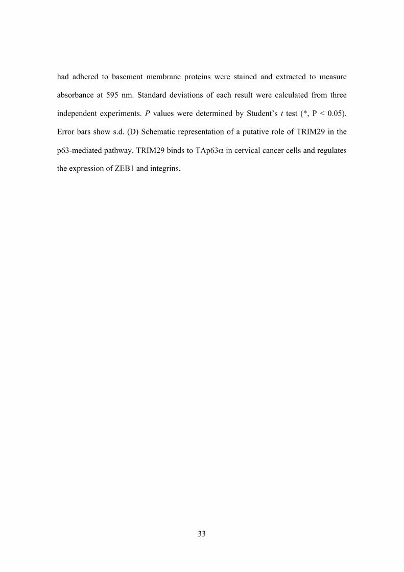
Fig. 1. TRIM29 regulates cell adhesion and invasion. (A,B,C) Effect of TRIM29
knockdown on cell adhesion. HeLa S3 (A), HeLa (B) and SiHa (C) cells transfected
with siCTRL or siTRIM29 were incubated on wells coated with fibronectin, fibrinogen,
collagen I, collagen IV and laminin I for 1 h. Cells that had adhered to basement
membrane proteins were stained and extracted to measure absorbance at 595 nm.
Standard deviations of each result were calculated from three independent experiments.
P values were determined by Student’s t test (*, P < 0.05). Error bars show s.d. (D)
Effect of TRIM29 knockdown on invasion to the basement membrane matrix. HeLa S3,
HeLa and SiHa cells transfected with siCTRL or siTRIM29 were incubated on
membranes coated with basement membrane matrix for 1 h. Invasive cells were stained
and extracted to measure absorbance at 595 nm. Standard deviations of each result were
calculated from three independent experiments. P values were determined by Student’s t
test (*, P < 0.05). Error bars show s.d. (E) Effect of TRIM29 depletion on cell-cell
interactions. HeLa S3, HeLa and SiHa cells were transfected with siCTRL and
siTRIM29 and then incubated for 48 h.
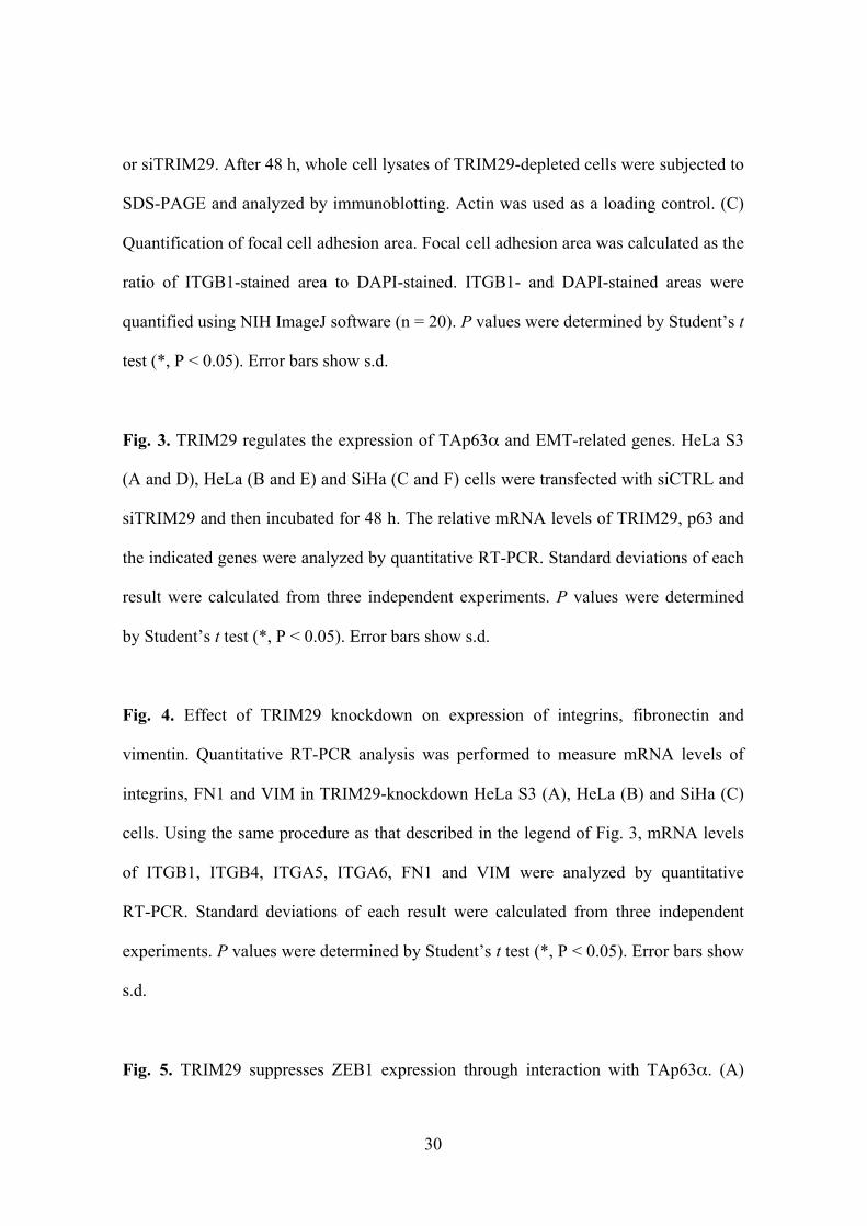
Fig. 2. TRIM29 regulates focal adhesion. (A) Effect of TRIM29 knockdown on focal
adhesion. HeLa S3, HeLa and SiHa cells were transfected with siCTRL or siTRIM29
and incubated on fibronectin-coated coverslips in a serum-free medium. After 48 h, cells
were fixed and stained with anti-actin and anti-ITGB1 antibodies. DNA was
counterstained with DAPI. Scale bar, 40 µm. (B) Immunoblot analysis to confirm
knockdown of TRIM29. HeLa S3, HeLa and SiHa cells were transfected with siCTRL
30
or siTRIM29. After 48 h, whole cell lysates of TRIM29-depleted cells were subjected to
SDS-PAGE and analyzed by immunoblotting. Actin was used as a loading control. (C)
Quantification of focal cell adhesion area. Focal cell adhesion area was calculated as the
ratio of ITGB1-stained area to DAPI-stained. ITGB1- and DAPI-stained areas were
quantified using NIH ImageJ software (n = 20). P values were determined by Student’s t
test (*, P < 0.05). Error bars show s.d.
Fig. 3. TRIM29 regulates the expression of TAp63α and EMT-related genes. HeLa S3
(A and D), HeLa (B and E) and SiHa (C and F) cells were transfected with siCTRL and
siTRIM29 and then incubated for 48 h. The relative mRNA levels of TRIM29, p63 and
the indicated genes were analyzed by quantitative RT-PCR. Standard deviations of each
result were calculated from three independent experiments. P values were determined
by Student’s t test (*, P < 0.05). Error bars show s.d.
Fig. 4. Effect of TRIM29 knockdown on expression of integrins, fibronectin and
vimentin. Quantitative RT-PCR analysis was performed to measure mRNA levels of
integrins, FN1 and VIM in TRIM29-knockdown HeLa S3 (A), HeLa (B) and SiHa (C)
cells. Using the same procedure as that described in the legend of Fig. 3, mRNA levels
of ITGB1, ITGB4, ITGA5, ITGA6, FN1 and VIM were analyzed by quantitative
RT-PCR. Standard deviations of each result were calculated from three independent
experiments. P values were determined by Student’s t test (*, P < 0.05). Error bars show
s.d.
Fig. 5. TRIM29 suppresses ZEB1 expression through interaction with TAp63α. (A)

31
Effect of TRIM29 knockdown on protein levels of TRIM29, TAp63α, ZEB1, ITGB1
and ITGB4 in HeLa S3, HeLa and SiHa cells. HeLa S3, HeLa and SiHa cells were
transfected with siCTRL and siTRIM29 and then incubated for 48 h. Cell lysates were
subjected to SDS-PAGE. Protein levels of TRIM29, p63, ZEB1, ITGB1 and ITGB4
were analyzed by immunoblotting. Actin was used as a loading control. (B) Effect of
TRIM29 knockdown on subcellular localization of p63. HeLa S3, HeLa and SiHa cells
were transfected with siCTRL or siTRIM29 and incubated on coverslips. After 48 h,
cells were fixed and stained with anti-actin and anti-p63 antibodies. DNA was
counterstained with DAPI. Scale bar, 40 µm. (C) Western blot analysis of proteins
associated with FLAG-tagged TRIM29. M2-agarose was incubated with nuclear
extracts of HeLa S3 cells stably expressing FLAG-tagged TRIM29. The bound proteins
were washed, eluted by 3×FLAG peptides, and then analyzed by Western blotting. Cells
that were not transfected were used as control cells.
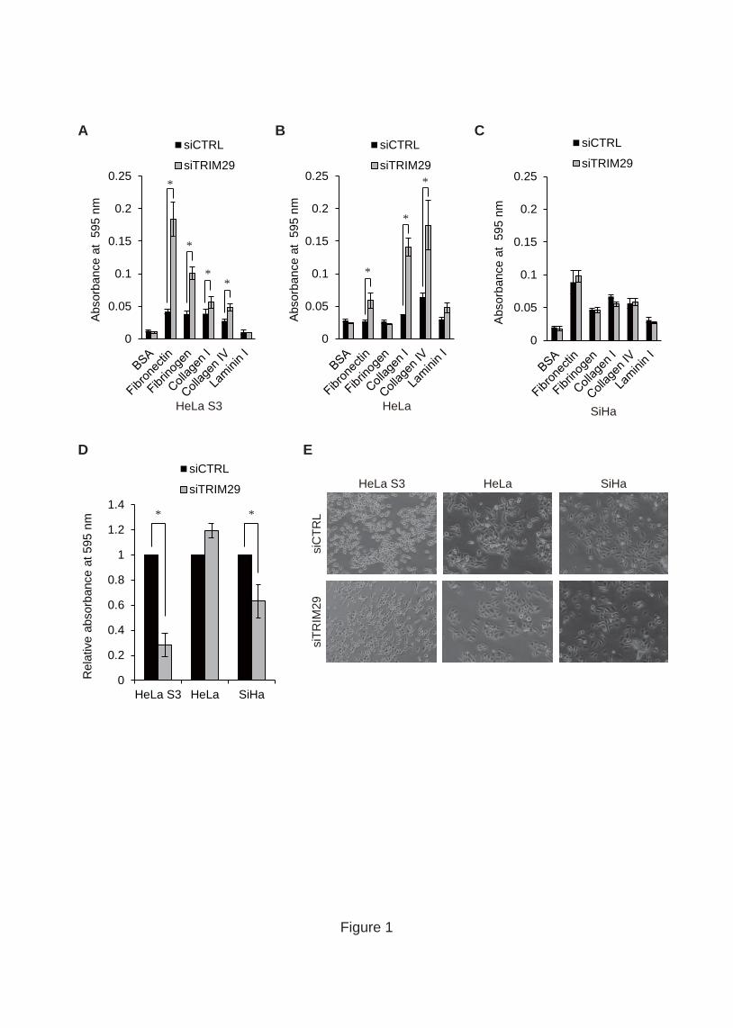
Fig. 6. Effect of overexpression of TRIM29 on p63 and integrin expression. (A-C)
Quantitative PCR analysis of the expression levels of TRIM29 (A), p63 isoforms (B)
and integrins (C) in HeLa S3 cells overexpressing TRIM29. P values were determined
by Student’s t test (*, P < 0.05). Error bars show s.d. (D) Effect of TRIM29
overexpression on protein levels of p63, ITGB1 and ITGB4 in HeLa S3. Cell lysates
from HeLa S3 cells expressing FLAG-tagged TRIM29 were subjected to SDS-PAGE.
Protein levels of p63, ITGB1 and ITGB4 were analyzed by immunoblotting. (E) Effect
of TRIM29 overexpression on cell invasion to the basement membrane matrix. HeLa S3
cells stably expressing FLAG-tagged TRIM29 were incubated on membranes coated
with basement membrane matrix for 1 h. Invasive cells were stained and extracted to

32
measure absorbance at 595 nm. Standard deviations of each result were calculated from
three independent experiments. P values were determined by Student’s t test (*, P <
0.05). Error bars show s.d. (F) Effect of TRIM29 overexpression on cell adhesion to
ECM proteins. HeLa S3 cells stably expressing FLAG-tagged TRIM29 were incubated
in wells coated with fibronectin, fibrinogen, collagen I, collagen IV and laminin I for 1 h.
Cells that had adhered to basement membrane proteins were stained and extracted to
measure absorbance at 595 nm. Standard deviations of each result were calculated from
three independent experiments. P values were determined by Student’s t test (*, P <
0.05). Error bars show s.d. (G) Effect of TRIM29 overexpression on cell-cell
interactions. Images of HeLa S3 cells expressing FLAG-tagged TRIM29 are shown.
Fig. 7. Functional relationship between TRIM29 and TAp63α. (A) Quantitative PCR
analysis of the expression levels of TRIM29, TAp63, ZEB1 and ITGB1 in HeLa S3
cells overexpressing TAp63α. HeLa S3 cells stably expressing FLAG-tagged TAp63
were transfected with siCTRL and siTRIM29 and then incubated for 48 h. The relative
mRNA levels of the indicated genes were analyzed by quantitative RT-PCR. Standard
deviations of each result were calculated from three independent experiments. P values
were determined by Student’s t test (*, P < 0.05). Error bars show s.d. (B) Effect of
TRIM29 knockdown on cells overexpressing TAp63α. HeLa S3 cells expressing
FLAG-tagged TAp63 were depleted of TRIM29 by siRNA transfection. Cell lysates
from the cells were subjected to SDS-PAGE followed by Western blotting with the
indicated antibodies. GAPDH was used as a loading control. (C) Effect of TAp63α
overexpression on cell adhesion to ECM proteins. Cells were incubated in wells coated
with fibronectin, fibrinogen, collagen I, collagen IV and laminin I for 1 h. The cells that
33
had adhered to basement membrane proteins were stained and extracted to measure
absorbance at 595 nm. Standard deviations of each result were calculated from three
independent experiments. P values were determined by Student’s t test (*, P < 0.05).
Error bars show s.d. (D) Schematic representation of a putative role of TRIM29 in the
p63-mediated pathway. TRIM29 binds to TAp63α in cervical cancer cells and regulates
the expression of ZEB1 and integrins.
0
0.2
0.4
0.6
0.8
1
1.2
1.4
HeLa S3 HeLa SiHa
Rel
ativ
e ab
sorb
ance
at 5
95 n
m
Figure 1
A B C
0
0.05
0.1
0.15
0.2
0.25
Abs
orba
nce
at 5
95 n
m
siCTRL
siTRIM29
SiHa
0
0.05
0.1
0.15
0.2
0.25
Abs
orba
nce
at 5
95 n
m
HeLa S3
siCTRL
siTRIM29*
*
**
0
0.05
0.1
0.15
0.2
0.25
Abs
orba
nce
at 5
95 n
m
siCTRL
siTRIM29
HeLa
*
*
*
DsiCTRL
siTRIM29
**
siC
TRL
siTR
IM29
HeLa SiHaHeLa S3
E

Figure 2
A
B
siCTRL siTRIM29 siCTRL siTRIM29 siCTRL siTRIM29
HeLa S3 HeLa SiHa
ITGB1
Actin
DAPI
Merge
C siCTRL
siTRIM29
0
0.5
1
1.5
2
2.5
3
3.5
HeLa S3 HeLa SiHa
Foca
l adh
esio
n ar
ea
* *
TRIM29
Actin
HeLa S3 HeLa SiHasiCTRL:
siTRIM29: --++ -
-++ -
-++

00.20.40.60.8
11.21.41.6
Rel
ativ
e m
RN
A le
vel
* *
* *
*
HeLa S3
HeLa SiHaC
00.20.40.60.8
11.21.41.61.8
Rel
ativ
e m
RN
A le
vel
0
0.5
1
1.5
2
2.5
3
Rel
ativ
e m
RN
A le
vel
B
A
Figure 3
* *
* *
*
* * *
*siTRIM29siCTRL
siTRIM29siCTRL
siTRIM29siCTRL
00.00005
0.00010.00015
0.00020.00025
0.00030.00035
0.00040.00045
TAp63Np6
3p6
3p6
3p6
30
0.000020.000040.000060.00008
0.00010.000120.000140.000160.00018
HeLa S3 HeLa
Rel
ativ
e m
RN
A le
vel
Rel
ativ
e m
RN
A le
vel
D E F
* *
*
*
*
*
0
0.00001
0.00002
0.00003
0.00004
0.00005
0.00006
0.00007
0.00008
Rel
ativ
e m
RN
A le
vel
*
*
SiHa
TAp63Np6
3p6
3p6
3p6
3TAp6
3Np6
3p6
3p6
3bp6
3
siTRIM29siCTRL
siTRIM29siCTRL
siTRIM29siCTRL

0
0.05
0.1
0.15
0.2
0.25
0.3
ITGB1 ITGB4 ITGA3 ITGA5 ITGA6 FN1 VIM
siCTRL
siTRIM29
Figure 4
HeLa S3
HeLa
SiHaC
A
B
*
siCTRL
siTRIM29
siCTRL
siTRIM29
* **
*
Rel
ativ
e m
RN
A le
vel
00.05
0.10.15
0.20.25
0.30.35
0.4
ITGB1 ITGB4 ITGA3 ITGA5 ITGA6 FN1 VIM
*
* *
Rel
ativ
e m
RN
A le
vel
0
0.05
0.1
0.15
0.2
0.25
0.3
ITGB1 ITGB4 ITGA3 ITGA5 ITGA6 FN1 VIM
*
*
Rel
ativ
e m
RN
A le
vel

Figure 5
A C
B
p63
siCTRL siTRIM29 siCTRL siTRIM29 siCTRL siTRIM29
Actin
DAPI
Merge
HeLa S3 HeLa SiHa
IP: FLAG Input
Con
trol
FLAG
-TR
IM29
Pol IIMED12
Con
trol
FLAG
-TR
IM29
TAp63
FLAGITGB1
TAp63
ITGB4
ZEB1
GAPDH
HeLa S3 HeLa SiHasiCTRL:
siTRIM29:TRIM29
--++-
-++ -
-++
Actin
0
0.00005
0.0001
0.00015
0.0002
0.00025
0.0003
0.00035
0.0004
TAp63 Np63 p63 p63 p63
Figure 6
CTRL
FLAG-TRIM29
DC
E F
FLAG
Con
trol
FLAG
-TR
IM29
TAp63ITGB1ITGB4GAPDH
B
Rel
ativ
e m
RN
A le
vel *
*
*
*0
0.25
0.5
0.75
1
1.25
1.5R
elat
ive
mR
NA
leve
l*
CTRL
FLAG-T
RIM29
GCTRL
FLAG-TRIM29
00.010.020.030.040.050.060.070.080.09
0.1
Abs
orba
nce
at 5
95 n
m
CTRL
FLAG-TRIM29
*
00.05
0.10.15
0.20.25
0.30.35
0.40.45
Abs
orba
nce
at 5
95 n
m
CTRL
FLAG-T
RIM29
*
0
0.02
0.04
0.06
0.08
0.1
0.12
0.14
0.16
ITGB1 ITGB4 ITGA3 ITGA5 ITGA6
CTRL
FLAG-TRIM29
*
*
*
*Rel
ativ
e m
RN
A le
vel
A
0
0.02
0.04
0.06
0.08
0.1
0
0.0005
0.001
0.0015
0.002
0.0025
0.003
0
0.1
0.2
0.3
0.4
0.5
0.6
0
0.01
0.02
0.03
0.04
0.05
0.06
0
0.02
0.04
0.06
0.08
0.1
0.12
0.14
Abs
orba
nce
at 5
95 n
m
B C
Figure 7
D
siTRIM29:
TAp63 -FLAG: - + - +siCTRL: + +
+ +- -- -
TRIM29TAp63
ITGB1ZEB1
FLAG
endo TAp63TAp63 -FLAG
GAPDH
HeLa S3
Change in cell adhesion
Suppression of ZEB1
Integrin expression TRIM29
TAp63
*
* *
Rel
ativ
e m
RN
A le
vel
A
Rel
ativ
e m
RN
A le
vel
Rel
ativ
e m
RN
A le
vel
Rel
ativ
e m
RN
A le
vel
siCTRLsiCTRL + TAp63 -FLAGsiTRIM29siTRIM29 + TAp63 -FLAG
siCTRLsiCTRL + TAp63 -FLAGsiTRIM29siTRIM29 + TAp63 -FLAG
TRIM29 TAp63 ZEB1 ITGB1
** *