trypanosoma livingstonei: a new species from african bats ... filethese trypanosomes as a new...
TRANSCRIPT
Universidade de São Paulo
2013
Trypanosoma livingstonei: a new species from
African bats supports the bat seeding
hypothesis for the Trypanosoma cruzi clade Parasites and Vectors, London, v.6, n.1, 2013http://www.producao.usp.br/handle/BDPI/44579
Downloaded from: Biblioteca Digital da Produção Intelectual - BDPI, Universidade de São Paulo
Biblioteca Digital da Produção Intelectual - BDPI
Departamento de Parasitologia - ICB/BMP Artigos e Materiais de Revistas Científicas - ICB/BMP

RESEARCH Open Access
Trypanosoma livingstonei: a new species fromAfrican bats supports the bat seeding hypothesisfor the Trypanosoma cruzi cladeLuciana Lima1, Oneida Espinosa-Álvarez1, Patrick B Hamilton2, Luis Neves3, Carmen SA Takata1, Marta Campaner1,Márcia Attias4, Wanderley de Souza4, Erney P Camargo1 and Marta MG Teixeira1*
Abstract
Background: Bat trypanosomes have been implicated in the evolutionary history of the T. cruzi clade, whichcomprises species from a wide geographic and host range in South America, Africa and Europe, includingbat-restricted species and the generalist agents of human American trypanosomosis T. cruzi and T. rangeli.
Methods: Trypanosomes from bats (Rhinolophus landeri and Hipposideros caffer) captured in Mozambique,southeast Africa, were isolated by hemoculture. Barcoding was carried out through the V7V8 region of SmallSubunit (SSU) rRNA and Fluorescent Fragment Length barcoding (FFLB). Phylogenetic inferences were based onSSU rRNA, glyceraldehyde phosphate dehydrogenase (gGAPDH) and Spliced Leader (SL) genes. Morphologicalcharacterization included light, scanning and transmission electron microscopy.
Results: New trypanosomes from bats clustered together forming a clade basal to a larger assemblage called theT. cruzi clade. Barcoding, phylogenetic analyses and genetic distances based on SSU rRNA and gGAPDH supportedthese trypanosomes as a new species, which we named Trypanosoma livingstonei n. sp. The large and highlypolymorphic SL gene repeats of this species showed a copy of the 5S ribosomal RNA into the intergenic region.Unique morphological (large and broad blood trypomastigotes compatible to species of the subgenusMegatrypanum and cultures showing highly pleomorphic epimastigotes and long and slender trypomastigotes) andultrastructural (cytostome and reservosomes) features and growth behaviour (when co-cultivated with HeLa cells at37°C differentiated into trypomastigotes resembling the blood forms and do not invaded the cells) complementedthe description of this species.
Conclusion: Phylogenetic inferences supported the hypothesis that Trypanosoma livingstonei n. sp. diverged from acommon ancestral bat trypanosome that evolved exclusively in Chiroptera or switched at independentopportunities to mammals of several orders forming the clade T. cruzi, hence, providing further support for the batseeding hypothesis to explain the origin of T. cruzi and T. rangeli.
Keywords: Chiroptera, Taxonomy, Phylogeny, Phylogeography, Evolution, Africa, Trypanosoma cruzi
* Correspondence: [email protected] de Parasitologia, Instituto de Ciências Biomédicas,Universidade de São Paulo, São Paulo, SP 05508-900, BrazilFull list of author information is available at the end of the article
© 2013 Lima et al.; licensee BioMed Central Ltd. This is an Open Access article distributed under the terms of the CreativeCommons Attribution License (http://creativecommons.org/licenses/by/2.0), which permits unrestricted use, distribution, andreproduction in any medium, provided the original work is properly cited.
Lima et al. Parasites & Vectors 2013, 6:221http://www.parasitesandvectors.com/content/6/1/221
BackgroundTrypanosomes (Euglenozoa: Kinetoplastea: Trypanosoma-tidae) are blood parasites widespread in all continents,adapted to all classes of vertebrates and transmitted byleeches and a variety of bloodsucking arthropods. Al-though Chiroptera harbour numerous trypanosome spe-cies with a high prevalence and worldwide distribution,species diversity, vectors, life cycles, distribution and try-panosome evolution remain poorly understood [1-10].The majority of trypanosomes reported in bats have
not been cultivated, and their classification has beenbased exclusively on the morphology of blood trypo-mastigotes. Large blood trypanosomes of the subgenusMegatrypanum, followed by small blood forms of the sub-genus Schizotrypanum, comprise the majority of the try-panosomes reported in bats throughout South America,Asia, Europe and, especially, Africa [1,3,5,7,8,10-14]. Thesubgenus Megatrypanum, originally comprising large bloodtrypanosomes from artiodactyls [15], was amended exclu-sively on a morphological basis to include any large tryp-anosome found in bats, monkeys and rodents [1,2,4,6].Molecular phylogenetic analysis has demonstrated thepolyphyly of the traditional subgenus Megatrypanum,which was revised as a clade comprising trypanosomesfrom ruminants headed by the type species T. theileri, acosmopolitan parasite of cattle [16-19]. However, in thereappraisal of this subgenus, other species from non-ruminant hosts that putatively belong to this subgenusneed to be phylogenetically positioned, especially thosefrom bats, which together with trypanosomes fromartiodactyls, account for most of the species assigned tothis subgenus [1,4,7].Most bat trypanosome species that have been
characterised by molecular approaches belong to thesubgenus Schizotrypanum [8-11,13,14,20-25]. With theexception of T. cruzi, there are no Schizotrypanumspecies in hosts other than bats. T. rangeli was foundin Brazilian bats [26], this species is infective to severalmammals and comprises distinct genotypes [26,27], whichclustered into a clade containing T. conorhini from rats, T.vespertilionis from a European bat and two African try-panosomes from monkey and civet. Although T. rangeli,T. conorhini and T. vespertilionis were morphologicallyclassified into the subgenera Herpetosoma, Megatrypanumand Schizotrypanum, respectively, molecular phylogeniesdemonstrated that they clustered together forming thestrongly supported sister clade of the Schizotrypanumclade. Trypanosoma sp. (T. sp. bat) from an Africanmegabat (suborder Megachiroptera) originally assigned tothe subgenus Megatrypanum was positioned at the edgeof this clade [8-10,18,20,23,24].The major assemblage formed by the subgenus
Schizotrypanum and the clade T. rangeli/T. conorhiniwas designated as the T. cruzi clade. The positioning
of a kangaroo trypanosome at its edge in association withvicariance has supported the southern supercontinenthypothesis for the origin of T. cruzi. Accordingly, thisspecies could have originated in marsupials at a time whenSouth America, Australia and Antarctica formed a singlecontinent. However, in conflict with this hypothesis, someAustralian trypanosomes from marsupials are morerelated to trypanosomes from non-Australian hosts[9,18,20,21,23,24]. The discovery of African terrestrialmammals infected with trypanosomes placed in theT. cruzi clade [18] has complicated the southern super-continent hypothesis.Taken together, the findings that T. c. marinkellei from
South American bats is the closest living relative ofT. cruzi [8,10,22,25,28] followed by T. erneyi from Africanbats [10], the close phylogenetic relationship betweenT. dionisii from Europe and South America [8,9,24], thepresence of T. rangeli in Brazilian bats [26] and the rela-tionships of this species with African (T. sp. bat) andEuropean (T. vespertilionis) bat trypanosomes, and thediscovery of Tcbat, a bat-associated T. cruzi genotypefound in South and Central America [14,22] all supportthe bat seeding hypothesis for the origin of the T. cruziclade [24]. In this scenario, which is the most parsimo-nious for explaining the relationships observed withinthe T. cruzi clade, an ancestral trypanosome parasite inbats diverged to lineages that evolved exclusively inbats, giving rise to the bat-restricted species, or evolvedthrough multiple switches at independent times in hostsof other mammalian orders, including the generalistsT. cruzi and T. rangeli, which also infect bats. Multipletrypanosome jumps between hosts were most likelyfacilitated by the sharing of niches by bats, haematopha-gous insects (vectors) and terrestrial mammals. Oralinfection through the predation of infected bats by othermammals and by the consumption of insect vectors bybats probably played important roles in the colonisationof new hosts by bat trypanosomes. Transmission of try-panosomes among bats is likely to occur by an oralroute when the vector insects are eaten by insectivorousbats. The grooming habits of the bats probably facili-tates the infection by the bat trypanosomes transmittedby ectoparasite cimicids [3,6,7]. Both vectorial and oraltransmission routes are important in the natural transmis-sion cycles of T. cruzi and other trypanosomes nested inthe T. cruzi clade [1,3,6-8].With the discovery of T. erneyi [10] and a new geno-
type of T. dionisii in the UK [9], bat trypanosomes fromthe Old World revealed to be more closely related toSouth American bat trypanosomes than showed by pre-vious studies [8,22,26]. These findings suggested move-ment of bat trypanosomes between the New and Oldworlds occurred in a relatively more recent time thanbat fossil records suggested [9,24].
Lima et al. Parasites & Vectors 2013, 6:221 Page 2 of 17http://www.parasitesandvectors.com/content/6/1/221
Trypanosomes from the T. cruzi clade are likely tohave started to diversify sometime after the great diver-sification of bats in the Eocene (70–58 mya) [29-31].However, the extant species of bat trypanosomes appearto have emerged during a short period and much morerecently than expected based on the fragmented paleon-tological history of bats [9,24].In this study, we isolated and characterised 14 new try-
panosomes from African bats captured in Mozambique,southeast Africa, by inferring phylogenetic relationshipsusing ribosomal SSU rRNA, gGAPDH and SL genes.Sequences from the new bat isolates were compared tothose from other bat trypanosomes determined in thisand in previous studies (including other isolates mor-phologically assignable to the subgenus Megatrypanum)to address taxonomic questions about bat trypanosomes.Comparison of bat trypanosomes by combining molecu-lar, morphological and behavioural information providesnew information on the evolutionary history of battrypanosomes and the origin of the T. cruzi clade.
MethodsCollection sites, capture and identification of batsBats were captured in Mozambique, southeastern Africa,in the district of Chupanga (S18°02′ E35°34′), Zambezivalley, and the Gorongosa National Park (S18°58′E34°21′), both of which are located in the Province ofSofala in central Mozambique (Table 1; Figure 1).Captures were carried out with mist nets; bats wereanaesthetised and blood samples were collected by cardiacpuncture as previously described [8,22]. For the molecularidentification of bats, liver tissue samples were fixed in100% ethanol, processed for genomic DNA and used tosequence the cytochrome b gene (Cyt b) as previouslydescribed [32]. Sequences were analysed by BLAST searchin GenBank.
Detection and culture of bat trypanosomesBat blood samples were examined for the presence oftrypanosomes by using the microhaematocrit (MH),Giemsa-stained blood smear examination and haemo-culture methods as employed before for trypanosomesfrom different vertebrate hosts [8,10,22,33,34]. For thehaemoculture, bat blood samples were transferred to tubescontaining a medium consisting of solid phase blood agarbase (BAB) with an overlay of LIT (liver infusion tryptose)medium containing 10% foetal bovine serum (FBS); thetubes were maintained at 25-28°C for 10–15 days. Positivecultures were transferred to culture flasks containing amonolayer of insect cells (Hi-5 from Trichoplusia ni) andepimastigotes from log-phase cultures were then trans-ferred to TC100 medium (= Grace’s medium) containing10% FBS, with incubation at 25°C. The utilization of insectfeeder cells for the isolation in culture of trypanosomes
largely improves the differentiation of blood trypo-mastigotes to epimastigotes and the multiplication ofepimastigotes in primary cultures [10,33,34]. The isolateswere grown in TC100 with 5.0% FBS for DNA preparationand cryopreservation at the Trypanosomatid CultureCollection (TCC) of the Department of Parasitology,University of São Paulo, Brazil.
Amplification, sequencing and data analysis of SSU rDNAand gGAPDHDNA was extracted from cultured bat trypanosomes byclassical phenol-chloroform method and used as tem-plates for the PCR amplification of DNA sequences.PCR amplification, cloning and sequencing of the vari-able V7-V8 region of SSU rRNA (employed as barcodes),whole SSU rRNA and gGAPDH genes were determinedas before [35-37]. Sequences were aligned using ClustalX [38] and the resulting alignments were manuallyrefined. We created the following alignments for phylo-genetic inferences: a) the V7V8 region of SSU rRNAsequences (~880 bp) from the new bat trypanosomesaligned with their closest Australian and bat trypano-somes, yielding a high similarity index for the new battrypanosomes by BLAST search; b) gGAPDH sequences(~830 bp) of trypanosomes representing all major cladesin the phylogenetic tree of Trypanosoma, using non-trypanosome trypanosomatids as an outgroup; c) conca-tenated gGAPDH and SSU rRNA sequences (~3.3 kb)from 6 new isolates and several species of T. cruzi cladeusing T. lewisi as an outgroup. The species included inthe phylogenetic trees and their respective host, geo-graphical origin and GenBank accession numbers areshown in Table 1. Phylogenies were inferred by usingmaximum likelihood (ML), Bayesian inferences (BI) andparsimony (P) analyses. Parsimony and bootstrap ana-lyses were carried out using PAUP version 4.0b10 [39]with 500 replicates of a random addition sequencefollowed by branch swapping (RAS-TBR) as previouslydescribed [36,37]. The ML analyses were performedusing RAxML v.2.2.3 [40]. Tree searches were performedwith GTRGAMMA, with 500 maximum parsimonystarting trees. Model parameters were estimated inRAxML for the duration of the tree search. Nodal sup-port was estimated with 500 bootstrap replicates inRAxML using GTRGAMMA and maximum parsimonystarting trees. MrBayes v3.1.2 [41] was used for BI infer-ences as described previously [36,37].
FFLB - Fluorescent fragment length barcodingDNA from cultured trypanosomes and from bat bloodsamples were tested by FFLB carried out using four primersets and PCR conditions described previously [10,42,43].
Lima et al. Parasites & Vectors 2013, 6:221 Page 3 of 17http://www.parasitesandvectors.com/content/6/1/221
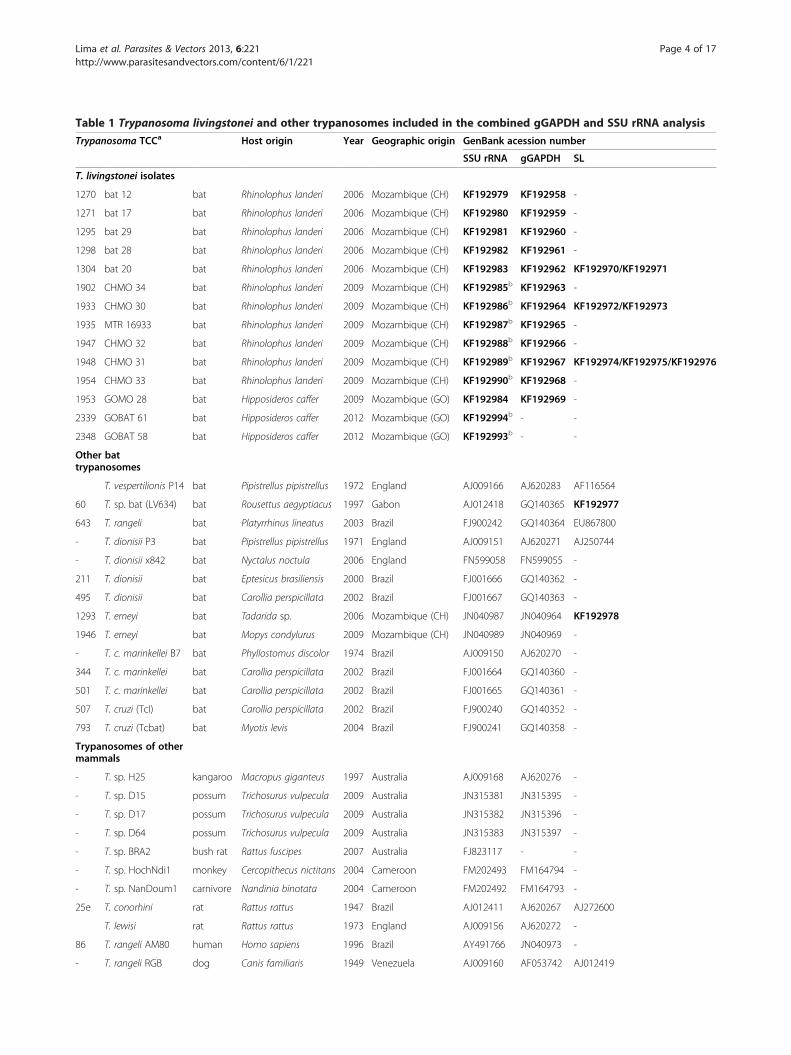
Table 1 Trypanosoma livingstonei and other trypanosomes included in the combined gGAPDH and SSU rRNA analysis
Trypanosoma TCCa Host origin Year Geographic origin GenBank acession number
SSU rRNA gGAPDH SL
T. livingstonei isolates
1270 bat 12 bat Rhinolophus landeri 2006 Mozambique (CH) KF192979 KF192958 -
1271 bat 17 bat Rhinolophus landeri 2006 Mozambique (CH) KF192980 KF192959 -
1295 bat 29 bat Rhinolophus landeri 2006 Mozambique (CH) KF192981 KF192960 -
1298 bat 28 bat Rhinolophus landeri 2006 Mozambique (CH) KF192982 KF192961 -
1304 bat 20 bat Rhinolophus landeri 2006 Mozambique (CH) KF192983 KF192962 KF192970/KF192971
1902 CHMO 34 bat Rhinolophus landeri 2009 Mozambique (CH) KF192985b KF192963 -
1933 CHMO 30 bat Rhinolophus landeri 2009 Mozambique (CH) KF192986b KF192964 KF192972/KF192973
1935 MTR 16933 bat Rhinolophus landeri 2009 Mozambique (CH) KF192987b KF192965 -
1947 CHMO 32 bat Rhinolophus landeri 2009 Mozambique (CH) KF192988b KF192966 -
1948 CHMO 31 bat Rhinolophus landeri 2009 Mozambique (CH) KF192989b KF192967 KF192974/KF192975/KF192976
1954 CHMO 33 bat Rhinolophus landeri 2009 Mozambique (CH) KF192990b KF192968 -
1953 GOMO 28 bat Hipposideros caffer 2009 Mozambique (GO) KF192984 KF192969 -
2339 GOBAT 61 bat Hipposideros caffer 2012 Mozambique (GO) KF192994b - -
2348 GOBAT 58 bat Hipposideros caffer 2012 Mozambique (GO) KF192993b - -
Other battrypanosomes
T. vespertilionis P14 bat Pipistrellus pipistrellus 1972 England AJ009166 AJ620283 AF116564
60 T. sp. bat (LV634) bat Rousettus aegyptiacus 1997 Gabon AJ012418 GQ140365 KF192977
643 T. rangeli bat Platyrrhinus lineatus 2003 Brazil FJ900242 GQ140364 EU867800
- T. dionisii P3 bat Pipistrellus pipistrellus 1971 England AJ009151 AJ620271 AJ250744
- T. dionisii x842 bat Nyctalus noctula 2006 England FN599058 FN599055 -
211 T. dionisii bat Eptesicus brasiliensis 2000 Brazil FJ001666 GQ140362 -
495 T. dionisii bat Carollia perspicillata 2002 Brazil FJ001667 GQ140363 -
1293 T. erneyi bat Tadarida sp. 2006 Mozambique (CH) JN040987 JN040964 KF192978
1946 T. erneyi bat Mopys condylurus 2009 Mozambique (CH) JN040989 JN040969 -
- T. c. marinkellei B7 bat Phyllostomus discolor 1974 Brazil AJ009150 AJ620270 -
344 T. c. marinkellei bat Carollia perspicillata 2002 Brazil FJ001664 GQ140360 -
501 T. c. marinkellei bat Carollia perspicillata 2002 Brazil FJ001665 GQ140361 -
507 T. cruzi (TcI) bat Carollia perspicillata 2002 Brazil FJ900240 GQ140352 -
793 T. cruzi (Tcbat) bat Myotis levis 2004 Brazil FJ900241 GQ140358 -
Trypanosomes of othermammals
- T. sp. H25 kangaroo Macropus giganteus 1997 Australia AJ009168 AJ620276 -
- T. sp. D15 possum Trichosurus vulpecula 2009 Australia JN315381 JN315395 -
- T. sp. D17 possum Trichosurus vulpecula 2009 Australia JN315382 JN315396 -
- T. sp. D64 possum Trichosurus vulpecula 2009 Australia JN315383 JN315397 -
- T. sp. BRA2 bush rat Rattus fuscipes 2007 Australia FJ823117 - -
- T. sp. HochNdi1 monkey Cercopithecus nictitans 2004 Cameroon FM202493 FM164794 -
- T. sp. NanDoum1 carnivore Nandinia binotata 2004 Cameroon FM202492 FM164793 -
25e T. conorhini rat Rattus rattus 1947 Brazil AJ012411 AJ620267 AJ272600
T. lewisi rat Rattus rattus 1973 England AJ009156 AJ620272 -
86 T. rangeli AM80 human Homo sapiens 1996 Brazil AY491766 JN040973 -
- T. rangeli RGB dog Canis familiaris 1949 Venezuela AJ009160 AF053742 AJ012419
Lima et al. Parasites & Vectors 2013, 6:221 Page 4 of 17http://www.parasitesandvectors.com/content/6/1/221

Amplification, sequencing and data analysis of splicedleader (SL) sequencesThe amplification and sequencing of whole SL generepeats from bat trypanosomes were performed usingprimers and reaction conditions as previously described[17,27]. PCR-amplified whole SL repeats were purifiedfrom agarose gels and cloned and at least 3 clones fromeach isolate were sequenced. The resulting sequenceswere aligned with ClustalX and the resulting alignmentwas manually refined. The phylogenetic analysis of SLsequences was performed using the NJ method as pre-viously described [17,19,26].
Culture behaviour and infectivity of T. livingstonei formice and triatomine insectsTwo new isolates from bats (TCC1270 and 1271) werecompared for their growth behaviour in TC100 and LITmedia during the logarithmic and stationary phases. Cul-tures containing a large number of trypomastigotes atstationary phase were transferred to monolayers of HeLacells to verify their ability to invade and develop withincells [10,22]. Epimastigotes of logarithmic cultures weretransferred to monolayers of mammalian cells (LLC-MK2) and incubated at 37°C to assess the differentiationof epimastigotes into large and wide trypomastigotes re-sembling blood forms.To analyse mouse infectivity, Balb/c mice were ino-
culated (i.p.) with T. livingstonei cultures containingtrypomastigote forms (~106/mouse) from TC100 cultures.Mouse blood samples were examined weekly from 3 to 20days p.i. by MH, and at the 20th day p.i. by haemoculturemethod (HE). To evaluate the behaviour of T. livingstoneiin triatomines, 15 4th-5th instar nymphs of each Rhodniusneglectus and Triatoma infestans were inoculated withstationary phase cultures containing epi- and metacyclictrypomastigotes, dissected at 10 and 30 days p.i., and thecontents of their digestive tubes were examined fortrypanosomes.
Light, and transmission (TEM) and scanning (SEM)electron microscopyFor light microscopical analysis, blood smears from na-turally infected bats and logarithmic and stationary
phase cultures in TC100 medium were fixed withmethanol and Giemsa-stained. For the analyses of theultrastructural organization by TEM and SEM, culturesfrom two isolates were processed as previously described[10,36,37]. TEM was performed with a JEOL 100CXelectron microscope. For the scanning electron micros-copy (SEM), flagellates fixed with glutaraldehyde wereadhered to poly-L-lysine-coated coverslips and processedfor observation on a ZEISS DSM 940 microscope aspreviously detailed [10,34].
Ethical approvalThe capture and handling of bats was performed inaccordance with the research project approved by theScientific Boards of the Veterinary Faculty of theUniversidade Eduardo Mondlane, Maputo, Mozambiqueand the Ethic Committee in Animal Experimentationfrom the Institute of Biomedical Center, University ofSão Paulo, São Paulo, Brazil.
ResultsTrypanosomes in blood samples and haemocultures frombatsIn this study, we evaluated trypanosome infection in 79bats from Mozambique: 48 Rhinolophus landeri fromChupanga, and 31 Hipposideros caffer from Gorongosa(Table 1; Figure 1). We determined the Cyt b genesequences from bat liver DNA to confirm the morpho-logical identification and ascertain the bat species byBLAST analyses from GenBank (Table 1).The examination of blood samples from 37 R. landeri
by microhaematocrit revealed the presence of trypano-somes in 15 bats, yielding a prevalence of ~40%. How-ever, the parasitemia was low and few trypomastigotescould be found in blood smears. Other blood samplesfrom this species and from H. caffer could not be exa-mined by this method because of fieldwork complica-tions. The blood samples from all bats were examinedby haemoculture, and cultures of 11 R. landeri isolatesand three from H. caffer were established. These newtrypanosomes from bats were first cultivated with amonolayer of Hi-5 cells in TC100 medium, and thengradually adapted to TC100 dispersing feeder cells.
Table 1 Trypanosoma livingstonei and other trypanosomes included in the combined gGAPDH and SSU rRNA analysis(Continued)
34 T. cruzi Y (TcII) human Homo sapiens 1953 Brazil AF301912 GQ140353 -
30 T. cruzi G (TcI) marsupial Didelphis marsupialis 1983 Brazil AF239981 GQ140351 -
Bat blood samples
- bat 19 bat Rhinolophus landeri 2006 Mozambique (CH) KF192992b - -
- bat 25 bat Rhinolophus landeri 2006 Mozambique (CH) KF192991b - -aNumber codes of cultures of trypanosomes cryopreserved in the Trypanosomatid Culture Collection (TCC).Sequences determined in this study and deposited in GenBank were indicated in bold; bV7V8 SSU rRNA; CH Chupanga, GO Gorongosa.
Lima et al. Parasites & Vectors 2013, 6:221 Page 5 of 17http://www.parasitesandvectors.com/content/6/1/221
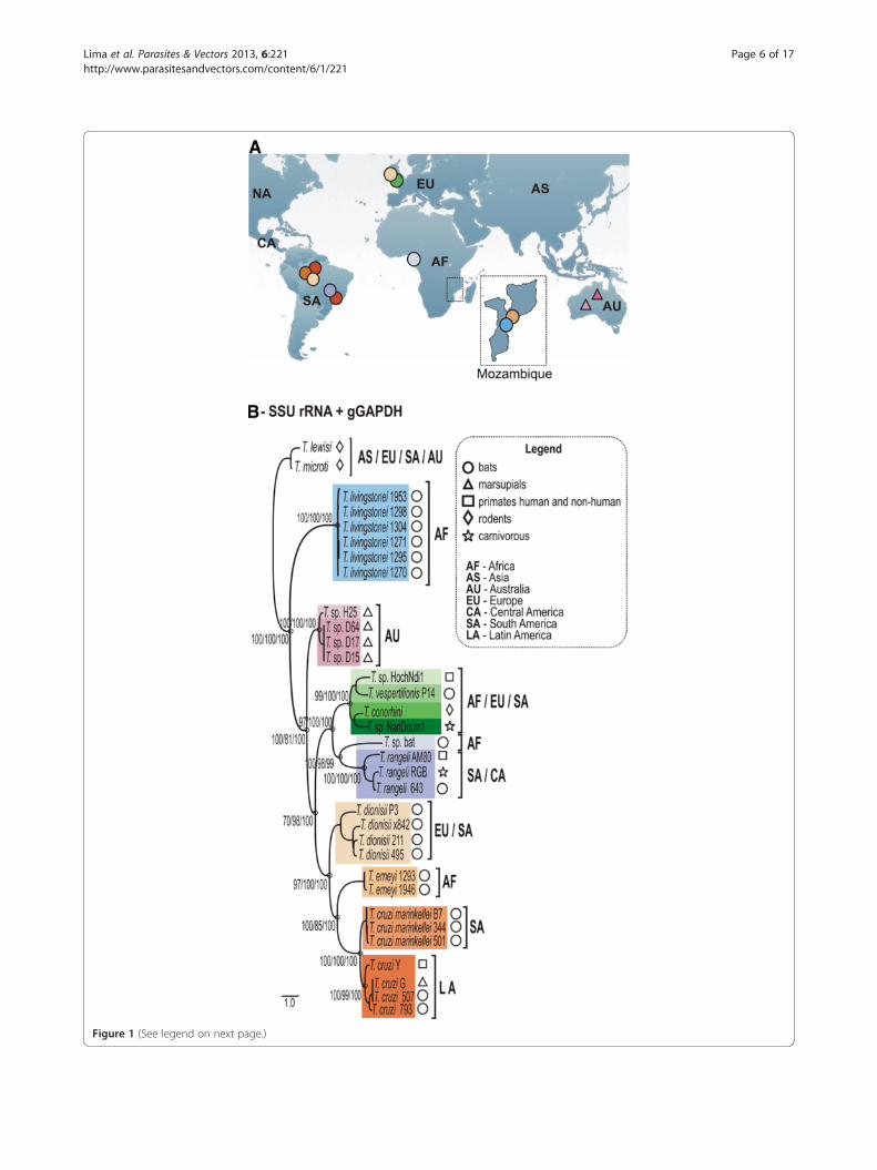
Figure 1 (See legend on next page.)
Lima et al. Parasites & Vectors 2013, 6:221 Page 6 of 17http://www.parasitesandvectors.com/content/6/1/221

(See figure on previous page.)Figure 1 Phylogeographical analysis of T. livingstonei, other bat trypanosomes and species from other mammals that nested into theclade T. cruzi. (a) Geographic origin of all trypanosomes included in the phylogenetic analysis, with the map from Mozambique (south-easternAfrica) where T. livingstonei was isolated in detail. (b) ML phylogenetic analysis based on concatenated sequences of SSU rRNA and gGAPDHsequences (3.318 characters, –Ln = 12694.460743) from 6 T. livingstonei isolates, 20 isolates from other species of bat trypanosomes, and 13trypanosomes from other mammalian orders; all the selected trypanosomes were previously positioned in the T. cruzi clade (GenBank accessionnumbers are listed on Table 1). Species from the T. lewisi clade were used as outgroups. Numbers are bootstrap values derived from 500replicates in the P/ML/BI analyses.
Figure 2 Barcoding of new African bat trypanosomes from cultures and bat blood samples. (a) Dendrogram inferred by using V7-V8 SSUrRNA sequences. The node numbers are bootstrap values derived from 100 replicates. (b) Fluorescent fragment length barcoding (FFLB) profilesof distinct isolates of the new species T. livingstonei and the FFLB pattern of T. sp. bat, a closely related bat trypanosome.
Lima et al. Parasites & Vectors 2013, 6:221 Page 7 of 17http://www.parasitesandvectors.com/content/6/1/221
Barcoding of the new African bat trypanosomes throughV7V8 rRNA sequencesThe analysis of the V7V8 variable region of the SSU rRNAgene for barcoding trypanosomes has demonstrated thatthis sequence is sufficiently polymorphic to distinguish allspecies from the several vertebrate classes examined todate [8,10,19,33,34]. In this study, barcoding using V7V8SSU rRNA revealed that all new isolates from African batsshared high sequence similarity; 2–3 cloned sequenceswere determined for each isolate, and they tightly clus-tered together and were virtually identical (~0.2% ofdivergence) and different from any previously reportedtrypanosome species. Regarding their closest relatives, thenew trypanosomes diverged ~9.5% from Australian try-panosomes from kangaroo, possums (marsupials) androdents, and ~12% from T. sp. bat (Gabon, Africa) andT. vespertilionis (UK bat). DNA from bat blood sampleswith negative haemoculture results were also used as a tem-plate for barcoding, and revealed trypanosome sequencesidentical to those of cultivated trypanosomes (Figure 2a).
Fluorescent Fragment Length Barcoding (FFLB) oftrypanosomes from culture and blood samplesThe FFLB techniques relies on the amplification andfluorescence detection of four small regions of rRNAgenes of variable length according to the species/isolates,and have been valuable to distinguish a wide range oftrypanosomes from cultures, blood and insect samples[10,42,43]. Here, we barcoded the new bat trypanosomesfrom culture and directly from blood samples. A com-parison was made of their FFLB profiles with those fromseveral previously barcoded trypanosomes, including thefollowing species found in bats: T. cruzi, T. c. marinkellei,T. dionisii, T. rangeli and T. erneyi [10,43], resulting inunique profiles for each species. Highly similar but non-identical FFLB profiles were found for all the new cul-tivated bat trypanosomes and the isolates from bat bloodsamples. The FFLB patterns permitted to distinguish thenew trypanosomes from all trypanosomes from bats andother hosts investigated in this (Figure 2b) and in previousstudies [10,42,43].
Phylogenetic analysis of new African bat trypanosomesbased on gGAPDH and SSU rRNA genesPhylogenies based on SSU rRNA and gGAPDH havebeen used for evolutionary and taxonomic studies oftrypanosomatids and it has been recommended that allnew trypanosome species are phylogenetically validatedusing at least these two genes [10,34,36,37]. Here, thenew bat isolates were initially positioned using inde-pendent gGAPDH (Figure 3) and SSU rRNA (data notshown) sequences in phylogenetic trees comprising re-presentative species of all major trypanosome clades.Concordant tree topologies from ML, P and BI analyses
were obtained by using these two genes. In all phyloge-netic trees, the new bat isolates formed a well-supportedclade close to Australian trypanosomes (10% divergence)and basal to the T. cruzi clade (Figure 3).We selected 6 new isolates (TCC1270, 1271, 1295,
1298, 1304 and 1953) to be positioned in the phylogenyof Trypanosoma using concatenated data set from wholeSSU rRNA and gGAPDH sequences (Figure 1). The useof these combined genes corroborated all the clades andtheir phylogenetic relationships as demonstrated inbroader phylogenies [10,23]. The new bat isolates arehighly homogeneous, diverging by only 0.3% in theirgGAPDH sequences. The clade formed by the new battrypanosomes was basal to the T. cruzi clade (100%bootstrap); their closest relatives were the Australian try-panosomes, whereas T. vespertilionis and T. sp. bat weremore closely related, despite being separated from thenew bat trypanosomes by ~13% gGAPDH sequencedivergence (Figures 1 and 3).Taken together, barcoding and phylogenetic analyses
demonstrated that the new African bat isolates belong toonly one species, which are exclusive to African bats sofar and display intra-specific variability (genotypes) in-sufficient to represent more than one species. Theresults supported the classification of this trypanosomeas a new species designated as Trypanosoma livingstonein. sp., which did not belong to any known subgenus.
Uniqueness of the primary and secondary structure of theSL gene from T. livingstoneiWe determined 3–4 sequences of full-length SL unitrepeats from each of three selected isolates from T.livingstonei. The results showed large repeats, varyingamong and within the isolates as follows: 1315 bp forTCC1304, 1322 and 1326 bp for TCC1933, and 1323,1347 and 1363 bp for TCC1948. The SL sequence align-ment revealed that the 39 bp exon, which is conservedin all trypanosome species, can display different nu-cleotides as observed in one sequence of the isolateTCC1933 (Figure 4a), whereas the intron sequences (110bp) were identical for all three isolates (Figure 4a). Theintergenic regions were quite variable in length and se-quence inter- and intra-isolates; TCC1948 showed themost highly divergent sequences (Figure 4b).Alignments restricted to the exon and intron sequences
(SL transcript) enabled an evaluation of the geneticrelatedness of T. livingstonei with all available trypano-somes permitting reliable alignments, namely T. sp. bat,T. vespertilionis, T. rangeli and T. conorhini. T. dionisiiand T. erneyi sequences could be partially aligned whereasall other species, including T. cruzi, resulted in inconsis-tent alignments. T. livingstonei largely diverged in theirintergenic sequences from all these species (data notshown). All SL transcript sequences from T. livingstonei
Lima et al. Parasites & Vectors 2013, 6:221 Page 8 of 17http://www.parasitesandvectors.com/content/6/1/221

Figure 3 Phylogenetic tree of the new trypanosome isolates from African bats. Phylogenetic tree inferred by maximum likelihood (ML) ofgGAPDH sequences from 12 isolates of T. livingstonei and 76 isolates of other species representative of all major clades with Trypanosoma usingtrypanosomatids of other genera as outgroups (832 characters, –Ln = −13552.439577). Numbers at nodes are support values derived from 500replicates in P/ML/Bayes analyses. Codes within parenthesis are GenBank accession numbers.
Lima et al. Parasites & Vectors 2013, 6:221 Page 9 of 17http://www.parasitesandvectors.com/content/6/1/221

isolates clustered together and their relationships with theother species (Figure 4c) corroborated SSU rRNA andgGAPDH data (Figures 1 and 3). Additionally, we inferredthe putative SL secondary structure (SL transcripts) fromT. livingstonei and compared it with that from T. sp. bat.The results showed a similar general secondary structureas a consequence of their similar SL transcript sequences(Figure 4d). The SL secondary structure of T. livingstonei
slightly differed from those inferred for T. vespertilionisand T. rangeli, whereas T. dionisii, T. cruzi and T. erneyiexhibited clearly different SL secondary structures[44; data not show].The sequence of whole SL repeats from T. livingstonei
revealed a copy of the 5S ribosomal RNA (5S rRNA)gene inserted into the intergenic region in the sameorientation as the SL gene. The same arrangement was
Figure 4 Characterisation and phylogenetic analysis of SL genes from T. livingstonei. Analysis of whole SL gene repeat (exon+intron+intergenicspacer) from isolates TCC 1304, 1933 and 1948. Comparison of primary (a – exon and partial intron sequences, b – a selected region of intergenicsequences) and secondary (d) structures, and relationships with phylogenetically related trypanosomes inferred using SL transcript sequences (c).
Lima et al. Parasites & Vectors 2013, 6:221 Page 10 of 17http://www.parasitesandvectors.com/content/6/1/221
demonstrated in this study on the SL repeats fromT. sp. bat and T. vespertilionis. The 5S rRNA sequencesfrom T. livingstonei were almost identical to those frommost trypanosomes [19]. However, one nucleotide substi-tution (A/G at position 18) was found in the sequencesfrom two (TCC1304 and 1948) of the three isolates fromthis species; this polymorphism was not detected in anyother trypanosome 5S rRNA.
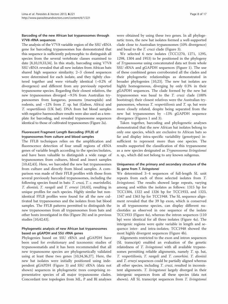
Light microscopy of T. livingstonei blood and cultureforms and behaviour in culturesTrypanosomes found in bat blood from which the T.livingstonei isolates were derived were large trypoma-stigotes with a broad body and a pointed posterior end, amarkedly frilled undulating membrane and a short freeflagellum. The small kinetoplast occupied a marginal po-sition adjacent to the rounded and nearly central nucleusand several surface striations (Figure 5a,b). Dividing formswere not observed in blood smears.The first forms observed in haemoculture were epi-
mastigotes arranged in rosettes and attached by their fla-gella (Figure 5c). Free epimastigotes in the supernatantpredominated during the log- and mid-phase cultures, andthese forms are pleomorphic, with bodies varying in length
from 16.0 to 29.0 μm (with an average of 22.3 μm) andfrom 1.1 to 2.0 μm in width (average of 1.56 μm), withmost forms displaying long free flagellum (average 15.5 μmlength) (Figure 5d). Both rounded forms and large epi-mastigotes were observed as dividing forms at log-phase(Figure 5d). Flagellates from the mid-log cultures aremostly large epimastigotes with a prominent undulantmembrane and long flagella, and they were long and slen-der trypomastigotes; both are dividing forms (Figure 5e).At stationary cultures, small and slim flagellates with alarge and nearly terminal kinetoplast resembling metacyclictrypomastigote forms were predominant (Figure 5f).When co-cultivated with a monolayer of HeLa mamma-
lian cells at 37°C, T. livingstonei epimastigotes developedinto wide free-swimming trypomastigotes in the super-natant (Figure 5g), with a resemblance but a smaller sizethan the bat blood forms (Figure 5a,b). These trypo-mastigotes were dividing in early cultures (Figure 5g), butattempts to successively culture them were unsuccessful.The development of epimastigotes into broad trypo-mastigotes under similar culture conditions is a feature ofMegatrypanum spp. [1,45]. T. livingstonei was unable toinvade and develop within mammalian cells, a feature ofall Schizotrypanum species [10].
Figure 5 Light microscopy of T. livingstonei (Giemsa-staining). (a, b) blood trypomastigotes in naturally infected bats; epimastigotes in log-phase cultures, arranged in rosettes attached by flagella (c) and free epimastigotes (d); large epimastigotes and trypomastigotes in mid-logcultures (e); metacyclic trypomastigotes in stationary cultures (f); trypomastigote forms developed in the supernatant of LLC-MK2 mammalian cellmonolayers at 37°C (g). Dividing flagellates are indicated by stars. Nucleus (N); kinetoplast (K) and flagellum (F). Scale bars: 10 μm.
Lima et al. Parasites & Vectors 2013, 6:221 Page 11 of 17http://www.parasitesandvectors.com/content/6/1/221
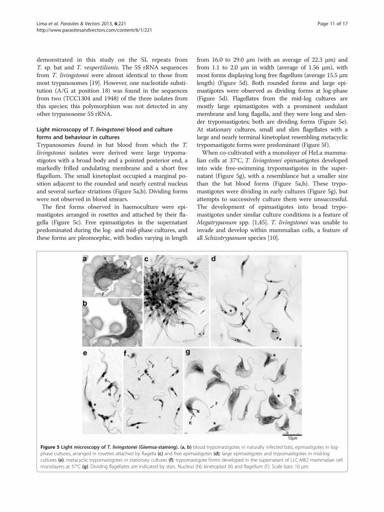
Scanning and transmission electron microscopy ofT. livingstoneiScanning electron microscopy (SEM) analyses of T.livingstonei cultures showed a diversity of forms includingthe following: a) large rosettes of epimastigotes united byflagella with one to three body torsions (Figure 6a); b) aruffled area near the cytostome, an invagination of themembrane close to the flagellar pocket shown by SEM asa small opening near the emergence of the flagellum(Figure 6b,c); c) long and slender epimastigotes with no-ticeable body torsions and a dilated anterior extremityconstituted by the joining of the flagellum and cell
membranes (“undulant membrane”) before the emergenceof the flagellum (Figure 6d,e).The ultrastructural organisation of T. livingstonei epi-
mastigotes (TEM analysis) revealed all common organellesof trypanosomatids. However, some features should bementioned as follows: a) The cytostome (Figure 7a,b,c),which forms together with the flagellar pocket the mainstructure involved in the endocytic process; b) a large num-ber of reservosomes, which are compartments that accu-mulate endocytosed macromolecules found at the posteriorregion of epimastigotes (Figure 7a); c) the compacted disk-shaped kinetoplast structure (Figure 7a,b,c).
Figure 6 Scanning electron microscopy of T. livingstonei. (a) Epimastigotes attached by their flagella forming a rosette; (b) free-swimmingepimastigote flagellar pocket and the emergence of the flagellum; (c) detail of the ruffled region with the cytostome opening; (d) long, slenderand twisted epimastigote; (e) dilated anterior extremity formed by joined cell and flagellar membranes is indicated by stars.
Lima et al. Parasites & Vectors 2013, 6:221 Page 12 of 17http://www.parasitesandvectors.com/content/6/1/221

T. livingstonei behaviour in mice and triatominesThe small trypomastigotes from end-phase T. livingstoneicultures (TC100 medium), which most likely correspondto metacyclic forms, were incapable of infecting Balb/cmice. This absence of infection was confirmed byblood examination using the microhaematocrit fromtwo to 15 days post-inoculation and, after that, byhaemoculture. Epi- and trypomastigotes were unableto infect triatomines (Rhodnius neglectus and Triatomainfestans).
Taxonomic summaryNew species description: Phylum Euglenozoa, Cavalier-Smith, 1981; class Kinetoplastea, Honigberg, 1963; orderTrypanosomatida (Kent, 1880) Hollande, 1952; familyTrypanosomatidae, Doflein, 1951. Trypanosoma living-stonei Teixeira and Camargo n. sp.Type material: hapantotype: culture TCC1270.
Paratypes: cultures TCC1271, 1295, 1298, 1304, 1902,1933, 1935, 1947, 1948, 1953, 1954, 2339 and 2348,whose bat hosts and locality of collection in Mozambique,Africa, are in Table 1. Type host: Chiroptera, Rhino-lophidae, Rhinolophus landeri. Additional host: Chi-roptera, Hipposideridae, Hipposideros caffer. Locality:
Mozambique, Province of Sofala, District of Chupanga(S18°02′ E35°34′), Zambezi valley and the Gorongosa Na-tional Park (S18°58′ E34°21′). Morphology: T. livingstoneiexhibits large and wide blood trypomastigotes (average32.4 μm length and 7.8 μm wide) with several striations,small kinetoplast, frilled undulating membrane and afree flagellum averaging 11.0 μm in length. Epimas-tigotes from log-phase cultures are mostly slender andpointed at posterior ends, ranging from 16.0 to 29.0 μmlength and 1.1 to 2.0 μm wide, with free flagellum aver-aging 15.5 μm length; the kinetoplast in general is closeto the nucleus. Diagnosis: DNA sequences unique toT. livingstonei are deposited in GenBank (accession num-bers): SSU rRNA (KF192979 - KF192994), gGAPDH(KF192958 - KF192969) and SL gene (KF192970 -KF192976). Cultures are cryopreserved at the Try-panosomatid Culture Collection of the University of SãoPaulo, TCC-USP. Glass slides of Giemsa-stained smearsfrom bat blood samples and cultures and DNA samplesare also kept at TCC-USP. To comply with the regulationsof the International Code of Zoological Nomenclature(ICZN), details of this species have been submitted toZooBank with the Life Science Identifier (LSID) zoobank.org:pub: D8714C8C-71F5-44ED-AC91-2106E49C1A6D.
Figure 7 Transmission electron microscopy (TEM) of T. livingstonei. (a) Ultrastructural organisation of epimastigotes in longitudinal andtransversal sections showing the nucleus, kinetoplast, reservosomes concentrated at the posterior region, basal body, flagellum and paraflagellarrod (b) longitudinal section of an dividing epimastigote exhibiting a new emerging flagella and a lengthened kinetoplast and the invagination ofthe flagellar pocket membrane forming the cytostome; (c) longitudinal section showing the cytostome opening and groove surrounded bymicrotubules penetrating deep into the cytoplasm; (c) compacted disk shaped kinetoplast. N, nucleus; K, kinetoplast; F, flagellum; R, reservosome;Cy, cytostome; Bb, basal body; PR, paraflagellar rod; MT, microtubules.
Lima et al. Parasites & Vectors 2013, 6:221 Page 13 of 17http://www.parasitesandvectors.com/content/6/1/221
Etymology: The name was given because Trypanosomalivingstonei n. sp. was first discovered in bats captured inChupanga, Mozambique, a small village in the margin ofthe Zambezi River, where Mary Livingstone, the wife ofDavid Livingstone, died of “fevers” in 1862; her grave re-mains in an small cemetery from a Portuguese Missionpractically destroyed by the Mozambique wars.
DiscussionFor a better appraisal of the genetic diversity and evolu-tionary history of trypanosomes, and for their reliableclassification and phylogenetic inferences, studies mustinclude trypanosomes from all vertebrate classes, repre-sentative of orders, genera and species, by using molecu-lar phylogenetic approaches. Bats are among the mostcommon hosts of a large variety of trypanosomes inAfrica, Asia, South America and Europe. However, ourknowledge of their genetic diversity, hosts, vectors, lifecycles, pathology, distribution and phylogenetic re-lationships is restricted to a few species. Almost all avai-lable data are about the species of the subgenusSchizotrypanum because bat trypanosomes in this sub-genus are the closest relatives of the human pathogenT. cruzi [8-11,13,14,22,25,28,46]. However, several batsaround the world harbour a plethora of trypanosomespecies, most of which are morphologically assigned tothe subgenus Megatrypanum [1,2,4,6,7,47].In this study, we surveyed trypanosomes in blood samples
from bats of old world-restricted families Rhinolophidaeand Hipposideridae captured in Mozambique. Weobtained 11 haemocultures from R. landeri and 3 fromH. caffer. Morphologically, the large trypomastigotesfound in bat blood smears would be assigned to the sub-genus Megatrypanum. However, multilocus phylogenyvalidated in this subgenus only the trypanosomes fromruminants allied to T. theileri [16,17,19,45]. With the ex-ception of artiodactyls, bats were the main hosts of try-panosomes morphologically classified in the subgenusMegatrypanum [1,7], so a thorough phylogenetic analysisof bat trypanosomes was required to warrant their exclu-sion from this subgenus.The phylogenetic positioning of T. livingstonei and
T. sp. bat, both of which are morphologically compatiblewith the subgenus Megatrypanum, support the exclusionof bats as hosts of species in this subgenus. The mor-phology of T. livingstonei blood and culture formslargely differs from those of the Megatrypanum species.However, epimastigotes of this species developed intolarge trypomastigotes resembling blood forms under amonolayer of mammalian cells at 37°C, a process alsoobserved for the Megatrypanum trypanosomes [1,45]. Infact, T. livingstonei blood and culture forms exhibitedunique morphological features as shown by light and SEMmicroscopy. This new species exhibited a cytostome,
reservosomes and a disk-shaped kinetoplast, all of whichare absent in species of the Megatrypanum and commonto those of Schizotrypanum. This is the first study to useTEM and SEM to analyse a bat trypanosome not classifiedinto Schizotrypanum. There are no unambiguous dif-ferences in the overall ultrastructural organisation thatwould be useful for distinguishing T. livingstonei fromSchizotrypanum trypanosomes, even though the new spe-cies strongly diverged with all molecular markers and inseveral biological features such as the inability to developinside mammalian cells and lack of infection in mice andtriatomine bugs.The vectors of T. livingstonei are so far unknown; in
this work we demonstrated its inability to infect T.infestans and Rhodnius neglectus in accordance with thefact that triatomines cannot be the vectors of this speciesbecause they do not occur in Africa. In Africa andEurope, bat bugs (cimicids) are the vectors of T. dionisiiand T. (Megatrypanum) incertum [6,7]. The bat re-stricted Stricticimex brevispinosus was found to beinfected by a Megatrypanum trypanosome in Africa [48].In addition, sand flies were incriminated as vectors of T.(Megatrypanum) leonidasdeanei in South America [49].The barcoding of new African bat trypanosomes mor-
phologically assignable to the subgenus Megatrypanumthrough both V7V8 SSU rRNA and FFLB has shownsimilar sequences and profiles for all the new isolates,which were shown to be highly different from thebarcodes of other trypanosomes from bats and otherhosts [10,42,43]. In all the inferred phylogenetic analyses,the new bat trypanosomes always tightly clusteredtogether, forming a homogeneous clade separated by suf-ficient genetic distances from all other trypanosomes toallow their description as a new species, that is,T. livingstonei n. sp., which does not nest within anyknown subgenera.For insect and plant trypanosomatids, SL genes have
proven to be valuable for identifying the genera and spe-cies of cultivated flagellates, as well as for the barcodingof trypanosomatids directly from their hosts [50-53]. SLRNA genes have also been used for species identificationand genotyping of T. cruzi and T. rangeli [22,26,27,54],T. vivax [55] and T. theileri [17,19]. The characterisationof whole SL gene repeats in T. livingstonei showed alarger length and more polymorphic sequences amongisolates of the same species and repeats of the same iso-late, when compared to other trypanosome species. Inaddition, this species enclosed a copy of 5S rRNA withinits intergenic region, as reported before for T. vivax,T. conorhini, T. rangeli, T. desterrensis, T. theileri and T.melophagium, but absent in T. cruzi, T. cruzi marinkellei,T. brucei and T. lewisi [17,19,21,44,45,55,56]. Here, thecomparison of primary and secondary structures from theSL rRNA of T. livingstonei and other trypanosomes
Lima et al. Parasites & Vectors 2013, 6:221 Page 14 of 17http://www.parasitesandvectors.com/content/6/1/221
corroborated its close relationships with the trypanosomesthat nested into a strongly supported (despite being highlyheterogeneous) major clade containing African, Europeand South American species from bats (T. sp. bat andT. vespertilionis), T. rangeli, T. conorhini and trypano-somes from monkeys and civets.The trypomastigotes we found in blood smears from
bats infected with T. livingstonei resembled thosedenominated as T. heybergi-type and described forthe African Megatrypanum trypanosomes T. leleupi,T. mpapuense, T. morinorum and T. thomasi. Thesespecies, which could all be synomies, were reported inbats from Congo, Zambia, Kenya and Tanzania[1,3,7,47]. Our findings corroborated that African speciesof Rhinolophus and Hipposideros bats harbour trypano-somes morphologically similar to T. heybergi. However, T.(Megatrypanum) leonidasdeanei and T. (Megatrypanum)pessoai were reported in South American bats and alsodescribed as resembling T. heybergi [49,57]. Nevertheless,no cultures, DNA sequences or blood smears were avai-lable from any T. heybergi-type trypanosomes, whichprevented the molecular comparison between previouslyreported species and our new isolates.Bat species harbouring T. livingstonei are endemic
to sub-Saharan African bats, although their genera,Rhinolophus and Hipposideros, are widespread throughoutAsia, Oceania, Europe and Africa, but both are absentfrom the New World. Bats may have originated inLaurasia (~ 65 MYA), and bat trypanosomes should havediverged since the great diversification/expansion of batsin the Eocene [29-31]. A long past and extensive bat ra-diation, recent movement of bats across large geographicdistances (even large oceanic barriers but not across theAtlantic Ocean), and incomplete bat palaeontology havecomplicated the studies about the origin and dispersion ofbat trypanosomes. Associations between bats and theirtrypanosomes, and an evaluation of possible paleonto-logical and eco-biogeographical scenarios could accountfor the origin, genetic diversity, relationships and currentdistribution of these parasites and are crucial for under-standing their evolutionary history.There is an urgent need for an extensive taxonomic
revision of the genera Trypanosoma on a strongly sup-ported phylogenetic basis that firstly requires themolecular analyses of a large sampling representative ofhost species and geographic ranges. This may allow forthe description of several new species and the creationof new subgenera to accommodate new species thatformed clades without correspondence to any subgenerapreviously proposed by Hoare [1]. To meet these objec-tives, new trypanosome cultures should be obtained anddeposited in reference collections. The naming of anynew trypanosomatid species should be considered validonly when supported by sound and broad phylogenies
(using at least SSU rRNA and gGAPDH genes). How-ever, the description of new trypanosome species basedon small DNA sequences, accompanied or not by themorphology of blood flagellates (mostly because cultiva-tion have failed), have been accepted [58-60]. We aredesignating the new African bat isolates as T. livingstoneion the basis of its position in the Trypanosoma phylo-genetic trees inferred using SSU rRNA and gGAPDHgenes, its genetic distances from other species and alsotaking into account its peculiar SL RNA gene repeats.Morphological features and information regarding hostspecies, and its behaviour in culture and in micecomplement the species description. These data can bevaluable for comparative studies of the cellular biology,host-parasite interactions, ecology and evolution oftrypanosomes.
ConclusionThe phylogenetic evidence produced by this study un-derscores the great genetic diversity of trypanosomes inbats around the world. T. livingstonei fell at the edge ofthe T. cruzi clade, which comprises all bat trypano-somes sampled to date regardless of whether they arefrom Africa, Europe or South America. The position ofT. livingstonei at the base of the T. cruzi clade furthersupports the hypothesis that the clade was ancestrally agroup of bat-restricted parasites that evolved exclu-sively in these hosts and later jumped at independenttimes to mammals of other orders. In the most likelyscenario, the trypanosomes from several mammalianorders nested into this clade, including those fromAfrican and Australian terrestrial mammals, evolvedfrom a bat trypanosome. Other explanations requiremultiple jumps into bats, which seem less probably.Apparently, this ancestral bat trypanosome gave risemorphologically, biologically (different life cycles andvectors), ecologically and genetically distinct species.The positioning of T. livingstonei in all inferred phylog-enies provides evidence that the T. cruzi clade derivedfrom a bat trypanosome, prior to the splits between T.cruzi, T. rangeli and the Australian groups, hence, lendsfurther support to the bat seeding evolutionary hypoth-esis for the origin of this clade [24]. This study alsoadds some additional support to T. cruzi itself evolvingfrom a bat trypanosome as the resulting data makemore likely that the common ancestor of T. rangeli andT. cruzi was a bat trypanosome.
Competing interestsThe authors declare that they have no competing interests.
Authors’ contributionsLL, EPC and MMGT conceived the study and designed the experiments; LL,OEA, PBH, LN, CSAT, MC, MA, WS assisted with sample collection, performedthe experiments and analyzed the data; LL, PBH, EPC, MMGT prepared the
Lima et al. Parasites & Vectors 2013, 6:221 Page 15 of 17http://www.parasitesandvectors.com/content/6/1/221
paper. All authors read, revised and approved the submitted version of themanuscript.
AcknowledgementsWe would like to thank many people who kindly helped us with thefieldwork in Mozambique. We are particularly grateful to our friends Carmene José Martins for their delightful company and hospitality in their house inChupanga. We are deeply indebted to Carlos Pereira and the staff of theGorongosa National Park. We are also grateful to Laerte B. Viola, Arlei Marcili,Bruno R. Fermino and many other students for their efforts in bat captureand sample collection. This work was supported by grants from theConselho Nacional de Desenvolvimento Científico and Tecnológico (CNPq)within the PROAFRICA, PROTAX, and UNIVERSAL Programs to MMGT andEPC. LL is a postdoctoral fellow sponsored by São Paulo State ResearchSupport Foundation (FAPESP) process nº 2012/14985-6, Espinosa-Álvarez O isa PhD student sponsored by CNPq (PROTAX).
Author details1Departamento de Parasitologia, Instituto de Ciências Biomédicas,Universidade de São Paulo, São Paulo, SP 05508-900, Brazil. 2Biosciences,College of Life and Environmental Sciences, University of Exeter, Exeter, UK.3Centro de Biotecnologia, Universidade Eduardo Mondlane, Maputo,Mozambique. 4Laboratório de Ultraestrutura Celular Hertha Meyer, Institutode Biofísica Carlos Chagas Filho, Universidade Federal do Rio de Janeiro, Riode Janeiro, RJ 21941-902, Brazil.
Received: 13 June 2013 Accepted: 1 August 2013Published: 3 August 2013
References1. Hoare CA: The trypanosomes of mammals: a zoological monograph. Oxford,
England: Blackwell Scientific Publishing; 1972.2. Baker JR: First European record of Trypanosoma (Megatrypanum) sp. of
bats. Nat New Biol 1973, 241:96.3. Marinkelle CJ: The biology of the trypanosomes of bats. In Biology of the
Kinetoplastida. Edited by Lumdsen WHR, Evans DA. New York: Academic;1976:175–216.
4. Marinkelle CJ: Trypanosoma (Megatrypanum) megachiropterum sp. n. fromthe flying fox, Pteropus tonganus, Quoy, Gaimard. J Protozool 1979,26:352–353.
5. Gardner RA, Molyneux DH: Schizotrypanum in British bats Parasitology1988, 97:43–50.
6. Gardner RA, Molyneux DH: Trypanosoma (Megatrypanum) incertum fromPipistrellus pipistrellus: development and transmission by cimicid bugs.Parasitology 1988, 96:433–447.
7. Molyneux DH: Trypanosomes of bats. In Parasitic Protozoa. Edited byKreier JP, Baker JR. New York: Academic; 1991:195–223.
8. Cavazzana M Jr, Marcili A, Lima L, da Silva FM, Junqueira AC, Veludo HH,Viola LB, Campaner M, Nunes VL, Paiva F, Coura JR, Camargo EP,Teixeira MMG: Phylogeographical, ecological and biological patternsshown by nuclear (ssrRNA and gGAPDH) and mitochondrial (Cyt b)genes of trypanosomes of the subgenus Schizotrypanum parasitic inBrazilian bats. Int J Parasitol 2010, 40:345–355.
9. Hamilton PB, Cruickshank C, Stevens JR, Teixeira MMG, Mathews F:Parasites reveal movement of bats between the new and old worlds.Mol Phylogenet Evol 2012, 63:521–526.
10. Lima L, Maia da Silva F, Neves L, Attias M, Takata CS, Campaner M,de Souza W, Hamilton PB, Teixeira MMG: Evolutionary insights from battrypanosomes: morphological, developmental and phylogeneticevidence of a new species, Trypanosoma (Schizotrypanum) erneyi sp.nov., in African bats closely related to Trypanosoma (Schizotrypanum)cruzi and allied species. Protist 2012, 163:856–872.
11. Lisboa CV, Pinho AP, Herrera HM, Gerhardt M, Cupolillo E, Jansen AM:Trypanosoma cruzi (Kinetoplastida, Trypanosomatidae) genotypes inneotropical bats in Brazil. Vet Parasitol 2008, 156:314–318.
12. Cottontail VM, Wellinghausen N, Kalko EK: Habitat fragmentation andhaemoparasites in the common fruit bat, Artibeus jamaicensis(Phyllostomidae) in a tropical lowland forest in Panamá. Parasitology2009, 136:1133–1145.
13. García L, Ortiz S, Osorio G, Torrico MC, Torrico F, Solari A: Phylogeneticanalysis of Bolivian bat trypanosomes of the subgenus Schizotrypanum
based on cytochrome B sequence and minicircle analyses. PLoS One2012, 7:e36578.
14. Pinto CM, Kalko EK, Cottontail I, Wellinghausen N, Cottontail VM: TcBat abat-exclusive lineage of Trypanosoma cruzi in the Panama Canal Zone,with comments on its classification and the use of the 18S rRNA genefor lineage identification. Infect Genet Evol 2012, 12:1328–1332.
15. Hoare CA: Morphological and taxonomic studies on mammaliantrypanosomes. X. Revision of the systematics. J Protozool 1964,11:200–207.
16. Rodrigues AC, Paiva F, Campaner M, Stevens JR, Noyes HA, Teixeira MMG:Phylogeny of Trypanosoma (Megatrypanum) theileri and relatedtrypanosomes reveals lineages of isolates associated with artiodactylhosts diverging on SSU and ITS ribosomal sequences. Parasitology 2006,132:215–224.
17. Rodrigues AC, Garcia HA, Batista JS, Minervino AH, Góes-Cavalcante G,Maia da Silva F, Ferreira RC, Campaner M, Paiva F, Teixeira MMG:Characterization of spliced leader genes of Trypanosoma(Megatrypanum) theileri: phylogeographical analysis of Brazilian isolatesfrom cattle supports spatial clustering of genotypes and parity withribosomal markers. Parasitology 2010, 137:111–122.
18. Hamilton PB, Adams ER, Njiokou F, Gibson WC, Cuny G, Herder S: Phylogeneticanalysis reveals the presence of the Trypanosoma cruzi clade in Africanterrestrial mammals. Infect Genet Evol 2009, 9:81–86.
19. Garcia HA, Rodrigues AC, Martinkovic F, Minervino AH, Campaner M,Nunes VL, Paiva F, Hamilton PB, Teixeira MMG: Multilocusphylogeographical analysis of Trypanosoma (Megatrypanum) genotypesfrom sympatric cattle and water buffalo populations supportsevolutionary host constraint and close phylogenetic relationships withgenotypes found in other ruminants. Int J Parasitol 2011, 41:1385–1396.
20. Stevens JR, Noyes HA, Dover GA, Gibson WC: The ancient and divergentorigins of the human pathogenic trypanosomes. Trypanosoma bruceiand T. cruzi. Parasitology 1999, 118:107–116.
21. Stevens JR, Teixeira MMG, Bingle LE, Gibson WC: The taxonomic positionand evolutionary relationships of Trypanosoma rangeli. Int J Parasitol1999, 29:749–757.
22. Marcili A, Lima L, Cavazzana M, Junqueira AC, Veludo HH, Maia Da Silva F,Campaner M, Paiva F, Nunes VL, Teixeira MMG: A new genotype ofTrypanosoma cruzi associated with bats evidenced by phylogeneticanalyses using SSU rDNA, cytochrome b and Histone H2B genes andgenotyping based on ITS1 rDNA. Parasitology 2009, 136:641–655.
23. Hamilton PB, Gibson WC, Stevens JR: Patterns of co-evolution betweentrypanosomes and their hosts deduced from ribosomal RNA andprotein-coding gene phylogenies. Mol Phylogenet Evol 2007, 44:15–25.
24. Hamilton PB, Teixeira MMG, Stevens JR: The evolution of Trypanosomacruzi: the ‘bat seeding’ hypothesis. Trends Parasitol 2012, 28:136–141.
25. Franzén O, Talavera-López C, Ochaya S, Butler CE, Messenger LA, Lewis MD,Llewellyn MS, Marinkelle CJ, Tyler KM, Miles MA, Andersson B: Comparativegenomic analysis of human infective Trypanosoma cruzi lineages with thebat-restricted subspecies T. cruzi marinkellei. BMC Genomics 2012, 13:531.
26. Maia da Silva F, Marcili A, Lima L, Cavazzana M Jr, Ortiz PA, Campaner M,Takeda GF, Paiva F, Nunes VL, Camargo EP, Teixeira MMG: Trypanosomarangeli isolates of bats from Central Brazil: genotyping and phylogeneticanalysis enable description of a new lineage using spliced-leader genesequences. Acta Trop 2009, 109:199–207.
27. Maia Da Silva F, Junqueira AC, Campaner M, Rodrigues AC, Crisante G,Ramirez LE, Caballero ZC, Monteiro FA, Coura JR, Añez N, Teixeira MMG:Comparative phylogeography of Trypanosoma rangeli and Rhodnius(Hemiptera: Reduviidae) supports a long coexistence of parasite lineagesand their sympatric vectors. Mol Ecol 2007, 16:3361–3373.
28. Lima L, Ortiz PA, da Silva FM, Alves JM, Serrano MG, Cortez AP, Alfieri SC,Buck GA, Teixeira MMG: Repertoire, genealogy and genomic organizationof cruzipain and homologous genes in Trypanosoma cruzi. T. cruzi likeand other trypanosome species. PLoS One 2012, 7:e38385.
29. Eick GN, Jacobs DS, Matthee CA: A nuclear DNA phylogenetic perspectiveon the evolution of echolocation and historical biogeography of extantbats (Chiroptera). Mol Biol Evol 2005, 22:1869–1886.
30. Teeling EC, Springer MS, Madsen O, Bates P, O'brien SJ, Murphy WJ: Amolecular phylogeny for bats illuminates biogeography and the fossilrecord. Science 2005, 307:580–584.
31. Simmons NB: Evolution. An Eocene big bang for bats. Science 2005,307:527–528.
Lima et al. Parasites & Vectors 2013, 6:221 Page 16 of 17http://www.parasitesandvectors.com/content/6/1/221

32. Cui J, Han N, Streicker D, Li G, Tang X, Shi Z, Hu Z, Zhao G, Fontanet A,Guan Y, Wang L, Jones G, Field HE, Daszak P, Zhang S: Evolutionaryrelationships between bat coronaviruses and their hosts. Emerg Infect Dis2007, 13:1526–1532.
33. Ferreira RC, De Souza AA, Freitas RA, Campaner M, Takata CS, Barrett TV,Shaw JJ, Teixeira MMG: A phylogenetic lineage of closely relatedtrypanosomes (Trypanosomatidae, Kinetoplastida) of anurans and sandflies (Psychodidae, Diptera) sharing the same ecotopes in BrazilianAmazonia. J Eukaryot Microbiol 2008, 55:427–435.
34. Viola LB, Attias M, Takata CS, Campaner M, De Souza W, Camargo EP,Teixeira MMG: Phylogenetic analyses based on small subunit rRNA andglycosomal glyceraldehyde-3-phosphate dehydrogenase genes andultrastructural characterization of two snake Trypanosomes:Trypanosoma serpentis n. sp. From Pseudoboa nigra and Trypanosomacascavelli from Crotalus durissus terrificus. J Euk Microbiol 2009, 56:594–602.
35. Hamilton PB, Stevens JR, Gaunt MW, Gidley J, Gibson WC: Trypanosomesare monophyletic: evidence from genes for glyceraldehyde phosphatedehydrogenase and small subunit ribosomal RNA. Int J Parasitol 2004,34:1393–1404.
36. Teixeira MMG, Borghesan TC, Ferreira RC, Santos MA, Takata CS,Campaner M, Nunes VL, Milder RV, de Souza W, Camargo EP: Phylogeneticvalidation of the genera Angomonas and Strigomonas oftrypanosomatids harboring bacterial endosymbionts with thedescription of new species of trypanosomatids and of Proteobacterialsymbionts. Protist 2011, 162:503–524.
37. Borghesan TC, Ferreira RC, Takata CS, Campaner M, Borda CC, Paiva F,Milder RV, Teixeira MMG, Camargo EP: Molecular phylogenetic redefinitionof Herpetomonas (Kinetoplastea, Trypanosomatidae), a genus of insectparasites associated with flies. Protist 2013, 164:129–152.
38. Thompson JD, Gibson TJ, Plewniak F, Jeanmougin F, Higgins DG: TheCLUSTAL_X windows interface: flexible strategies for multiple sequencealignment aided by quality analysis tools. Nucleic Acids Res 1997,25:4876–4882.
39. Swofford DL: PAUP*. Phylogenetic analysis using parsimony (* and othermethods). Version 4. Sunderland MA: Sinauer Associates; 2002.
40. Stamatakis A: RAxML-VI-HPC: maximum likelihood-based phylogeneticanalyses with thousands of taxa and mixed models. Bioinformatics 2006,22:2688–2690.
41. Huelsenbeck JP, Ronquist F: MRBAYES: bayesian inference of phylogenetictrees. Bioinformatics 2001, 17:754–755.
42. Hamilton PB, Adams ER, Malele II, Gibson WC: A novel, high-throughputtechnique for species identification reveals a new species oftsetse-transmitted trypanosome related to the Trypanosoma bruceisubgenus Trypanozoon. Infect Genet Evol 2008, 8:26–33.
43. Hamilton PB, Lewis MD, Cruickshank C, Gaunt MW, Yeo M, Llewellyn MS,Valente SA, Maia da Silva F, Stevens JR, Miles MA, Teixeira MMG:Identification and lineage genotyping of South American trypanosomesusing fluorescent fragment length barcoding. Infect Genet Evol 2011,11:44–51.
44. Gibson W, Bingle L, Blendeman W, Brown J, Wood J, Stevens J: Structureand sequence variation of the trypanosome spliced leader transcript.Mol Biochem Parasitol 2000, 107:269–277.
45. Martinković F, Matanović K, Rodrigues AC, Garcia HA, Teixeira MMG:Trypanosoma (Megatrypanum) melophagium in the sheep kedMelophagus ovinus from organic farms in Croatia: phylogeneticinferences support restriction to sheep and sheep keds and closerelationship with trypanosomes from other ruminant species. J EukaryotMicrobiol 2012, 59:134–144.
46. Barnabé C, Brisse S, Tibayrenc M: Phylogenetic diversity of battrypanosomes of subgenus Schizotrypanum based on multilocus enzymeelectrophoresis, random amplified polymorphic DNA, and cytochrome bnucleotide sequence analyses. Inf Gen Evol 2003, 2:201–208.
47. Keymer IF: Bood protozoa of insectivores, bats and primates in CentralAfrica. J Zoology 1971, 163:421–441.
48. Van der Berge L, Chardome M, Péel E: An African bat trypanosome inStricticimex brevispinosus Usinger. 1959. J Protozool 1963, 10:135–138.
49. Zeledón R, Rosabal R: Trypanosoma leonidasdeanei sp. nov. ininsectivorous bats of Costa Rica. Ann Trop Med Parasitol 1969, 63:221–228.
50. Teixeira MMG, Serrano MG, Nunes LR, Campaner M, Buck GA, Camargo EP:Trypanosomatidae: a spliced-leader-derived probe specific for the genusPhytomonas. Exp Parasitol 1996, 84:311–319.
51. Serrano MG, Nunes LR, Campaner M, Buck GA, Camargo EP, Teixeira MMG:Trypanosomatidae: Phytomonas detection in plants and phytophagousinsects by PCR amplification of a genus-specific sequence of the splicedleader gene. Exp Parasitol 1999, 91:268–279.
52. Westenberger SJ, Sturm NR, Yanega D, Podlipaev SA, Zeledón R,Campbell DA, Maslov DA: Trypanosomatid biodiversity in Costa Rica:genotyping of parasites from Heteroptera using the spliced leader RNAgene. Parasitology 2004, 129:537–547.
53. Maslov DA, Westenberger SJ, Xu X, Campbell DA, Sturm NR: Discovery andbarcoding by analysis of spliced leader RNA gene sequences of newisolates of Trypanosomatidae from Heteroptera in Costa Rica andEcuador. J Eukaryot Microbiol 2007, 54:57–65.
54. Urrea DA, Carranza JC, Cuba CA, Gurgel-Gonçalves R, Guhl F, Schofield CJ,Triana O, Vallejo GA: Molecular characterisation of Trypanosoma rangelistrains isolated from Rhodnius ecuadoriensis in Peru, R. colombiensis inColombia and R. pallescens in Panama, supports a co-evolutionaryassociation between parasites and vectors. Infect Genet Evol 2005,5:123–129.
55. Ventura RM, Paiva F, Silva RA, Takeda GF, Buck GA, Teixeira MMG:Trypanosoma vivax: characterization of the spliced-leader gene of aBrazilian stock and species-specific detection by PCR amplification of anintergenic spacer sequence. Exp Parasitol 2001, 99:37–48.
56. Grisard EC, Sturm NR, Campbell DA: A new species of trypanosome,Trypanosoma desterrensis sp. n., isolated from South American bats.Parasitology 2003, 127:265–271.
57. Deane LM, Sugay W: Trypanosoma pessoai n. sp., in vampire batsDesmodus rotundus rotundus from the State of São Paulo, Brazil.Rev Inst Med Trop Sao Paulo 1963, 5:165–169.
58. McInnes LM, Hanger J, Simmons G, Reid SA, Ryan UM: Novel trypanosomeTrypanosoma gilletti sp. (Euglenozoa: Trypanosomatidae) and theextension of the host range of Trypanosoma copemani to include thekoala (Phascolarctos cinereus). Parasitology 2011, 138:59–70.
59. McInnes LM, Gillett A, Ryan UM, Austen J, Campbell RS, Hanger J, Reid SA:Trypanosoma irwini n. sp (Sarcomastigophora: Trypanosomatidae) fromthe koala (Phascolarctos cinereus). Parasitology 2009, 136:875–885.
60. Thompson CK, Botero A, Wayne AF, Godfrey SS, Lymbery AJ, Thompson RA:Morphological polymorphism of Trypanosoma copemani and descriptionof the genetically diverse T. vegrandis sp. nov. from the criticallyendangered Australian potoroid, the brush-tailed bettong (Bettongiapenicillata) (Gray,1837). Parasit Vectors 2013, 6:121.
doi:10.1186/1756-3305-6-221Cite this article as: Lima et al.: Trypanosoma livingstonei: a new speciesfrom African bats supports the bat seeding hypothesis for theTrypanosoma cruzi clade. Parasites & Vectors 2013 6:221.
Submit your next manuscript to BioMed Centraland take full advantage of:
• Convenient online submission
• Thorough peer review
• No space constraints or color figure charges
• Immediate publication on acceptance
• Inclusion in PubMed, CAS, Scopus and Google Scholar
• Research which is freely available for redistribution
Submit your manuscript at www.biomedcentral.com/submit
Lima et al. Parasites & Vectors 2013, 6:221 Page 17 of 17http://www.parasitesandvectors.com/content/6/1/221