ts karyotype, pedigree and cone-beam computerized
TRANSCRIPT
Case Reports
468Journal of Natural Science, Biology and Medicine | July 2015 | Vol 6 | Issue 2
Karyotype, Pedigree and cone-beam computerized tomography analysis of a case of nonsyndromic pandental anomalies
AbstractThis case report presented a karyotype and pedigree analysis of a case with unusual combination of dental anomalies: Generalized short roots, talon cusps, dens invagination, low alveolar bone heights, very prominent cusp of carabelli and protostylid on first permanent molars, taurodontism of second permanent molars, rotated, missing and impacted teeth. None of the anomalies alone are rare. However, until date, nonsyndromic pandental anomalies that are affecting entire dentition with detailed karyotype, pedigree and cone-beam computerized tomography analysis have not been reported. The occurrence of these anomalies is probably incidental as the conditions are etiologically unrelated.
Key words: Cone-beam computerized tomography, conventional karyotyping, dense invaginatus, short root anomalies, talon cusp, taurodontism
Umesh Dharmani, Ganesh Ranganath
Jadhav1, Charan Kamal
Kaur Dharmani2, Akhil Rajput3, Priya Mittal4,
Sathish Abraham5, Vinay Soni
Departments of Conservative Dentistry and Endodontics and 2Pedodontics and Preventive Dentistry, Dental College, RIMS, Imphal, Manipur, 1Department of Conservative Dentistry and Endodontics, Sinhgad Dental College and Hospital, Pune, 4Department of Conservative Dentistry and Endodontics, SMBT Dental College, Sangamner, Maharashtra, 3Department of Conservative Dentistry and Endodontics, Maulana Azad Institute of Dental Sciences, New Delhi, 5Department of Orthodontics, Oral Health Science Centre, Postgraduate Institute of Education and Research, Chandigarh, India
Address for correspondence: Dr. Ganesh Ranganath Jadhav, Department of Conservative Dentistry and Endodontics, Sinhgad Dental College and Hospital, Pune, Maharashtra, India. E-mail: [email protected]
INTRODUCTION
During tooth morphogenesis, hereditary disturbances or environmenta l factors are responsible for various dental anomalies.[1,2] Usually, the occurrence of multiple dental anomalies in individuals or families is associated with other systemic manifestations or syndromes.[3] The commonly occurring dental anomalies are include talon cusp, tooth agenesis, transmigration, idiopathic generalized short root, microdontia, macrodontia, taurodontism, obliterated pulp chambers, dens invaginatus (DI), generalized enamel hypoplasia, root resorption, etc. To the authors best of knowledge, this was the first reported case that highlighted a karyotype, pedigree and cone-beam computerized tomography (CBCT) analysis of a case of nonsyndromic pandental anomalies that is affecting entire permanent dentition.
CASE REPORT
A 22-year-old boy reported to the department with the chief complaint of irregularly placed maxillary anterior teeth for
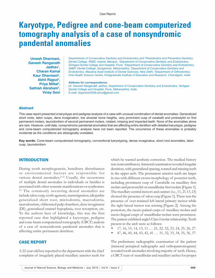
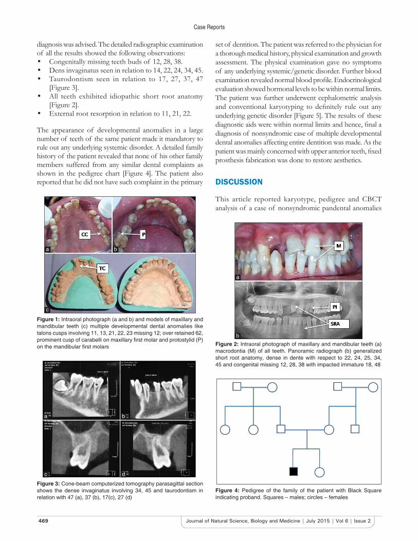
which he wanted aesthetic correction. The medical history was noncontributory. Intraoral examination revealed irregular dentition, with generalized spacing, rotated and missing teeth in the upper arch. The permanent anterior teeth are larger in size with different crown morphology of posterior teeth, including prominent cusp of Carrabelle on maxillary first molars and protostylid on mandibular first molars [Figure 1]. The maxillary central incisors and canines (i.e., 11, 21.13, 23) showed the presence of talon cusp [Figure 1]. There was the presence of over-retained left lateral primary incisor while the right lateral incisor was missing [Figure 2]. Among the posteriors, the mesio-palatal cusps of maxillary molars and mesio-lingual cusps of mandibular molars were prominent. The patient exhibited angle’s Class I molar relationship. Teeth present in the arch were as follows:• 17, 16, 15, 14, 13, 11 … 21, 22, 52, 23, 24, 25, 26, 27.• 47, 46, 45, 44, 43, 42, 41 … 31, 32, 33, 34, 35, 36, 37.
The preliminary radiographic examination of the patient (intraoral periapical radiographs and orthopantomogram) revealed tooth anomalies involving majority of teeth. Hence, a CBCT scan of mandibular and maxillary arches for proper
Case Reports
469 Journal of Natural Science, Biology and Medicine | July 2015 | Vol 6 | Issue 2
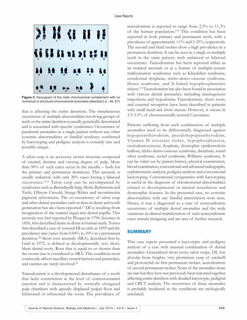
diagnosis was advised. The detailed radiographic examination of all the results showed the following observations:• Congenitally missing teeth buds of 12, 28, 38.• Dens invaginatus seen in relation to 14, 22, 24, 34, 45.• Taurodontism seen in relation to 17, 27, 37, 47
[Figure 3].• All teeth exhibited idiopathic short root anatomy
[Figure 2].• External root resorption in relation to 11, 21, 22.
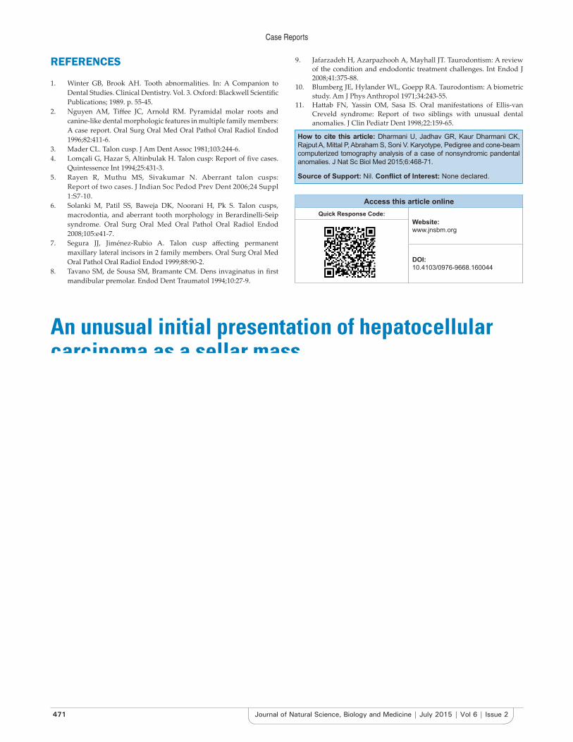
The appearance of developmental anomalies in a large number of teeth of the same patient made it mandatory to rule out any underlying systemic disorder. A detailed family history of the patient revealed that none of his other family members suffered from any similar dental complaints as shown in the pedigree chart [Figure 4]. The patient also reported that he did not have such complaint in the primary
set of dentition. The patient was referred to the physician for a thorough medical history, physical examination and growth assessment. The physical examination gave no symptoms of any underlying systemic/genetic disorder. Further blood examination revealed normal blood profile. Endocrinological evaluation showed hormonal levels to be within normal limits. The patient was further underwent cephalometric analysis and conventional karyotyping to definitely rule out any underlying genetic disorder [Figure 5]. The results of these diagnostic aids were within normal limits and hence, final a diagnosis of nonsyndromic case of multiple developmental dental anomalies affecting entire dentition was made. As the patient was mainly concerned with upper anterior teeth, fixed prosthesis fabrication was done to restore aesthetics.
DISCUSSION
This article reported karyotype, pedigree and CBCT analysis of a case of nonsyndromic pandental anomalies
Figure 4: Pedigree of the family of the patient with Black Square indicating proband. Squares – males; circles – females
Figure 1: Intraoral photograph (a and b) and models of maxillary and mandibular teeth (c) multiple developmental dental anomalies like talons cusps involving 11, 13, 21, 22, 23 missing 12; over retained 62, prominent cusp of carabelli on maxillary first molar and protostylid (P) on the mandibular first molars
a b
c
Figure 2: Intraoral photograph of maxillary and mandibular teeth (a) macrodontia (M) of all teeth. Panoramic radiograph (b) generalized short root anatomy, dense in dente with respect to 22, 24, 25, 34, 45 and congenital missing 12, 28, 38 with impacted immature 18, 48
a
b
Figure 3: Cone-beam computerized tomography parasagittal section shows the dense invaginatus involving 34, 45 and taurodontism in relation with 47 (a), 37 (b), 17(c), 27 (d)
a b
c d
Case Reports
470Journal of Natural Science, Biology and Medicine | July 2015 | Vol 6 | Issue 2
that is affecting the entire dentition. The simultaneous occurrence of multiple abnormalities involving groups of teeth or the entire dentition is usually genetically determined and is associated with specific syndromes. Occurrence of pandental anomalies in a single patient without any other systemic abnormalities or familial tendency confirmed by karyotyping and pedigree analysis is certainly rare and possibly unique.
A talon cusp is an accessory crown structure composed of enamel, dentine and varying degree of pulp. More than 90% of such cases occur in the maxilla – both for the primary and permanent dentitions. This anomaly is usually unilateral, with only 20% cases having a bilateral occurrence.[4-6] Talon cusp can be accompanied by syndromes such as Berardinelli-Seip, Mohr, Rubinstein and Taybi, Ellisvan Creveld, Sturge-Weber and incontinentia pigmenti achromians. The co-occurrence of talon cusp and other dental anomalies such as dens in dente and tooth gemination has also been reported.[7] DI is resulting from invagination of the enamel organ into dental papilla. This anomaly was first reported by Ploquet in 1794. Socrates in 1856, first described dense in dente in human teeth. Tomes first described a case of coronal DI as early as 1859 and the prevalence rate varies from 0.04% to 10% in a permanent dentition.[8] Short root anomaly (SRA), described first by Lind in 1972, is defined as developmentally very short, blunt dental roots. Root that is equal to or shorter than the crown size is considered as SRA. This condition most commonly affects maxillary central incisors and premolars, and canines are rarely involved.[9]
Taurodontism is a developmental disturbance of a tooth that lacks constriction at the level of cementoenamel junction and is characterized by vertically elongated pulp chambers with apically displaced pulpal floor and bifurcated or trifurcated the roots. The prevalence of
taurodontism is reported to range from 2.5% to 11.3% of the human population.[10] This condition has been reported in both primary and permanent teeth, with a prevalence of approximately <1% and 3-35%, respectively. The second and third molars show a high prevalence in a permanent dentition. It can be seen in a single or multiple teeth in the same patient, with unilateral or bilateral occurrence. Taurodontism has been reported either as an isolated anomaly or as a feature of multiple-system malformation syndromes such as Klinefelter syndrome, ectodermal dysplasia, tricho-dento-osseous syndrome, Down syndrome, and X-linked hypophosphatemic rickets.[11] Taurodontism has also been found in association with various dental anomalies, including amelogenesis imperfecta and hypodontia. Taurodontism, short roots, and external resorption have been described in patients with small head and short stature. However, it occurs in 2.5-3.5% of chromosomally normal Caucasians.
Patients suffering from such combination of multiple anomalies need to be differentially diagnosed against hypopararthyrodisim, pseudohypoparathyrodism, Vitamin D resistant r icket , hypophosphatasia , oculodentoosseous dysplasia, dystrophic epidermolysis bullosa, tricho-dento-osseous syndrome, dwarfism, russel silver syndrome, seckel syndrome, Williams syndrome. It can be ruled out by patient history, physical examination, blood examination, conventional and advanced radiographs, cephalometric analysis, pedigree analysis and conventional karyotyping. Conventional cytogenetics with karyotyping is useful in the diagnosis of chromosomal abnormalities related to developmental or mental retardation and dysmorphic features. In the presented case, no systemic abnormalities with any familial interrelation were seen. Hence, it was a diagnosed as a case of nonsyndromic occurrence of multiple dental anomalies and the wide variations in clinical manifestation of such nonsyndromic cases remain intriguing and are area of further research.
SUMMARY
This case report presented a karyotypic and pedigree analysis of a case with unusual combination of dental anomalies: Generalized short roots, talon cusps, DI, low alveolar bone heights, very prominent cusp of carabelli and protostylid on first permanent molars, taurodontism of second permanent molars. None of the anomalies alone are rare but they have not previously been reported together affecting entire dentition with detailed karyotypic, pedigrial and CBCT analysis. The occurrence of these anomalies is probably incidental as the conditions are etiologically unrelated.
Figure 5: Karyogram of the male chromosomal complement with no numerical or structural chromosomal anomalies detected (i.e., 46, XY)
Case Reports
471 Journal of Natural Science, Biology and Medicine | July 2015 | Vol 6 | Issue 2
REFERENCES
1. Winter GB, Brook AH. Tooth abnormalities. In: A Companion to Dental Studies. Clinical Dentistry. Vol. 3. Oxford: Blackwell Scientific Publications; 1989. p. 55-45.
2. Nguyen AM, Tiffee JC, Arnold RM. Pyramidal molar roots and canine-like dental morphologic features in multiple family members: A case report. Oral Surg Oral Med Oral Pathol Oral Radiol Endod 1996;82:411-6.
3. Mader CL. Talon cusp. J Am Dent Assoc 1981;103:244-6.4. Lomçali G, Hazar S, Altinbulak H. Talon cusp: Report of five cases.
Quintessence Int 1994;25:431-3.5. Rayen R, Muthu MS, Sivakumar N. Aberrant talon cusps:
Report of two cases. J Indian Soc Pedod Prev Dent 2006;24 Suppl 1:S7-10.
6. Solanki M, Patil SS, Baweja DK, Noorani H, Pk S. Talon cusps, macrodontia, and aberrant tooth morphology in Berardinelli-Seip syndrome. Oral Surg Oral Med Oral Pathol Oral Radiol Endod 2008;105:e41-7.
7. Segura JJ, Jiménez-Rubio A. Talon cusp affecting permanent maxillary lateral incisors in 2 family members. Oral Surg Oral Med Oral Pathol Oral Radiol Endod 1999;88:90-2.
8. Tavano SM, de Sousa SM, Bramante CM. Dens invaginatus in first mandibular premolar. Endod Dent Traumatol 1994;10:27-9.
9. Jafarzadeh H, Azarpazhooh A, Mayhall JT. Taurodontism: A review of the condition and endodontic treatment challenges. Int Endod J 2008;41:375-88.
10. Blumberg JE, Hylander WL, Goepp RA. Taurodontism: A biometric study. Am J Phys Anthropol 1971;34:243-55.
11. Hattab FN, Yassin OM, Sasa IS. Oral manifestations of Ellis-van Creveld syndrome: Report of two siblings with unusual dental anomalies. J Clin Pediatr Dent 1998;22:159-65.
How to cite this article: Dharmani U, Jadhav GR, Kaur Dharmani CK, Rajput A, Mittal P, Abraham S, Soni V. Karyotype, Pedigree and cone-beam computerized tomography analysis of a case of nonsyndromic pandental anomalies. J Nat Sc Biol Med 2015;6:468-71.
Source of Support: Nil. Conflict of Interest: None declared.
Access this article onlineQuick Response Code:
Website: www.jnsbm.org
DOI: 10.4103/0976-9668.160044
An unusual initial presentation of hepatocellular carcinoma as a sellar mass
AbstractSellar masses are frequently adenomatous pituitary tumors. Metastatic disease is unusual, often mimicking the presentations of adenomas. Hepatocellular carcinoma (HCC) is the most common primary hepatic malignancy but unusual to have a pituitary metastasis (PM). A 65-year-old man presented with headache, diplopia, ptosis, decreased vision in the right eye and unintentional weight loss of 32lbs. Preliminary out-patient work-up revealed a mass in the pituitary region. Cranial imaging showed 3.1 cm × 3.2 cm × 4.4 cm lesion. Abdominal imaging (computed tomography and magnetic resonance imaging) demonstrated a lobulated, nodular and heterogeneous right lobe of the liver. Trans-sphenoidal resection of the sellar mass favored metastatic HCC on histology. Liver biopsy confirmed HCC. We recommend maintaining an increased clinical suspicion upon evaluation of nonclassical clinical and radiological presentations of suspected PM/malignancy; as well as pursuing additional investigations in all early cases.
Key words: Hepatocellular carcinoma, metastasis, pituitary gland
Nihar Shah, Yana Cavanagh,
Hamid Shaaban1, Beth Stein,
Sohail N. Shaikh, Dharmesh H. Kaswala2,
Walid Baddoura1
Department of Gastroenterology, St. Joseph’s Regional Medical Center, Paterson, 1Department of Gastroenterology, St Michael’s Medical Center, Newark, NJ, 2Department of Gastroenterology, Beth Israel Deaconess Medical Center, Harvard Medical School, Harvard University, Boston, MA, USA
Address for correspondence: Dr. Nihar Shah, St. Joseph’s Regional Medical Center, Paterson, NJ, USA. E-mail: [email protected]
INTRODUCTION
Sellar masses are most frequently primary pituitary tumors of the adenomatous type. Metastatic malignancies are
an unusual clinical presentation.[1] However, metastatic disease (including isolated pituitary metastasis [PM]) may mimic the clinical, endocrinological and radio graphical presentations of primary pituitary adenomas (PA)[2] which