tumor and stem cell biology cancer research intratumoral ... · yasuhiro miki1, takashi suzuki1,...
TRANSCRIPT

Tum
IntrbetwCel
YasuhShukoHisafu
Abst
Intro
Non∼80%neousnoma,compr
AuthorGraduaInstituSendaiHospitaPrefectCompaResear
Note: SOnline
CurrentOral MDentist
CorresTohokuAoba-kFax: 81
doi: 10
©2010
www.a
Canceresearch
or and Stem Cell Biology
atumoral Localization of Aromatase and Interactioneen Stromal and Parenchymal Cells in the Non–Small
R
l Lung Carcinoma Microenvironment
iro Miki1, Takashi Suzuki1, Keiko Abe1, Satoshi Suzuki3, Hiromichi Niikawa4, Shinya Iida1,
Hata1, Jun-ichi Akahira1, Kazushige Mori5, Dean B. Evans6, Takashi Kondo2, mi Yamada-Okabe5, and Hironobu Sasano1ractEstr
roles iunknofollowcasesnohiston arosystemarraysBoth acells. ASeveraible faboth o
and largise 57% o
s' Affiliatiote Schoolte of Deve, Japan; 3Del, Ishinomaural Centralny, Shizuoch Basel, O
upplementa(http://cance
address foedicine anry, 4-1 Seiry
ponding AUniversity
u, Sendai,-22-717-80
.1158/0008-
American A
acrjourna
Download
ogens produced as a result of intratumoral aromatization has been recently shown to play importantn proliferation of human non–small cell lung carcinomas (NSCLC), but the details have remained largelywn. Therefore, in this study, we evaluated the possible roles of intratumoral aromatase in NSCLCs ass: (a) evaluation of intratumoral localization of aromatase mRNA/protein in six lung adenocarcinomausing laser capture microdissection combined with quantitative reverse transcriptase-PCR and immu-ochemistry; (b) examination of the possible effects of isolated stromal cells from lung carcinoma tissuesmatase mRNA transcript expression in lung carcinoma cell lines (A549 and LK87) through a coculture; and (c) screening of cytokines derived from stromal LK001S and LK002S cells using cytokine antibodyand subsequent evaluation of effects of these cytokines on aromatase expression in A549 and LK87.romatase mRNA and protein were mainly detected in intratumoral carcinoma cells but not in stromalromatase expression of A549 and LK87 was upregulated in the presence of LK001S or LK002S cells.l cytokines such as interleukin-6 (IL-6), oncostatin M, and tumor necrosis factor-α, all known as induc-ctors of aromatase gene, were detected in conditioned media of LK001S and LK002S cells. Treatment ofncostatin M and IL-6 induced aromatase gene expression in A549 an LK87, respectively. These results allted that intratumoral microenvironments, especially carcinoma-stromal cell interactions, play a pivotal
indicarole in the regulation of intratumoral estrogen synthesis through aromatase expression in human lungadenocarcinomas. Cancer Res; 70(16); 6659–69. ©2010 AACR.
where55% inestrogadenotients,
duction
–small cell lung carcinomas (NSCLC) account forof all lung carcinomas and are composed of heteroge-groups such as adenocarcinoma, squamous cell carci-
e-cell carcinoma. Squamous cell carcinomasf all lung cancer in men and 25% in women,
of aroby theWeinbprolife(4). Sinaromafor lunportedverseNSCLCplays pas wecombitor in(ER) bpatienNCT0rando(anast
ns: 1Department of Pathology, Tohoku Universityof Medicine; 2Department of Thoracic Surgery,lopment, Aging and Cancer, Tohoku University,partment of Thoracic Surgery, Ishinomaki Red Crosski, Japan; 4Department of Thoracic Surgery, IwateHospital, Morioka, Japan; 5CHUGAI Pharmaceuticalka, Japan; and 6Novartis Institutes for BioMedicalncology Research, Basel, Switzerland
ry data for this article are available at Cancer Researchrres.aacrjournals.org/).
r Y. Miki: Division of Oral Pathology, Department ofd Surgery, Tohoku University Graduate School ofo-machi, Aoba-ku, Sendai 980-8575, Japan
uthor: Hironobu Sasano, Department of Pathology,Graduate School of Medicine, 2-1 Seiryo-machi,Miyagi 980-8575, Japan. Phone: 81-22-717-8050;51; E-mail: [email protected].
5472.CAN-09-4653
ssociation for Cancer Research.
ls.org
Research. on January 19, 20cancerres.aacrjournals.org ed from
as adenocarcinomas comprise 30% cases in men andwomen (1). Therefore, sex steroid hormones, such as
en, may also play an important role in NSCLC, especiallycarcinoma. In postmenopausal breast carcinoma pa-intratumoral production of estrogens occurs as a resultmatization of androgens into estrogens and is catalyzedcytochrome P450 aromatase enzyme (2, 3). In 2005,erg and colleagues showed aromatase-dependent cellration of lung carcinoma using a mice xenograft modelce the pioneering study of Weinberg and colleagues (4),tase has been studied as one of the therapeutic targetsg carcinoma patients (5–7). Mah and colleagues (5) re-an association between aromatase expression and ad-clinical outcome in female patients with early-stage. These findings all suggest that intratumoral aromataseivotal roles in estrogen-dependent pathways in NSCLCll as in breast carcinoma. Phase II clinical trials of anation therapy using an epidermal growth factor recep-hibitor [erlotinib (Tarceva)] and an estrogen receptorlocker (fulvestrant) versus erlotinib alone in NSCLCts are now under way (ClinicalTrials.gov identifier:0100854 and NCT00592007). In addition, a phase II
mized trial of fulvestrant and an aromatase inhibitorrozole) as consolidation therapy in postmenopausal6659
20. © 2010 American Association for Cancer

womeplatinuwill aNCT00such asion inconsidtherapThe
of aromcell ca(LCM)(RT-PCtosterinhibitprolifethe prtosterIn h
both sed bykines (effectssion ofcocultfactorsinterleα (TNFarraystase ex
Mate
LaserSix
77 yeathree76, anDeparpital. REthicsMedicly emb(Sakurthicknthis thof relainationwere lal strowas pe& IndMicroscripti
QuanQua
System
used iwardreversrepresage ra
ImmuSeq
aboveand pcanceantibohistocpropo(15) wcentagsifiedand 3,omataimmunoreactotal snointedepen
LungWe
BEAS-tissuetary TmenteMCF-7previoCell RversitERα atreatmwas thand tER blblockeTocrisrespeced byals weIndust
Cell pBot
assay.determSupplered–frCo.) wbeforeon th
Miki et al.
Cance6660
n with advanced NSCLC who have received first-linem-based chemotherapy with or without bevacizumablso be scheduled (ClinicalTrials.gov identifier:932152). However, the basic biological characteristics,s tissue localization and regulation of aromatase expres-the lung carcinoma microenvironment, are generallyered prerequisites for successful outcome of these novelies but have remained relatively unknown.refore, in this study, we first examined the localizationatase in six lung adenocarcinoma and three squamousrcinoma tissues using laser capture microdissectiontogether with quantitative reverse transcriptase-PCRR) and immunohistochemistry (8, 9). The effects of tes-
one on both A549 and LK87 cell proliferation, and theory effects of the aromatase inhibitor letrozole on cellration, were subsequently evaluated. We also showedoduction of estrogen as a result of conversion from tes-one in both A549 and LK87 cells.uman breast carcinoma tissue, aromatase expression intromal and parenchymal cells was reported to be regulat-various factors such as cell-to-cell interactions and cyto-9–12). Therefore, in this study, we examined the possibleof isolated stromal cells from NSCLC tissues on expres-aromatase mRNA transcripts in NSCLC cell lines using aure system (9, 12). We then screened the expression ofthat have been reported to induce aromatase, such asukin-6 (IL-6), oncostatin M, and tumor necrosis factor--α), derived from stromal cells using cytokine antibodies, and evaluated the effects of these cytokines on aroma-pression in A549 and LK87, respectively.
rials and Methods
capture microdissectionlung adenocarcinoma cases [female, A1–A3 (48, 65, andrs old); male, A4–A6 (53, 58, and 70 years old)] andlung squamous cell carcinoma cases [male, S1–S3 (55,d 60 years old)] were obtained from the patients in thetment of Thoracic Surgery at Tohoku University Hos-esearch protocols for this study were approved by theCommittee of Tohoku University Graduate School ofine (no. 2008-444). Lung carcinoma tissues were rapid-edded in Tissue-Tek optimal temperature compounda Finetechnical Co., Ltd.) and frozen sectioned at aess of 8 μm (9). We used 8 μm in this study becauseickness is generally considered to contain a monolayertively homogeneous cell populations without contam-of other components (13). Approximately 5,000 cells
aser transferred from both carcinoma and intratumor-mal cells under light microscopic evaluation (9). LCMrformed using mmi CellCut (MMI Molecular Machinesustries AG). Total RNA was extracted using RNeasyKit (Qiagen GmbH), and a QuantiTect Reverse Tran-on Kit (Qiagen) was used in the synthesis of cDNA.
titative RT-PCR
ntitative RT-PCR was carried out using the LightCycler(Roche Diagnostics GmbH). The primer positionsdepenassays
r Res; 70(16) August 15, 2010
Research. on January 19, 20cancerres.aacrjournals.org Downloaded from
n this study were as follows: RPL13A (NM_012423), for-487–reverse 612 (9); aromatase (X13589), forward 691–e 806 (9). Aromatase mRNA levels in each case wereented as a ratio of RPL13A and evaluated as a percent-tio (9).
nohistochemistryuential frozen tissues, also used in the LCM analyses, were used to examine the correlation between mRNArotein in individual cellular compartments of the lungr tissues with immunohistochemistry using monoclonaldy no. 677 (9, 14, 15). Evaluation of aromatase immuno-hemistry was performed based on staining intensityrtion scoring systems used for breast carcinoma tissuesith some modifications (9, 14). The approximate per-e of immunopositive cells (proportion score) were clas-into the following four groups: 0, <1%; 1, −25%; 2, −50%;>50% immunopositive cells. The relative intensity of ar-se immunopositive cells was classified as follows: 0, nonoreactivity; 1, weak; 2, moderate; and 3, intense immu-tivity. Aromatase immunoreactivity was evaluated as acore composed of the proportion score + relative immu-nsity score. Results of immunohistochemistry were in-dently evaluated by two of the authors (Y.M. and K.A.).
carcinoma cell lines and culture conditionsused A549, LCSC#1, RERF-LC-OK, LK87, LK2, WI-26,2B, NCI-H727, RERF-LC-AI, and LCAM1. The original, source, and medium were summarized in Supplemen-able S1. Cells were maintained in each medium supple-dwith 10% fetal bovine serum (FBS; JRHBiosciences Co.).was used as a positive control of aromatase. In our
us studies (7, 16), we showed that A549, provided byesource Center for Biomedical Research (Tohoku Uni-y, Sendai, Japan), did not have detectable levels ofnd ERβ proteins and was unresponsive to estradiolent in reporter gene assay. A549 used in this studye same stock as in the previous study (16). Estradiolestosterone were obtained from Sigma-Aldrich. Theocker, ICI 182,780, and the androgen receptor (AR)r, hydroxyflutamide (flutamide), were obtained fromCookson Ltd. and Toronto Research Chemicals, Inc.,tively. The aromatase inhibitor, letrozole, was provid-Novartis Pharmaceutical Corporation. These materi-re dissolved in 99.5% ethanol (Wako Pure Chemicalries).
roliferation assayh A549 and LK87 were used in the cell proliferationThe amounts of ERα, ERβ, and AR mRNA expressionined by quantitative RT-PCR were summarized inmentary Fig. S1A. The cells were cultured in phenolee RPMI 1640 (modified RPMI 1640, Sigma-Aldrichith dextran-coated charcoal (DCC)-FBS for 2 dayseach experiment. The effects of steroids and drugs
ese cell lines were evaluated using both anchorage-
dent and anchorage-independent cell proliferation(17).Cancer Research
20. © 2010 American Association for Cancer

AncwereTECHtaininand tewheneratioKit-8;Anc
cells wTECH10% Dtest cocells wusing
SteroA54
incubataininThe eon steconcedehydchromanalystreateAfterIsotopChemnal stfrom traphywith a(Appliizationsepara(Imtakare prcells (
EffectcarcinIntr
usedLK002mentdisagg1 to 1subseqgrowt5 daysmal cblastschangand 8Coc
carcin
in 100(0.4-μand Labsenplacedtion uratedexamiCon
culturwas r24 holectedMacrowere cing 5(CC-mlevelsabove
CytokIn t
(RayBleasedwere iactedconjugperoxdots wchemiHum
purch(20 ngwas aChemexamAromimmuglasseAGCscribefor ardensi(Scien
NeutrThe
IL-6 (neutrInc. Nof an24 houimmoby usiAfterDynab
Aromatase in the Lung Carcinoma Microenvironment
www.a
horage-dependent cell proliferation assay. The cellsseeded onto normal-adhesion 96-well plates (AGCNO GLASS CO., LTD.) in phenol red–free medium con-g 10% DCC-FBS. The cells were treated with steroidsst compounds for 24, 48, and 72 hours, respectively,these cells were harvested and evaluated for cell prolif-n using the WST-8 colorimetric assay (Cell CountingDojindo Laboratories; ref. 18).horage-independent cell proliferation assay. Theere cultured in low-adhesion 24-well plates (AGCNO GLASS) in phenol red–free medium containingCC-FBS. The cells were then treated with steroids andmpounds for 3, 6, and 9 days, respectively, when theseere harvested. The cell number was determined bya cell counter (Sysmex CDA-500, Sysmex Corporation).
id production assays9 and LK87 cells cultured onto 100-mm dishes wereted at 37°C in phenol red– and FBS-free medium con-g 100 nmol/L testosterone as substrates for 24 hours.ffects of 5 μmol/L flutamide and 10 nmol/L letrozoleroid hormone production were also examined. Thentrations of estrone, estradiol, testosterone, and 5α-rotestosterone (5α-DHT) were evaluated by liquidatography-electrospray tandem mass spectrometryis (ASUKA Pharma Medicals Co.; refs. 9, 19). All cellsd with substrates were counted by the cell counter.addition of 100 pg of androstenedione-2H7 (C/D/Nes) and estrone-13C4 and estradiol-13C4 (Hayashi Pureical Industrial Company Ltd., Osaka, Japan) as inter-andards, steroids were extracted with diethyl etherhe medium. In this study, we used liquid chromatog-(Agilent 1100; Agilent Technologies, Inc.) coupledn API 4000 triple-stage quadruple mass spectrometered Biosystems, Inc.) operated with electron spray ion-in the positive-ion mode, and the chromatographiction was performed on Cadenza CD-C18 columnst Corporation). Steroid hormone levels in each caseesented as pg/mL/104 cells (estrogen) or ng/mL/104
androgen).
s of stromal cells on aromatase expression inoma cellsatumoral stromal cells. The primary stromal cellsin our present study were designated LK001S andS, respectively, and isolated using collagenase treat-(20) from human lung carcinoma tissues. After tissueregation with collagenase S-I (Nitta Gelatin, Inc.) for.5 hours, these stromal cells grown to confluence wereuently cultured in RPMI 1640 with 10% FBS. The out-h of these cells was detected at 3 days (LK002S) or(LK001S) of cell culture. These lung intratumoral stro-ells showed typical morphological features of fibro-under light microscopy. There were no morphologices and cell viabilities between the cells in passages 5used in the experiment.
ulture system. For physical separation of stromal andoma cell lines, transwell cultures were establishedwithoCC-m
acrjournals.org
Research. on January 19, 20cancerres.aacrjournals.org Downloaded from
-mm dishes using Transwell Permeable Supportsm pore; Corning Incorporated; refs. 9, 12). Both A549K87 cells were cultured in transwell chambers in thece or presence of LK001S and LK002S cells and wereat the bottom of the dishes. After 72 hours of cultiva-sing this coculture system, carcinoma cells were sepa-and the levels of aromatase mRNA expression werened by real-time RT-PCR analysis as described above.ditioned medium. Both LK001S and LK002S wereed in 155-cm2 culture flasks. The culture mediumeplaced by FBS and phenol red–free medium. Afterurs, the conditioned medium (total 80 mL) was col-and concentrated to a total volume of 16 mL usingsep Centrifugal Devices (Pall Corporation). A549 cellsultured onto a six-well plate in the medium contain-%, 10%, and 20% condensed conditioned mediumedium). After 72 hours of culture, aromatase mRNAwere evaluated by real-time RT-PCR as described.
ine analysishis study, we used Human Cytokine Antibody Array 5iotech, Inc.; refs. 21–23) to identify the cytokines re-from stromal cells. Cytokine antibody membranes
ncubated for 2 hours with 2 mL of CC-medium. The re-membranes were then incubated for 1 hour with biotin-ated anti-cytokines and then developed with horseradishidase–streptavidin and chemiluminescence. Proteinere visualized with Las-1000 cooled CCD-camera
luminescent image analyzer (Fuji Photo Film Co., Ltd.).an recombinant TNF-α, IL-6, and oncostatin M wereased from Wako Pure Chemical Industries. TNF-α/mL), IL-6 (50 ng/mL), or oncostatin M (50 ng/mL)dded with 100 nmol/L dexamethasone (Wako Pureical Industries). Aromatase mRNA expression wasined using quantitative RT-PCR described above.atase protein expression was also evaluated usingnocytochemistry. Cells were grown directly on covers (collagen type I–coated cover glass, round, 12 mm;TECHNO GLASS) under the culture conditions de-d above. The relative abundance of reacted signalsomatase was subsequently quantified as the opticalty (OD) value by using the Multi Gauge softwarece Lab 2005 version 3.0; Fuji Photo Film).
alization assay of cytokinesmonoclonal antibodies against TNF-α (clone 28401),
clone 6708), and oncostatin M (clone 17001) used inalization assays were obtained from R&D Systems,eutralizations were performed by adding 50 mg/mLtibodies to 2 mL CC-medium described above forrs on ice, with gentle shaking. All antibodies used werebilized to magnetic Dynabeads Protein G (Invitrogen)ng 50 μg IgG for every 100 μL Dynabeads Protein G.2 hours of incubation on ice, the antibody-boundeads were collected using a magnet. The supernatants
ut antibody-bound Dynabeads, termed neutralizingedium (NCC-medium), were also used for aromataseCancer Res; 70(16) August 15, 2010 6661
20. © 2010 American Association for Cancer

exprescontailevelsof cell
StatisRes
was pInstituwas dweretest. Asignifi
Resu
Locallung cIn
transc
casesAromain carlow lecells.analyscytoplsix camunothesenohist
Figurelung adadenocand no
Miki et al.
Cance6662
sion assay. A549 cells were cultured in DCC-mediumning 20% NCC-medium. Aromatase mRNA expressionwere examined by quantitative RT-PCR after 72 hoursculture.
tical analysisults were expressed as mean ± SD. Statistical analysiserformed using the StatView 5.0 J software (SASte, Inc.). The statistical difference of two groupsetermined by Student's t test. Multiple comparisonsperformed with Bonferroni/Dunn test or Dunnett's
P value of <0.05 was considered to indicate statistical A1, 3;LCM/tase wdetectma catransctumoradditi
cance.
lts
ization of aromatase mRNA and protein inarcinomas
six lung adenocarcinoma cases, aromatase mRNAripts were detected in carcinoma cells from fivethesepresen
arcinoma tissues. Aromatase immunoreactivity was detected in carcinoma cells. Irmal lung cell lines. MCF-7, breast carcinoma cell line.
r Res; 70(16) August 15, 2010
Research. on January 19, 20cancerres.aacrjournals.org Downloaded from
and in stromal cells from two cases (Fig. 1A).tase mRNA expression was predominantly detectedcinoma or parenchymal cells. In case A3, a relativelyvel of aromatase mRNA was detected only in stromalIn the same cases evaluated by aromatase LCM/PCRis, aromatase immunoreactivity was detected in theasm of carcinoma cells but not stromal cells from allses examined in this study (Fig. 1B). Aromatase im-reactivity was not detected in all the stromal cells ofsix cases. The cumulative or total aromatase immu-ochemistry score of each case was as follows: caseA2, 6; A3, 4; A4, 6; A5, 2; A6, 0. The results betweenPCR analysis and immunohistochemistry of aroma-ere consistent in five cases, but a discrepancy wased in case A3. In three lung squamous cell carcino-ses (S1, S2, and S3), the levels of aromatase mRNAripts were below the detection limits in both intra-al carcinoma and stromal cells (data not present). Inon, no aromatase immunoreactivity was detected in
three cases of squamous cell carcinoma (data nott).1. Aromatase expression in lung adenocarcinoma tissues and lung carcinoma cell lines. A, aromatase mRNA level in each case (cases A1–A6) ofenocarcinoma (white columns) and stromal (black columns) cells. B, aromatase immunohistochemistry for each case (cases A1–A6) of lung
HC, immunohistochemistry. C, aromatase levels in lung carcinoma
Cancer Research
20. © 2010 American Association for Cancer

AromcarcinAro
lines eRelativel as iWe thexpreA549expresthan ttissues
EffectprolifIn
LK87treatminduce
0.001)liferat(Fig. 2tion w1 μmoand Bicantlyited byflutamwith ttamidthe cecompacreaseditioninhibimide,
FigureA, anch[ICI (+)]testostecontrolwhite sD, anchwith 1 μmol/L flutamide (TST + FUL); white diamonds, 10 nmol/L testosterone with 1 μmol/L flutamide and 10 nmol/L letrozole (TST + FLU + LET);black s (TST +
Aromatase in the Lung Carcinoma Microenvironment
www.a
atase mRNA expression levels in human lungoma cell linesmatase mRNA was detected in all lung carcinoma cellxcept for LCSC#1 examined in this study (Fig. 1C).ely high levels of aromatase mRNA, with the same lev-n MCF-7, were detected in both LK87 and NCI-H727.erefore used the relatively higher aromatase mRNA–ssing LK87 and low aromatase mRNA–expressingin further evaluations. The level of aromatase mRNAsion in lung carcinoma cell lines was ∼10 times lowerhat in intratumoral carcinoma cells of lung carcinoma.
s of estrogen and aromatase on celleration in LK87the anchorage-independent assay, the number ofcells was significantly increased following estradiol
quares, 10 nmol/L testosterone with 1 μmol/L flutamide and 5 μmol/L ICI
ent (1 nmol/L–1 μmol/L) for 6 days (Fig. 2A). Estradiol-d cell proliferation was significantly inhibited (P <
cell pSimila
acrjournals.org
Research. on January 19, 20cancerres.aacrjournals.org Downloaded from
by ICI 182,780 (1 μmol/L) treatment, with cell pro-ion nearly comparable with that at the basal levelA). Estradiol- or testosterone-mediated cell prolifera-as not detected in A549 cells treated with 1 nmol/L tol/L estradiol or testosterone (Supplementary Fig. S2A). Testosterone treatment (1 nmol/L–1 μmol/L) signif-decreased the number of LK87, which was also inhib-treatment with the specific androgen receptor blockeride (5 μmol/L; Fig. 2B). When LK87 cells were treatedestosterone (1 nmol/L–1 μmol/L) and 5 μmol/L flu-e simultaneously for 3, 6, and 9 days, the number oflls was significantly increased by these treatmentred with that of the control level (Fig. 2B). This in-d cell proliferation was significantly inhibited by ad-of ICI 182,780 (1 μmol/L; Fig. 2C) or the aromatasetor letrozole (10 nmol/L; Fig. 2D). ICI 182,780, fluta-and letrozole alone did not significantly change the
FLU + ICI); *, P < 0.05 versus TST + FLU.
2. Summary of experiments of the proliferation of lung carcinoma cell line LK-87 through the aromatase-dependent estrogen synthesis.orage-independent cell proliferation of LK-87 treated by estradiol. White columns, estradiol alone [ICI (−)]; shaded column, estradiol with ICI182,720, *, P < 0.05 versus control (0); †, P < 0.05 versus 1 μmol/L estradiol alone. B, anchorage-independent proliferation of LK87 treated byrone with flutamide for 6 d. Black columns, testosterone alone [FLU (−)]; shaded columns, testosterone with flutamide [FLU (+)]; *, P < 0.05 versus[0, FLU (−)]. C, anchorage-independent proliferation of LK87 treated by testosterone with flutamide for 3 to 6 d. White circles, control (CTL);quares, 10 nmol/L testosterone alone (TST); black circles, 10 nmol/L testosterone with 1 μmol/L flutamide (TST + FLU). *, P < 0.05 versus CTL.orage-independent proliferation of LK87 treated by testosterone with flutamide and letrozole/ICI for 3 to 6 d. Black circles, 10 nmol/L testosterone
roliferation of LK87 cells (Supplementary Fig. S2C).r findings of cell proliferation were also obtained
Cancer Res; 70(16) August 15, 2010 6663
20. © 2010 American Association for Cancer

whenexperi
EstroFoll
conve5.95 ±n = 3)nificanletrozocentrawith 10.21 m(Fig. 3creasement
EffectmRNAThe
LK87marizecocultmono
cocultcantly
EffectaromThe
TNF-αLK 00costatanalyscreaseIL-6 afrom LtectionlowingLK87.with bAdd
and LmRNAwas sIL-6 (L
Miki et al.
Cance6664
anchorage-dependent/WST-8 assay was used in thesements (Supplementary Fig. S1B–E).
gen and androgen production in LK87owing testosterone treatment (288.4 mg/mL), thersion rate into estradiol and estrone in LK87 was0.55 and 0.20 ± 0.02 mg/mL/10−6 cells (mean ± SD,, respectively (Fig. 3A). Estrogen production was sig-tly decreased by 5μmol/L flutamide alone or 10 nmol/Lle+5 μmol/L flutamide treatment (Fig. 3A). The con-tion of testosterone and 5α-DHT in LK87 treated0 nmol/L testosterone was 10.43 ± 0.79 and 1.41 ±g/mL/10−6 cells (mean ± SD, n = 3), respectivelyB). These androgen productions were significantly in-d by 10 nmol/L letrozole + 5 μmol/L flutamide treat-(Fig. 3B).
s of coculture with stromal cells on aromataseexpression in LK87 and A549results of the effects of the coculture of stromal cells onand A549 aromatase mRNA expression levels are sum-d in Fig. 4A. Aromatase mRNA levels of LK87 following
ure with LK002S were significantly higher than those incultured LK87. Aromatase mRNA levels in A549 (afterpareddiffere
r Res; 70(16) August 15, 2010
Research. on January 19, 20cancerres.aacrjournals.org Downloaded from
ure with both LK001S and LK002S) were also signifi-higher than those in monocultured A549 cells.
s of cytokines derived from stromal cells onatase mRNA expression in LK87 and A549results of the expression of IL-6, oncostatin M, andderived from lung intratumoral stromal LK001S and
2S cells are summarized in Fig. 4B. In LK002S, IL-6, on-in M, and TNF-α were all detected by cytokine arrayis. The levels of all these three cytokines were in-d by coculture with both A549 and LK87 cells. Bothnd oncostatin M were also detected in the cells derivedK001S. However, TNF-α was under the level of the de-limit in LK001S. The level of IL-6 was increased fol-coculture with A549 and decreased by coculture withThe level of oncostatin M was increased by cocultureoth A549 and LK87 cells.ition of CC-medium (10% and 20%) from both LK001SK002S resulted in significant increment of aromataselevels in A549 (Fig. 4C). The aromatase mRNA level
ignificantly decreased by addition of NCC-medium ofK001S) or both IL-6 and oncostatin M (LK002S) com-
with that of CC-medium alone (Fig. 4D). There were nonces on the degrees of aromatase mRNA inductionureestrducstroronendrtost< 0.05.
FigonproA, eestB, ates*, P
20. © 2010 American Associa
3. Summary of experimentsogen and androgention in LK87 cells.gen production. Left,; right, estradiol; *, P < 0.05.ogen production. Left,erone; right, 5α-DHT;
Cancer Research
tion for Cancer

betweA549 cThe
aromatreatmincrea
mentexpresmRNAment
Figure*, P < 0C, effec10%, aaromat(P < 0.05) lower than treatments of CC-medium (CC) alone. CTL, control (without CC-medium and NCC-medium); IL-6, NCC-medium of IL-6; OSM,NCC-medium of oncostatin M; TNF, NCC-medium of TNF-α.
Aromatase in the Lung Carcinoma Microenvironment
www.a
en NCC-medium of TNF-α and CC-medium alone inells (Fig. 4D).effects of these cytokine treatments on LK87 and A549tase levels are summarized in Fig. 5. In LK87 cells,
ent with oncostatin M + dexamethazone significantlysed aromatase mRNA transcript levels, but the treat-trol ldexam
acrjournals.org
Research. on January 19, 20cancerres.aacrjournals.org Downloaded from
of dexamethazone alone did not influence aromatasesion in LK87 (Fig. 5A). In A549 cells, the aromatasetranscript level was significantly increased by the treat-
of dexamethazone alone compared with that at the con-
4. Summary of experiments of coculture or cytokine assay. A, effects of coculture on aromatase mRNA levels in lung carcinoma cell lines..05 versus monoculture. B, expression of IL-6, oncostatin M (OSM), and TNF-α (TNF) in conditioned media from stromal LK001S or LK002S cultures.ts of CC-medium derived from LK001S or LK002S on aromatase mRNA expression in A549 cells. The concentrations of CC-medium were 5%,nd 20%. *, P < 0.05 versus control (CTL, without CC-medium). D, effects of NCC-medium (NCC-med.) derived from LK001S or LK002S onase mRNA expression in A549 cells. Treatments of NCC-medium with IL-6 (LK001S) or both oncostatin M and IL-6 (LK002S) were significantly
evel (Fig. 5A). Treatment of both oncostatin M +ethasone and IL-6 + dexamethasone significantly
Cancer Res; 70(16) August 15, 2010 6665
20. © 2010 American Association for Cancer
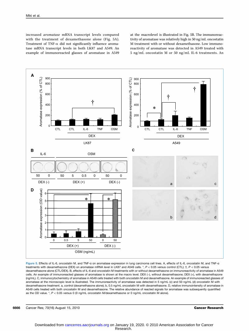
increawithTreatmtase mexamp
at thetivity oM trea
Figuretreatmedexamecells. A(ng/mLaromatdexameA549 cas the
Miki et al.
Cance6666
sed aromatase mRNA transcript levels comparedthe treatment of dexamethasone alone (Fig. 5A).ent of TNF-α did not significantly influence aroma-
RNA transcript levels in both LK87 and A549. Anle of immunoreacted glasses of aromatase in A549reacti5 ng/
ells treated with both oncostatin M and dexamethasone. The relative abundanceOD value. *, P < 0.05 versus 0 (0 ng/mL oncostatin M/dexamethasone or 0 ng/m
r Res; 70(16) August 15, 2010
Research. on January 19, 20cancerres.aacrjournals.org Downloaded from
macrolevel is illustrated in Fig. 5B. The immunoreac-f aromatase was relatively high in 50 ng/mL oncostatintment with or without dexamethasone. Low immuno-
vity of aromatase was detected in A549 treated withmL oncostatin M or 50 ng/mL IL-6 treatments. An5. Effects of IL-6, oncostatin M, and TNF-α on aromatase expression in lung carcinoma cell lines. A, effects of IL-6, oncostatin M, and TNF-αnts with dexamethazone (DEX) on aromatase mRNA level in LK87 and A549 cells. *, P < 0.05 versus control (CTL); †, P < 0.05 versusthasone alone (CTL/DEX). B, effects of IL-6 and oncostatin M treatments with or without dexamethasone on immunoreactivity of aromatase in A549n example of immunoreacted glasses of aromatase is shown at the macro level. DEX (−), without dexamethasone; DEX (+), with dexamethazone). C, immunocytochemistry of aromatase in A549 cells treated with both oncostatin M and dexamethasone. An example of immunoreacted glasses ofase at the microscopic level is illustrated. The immunoreactivity of aromatase was detected in 5 ng/mL (c) and 50 ng/mL (d) oncostatin M withthasone treatment. a, control (dexamethasone alone); b, 0.5 ng/mL oncostatin M with dexamethasone. D, relative immunointensity of aromatase in
of reacted signals for aromatase was subsequently quantifiedL oncostatin M alone).
Cancer Research
20. © 2010 American Association for Cancer

exampcroscoof arotreateimmucreasemetha
Discu
Aropressehumannoma,in parThereintratucausechemantibothe rearomamRNAmal ction imethobeenmouswas atumor(31) cThesedepencanceraromaThe
lines wstromrelativin LK8in thisdepenthat “depenour prtamidtion awerethrou(32, 33blockicell lincarcintor wa(34). Ialso streatmto inc
ma tiscreasiandroprolif(35). Tcer paERβ),inhibifects olargelyThe
tase ecarcinto thecell-toanalysthis dcarcinwe foccells oever, ifactorformininducestromsecretder thlung cThe
varioureleasquentsion iIn ouwas aM treIL-11,throuscriptition spromorelativoncostainedresultlevelsbelowalso sstromin thetralizaily deFurtheisms olung cIn s
Aromatase in the Lung Carcinoma Microenvironment
www.a
le of immunoreacted glasses of aromatase at the mi-pic level is illustrated in Fig. 5C. The immunoreactivitymatase was detected in the cytoplasm of A549 cellsd with 5 and 50 ng/mL of oncostatin M. The relativenointensity of aromatase in A549 was significantly in-d by 50 ng/mL oncostatin M with or without dexa-sone treatment (Fig. 5D).
ssion
matase has been reported to be predominantly ex-d in intratumoral stromal cells and adipocytes ofbreast carcinoma tissues (9, 10, 24). In lung carci-however, several studies showed aromatase proteinenchymal cells but not in stromal cells (4, 6, 25).have been controversies as to the localization ofmoral aromatase in human breast carcinoma (3) be-many results have been obtained by immunohisto-istry, which were influenced by the nature ofdies and specimen preparation. In our present study,sults of combined LCM and RT-PCR analysis oftase showed that in lung carcinoma, aromatase/protein was detected predominantly in parenchy-ells of lung cancer tissues in contrast to its localiza-n human breast carcinoma evaluated by the samedology (8). Similar localization of aromatase has alsoreported in colon (26), gastric (27), and oral squa-cell carcinomas (28). Aromatase immunoreactivitylso reported to be predominantly present in intra-al stromal cells in endometrial (29, 30) and prostaticarcinomas as well as in breast carcinoma (9, 10, 13).data all suggest that stromal cells in classic estrogen-dent malignancies, such as breast and endometrials, are all associated with abundant overexpression oftase.amount of aromatase mRNA in lung carcinoma cellas ∼10 times lower than that detected in intratumoralal cells of breast carcinomas (9). In our present study, aely high rate of conversion into estradiol was detected7 following the treatment of testosterone. LK87 usedstudy has relatively high levels of ERβ and estrogen-
dent cell proliferation. This finding above all suggestsfunctional” aromatase and subsequent estrogen-dent cell proliferation pathways exist in LK87. Inesent study, we also showed that the AR blocker flu-e also exerted inhibitory effects on estradiol produc-s a result of conversion from testosterone. Androgensreported to stimulate aromatase expression/activitygh an AR-dependent pathway in rodent brains). Therefore, aromatase expression was inhibited byng of AR-dependent transcription in lung carcinomaes. Intratumoral concentration of 5α-DHT in breastoma tissues following treatment of aromatase inhibi-s significantly higher than in those without treatmentn our present study, 5α-DHT production in LK87 wasignificantly increased by letrozole with flutamide
ent. Aromatase inhibitors are therefore consideredrease in situ availability of androgens in lung carcino-ered tplays
acrjournals.org
Research. on January 19, 20cancerres.aacrjournals.org Downloaded from
sues as well as in breast carcinoma, possibly by in-ng testosterone concentrations. In breast carcinoma,gens have been shown to predominantly exert anti-erative effects through an AR-dependent pathwayherefore, all of these findings suggest that lung can-tients, who expressed aromatase, ER (ERα and/orand possibly AR, may benefit more from aromatasetor therapy. However, in contrast to estrogen, the ef-f androgens on lung carcinoma cells have remainedunclear.
re was a “discrepancy” in terms of amounts of aroma-xpression between the tissues of the patients and lungoma cell lines. This discrepancy is considered to be dueconditions of cell culture such as culture period and-cell contact. In addition, results from our coculturees in lung and breast carcinoma (9) cells suggest thatiscrepancy is partly due to the lack of interaction ofoma cells and stromal cells. Therefore, in this study,used on the effects of cytokines derived from stromaln aromatase induction in lung carcinoma cells. How-t is true that several investigators showed that solubles, including platelet-derived growth factor and trans-g growth factor β, derived from lung carcinoma cellsd differentiation and cell proliferation of fibroblastical cells (36). Therefore, aromatase-inducible cytokinesed from stromal cells of carcinoma tissues may be un-e control of an interaction with carcinoma cells in theancer microenvironment.results of previously reported studies all showed thats aromatase-stimulating factors (10, 11, 37, 38) wereed from parenchymal or carcinoma cells, which subse-ly resulted in the upregulation of aromatase expres-n stromal cells of human breast carcinoma tissues.r present study, aromatase mRNA/protein in A549lso shown to be induced by both IL-6 and oncostatinatments. In adipose stromal cells, the effects of IL-6,and oncostatin M were all reported to be mediatedgh the Jak/signal transducer and activator of tran-on 3 signaling pathway (39), and interferon-γ activa-ite element was located in the upstream of aromataseter 1.4 (39). We also showed in our present study thately high levels of cytokines such as IL-6, TNF-α, andtatin M were detected in conditioned medium ob-from LK001S and LK002S cultures. In addition, the
s of these cytokine analyses also showed that theof IL-1 and IL-11 secretion from stromal cells werethe detection limits (data not present). These resultsuggest that both IL-6 and oncostatin M secreted fromal cells were required to induce aromatase expressionlung intratumoral microenvironment. However, neu-tion of both IL-6 and oncostatin M did not necessar-crease aromatase expression to the control levels.r investigations are required to clarify the mechan-f regulation of intratumoral aromatase in the humanarcinoma microenvironment.ummary, some human lung carcinomas are consid-
o be estrogen-dependent carcinomas, and aromatasea pivotal role in intratumoral estrogen synthesis. InCancer Res; 70(16) August 15, 2010 6667
20. © 2010 American Association for Cancer

lung cnomafromour psuch athe aMCF-aromapatienwith i
Disclo
TheCHUGANovartis
(K. Morno pote
Ackn
WeMedicin
Grant
Refe1. Se
ma2. Sa
en59
3. Mikinroi
4. Weinh112
5. MavivRe
6. AbaroHu
7. NiigenRe
8. SaarorelJ S
9. Mikbrestr
10. Saaro79:
11. SimAn
12. YathrCa
13. Pintion140
14. Satasrep
15. Lyktastorad
16. Ish
Miki et al.
Cance6668
arcinoma, this aromatization mainly occurs in carci-cells under the influence of various cytokines derivedintratumoral fibroblastic stromal cells. The results ofrevious study (9) showed that aromatase inhibitorss letrozole and exemestane were more effective onromatase enzymatic activity of breast carcinoma7 cells than in stromal 32N cells alone. Therefore,tase inhibitors used in ER-positive breast carcinoma
ts may also be effective in NSCLC patients associated GranResearcPharmaChemic
Theof pageaccorda
ntratumoral aromatase.
sure of Potential Conflicts of Interest
authors (Y. Miki and H. Sasano) received research funding fromI Pharmaceutical Company. The authors have ownership interests,Pharma, AG (D.B. Evans) and CHUGAI Pharmaceutical Company
Receonline 0
expression but is not predict of response to endocrine therapy invanced breast cancer. BMC Cancer 2009;9:185.ibashi H, Suzuki T, Suzuki S, et al. Progesterone receptor in non-
smge
17. PaphSrc20
18. Isozoltur
19. SuinJ C
20. Scininf
21. Ohtraregca
22. Wuortstimas
23. CethenoCe
24. Sabre19
25. Pierecce
26. Sasulge69
27. Izage11
28. ChmaArc
29. Sage15
r Res; 70(16) August 15, 2010
Research. on January 19, 20cancerres.aacrjournals.org Downloaded from
i and H. Yamada-Okabe), respectively. The other authors disclosedntial conflicts of interest.
owledgments
thank Miki Mori and Katsuhiko Ono (Tohoku University School ofe) for their skillful technical assistance.
Support
t-in-Aid for Young Scientist (B) and Grant-in-Aid for Scientifich (B) MEXT, Tokyo, Japan; Risk Analysis Research on Food andceuticals for Health and Labor Science Research Grants (H19-als-004); and The KANEHARA Foundation, Tokyo, Japan.costs of publication of this article were defrayed in part by the paymentcharges. This article must therefore be hereby marked advertisement innce with 18 U.S.C. Section 1734 solely to indicate this fact.
ived 12/21/2009; revised 06/28/2010; accepted 06/28/2010; published8/15/2010.
renceslvaggi G, Scagliotti GV. Histologic subtype in NSCLC: does ittter? Oncology (Williston Park) 2009;23:1133–40.sano H, Harada N. Intratumoral aromatase in human breast,dometrial, and ovarian malignancies. Endocr Rev 1998;19:3–607.i Y, Suzuki T, Sasano H. Controversies of aromatase localizationhuman breast cancer—stromal versus parenchymal cells. J Ste-d Biochem Mol Biol 2007;106:97–101.inberg OK, Marquez-Garban DC, Fishbein MC, et al. Aromataseibitors in human lung cancer therapy. Cancer Res 2005;65:87–91.h V, Seligson DB, Li A, et al. Aromatase expression predicts sur-al in women with early-stage non-small cell lung cancer. Cancers 2007;67:10484–90.e K, Miki Y, Ono K, et al. Highly concordant coexpression ofmatase and estrogen receptor β in non-small cell lung cancer.m Pathol 2010;41:190–8.kawa H, Suzuki T, Miki Y, et al. Intratumoral estrogens and estro-receptors in human non-small cell lung carcinoma. Clin Cancer
s 2008;14:4417–26.sano H, Anderson TJ, Silverberg SG, et al. The validation of newmatase monoclonal antibodies for immunohistochemistry—a cor-ation with biochemical activities in 46 cases of breast cancer.teroid Biochem Mol Biol 2005;95:35–9.i Y, Suzuki T, Tazawa C, et al. Aromatase localization in humanast cancer tissues: possible interactions between intratumoralomal and parenchymal cells. Cancer Res 2007;67:3945–54.nten RJ, Martel J, Hoagland M, et al. Stromal spindle cells containmatase in human breast tumors. J Clin Endocrinol Metab 1994;627–32.pson ER, Clyne C, Rubin G, et al. Aromatase—a brief overview.
nu Rev Physiol 2002;64:93–127.maguchi Y, Takei H, Suemasu K, et al. Tumor-stromal interactionough the estrogen-signaling pathway in human breast cancer.ncer Res 2005;65:4653–62.zani P, Orlando C, Pazzagli M. Laser-associated microdissec-for real-time PCR sample preparation. Mol Asp Med 2006;27:–59.sano H, Edwards DP, Anderson TJ, et al. Validation of new aroma-e monoclonal antibodies for immunohistochemistry: progressort. J Steroid Biochem Mol Biol 2003;86:239–44.kesfeldt AE, Henriksen KL, Rasmussen BB, et al. In situ aroma-e expression in primary tumor is associated with estrogen recep-
all cell lung cancer—a potent prognostic factor and possible tar-t for endocrine therapy. Cancer Res 2005;65:6450–8.twardhan P, Shen Y, Goldberg GS, Miller WT. Individual Cas phos-orylation sites are dispensable for processive phosphorylation byand anchorage-independent cell growth. J Biol Chem 2006;281:
689–97.be I, Michikawa M, Yanagisawa K. Enhancement of MTT, a tetra-ium salt, exocytosis by amyroid β-protein and chloroquine in cul-ed rat astrocytes. Neurosci Lett 1999;266:129–32.zuki T, Miki Y, Moriya T, et al. 5α-Reductase type 1 and aromatasebreast carcinoma as regulators of in situ androgen production. Intancer 2007;120:285–91.hrey MP, Patel KV. Prostaglandin E2 production and metabolismhuman breast cancer cells and breast fibroblasts. Regulation bylammatory mediators. Br J Cancer 1995;72:1412–9.HS, Moharita A, Potian JG, et al. Bone marrow stroma influencesnsforming growth factor-β production in breast cancer cells toulate c-myc activation of the preprotachykinin-I gene in breastncer cells. Cancer Res 2004;64:6327–36.Q, Esuvaranathan K, Mahendran R. Monitoring the response of
hotopic bladder tumors to granulocyte macrophage colony-ulating factor therapy using the prostate-specific antigen gene
a reporter. Clin Cancer Res 2004;10:6977–84.lis JE, Gromov P, Cabezón T, et al. Proteomic characterization ofinterstitial fluid perfusing the breast tumor microenvironment: a
vel resource for biomarker and therapeutic target discovery. Molll Proteomics 2004;3:327–44.sano H, Murakami H. Immunolocalization of aromatase in humanast disorders using different antibodies. Breast Cancer Res Treat98;49:S79–84.tras RJ, Márquez DC, Chen HW, et al. Estrogen and growth factoreptor interactions in human breast and non-small cell lung cancerlls. Steroids 2005;70:372–81.to R, Suzuki T, Katayose Y, et al. Steroid sulfatase and estrogenfotransferase in colon carcinoma: regulators of intratumoral estro-n concentrations and potent prognostic factors. Cancer Res 2009;:914–22.wa M, Inoue M, Osaki M, et al. Cytochrome P450 aromatasene (CYP19) expression in gastric cancer. Gastric Cancer 2008;:103–10.eng YS, Mues G, Wood D, Ding J. Aromatase expression in nor-l human oral keratinocytes and oral squamous cell carcinoma.h Oral Biol 2006;51:612–20.
sano H, Kaga K, Sato S, et al. Aromatase cytochrome P450ne expression in endometrial carcinoma. Br J Cancer 1996;74:41–4.Cancer Research
20. © 2010 American Association for Cancer

30. Sestrpo
31. Hirsia11
32. Road114
33. Peandbrame
34. Takintan
35. Mik
caduBio
36. Mifib20
37. Sinbresyn
38. ZhtheAMge
39. Zh
Aromatase in the Lung Carcinoma Microenvironment
www.a
gawa T, Shozu M, Murakami K, et al. Aromatase expression inomal cells of endometrioid endometrial cancer correlates withor survival. Clin Cancer Res 2005;11:2188–94.amatsu M, Maehara I, Ozaki M, et al. Aromatase in hyperpla-and carcinoma of the human prostate. Prostate 1997;31:
8–24.selli CE, Resko JA. Androgens regulate brain aromatase activity inult male rats through a receptor mechanism. Endocrinology 1984;:2183–9.natti CA, Porter DM, Henderson LP. Chronic exposure to anabolicrogenic steroids alters neuronal function in the mammalian fore-in via androgen receptor- and estrogen receptor-mediatedchanisms. J Neurosci 2009;29:12484–96.agi K, Miki Y, Nagasaki S, et al. Increased intratumoral androgens
human breast carcinoma following aromatase inhibitor exemes-e treatment. Endocr Relat Cancer 2010;17:415–30.i Y, Suzuki T, Sasano H. Intracrinology of sex steroids in ductaltasSTCh
acrjournals.org
Research. on January 19, 20cancerres.aacrjournals.org Downloaded from
rcinoma in situ (DCIS) of human breast: comparison to invasivectal carcinoma (IDC) and non-neoplastic breast. J Steroidchem Mol Biol 2009;114:68–71.cke P, Ostman A. Tumour-stroma interaction: cancer-associatedroblasts as novel targets in anti-cancer therapy? Lung Cancer04;45:S163–75.gh A, Purohit A, Wang DY, et al. IL-6sR: release from MCF-7ast cancer cells and role in regulating peripheral oestrogenthesis. J Endocrinol 1995;147:R9–12.ao Y, Agarwal VR, Mendelson CR, Simpson ER. Estrogen biosyn-sis proximal to a breast tumor is stimulated by PGE2 via cyclicP, leading to activation of promoter II of the CYP19 (aromatase)ne. Endocrinology 1996;137:5739–42.ao Y, Nichols JE, Bulun SE, Mendelson CR, Simpson ER. Aroma-e P450 gene expression in human adipose tissue. Role of a Jak/
AT pathway in regulation of the adipose-specific promoter. J Biolem 1995;270:16449–57.Cancer Res; 70(16) August 15, 2010 6669
20. © 2010 American Association for Cancer

2010;70:6659-6669. Cancer Res Yasuhiro Miki, Takashi Suzuki, Keiko Abe, et al. Lung Carcinoma Microenvironment
Small Cell−between Stromal and Parenchymal Cells in the Non Intratumoral Localization of Aromatase and Interaction
Updated version
http://cancerres.aacrjournals.org/content/70/16/6659
Access the most recent version of this article at:
Material
Supplementary
http://cancerres.aacrjournals.org/content/suppl/2010/08/06/70.16.6659.DC1
Access the most recent supplemental material at:
Cited articles
http://cancerres.aacrjournals.org/content/70/16/6659.full#ref-list-1
This article cites 39 articles, 16 of which you can access for free at:
Citing articles
http://cancerres.aacrjournals.org/content/70/16/6659.full#related-urls
This article has been cited by 2 HighWire-hosted articles. Access the articles at:
E-mail alerts related to this article or journal.Sign up to receive free email-alerts
Subscriptions
Reprints and
To order reprints of this article or to subscribe to the journal, contact the AACR Publications
Permissions
Rightslink site. Click on "Request Permissions" which will take you to the Copyright Clearance Center's (CCC)
.http://cancerres.aacrjournals.org/content/70/16/6659To request permission to re-use all or part of this article, use this link
Research. on January 19, 2020. © 2010 American Association for Cancercancerres.aacrjournals.org Downloaded from