tumor dormancy and the neuroendocrine system: an undisclosed connection?
TRANSCRIPT
NON-THEMATIC REVIEW
Tumor dormancy and the neuroendocrine system:an undisclosed connection?
Giovanna Zappalà & Paige Green McDonald &
Steve W. Cole
# Springer Science+Business Media New York (outside the USA) 2012
Abstract Tumor dormancy is a poorly understood phenom-enon conceptualized as a protracted quiescent state duringwhich cancer cells are present but clinical disease is notapparent, a condition referred to as “cancer without disease”by Folkman. Examples include the incidental detection ofoccult in situ tumors in post-mortem organ analysis andcancer recurrence after long disease-free periods. Lack ofangiogenic competency has been proposed as a major de-terminant of the fate of dormant tumors. Other proposedprocesses include establishment of homeostatic equilibriumbetween tumor cells and the host’s immune system responseand a non-permissive microenvironment for tumor growth.Recent cellular and molecular studies suggest that neuroen-docrine mediators regulate the biology of tumor progressionand act as endogenous modulators of angiogenesis, inflam-mation, and other molecular processes involved in tumorreactivation from dormancy. We review experimental and
clinical evidence and propose that neuroendocrine dynamicsof the sympathetic nervous system and the hypothalamic–pituitary–adrenal axis might contribute to the loss of tumordormancy.
Keywords Tumor dormancy . Neuroendocrine system .
Tumor microenvironment . Immunosurveillance . Cancerrecurrence
1 Perspective
Recent research on cancer dormancy and recurrence hasseen the convergence of two of Rudolf Virchow’s mostenduring scientific contributions: the link between inflam-mation and cancer and the role of socio-environmentalfactors in the alteration of disease risks. The molecular
G. ZappalàBasic Biobehavioral and Psychological Sciences Branch,Clinical Research Directorate/CMRP, SAIC-Frederick, Inc.,Frederick National Laboratory for Cancer Research,Frederick, MD 21702, USA
P. G. McDonaldBasic Biobehavioral and Psychological Sciences Branch,Behavioral Research Program, Division of Cancer Control andPopulation Sciences, National Cancer Institute, National Institutesof Health, Department of Health and Human Services,Bethesda, MD 20892, USA
S. W. ColeDivision of Hematology–Oncology, Department of Medicine,University of California at Los Angeles (UCLA)School of Medicine,Los Angeles, CA 90095, USA
S. W. ColeJonsson Comprehensive Cancer Center,Los Angeles, CA 90095, USA
S. W. ColeNorman Cousins Center,Los Angeles, CA 90095, USA
S. W. ColeUCLA Molecular Biology Institute,Los Angeles, CA 90095, USA
G. Zappalà (*)Basic Biobehavioral and Psychological Sciences Branch,Clinical Research Directorate/CMRP, SAIC-Frederick, Inc.,Frederick National Laboratory for Cancer Research,6130 Executive Blvd, Room 4054,Bethesda, MD 20892, USAe-mail: [email protected]
Cancer Metastasis RevDOI 10.1007/s10555-012-9400-x
dynamics connecting psychosocial factors and neuroendo-crine system with the dormancy status of a tumor begin tomap the mechanisms of the relationship that Virchow pio-neered more than 150 years ago.
2 The neuroendocrine/tumor dormancy hypothesis
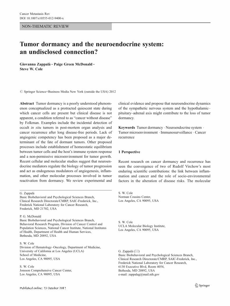
Cancers have long been known to have the capacity toenter a state of dormancy in which they cease growthand disease progression but do not die or regress. Dor-mant tumors retain the capacity to reactivate (escapefrom dormancy) and resume active growth and progres-sion and thus remain a significant health threat. How-ever, the general physiologic factors that influence atumor’s entry into and exit from dormancy remain poor-ly understood. A growing body of research has begunto identify some of the specific molecular processesinvolved in the establishment and escape from tumordormancy. Remarkably, a separate body of cancer re-search has begun to show that many of the same mo-lecular dynamics involved in tumor dormancy aresubject to regulation by the neural and endocrine sys-tems. The goal of this review is to highlight that mo-lecular intersection (Fig. 1a–c) and prompt new researchto join these two literatures and test the hypothesis thatneural and endocrine dynamics represent key generalphysiologic conditions that modulate tumor dormancyand thus direct the fate of clinically occult cancers.
3 Tumor dormancy: cancer without disease
Tumor dormancy reflects a period of arrested tumor growththat can be categorized based on the underlying cellular mech-anisms. “Tumor mass dormancy” occurs in a population ofcells undergoing indolent proliferation but not expanding inmass because the process is offset by a concomitant rate of celldeath [1]. This dormant state has been hypothesized to stemfrom lack of adequate vascularization and immune systemcontrol activity, among other processes. “Tumor cell dorman-cy” refers to cancer cells in a state of cell growth arrestresulting from microenvironment signals unfavorable to tu-mor cell proliferation [1]. Both tumor mass dormancy andtumor cell dormancy may be reversed when microenviron-mental conditions shift to support tumor expansion.
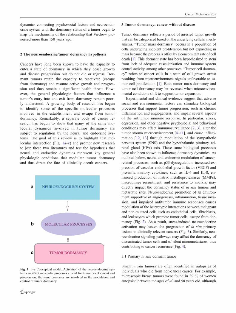
Experimental and clinical evidence suggest that adversesocial and environmental factors can stimulate biologicalprocesses that support tumor progression, such as chronicinflammation and angiogenesis, and impair several aspectsof the antitumor immune response. In particular, stress,depression, and other negative psychosocial and behavioralconditions may affect immunosurveillance [2, 3], alter thetumor stroma microenvironment [4–11], and cause inflam-mation [12, 13] through modulation of the sympatheticnervous system (SNS) and the hypothalamic–pituitary–ad-renal gland (HPA) axis. These same biological processeshave also been shown to influence dormancy dynamics. Asoutlined below, neural and endocrine modulation of cancer-related processes, such as p53 dysregulation, increased ex-pression of vascular endothelial growth factor (VEGF) andpro-inflammatory cytokines, such as IL-6 and IL-8, en-hanced production of matrix metalloproteinases (MMPs),macrophage recruitment, and resistance to anoikis, maydirectly impact the dormancy status of in situ tumors andmetastatic sites. Neuroendocrine promotion of an environ-ment supportive of angiogenesis, inflammation, tissue inva-sion, and impaired antitumor immune responses causesmodulation of the heterotypic interactions between malignantand non-mutated cells such as endothelial cells, fibroblasts,and leukocytes which promote tumor cells’ escape from dor-mancy (Fig. 2). As a result, stress-induced neuroendocrineactivation may hasten the progression of in situ primarylesions to clinically relevant cancers (Fig. 3). Similarly, neu-roendocrine signaling pathways may affect the dormancy ofdisseminated tumor cells and of silent micrometastases, thuscontributing to cancer recurrence (Fig. 4).
3.1 Primary in situ dormant tumor
Small in situ tumors are often identified in autopsies ofindividuals who die from non-cancer causes. For example,microscopic breast tumors were found in 39 % of womenautopsied between the ages of 40 and 50 years old, although
Fig. 1 a–c Conceptual model. Activation of the neuroendocrine sys-tem can affect molecular processes crucial for tumor development andprogression; the same processes are involved in the modulation andcontrol of tumor dormancy
Cancer Metastasis Rev
the clinical incidence of breast cancer in women in the sameage range is only 1 % [14, 15]. Similar data have been foundfor other types of cancer [16–18] indicating a higher fre-quency of silent tumors compared to the prevalence of theovert cancers. These small tumors can persist as microscopiclesions without detection or clinical manifestation, a condi-tion referred to as “cancer without disease” by Folkman[19]. As physiologic conditions change, these tumors maymove from a dormant state to become progressive invasivecancers (i.e., reactivation from dormancy).
3.2 Dormant metastasis
Cancer recurrence may occur years or decades after surgicalexcision and treatment of the primary tumor. Persistentminimal residual disease occurs for most types of cancer
[1] and vital organ failure due to metastases represents themain cause of cancer death. This nonrandom and predictabletumor colonization of distant tissues and organs is mediatedby disseminated tumor cells (DTCs). Bone marrow consti-tutes a common homing site and reservoir of DTCs origi-nating from various epithelial tumors. Clinical detection andmolecular characterization of DTCs or peripheral bloodcirculating tumor cells (CTCs) may constitute unique prog-nostic factors for follow-up risk assessment and therapymonitoring [20]. An extensive pooled analysis of 4,703breast cancer patients from eight large studies found thatthe presence of DTCs in bone marrow at the time of cancerdiagnosis was associated with poor prognosis and survival[21]. In addition, a European study showed that persistentbone marrow DTCs during postoperative clinical follow-upconstituted an independent prognostic factor for breast
Fig. 2 Neuroendocrineinfluences on tumor dormancy.Psychosocial and/orenvironmental stressors maytrigger activation of theneuroendocrine systeminitiating a cascade ofneuroendocrine mediators thatcan affect themicroenvironment ofpreexisting in situ dormantlesions, enhancinginflammatory states andinterfering with the immuneresponse. Ultimately, theseprocesses may lead toangiogenic switch, tumor cellsovercoming immune control,and escape from dormancy.Affective states may also resultin the activation of crucialsignaling pathways in tumorcells and the microenvironmentof dormant metastases, alteringtheir biology and leading topatient clinical relapse
Cancer Metastasis Rev

cancer recurrence and poor survival [22]. Results frommeta-analyses of patients with colorectal or melanoma can-cer revealed a clinically prognostic significance for CTCs[23, 24]. CTCs have been found in patients up to 22 yearspostsurgical removal of primary breast tumor [25]. This lagtime does not reflect the primary growth kinetics of thetumor cell population, thus it is reasonable to hypothesizethat sustained shedding of CTCs (which themselves survivefor only hours in circulation [25]) reflects ongoing sheddingfrom a clinically dormant population of DTCs. Accumulat-ing molecular and clinical evidence supports a new concep-tualization of the metastatic process in which metastases ofearly disseminated, primordial cancer cells are seeded in
parallel with primary tumor progression [26]. Eyles [27]has proposed that this early tumor cell migration and dis-semination is necessary to accommodate cell–cell competi-tion for space and nutrients. In this context, dormancy ofdisseminated cancer cells might exemplify a potential ther-apeutic target to control disease course and the patient’ssubsequent fate (i.e., morbidity and survival).
3.3 Framing tumor dormancy in the context of the tumormicroenvironment
The traditional view of carcinogenesis as a phenomenon thatoccurs at the subcellular level is evolving and cancer is now
TTumor MassDormancy
Tumor Progression
SNS
VEGF, IL-6, MMPs, IL-8
Angiogenic Sw i t c h
Tumor cell
Stromal cell
SNS
ADRB2(STAT -3, CREB)
ADRB2(FAK)
Anoikis
Tumor Cell MigrationAnd Dissemination
SNS
TAMs
Fig. 3 Mechanistic model.Neuroendocrine influences ontumor mass dormancy. Socialenvironmental influences cancause activation of thesympathetic nervous systemwhich, in turn, can affect tumormass dormancy and hasten theprogression of in situ lesions toclinically relevant cancers. Seetext for detailed explanation.ADRB2 β2-adrenergicreceptors; STAT-3 signaltransducer and activator oftranscription-3; CREB cAMPresponse element-binding pro-tein; TAMs tumor-associatedmacrophages
Cancer Metastasis Rev
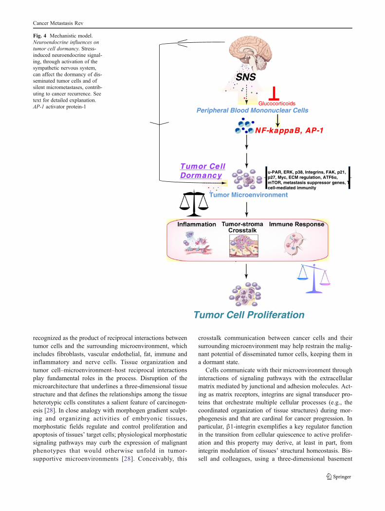
recognized as the product of reciprocal interactions betweentumor cells and the surrounding microenvironment, whichincludes fibroblasts, vascular endothelial, fat, immune andinflammatory and nerve cells. Tissue organization andtumor cell–microenvironment–host reciprocal interactionsplay fundamental roles in the process. Disruption of themicroarchitecture that underlines a three-dimensional tissuestructure and that defines the relationships among the tissueheterotypic cells constitutes a salient feature of carcinogen-esis [28]. In close analogy with morphogen gradient sculpt-ing and organizing activities of embryonic tissues,morphostatic fields regulate and control proliferation andapoptosis of tissues’ target cells; physiological morphostaticsignaling pathways may curb the expression of malignantphenotypes that would otherwise unfold in tumor-supportive microenvironments [28]. Conceivably, this
crosstalk communication between cancer cells and theirsurrounding microenvironment may help restrain the malig-nant potential of disseminated tumor cells, keeping them ina dormant state.
Cells communicate with their microenvironment throughinteractions of signaling pathways with the extracellularmatrix mediated by junctional and adhesion molecules. Act-ing as matrix receptors, integrins are signal transducer pro-teins that orchestrate multiple cellular processes (e.g., thecoordinated organization of tissue structures) during mor-phogenesis and that are cardinal for cancer progression. Inparticular, β1-integrin exemplifies a key regulator functionin the transition from cellular quiescence to active prolifer-ation and this property may derive, at least in part, fromintegrin modulation of tissues’ structural homeostasis. Bis-sell and colleagues, using a three-dimensional basement
Tumor CellDormancy
NF-kappaB, AP-1
Tumor Microenvironment
Tumor Cell Proliferation
SNS
Glucocorticoids
u-PAR, ERK, p38, Integrins, FAK, p21, p27, Myc, ECM regulation, ATF6 , mTOR, metastasis suppressor genes, T cell-mediated immunity
Peripheral Blood Mononuclear Cells
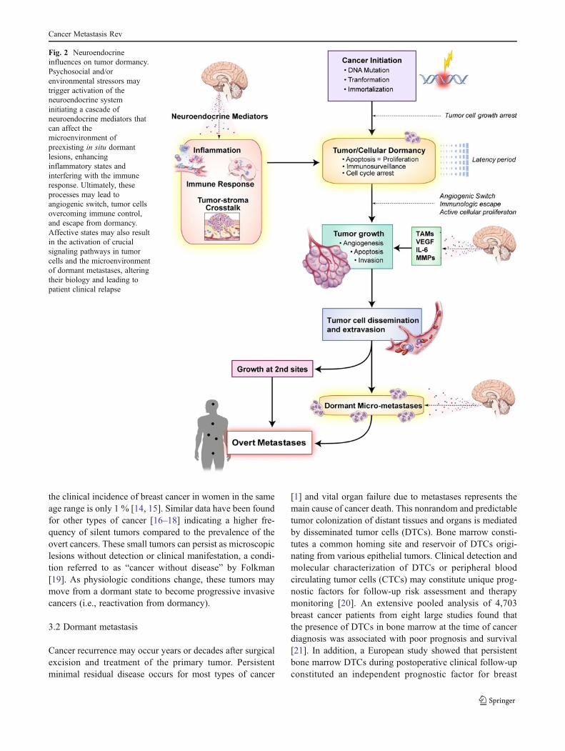
Fig. 4 Mechanistic model.Neuroendocrine influences ontumor cell dormancy. Stress-induced neuroendocrine signal-ing, through activation of thesympathetic nervous system,can affect the dormancy of dis-seminated tumor cells and ofsilent micrometastases, contrib-uting to cancer recurrence. Seetext for detailed explanation.AP-1 activator protein-1
Cancer Metastasis Rev
membrane in vitro and an in vivo models, demonstrated thatinhibition of β1-integrin function reverted the malignantbehavior of mammary cancer cells and induced cellularquiescence [29]. Removal of the inhibition rescued themalignant phenotype pointing out the reversibility of thephenomenon and supporting the thesis that stabilizing(preserving) homeostatic and architectural structure of atissue may cause a cellular phenotype to prevail over themalignant genotype [29] in contrast with the somatic muta-tion theory of cancer [30]. Several other studies [31–36]provided further evidence that β1-integrin and its signalingpathways encompassing Src and focal adhesion kinase(FAK), by regulating cells’ proliferative behavior, candictate tumor fate, and inhibition of the β1-integrin-FAK signaling axis may retain cancer cells in a dor-mant state. As outlined later, Src activation and FAKphosphorylation fall under the regulation of stress-mediatedneuroendocrine activation of β2-adrenoreceptors with in-creased neural activation augmenting FAK phosphorylation,emphasizing the striking network of mechanistic relationshipsexisting between tumor dormancy and neuroendocrineregulation.
Additionally, the microenvironment may dictate the tran-sition from dormancy to active proliferation through themiscellaneous actions of MMPs, a family of proteases withextracellular matrix (ECM) degrading properties that aremainly secreted by stromal cells such as vascular endothelialcells, pericytes, fibroblasts, macrophages, and other inflam-matory cells [37]. In addition to their actions on the ECM,MMPs proteolytically cleave several non-matrix substratesincluding precursors of biologically active fragments [38].For example MMP-9, by cleaving the heparin-bindingdomains of VEGF165 and VEGF121, enhances the bioavail-ability of these factors in the tumor microenvironment thuspotentially intensifying the chances to activate the angio-genic switch necessary for the shift of dormant tumors to anactive state [37]. MMPs may also cause the release from theECM of angiostatic factors such as endostatin [39] andangiostatin [40] further adding to the complexity of thesystem. As noted earlier, MMPs are produced by severalmicroenvironment components including tumor-associatedmacrophages (TAMs). Besides the production of MMPs,TAMs support tumorigenesis through plentiful actions di-rected to several cells that populate the tumor microenviron-ment resulting in the promotion of tumor angiogenesis,cancer cell survival, proliferation and invasion, and sup-pression of adaptive immune responses. A recent inves-tigation [41] demonstrated that the extent of TAMinfiltration and expression of pro-metastatic genes suchas Mmp9, Vegf, Cox2, and Tgfb falls under the controlof stress-associated SNS activation. In the same study,TAMs were shown to mediate tumor cell metastaticseeding and colonization [41].
3.4 Mechanisms of cancer dormancy
Folkman and colleagues [42–44] identified the switch froma phenotype unable to recruit new vasculature to an angio-genic phenotype as one of the principal molecular regulatorsof escape from tumor dormancy. Angiogenic proteins (e.g.,VEGF, basic fibroblast growth factor), as well asangiogenesis-related proteins (e.g., ras, c-Myc, and p53),are involved in the process [45]. Additional “niche” mech-anisms of dormancy may also exist. For example, an Italianstudy [46] posits that the microenvironment crosstalk be-tween tumor and vascular endothelial cells, through Notch-Dll4 interactions and activation of the NF-κB pathway,deliver survival signals that trigger the escape of leukemiaand colorectal cancer cells from dormancy. This investiga-tion supports the existence of a vascular niche that embedsdormant cancer cells and elicits tumor-promoting activities[47]. Bidirectional signaling with the niches they inhabitmay determine the fate of dormant cells, stressing the im-portance of microenvironmental control over the release oftumor cells from dormancy. Using an artificial liver metas-tasis model [48], Guba proposed that the presence of aprimary tumor might halt the metastatic progression ofsolitary disseminated tumor cells by forcing them to enterinto a state of dormancy at an early stage, before angiogenesistakes control and causes them to advance to macroscopiclesions.
Several studies highlighted a role for p53 in tumor dor-mancy through the modulation of angiogenesis. Holmgrenet al. [49] showed that p53 gene therapy altered the angio-genic potential of a tumor and induced a state of dormancyin a mouse fibrosarcoma model, independent of p53’s directeffects on cell cycle and apoptosis. The MAP kinase p38 is astress-activated kinase involved in tumor cell dormancy; in arecent study [50], p53 transcriptional regulation was foundto be important for p-38-induced cellular quiescence.
“Oncogene addiction” refers to the dependency of specificactivated or overexpressed oncogenes for the maintenance ofcancer cells’ malignant phenotype [51]. Fluctuations in onco-gene expression have been implicated in tumor dormancy.The ErbB2 (Her2) receptor is involved in the induction ofseveral cancers and its upregulation correlates with poor prog-nosis in breast cancer patients [52]. Conditional activation ofthe ErbB2 rat homologue NEU in transgenic mice induces thedevelopment of invasive mammary adenocarcinoma while itsdownregulation causes the disappearance of all primary andsecondary tumors [53], showing cancer cells’ dependency onthe continued expression of the oncogene. However, afterregression of the tumors following abrogation of NEU expres-sion, most of the animals harbored residual cancer cells thatultimately generated NEU-independent tumors [53]. Ursini-Siegel et al. argued that these recurrent tumors may derivefrom the reactivation of dormant tumor cells within the
Cancer Metastasis Rev
primary tumor [52] that may have become sensitive to adifferent addictive pathway than NEU [54].
In a conditional transgenic mice model, the inactivation ofthe oncogene MYC caused liver tumor regression, tumor celldifferentiation, and reversion to a dormant state [55]. Hepato-cellular carcinoma cells were induced to differentiate into livercells, causing tumor cell proliferation to arrest while retaininglatent tumorigenic potential upon MYC reactivation. Thesefindings support the existence of a link between tumor dorman-cy and epigenetic reprogramming associated with tumor celldifferentiation and suggest that dormancy may involve differ-entiation of the transformed cells rather than only mere prolif-eration arrest. Shachaf and Felsher [56] proposed that MYCinactivation in hepatocellular carcinoma cells and their subse-quent differentiation uncovers stem cell properties in livertumor cells due to their ability to reemerge from dormancyand regain neoplastic proprieties uponMYC reactivation. Pont-ier and Muller postulated that single dormant cells may bederived from migrating cancer stem cells and that migrationoutside their niche requires integrin involvement, particularlyα5β1 integrin heterodimer formation [57]. The extracellularmatrix may in fact dictate the fate of incipient dormant cancercells by regulating the switch from quiescence to proliferation.Barkan et al. showed that the transition from dormancy toproliferation involved fibronectin-driven cytoskeletal architec-ture reorganization and actin stress fiber formation through theengagement of integrin β1 [33]. Furthermore, enrichment intype I collagen and the associated fibrosis induction may pro-vide a fertile soil for the switch from dormancy to activeproliferation [34]. The functional role of integrins and fibronec-tin in the regulation of tumor dormancy was previously high-lighted through pioneering studies of Aguirre-Ghiso [58].These investigations demonstrated that high levels of uPAR/α5β1-integrin association, through fibronectin production, p38activity suppression, and subsequent imbalance between ERKand p38, triggered head and neck carcinoma cells to escapefrom dormancy. Subsequent studies [59] identified a pivotalrole for the transcription factor ATF6α as a survival elementallowing dormant cancer cells to endure adverse microenvir-onmental conditions and nutritional or chemotherapy-inducedstress, hence identifying the ATF6α–Rheb–mTOR axis as adeterminant for the survival of dormant tumor cells [59].
The debate about the validity of the notion of host immu-nosurveillance has recently led to the concept of immunoedit-ing, which recognizes that the immune system contributes toboth impeding and aiding tumor progression through itsimmunogenic-sculpting actions [60]. The equilibrium phaseof the cancer immunoediting process, during which host im-munity is credited with restraining the outgrowth of occultcancer cells, corresponds to the tumor dormancy phase, whileinhibition of T cell-mediated immunity may contribute toescape from dormancy. Koebel et al. showed that adaptive Tcell immunity, through cytostatic and cytolytic actions, holds
highly immunogenic tumor cells in a dormant phase of dy-namic equilibrium in mouse models of sarcoma [61]. Targetedadaptive immunosuppression, however, can break down thisequipoise state causing the edited, immunogenic-attenuatedtumor cells to expand and grow [61]. In a transgenic melano-mamouse model, CD8+ Tcells were required for maintenanceof the dormancy status of early disseminated tumor cells invisceral organs [27]. Furthermore, Zhang et al. demonstratedthat adoptive CD8+ T cell transfer is able to establish astationary phase of equilibrium between host and cancer cells[62]. This induction of dormancy may be the result of effectorT cell-mediated destruction of stromal myeloid-derived sup-pressor cells with subsequent reversion of the pro-angiogenic,inflammatory, and immunosuppressive phenotype. Kraman etal. found that deletion of a subpopulation of stromal cellsexpressing fibroblast activation protein-α caused immuno-genic tumors to arrest growth and enter a dormant state byallowing host immunological control of tumor growth [63].TNF-α and IFN-γ-induced hypoxic necrosis of both cancerand stromal cells was involved in the process.
4 Neuroendocrine regulation of molecular pathwaysinvolved in tumor dormancy
4.1 Neuroendocrine regulation of the adaptive stressresponse
Neuroendocrine and autonomic functional responses repre-sent the essential components of the body’s adaptive mecha-nisms to restore homeostasis after environmental andpsychosocial challenges. As an integral component of thehormonal response to threatening stimuli, HPA axis activationis molecularly triggered by the release of corticotrophin-releasing hormone, along with arginin vasopressin; both ofwhich, in turn, stimulate the release of adrenocorticotropinfrom the anterior pituitary gland. The final output of thesystem is mediated by the subsequent production of glucocor-ticoids [2, 3]. The sympathetic division of the autonomicnervous system (sympathetic nervous system), together withthe adrenal medulla, elicits the production of epinephrine andnorepinephrine, signaling physiological adaptive changes to athreatening situation [2, 3]. Both the HPA axis and the SNShave been shown to modulate tumor growth and dissemina-tion [2, 64] and many of the specific molecular mechanismsinvolved in these dynamics are also hypothesized to modulatetumor dormancy.
4.2 Neuroendocrine regulation of crosstalkbetween the tumor cell and its microenvironment
Neuroendocrine dynamics can affect angiogenic, inflamma-tory, and invasion pathways crucial for tumor development
Cancer Metastasis Rev
and progression. Norepinephrine and epinephrine can stimu-late angiogenesis through the activation of STAT-3 [65] andby upregulating the expression of angiogenic factors such asVEGF, IL-6, and IL-8 [5–8]. Complementary findings havebeen documented in the clinical setting. In particular, in-creased amounts of norepinephrine were detected in the tumormicroenvironment [65, 66] of ovarian cancer patients report-ing higher levels of chronic stress and lower social support,while higher levels of VEGF were observed in plasma [67]and in tumor tissues [68]. Additionally, in an orthotopicmousemodel of ovarian carcinoma [4], chronic stress upregulatedtumor cell expression of VEGF, subsequently increasing vas-cularization and aggravating tumor burden. These effects weremediated through SNS activation and β2 adrenergic receptorsignaling. β-adrenergic receptor activation of the cyclic 3′,5′-adenosine monophosphate/protein kinase A (cAMP/PKA)may regulate gene expression via phosphorylation of multipletranscription factors and, under selected circumstances, PKAcan cross-regulate the activity of the pro-inflammatory NF-κB[64, 66]. Implications for dormancy: Given the key role ofangiogenesis in dormancy (reviewed above), SNS-mediatedβ-adrenergic signaling represents one major molecular path-way by which the nervous system could regulate tumor dor-mancy dynamics.
Neuroendocrine mediators also play a role in cellularDNA repair mechanisms by impairing the ability to correctgenetic damage following stress or radiation [11, 69], alter-ing the regulation of apoptosis mechanisms and even favor-ing mutagenesis through reactive oxygen species-deriveddamage [70]. A recent study [71] showed that catechol-amine engagement of β2-adrenoreceptors, via β-arrestinsignaling, triggers DNA damage and promotes p53 degra-dation, leading to compromised genome maintenance andsuggesting that stress pathways might potentially affectcancer initiation. Feng et al. showed that chronic stress,through glucocorticoid signaling, decreased p53 functionand promoted the growth of xenograft tumors in a mousemodel of colorectal cancer [72]. Implications for dormancy:Given the key role of p53 in the induction of tumor dor-mancy [49], HPA axis-induced p53 degradation constitutesanother potential pathway by which endocrine dynamicsmight modulate tumor dormancy.
Growth factor signaling is also subject to regulation bythe neural and endocrine systems. Shi et al. showed that theβ2-adrenergic receptor and ErbB2 are part of a positivefeedback loop in human breast cancer cells [73]. Chroniccatecholamine stimulation induces β2-adrenoreceptor-mediated overexpression of ErbB2, triggering strongmitogen-ic effects; in turn, ErbB2 upregulation induces autocrine epi-nephrine release and upregulation of β2-adrenergic receptor[73]. Furthermore, ErbB2 overexpression activated a tran-scriptional pro-inflammatory profile, involving IL-6 andSTAT3, required for ErbB-mediated tumorigenesis in vitro,
as well as in an in vivo mouse model of ErbB overexpression[74]. Implications for dormancy: Given the key role of ErbB2in tumor dormancy [52], β-adrenergic regulation of this keygrowth control pathway represents yet another molecularmechanism by which the SNS might modulate tumordormancy.
Anoikis represents a form of apoptosis induced by inap-propriate cell–cell and/or cell–matrix interactions; its cir-cumvention enhances the metastatic potential of malignantcells [75]. Sood and colleagues demonstrated that ovariancancer cells in vitro and in an in vivo orthotopic mousemodel are protected from anoikis following exposure toepinephrine or norepinephrine [76]. This effect is mediatedby FAK through involvement of β2-adrenoreceptors andsubsequent Src activation and FAK phosphorylation. Impor-tantly, these results mirrored clinical data from ovariancancer patients showing positive associations betweennorepinephrine, increased FAK activation, and acceleratedcancer mortality [76]. Implications for dormancy: Given thekey role of FAK in tumor dormancy [32–34], β-adrenergicregulation of FAK and related cell survival processes repre-sents an additional molecular mechanism by which the SNSmight modulate tumor dormancy.
MMPs are modulators of the tumor microenvironmentand represent key players in the molecular communicationbetween tumor and stroma. Norepinephrine and epinephrinemodulate cell migration and invasion by stimulating theproduction of MMPs-2 and -9 in ovarian [9] and nasopha-ryngeal carcinoma cancer cells [6] through involvement ofβ-adrenergic receptors. In ovarian cancer patients chronicstress, high levels of depression and low social supportcorrelated with elevated MMP-9 expression in tumor-associated macrophages [68]. Implications for dormancy:Given the key role of MMP-2 and -9 in tumor dormancy[77, 78], β-adrenergic regulation of MMP expression rep-resents yet another molecular mechanism by which the SNSmight modulate tumor dormancy.
Tumor dormancy may result from T cell response to thetumor mass, and T cell inhibition may tip the balance to-wards tumor mass escape from dormancy [79]. Neuroendo-crine stress responses may promote tumor growth byimpairing immune cell function. Stress-signaling pathwaysmediate an increase in circulating IL-6 and VEGF [5–7] andboth VEGF and IL-6 reduce T cell number and activity[80, 81]. Additionally, stress-mediated dynamics may di-rectly inhibit cytotoxic T lymphocyte and natural killercell responses [82, 83]. Implications for dormancy: Tothe extent that tumor dormancy depends on the ability ofthe immune system to control tumor cells [60, 61, 84],neural and endocrine regulation of antitumor cellularimmune responses represents yet another molecularmechanism by which the neuroendocrine system mightmodulate tumor dormancy.
Cancer Metastasis Rev
5 Future directions
Converging evidence from in vitro, in vivo, and clinicalstudies strongly point out a role for stress-mediated neuro-endocrine regulation of several molecular pathways whosedysregulation is also cardinal for the fate of dormant tumors.At the moment, these observations exist in two separatebodies of literature within cancer research: one connectingneuroendocrine dynamics to cancer molecular mechanismsand a separate literature connecting those molecular mech-anisms to tumor dormancy. These literatures could be inte-grated to include analyses of tumor dormancy dynamics inthe context of experimental models of neural and endocrineregulation of tumor biology.
We suggest that the time is right to initiate a definedresearch agenda to explore both cellular and tumor massdormancy dynamics and neuroendocrine influences on can-cer progression in novel preclinical laboratory models, usingnew and improved in vivo paradigms. This research agendamay be initiated in the context of diseases, such as breastcancer, in which dormancy dynamics are relatively commonand of substantial clinical significance in determining long-term health outcomes. Breast cancer might also be a partic-ularly appealing model because previous research providesboth observational clinical studies and preclinical experi-mental studies supporting a significant role of neuroendo-crine dynamics modulating overall disease progression anddormancy-relevant molecular dynamics.
In a breast cancer mouse model, Sloan et al. [41] showedthat stress-induced neuroendocrine activation of β-adrenergic signaling caused macrophage recruitment and apro-metastatic gene expression signature indicative of M2differentiation (associated with immunoregulatory, tissueremodeling, and tumor-promoting properties [85]). Theseresults, in consort with other studies showing that stress-induced increase in tumor VEGF and angiogenesis washalted by the β-blocker propanolol [4–6], suggest that noveland promising experimental strategies to prevent cancerrecurrence might leverage the use of β-blockers, inexpen-sive and well-understood drugs with minimal and easilymanaged side effects. Breast cancer would be particularlysuitable to this purpose because of its late recurrence and thepresence of noninvasive or low-grade cancers that mayremain dormant for a long time. Two recent observationalstudies investigated the effects of β-blockers on breast can-cer progression and mortality [86, 87]. Both reports con-cluded that β-blocker use was associated with reduction inmetastasis development and tumor recurrence and improvedsurvival. β-blocker treatment may inhibit signaling path-ways important for tumor cell escape from dormancy, thusreducing cancer recurrence and mortality; hence, pharmaco-logic control of neuroendocrine activity may represent auseful adjuvant to traditional therapy.
A growing body of research is revealing the influences ofstress-mediated neuroendocrine mediators on cancer progres-sion and recurrence, while a similarly expanding body ofliterature investigating the molecular underpinnings of tumordormancy is rapidly emerging. As we began to uncover themolecular mechanisms that cause tumor escape from dorman-cy, the general physiologic processes that promote the activa-tion of those mechanisms remain unclear. We propose thatstress and other psychological and social conditions representcontributing factors via their modulation of the neural andendocrine system. This framework challenges us with inte-grating the connections into a comprehensive cancer caresetting that would allow manipulation and control of systemicneural and endocrine influences in order to maximally inhibitcancer progression and disease recurrence.
6 Concluding remarks
Due to his outspoken support in favor of social reforms andadvancements in public health to improve economic and socialconditions during the nineteenth century, Rudolf Virchow isconsidered the Father of Social Medicine. With his theorytracing social influences on the origin of diseases, and theidentification of inflammation as a predisposing factor fortumorigenesis, Virchow pioneered a paradigm shift in mecha-nistic insights in the etiopathology of illnesses. By embracinghis view of “disease” as “an expression of individual life underunfavorable circumstances,” we begin to disentangle the dy-namics connecting social conditions with the recurrence ofcancer through the biological underpinning of tumor dormancy.
Acknowledgments We gratefully thank Jerry Suls for critical readingof the manuscript and Kathleen Igo for professional editing. This projecthas been funded in whole or in part with federal funds from the NationalCancer Institute, National Institutes of Health, under contract no.HHSN261200800001E. The content of this publication does not neces-sarily reflect the views or policies of the Department of Health andHuman Services, nor does mention of trade names, commercial products,or organizations imply endorsement by the U.S. Government.
Conflict of interest The authors declare that they have no conflict ofinterest.
References
1. Aguirre-Ghiso, J. A. (2007). Models, mechanisms and clinicalevidence for cancer dormancy. Nature Reviews. Cancer, 7(11),834–846. doi:10.1038/nrc2256.
2. Antoni, M. H., Lutgendorf, S. K., Cole, S. W., Dhabhar, F. S.,Sephton, S. E., McDonald, P. G., et al. (2006). The influence ofbio-behavioural factors on tumour biology: pathways andmechanisms. Nature Reviews. Cancer, 6(3), 240–248. doi:10.1038/nrc1820.
Cancer Metastasis Rev

3. Irwin, M. R., & Cole, S. W. (2011). Reciprocal regulation of theneural and innate immune systems. Nature Reviews Immunology,11(9), 625–632. doi:10.1038/nri3042.
4. Thaker, P. H., Han, L. Y., Kamat, A. A., Arevalo, J. M., Takahashi,R., Lu, C., et al. (2006). Chronic stress promotes tumor growth andangiogenesis in a mouse model of ovarian carcinoma. NatureMedicine, 12(8), 939–944. doi:10.1038/nm1447.
5. Lutgendorf, S. K., Cole, S., Costanzo, E., Bradley, S., Coffin, J.,Jabbari, S., et al. (2003). Stress-related mediators stimulate vascu-lar endothelial growth factor secretion by two ovarian cancer celllines. Clinical Cancer Research, 9(12), 4514–4521.
6. Yang, E. V., Sood, A. K., Chen, M., Li, Y., Eubank, T. D., Marsh,C. B., et al. (2006). Norepinephrine up-regulates the expression ofvascular endothelial growth factor, matrix metalloproteinase(MMP)-2, and MMP-9 in nasopharyngeal carcinoma tumor cells.Cancer Research, 66(21), 10357–10364. doi:10.1158/0008-5472.CAN-06-2496.
7. Nilsson, M. B., Armaiz-Pena, G., Takahashi, R., Lin, Y. G., Trevino,J., Li, Y., et al. (2007). Stress hormones regulate interleukin-6expression by human ovarian carcinoma cells through a Src-dependent mechanism. Journal of Biological Chemistry, 282(41), 29919–29926. doi:10.1074/jbc.M611539200.
8. Shahzad, M. M., Arevalo, J. M., Armaiz-Pena, G. N., Lu, C.,Stone, R. L., Moreno-Smith, M., et al. (2010). Stress effects onFosB- and interleukin-8 (IL8)-driven ovarian cancer growth andmetastasis. Journal of Biological Chemistry, 285(46), 35462–35470. doi:10.1074/jbc.M110.109579.
9. Sood, A. K., Bhatty, R., Kamat, A. A., Landen, C. N., Han, L.,Thaker, P. H., et al. (2006). Stress hormone-mediated invasion ofovarian cancer cells. Clinical Cancer Research, 12(2), 369–375.doi:10.1158/1078-0432.CCR-05-1698.
10. Fang, C. Y., Miller, S. M., Bovbjerg, D. H., Bergman, C., Edelson,M. I., Rosenblum, N. G., et al. (2008). Perceived stress isassociated with impaired T-cell response to HPV16 in womenwith cervical dysplasia. Annals of Behavioral Medicine, 35(1),87–96. doi:10.1007/s12160-007-9007-6.
11. Yang, E. V., &Glaser, R. (2003). Stress-induced immunomodulation:implications for tumorigenesis. Brain, Behavior, and Immunity, 17(Suppl 1), S37–S40.
12. Black, P. H. (2002). Stress and the inflammatory response: areview of neurogenic inflammation. Brain, Behavior, and Immunity,16(6), 622–653.
13. Garcia-Bueno, B., Caso, J. R., & Leza, J. C. (2008). Stress as aneuroinflammatory condition in brain: damaging and protectivemechanisms. Neuroscience and Biobehavioral Reviews, 32(6),1136–1151. doi:10.1016/j.neubiorev.2008.04.001.
14. Black, W. C., & Welch, H. G. (1993). Advances in diagnosticimaging and overestimations of disease prevalence and the benefitsof therapy. The New England Journal of Medicine, 328(17), 1237–1243. doi:10.1056/NEJM199304293281706.
15. Nielsen, M., Thomsen, J. L., Primdahl, S., Dyreborg, U., & Andersen,J. A. (1987). Breast cancer and atypia among young and middle-agedwomen: a study of 110 medicolegal autopsies. British Journal ofCancer, 56(6), 814–819.
16. Harach, H. R., Franssila, K. O., & Wasenius, V. M. (1985). Occultpapillary carcinoma of the thyroid. A “normal” finding in Finland.A systematic autopsy study. Cancer, 56(3), 531–538.
17. Montie, J. E., Wood, D. P., Jr., Pontes, J. E., Boyett, J. M., & Levin,H. S. (1989). Adenocarcinoma of the prostate in cystoprostatectomyspecimens removed for bladder cancer. Cancer, 63(2), 381–385.
18. Kimura, W., Morikane, K., Esaki, Y., Chan, W. C., & Pour, P. M.(1998). Histologic and biologic patterns of microscopic pancreaticductal adenocarcinomas detected incidentally at autopsy. Cancer,82(10), 1839–1849. doi:10.1002/(SICI)1097-0142.
19. Folkman, J., & Kalluri, R. (2004). Cancer without disease. Nature,427(6977), 787. doi:10.1038/427787a.
20. Pantel, K., Alix-Panabieres, C., & Riethdorf, S. (2009). Cancermicrometastases. Nature Reviews. Clinical Oncology, 6(6), 339–351. doi:10.1038/nrclinonc.2009.44.
21. Braun, S., Vogl, F. D., Naume, B., Janni, W., Osborne, M. P.,Coombes, R. C., et al. (2005). A pooled analysis of bone marrowmicrometastasis in breast cancer. The New England Journal ofMedicine, 353(8), 793–802. doi:10.1056/NEJMoa050434.
22. Janni, W. J., Vogl, F. D., Wiedswang, G., Synnestvedt, M., Fehm,T. N., Jueckstock, J., et al. (2011). Persistence of disseminatedtumor cells in the bone marrow of breast cancer patients predictsincreased risk for relapse—a European pooled analysis. ClinicalCancer Research. doi:10.1158/1078-0432.CCR-10-2515.
23. Rahbari, N. N., Aigner, M., Thorlund, K., Mollberg, N., Motschall,E., Jensen, K., et al. (2010). Meta-analysis shows that detection ofcirculating tumor cells indicates poor prognosis in patients withcolorectal cancer. Gastroenterology, 138(5), 1714–1726.doi:10.1053/j.gastro.2010.01.008.
24. Mocellin, S., Hoon, D., Ambrosi, A., Nitti, D., & Rossi, C. R.(2006). The prognostic value of circulating tumor cells in patientswith melanoma: a systematic review and meta-analysis. ClinicalCancer Research, 12(15), 4605–4613. doi:10.1158/1078-0432.CCR-06-0823.
25. Meng, S., Tripathy, D., Frenkel, E. P., Shete, S., Naftalis, E. Z.,Huth, J. F., et al. (2004). Circulating tumor cells in patients withbreast cancer dormancy. Clinical Cancer Research, 10(24), 8152–8162. doi:10.1158/1078-0432.CCR-04-1110.
26. Klein, C. A. (2009). Parallel progression of primary tumours andmetastases. Nature Reviews. Cancer, 9(4), 302–312. doi:10.1038/nrc2627.
27. Eyles, J., Puaux, A. L., Wang, X., Toh, B., Prakash, C., Hong, M.,et al. (2010). Tumor cells disseminate early, but immunosurveil-lance limits metastatic outgrowth, in a mouse model of melanoma.The Journal of Clinical Investigation, 120(6), 2030–2039.doi:10.1172/JCI42002.
28. Potter, J. D. (2007). Morphogens, morphostats, microarchitectureand malignancy. Nature Reviews. Cancer, 7(6), 464–474.doi:10.1038/nrc2146.
29. Weaver, V. M., Petersen, O. W., Wang, F., Larabell, C. A., Briand,P., Damsky, C., et al. (1997). Reversion of the malignant pheno-type of human breast cells in three-dimensional culture and in vivoby integrin blocking antibodies. The Journal of Cell Biology, 137(1), 231–245.
30. Soto, A. M., & Sonnenschein, C. (2004). The somatic mutationtheory of cancer: growing problems with the paradigm? Bioessays,26(10), 1097–1107. doi:10.1002/bies.20087.
31. Aguirre Ghiso, J. A., Kovalski, K., & Ossowski, L. (1999). Tumordormancy induced by downregulation of urokinase receptor inhuman carcinoma involves integrin and MAPK signaling. TheJournal of Cell Biology, 147(1), 89–104.
32. Aguirre Ghiso, J. A. (2002). Inhibition of FAK signaling activatedby urokinase receptor induces dormancy in human carcinomacells in vivo. Oncogene, 21(16), 2513–2524. doi:10.1038/sj.onc.1205342.
33. Barkan, D., Kleinman, H., Simmons, J. L., Asmussen, H.,Kamaraju, A. K., Hoenorhoff, M. J., et al. (2008). Inhibitionof metastatic outgrowth from single dormant tumor cells bytargeting the cytoskeleton. Cancer Research, 68(15), 6241–6250. doi:10.1158/0008-5472.CAN-07-6849.
34. Barkan, D., El Touny, L. H., Michalowski, A. M., Smith, J. A.,Chu, I., Davis, A. S., et al. (2010). Metastatic growth from dormantcells induced by a col-I-enriched fibrotic environment. CancerResearch, 70(14), 5706–5716. doi:10.1158/0008-5472.CAN-09-2356.
35. Shibue, T., & Weinberg, R. A. (2009). Integrin beta1-focal adhesionkinase signaling directs the proliferation of metastatic cancer cellsdisseminated in the lungs. Proceedings of the National Academy of
Cancer Metastasis Rev

Sciences of the United States of America, 106(25), 10290–10295.doi:10.1073/pnas.0904227106.
36. White, D. E., Kurpios, N. A., Zuo, D., Hassell, J. A., Blaess, S.,Mueller, U., et al. (2004). Targeted disruption of beta1-integrin in atransgenic mouse model of human breast cancer reveals an essentialrole in mammary tumor induction. Cancer Cell, 6(2), 159–170.doi:10.1016/j.ccr.2004.06.025.
37. Jodele, S., Blavier, L., Yoon, J. M., & DeClerck, Y. A. (2006).Modifying the soil to affect the seed: role of stromal-derived matrixmetalloproteinases in cancer progression. Cancer MetastasisReviews, 25(1), 35–43. doi:10.1007/s10555-006-7887-8.
38. McCawley, L. J., & Matrisian, L. M. (2001). Matrix metallopro-teinases: they’re not just for matrix anymore! Current Opinion inCell Biology, 13(5), 534–540.
39. Heljasvaara, R., Nyberg, P., Luostarinen, J., Parikka, M., Heikkila,P., Rehn, M., et al. (2005). Generation of biologically active endo-statin fragments from human collagen XVIII by distinct matrixmetalloproteases. Experimental Cell Research, 307(2), 292–304.doi:10.1016/j.yexcr.2005.03.021.
40. Pozzi, A., Moberg, P. E., Miles, L. A., Wagner, S., Soloway, P., &Gardner, H. A. (2000). Elevated matrix metalloprotease andangiostatin levels in integrin alpha 1 knockout mice cause reducedtumor vascularization. Proceedings of the National Academy ofSciences of the United States of America, 97(5), 2202–2207.doi:10.1073/pnas.040378497.
41. Sloan, E. K., Priceman, S. J., Cox, B. F., Yu, S., Pimentel, M. A.,Tangkanangnukul, V., et al. (2010). The sympathetic nervoussystem induces a metastatic switch in primary breast cancer.Cancer Research, 70(18), 7042–7052. doi:10.1158/0008-5472.CAN-10-0522.
42. Udagawa, T., Fernandez, A., Achilles, E. G., Folkman, J., &D’Amato, R. J. (2002). Persistence of microscopic human cancersin mice: alterations in the angiogenic balance accompanies loss oftumor dormancy. The FASEB Journal, 16(11), 1361–1370.doi:10.1096/fj.01-0813com.
43. Naumov, G. N., Folkman, J., & Straume, O. (2009). Tumordormancy due to failure of angiogenesis: role of the microen-vironment. Clinical & Experimental Metastasis, 26(1), 51–60.doi:10.1007/s10585-008-9176-0.
44. Naumov, G. N., Akslen, L. A., & Folkman, J. (2006). Role ofangiogenesis in human tumor dormancy: animal models of theangiogenic switch. Cell Cycle, 5(16), 1779–1787.
45. Naumov, G. N., Folkman, J., Straume, O., & Akslen, L. A. (2008).Tumor-vascular interactions and tumor dormancy. APMIS, 116(7–8), 569–585. doi:10.1111/j.1600-0463.2008.01213.x.
46. Indraccolo, S., Minuzzo, S., Masiero, M., Pusceddu, I., Persano,L., Moserle, L., et al. (2009). Cross-talk between tumor andendothelial cells involving the Notch3-Dll4 interaction marksescape from tumor dormancy. Cancer Research, 69(4), 1314–1323. doi:10.1158/0008-5472.CAN-08-2791.
47. Favaro, E., Amadori, A., & Indraccolo, S. (2008). Cellular inter-actions in the vascular niche: implications in the regulation oftumor dormancy. APMIS, 116(7–8), 648–659. doi:10.1111/j.1600-0463.2008.01025.x.
48. Guba, M., Cernaianu, G., Koehl, G., Geissler, E. K., Jauch, K. W.,Anthuber, M., et al. (2001). A primary tumor promotes dormancyof solitary tumor cells before inhibiting angiogenesis. CancerResearch, 61(14), 5575–5579.
49. Holmgren, L., Jackson, G., & Arbiser, J. (1998). p53 inducesangiogenesis-restricted dormancy in amouse fibrosarcoma.Oncogene,17(7), 819–824. doi:10.1038/sj.onc.1201993.
50. Adam, A. P., George, A., Schewe, D., Bragado, P., Iglesias, B. V.,Ranganathan, A. C., et al. (2009). Computational identification ofa p38SAPK-regulated transcription factor network required fortumor cell quiescence. Cancer Research, 69(14), 5664–5672.doi:10.1158/0008-5472.can-08-3820.
51. Weinstein, I. B. (2002). Cancer. Addiction to oncogenes—theAchilles heal of cancer. Science, 297(5578), 63–64. doi:10.1126/science.1073096.
52. Ursini-Siegel, J., Schade, B., Cardiff, R. D., & Muller, W. J.(2007). Insights from transgenic mouse models of ERBB2-induced breast cancer. Nature Reviews. Cancer, 7(5), 389–397.doi:10.1038/nrc2127.
53. Moody, S. E., Sarkisian, C. J., Hahn, K. T., Gunther, E. J., Pickup,S., Dugan, K. D., et al. (2002). Conditional activation of Neu in themammary epithelium of transgenic mice results in reversiblepulmonary metastasis. Cancer Cell, 2(6), 451–461.
54. Campone, M., Juin, P., Andre, F., & Bachelot, T. (2011). Resis-tance to HER2 inhibitors: is addition better than substitution?rationale for the hypothetical concept of drug sedimentation.Critical Reviews in Oncology/Hematology, 78(3), 195–205.doi:10.1016/j.critrevonc.2010.04.012.
55. Shachaf, C. M., Kopelman, A. M., Arvanitis, C., Karlsson, A.,Beer, S., Mandl, S., et al. (2004). MYC inactivation uncoverspluripotent differentiation and tumour dormancy in hepatocellularcancer. Nature, 431(7012), 1112–1117. doi:10.1038/nature03043.
56. Shachaf, C. M., & Felsher, D. W. (2005). Tumor dormancy andMYC inactivation: pushing cancer to the brink of normalcy.Cancer Research, 65(11), 4471–4474. doi:10.1158/0008-5472.CAN-05-1172.
57. Pontier, S. M., & Muller, W. J. (2008). Integrins in breast cancerdormancy. APMIS, 116(7–8), 677–684. doi:10.1111/j.1600-0463.2008.01026.x.
58. Aguirre-Ghiso, J. A., Liu, D., Mignatti, A., Kovalski, K., &Ossowski, L. (2001). Urokinase receptor and fibronectin regulatethe ERK(MAPK) to p38(MAPK) activity ratios that determinecarcinoma cell proliferation or dormancy in vivo. MolecularBiology of the Cell, 12(4), 863–879.
59. Schewe, D. M., & Aguirre-Ghiso, J. A. (2008). ATF6alpha-Rheb-mTOR signaling promotes survival of dormant tumor cells in vivo.Proceedings of the National Academy of Sciences of the UnitedStates of America, 105(30), 10519–10524. doi:10.1073/pnas.0800939105.
60. Dunn, G. P., Bruce, A. T., Ikeda, H., Old, L. J., & Schreiber, R. D.(2002). Cancer immunoediting: from immunosurveillance to tu-mor escape. Nature Immunology, 3(11), 991–998. doi:10.1038/ni1102-991.
61. Koebel, C. M., Vermi, W., Swann, J. B., Zerafa, N., Rodig, S. J.,Old, L. J., et al. (2007). Adaptive immunity maintains occultcancer in an equilibrium state. Nature, 450(7171), 903–907.doi:10.1038/nature06309.
62. Zhang, B., Zhang, Y., Bowerman, N. A., Schietinger, A., Fu, Y. X.,Kranz, D. M., et al. (2008). Equilibrium between host and cancercaused by effector T cells killing tumor stroma. Cancer Research,68(5), 1563–1571. doi:10.1158/0008-5472.CAN-07-5324.
63. Kraman, M., Bambrough, P. J., Arnold, J. N., Roberts, E. W., Magiera,L., Jones, J. O., et al. (2010). Suppression of antitumor immunity bystromal cells expressing fibroblast activation protein-alpha. Science,330(6005), 827–830. doi:10.1126/science.1195300.
64. Cole, S. W., & Sood, A. K. (2012). Molecular pathways: beta-adrenergic signaling in cancer. Clinical Cancer Research, 18(5),1201–1206. doi:10.1158/1078-0432.ccr-11-0641.
65. Lutgendorf, S. K., DeGeest, K., Dahmoush, L., Farley, D., Penedo,F., Bender, D., et al. (2011). Social isolation is associated withelevated tumor norepinephrine in ovarian carcinoma patients.Brain, Behavior, and Immunity, 25(2), 250–255. doi:10.1016/j.bbi.2010.10.012.
66. Lutgendorf, S. K., DeGeest, K., Sung, C. Y., Arevalo, J. M.,Penedo, F., Lucci, J., 3rd, et al. (2009). Depression, social support,and beta-adrenergic transcription control in human ovarian cancer.Brain, Behavior, and Immunity, 23(2), 176–183. doi:10.1016/j.bbi.2008.04.155.
Cancer Metastasis Rev
67. Lutgendorf, S. K., Johnsen, E. L., Cooper, B., Anderson, B.,Sorosky, J. I., Buller, R. E., et al. (2002). Vascular endothelialgrowth factor and social support in patients with ovarian carcino-ma. Cancer, 95(4), 808–815. doi:10.1002/cncr.10739.
68. Lutgendorf, S. K., Lamkin, D. M., Jennings, N. B., Arevalo, J. M.,Penedo, F., DeGeest, K., et al. (2008). Biobehavioral influences onmatrix metalloproteinase expression in ovarian carcinoma. ClinicalCancer Research, 14(21), 6839–6846. doi:10.1158/1078-0432.ccr-08-0230.
69. Kiecolt-Glaser, J. K., Stephens, R. E., Lipetz, P. D., Speicher, C.E., & Glaser, R. (1985). Distress and DNA repair in humanlymphocytes. Journal of Behavioral Medicine, 8(4), 311–320.
70. Flint, M. S., Baum, A., Chambers, W. H., & Jenkins, F. J. (2007).Induction of DNA damage, alteration of DNA repair and transcrip-tional activation by stress hormones. Psychoneuroendocrinology,32(5), 470–479. doi:10.1016/j.psyneuen.2007.02.013.
71. Hara, M. R., Kovacs, J. J., Whalen, E. J., Rajagopal, S., Strachan,R. T., Grant, W., et al. (2011). A stress response pathway regulatesDNA damage through beta2-adrenoreceptors and beta-arrestin-1.Nature, 477(7364), 349–353. doi:10.1038/nature10368.
72. Feng, Z., Liu, L., Zhang, C., Zheng, T., Wang, J., Lin, M., et al.(2012). Chronic restraint stress attenuates p53 function andpromotes tumorigenesis. Proceedings of the National Academyof Sciences of the United States of America, 109(18), 7013–7018. doi:10.1073/pnas.1203930109.
73. Shi, M., Liu, D., Duan, H., Qian, L., Wang, L., Niu, L., et al. (2011).The beta2-adrenergic receptor andHer2 comprise a positive feedbackloop in human breast cancer cells. Breast Cancer Research andTreatment, 125(2), 351–362. doi:10.1007/s10549-010-0822-2.
74. Hartman, Z. C., Yang, X. Y., Glass, O., Lei, G., Osada, T., Dave, S.S., et al. (2011). HER2 overexpression elicits a proinflammatory IL-6autocrine signaling loop that is critical for tumorigenesis. CancerResearch, 71(13), 4380–4391. doi:10.1158/0008-5472.can-11-0308.
75. Simpson, C. D., Anyiwe, K., & Schimmer, A. D. (2008). Anoikisresistance and tumor metastasis. Cancer Letters, 272(2), 177–185.doi:10.1016/j.canlet.2008.05.029.
76. Sood, A. K., Armaiz-Pena, G. N., Halder, J., Nick, A. M., Stone, R.L., Hu, W., et al. (2010). Adrenergic modulation of focal adhesionkinase protects human ovarian cancer cells from anoikis. The Journalof Clinical Investigation, 120(5), 1515–1523. doi:10.1172/JCI40802.
77. Katori, H., Baba, Y., Imagawa, Y., Nishimura, G., Kagesato, Y.,Takagi, E., et al. (2002). Reduction of in vivo tumor growth byMMI-166, a selective matrix metalloproteinase inhibitor, throughinhibition of tumor angiogenesis in squamous cell carcinoma celllines of head and neck. Cancer Letters, 178(2), 151–159.
78. Shintani, T., Komaki, R., Itasaka, S., Isobe, T., Shibuya, K., Wu,W., et al. (2004). Clinical tumor dormancy: biology and regulationof dormant lung metastases in a characterization of the dormanttumor; B16F10 murine melanoma model. AACR MeetingAbstracts, 2004(1), 1139-c-1140.
79. Farrar, J. D., Katz, K. H., Windsor, J., Thrush, G., Scheuermann,R. H., Uhr, J. W., et al. (1999). Cancer dormancy. VII. A regulatoryrole for CD8+ T cells and IFN-gamma in establishing and main-taining the tumor-dormant state. Journal of Immunology, 162(5),2842–2849.
80. Ohm, J. E., Gabrilovich, D. I., Sempowski, G. D., Kisseleva, E.,Parman, K. S., Nadaf, S., et al. (2003). VEGF inhibits T-celldevelopment and may contribute to tumor-induced immune sup-pression. Blood, 101(12), 4878–4886. doi:10.1182/blood-2002-07-1956.
81. Trikha, M., Corringham, R., Klein, B., & Rossi, J. F. (2003).Targeted anti-interleukin-6 monoclonal antibody therapy forcancer: a review of the rationale and clinical evidence. ClinicalCancer Research, 9(13), 4653–4665.
82. Zorrilla, E. P., Luborsky, L., McKay, J. R., Rosenthal, R., Houldin,A., Tax, A., et al. (2001). The relationship of depression andstressors to immunological assays: a meta-analytic review. Brain,Behavior, and Immunity, 15(3), 199–226. doi:10.1006/brbi.2000.0597.
83. Cohen, M., Klein, E., Kuten, A., Fried, G., Zinder, O., & Pollack,S. (2002). Increased emotional distress in daughters of breastcancer patients is associated with decreased natural cytotoxicactivity, elevated levels of stress hormones and decreasedsecretion of Th1 cytokines. International Journal of Cancer,100(3), 347–354. doi:10.1002/ijc.10488.
84. Teng, M. W., Swann, J. B., Koebel, C. M., Schreiber, R. D., &Smyth, M. J. (2008). Immune-mediated dormancy: an equilibriumwith cancer. Journal of Leukocyte Biology, 84(4), 988–993.doi:10.1189/jlb.1107774.
85. Biswas, S. K., & Mantovani, A. (2010). Macrophage plasticity andinteraction with lymphocyte subsets: cancer as a paradigm. NatureImmunology, 11(10), 889–896. doi:10.1038/ni.1937.
86. Barron, T. I., Connolly, R.M., Sharp, L., Bennett, K., &Visvanathan,K. (2011). Beta blockers and breast cancer mortality: a population-based study. Journal of Clinical Oncology, 29(19), 2635–2644.doi:10.1200/jco.2010.33.5422.
87. Powe, D. G., Voss, M. J., Zanker, K. S., Habashy, H. O., Green, A.R., Ellis, I. O., et al. (2010). Beta-blocker drug therapy reducessecondary cancer formation in breast cancer and improves cancerspecific survival. Oncotarget, 1(7), 628–638.
Cancer Metastasis Rev