tumor necrosis factor-a-induced apoptosis in prostate...
TRANSCRIPT
Tumor Necrosis Factor-a-induced Apoptosis in Prostate CancerCells through Inhibition of Nuclear Factor- kB by anIkBa “Super-Repressor”1
Heather J. Muenchen,2 Din-Lii Lin,Michael A. Walsh, Evan T. Keller, andKenneth J. PientaDepartment of Internal Medicine, Division of Hematology/Oncology[H. J. M., M. A. W., K. J. P.], Department of Animal Medicine,Division of Pathology, [D-L. L., E. T. K.], and Department ofSurgery, Division of Urology [K. J. P.], University of Michigan, AnnArbor, Michigan 48109
ABSTRACTProstate cancer patients experiencing a relapse in dis-
ease often express high serum tumor necrosis factor-a(TNF-a) levels. Many androgen-insensitive prostate cancercells are TNF-a insensitive because of the expression ofantiapoptotic genes as part of the nuclear factor-kB (NF-kB)family of transcription factors. NF-kB stimulates gene tran-scription when expressed in the nucleus; however, in restingcells, this nuclear import is prevented by association with thecytoplasmic inhibitor I kBa. This cytoplasmic retention ofNF-kB is uncoupled by many extracellular signals includinglow levels of TNF-a. During normal cell activation, nuclear
degradation of IkBa. When phosphorylation is blocked,IkBa remains intact, thereby blocking NF-kB translocationto the nucleus and subsequent activation of antiapoptoticgenes that cause TNF-a insensitivity. We tested whether a“super-repressor” of NF-kB activity could be transfectedinto prostate cancer cells and make them TNF-a sensitive.PC-3 and LNCaP cells were stimulated with TNF-a (10ng/ml) for 24 h in the presence or absence of the IkBa“super-repressor” (p6R-IkBS32A1 S36A). NF-kB activity wasmeasured by electrophoretic mobility shift assay and thesteady state levels of the cytoplasmic IkBa protein weremeasured by Western blot. Secretory IL-6 and IL-6 mRNAwere measured by ELISA. p6R-IkBS32A 1 S36A blocked thestimulation of NF-kB activity by TNF-a in prostate cancercells. It also subsequently decreased IL-6 production byTNF-a. We conclude that these data demonstrate that inhi-
bition of NF-kB selectively sensitizes previously insensitiveprostate cancer cells to TNF-a.
INTRODUCTIONTNF-a3 is known to possess potent antitumor activity both
in vivo (1) andin vitro (2, 3). TNF-a is a polypeptide mediatorof a variety of cellular responses, including apoptotic or necroticcell lysis and proliferation (4, 5). It is predominately releasedfrom macrophages in response to foreign microbial componentsand has been established as an important mediator of tumor celldeath, as demonstrated in previous experiments by others aswell as the principal investigator and colleagues (3, 6–8).
TNF-a is not released from prostate cancer cells them-selves but from associated macrophages after a relapse in dis-ease (9). Studies have shown that patients with hormone-refrac-tory prostate cancer demonstrate high serum TNF-a levels ascompared with untreated patients (9–12). The relationship be-tween TNF-asensitivity and hormone responsiveness has notyet been explored. However, androgen-insensitive prostate can-cer cells, PC-3 and JCA-1, have proven to be TNF-a insensitive,whereas androgen-sensitive prostate cancer cells, LNCaP, areTNF-a sensitive (13).
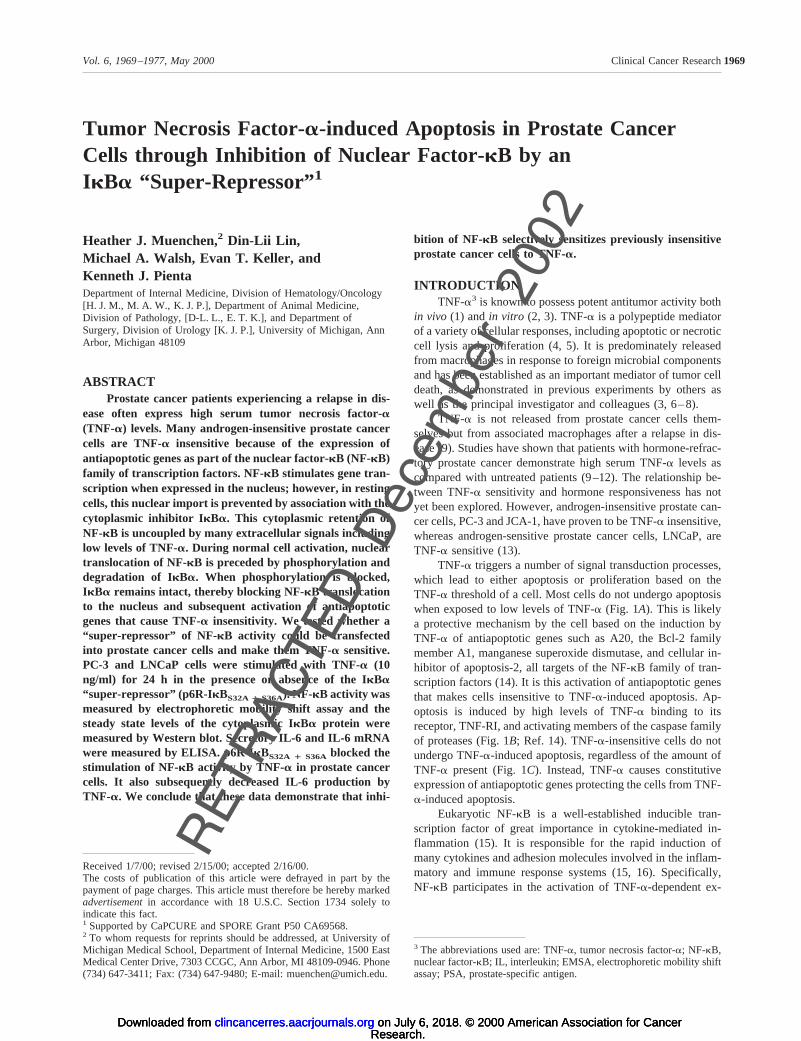
TNF-a triggers a number of signal transduction processes,which lead to either apoptosis or proliferation based on theTNF-a threshold of a cell. Most cells do not undergo apoptosiswhen exposed to low levels of TNF-a (Fig. 1A). This is likelya protective mechanism by the cell based on the induction byTNF-a of antiapoptotic genes such as A20, the Bcl-2 familymember A1, manganese superoxide dismutase, and cellular in-hibitor of apoptosis-2, all targets of the NF-kB family of tran-scription factors (14). It is this activation of antiapoptotic genesthat makes cells insensitive to TNF-a-induced apoptosis. Ap-optosis is induced by high levels of TNF-a binding to itsreceptor, TNF-RI, and activating members of the caspase familyof proteases (Fig. 1B; Ref. 14). TNF-a-insensitive cells do notundergo TNF-a-induced apoptosis, regardless of the amount ofTNF-a present (Fig. 1C). Instead, TNF-a causes constitutiveexpression of antiapoptotic genes protecting the cells from TNF-a-induced apoptosis.
Eukaryotic NF-kB is a well-established inducible tran-scription factor of great importance in cytokine-mediated in-flammation (15). It is responsible for the rapid induction ofmany cytokines and adhesion molecules involved in the inflam-matory and immune response systems (15, 16). Specifically,NF-kB participates in the activation of TNF-a-dependent ex-
Received 1/7/00; revised 2/15/00; accepted 2/16/00.The costs of publication of this article were defrayed in part by thepayment of page charges. This article must therefore be hereby markedadvertisementin accordance with 18 U.S.C. Section 1734 solely toindicate this fact.1 Supported by CaPCURE and SPORE Grant P50 CA69568.2 To whom requests for reprints should be addressed, at University ofMichigan Medical School, Department of Internal Medicine, 1500 EastMedical Center Drive, 7303 CCGC, Ann Arbor, MI 48109-0946. Phone(734) 647-3411; Fax: (734) 647-9480; E-mail: [email protected].
3 The abbreviations used are: TNF-a, tumor necrosis factor-a; NF-kB,nuclear factor-kB; IL, interleukin; EMSA, electrophoretic mobility shiftassay; PSA, prostate-specific antigen.
1969Vol. 6, 1969–1977, May 2000 Clinical Cancer Research
RETRACT
Dec
embe
r 20
02translocation of NF-kB is preceded by phosphorylation and
ED
Research. on July 6, 2018. © 2000 American Association for Cancerclincancerres.aacrjournals.org Downloaded from
Research. on July 6, 2018. © 2000 American Association for Cancerclincancerres.aacrjournals.org Downloaded from
Research. on July 6, 2018. © 2000 American Association for Cancerclincancerres.aacrjournals.org Downloaded from
pression of IL-6, intercellular adhesion molecule-1, and matrixmetalloproteinase-9, among others, in various cell systems (17,18). IL-6, an inflammatory cytokine, is a known autocrine andparacrine growth factor in androgen-insensitive human prostatecarcinomas (17). Intercellular adhesion molecule-1 is an estab-lished adhesion molecule involved in prostate cancer metastasis(17), whereas matrix metalloproteinase-9 plays an essential rolein the destruction of the basement membrane because of itsability to proteolyze type IV collagen (18).
The prototypical form of NF-kB is a heterodimeric induc-ible complex containing two DNA binding subunits, p50 (NF-kB1) and p65 (RelA), both of which belong to the Rel family oftranscription factors (19, 20). This heterodimer is the mostpotent gene transactivator within the NF-kB family (21). WhenNF-kB is expressed in the nucleus, it stimulates gene transcrip-tion via the potent transactivation domain located within theCOOH-terminal half of RelA (22). However, in resting cells, thenuclear import of NF-kB is prevented because of a high-affinityassociation of its RelA subunit with a labile cytoplasmic inhib-itor called IkBa (19, 23). This IkBa-dependent mechanism forthe cytoplasmic retention of NF-kB is uncoupled by manyextracellular signals including low levels of TNF-a (24). Afterthis cellular stimulation, IkBa is phosphorylated at serines 32and 36 by a specific kinase IKK (25), ubiquitinated, and under-goes proteolysis in proteosomes, enabling NF-kB to translocateto the nucleus, where it binds to NF-kB DNA binding sites andstimulates transcription of many cytokines, chemokines, andadhesion molecules (26).
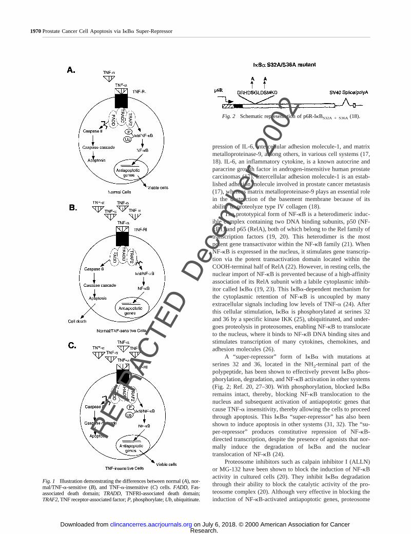
A “super-repressor” form of IkBa with mutations atserines 32 and 36, located in the NH2-terminal part of thepolypeptide, has been shown to effectively prevent IkBa phos-phorylation, degradation, and NF-kB activation in other systems(Fig. 2; Ref. 20, 27–30). With phosphorylation, blocked IkBaremains intact, thereby, blocking NF-kB translocation to thenucleus and subsequent activation of antiapoptotic genes thatcause TNF-ainsensitivity, thereby allowing the cells to proceedthrough apoptosis. This IkBa “super-repressor” has also beenshown to induce apoptosis in other systems (31, 32). The “su-per-repressor” produces constitutive repression of NF-kB-directed transcription, despite the presence of agonists that nor-mally induce the degradation of IkBa and the nucleartranslocation of NF-kB (24).
Proteosome inhibitors such as calpain inhibitor I (ALLN)or MG-132 have been shown to block the induction of NF-kBactivity in cultured cells (20). They inhibit IkBadegradationthrough their ability to block the catalytic activity of the pro-teosome complex (20). Although very effective in blocking theinduction of NF-kB-activated antiapoptotic genes, proteosome
Fig. 1 Illustration demonstrating the differences between normal (A), nor-mal/TNF-a-sensitive (B), and TNF-a-insensitive (C) cells. FADD, Fas-associated death domain;TRADD, TNFRI-associated death domain;TRAF2,TNF receptor-associated factor;P,phosphorylate;Ub,ubiquitinate.
Fig. 2 Schematic representation of p6R-IkBS32A 1 S36A (18).
1970Prostate Cancer Cell Apoptosis via IkBa Super-Repressor
RETRACT
Dec
embe
r 20
02
ED
Research. on July 6, 2018. © 2000 American Association for Cancerclincancerres.aacrjournals.org Downloaded from
inhibitors are nonspecific and therefore potentially very toxic.However, the IkBa“super-repressor,” p6R-IkBS32A 1 S36A, hasalso proven to be very effective in inhibiting activation ofNF-kB20. Its ability to be genetically engineered with a PSApromoter provides specificity for prostate cancer cells and there-fore makes it an ideal candidate for hormone-refractory prostatecancer gene therapy.
We report an efficient transduction of exogenous “super-repressor,” p6R-IkBS32A 1 S36A, NF-kB into both TNF-a-insensi-tive PC-3 cells and TNF-a-sensitive LNCaP cells. This “super-repressor” blocked IkBa phosphorylation, NF-kB translocationand activation, IL-6 production, and induced apoptosis in trans-fected PC-3 and LNCaP cells exposed to TNF-a. Our data dem-onstrate that p6R-IkBS32A 1 S36A is a powerful tool that cansensitize TNF-a-insensitive prostate cancer cells to undergo apo-ptosis. Moreover, blockage of IL-6 production decreases cell pro-liferation of androgen-independent prostate cancer cells.
MATERIALS AND METHODSCell Cultures. Androgen-sensitive LNCaP and andro-
gen-insensitive PC-3 cells (American Type Culture Collection,Rockville, MD) were maintained at 37°C in an atmosphere of5% CO2 in RPMI 1640 (Life Technologies, Grand Island, NY)containing 1% antibiotic-antimycotic (10,000 units/ml penicillinG, 10,000mg/ml streptomycin sulfate, and 25mg/ml amphoter-icin B; Life Technologies) and supplemented with 10% fetalbovine serum (Life Technologies).
Transfections. The IkBaS32A/S36Aplasmid used in thisstudy was described previously (21, 33). Transfections wereconducted by using SuperFect (Qiagen, Valencia, CA). Theprocedure was followed according to the manufacturer’s proto-col. Briefly, cells were seeded overnight at 60% confluency. TheDNA plasmid (10mg/10-cm dish or 2mg/6-cm dish) containingthe p6R-IkBS32A1 S36A“super-repressor” was mixed with 10 or
25 ml of SuperFect, respectively. In control experiments, theempty eukaryotic expression vector, p6R, was similarly intro-duced into cells. Cells were incubated with the DNA mixture for2 h at 37°C. Equal amounts of additional fresh media were thenadded to the cells. The cultures were incubated at 37°C for 24 hand replaced with fresh media. Cells were then treated withTNF-a (10 ng/ml) for 24 h.
Cytosolic and Nuclear Extracts. Cell pellets werewashed in PBS, pelleted again, resuspended in buffer A [10 mM
HEPES (pH 7.9), 1.5 mM MgCl2, 10 mM KCl, 5 mM DTT, 5 mM
phenylmethylsulfonyl fluoride and protease inhibitors (50mg/ml antipain, 2mg/ml aprotinin, 1mg/ml leupeptin, and 1mg/ml pepstatin)] and placed on ice for 10 min. The cells werethen vortexed and centrifuged for 10 s. The supernatant wasplaced in a separate tube, and 10 mM EDTA, 120 mM KCl, and20% glycerol were added. This mixture was designated cytoso-lic extracts and stored at280°C. The pellets were resuspendedin equal volumes of buffer C [10 mM HEPES (pH 7.9), 25%glycerol, 1.5 mM MgCl2, 0.2 mM EDTA, 0.8 M KCl, and pro-tease inhibitors] and incubated for 20 min on ice. Samples werecentrifuged for 5 min at 10,0003 g at 4°C. Supernatants weredesignated nuclear extracts and were stored at280°C.
Western Blot Analysis. Equal amounts of cytosolic ex-tracts (50mg) were analyzed by SDS-PAGE, followed by West-ern blotting using a polyclonal rabbit IkBa antibody (SantaCruz Biotechnology, Santa Cruz, CA). Immunoreactive IkBawas detected using the enhanced chemiluminescence (ECL)light detecting kit (Amersham).
EMSA. NF-kB oligonucleotide probe (Santa Cruz Bio-technology) was labeled with [g32P]ATP to 50,000 cpm/ngusing polynucleotide kinase. Nuclear extracts (5mg) were in-cubated with 1mg of poly(deoxyinosinic-deoxycytidylic acid) a20-ml volume of gel shift reaction buffer [10 mM Tris (pH 7.5),50 mM NaCl, 1 mM DTT, 1 mM EDTA, and 5% glycerol] and 0.5
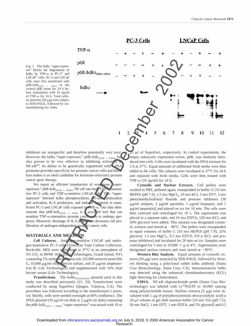
Fig. 3 The IkBa “super-repres-sor” blocks the degradation ofIkBa by TNF-a in PC-3T andLNCaPT cells. PC-3 and LNCaPcells were first transfected withp6R-IkBS32A 1 S36A or thecontrol p6R alone for 24 h be-fore stimulation with 10 ng/mlof TNF-a for 24 h. Total cellu-lar proteins (50mg) were subjectto SDS-PAGE, followed by im-munoblotting for IkBa.
1971Clinical Cancer Research
RETRACT
Dec
embe
r 20
02
ED
Research. on July 6, 2018. © 2000 American Association for Cancerclincancerres.aacrjournals.org Downloaded from
ng of labeled oligonucleotide probe for 20 min at room tempera-ture. For supershifts with p65 antibody, nuclear extracts fromuntransfected PC-3 cells were preincubated with 1ml of RelAantibody against the COOH-terminal portion of the molecule (San-ta Cruz Biotechnology) for 15 min at room temperature before theaddition of binding buffer and probe. DNA-protein complexeswere resolved by electrophoresis through a 4% polyacrylamide gelcontaining 50 mM Tris (pH 7.5), 0.38M glycine, and 2 mM EDTA.The gel was then dried and visualized by autoradiography.
RNA Extraction and Amplification. RNA was isolatedfrom control, p6R, and mutant, p6R-IkBS32A1 S36A, transfectedPC-3 cells using the TRIzol method (Life Technologies). TotalRNA (10 mg) was amplified and measured using a mRNA IL-6Quantikine ELISA (R&D Systems, Minneapolis, MN).
IL-6 ELISA. An IL-6 ELISA of cell culture supernatantsfrom control, p6R, and mutant, p6R-IkBS32A 1 S36A, transfectedPC-3 cells was performed in triplicate according to the manufac-
turer’s specifications (R&D Systems). Supernatants were removedat various time points after TNF-a (10 ng/ml) stimulation.
Apoptosis Detection. Annexin V fluorescent stainingwas assayed using an Annexin V apoptosis kit (Santa CruzBiotechnology), and Apoptag fluorescent staining was detectedusing an immunohistochemistry kit (Intergen, Purchase, NY),according to manufacturer’s protocol.
Statistical Analysis. Data are expressed as a means6SE. Statistical significance was performed by the two-tailedStudentt test for paired data and considered significant ifPswere,0.05.
RESULTSBlocked Degradation of IkBa in Transfected LNCaP
(LNCaPT) and PC-3 (PC-3T) Cells. To inhibit TNF-a in-duced IkBa degradation, we transfected LNCaP and PC-3 cells
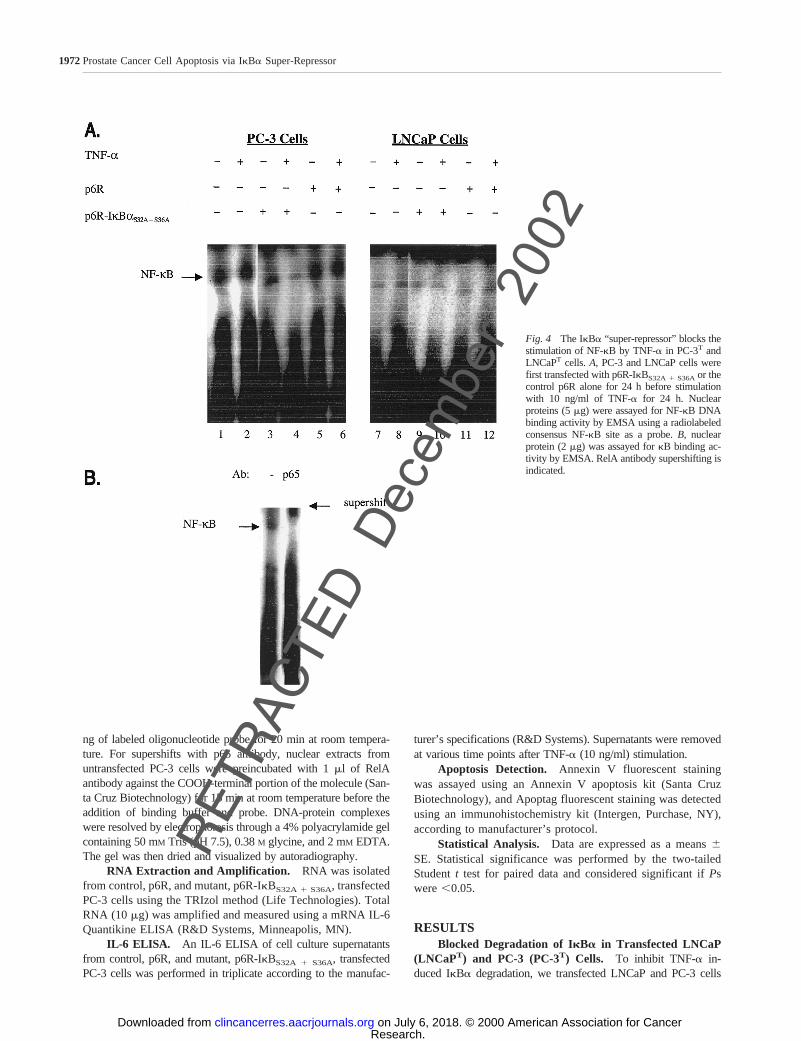
Fig. 4 The IkBa “super-repressor” blocks thestimulation of NF-kB by TNF-a in PC-3T andLNCaPT cells.A, PC-3 and LNCaP cells werefirst transfected with p6R-IkBS32A1 S36Aor thecontrol p6R alone for 24 h before stimulationwith 10 ng/ml of TNF-a for 24 h. Nuclearproteins (5mg) were assayed for NF-kB DNAbinding activity by EMSA using a radiolabeledconsensus NF-kB site as a probe.B, nuclearprotein (2mg) was assayed forkB binding ac-tivity by EMSA. RelA antibody supershifting isindicated.
1972Prostate Cancer Cell Apoptosis via IkBa Super-Repressor
RETRACT
Dec
embe
r 20
02
ED
Research. on July 6, 2018. © 2000 American Association for Cancerclincancerres.aacrjournals.org Downloaded from
with the IkBa “super-repressor,” p6R-IkBS32A1 S36A, which is notsusceptible to phosphorylation at NH2-terminal serines 32 and 36and is therefore resistant to subsequent degradation. Western blotanalysis of cells transfected with p6R-IkBS32A 1 S36A and treatedwith 10 ng/ml of TNF-a for 24 h demonstrated blockage of IkBaphosphorylation and degradation (Fig. 3). The TNF-a-insensitivePC-3 cells constitutively degraded IkBa until its phosphorylationwas blocked (Fig. 3,Lane 4). This phenomenon was present 24 hafter TNF-a was added to the cultures. After 48 h in culture,p6R-IkBS32A 1 S36A alone could stop degradation of IkBa (datanot shown). On the basis of these data, it appears that TNF-a
expedites the blocked degradation of IkBa. The TNF-a-sensitiveLNCaP cells only degraded IkBa in the presence of 10 ng/mlTNF-a (Fig. 3,Lanes 8and12), except where phosphorylation wasblocked by the IkBa “super-repressor” (Fig. 3,Lane 10).
Inhibition of NF- kB Translocation and Activation.Nuclear extracts prepared from LNCaPT and PC-3T cellswere analyzed by EMSA for their NF-kB DNA bindingactivity using a radiolabeled binding site as akB probe.TNF-a strongly induced NF-kB activity in untransfected andcontrol cells (Fig. 4A). Transfection with p6R-IkBS32A 1 S36A
and stimulation with TNF-a (10 ng/ml) for 24 h significantly
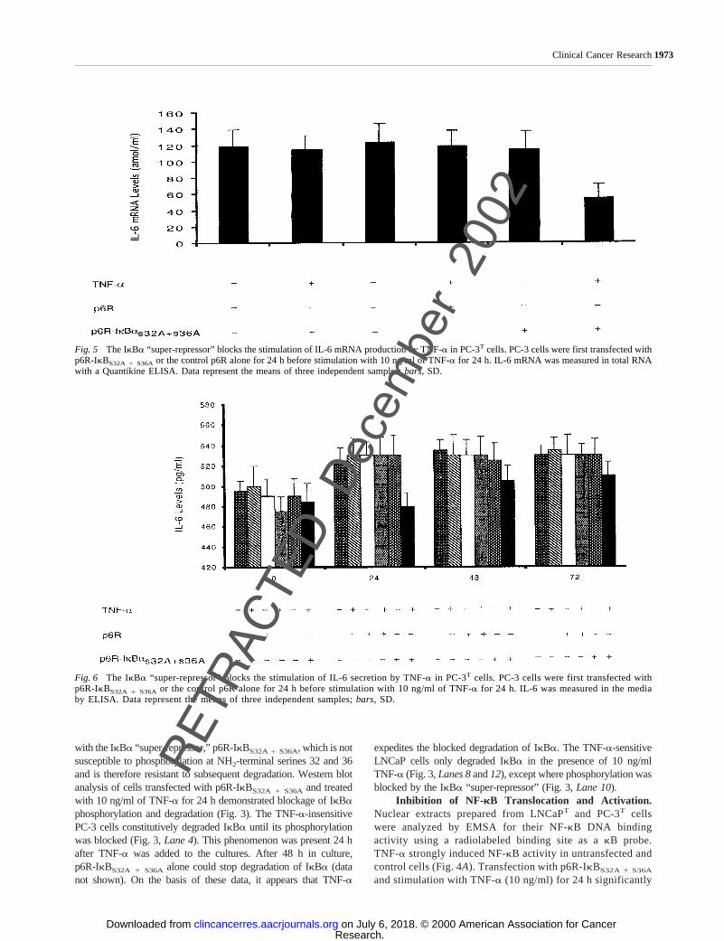
Fig. 5 The IkBa “super-repressor” blocks the stimulation of IL-6 mRNA production by TNF-a in PC-3T cells. PC-3 cells were first transfected withp6R-IkBS32A 1 S36A or the control p6R alone for 24 h before stimulation with 10 ng/ml of TNF-a for 24 h. IL-6 mRNA was measured in total RNAwith a Quantikine ELISA. Data represent the means of three independent samples;bars,SD.
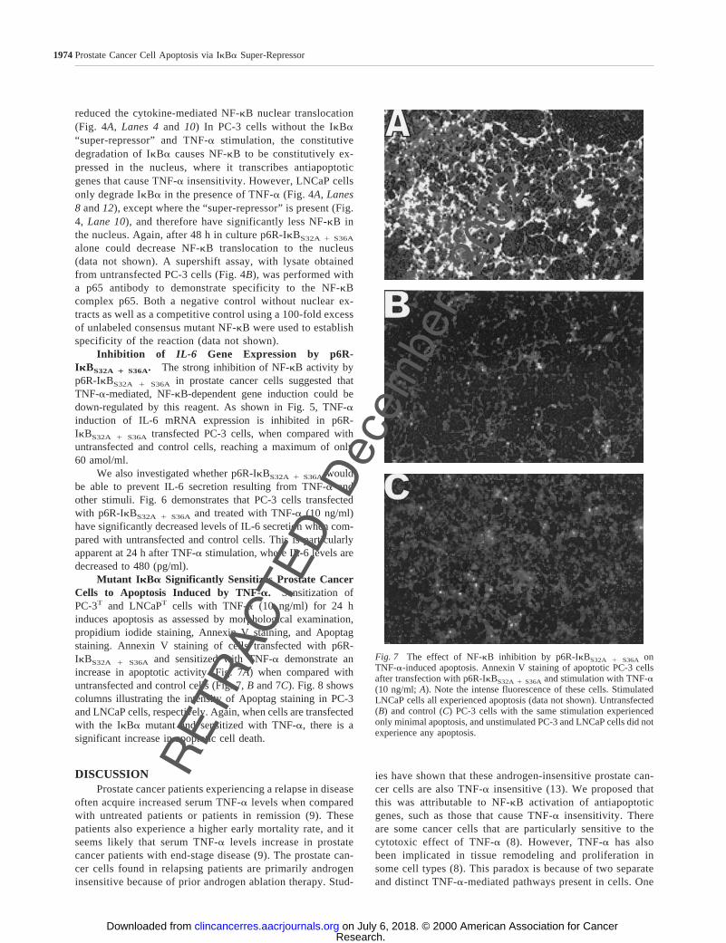
Fig. 6 The IkBa “super-repressor” blocks the stimulation of IL-6 secretion by TNF-a in PC-3T cells. PC-3 cells were first transfected withp6R-IkBS32A 1 S36A or the control p6R alone for 24 h before stimulation with 10 ng/ml of TNF-a for 24 h. IL-6 was measured in the mediaby ELISA. Data represent the means of three independent samples;bars, SD.
1973Clinical Cancer Research
RETRACT
Dec
embe
r 20
02
ED
Research. on July 6, 2018. © 2000 American Association for Cancerclincancerres.aacrjournals.org Downloaded from
reduced the cytokine-mediated NF-kB nuclear translocation(Fig. 4A, Lanes 4and 10) In PC-3 cells without the IkBa“super-repressor” and TNF-astimulation, the constitutivedegradation of IkBa causes NF-kB to be constitutively ex-pressed in the nucleus, where it transcribes antiapoptoticgenes that cause TNF-a insensitivity. However, LNCaP cellsonly degrade IkBa in the presence of TNF-a (Fig. 4A, Lanes8 and12), except where the “super-repressor” is present (Fig.4, Lane 10), and therefore have significantly less NF-kB inthe nucleus. Again, after 48 h in culture p6R-IkBS32A 1 S36A
alone could decrease NF-kB translocation to the nucleus(data not shown). A supershift assay, with lysate obtainedfrom untransfected PC-3 cells (Fig. 4B), was performed witha p65 antibody to demonstrate specificity to the NF-kBcomplex p65. Both a negative control without nuclear ex-tracts as well as a competitive control using a 100-fold excessof unlabeled consensus mutant NF-kB were used to establishspecificity of the reaction (data not shown).
Inhibition of IL-6 Gene Expression by p6R-IkBS32A 1 S36A. The strong inhibition of NF-kB activity byp6R-IkBS32A 1 S36A in prostate cancer cells suggested thatTNF-a-mediated, NF-kB-dependent gene induction could bedown-regulated by this reagent. As shown in Fig. 5, TNF-ainduction of IL-6 mRNA expression is inhibited in p6R-IkBS32A 1 S36A transfected PC-3 cells, when compared withuntransfected and control cells, reaching a maximum of only60 amol/ml.
We also investigated whether p6R-IkBS32A 1 S36A wouldbe able to prevent IL-6 secretion resulting from TNF-a andother stimuli. Fig. 6 demonstrates that PC-3 cells transfectedwith p6R-IkBS32A 1 S36A and treated with TNF-a (10 ng/ml)have significantly decreased levels of IL-6 secretion when com-pared with untransfected and control cells. This is particularlyapparent at 24 h after TNF-a stimulation, where IL-6 levels aredecreased to 480 (pg/ml).
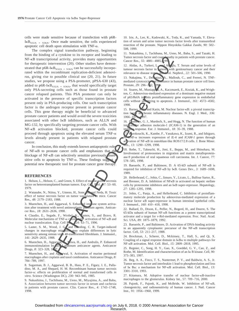
Mutant IkBa Significantly Sensitizes Prostate CancerCells to Apoptosis Induced by TNF-a. Sensitization ofPC-3T and LNCaPT cells with TNF-a (10 ng/ml) for 24 hinduces apoptosis as assessed by morphological examination,propidium iodide staining, Annexin V staining, and Apoptagstaining. Annexin V staining of cells transfected with p6R-IkBS32A 1 S36A and sensitized with TNF-a demonstrate anincrease in apoptotic activity (Fig. 7A) when compared withuntransfected and control cells (Fig. 7,B and 7C). Fig. 8 showscolumns illustrating the intensity of Apoptag staining in PC-3and LNCaP cells, respectively. Again, when cells are transfectedwith the IkBa mutant and sensitized with TNF-a, there is asignificant increase in apoptotic cell death.
DISCUSSIONProstate cancer patients experiencing a relapse in disease
often acquire increased serum TNF-a levels when comparedwith untreated patients or patients in remission (9). Thesepatients also experience a higher early mortality rate, and itseems likely that serum TNF-a levels increase in prostatecancer patients with end-stage disease (9). The prostate can-cer cells found in relapsing patients are primarily androgeninsensitive because of prior androgen ablation therapy. Stud-
ies have shown that these androgen-insensitive prostate can-cer cells are also TNF-a insensitive (13). We proposed thatthis was attributable to NF-kB activation of antiapoptoticgenes, such as those that cause TNF-ainsensitivity. Thereare some cancer cells that are particularly sensitive to thecytotoxic effect of TNF-a (8). However, TNF-a has alsobeen implicated in tissue remodeling and proliferation insome cell types (8). This paradox is because of two separateand distinct TNF-a-mediated pathways present in cells. One
Fig. 7 The effect of NF-kB inhibition by p6R-IkBS32A 1 S36A onTNF-a-induced apoptosis. Annexin V staining of apoptotic PC-3 cellsafter transfection with p6R-IkBS32A 1 S36A and stimulation with TNF-a(10 ng/ml;A). Note the intense fluorescence of these cells. StimulatedLNCaP cells all experienced apoptosis (data not shown). Untransfected(B) and control (C) PC-3 cells with the same stimulation experiencedonly minimal apoptosis, and unstimulated PC-3 and LNCaP cells did notexperience any apoptosis.
1974Prostate Cancer Cell Apoptosis via IkBa Super-Repressor
RETRACT
Dec
embe
r 20
02
ED
Research. on July 6, 2018. © 2000 American Association for Cancerclincancerres.aacrjournals.org Downloaded from

pathway leads to apoptosis and the other to activation ofprotective antiapoptotic genes through NF-kB. Which path-way is activated depends on the TNF-a threshold of thatparticular cell. Typically, lower or normal levels of TNF-ainduce the NF-kB protective pathway to keep the cell fromundergoing unnecessary apoptosis, where higher or toxiclevels induce apoptosis. TNF-a-insensitive cells, such asandrogen-insensitive prostate cancer cells, do not experienceTNF-a-induced apoptosis, even at the LD10
This is attributable to the constitutive degradation of IkBaand therefore NF-kB activation of antiapoptotic genes. EvenTNF-a levels of 100 ng/ml could not force the TNF-a-insensitive cells into apoptosis. The fact that the androgen-insensitive prostate cancer cells were also TNF-a insensitive,coupled with the increased serum TNF-a levels of relapsingprostate cancer patients, lead us to believe that these cellswere thriving in a toxic TNF-a environment attributable tothe induction of NF-kB antiapoptotic genes.
This study provides important new insights into the effectsof inhibiting NF-kB in TNF-a-insensitive prostate cancer cells.A wide range of intracellular components are implicated inTNF-a-induced cell killing, including pertussis toxin-sensitiveguanine nucleotide binding protein (34), phospholipase A2 (35),phospholipase D activation (36), and DNA damage (37). De-spite which receptor sets off the cytotoxic effect, the variousintracellular signals activated by TNF-a lead to activation ofNF-kB (4). Prostate cancer cells up-regulate multiple NF-kB-responsive genes in response to TNF-a stimulation. These mol-ecules may be involved in cell proliferation and metastasis,where modulation of their expression could be clinically bene-ficial. We have targeted our intervention at the IkB/NF-kBpathway because most of these genes are predominately regu-lated at the level of transcription.
After TNF-a stimulation, NF-kB maintains a balance be-tween an inactive and active state that relies mostly on IkBa(21). NF-kB can be shifted to an inactive or active state byoverexpression or degradation of IkBa (21). Previous ap-
proaches to inhibit NF-kB activity have focused on endothelialor mononuclear hemopoietic cells (21). Most strategies targetIkBa through proteosome blockades, phosphorylation inhibi-tion, and protein overexpression (38, 39). The method we usedto block the NF-kB pathway in TNF-a-insensitive prostatecancer cells was transfection with an IkBa “super-repressor”that resists TNF-a induced phosphorylation and degradation.The IkBa protein used was a S32A/S36A mutant form of IkBathat mutated the inducible amino acid phosphoreceptor andtherefore abolished the degradation process (21). Our approachwas successful in maintaining IkBa levels, which selectivelyinhibited NF-kB p65 subunit nuclear translocation, NF-kBDNA binding activity, down-regulated the induction of NF-kBresponsive geneIL-6, and induced apoptosis.
Androgen-independent prostate cancer cells spontane-ously release high levels of IL-6 into the cell supernatantwithout exogenous stimulation (13). IL-6 is a cytokine withpleiotropic activities and has been shown to play a centralrole in immune host-defense mechanisms (40). This cytokinehas been implicated in growth differentiation, inhibition, andproliferation, depending upon the nature of the responsivetarget cells (41). IL-6 has been shown to promote cell pro-liferation in androgen-independent prostate cancer cells,PC-3 (42). When these cells were transfected with thep6R-IkBS32A 1 S36A “super-repressor” and stimulated withTNF-a, IL-6 secretion and IL-6 mRNA production was de-creased. Although our results demonstrate only partial inhi-bition of IL-6 production, we believe that this IL-6 is resid-ual, because of the fact that the “super-repressor” onlyinhibits new gene transcription, and that we have demon-strated that p6R-IkBS32A 1 S36A significantly suppresses newIL-6 production by TNF-a-stimulated PC-3 cells.
Transfected prostate cancer cells stimulated with TNF-awere induced to proceed through apoptosis. With the NF-kBpathway blocked by the IkBa “super-repressor,” the cells wereforced to proceed though TNF-a-induced apoptosis. Our resultsindicate that the previously TNF-a-insensitive prostate cancer
Fig. 8 The effect of NF-kB inhibition by p6R-IkBS32A 1 S36A on TNF-a-induced apoptosis.Apoptosis was examined by Apoptag fluores-cence staining of PC-3 and LNCaP cells. Thenumber of apoptotic and healthy cells wascounted by a fluorescent microscope in ran-domly selected three fields/well, and the data arepresented as a percentage of cells appearing ap-optotic. Triplicate wells in each experiment wereexamined (means;bars,SE; n 5 4).
1975Clinical Cancer Research
RETRACT
Dec
embe
r 20
02dose of 10 ng/ml.
ED
Research. on July 6, 2018. © 2000 American Association for Cancerclincancerres.aacrjournals.org Downloaded from
cells were made sensitive because of transfection with p6R-IkBS32A 1 S36A. Once made sensitive, the cells experiencedapoptotic cell death upon stimulation with TNF-a.
The complex signal transduction pathway, beginningfrom the binding of a cytokine to its receptor and leading toNF-kB transcriptional activity, provides many opportunitiesfor therapeutic intervention (20). Other studies have demon-strated that p6R-IkBS32A 1 S36A can be successfully incorpo-rated within the recombinant replication-deficient adenovi-rus, giving rise to possible clinical use (20, 21). In futurestudies, we propose using a PSA-promoter, pPSA-630 (43),added to p6R-IkBS32A 1 S36A, that would specifically targetonly PSA-secreting cells such as those found in prostatecancer relapsed patients. This PSA promoter can only beactivated in the presence of specific transcription factorspresent only in PSA-producing cells. One such transcriptionfactor is the androgen receptor present in prostate cancercells. This gene therapy might be beneficial to advancedprostate cancer patients and would avoid the severe toxicitiesassociated with other IkB inhibitors, such as ALLN andMG-132, by specifically targeting prostate cancer cells. WithNF-kB activation blocked, prostate cancer cells couldproceed through apoptosis using the elevated serum TNF-alevels already present in patients with relapsed prostatecancer.
In conclusion, this study extends known antiapoptotic rolesof NF-kB to prostate cancer cells and emphasizes that theblockage of NF-kB can selectively sensitize previously insen-sitive cells to apoptosis by TNF-a. These findings suggest apotential new therapeutic tool for prostate cancer gene therapy.
REFERENCES1. Helson, L., Helson, C., and Green, S. Effect of murine tumor necrosisfactor on heterotransplanted human tumors. Exp. Cell. Biol.,47: 53–60,1979.2. Watanabe, N., Niitsu, Y., Umeno, H., Sone, H., and Neda, H. Toxiceffect of tumor necrosis factor on tumor vasculature in mice. CancerRes.,48: 2179–2183, 1988.3. Muenchen, H., and Aggarwal, S. Enhanced immune system activa-tion after treatment with novel antineoplastic platinum agents. Antican-cer Res.,18: 2631–2636, 1998.4. Claudio, E., Segade, F., Wrobel, K., Ramos, S., and Bravo, R.Molecular mechanisms of TNF-acytotoxicity: activation of NF-kB andnuclear translocation. Exp. Cell Res.,224: 63–71, 1996.5. Laster, S. M., Wood, J. G., and Gooding, C. R. Target-inducedchanges in macrophage migration may explain differences in lyticsensitivity among simian virus 40-transformed fibroblasts. J. Immunol.,141: 2629–2635, 1988.6. Muenchen, H., Aggarwal, S., Misra, H., and Andrulis, P. Enhancedimmunostimulation by novel platinum anticancer agents. AnticancerDrugs,8: 323–328, 1997.7. Muenchen, H., and Aggarwal, S. Activation of murine peritonealmacrophages after cisplatin and taxol combination. Anticancer Drugs,8:784–789, 1997.8. Sugarman, B. J., Aggarwal, B. B., Hass, P. E., Figary, I. E., Palla-dino, M. A., and Shepard, H. M. Recombinant human tumor necrosisfactor-a: effects on proliferation of normal and transformed cellsinvitro. Science (Washington DC),230: 943–945, 1985.9. Nakashima, J., Tachibana, M., Ueno, M., Miyajima, A., and Baba,S. Association between tumor necrosis factor in serum and cachexiain patients with prostate cancer. Clin. Cancer Res.,4: 1743–1748,1998.
10. Irie, A., Lee, K., Kadowaki, K., Toda, K., and Yamada, Y. Eleva-tion of serum and urine tumor necrosis factor levels after transurethralresection of the prostate. Nippon Hinyokika Gakkai Zasshi,90: 502–508, 1999.
11. Nakashima, J., Tachibana, M., Ueno, M., Baba, S., and Tazaki, H.Tumor necrosis factor and coagulopathy in patients with prostate cancer.Cancer Res.,55: 4881–4885, 1995.
12. Akdas, A., Turkeri, L., and Akoglu, T. Serum and urine levels oftumour necrosis factor in patients with genitourinary cancer and theirrelevance to disease status. Int. Urol. Nephrol.,22: 501–506, 1990.
13. Nakajima, Y., Dellipizzi, A., Mallouh, C., and Ferreri, N. TNF-mediated cytotoxicity and resistance in human prostate cancer cell lines.Prostate,29: 296–302, 1996.
14. Soares, M., Muniappan, A., Kaczmarek, E., Koziak, K., and Wrigh-ton, C. Adenovirus-mediated expression of a dominant negative mutantof p65/RelA inhibits proinflammatory gene expression in endothelialcells without sensitizing to apoptosis. J. Immunol.,161: 4572–4582,1998.
15. Barnes, P. J., and Karin, M. Nuclear factor-kB: a pivotal transcrip-tion factor in chronic inflammatory diseases. N. Engl. J. Med.,336:1066–1071, 1997.
16. Douherty, G. J., Murdoch, S., and Hogg, N. The function of humanintercellular adhesion molecule-1 (ICAM-1) in the generation of animmune response. Eur. J. Immunol.,18: 35–39, 1988.
17. Kurokouchi, K., Kambe, F., Yasukawa, K., Izumi, R., and Ishiguro,N. TNF-a increases expression ofIL-6 and ICAM-1 genes throughactivation of NF-kB in osteoblast-like ROS17/2.8 cells. J. Bone Miner.Res.,13: 1290–1299, 1998.
18. Ikebe, T., Takeuchi, H., Jimi, E., Beppu, M., and Shinohara, M.Involvement of proteosomes in migration and matrix metalloprotein-ase-9 production of oral squamous cell carcinoma. Int. J. Cancer,77:578–585, 1998.
19. Baeuerle, P., and Baltimore, D. A 65-kD subunit of NF-kB isrequired for inhibition of NF-kB by IkB. Genes Dev.,3: 1689–1698,1989.
20. Hellerbrand, C., Jobin, C., Iimuro, Y., Licato, L., Balfour Sartor, R.,and Brenner, D. A. Inhibition of NFkB in activated rat hepatic stellatecells by proteosome inhibitors and an IkB super-repressor. Hepatology,27: 1285–1295, 1998.
21. Jobin, C., Panja, A., and Hellerbrand, C. Inhibition of proinflam-matory molecule production by adenovirus-mediated expression of anuclear factorkB super-repressor in human intestinal epithelial cells.J. Immunol.,160: 410–418, 1998.
22. Ballard, D., Dixon, E., Peffer, N., Bogerd, H., and Doerre, S. The65-kDa subunit of human NF-kB functions as a potent transcriptionalactivator and a target for v-Rel-mediated repression. Proc. Natl. Acad.Sci. USA,89: 1875–1879, 1992.
23. Baeuerle, P., and Baltimore, D. Activation of DNA-binding activityin an apparently cytoplasmic precursor of the NF-kB transcriptionfactor. Cell,53: 211–217, 1988.
24. Brockman, J., Scherer, D., Mckinsey, T., Hall, S., and Qi, X.Coupling of a signal response domain in IkBa to multiple pathways forNF-kB activation. Mol. Cell. Biol.,15: 2809–2818, 1995.
25. Regnier, C., Song, H. Y., Gao, X., Goeddel, G. V., Cao, Z., andRothe, M. Identification and characterization of an Ik B kinase. Cell,90:373–383, 1997.
26. Beg, A. A., Finco, T. S., Nantermet, P. V., and Baldwin, A. S., Jr.Tumor necrosis factor and interleukin-1 lead to phosphorylation and lossof Ik Ba: a mechanism for NF-kB activation. Mol. Cell. Biol.,13:3301–3310, 1993.
27. Kitamura, M. Adoptive transfer of nuclear factor-kB-inactivemacrophages to the glomerulus. Kidney Int., 57: 709–716, 2000.
28. Pajonk, F., Pajonk, K., and McBride, W. Inhibition of NF-kB,clonogenicity, and radiosensitivity of human cancer. J. Natl. CancerInst., 91: 1956–1960, 1999.
1976Prostate Cancer Cell Apoptosis via IkBa Super-Repressor
RETRACT
Dec
embe
r 20
02
ED
Research. on July 6, 2018. © 2000 American Association for Cancerclincancerres.aacrjournals.org Downloaded from
29. Li, N., and Karin, M. Ionizing radiation and short wavelength UVactivate NF-kB through two mechanisms. Proc. Natl. Acad. Sci. USA,95: 13012–13017, 1998.30. Brown, K., Gersberger, S., Carlson, L., Franzoso, G., and Sieben-list, U. Control of IkB-a proteolysis by site-specific, signal-inducedphosphorylation. Science (Washington DC),267: 1485–1488, 1995.31. van Hogerlindin, M., Rozell, B., Ahrlund-Richter, L., and Toftgard, R.Squamous cell carcinomas and increased apoptosis in skin with inhibitedRel/nuclear factor-kB signaling. Cancer Res.,59: 3299–3303, 1999.32. Miagkov, A., Kovalenko, D., Brown, C., Didsbury, J., Cogswell, J.,Stimpson, S., Baldwin, A., and Makarov, S. NF-kB activation providesthe potential link between inflammation and hyperplasia in the arthriticjoint. Proc. Natl. Acad. Sci. USA,95: 13859–13864, 1998.33. Didonato, J., Mercurio, F., Rosette, C., Wu-Li, J., Suyang, H.,Ghosh, S., and Karin, M. Mapping of inducible IkB phosphorylationsites that signal its ubiquitination and degradation. Mol. Cell. Biol.,16:1295–1304, 1996.34. Imamura, K., Sherman, M. L., Sprigg, D., and Kufe, D. Effect oftumor necrosis factor on GTP binding and GTPase activity in HL-60 andL929 cells. J. Biol. Chem.,263: 10247–10253, 1988.35. Miele, L., Cordella-Miele, E., and Mukherjee, A. B. Novel anti-inflammatory peptides from the region of highest similarity betweenuteroglobin and lipocortin I. Nature (Lond.),335: 726–730, 1988.36. De Valck, D., Beyaert, R., Van Roy, F., and Fiers, W. Tumornecrosis factor cytotoxicity is associated with phospholipase D activa-tion. Eur. J. Biochem.,202: 491–497, 1993.
37. Dealtry, C. B., Naylor, M. S., Fiers, W., and Balkwill, F. R. DNAfragmentation and cytotoxicity caused by tumor necrosis factor is en-hanced by interferon-g. Eur. J. Immunol.,17: 689–693, 1987.
38. Wrighton, C. J., Hofer-Warbinek, R., Moll, T., Eytner, R., Bach,F. H., and de Martin, R. Inhibition of endothelial cell activation byadenovirus-mediated expression of IkBa, an inhibitor of transcriptionfactor NF-kB. J. Exp. Med.,183: 1013–1022, 1996.
39. Read, M. A., Neish, A. S., Gerritsen, M. E., and Collins, T.Post-induction of transcriptional repression of E-selectin and vascularcell adhesion molecule-1. J. Immunol.,157: 3472–3479, 1996.
40. Akira, S., Taga, T., and Kishimoto, T. Interleukin-6 in biology andmedicine. Adv. Immunol.,54: 1–78, 1993.
41. Borsellino, N., Belldegrun, A., and Bonavida, B. Endogenous in-terleukin 6 is a resistance factor ofcis-diamminedichloroplatinum andetoposide-mediated cytotoxicity of human prostate carcinoma cell lines.Cancer Res.,55: 4633–4639, 1995.
42. Hobisch, A., Eder, I. E., Putz, T., Horninger, W., Bartsch, G.,Klocker, H., and Culig, Z. Interleukin-6 regulates prostate-specific pro-tein expression in prostate carcinoma cells by activation of the androgenreceptor. Cancer Res.,58: 4640–4645, 1998.
43. Sato, N., Sadar, M., Bruchovsky, N., Saatcioglu, F., and Rennie, P.Androgenic induction of prostate-specific antigen gene is repressed byprotein-protein interaction between the androgen receptor and AP-1/c-Jun in the human prostate cancer cell line LNCaP. J. Biol. Chem.,272:17485–17494, 1997.
1977Clinical Cancer Research
RETRACT
Dec
embe
r 20
02
ED
Research. on July 6, 2018. © 2000 American Association for Cancerclincancerres.aacrjournals.org Downloaded from

RetractionAn article published in the May 2000 issue of Clinical Cancer Research (H. J. Muenchen, D-L. Lin, M. A. Walsh, E. T. Keller,
K. J. Pienta: Tumor necrosis factor-�-induced apoptosis in prostate cancer cells through inhibition of nuclear factor-�B by an I�B�“super-repressor,” Clin. Cancer Res., 6: 1969–1977, 2000), is hereby retracted from the literature. The Office of Research Integrityhas determined that the data in Figure 3, and Figures 4A and 4B were falsified, and the coauthors have requested that the entire articlebe retracted.
3961Retraction Clinical Cancer Research

2000;6:1969-1977. Clin Cancer Res Heather J. Muenchen, Din-Lii Lin, Michael A. Walsh, et al.
''Super-Repressor''αBκB by an IκCancer Cells through Inhibition of Nuclear Factor-
-induced Apoptosis in ProstateαTumor Necrosis Factor-
Updated version
http://clincancerres.aacrjournals.org/content/6/5/1969
Access the most recent version of this article at:
Cited articles
http://clincancerres.aacrjournals.org/content/6/5/1969.full#ref-list-1
This article cites 43 articles, 22 of which you can access for free at:
Citing articles
http://clincancerres.aacrjournals.org/content/6/5/1969.full#related-urls
This article has been cited by 16 HighWire-hosted articles. Access the articles at:
E-mail alerts related to this article or journal.Sign up to receive free email-alerts
Subscriptions
Reprints and
To order reprints of this article or to subscribe to the journal, contact the AACR Publications
Permissions
Rightslink site. Click on "Request Permissions" which will take you to the Copyright Clearance Center's (CCC)
.http://clincancerres.aacrjournals.org/content/6/5/1969To request permission to re-use all or part of this article, use this link
Research. on July 6, 2018. © 2000 American Association for Cancerclincancerres.aacrjournals.org Downloaded from